ULTRASOUND OF HERNIAS DR MOHIT GOEL JRI 10/4/2013

Welcome message from author
This document is posted to help you gain knowledge. Please leave a comment to let me know what you think about it! Share it to your friends and learn new things together.
Transcript

ULTRASOUND OF HERNIAS
DR MOHIT GOELJRI10/4/2013

Sonography of Inguinal Region HerniasANATOMY
The inguinal ligament, the folded and thickened lower border of the external oblique aponeurosis, attaches at the anterior superior iliac spine and pubic tubercle and medially forms the inferior floor of the inguinal canal.
The deep inguinal ring is an anatomic defect in the transversalis fascia.
The superficial inguinal ring is a triangle-shaped anatomic defect in the external oblique aponeurosis immediately superior and lateral to the pubic tubercle.

Condensation of the internal oblique and trans-versus abdominis aponeuroses forms the conjoint tendon, and a reflection of the inguinal ligament forms the lacunar ligament.
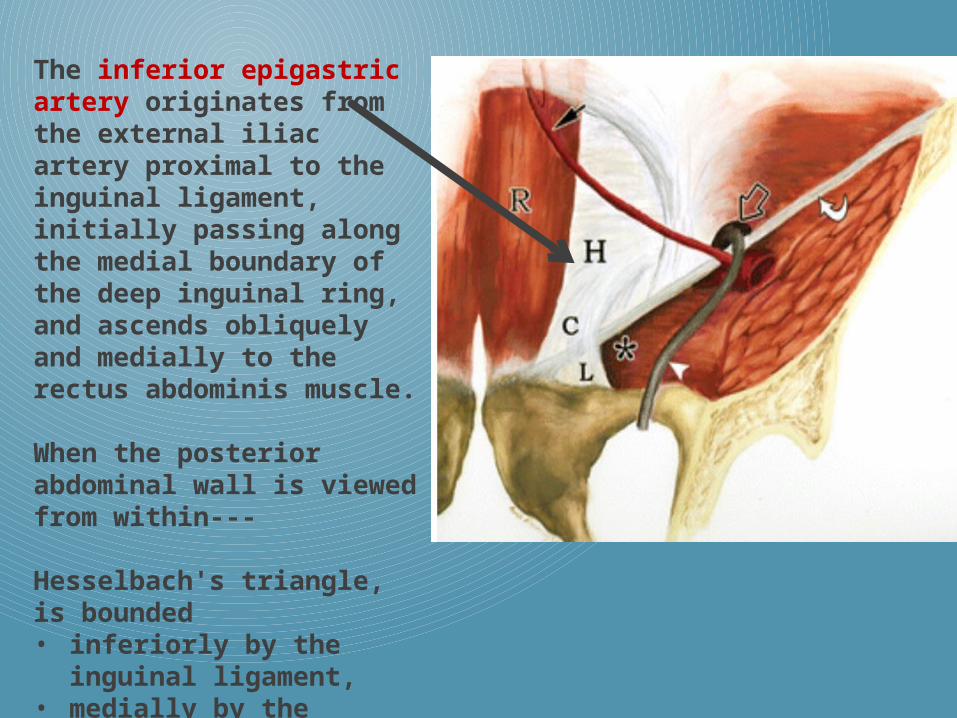
The inferior epigastric artery originates from the external iliac artery proximal to the inguinal ligament, initially passing along the medial boundary of the deep inguinal ring, and ascends obliquely and medially to the rectus abdominis muscle.
When the posterior abdominal wall is viewed from within---
Hesselbach's triangle, is bounded • inferiorly by the
inguinal ligament, • medially by the lateral
margin of the rectus abdominis and
• superiorly by the inferior epigastric artery.

Illustration of right inguinal region from anterior view shows transducer position to evaluate for
spigelian hernia (1), indirect inguinal hernia (2), direct inguinal hernia (3),and femoral hernia (4).
Inguinal ligament (curved arrow), rectus abdominis muscle (R), lateral boundary of Hesselbach's triangle (H) defined by inferior epigastric artery (open arrow), and spermatic cord (arrowhead).

40-year-old man with right inguinal anatomy. Sonogram of inguinal region parallel and cranial to inguinal ligament corresponding to transducer position 2 shows spermatic cord (C), external iliac artery (A), inferior epigastric artery (E), femoral vein (V), and superior pubic ramus (curved arrow).
Patients with groin hernias typically present with an obvious lump or bulge and are often diagnosed clinically and infrequently require imaging
On the other hand, patients with hernias who present with pain but without a lump or bulge are more often referred for diagnostic imaging.
More recently, CT and MRI have been used to identify and describe hernias
However, real-time ultrasound has advantages over other imagingmodalities:
• The ability to scan the patient in both upright and supine positions.
• To use dynamic manoeuvres such as Valsalva and compression.
Hernias occur
--in areas of natural weakness
--in areas where vessels penetrate the abdominal wall (femoral and spigelian)
--where fetal migration of testis, spermatic cord, or round ligament have occurred (indirect inguinal)--through broad flat tendons called aponeuroses (direct inguinal).
Hernias do not occur through the belly of abdominal wall muscles unless they have been surgically
Why does hernia occurs ?

HERNIA CONTENTS
Most Sonographically detected hernias do not contain bowel. In fact, most hernias contain only fat.The fat may be intraperitoneal (mesenteric or omental)or preperitoneal in origin.
Generally, it is not possible sonographically to distinguish whether the hernia contains intraperitoneal or preperitoneal fat.Hernias that contain intraperitoneal fat may contain bowel later and thus may be a greater risk than those that contain only preperitoneal fat
Hernias that contain bowel are considered higher risk because strangulation may lead to infarction of bowel.
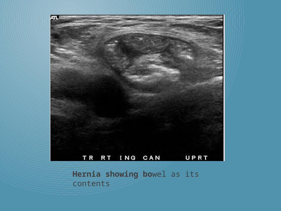
Hernia showing bowel as its contents

Spigelian hernias that present clinically are rare.
Sonographically detected spigelian hernias are more common than the literature would suggest.
Spigelian Hernias
All hernias occur along the course of the spigelian fascia, the complex aponeurotic tendon that lies between the oblique muscles laterally and the rectus muscles medially.
However, almost all spigelian hernias occur where the posterior rectus sheath is absent, and where the spigelian fascia is penetrated and weakened by the inferior epigastric vessels.

Almost all spigelian hernias arise from the inferior end of the spigelian fascia just lateral to whereit is penetrated by the inferior epigastric vessels, lateral to the lateral edge of the rectus abdominis muscle.

Image 1 -The inferior epigastric artery and its paired veins lie along the midlateral posterior surface of the rectus abdominis muscle. Image 2 - IEVs lie more laterally. Image 3 - is obtained at a level where the IEVs (arrow)lie at the edge of the rectus muscle. This is the level at which most spigelian hernias occur.

25-year-old man with right spigelian hernia. Pre-Valsalva maneuve over linea semilunaris in axial plane corresponding to transducer position 1 in Figure 4 (hernia not visible) showing right rectus abdominis muscle (R), inferior epigastric artery (curved arrow), peritoneal fat stripe (straight arrows), and lateral abdominal muscles (M).

Post-Valsalva maneuver sonogram in same location showing peritoneal fat stripe distorted by fat-containing spigelian hernia (arrows) at linea semilunaris.
Note rectus abdominis muscle (R) and lateral abdominal muscles (M).
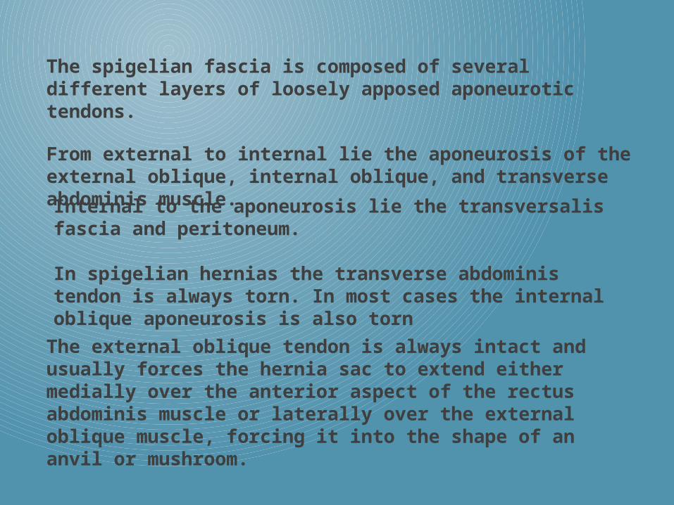
The spigelian fascia is composed of several different layers of loosely apposed aponeurotic tendons.
From external to internal lie the aponeurosis of the external oblique, internal oblique, and transverse abdominis muscle.
Internal to the aponeurosis lie the transversalis fascia and peritoneum.
In spigelian hernias the transverse abdominis tendon is always torn. In most cases the internal oblique aponeurosis is also torn
The external oblique tendon is always intact and usually forces the hernia sac to extend either medially over the anterior aspect of the rectus abdominis muscle or laterally over the external oblique muscle, forcing it into the shape of an anvil or mushroom.


Small, spigelian hernia in which the aponeuroses of both the transverseabdominis and internal oblique muscles are torn, but in which the external oblique aponeurosis, is intact.

Nonreducible left spigelian hernia contains bowel and has a narrow neck and broad fundus, the typical shape for spigelian hernias.

In indirect inguinal hernia - herniated structures
enter the inguinal canal lateral to the inferior
epigastric artery and superior to the inguinal
ligament, and extend for a variable distance
through the inguinal canal.
A second site of herniation is at the inferior aspect
of the Hesselbach's triangle, where a direct
inguinal hernia usually occurs. This weakened area
is just lateral to the conjoint tendon and medial to
the inferior epigastric artery, in contrast to the
indirect inguinal hernia that originates lateral to
the inferior epigastric artery.
INGUINAL HERNIA
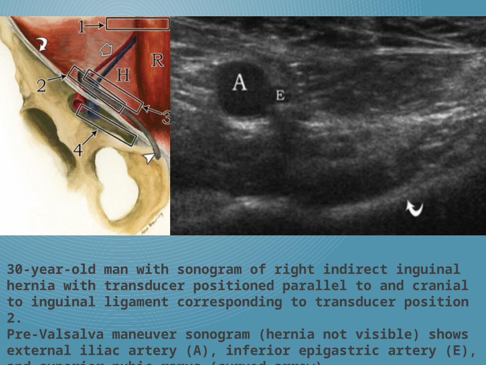
30-year-old man with sonogram of right indirect inguinal hernia with transducer positioned parallel to and cranial to inguinal ligament corresponding to transducer position 2.
Pre-Valsalva maneuver sonogram (hernia not visible) shows external iliac artery (A), inferior epigastric artery (E), and superior pubic ramus (curved arrow).

Post-Valsalva maneuver sonogram shows external iliac artery (A), inferior epigastric artery (E), dilated external iliac vein (V), superior pubic ramus (curved arrow), and indirect inguinal hernia (H) originating from lateral to external iliac artery (arrowhead) and traversing inguinal canal from lateral to medial. (Left = lateral).

Indirect inguinal hernia.Long-axis view shows that neck of the hernia lies in the internal inguinal ring (IIR),which lies superior and lateral to the proximal inferior epigastric artery (IEA).Hernia sac then courses horizontally in an inferomedial direction within the inguinal canal (IC).Indirect inguinal hernias always pass superficial to the IEA.
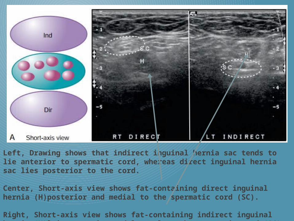
Left, Drawing shows that indirect inguinal hernia sac tends to lie anterior to spermatic cord, whereas direct inguinal hernia sac lies posterior to the cord.
Center, Short-axis view shows fat-containing direct inguinal hernia (H)posterior and medial to the spermatic cord (SC).
Right, Short-axis view shows fat-containing indirect inguinal hernia (H)lying anterior and lateral to the spermatic cord (SC).

Long-axis views
Left, Image shows the right direct inguinal hernia sac lying posterior to the spermatic cord (SC). Right, Image shows the left indirect inguinal hernia sac lying anterior to the spermatic cord (SC).

Indirect inguinal hernia.
Short-axis view shows indirect inguinal hernia displacing and compressing the hyperechoic spermatic cord posteriorly.
Direct inguinal hernia.
Short-axis view shows direct inguinal hernia displacing and compressing the hyperechoic spermatic cord anteriorly and laterally.

FEMORAL HERNIAS
Femoral hernias are rare, because they are difficult to diagnose clinically unless strangulated, and in fact are much less common that inguinal hernias.
Unlike inguinal hernias, femoral hernias are more common in women than men.
It is thought that the increased intrapelvic pressure that occurs during the third trimester of pregnancy together with the hormone induced Softening of tissues, predisposes to the development of femoral hernias.
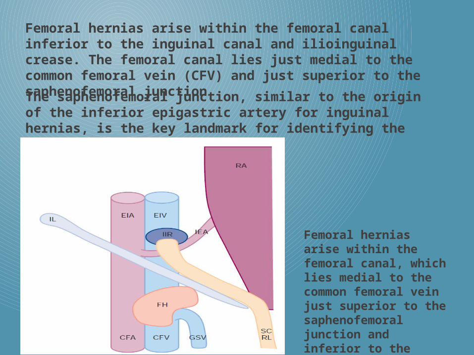
Femoral hernias arise within the femoral canal inferior to the inguinal canal and ilioinguinal crease. The femoral canal lies just medial to the common femoral vein (CFV) and just superior to the saphenofemoral junctionThe saphenofemoral junction, similar to the origin of the inferior epigastric artery for inguinal hernias, is the key landmark for identifying the femoral
Femoral hernias arise within the femoral canal, which lies medial to the common femoral vein just superior to the saphenofemoral junction and inferior to the inguinal ligament.

31-year-old woman with femoral hernia. Sonogram of right inguinal region parallel to and caudad to inguinal ligament corresponding to transducer position 4. Pre-Valsalva maneuver sonogram shows (hernia not visible) femoral artery (A), femoral vein (V), and superior pubic ramus (curved arrow).
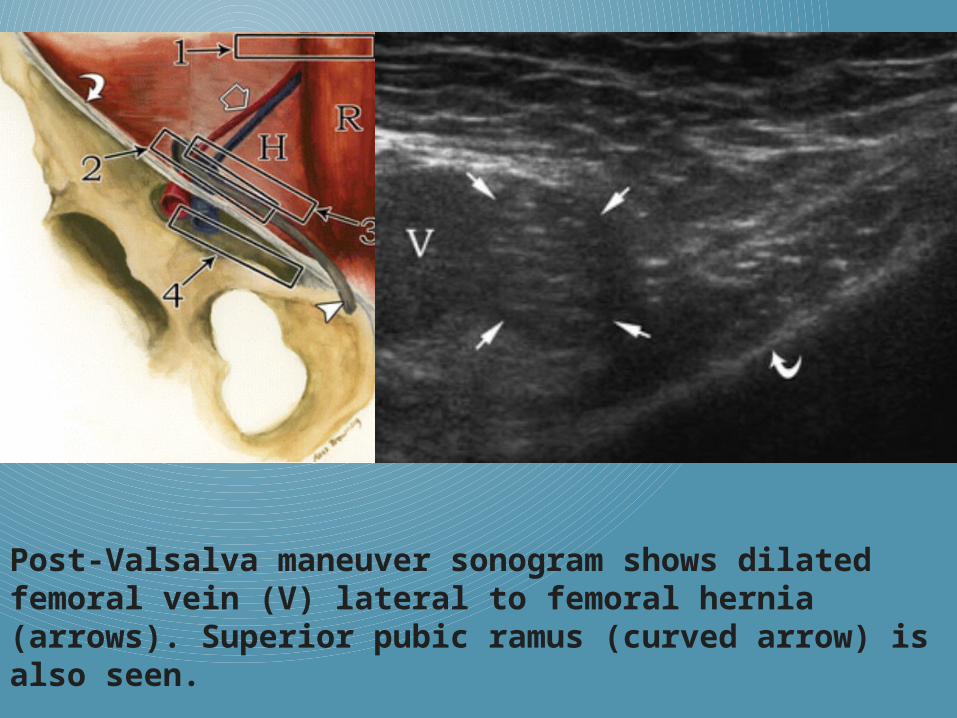
Post-Valsalva maneuver sonogram shows dilated femoral vein (V) lateral to femoral hernia (arrows). Superior pubic ramus (curved arrow) is also seen.

Linea Alba Hernias
Linea alba hernias are anterior abdominal wall hernias that protrude through the linea alba.
Those that occur superior to the umbilicus are called epigastric hernias, and those that occur inferior to the umbilicus are called hypogastric hernias.
Hypogastric hernias are much less common than epigastric hernias because the linea alba is much narrower and shorter, inferior to the umbilicus than superior to the umbilicus.
The linea alba is a thick layer of aponeurosis that separates the rectus abdominis muscles. It is formed by fusion and interlacing of fibers of the anterior and posterior sheaths of the right and left rectus muscles.

Transverse views. A, Normal, thick linea alba. B, Thinner but wider linea alba, possibly resulting from fewer decussations of rectus sheath fibres or representing diastasis recti.C, Marked thinning and bulging of the linea alba that occurs in diastasis recti. D, Typical small, epigastric linea alba hernia with its neck near the midline of the linea alba. E, Small linea alba hernia with neck occurring eccentrically near the right edge of the linea alba.
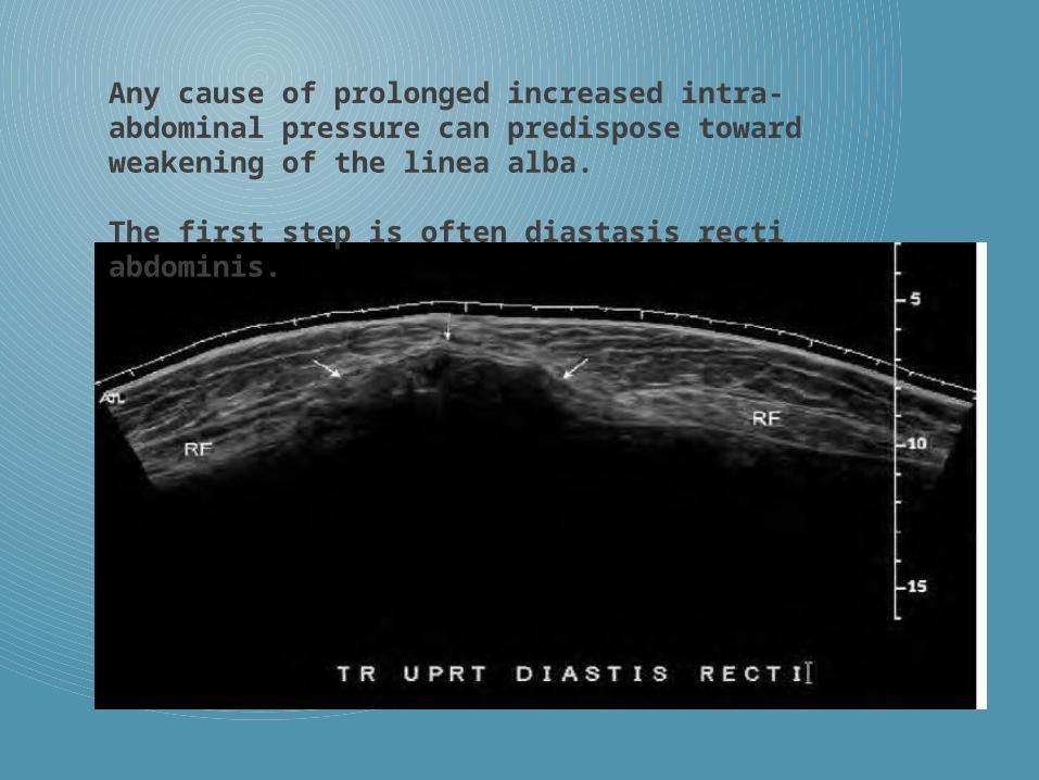
Any cause of prolonged increased intra-abdominal pressure can predispose toward weakening of the linea alba.
The first step is often diastasis recti abdominis.

Epigastric linea alba hernias are easier to diagnose than are groin hernias.
The defect through the linea alba is usually quite conspicuous because it is either isoechoic or hypoechoic compared with the extremely hyperechoic linea alba.
The defect is usually very near the midline.

Umbilical Hernias
Umbilical hernias occur through a widened umbilical ring.
Umbilical hernias can, however, develop at any time during life.
Any cause of chronically increased intraabdominal pressure or connective tissue weakness can lead to dilation of the umbilical ring and formation ofan umbilical hernia.
Umbilical hernias contain intraperitoneal contents, but smaller umbilical hernias usually contain only intraperitoneal fat.Untreated umbilical hernias tend to increase in size over time.
They are usually reducible but may become non-reducible and can also become strangulated.
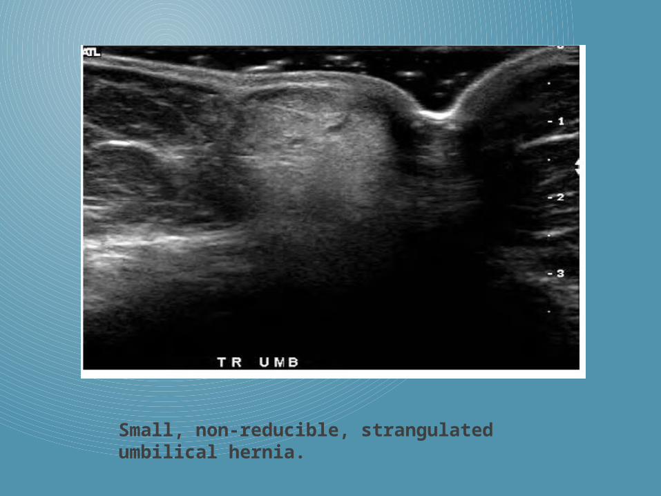
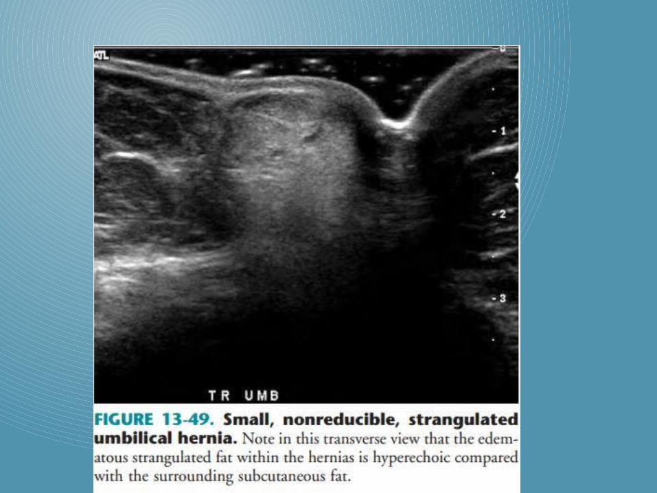
Small, non-reducible, strangulated umbilical hernia.

Incisional Hernias
Incisional hernias occur through surgical scars.
Herniation can occur through any type of surgical scar, including laparoscopy ports and stomal sites.
Incisional hernias can occur in any area along the anterior abdominal wallwhere an incision is made.
Incisional hernias resulting from thinning and stretching of the scar have wide necks and are reducible, whereas those resulting from tears in the scar are more likely to have narrow necks and to be nonreducible.

Incisional hernias can occur where natural hernias cannot, through thebellies of muscles that have been incised.

Narrow-necked, fat containing ventral incisional hernia that is incompletely reducible, with no compression on the right, but with compression on the left.

Hernia Complications
Hernia complications include incarceration, obstruction, and strangulation.
Incarcerated hernias are simply hernias that are nonreducible.
Obstructed hernias contain incarcerated bowel loops that have becomemechanically obstructed.
Strangulated hernias contain incarcerated contents with compromised vascularityNot all strangulated hernias contain bowel loops; even preperitoneal fat can become strangulated.
Most incarcerated hernias are neither obstructed nor strangulated,but all obstructed and strangulated hernias are also incarcerated.

It is prefered not to use the term “incarcerated” because many referring clinicians confuse incarceration with obstruction and strangulation, often believing that incarceration is a surgical emergency when it is not.Even strangulated hernias that contain only pre-peritoneal fat may not be emergencies.
It is the presence of bowel loops within strangulated hernias that makes them emergent.
The shape of hernias affects their reducibility and their likelihood of becoming obstructed or strangulated in the future.
Hernia types that typically have narrow necks and are at high risk for strangulation include
• femoral
• spigelian
• linea alba
• umbilical
• indirect inguinal hernias.

Although vascular compromise is the hallmark of strangulation ,Doppler ultrasound is not the most sensitive modality for demonstrating signs of strangulation.
Doppler ultrasound shows arterial flow within hernias with some success, but generally is not sensitive enough to demonstrate venous flow and cannot show lymphatic flow at all.
Thus, in strangulated hernias, the lymphatics and veins becomeobstructed long before arterial flow decreases.

The most sensitive findings of strangulation are the presenceof the following:
• Hyperechoic fat
• Isoechoic thickening of the normally thin and echogenic hernia sac
• Fluid within the sac
• Thickening of bowel wall in bowel-containing hernias.
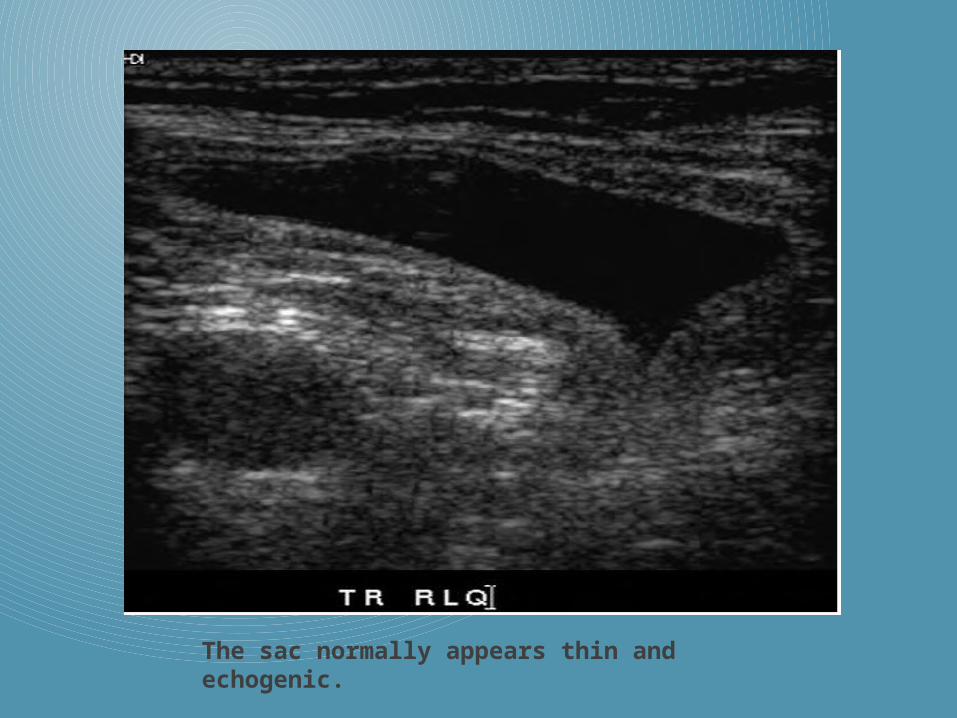
The sac normally appears thin and echogenic.
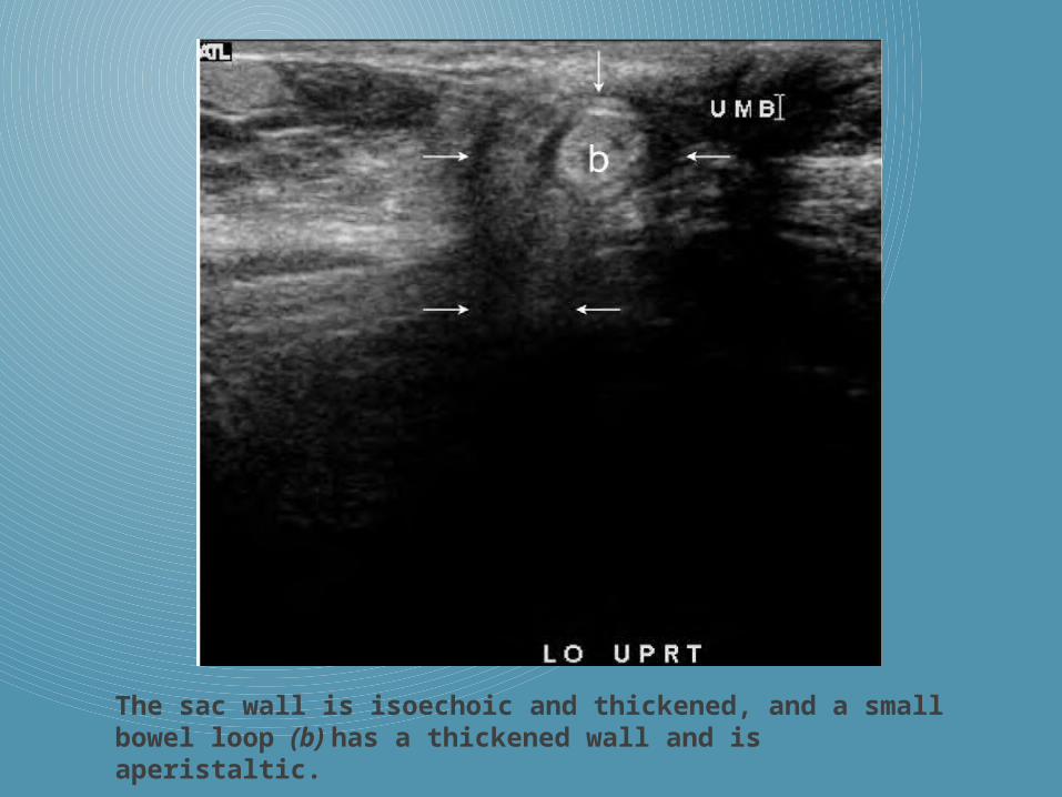
The sac wall is isoechoic and thickened, and a small bowel loop (b) has a thickened wall and is aperistaltic.
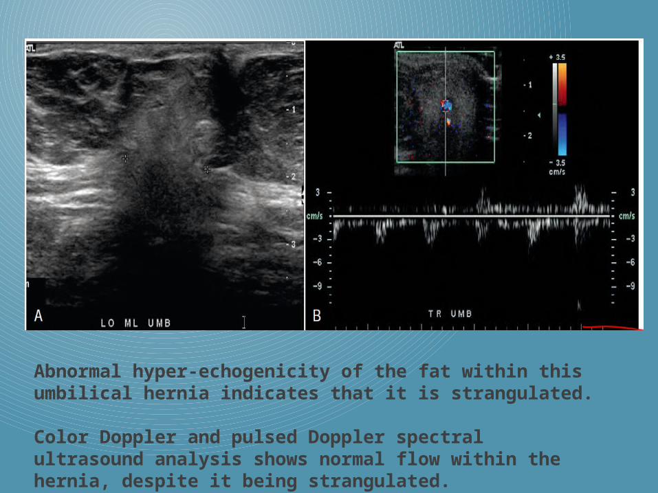
Abnormal hyper-echogenicity of the fat within this umbilical hernia indicates that it is strangulated.
Color Doppler and pulsed Doppler spectral ultrasound analysis shows normal flow within the hernia, despite it being strangulated.

THANK YOU
Related Documents