Ultrafast ligand dynamics in the heme-based GAF sensor domains of the histidine kinases DosS and DosT from Mycobacterium tuberculosis † Marten H. Vos a,b,* , Latifa Bouzhir-Sima a,b , Jean-Christophe Lambry a,b , Hao Luo c , Julian J. Eaton-Rye c , Alexandra Ioanoviciu d , Paul R. Ortiz de Montellano d , and Ursula Liebl a,b a Laboratory for Optics and Biosciences, CNRS Ecole Polytechnique, 91128 Palaiseau, France b INSERM U696, 91128 Palaiseau, France c Department of Biochemistry, University of Otago, Dunedin, New Zealand d Department of Pharmaceutical Chemistry, University of California, 600 16th Street, San Francisco, CA 94158, U.S.A Abstract The transcriptional regulator DosR from M. tuberculosis plays a crucial role in the virulence to dormancy transition of the pathogen. DosR can be activated by DosT and DosS, two histidine kinases with heme-containing sensor GAF domains, capable of diatomic ligand binding, To investigate the initial processes occurring upon ligand dissociation, we performed ultrafast time- resolved absorption spectroscopy of the isolated sensor domains ligated with O 2 , NO and CO. The results reveal a relatively closed heme pocket for both proteins. For DosT the yield of O 2 escape from the heme pocket on the picoseconds timescale upon photodissociation was found to be very low (1.5%), similar to other heme-based oxygen sensor proteins, implying that this sensor acts as an effective O 2 trap. Remarkably, this yield is an order of magnitude higher in DosS (18%). For CO, by contrast, the fraction of CO rebinding within the heme pocket is higher in DosS. Experiments with mutant DosT sensor domains and molecular dynamics simulations indicate an important role in ligand discrimination of the distal tyrosine, present in both proteins, which forms a hydrogen bond with heme-bound O 2 . We conclude that despite their similarity, DosT and DosS display ligand-specific different primary dynamics during the initial phases of intra-protein signaling. The distal tyrosine, present in both proteins, plays an important role in these processes. Mycobacterium tuberculosis is a widespread pathogen that is thought to have latently infected as much as one third of the human population worldwide, and to cause over 9 million new cases of tuberculosis each year (1). It has the capacity to switch from an actively replicating form to a dormant non-active form. The latter form can be maintained for extended periods of time within the host and is highly resistant to drugs, a combination of properties that largely contributes to the difficulty in eradicating tuberculosis. The transition between the two states is activated by stimuli including hypoxia (2) and exposure to NO (3). A crucial role in this transition is played by the transcriptional regulator DosR that induces the expression of a number of genes required for entry to dormancy (4). This response regulator is part of a two-component system that also comprises the signaling histidine kinases DosT and DosS (5), each of which can transfer phosphate to DosR. DosT † This work was supported by National Institutes of Health grant AI074824 to P.R.O.d M. and by a New Zealand Lottery Health Research Grant to J.J. E.-R. * Corresponding author, [email protected], tel. +33169335066, FAX +33169335084. NIH Public Access Author Manuscript Biochemistry. Author manuscript; available in PMC 2013 January 10. Published in final edited form as: Biochemistry. 2012 January 10; 51(1): 159–166. doi:10.1021/bi201467c. NIH-PA Author Manuscript NIH-PA Author Manuscript NIH-PA Author Manuscript

Welcome message from author
This document is posted to help you gain knowledge. Please leave a comment to let me know what you think about it! Share it to your friends and learn new things together.
Transcript

Ultrafast ligand dynamics in the heme-based GAF sensordomains of the histidine kinases DosS and DosT fromMycobacterium tuberculosis†
Marten H. Vosa,b,*, Latifa Bouzhir-Simaa,b, Jean-Christophe Lambrya,b, Hao Luoc, Julian J.Eaton-Ryec, Alexandra Ioanoviciud, Paul R. Ortiz de Montellanod, and Ursula Liebla,b
aLaboratory for Optics and Biosciences, CNRS Ecole Polytechnique, 91128 Palaiseau, FrancebINSERM U696, 91128 Palaiseau, France cDepartment of Biochemistry, University of Otago,Dunedin, New Zealand dDepartment of Pharmaceutical Chemistry, University of California, 60016th Street, San Francisco, CA 94158, U.S.A
AbstractThe transcriptional regulator DosR from M. tuberculosis plays a crucial role in the virulence todormancy transition of the pathogen. DosR can be activated by DosT and DosS, two histidinekinases with heme-containing sensor GAF domains, capable of diatomic ligand binding, Toinvestigate the initial processes occurring upon ligand dissociation, we performed ultrafast time-resolved absorption spectroscopy of the isolated sensor domains ligated with O2, NO and CO. Theresults reveal a relatively closed heme pocket for both proteins. For DosT the yield of O2 escapefrom the heme pocket on the picoseconds timescale upon photodissociation was found to be verylow (1.5%), similar to other heme-based oxygen sensor proteins, implying that this sensor acts asan effective O2 trap. Remarkably, this yield is an order of magnitude higher in DosS (18%). ForCO, by contrast, the fraction of CO rebinding within the heme pocket is higher in DosS.Experiments with mutant DosT sensor domains and molecular dynamics simulations indicate animportant role in ligand discrimination of the distal tyrosine, present in both proteins, which formsa hydrogen bond with heme-bound O2. We conclude that despite their similarity, DosT and DosSdisplay ligand-specific different primary dynamics during the initial phases of intra-proteinsignaling. The distal tyrosine, present in both proteins, plays an important role in these processes.
Mycobacterium tuberculosis is a widespread pathogen that is thought to have latentlyinfected as much as one third of the human population worldwide, and to cause over 9million new cases of tuberculosis each year (1). It has the capacity to switch from anactively replicating form to a dormant non-active form. The latter form can be maintainedfor extended periods of time within the host and is highly resistant to drugs, a combinationof properties that largely contributes to the difficulty in eradicating tuberculosis.
The transition between the two states is activated by stimuli including hypoxia (2) andexposure to NO (3). A crucial role in this transition is played by the transcriptional regulatorDosR that induces the expression of a number of genes required for entry to dormancy (4).This response regulator is part of a two-component system that also comprises the signalinghistidine kinases DosT and DosS (5), each of which can transfer phosphate to DosR. DosT
†This work was supported by National Institutes of Health grant AI074824 to P.R.O.d M. and by a New Zealand Lottery HealthResearch Grant to J.J. E.-R.*Corresponding author, [email protected], tel. +33169335066, FAX +33169335084.
NIH Public AccessAuthor ManuscriptBiochemistry. Author manuscript; available in PMC 2013 January 10.
Published in final edited form as:Biochemistry. 2012 January 10; 51(1): 159–166. doi:10.1021/bi201467c.
NIH
-PA Author Manuscript
NIH
-PA Author Manuscript
NIH
-PA Author Manuscript

and DosS show high similarity and both contain, apart from the C-terminal enzymaticdomains, two GAF domains, one of which (the N-terminal GAF A domain) is a heme-basedsensor domain (6, 7). DosT and DosS appear not to be redundant, but active in the initial andfinal phases of the transition to dormancy, respectively (8, 9). The sensor domains of DosTand DosS carry hemes that, in the ferrous state, are able to bind the external ligands O2, NO,and CO and are 5-coordinate in the absence of these ligands (6, 7, 10–12). Both kinases areinactive in the ferrous O2-bound state and active in other ferrous forms (7, 12). Theautoxidation rates of the proteins are very low (7, 11). The proteins have been proposed toact as oxygen (or hypoxia) sensor (7, 11) or nitric oxide sensor (DosS) (13); alternatively forDosS a role as redox sensor has also been proposed (12, 14). The ligand binding and liganddiscrimination properties of both proteins are somewhat different (7). Crystal structures ofthe GAF A domains of DosT and DosS have been reported, for the former including theoxycomplex (14, 15) (Inset Fig. 1B). A specific role for a distal tyrosine present in bothproteins (Tyr 169 in DosT) that is in hydrogen bonding interaction with heme-boundmolecular oxygen has been evoked for the ligand sensing, discrimination and transferprocesses, although at least in DosS the oxygen binding properties are not very sensitive toits replacement (16). In the close environment of this tyrosine residue a few amino acids arenot conserved; they may play a role in the different reactivities of the two sensor proteins.
The molecular mechanism of intra-protein signal transmission from the heme pocket in theGAF A sensor domain to the kinase domain is unknown. The initial phases followingbinding or dissociation of the ligand to/from the heme are presumably triggered by structuralchanges of the heme or its close environment. Heme-ligand dissociation processes can besynchronized using short photodissociation light pulses, and therefore ultrafast spectroscopyis a powerful technique to study these early events (17). Femtosecond transient visibleabsorption spectroscopy has been used to study the reactivity of dissociated ligands withinthe sensor domains of various heme-based gas sensor proteins including the NO receptorguanylate cyclase (18), the CO sensor CooA (19) and the oxygen sensors FixL and EcDos(20–25). These studies have revealed a high reactivity of these sensors, in particular for thephysiologically sensed ligand, as witnessed by efficient heme-ligand geminaterecombination or low dissociation yields (17). Combined with site-directed mutagenesisapproaches, insight in the structural origin of these features has been obtained, especially forthe oxygen sensor FixL (20, 21). In the present work, we compare ligand dissociation andgeminate rebinding properties on the picosecond and early nanosecond timescale in theisolated DosS and DosT sensor domains from M. tuberculosis in order to get insight into theligand dynamics and intra-protein signalling. The results reveal a relatively closed hemepocket for both proteins and remarkable differences in oxygen dissociation properties. Theorigin of these features and possible functional implications are discussed based on ourstudies on genetically modified DosT heme domains and molecular dynamics simulations.
Materials and MethodsThe DosS GAF A domain from Mb. tuberculosis was expressed and purified as described(10) and suspended in 50 mM sodium phosphate buffer, pH 7.6 and 200 mM NaCl. The 444bp DNA fragment corresponding to the sequence of the DosT GAF A domain from M.tuberculosis, comprising amino acids 61 to 208, was optimized for expression inEscherichia coli and cloned into the vector pQE 80 (QIAGEN) using BamHI and PstIrestriction sites. The construct was confirmed by DNA sequencing, and the correspondingpolypeptide was expressed after induction with 1mM IPTG in E. coli strain BL 21 in 1 literof LB medium containing 1mg/liter ampicillin. The resulting 6xHis-tagged polypeptide waspurified from cell-free extracts by affinity gravity-flow chromatography on Ni-NTA agarose(Marcherey-Nagel), followed by size exclusion chromatography on Econo-Pac 10 DGcolumns (Bio-Rad). The polypeptides corresponding to the WT and mutant DosT GAF A
Vos et al. Page 2
Biochemistry. Author manuscript; available in PMC 2013 January 10.
NIH
-PA Author Manuscript
NIH
-PA Author Manuscript
NIH
-PA Author Manuscript
domains were suspended in 50 mM Tris, pH 7.6 and up to 250 mM NaCl. The desiredamino acid substitutions were introduced following the QuikChange site-directedmutagenesis protocol (Stratagene) and confirmed by DNA sequencing prior to furtheranalyses.
The experiments were carried out at a protein concentration of ~40 µM in 1-mm pathlengthgastight optical cells. All samples were degassed in the measuring cells. For the CO-boundforms, the samples were reduced with sodium dithionite and subsequently equilibrated with1 atm CO. For the ferric NO-bound form, the samples were equilibrated with 0.1 atm NO.The ferrous NO-bound form was generated for DosS, by adding sodium nitrite and sodiumdithionite (both 2 mM), and for DosT by reducing with sodium dithionite and equilibrationwith 0.01 atm NO for 1h. For DosS, the oxycomplex was generated by reduction with aslight excess of dithionite and equilibration with 1 atm O2. The WT DosT protein afterisolation and purification was in the oxycomplex form (see Results section). Theoxycomplex of the mutant DosT proteins was generated by reduction with ascorbate (5mM), using ruthenium hexamine (10 µM) as a mediator in some cases, and exposure to airor 1 atm. O2. For the R87A DosT mutant only ~80% oxycomplex formation could beachieved this way and 20% remained in the deoxy form. This incomplete feature was takeninto account when calculating the quantum yield of oxygen escape.
Multicolor femtosecond absorption experiments were performed as described (26) with a 30fs pump pulse centered at 565 nm and a <30 fs white light continuum probe pulse, at arepetition rate of 30 Hz. Full spectra of the test and reference beams were recorded using acombination of a polychromator and a CCD camera. The sample was continuously movedperpendicular to the beams to ensure sample renewal between shots.
Molecular dynamics simulations on the oxycomplex of the WT and Y169F sensor domain ofDosT were performed using CHARMM (27) (version 33b2) with the parameter set ofversion 22. The models were based on the oxycomplex subunit of the crystal structure of theGAF A domain of WT DosT (PDB entry 2VZW) (15). The model includes 149 residues(60–208), one heme, one dioxygen ligand, 11 sodium ions and 3337 water moleculessurrounding the protein. Nonbonded interactions were gradually set to zero between 10 and14 Å. After energy minimization of the model, the Langevin equations of motion of theatoms were resolved using the LEAP integrator at 300 K, with 1 fs time steps over 2.5 ns.The simulation protocol was similar to that in Ref. (20). Rms deviations from the X-raystructure remained less than 1.6 Å during the simulations. The Y169F model was generatedby deleting the Tyr169 Oη atom and switching Tyr169 to Phe169 in the coordinates inputfiles. After energy minimization, the position of the Phe was found to be very close to that ofTyr169 in the WT structure (Fig. 1B, inset). The same protocol was used to simulate the twomodels.
O2 dissociation was simulated by deletion of the bond between the heme iron and theoxygen ligand from the bond list and simultaneous switching of heme parameters from six-coordinate to five-coordinate. Independent starting conditions of the model before liganddissociation were obtained by prolongation of the free dynamic phase of the O2-bound formby subsequent periods of 250 ps. We performed nine dioxygen dissociations for each model.Structural coordinates were saved every 1 ps, and every 10 fs during 2.5 ps after eachdissociation event.
Vos et al. Page 3
Biochemistry. Author manuscript; available in PMC 2013 January 10.
NIH
-PA Author Manuscript
NIH
-PA Author Manuscript
NIH
-PA Author Manuscript
ResultsGround-state absorption spectra
The ground-state spectra of the different ligation states studied in the time-resolvedexperiments below, as well as the spectrum of the ferrous unliganded state, are shown inFig. 1 for the GAF A domains of M. tuberculosis DosS (Fig. 1A) and DosT (Fig. 1B) wild-type proteins. These spectra are similar to those reported previously for isolated GAFdomains and full-length proteins (7, 10, 13, 15, 16). The spectrum of the DosS oxycomplex(obtained after reduction with a small excess of dithionite) was also very similar to theFMN-reduced spectrum reported by Cho et al. (14) that was assigned to a modified form ofthe ferrous deoxy-complex. The spectra of the different ligations states are similar for bothcomplexes, but small spectral differences are present.
We found that the oxycomplex of the DosT GAF domain was directly formed uponpurification. The spectrum of this form, with distinct peaks in the α band region at 577 and542 nm and a Soret peak at 414 nm, is very similar to that of other 5-coordinate O2-bindingheme proteins, including DosS, FixL and myoglobin (11, 23). It differs substantiallyhowever, from the spectrum of the isolated full-length protein reported by Kumar et al. (12)that, although assigned to the oxycomplex, appears as a mixture of the ferric form and theferrous oxycomplex.
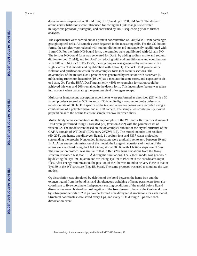
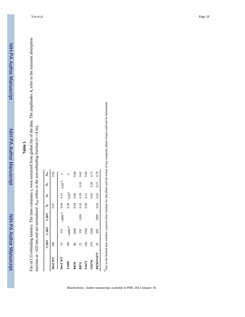
CO complexIn the oxygen transport and storage proteins hemoglobin and myoglobin, dissociated COdoes not rebind to a significant extent on the timescale prior to ~100 ns, but in sensorproteins like FixL, EcDos (24, 25), and CooA (19) modest to strong rebinding has beenobserved. Dissociation of CO from the GAF domain of DosS leads to geminate rebinding ofonly ~7% of CO, in a single phase with a time constant of 280 ps (Fig. 2, Table 1). In DosT,the main rebinding phase occurs with a similar time constant, 310 ps, but the rebindingduring the time window of our experiment is three times more extensive (~23% has reboundat 4 ns) and additional rebinding phases with time constants of 33 ps and >4 ns (the limitedtime window does not allow determination of the precise extent and rate of this phase) areobserved. These results suggest that the heme pocket is more tightly closed in DosT and/orthat the dissociated CO is maintained in a more favorable position for rebinding.
For both proteins, spectral evolution within a few picoseconds also occurs (not shown),which can be ascribed to heme photophysics (28). After decay of these phases, the spectra(Fig. 2, Inset) become similar to the steady state deoxy-minus-CO spectra.
NO complexesDissociation of the heme-NO bond in the ferrous (Fe2+) NO-complex leads to almostcomplete rebinding in a multiexponential manner in both proteins (Fig. 3A, Table 2), with afastest and dominant phase of ~5 ps. Such features are observed in many heme proteins andare thought to reflect recombination from distinct conformational states (17). The kinetics inboth proteins is similar, with overall rebinding in DosT somewhat faster.
NO can also be dissociated from the ferric (Fe3+) heme-NO complexes. In contrast to theferrous complexes, in the difference spectrum this gives rise to a shift feature of the Soretband to lower wavelengths with a broad maximum below 400 nm (Fig. 3B, Inset), as theSoret absorption of 5-coordinate ferric heme consists of a broad band with a maximumaround 395 nm (29), compared to the ~420 nm absorption maximum of the ferric heme-NO(6-coordinate) complex. We note that the transient spectra are quite different from the steadystate ferric-minus-ferric NO spectra (not shown), as for both, DosT and DosS, the stable
Vos et al. Page 4
Biochemistry. Author manuscript; available in PMC 2013 January 10.
NIH
-PA Author Manuscript
NIH
-PA Author Manuscript
NIH
-PA Author Manuscript
ferric protein is in the aquamet form with a Soret absorption around 406 nm (10, 15). Thesmall induced absorption at the red side of the bleaching, observed at short delay times, isascribed to excited state decay and heme cooling.
NO rebinding to the ferric heme occurs on the tens of picoseconds time scale, slower than toferrous heme (a similar observation was made for horseradish peroxidase (30)) and in a non-exponential way (Fig. 3B). The kinetics are again similar for DosT and DosS, with the latterbeing somewhat slower and with a higher probability for NO to escape from the hemepocket (further rebinding on the timescale 300 ps-4 ns does not occur, not shown).Altogether, for the CO and NO complexes the rebinding kinetics is qualitatively similar forboth sensor domains, but somewhat slower for DosS.
O2 complexExcitation of the oxycomplex of DosT initially leads to a highly asymmetric, red-shiftedspectrum, with a weak induced absorption lobe that mostly disappears with a time constantof ~ 5 ps, and the remaining spectrum being more symmetric (Fig. 4B,C). The initialdifference spectrum is strongly perturbed with respect to the steady-state deoxy-minus-O2spectrum, in a way very similar as in the bacterial oxygen sensor FixL (23). Based on ourtransient Raman analysis of the latter complex (21), we assign the analogous state in DosTpredominantly to a thermally excited 6-coordinate O2-bound state rather than an actual O2-dissociated state, and the corresponding decay to thermal cooling. In DosS, the initialspectrum is also rather asymmetric. However, after decay of the 5-ps phase, the amplitude ofthe remaining spectrum, which is close in shape to the steady-state deoxy-minus-O2spectrum, is strikingly higher compared to DosT, indicating that the dissociation yield on thetimescale > 5 ps is much higher in DosS.
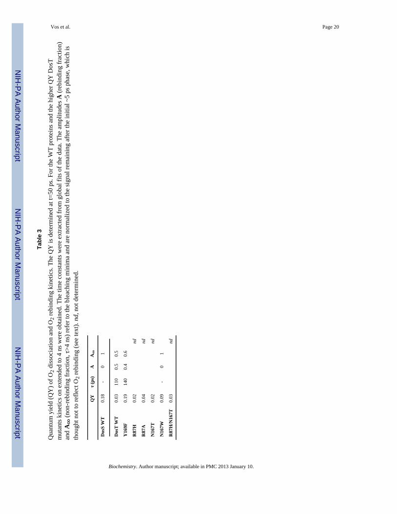
When we determined the quantum yield (QY) of dissociated O2 per absorbed photon at t =50 ps using MbCO as a reference (21, 31), we indeed found a value for DosS of ~0.18 that isalmost an order of magnitude higher than that for DosT (0.03, see Table 3). For comparison,the corresponding value for Mb is 0.26 (31). We conclude that the protein environment ofthe heme in DosT strongly prevents O2 from dissociation.
On the timescale 50 ps-4 ns no O2 recombination was observed for DosS, indicating that the~18% dissociated O2 can escape the protein. By contrast, for DosT about half of the ~3%dissociated O2 rebinds in ~100 ps (see below), further reducing the escape yield of O2 fromthe protein. This difference in O2 escape yield suggests differences in signalling reactivity ofthe proteins upon thermal heme-O2 dissociation (see Discussion).
Mutant DosT GAF A domainsTo further investigate the origin of the relatively low yields of ligand escape in DosT, weperformed experiments on the CO and O2 complexes of mutant DosT GAF A domains. Adistal tyrosine (Tyr169 in DosT) is in close interaction with heme-bound O2 via its OHgroup (15) (Inset Fig. 1B); this residue is part of a ligand-binding GAF motif and alsoconserved in DosS, where it has been shown to play an important role in liganddiscrimination (16). Non-conserved residues in close contact with Tyr169 include Arg87and Asp167, which are respectively His and Thr in DosS; both residues are thought to aligna ligand exchange pathway (15). Consequently we constructed mutants Y169F, R87H,N167T and the R87H/N167T double mutant and, in order to investigate steric effects, thesingle mutants R87A and N167W.
Fig. 5 shows the CO rebinding data for these mutant sensor domains; fit results are collectedin Table 1. All proteins show significant and multiexponential geminate rebinding. In theR87H, N167T and R87H/N167T mutant proteins that are altered towards the corresponding
Vos et al. Page 5
Biochemistry. Author manuscript; available in PMC 2013 January 10.
NIH
-PA Author Manuscript
NIH
-PA Author Manuscript
NIH
-PA Author Manuscript

residues in DosS, recombination is somewhat less extensive than in WT DosT. Interestingly,only the R87H single mutation suppresses all longer (> 1 ns) recombination phases; its COrebinding properties are similar to those of WT DosS. This finding suggests that His in placeof the more flexible Arg at position 87 allows the creation of a CO docking site where, afterinitial relaxation phases, recombination is strongly hindered. The much stronger andcontinuing recombination observed in the R87A mutant is consistent with a steric effect ofthe residue at position 87. In contrast, the influence of the residue at position 167 appearsmore subtle, as replacement of Asp167 by the bulky Trp only has a modest effect. Finally, inthe Y169F mutant protein, CO rebinding is markedly enhanced, in general agreement withthe close proximity of Tyr169 with the heme-bound ligand.
The characteristics of oxygen photodissociation and rebinding are summarized in Table 3.With the exception of the N167W substitution, the photodissociation QY for the mutationsinvolving Arg87 and Asp167 is similar as for WT, indicating that the amino acid differencesat positions 87 and 167 between DosS and DosT are not at the origin of the differences inQY. The higher QY (0.09) observed for N167W points at a steric effect disrupting theinteraction of bound oxygen with its environment.
The most dramatic effect, an increase in the dissociation QY of nearly an order of magnitudecompared to WT, was observed in the DosT Y169F substitution (Fig. 6, Table 3). Thisfinding points to a crucial role of Tyr169 in maintaining O2 in a favourable position forbinding. The rebinding kinetics of the dissociated O2, on the timescale > 5 ps, is very similarin Y169F and WT DosT. Altogether, remarkably, the overall effect of the Y169F mutation isan inversion of the ligand escape properties for CO and O2 in this sensor.
Molecular dynamics simulationsTo better understand the role of Tyr169 in the low QY of O2 dissociation, moleculardynamics simulations were performed on the WT and Y169F GAF domain models. In theseclassical simulations, impulsive Fe-O2 bond dissociation was imposed in independenttrajectories, but bond reformation was not taken into account. Therefore rather thanmonitoring directly the yield of dissociated O2 on the picosecond timescale (as measured inour experiments), information on the positioning of O2 immediately after dissociation isobtained.
Fig. 7 shows the evolution of characteristic parameters in the Fe-O2 interaction followingdissociation. The evolution appears generally similar, and in particular in both modelswithin the first ~100 fs the heme-binding oxygen atom (O1) moves away from the heme ironand the O2 molecule rotates towards the heme plane. However, close comparison shows thatin the Y169F substitution, both the Fe-O1 distancing and the O2 rotational motions can besignificantly further extended than in WT. These observations indicate that the H-bondinginteraction between Tyr169 and the O2 molecule keeps the dissociated ligand closer to theheme-bound configuration in the WT model. These features may allow O2 to moreefficiently reform a bond with the heme on the timescale of 100 fs or less, and are thereforein qualitative agreement with the experimental QY findings described above.
DiscussionLike in other sensor proteins (19, 23–25), rebinding of NO and CO with heme was found tobe efficient, indicating that the heme pocket acts as an effective ligand trap. Ultrafastrecombination of NO and heme is often used to probe the heme environment (17). For DosSand DosT, picosecond heme-NO rebinding is very efficient and the kinetic properties arevery similar, both in the ferrous and ferric state (Fig. 3), although the overall kinetics are
Vos et al. Page 6
Biochemistry. Author manuscript; available in PMC 2013 January 10.
NIH
-PA Author Manuscript
NIH
-PA Author Manuscript
NIH
-PA Author Manuscript

somewhat slower in DosS and in the ferric state the escape probability is also higher forDosS, pointing at a higher ligand mobility.
Picosecond CO rebinding in DosS is sizeable but modest (7%) (in many heme proteins,geminate CO rebinding does not occur (17)), and can be described by a single exponential(280 ps). Here, in the simplest model, upon CO dissociation competition occurs betweenoccupation (in ~300 ps) of a site from which rebinding is very inefficient and rebinding withheme (in ~4 ns). In contrast, in DosT, more extensive and multiexponential heme-COrebinding is observed, suggesting transiently a heterogeneous distribution of configurationsfrom which rebinding is possible. Investigation of the DosT mutants (Fig. 5) indicates thatthe R87H mutation alters the CO rebinding kinetics towards those observed for DosS. Thisindicates that the flexibility of Arg87 (His in DosS) in particular allows dissociated CO tooccupy a range of conformations in the distal heme cavity, including those from whichrebinding to the heme can occur in competition with escape through a ligand entry and exittunnel, which has been proposed to start out near this residue (15). In DosS and in the DosTR87H substitution, the rigid His residue may block access of dissociated CO to the hemeand/or facilitate access to the ligand tunnel. The opposite effect of the R87A mutation is inagreement with the proposed steric role of this residue at position 87. We remark that thedifference in CO binding properties between DosT and DosS must involve the subtleinterplay between a number of residues and not uniquely the His residue, as in the R87H/N167T double mutant CO rebinding is more extensive than in the R87H single substitutionand the WT DosS protein. Furthermore the effects of the R87H and N167T mutations arenot additive.
The most remarkable differences between DosT and DosS that we observed concern O2dissociation patterns. DosT displays a very low O2 photodissociation yield, similar to whathas been observed in the PAS-domain sensor proteins FixL and EcDos (23), implying thatthe heme domain acts as an effective O2 trap. By contrast, in DosS the yield is much higherand approaches that of the oxygen storage protein myoglobin (Fig. 4, Table 3). Aspreviously shown for FixL (21), these differences probably arise from very early dynamicprocesses occurring within the 100 fs timescale and involving interactions with distalresidues that can keep O2 close to the heme after dissociation, thus favoring ultrafastrebinding. In FixL, a hydrogen bond between a distal arginine and O2 plays an importantrole in this interaction (20, 21). In DosT, Tyr169, that forms a strong hydrogen bond withthe heme-ligated O2 (15), is likely implicated in such interactions. Indeed, mutation of thistyrosine to phenylalanine, which eliminates this H-bond, results in a much higher O2 escapeyield (Fig. 7, Table 3). Our MD simulations indicate that this occurs by releasing themotional restrictions on the 100 fs timescale imposed by the H-bond (Fig. 7). Yet, thisfinding does not explain the difference between DosT and DosS in this respect, as DosS alsocarries Tyr at the corresponding position. Mutation of the nearby non-conserved residuesArg87 and Asp167 in DosT did not lead to significantly higher escape yields, indicating thatyet to be identified further differences between DosT and DosS are involved. However, theensemble of our results does strongly suggest that the hydrogen bond between the distaltyrosine and O2, although clearly present (16), is weaker in DosS than in DosT. Theobserved insensitivity of the oxygen binding and autoxidation properties of DosS towardsmutation of the distal tyrosine (11) is in agreement with this proposal. Determination of thecrystal structure of the DosS sensor domain oxycomplex may further clarify this issue.
The rate of O2 escape from the protein due to thermal dissociation of the heme Fe-O2 bond(the oxygen off rate) has been suggested to be similar for DosT and DosS, although adirectly determined O2 off rate for DosT is not available (7). If this is correct, and assumingthat the intrinsic rate for thermal dissociation of the Fe-O2 bond is similar for DosT andDosS, our data, showing a much higher escape yield for DosS than for DosT on the time
Vos et al. Page 7
Biochemistry. Author manuscript; available in PMC 2013 January 10.
NIH
-PA Author Manuscript
NIH
-PA Author Manuscript
NIH
-PA Author Manuscript

scale of 4 ns, suggests that in DosS additional O2 rebinding phases take place in the timewindow of 4 ns to ~1 ms (that has not been investigated so far). This would imply that inDosS rebinding may take place simultaneously while the ligand switching process is alreadyin progress, a feature that may make fine tuning of the sensor efficiency more sensitive toenvironmental factors. By contrast, the DosT sensor functions with a mechanism where theswitch would fully proceed once the oxygen has escaped the heme pocket (a relatively rareevent), as we previously proposed for FixL and EcDos (23). In this view, the initiallyoperating DosT sensor acts more like other heme-based gas sensors (see Introduction).
The most prominent effect of our mutation studies is observed for the Y169F mutant, bothon CO and O2 dynamics. This is not surprising, as Tyr169 is in closest contact with theligand in the available X-ray structures and is proposed to be involved in the switchingpathway (15). However it is interesting to note that the effect is inverted for CO and O2:whereas the hydrogen bond with Tyr169 clearly constrains O2 in the heme pocket asdiscussed above, allowing much more O2 escape from the heme pocket in the mutant, thesame Y169F mutation leads to a much lower escape yield for CO. Assuming this lattereffect is due to the difference in hydrogen bonding capacity, it may reflect suppression of ahydrogen bond of Tyr169 with CO or, alternatively, a different site. The first possibility isunlikely as in this case hydrogen bonding would be expected to lead to less rather than moreCO escape, as for instance observed in the R220H mutation in FixL (20). Furthermore, atleast in DosS, hydrogen bonding of the distal tyrosine to heme-bound CO hardly occurs(16). Regarding the second possibility, Tyr169 has been invoked to play a key role in theinitial signalling pathway. One possible mechanism involves locking the residue in adifferent configuration in the presence of other ligands than O2 via interaction with anotherhydrogen-bond partner, either a residue or a water molecule. Such a mechanism has beeninvoked to explain the ligand discrimination effect of the corresponding tyrosine in DosS(16). In this mechanism, mutation to Phe would unlock the distal residue so that it canaccommodate a position similar to that in the oxygen bound form and favour COmaintenance close to the heme iron after dissociation and thus enhance fast geminaterecombination and decrease the escape yield. Our results suggest that such a hydrogenbonding “switch” takes place in DosT signal transmission and also that Tyr169 plays animportant role in ligand discrimination, as it does in DosS (16). Further detailing of themolecular mechanism, and in particular possible hydrogen bond partners in the active form,must await determination of the crystal structure of the CO-bound form or application ofappropriate time-resolved vibrational spectroscopic techniques.
Abbreviations
Mb myoglobin
QY quantum yield
WT wild type
References1. World Health Organization. Global Tuberculosis Control. 2010.2. Wayne LG, Sohaskey CD. Nonreplicating persistence of Mycobaterium tuberculosis. Ann. Rev.
Microbiol. 2001; 55:139–163. [PubMed: 11544352]3. Nathan C, Shiloh MU. Reactive oxygen and nitrogen intermediates in the relationship between
mammalian hosts and microbial pathogens. Proc. Natl. Acad. Sci. U.S.A. 2000; 97:8841–8848.[PubMed: 10922044]
Vos et al. Page 8
Biochemistry. Author manuscript; available in PMC 2013 January 10.
NIH
-PA Author Manuscript
NIH
-PA Author Manuscript
NIH
-PA Author Manuscript

4. Sherman DR, Voskuil M, Schnappinger D, Liao R, Harrell MI, Schoolnik GK. Regulation of theMycobacterium tuberculosis hypoxic response gene encoding α-crystallin. Proc. Natl. Acad. Sci.U.S.A. 2001; 98:7534–7539. [PubMed: 11416222]
5. Roberts DM, Liao RP, Wisedchaisri G, Hol WGJ, Sherman DR. Two Sensor Kinases Contribute tothe Hypoxic Response of Mycobacterium tuberculosis. J. Biol. Chem. 2004; 279:23082–23087.[PubMed: 15033981]
6. Sardiwal S, Kendall SL, Movahedzadeh F, Rison SCG, Stoker NG, Djordjevic S. A GAF Domain inthe Hypoxia/NO-inducible Mycobacterium tuberculosis DosS Protein Binds Haem. J. Mol. Biol.2005; 353:929–936. [PubMed: 16213520]
7. Sousa EHS, Tuckerman JR, Gonzalez G, Gilles-Gonzalez MA. DosT and DevS are oxygen-switched kinases in Mycobacterium tuberculosis. Protein Sci. 2007; 16:1708–1719. [PubMed:17600145]
8. Honaker RW, Leistikow RL, Bartek IL, Voskuil MI. Unique Roles of DosT and DosS in DosRRegulon Induction and Mycobacterium tuberculosis Dormancy. Infect. Immun. 2009; 77:3258–3263. [PubMed: 19487478]
9. Kim M-J, Park K-J, Ko I-J, Kim YM, Oh J-I. Different Roles of DosS and DosT in the HypoxicAdaptation of Mycobacteria. J. Bacteriol. 2010; 192:4868–4875. [PubMed: 20675480]
10. Ioanoviciu A, Yukl ET, Moënne-Loccoz P, Ortiz de Montellano PR. DevS, a Heme-ContainingTwo-Component Oxygen Sensor of Mycobacterium tuberculosis. Biochemistry. 2007; 46:4250–4260. [PubMed: 17371046]
11. Ioanoviciu A, Meharenna YT, Poulos TL, Ortiz de Montellano PR. DevS Oxy Complex StabilityIdentifies This Heme Protein as a Gas Sensor in Mycobacterium tuberculosis Dormancy.Biochemistry. 2009; 48:5839–5848. [PubMed: 19463006]
12. Kumar A, Toledo JC, Patel RP, Lancaster JR, Steyn AJC. Mycobacterium tuberculosis DosS is aredox sensor and DosT is a hypoxia sensor. Proc. Natl. Acad. Sci. U.S.A. 2007; 104:11568–11573.[PubMed: 17609369]
13. Yukl ET, Ioanoviciu A, Sivaramakrishnan S, Nakano MM, Ortiz de Montellano PR, Moënne-Loccoz P. Nitric Oxide Dioxygenation Reaction in DevS and the Initial Response to Nitric Oxidein Mycobacterium tuberculosis. Biochemistry. 2011; 50:1023–1028. [PubMed: 21250657]
14. Cho HY, Cho HJ, Kim YM, Oh JI, Kang BS. Structural Insight into the Heme-based RedoxSensing by DosS from Mycobacterium tuberculosis. J. Biol. Chem. 2009; 284:13057–13067.[PubMed: 19276084]
15. Podust LM, Ioanoviciu A, Ortiz de Montellano PR. 2.3 Ã X-ray Structure of the Heme-BoundGAF Domain of Sensory Histidine Kinase DosT of Mycobacterium tuberculosis. Biochemistry.2008; 47:12523–12531. [PubMed: 18980385]
16. Yukl ET, Ioanoviciu A, Nakano MM, Ortiz de Montellano PR, Moënne-Loccoz P. A DistalTyrosine Residue Is Required for Ligand Discrimination in DevS from Mycobacteriumtuberculosis. Biochemistry. 2008; 47:12532–12539. [PubMed: 18975917]
17. Vos MH. Ultrafast dynamics of ligands within heme proteins. Biochim. Biophys. Acta. 2008;1777:15–31. [PubMed: 17996720]
18. Négrerie M, Bouzhir-Sima L, Martin J-L, Liebl U. Control of nitric oxide dynamics by guanylatecyclase in its activated state. J. Biol. Chem. 2001; 276:46815–46821. [PubMed: 11590135]
19. Kumazaki S, Nakajima H, Sakaguchi T, Nakagawa E, Shinahara H, Yoshihara K, Aono S.Dissociation and recombination between ligands and heme in a CO-sensing transcriptionalactivator CooA. J. Biol. Chem. 2000; 275:38378–38383. [PubMed: 10978334]
20. Jasaitis A, Hola K, Bouzhir-Sima L, Lambry J-C, Balland V, Vos MH, Liebl U. Role of distalarginine in early sensing intermediates in the heme domain of the oxygen sensor FixL.Biochemistry. 2006; 45:6018–6026. [PubMed: 16681374]
21. Kruglik SG, Jasaitis A, Hola K, Yamashita T, Liebl U, Martin J-L, Vos MH. SubpicosecondOxygen Trapping in the Heme Pocket of the Oxygen Sensor FixL Observed by Time-ResolvedResonance Raman Spectroscopy. Proc. Natl. Acad. Sci. U.S.A. 2007; 104:7408–7413. [PubMed:17446273]
22. Lechauve C, Bouzhir-Sima L, Yamashita T, Marden MC, Vos MH, Liebl U, Kiger L. HemeLigand Binding Properties and Intradimer Interactions in the Full-length Sensor Protein Dos from
Vos et al. Page 9
Biochemistry. Author manuscript; available in PMC 2013 January 10.
NIH
-PA Author Manuscript
NIH
-PA Author Manuscript
NIH
-PA Author Manuscript

Escherichia coli and Its Isolated Heme Domain. J. Biol. Chem. 2009; 284:36146–36159.[PubMed: 19864414]
23. Liebl U, Bouzhir-Sima L, Négrerie M, Martin J-L, Vos MH. Ultrafast ligand rebinding in the hemedomain of the oxygen sensors FixL and Dos: general regulatory implications for heme-basedsensors. Proc. Natl. Acad. Sci. U.S.A. 2002; 99:12771–12776. [PubMed: 12271121]
24. Liebl U, Bouzhir-Sima L, Kiger L, Marden MC, Lambry J-C, Négrerie M, Vos MH. Ligandbinding dynamics to the heme domain of the oxygen sensor Dos from Escherichia coli.Biochemistry. 2003; 42:6527–6535. [PubMed: 12767236]
25. Yamashita T, Bouzhir-Sima L, Lambry J-C, Liebl U, Vos MH. Ligand dynamics and earlysignalling events in the heme domain of the sensor protein Dos from Escherichia coli. J. Biol.Chem. 2008; 283:2344–2352. [PubMed: 18039668]
26. Silkstone G, Jasaitis A, Wilson MT, Vos MH. Ligand dynamics in an electron-transfer protein:picosecond geminate recombination of carbon monoxide to heme in mutant forms of cytochromec. J. Biol. Chem. 2007; 282:1638–1649. [PubMed: 17114183]
27. Brooks BR, Bruccoleri RE, Olafson BD, Swaminathan S, Karplus M. CHARMM: A Program forMacromolecular Energy, Minimization, and Dynamics Calculations. J. Comput. Chem. 1983;4:187–212.
28. Petrich JW, Poyart C, Martin J-L. Photophysics and reactivity of heme proteins: a femtosecondabsorption study of hemoglobin, myoglobin and protoheme. Biochemistry. 1988; 27:4049–4060.[PubMed: 3415972]
29. Gilles-Gonzalez MA, Gonzalez G, Perutz MF, Kiger L, Marden MC, Poyart C. Heme-basedsensors, exemplified by the kinase FixL, are a new class of heme protein with distinctive ligandbinding and autoxidation. Biochemistry. 1994; 33:8067–8073. [PubMed: 8025112]
30. Ye X, Yu A, Champion PM. Dynamics of Nitric Oxide Rebinding and Escape in HorseradishPeroxidase. J. Am. Chem. Soc. 2006; 128:1444–1445. [PubMed: 16448103]
31. Ye X, Demidov A, Champion PM. Measurements of the Photodissociation Quantum Yields ofMbNO and MbO2 and the Vibrational Relaxation of the Six-Coordinate Heme Species. J. Am.Chem. Soc. 2002; 124:5914–5924. [PubMed: 12010067]
32. Pettersen EF, Goddard TD, Huang CC, Couch GS, Greenblatt DM, Meng EC, Ferrin TE. UCSFChimera—A visualization system for exploratory research and analysis. J. Comp. Chem. 2004;25:1605–1612. [PubMed: 15264254]
Vos et al. Page 10
Biochemistry. Author manuscript; available in PMC 2013 January 10.
NIH
-PA Author Manuscript
NIH
-PA Author Manuscript
NIH
-PA Author Manuscript

Fig. 1.Ground-state absorption spectra of different ligation states of GAF domains of DosS (A) andDosT (B): ferrous unliganded (black), ferrous CO-bound (green), ferrous O2-bound (red),ferrous NO-bound (blue) and ferric NO-bound (cyan). Inset: Structure of the distal hemepocket of the oxycomplex of the DosT GAF A domain (PDB 2VZW). The position ofPhe169 in the Y169F mutant protein, as modelled by our molecular dynamics simulations, isalso represented, in violet. Figure produced with UCSF Chimera (32).
Vos et al. Page 11
Biochemistry. Author manuscript; available in PMC 2013 January 10.
NIH
-PA Author Manuscript
NIH
-PA Author Manuscript
NIH
-PA Author Manuscript
Fig. 2.Kinetics of CO rebinding to the GAF domains of DosS (red) and DosT (blue), measured at435 nm, the maximum of the transient absorption spectra. Inset Transient spectra of DosT-CO at delay times of 5 ps (cyan), 45 ps (orange), 200 ps (red), 1 ns (blue) and 4 ns (green).
Vos et al. Page 12
Biochemistry. Author manuscript; available in PMC 2013 January 10.
NIH
-PA Author Manuscript
NIH
-PA Author Manuscript
NIH
-PA Author Manuscript

Fig. 3.Kinetics of NO rebinding to the ferrous (A) and ferric (B) GAF domains of DosS (red) andDosT (blue), measured at the bleaching maxima at 418 nm (A) and 420 nm (B). The Insetsshow transient spectra for ferrous DosT-NO (A) at delay times of 0.4 ps (blue), 3 ps (black),9 ps (green), 50 ps (red) and 300 ps (cyan) and ferric DosS-NO (B) at delay times of 0.8 ps(blue), 3 ps (black), 20 ps (green), 50 ps (red) and 300 ps (cyan).
Vos et al. Page 13
Biochemistry. Author manuscript; available in PMC 2013 January 10.
NIH
-PA Author Manuscript
NIH
-PA Author Manuscript
NIH
-PA Author Manuscript

Fig. 4.Transient spectra after excitation of the O2 complex of GAF domains of DosS (A) and DosT(B) at delay times of 0.5 ps (straight line) and 50 ps (dashed line). C: Normalized kinetics at435 nm for DosS (straight line) and DosT (dashed line).
Vos et al. Page 14
Biochemistry. Author manuscript; available in PMC 2013 January 10.
NIH
-PA Author Manuscript
NIH
-PA Author Manuscript
NIH
-PA Author Manuscript

Fig. 5.Kinetics of CO rebinding to the WT and mutant GAF domains of DosT, measured at 435nm.
Vos et al. Page 15
Biochemistry. Author manuscript; available in PMC 2013 January 10.
NIH
-PA Author Manuscript
NIH
-PA Author Manuscript
NIH
-PA Author Manuscript

Fig. 6.Kinetics at 435 nm after excitation of the O2 complex of WT (open circles) and Y169F(closed circles) DosT. The decay after ~20 ps reflects O2 recombination.
Vos et al. Page 16
Biochemistry. Author manuscript; available in PMC 2013 January 10.
NIH
-PA Author Manuscript
NIH
-PA Author Manuscript
NIH
-PA Author Manuscript

Fig. 7.Molecular dynamics simulations of O2 dissociation in WT (A) and Y169F (B) DosT. O2was dissociated at t=0 ps. Dotted curves represent independent trajectories and bold solidcurves averaged trajectories.
Vos et al. Page 17
Biochemistry. Author manuscript; available in PMC 2013 January 10.
NIH
-PA Author Manuscript
NIH
-PA Author Manuscript
NIH
-PA Author Manuscript
NIH
-PA Author Manuscript
NIH
-PA Author Manuscript
NIH
-PA Author Manuscript
Vos et al. Page 18
Tabl
e 1
Fits
of C
O re
bind
ing
kine
tics.
The
time
cons
tant
s τi w
ere
extra
cted
from
glo
bal f
its o
f the
dat
a. T
he a
mpl
itude
s Ai r
efer
to th
e tra
nsie
nt a
bsor
ptio
nm
axim
a at
~43
5 nm
and
are
nor
mal
ized
. A∞
refe
rs to
the
non-
rebi
ndin
g fr
actio
n (τ
>>4
ns).
τ 1 (p
s)τ 2
(ps)
τ 3 (p
s)A
1A
2A
3A∞
Dos
S W
T28
00.
070.
93
Dos
T W
T33
310
>400
0 a
0.04
0.13
0.83
aa
Y16
9F10
0>4
000
a0.
380.
62a
a
R87
H88
1000
0.04
0.08
0.88
R87
A12
150
2200
0.10
0.19
0.10
0.61
N16
7T33
037
000.
090.
110.
80
N16
7W27
025
000.
050.
200.
75
R87
H/N
167T
2640
028
000.
040.
020.
150.
79
a Due
to th
e lim
ited
time
win
dow
a p
reci
se ti
me
cons
tant
for t
his p
hase
and
the
exte
nt o
f any
sym
ptot
ic p
hase
(A∞
) cou
ld n
ot b
e de
term
ined
.
Biochemistry. Author manuscript; available in PMC 2013 January 10.

NIH
-PA Author Manuscript
NIH
-PA Author Manuscript
NIH
-PA Author Manuscript
Vos et al. Page 19
Tabl
e 2
Fits
of N
O re
bind
ing
kine
tics.
The
time
cons
tant
s τi w
ere
extra
cted
from
glo
bal f
its o
f the
dat
a. T
he a
mpl
itude
s Ai r
efer
to th
e bl
each
ing
min
ima
and
are
norm
aliz
ed. A
∞ re
fers
to th
e no
n-re
bind
ing
frac
tion
(τ>4
ns)
.
τ 1 (p
s)τ 2
(ps)
τ 3 (p
s)A
1A
2A
3A∞
Ferr
ous
Dos
S W
T4.
616
380
0.65
0.27
0.07
0.01
Dos
T W
T5.
741
0.79
0.19
0.02
Ferr
ic
Dos
S W
T8
470.
230.
700.
07
Dos
T W
T17
640.
640.
350.
01
Biochemistry. Author manuscript; available in PMC 2013 January 10.
NIH
-PA Author Manuscript
NIH
-PA Author Manuscript
NIH
-PA Author Manuscript
Vos et al. Page 20
Tabl
e 3
Qua
ntum
yie
ld (Q
Y) o
f O2 d
isso
ciat
ion
and
O2 r
ebin
ding
kin
etic
s. Th
e Q
Y is
det
erm
ined
at t
=50
ps. F
or th
e W
T pr
otei
ns a
nd th
e hi
gher
QY
Dos
Tm
utan
ts k
inet
ics o
n ex
tend
ed to
4 n
s wer
e ob
tain
ed. T
he ti
me
cons
tant
s wer
e ex
tract
ed fr
om g
loba
l fits
of t
he d
ata.
The
am
plitu
des A
(reb
indi
ng fr
actio
n)an
d A∞
(non
-reb
indi
ng fr
actio
n, τ>
4 ns
) ref
er to
the
blea
chin
g m
inim
a an
d ar
e no
rmal
ized
to th
e si
gnal
rem
aini
ng a
fter t
he in
itial
~5
ps p
hase
, whi
ch is
thou
ght n
ot to
refle
ct O
2 reb
indi
ng (s
ee te
xt).
nd, n
ot d
eter
min
ed.
QY
τ (p
s)A
A∞
Dos
S W
T0.
18-
01
Dos
T W
T0.
0311
00.
50.
5
Y16
9F0.
1914
00.
40.
6
R87
H0.
02nd
R87
A0.
04nd
N16
7T0.
02nd
N16
7W0.
09-
01
R87
H/N
167T
0.03
nd
Biochemistry. Author manuscript; available in PMC 2013 January 10.
Related Documents


