Ultra-thin Descemet Stripping Automated Endothelial Keratoplasty (UT-DSAEK) – why I prefere this technique? prof. Iva Dekaris University Eye Hospital “Svjetlost”, Zagreb, Medical School, University of Rijeka, Croatia 5th International Conference on Clinical & Experimental Ophthalmology, Valencia, Spain, 2015

Welcome message from author
This document is posted to help you gain knowledge. Please leave a comment to let me know what you think about it! Share it to your friends and learn new things together.
Transcript
Ultra-thin Descemet Stripping Automated
Endothelial Keratoplasty (UT-DSAEK)
– why I prefere this technique?
prof. Iva Dekaris
University Eye Hospital “Svjetlost”, Zagreb,
Medical School, University of Rijeka, Croatia
5th International Conference on Clinical & Experimental Ophthalmology,
Valencia, Spain, 2015
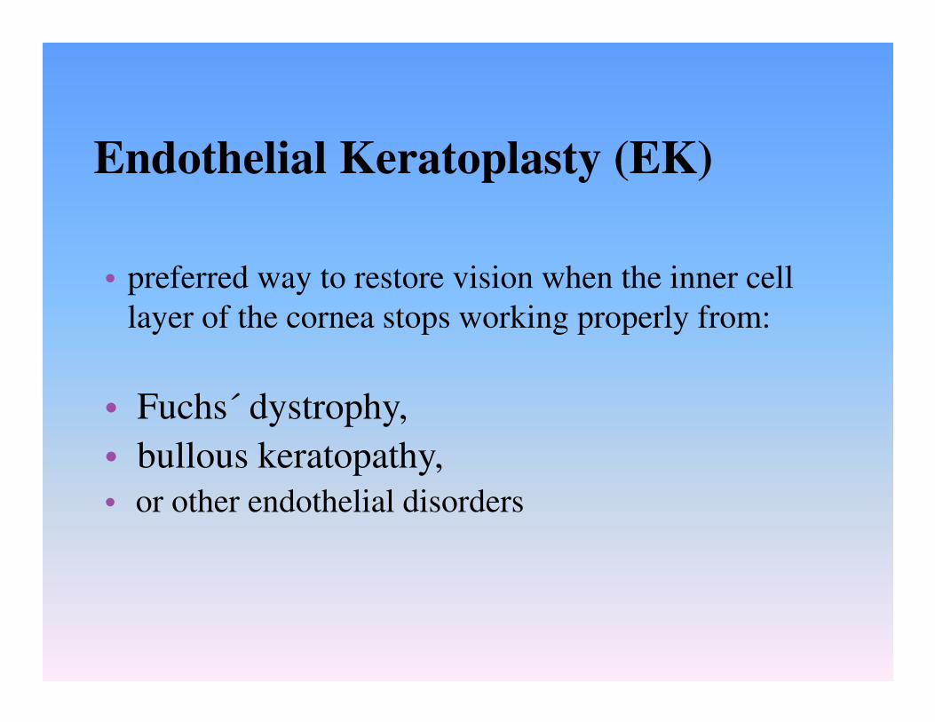
Endothelial Keratoplasty (EK)
• preferred way to restore vision when the inner cell
layer of the cornea stops working properly from:
• Fuchs´ dystrophy,
• bullous keratopathy,
• or other endothelial disorders
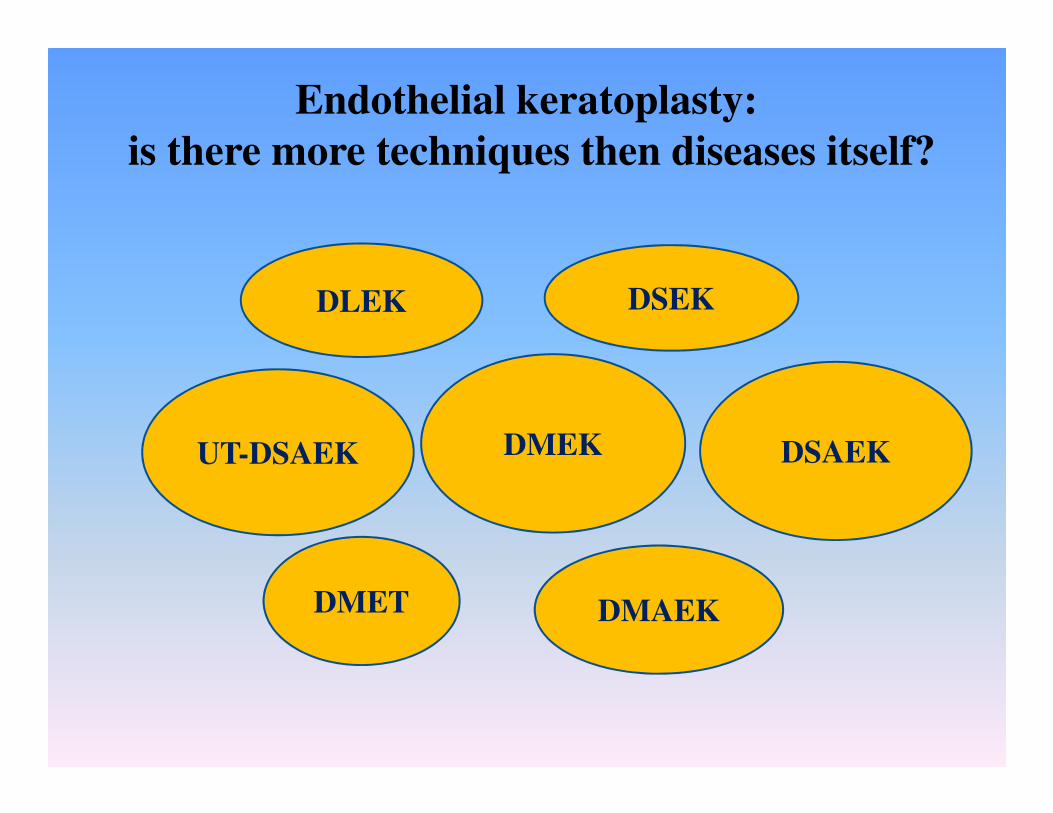
DLEK
DSAEKDMEK
DMAEK
Endothelial keratoplasty:
is there more techniques then diseases itself?
UT-DSAEK
DMET
DSEK
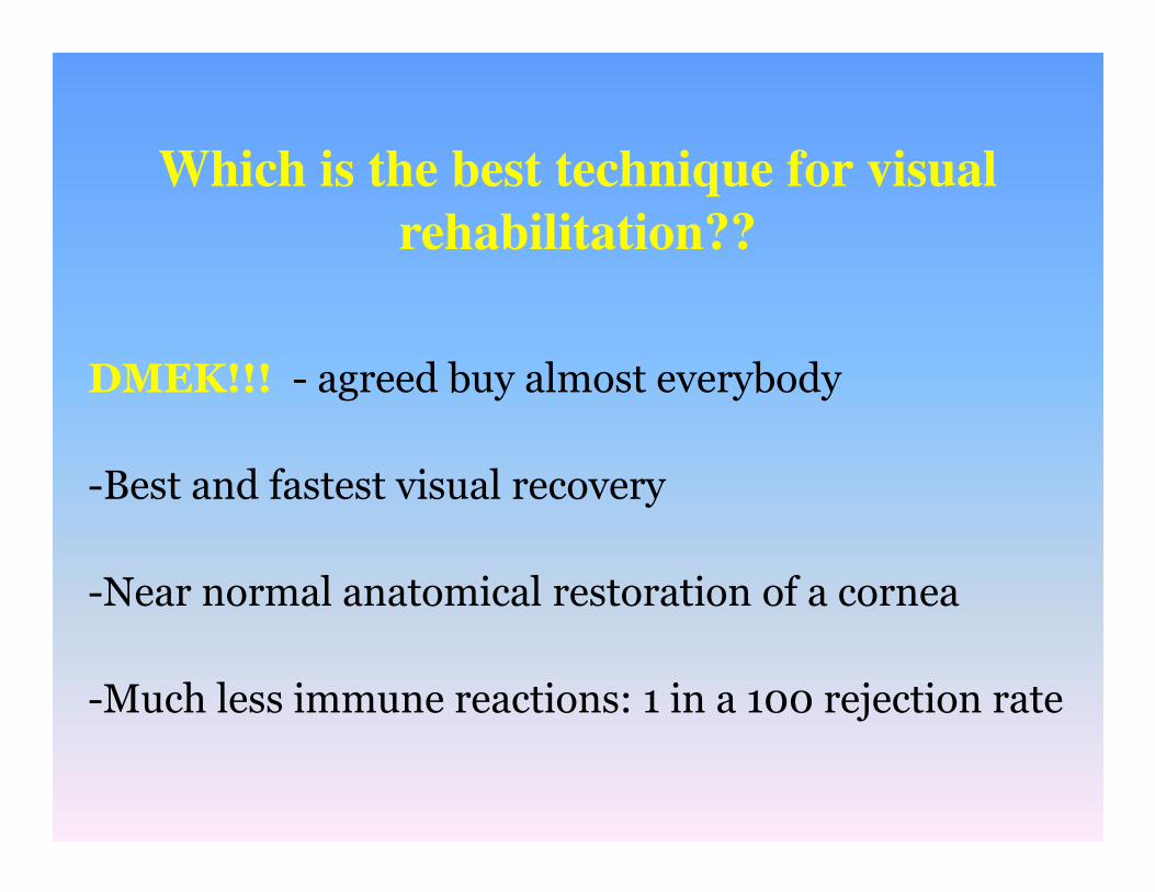
Which is the best technique for visual
rehabilitation??
DMEK!!! - agreed buy almost everybody
-Best and fastest visual recovery
-Near normal anatomical restoration of a cornea
-Much less immune reactions: 1 in a 100 rejection rate
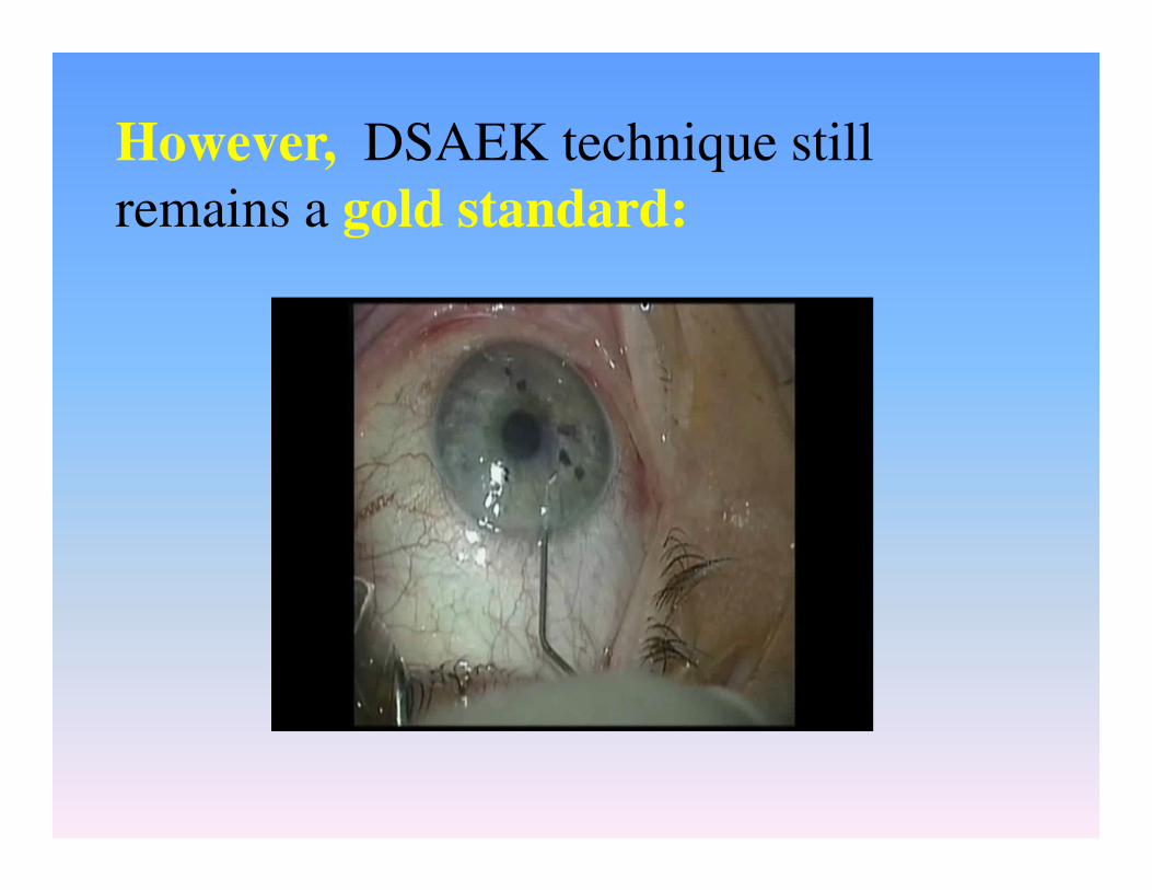
However, DSAEK technique still
remains a gold standard:
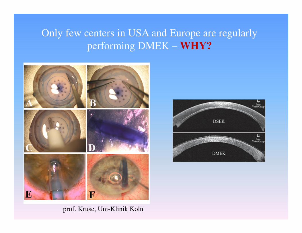
Only few centers in USA and Europe are regularly
performing DMEK – WHY?
prof. Kruse, Uni-Klinik Koln
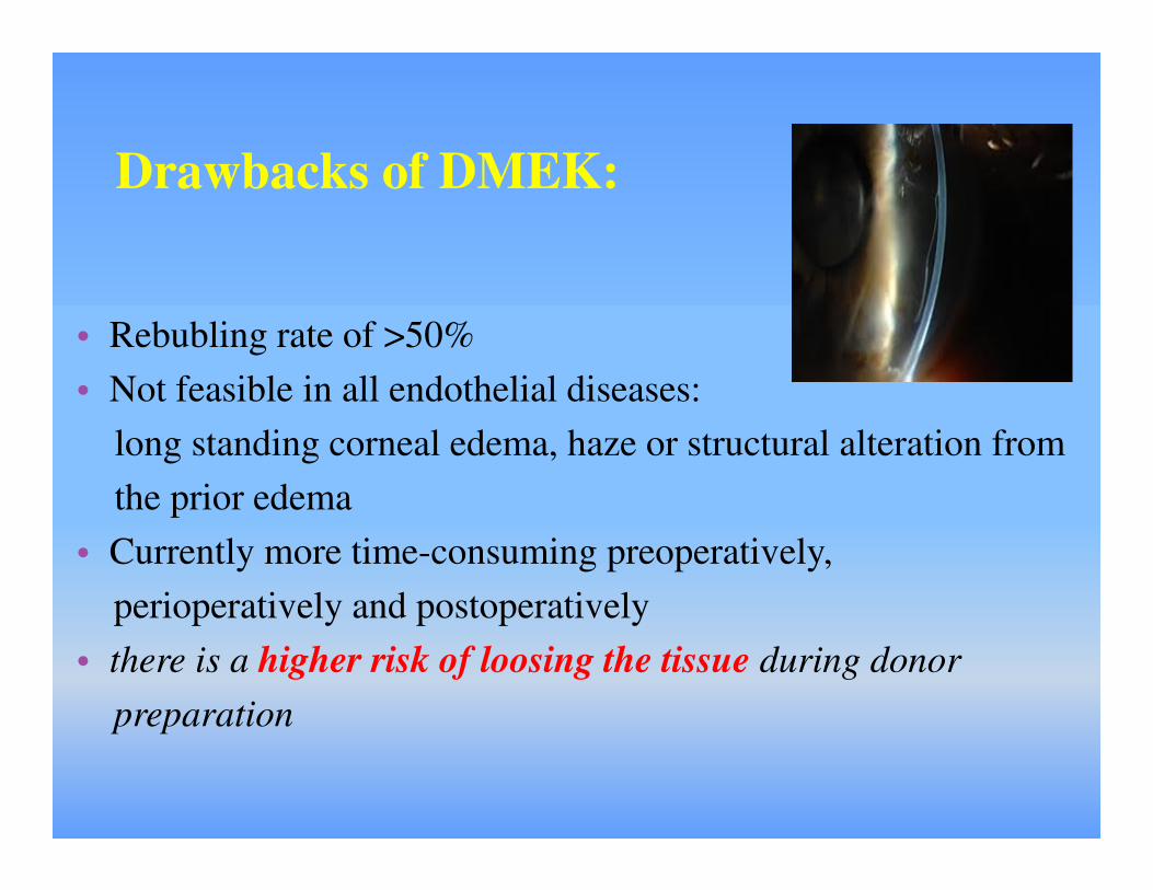
Drawbacks of DMEK:
• Rebubling rate of >50%
• Not feasible in all endothelial diseases:
long standing corneal edema, haze or structural alteration from
the prior edema
• Currently more time-consuming preoperatively,
perioperatively and postoperatively
• there is a higher risk of loosing the tissue during donor
preparation
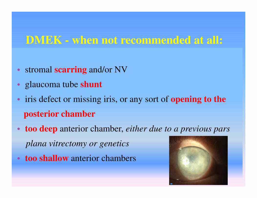
DMEK - when not recommended at all:
• stromal scarring and/or NV
• glaucoma tube shunt
• iris defect or missing iris, or any sort of opening to the
posterior chamber
• too deep anterior chamber, either due to a previous pars
plana vitrectomy or genetics
• too shallow anterior chambers
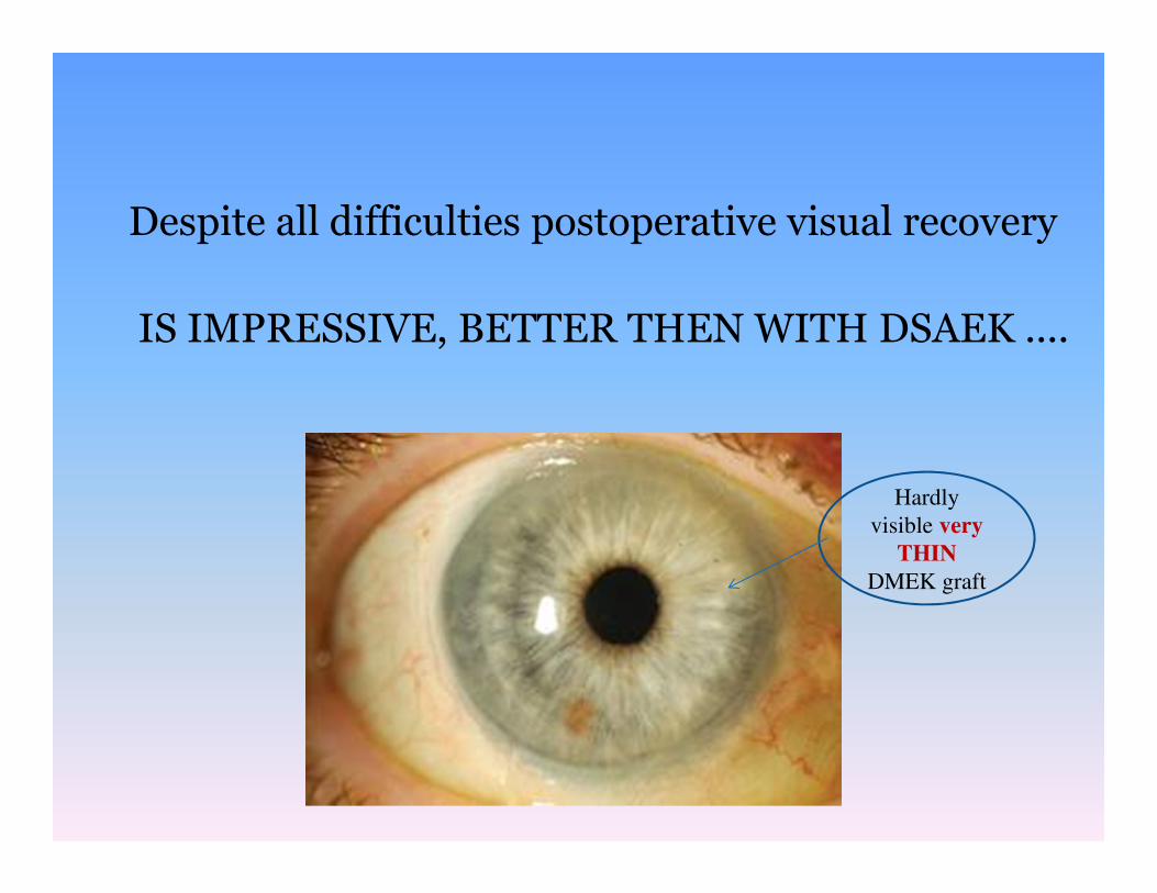
Despite all difficulties postoperative visual recovery
IS IMPRESSIVE, BETTER THEN WITH DSAEK ....
Hardly
visible very
THIN
DMEK graft
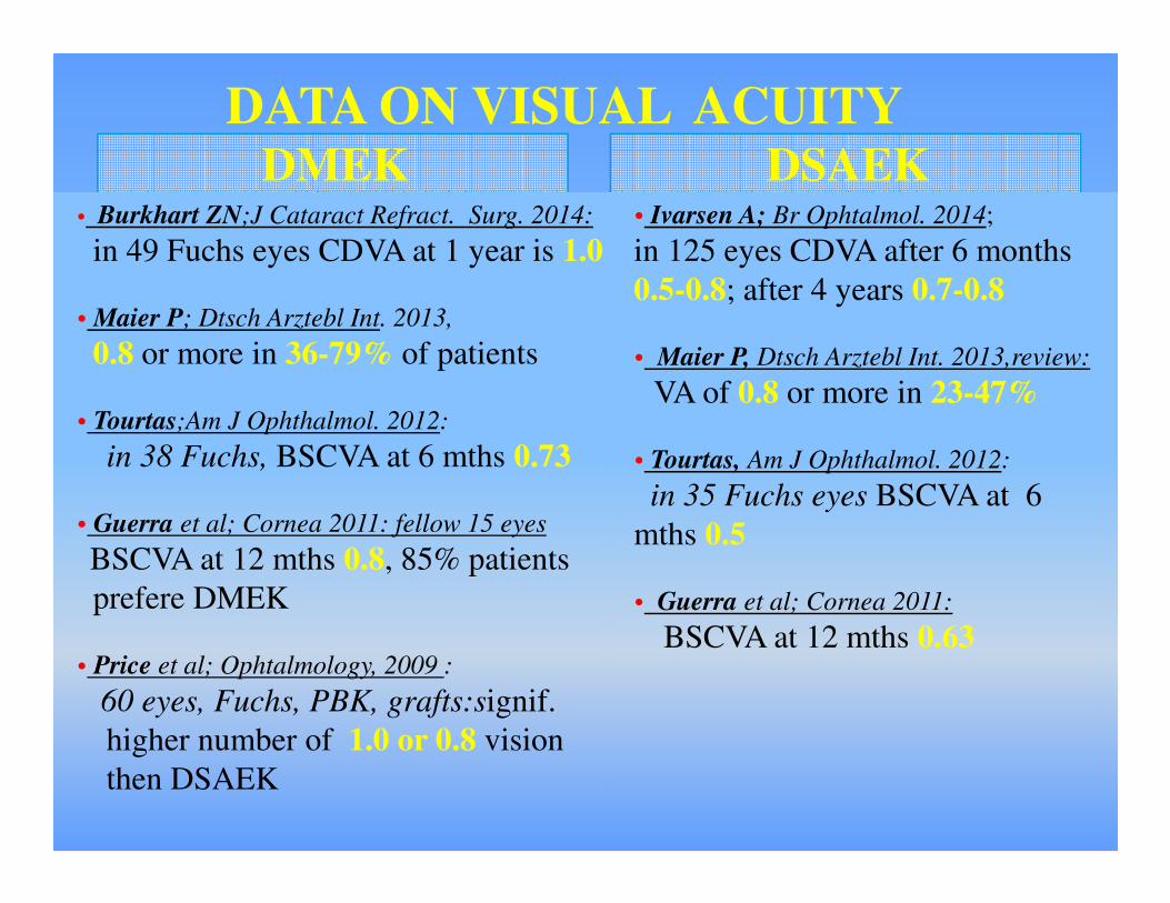
DMEK DSAEK• Burkhart ZN;J Cataract Refract. Surg. 2014:
in 49 Fuchs eyes CDVA at 1 year is 1.0
• Maier P; Dtsch Arztebl Int. 2013,
0.8 or more in 36-79% of patients
• Tourtas;Am J Ophthalmol. 2012:
in 38 Fuchs, BSCVA at 6 mths 0.73
• Guerra et al; Cornea 2011: fellow 15 eyes
BSCVA at 12 mths 0.8, 85% patients
prefere DMEK
• Price et al; Ophtalmology, 2009 :
60 eyes, Fuchs, PBK, grafts:signif.
higher number of 1.0 or 0.8 vision
then DSAEK
• Ivarsen A; Br Ophtalmol. 2014;
in 125 eyes CDVA after 6 months
0.5-0.8; after 4 years 0.7-0.8
• Maier P, Dtsch Arztebl Int. 2013,review:
VA of 0.8 or more in 23-47%
• Tourtas, Am J Ophthalmol. 2012:
in 35 Fuchs eyes BSCVA at 6
mths 0.5
• Guerra et al; Cornea 2011:
BSCVA at 12 mths 0.63
DATA ON VISUAL ACUITY
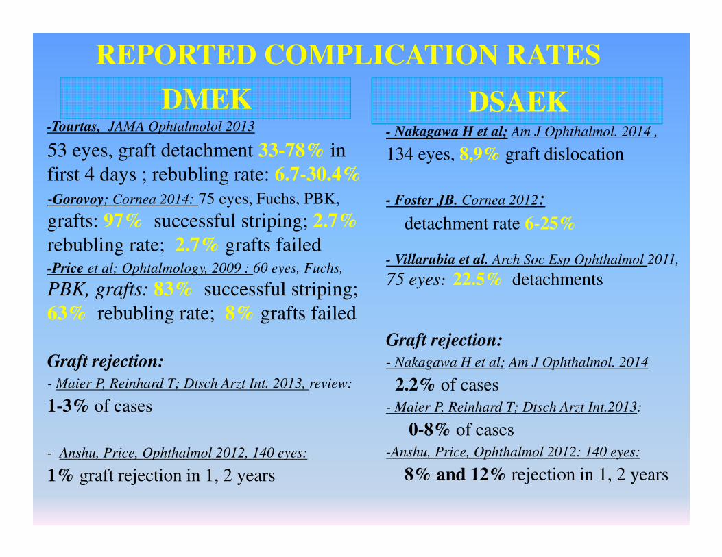
DMEK-Tourtas, JAMA Ophtalmolol 2013
53 eyes, graft detachment 33-78% in
first 4 days ; rebubling rate: 6.7-30.4%
-Gorovoy; Cornea 2014: 75 eyes, Fuchs, PBK,
grafts: 97% successful striping; 2.7%
rebubling rate; 2.7% grafts failed-Price et al; Ophtalmology, 2009 : 60 eyes, Fuchs,
PBK, grafts: 83% successful striping;
63% rebubling rate; 8% grafts failed
Graft rejection:
- Maier P, Reinhard T; Dtsch Arzt Int. 2013, review:
1-3% of cases
- Anshu, Price, Ophthalmol 2012, 140 eyes:
1% graft rejection in 1, 2 years
- Nakagawa H et al; Am J Ophthalmol. 2014 ,
134 eyes, 8,9% graft dislocation
- Foster JB. Cornea 2012:
detachment rate 6-25%
- Villarubia et al. Arch Soc Esp Ophthalmol 2011,
75 eyes: 22.5% detachments
Graft rejection:
- Nakagawa H et al; Am J Ophthalmol. 2014
2.2% of cases
- Maier P, Reinhard T; Dtsch Arzt Int.2013:
0-8% of cases
-Anshu, Price, Ophthalmol 2012: 140 eyes:
8% and 12% rejection in 1, 2 years
DSAEK
REPORTED COMPLICATION RATES
However, THERE ARE LACKING COMPARISONS
WITH ultra-thin DSAEK, ......
DMEK UT-DSAEK
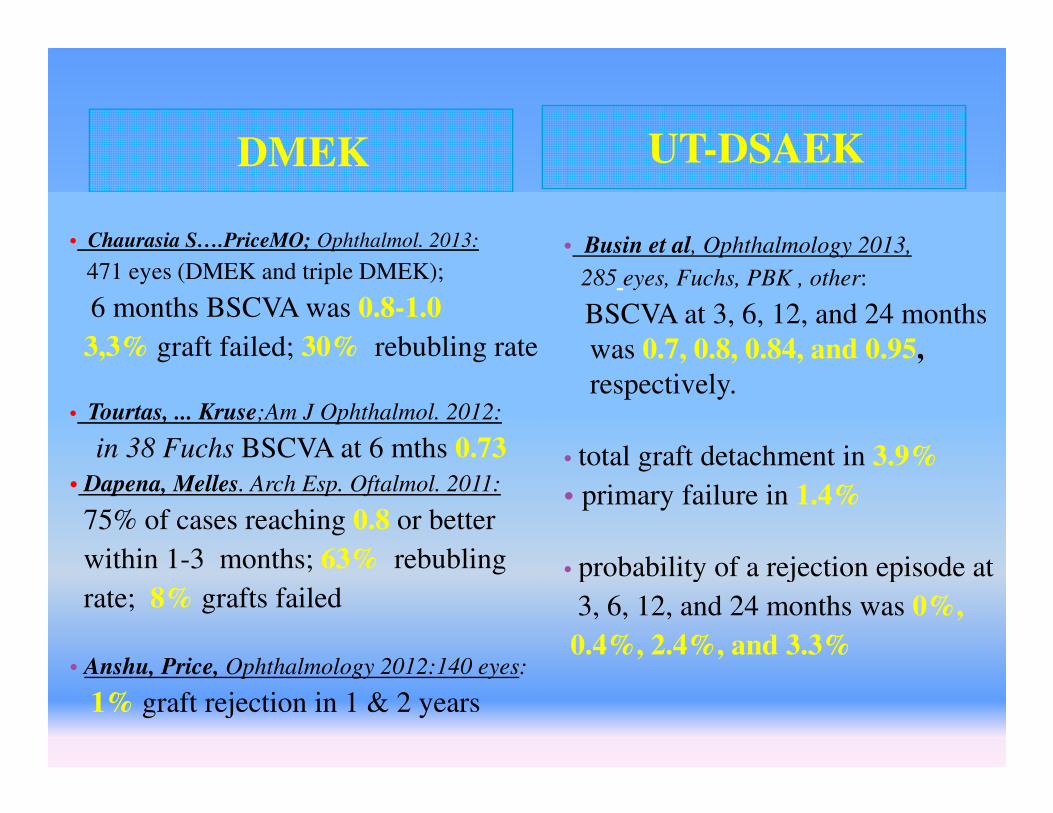
• Chaurasia S….PriceMO; Ophthalmol. 2013:
471 eyes (DMEK and triple DMEK);
6 months BSCVA was 0.8-1.0
3,3% graft failed; 30% rebubling rate
• Tourtas, ... Kruse;Am J Ophthalmol. 2012:
in 38 Fuchs BSCVA at 6 mths 0.73
• Dapena, Melles. Arch Esp. Oftalmol. 2011:
75% of cases reaching 0.8 or better
within 1-3 months; 63% rebubling
rate; 8% grafts failed
• Anshu, Price, Ophthalmology 2012:140 eyes:
1% graft rejection in 1 & 2 years
• Busin et al, Ophthalmology 2013,
285 eyes, Fuchs, PBK , other:
BSCVA at 3, 6, 12, and 24 months
was 0.7, 0.8, 0.84, and 0.95,
respectively.
• total graft detachment in 3.9%
• primary failure in 1.4%
• probability of a rejection episode at
3, 6, 12, and 24 months was 0%,
0.4%, 2.4%, and 3.3%
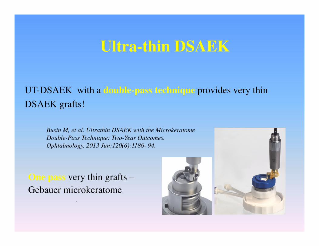
Ultra-thin DSAEK
UT-DSAEK with a double-pass technique provides very thin
DSAEK grafts!
Busin M, et al. Ultrathin DSAEK with the Microkeratome
Double-Pass Technique: Two-Year Outcomes.
Ophtalmology. 2013 Jun;120(6):1186- 94.
One pass very thin grafts –
Gebauer microkeratome.
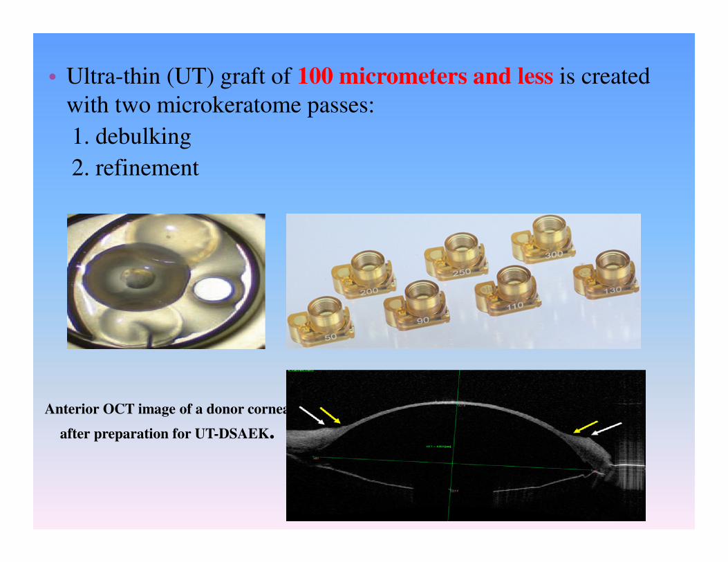
• Ultra-thin (UT) graft of 100 micrometers and less is created
with two microkeratome passes:
1. debulking
2. refinement
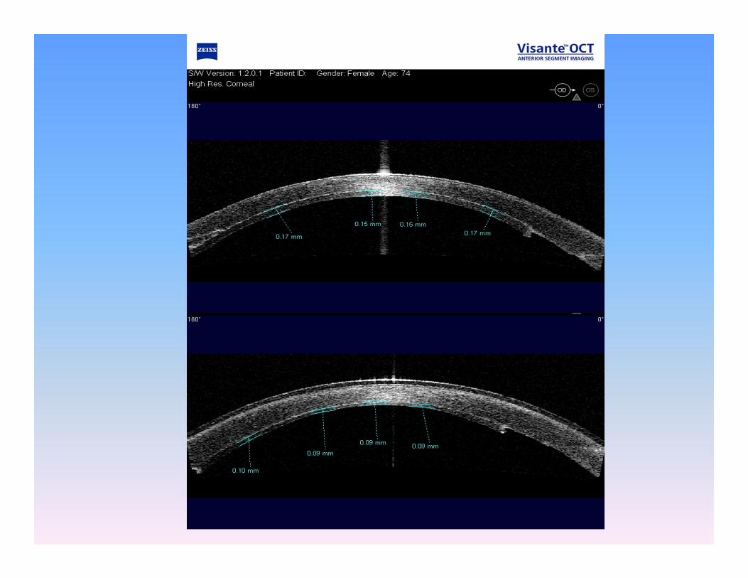
Anterior OCT image of a donor cornea
after preparation for UT-DSAEK.
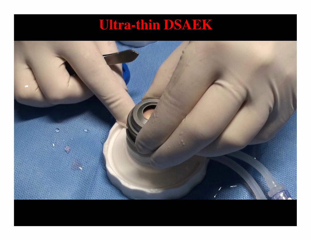
Ultra-thin DSAEK
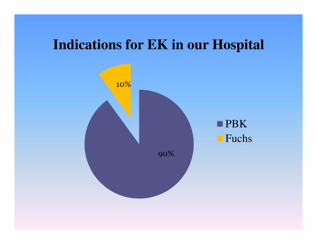
PBK
Fuchs
90%
10%
Indications for EK in our Hospital
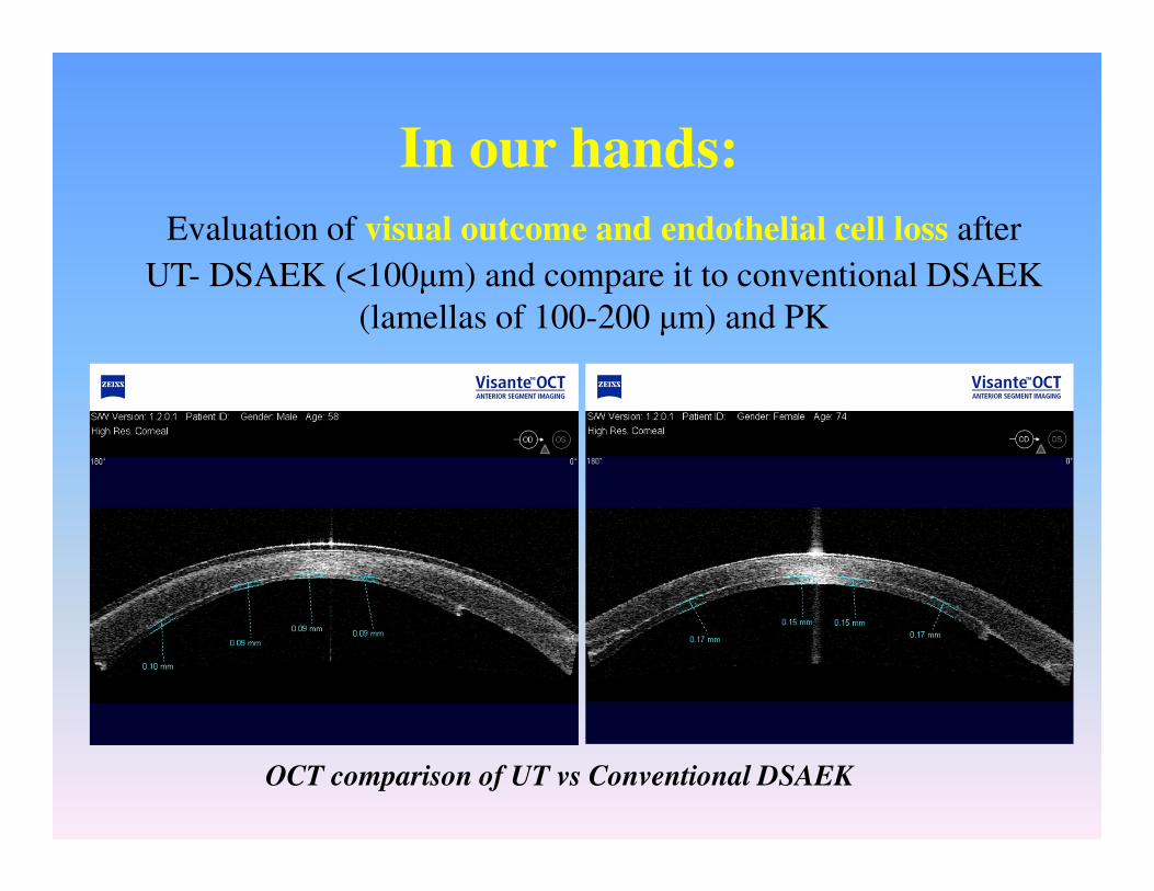
In our hands:
OCT comparison of UT vs Conventional DSAEK
Evaluation of visual outcome and endothelial cell loss after
UT- DSAEK (<100µm) and compare it to conventional DSAEK
(lamellas of 100-200 µm) and PK
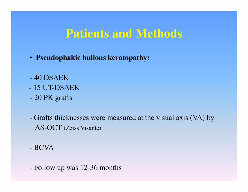
Patients and Methods
• Pseudophakic bullous keratopathy:
- 40 DSAEK
- 15 UT-DSAEK
- 20 PK grafts
- Grafts thicknesses were measured at the visual axis (VA) by
AS-OCT (Zeiss Visante)
- BCVA
- Follow up was 12-36 months
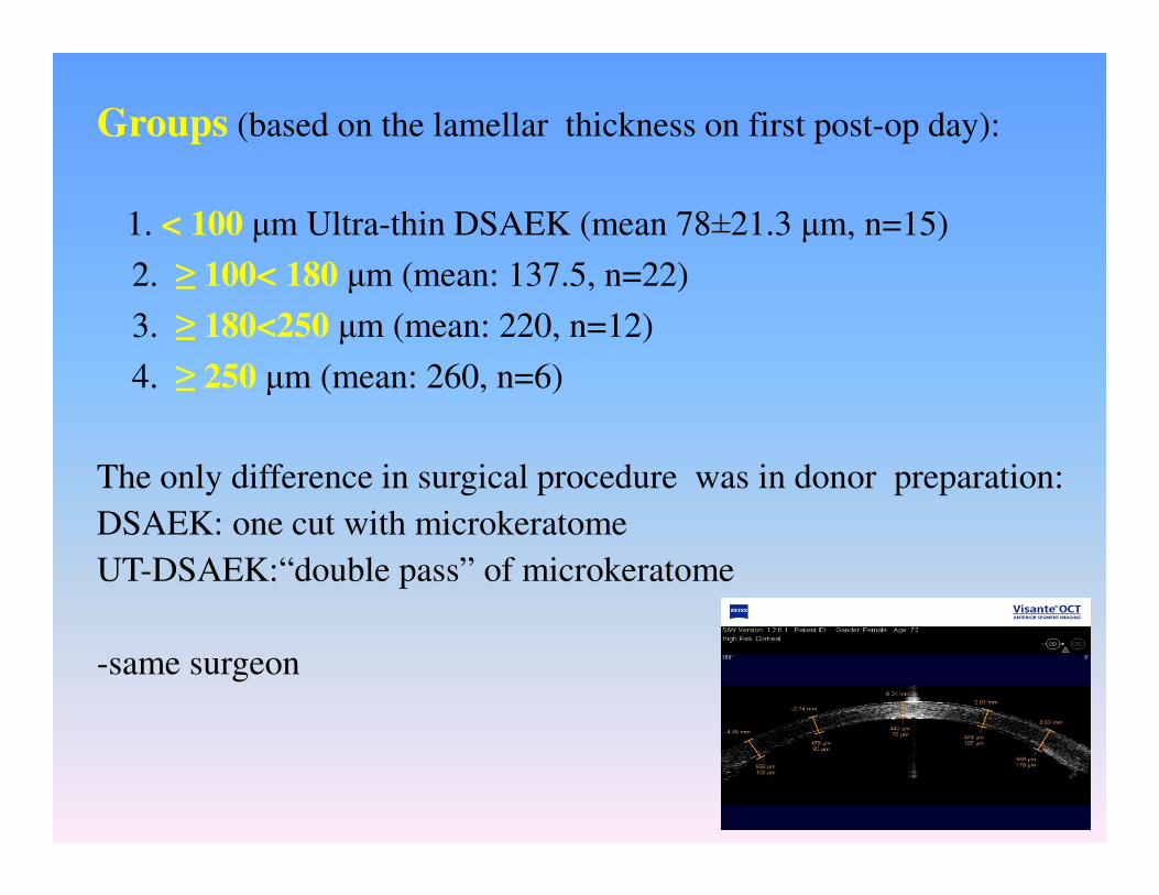
Groups (based on the lamellar thickness on first post-op day):
1. < 100 µm Ultra-thin DSAEK (mean 78±21.3 µm, n=15)
2. ≥ 100< 180 µm (mean: 137.5, n=22)
3. ≥ 180<250 µm (mean: 220, n=12)
4. ≥ 250 µm (mean: 260, n=6)
The only difference in surgical procedure was in donor preparation:
DSAEK: one cut with microkeratome
UT-DSAEK:“double pass” of microkeratome
-same surgeon
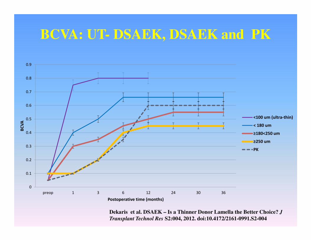
BCVA: UT- DSAEK, DSAEK and PK
0
0.1
0.2
0.3
0.4
0.5
0.6
0.7
0.8
0.9
preop 1 3 6 12 24 30 36
BC
VA
Postoperative time (months)
<100 um (ultra-thin)
< 180 um
≥180<250 um
≥250 um
PK
Dekaris et al. DSAEK – Is a Thinner Donor Lamella the Better Choice? J
Transplant Technol Res S2:004, 2012. doi:10.4172/2161-0991.S2-004
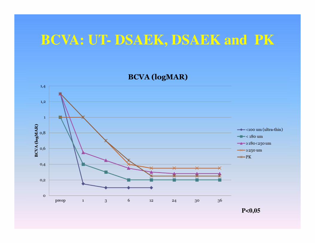
BCVA: UT- DSAEK, DSAEK and PK
P<0,05
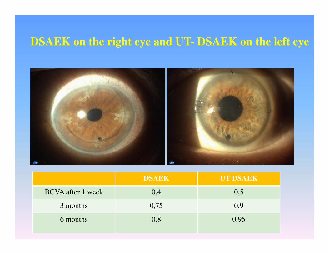
DSAEK on the right eye and UT- DSAEK on the left eye
DSAEK UT DSAEK
BCVA after 1 week 0,4 0,5
3 months 0,75 0,9
6 months 0,8 0,95
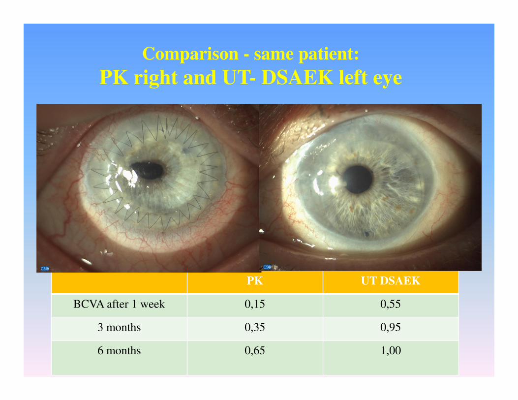
Comparison - same patient:
PK right and UT- DSAEK left eye
PK UT DSAEK
BCVA after 1 week 0,15 0,55
3 months 0,35 0,95
6 months 0,65 1,00
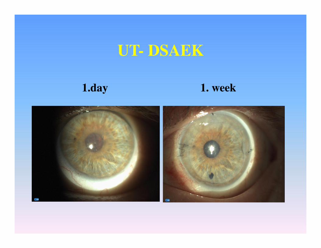
UT- DSAEK
1. week1.day
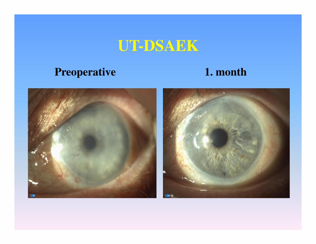
UT-DSAEK
1. monthPreoperative
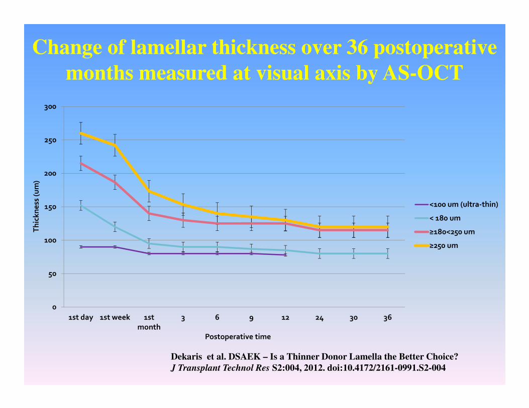
Change of lamellar thickness over 36 postoperative
months measured at visual axis by AS-OCT
0
50
100
150
200
250
300
1st day 1st week 1st
month
3 6 9 12 24 30 36
Th
ick
ne
ss (
um
)
Postoperative time
<100 um (ultra-thin)
< 180 um
≥180<250 um
≥250 um
Dekaris et al. DSAEK – Is a Thinner Donor Lamella the Better Choice?
J Transplant Technol Res S2:004, 2012. doi:10.4172/2161-0991.S2-004
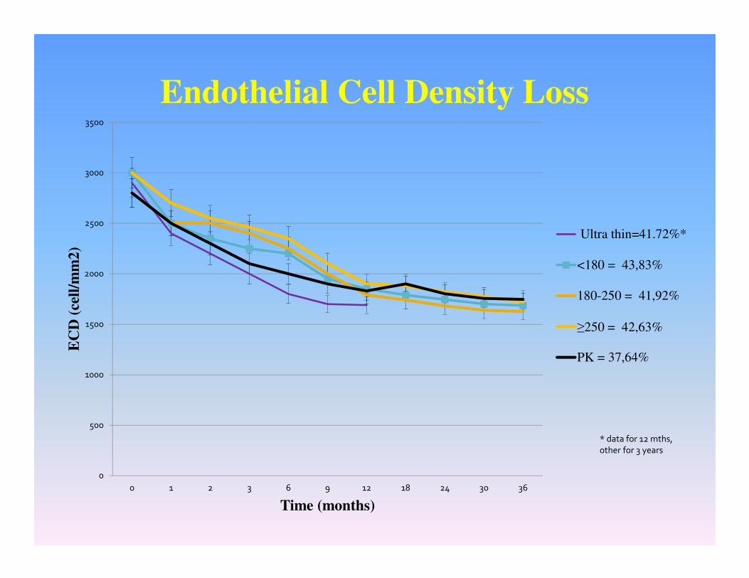
0
500
1000
1500
2000
2500
3000
3500
0 1 2 3 6 9 12 18 24 30 36
EC
D (
cell
/mm
2)
Time (months)
Endothelial Cell Density Loss
Ultra thin=41.72%*
<180 = 43,83%
180-250 = 41,92%
≥250 = 42,63%
PK = 37,64%
* data for 12 mths,
other for 3 years
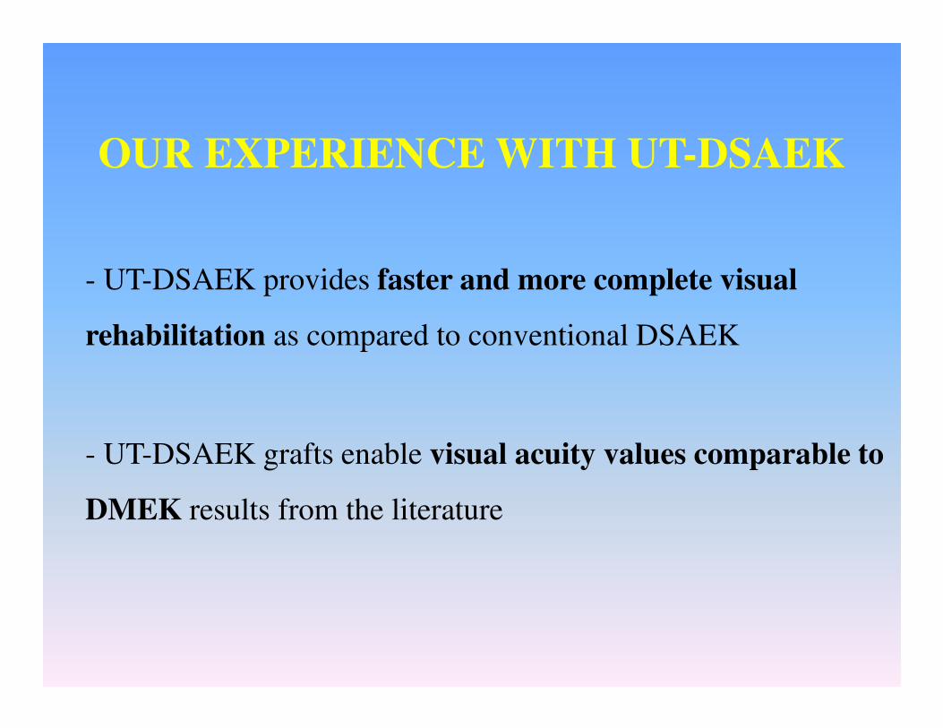
OUR EXPERIENCE WITH UT-DSAEK
- UT-DSAEK provides faster and more complete visual
rehabilitation as compared to conventional DSAEK
- UT-DSAEK grafts enable visual acuity values comparable to
DMEK results from the literature
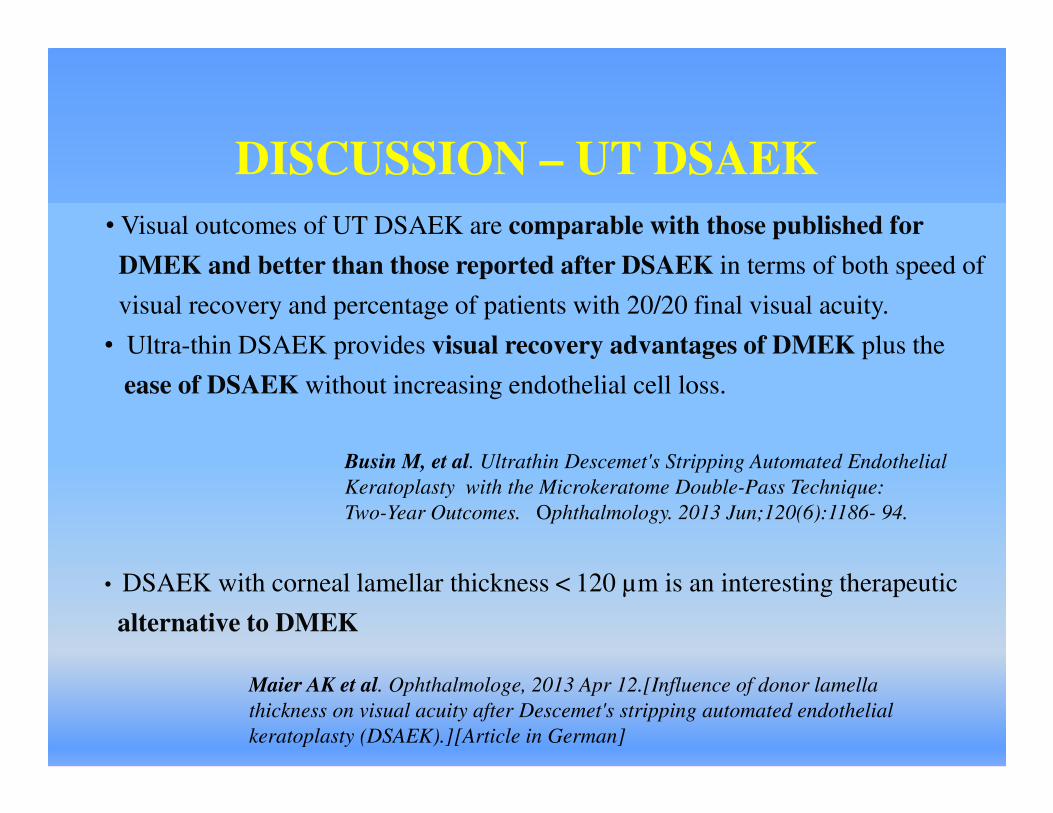
DISCUSSION – UT DSAEK
• Visual outcomes of UT DSAEK are comparable with those published for
DMEK and better than those reported after DSAEK in terms of both speed of
visual recovery and percentage of patients with 20/20 final visual acuity.
• Ultra-thin DSAEK provides visual recovery advantages of DMEK plus the
ease of DSAEK without increasing endothelial cell loss.
Busin M, et al. Ultrathin Descemet's Stripping Automated Endothelial
Keratoplasty with the Microkeratome Double-Pass Technique:
Two-Year Outcomes. Ophthalmology. 2013 Jun;120(6):1186- 94.
• DSAEK with corneal lamellar thickness < 120 µm is an interesting therapeutic
alternative to DMEK
Maier AK et al. Ophthalmologe, 2013 Apr 12.[Influence of donor lamella
thickness on visual acuity after Descemet's stripping automated endothelial
keratoplasty (DSAEK).][Article in German]
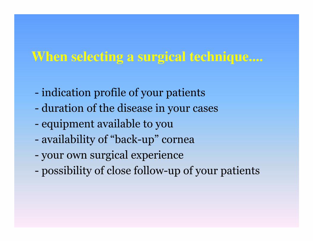
When selecting a surgical technique....
- indication profile of your patients
- duration of the disease in your cases
- equipment available to you
- availability of “back-up” cornea
- your own surgical experience
- possibility of close follow-up of your patients
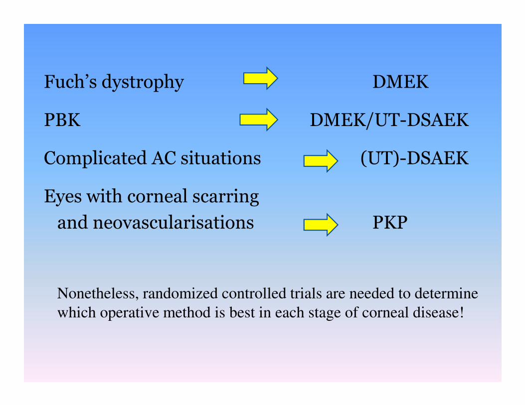
Fuch’s dystrophy DMEK
PBK DMEK/UT-DSAEK
Complicated AC situations (UT)-DSAEK
Eyes with corneal scarring
and neovascularisations PKP
Nonetheless, randomized controlled trials are needed to determine
which operative method is best in each stage of corneal disease!
If you end up with (UT)-DSAEK as a choice, always
try to keep your grafts AS THIN AS POSSIBLE .....
Chapter 7
Ultra-Thin Descemet's
Stripping
Automated Endothelial
Keratoplasty
(UT-DSAEK)
Iva Dekaris
University Eye Hospital „Svjetlost‟,
Zagreb, Croatia

Thank you!
Related Documents