RESEARCH ARTICLE Open Access Ubiquitous presence of piscidin-1 in Atlantic cod as evidenced by immunolocalisation Jareeporn Ruangsri 1 , Jorge M O Fernandes 1 , Jan H W M Rombout 1,2 , Monica F Brinchmann 1 and Viswanath Kiron 1* Abstract Background: Antimicrobial peptides (AMPs), the natural antibiotics bestowed upon all forms of life, consist of small molecular weight proteins with a broad spectrum antimicrobial activity against a variety of pathogenic microorganisms. Piscidins are one of the AMP families that are imperative for the innate defence mechanisms of teleosts. Atlantic cod, a basal fish belonging to the superorder Paracanthopterygii also possesses multiple piscidin peptides. Two piscidin paralogues (pis1 and pis2) and a novel alternative splice variant of pis2 of this fish were previously described by us. To shed light on other potent roles of these molecules, now we have mapped the distribution of piscidin 1 (Pis1), in different tissues and organs of cod through immunohistochemistry (IHC) employing an affinity purified polyclonal antibody specific to Pis1. Results: Various cell types and tissues of Atlantic cod including those from the immune organs of naïve fish are armed with Pis1 peptide. Different types of the blood leucocytes and phagocytic cells among the leucocytes examined gave a relatively strong indication of Pis1 immunopositivity. In addition, other cell types such as hematopoietic cells, epithelial cells and multi-granular cells located in the mucosal and hematopoietic tissues were also Pis1-immunoreactive. More interestingly, chondrocytes appear to produce Pis1 and this is the first report on the presence of an AMP in cartilage tissue of fish. Furthermore, Pis1 immunopositivity was detected in other tissues and organs of naïve fish including neural tissues, exocrine and endocrine glands, compound gland cells, excretory kidney, intestinal and respiratory epithelial cells, swim bladder, skin and hypodermis layer, myosepta, liver, heart, eye and oocytes. Conclusions: Pis1 peptide is produced by various cell types located in different tissues and organs of Atlantic cod. It is present in all immune-related organs of naïve fish and the elevated peptide expression following phagocytosis strongly suggest their involvement in innate defence. Further, its widespread occurrence in non-immune tissues and organs of apparently healthy fish implies that piscidin may have other functions in addition to its role as an immune effector molecule. Keywords: Piscidin, Antimicrobial peptide, Innate immunity, Multi-functionality, Gadus morhua Background Atlantic cod (Gadus morhua L.) is a demersal fish that is widely distributed in the North Atlantic region, the Baltic Sea and the Barents Sea. Commercial production of this fish has been undertaken mainly by Norway, though fraught with several challenges. There has been great inter- est in understanding the immune system of this fish spe- cies. It has been confirmed recently that cod has a unique immune architecture compared to other vertebrates as they are devoid of genes for major histocompatibility complex (MHC) II, cluster of differentiation 4 (CD4) and invariant chain (Ii) [1]; all of them are attributed to a normal func- tioning of adaptive immunity. Earlier studies [2-4] that examined the antibody responses of Atlantic cod have revealed that cod relies more on innate than adaptive de- fence mechanisms. On the other hand, cod exhibits an in- credible ability to defend itself against pathogens [5]. The effective functioning of the innate immune system could be due to the presence of a number of MHC I loci and the unique organization of Toll-like receptor (TLR) families in the genome [1,6]. Furthermore, our contribution to the knowledge on the innate immune components of Atlantic * Correspondence: [email protected] 1 Faculty of Biosciences and Aquaculture, University of Nordland, 8049, Bodø, Norway Full list of author information is available at the end of the article © 2012 Ruangsri et al.; licensee BioMed Central Ltd. This is an Open Access article distributed under the terms of the Creative Commons Attribution License (http://creativecommons.org/licenses/by/2.0), which permits unrestricted use, distribution, and reproduction in any medium, provided the original work is properly cited. Ruangsri et al. BMC Veterinary Research 2012, 8:46 VETERINARY RESEARCH http://www.biomedcentral.com/1746-6148/8/46

Welcome message from author
This document is posted to help you gain knowledge. Please leave a comment to let me know what you think about it! Share it to your friends and learn new things together.
Transcript

Ruangsri et al. BMC Veterinary Research 2012, 8:46
VETERINARY RESEARCHhttp://www.biomedcentral.com/1746-6148/8/46
RESEARCH ARTICLE Open Access
Ubiquitous presence of piscidin-1 in Atlantic codas evidenced by immunolocalisationJareeporn Ruangsri1, Jorge M O Fernandes1, Jan H W M Rombout1,2, Monica F Brinchmann1 and Viswanath Kiron1*
Abstract
Background: Antimicrobial peptides (AMPs), the natural antibiotics bestowed upon all forms of life, consist of smallmolecular weight proteins with a broad spectrum antimicrobial activity against a variety of pathogenicmicroorganisms. Piscidins are one of the AMP families that are imperative for the innate defence mechanisms ofteleosts. Atlantic cod, a basal fish belonging to the superorder Paracanthopterygii also possesses multiple piscidinpeptides. Two piscidin paralogues (pis1 and pis2) and a novel alternative splice variant of pis2 of this fish werepreviously described by us. To shed light on other potent roles of these molecules, now we have mapped thedistribution of piscidin 1 (Pis1), in different tissues and organs of cod through immunohistochemistry (IHC)employing an affinity purified polyclonal antibody specific to Pis1.
Results: Various cell types and tissues of Atlantic cod including those from the immune organs of naïve fish arearmed with Pis1 peptide. Different types of the blood leucocytes and phagocytic cells among the leucocytesexamined gave a relatively strong indication of Pis1 immunopositivity. In addition, other cell types such ashematopoietic cells, epithelial cells and multi-granular cells located in the mucosal and hematopoietic tissues werealso Pis1-immunoreactive. More interestingly, chondrocytes appear to produce Pis1 and this is the first report on thepresence of an AMP in cartilage tissue of fish. Furthermore, Pis1 immunopositivity was detected in other tissues andorgans of naïve fish including neural tissues, exocrine and endocrine glands, compound gland cells, excretorykidney, intestinal and respiratory epithelial cells, swim bladder, skin and hypodermis layer, myosepta, liver, heart, eyeand oocytes.
Conclusions: Pis1 peptide is produced by various cell types located in different tissues and organs of Atlantic cod.It is present in all immune-related organs of naïve fish and the elevated peptide expression following phagocytosisstrongly suggest their involvement in innate defence. Further, its widespread occurrence in non-immune tissuesand organs of apparently healthy fish implies that piscidin may have other functions in addition to its role as animmune effector molecule.
Keywords: Piscidin, Antimicrobial peptide, Innate immunity, Multi-functionality, Gadus morhua
BackgroundAtlantic cod (Gadus morhua L.) is a demersal fish that iswidely distributed in the North Atlantic region, the BalticSea and the Barents Sea. Commercial production of thisfish has been undertaken mainly by Norway, thoughfraught with several challenges. There has been great inter-est in understanding the immune system of this fish spe-cies. It has been confirmed recently that cod has a uniqueimmune architecture compared to other vertebrates as they
* Correspondence: [email protected] of Biosciences and Aquaculture, University of Nordland, 8049, Bodø,NorwayFull list of author information is available at the end of the article
© 2012 Ruangsri et al.; licensee BioMed CentraCommons Attribution License (http://creativecreproduction in any medium, provided the or
are devoid of genes for major histocompatibility complex(MHC) II, cluster of differentiation 4 (CD4) and invariantchain (Ii) [1]; all of them are attributed to a normal func-tioning of adaptive immunity. Earlier studies [2-4] thatexamined the antibody responses of Atlantic cod haverevealed that cod relies more on innate than adaptive de-fence mechanisms. On the other hand, cod exhibits an in-credible ability to defend itself against pathogens [5]. Theeffective functioning of the innate immune system could bedue to the presence of a number of MHC I loci and theunique organization of Toll-like receptor (TLR) families inthe genome [1,6]. Furthermore, our contribution to theknowledge on the innate immune components of Atlantic
l Ltd. This is an Open Access article distributed under the terms of the Creativeommons.org/licenses/by/2.0), which permits unrestricted use, distribution, andiginal work is properly cited.
Ruangsri et al. BMC Veterinary Research 2012, 8:46 Page 2 of 13http://www.biomedcentral.com/1746-6148/8/46
cod is that several tissues of the fish are armed with a bat-tery of peptides with antimicrobial activity [7].Antimicrobial peptides (AMPs), the natural antibiotics
bestowed upon all forms of life, consist of small molecu-lar weight proteins with a broad spectrum antimicrobialactivity against a variety of pathogenic microorganisms[8]. Several fish AMPs, are described as essential innatedefence molecules [9,10]. Piscidins are one of the mostpotent AMPs found in both freshwater and marine tele-osts [11-17] and their antimicrobial properties enablethem to inhibit the growth of bacteria, fungi, viruses andparasites [14,18-21]. Immunohistochemical studies haveshown that various cell types in different tissues andorgans, particularly the interface tissues that are in con-stant interaction with the environment (e.g. gills, skin,alimentary tract) and the hematopoietic tissues areinvolved in the production of piscidin peptides[13,14,22,23].Recently two piscidin paralogues (pis1 and pis2) and a
novel alternative splice variant of piscidin (pis2b) [17,24]were described for cod. Moreover, we have reported thatpis genes of Atlantic cod have undergone structural diver-sifications through positive selection [17]. Our additionalstudies have shed light on the variation in their geneexpressions in different tissues of adult fish and during de-velopmental stages and on the broad antibacterial proper-ties of the synthetic peptides of Atlantic cod piscidin [24] .To further understand the potent role of piscidin peptidesin Atlantic cod, immunohistochemistry (IHC) was used toidentify tissue and cell distribution of Pis1, using an anti-Pis1 antibody.
MethodsAnti-Pis1 antibodyAffinity-purified rabbit polyclonal anti-Pis1 antibodyraised against the whole mature peptide sequence ofAtlantic cod Pis1, prepared on demand (GenScript, NewJersey, USA) was used in the present study. The peptideantigen corresponding to C-FIHHIIGWISHGVRAIHRAIHG was used later for polyclonal antibody productionfollowing the manufacturer’s procedures (GenScript).Briefly, synthetic peptide was conjugated to keyhole lim-pet hemocyanin (KLH) and then injected into rabbits.The antiserum was then affinity purified by running overa column having the 23-mer Pis1 fragment conjugated tocyanogen bromide-activated agarose as an immunosorb-ent. The resulting titer of the affinity-purified antibodywas 1:64,000, confirmed by ELISA. The peptide-specificantibody had less than 1 % cross-reactivity in the ELISA,where 1 % cross-reactivity is 100 times more antibodythan is required to produce the same optical density witheither free KLH, conjugated KLH, or free peptide thatshares less than three amino acids in the sequence(according to GenScript). Furthermore, through Western
blot analysis it was determined that anti-Pis1 antibody didnot react with either synthetic Pis2 or Pis2b peptide (1 μgof Pis2; FLHHIVGLIHHGLSLFGDRAD or Pis2b;FLHHIVGLIHHGKLDMYRSNN) of Atlantic cod, whileits immunoreactivity was strong with 1 μg of Pis1 peptide(Additional file 1: Figure S1).
Fish and their maintenanceApparently healthy hatchery produced Atlantic codwere maintained in the indoor facilities at the researchstation of University of Nordland, Bodø, Norway. Thefish (200-300 g) selected for the study were from amongthose reared at 7-8°C in 2800 l fiberglass tanks and feddaily on a commercial feed (Amber Neptun; Skretting AS,Norway). All animal handling protocols were approved bythe National Animal Research Authority (Forsøksdyrutval-get, FDU; id numbers: 2453 and 3207) in Norway.
Leucocyte preparation and fixationExperimental fish were anesthetized using a sub-lethaldose (100 mg�l−1) of tricaine methanesulphonate (MS-222,Argent Chemical Laboratories, Washington, USA) and im-mediately euthanised. Blood was collected from the caudalvein using a heparinised (Heparin 150 Unit⋅ml−1, Sigma,Missouri, USA) syringe. Following aseptic procedures,freshly dissected head kidney and spleen were put in L-15medium (Leibovitz, Sigma) containing 2 % fetal bovineserum (Sigma) and 100 U⋅ml−1 heparin. Subsequently, thetissues were placed on a 100 μm filter and teased gentlywith a sterile syringe plunger to prepare cell suspensionsin the medium contained in a Petri dish. The cells col-lected were transferred to a 15 ml sterile tube. In order toachieve a good quality harvest of leucocytes from the per-ipheral blood, head kidney or spleen cells, their cell sus-pensions were centrifuged at 1000 g for 5 min at 4°C.Thereafter the buffy coat of leucocytes was put into a new1.5 ml sterile tube and was resuspended in L-15 mediumto get an approximate concentration of 106 cells.ml−1.An aliquot of the blood sample was applied onto a poly-
L-lysine coated glass slide (VWR International BVBA,Leuven, Belgium), smeared and air dried for about 2 h.The slides were then fixed in 100 % grade methanolfor few minutes, air dried and kept at 4°C until use.The remaining blood, head kidney or spleen leucocyteswere mixed with latex beads (FluoresbriteW YG Carboxyl-ate Microspheres 1.0 μm, Polysciences Europe GmbH,Germany) at a concentration of 5 × 10⁷ beads.ml−1cells, toget an approximate concentration of 50 beads per cell.They were incubated at 14°C for 4 h, after which eachsample was applied onto a poly-L-lysine coated glass slide,smeared, dried and preserved using the same procedure asmentioned above.
Ruangsri et al. BMC Veterinary Research 2012, 8:46 Page 3 of 13http://www.biomedcentral.com/1746-6148/8/46
Tissue fixation and paraffin sectionDifferent tissues and organs - dorsal skin, gill, peritonealtissue, head kidney, trunk kidney, spleen, swim bladder,liver, pyloric caeca, intestine, rectum, heart and ovary -were sampled and fixed overnight in 4 % paraformalde-hyde prepared in phosphate buffer saline (PBS, pH 7.4)treated with 0.1 % diethylpyrocarbonate (Sigma). Stand-ard histological procedures were adopted to processthese samples and embed them in paraffin (ParaplastW
Tissue Embedding Media, Pennsylvania, USA). Later theparaffin-blocked tissues were used to prepare 4-5 μmsections employing a microtome (Shandon Finesse,Thermo Scientific, Barrington, USA) and these sectionswere then mounted on poly-L-lysine coated slides. Theslides were subsequently incubated overnight at 50°Cand kept at room temperature until they were used inIHC analysis.
Specificity of the anti-Pis1 antibodyThe specificity of the anti-Pis1 antibody was confirmedby Western blot analysis using synthetic piscidins andtissue extracts as test samples, following the procedurereported by Corrales et al. [23]. In tissue sections, theantibody specificity was determined by incubating theserial section samples from the fry stage of cod in differ-ent preparations of primary antibody. These preparationswere: (i) untreated anti-Pis1 antibody (the positive assay),(ii) antibody pre-incubated with the cod Pis1 peptide,(iii) dilution buffer (1.5 % bovine serum albumin (BSA)in 0.1 M PBS) alone, (iv) antibody pre-incubated with arelated peptide (cod Pis2) or (v) a non-related peptide(cecropin P1) to check cross-reactivity or (vi) an anti-codgalectin antibody, to detect false positive reactions insome test tissues and organs. The pre-incubation of anti-body with each peptide was performed following theprocedure reported by Sompuram et al. [25]. Briefly, un-diluted anti-Pis1 antibody (727 μg⋅ml-1, according toGenScript) was mixed with an equal volume of each pep-tide solution, maintaining a ratio of 1:2 for the concen-tration of the antibody and the peptide, and incubated at37°C for 45 min. The pre-absorbed antibody was laterdiluted in dilution buffer to make the final concentrationof ~14.5 μg⋅ml−1 before being used for IHC.
ImmunohistochemistryIHC was performed on both smear and section samples,using procedures of Mulero et al. [22], but with somemodifications. Briefly, paraffin sections prepared asdescribed under tissue fixation were dewaxed with xylenefollowed by rehydration through decreasing gradients ofethyl alcohol. In the standardization steps of IHC analyses,two different antigen retrieval (AR) solutions, which arewidely used for antigen retrieval in IHC studies [26], wereevaluated - citrate buffer (10 mM sodium citrate, 0.05 %
Tween 20, pH 6.0) and Tris-EDTA buffer (10 mM Tris-Base, 1 mM EDTA solution, pH ~9), at high (autoclave at100°C, 10 min) and low (water bath at 65°C, 1 h) tempera-tures. Immunostaining of Pis1 in the tissue sectionsretrieved with citrate buffer at both low and high tempera-tures was faint, while the sections retrieved with Tris-EDTA buffer gave stronger Pis1 immunoreactivity and adesirable light background at high temperature. The lastset of conditions was chosen for the IHC studies. The anti-genic epitope of rehydrated specimens were retrieved byimmersing slides in a staining dish containing Tris-EDTAbuffer. The dish with the slides was autoclaved at 100°C for10 min and subsequently sections were kept at roomtemperature for 20 min.The tissue sections and all the leucocyte samples referred
to previously were washed thrice for 5 min each time withde-ionized water. They were later incubated with 3 %hydrogen peroxide in 100 % methanol for 10 min at roomtemperature to quench endogenous peroxidase activity.This step was followed by washing each slide three timeswith washing buffer (1.5 % Tween 20 in 0.1 M PBS) for10 min and incubating the slides in blocking solution (5 %BSA in 0.1 M PBS) for 1 h at room temperature. After re-moving the blocking solution the specimens were incu-bated overnight at 4°C with primary antibody at a dilutionof 1:50 (~14.5 μg⋅ml−1, the best titer that gave good immu-nostaining and light background from among the differentdilutions tested: 1:25, 1:50, 1:100, 1:400 and 1:800) with thedilution buffer. Sections were also incubated with a 1:50 di-lution of Pis-1 pre-incubated antibody or with the dilutionbuffer alone (omitting primary antibody). After rinsing for10 min with washing buffer (3 times), sections were incu-bated for 30 min with HRP conjugated secondary anti-rabbit antibody (Santa Cruz Biotechnology, California,USA) at 1:800 (the best titer which produced good immu-nostaining and light background from among those tested:1:400, 1:800, 1:1200 and 1:2000) dilution in buffer. Follow-ing another three-step washing cycle as mentioned earlierwith the buffer, the presence of the primary antibodybound to the tissue antigen was detected using liquid-stable 3,3´-diaminodbenzidine tetrahydrochloride (DAB,Sigma) solution as a dye substrate for alkaline phosphatase.The desired signal level was achieved after 3–10 min ofincubation. Sections were then slightly counterstainedin hematoxylin (MerckKgaA, Darmstadt, Germany),dehydrated and mounted with Eukitt mounting medium(Eukitt O. Kindler GmbH, Freiburg, Germany). The slideswere observed under a binocular microscope equippedwith a Leica camera (Leica Microsystem, Germany).
ResultsAnti-Pis1 antibody is specific for Atlantic cod Pis1 peptideWestern blot analysis indicated that Pis1-antibody wasimmunoreactive only with Pis1 and with peptides that have
Ruangsri et al. BMC Veterinary Research 2012, 8:46 Page 4 of 13http://www.biomedcentral.com/1746-6148/8/46
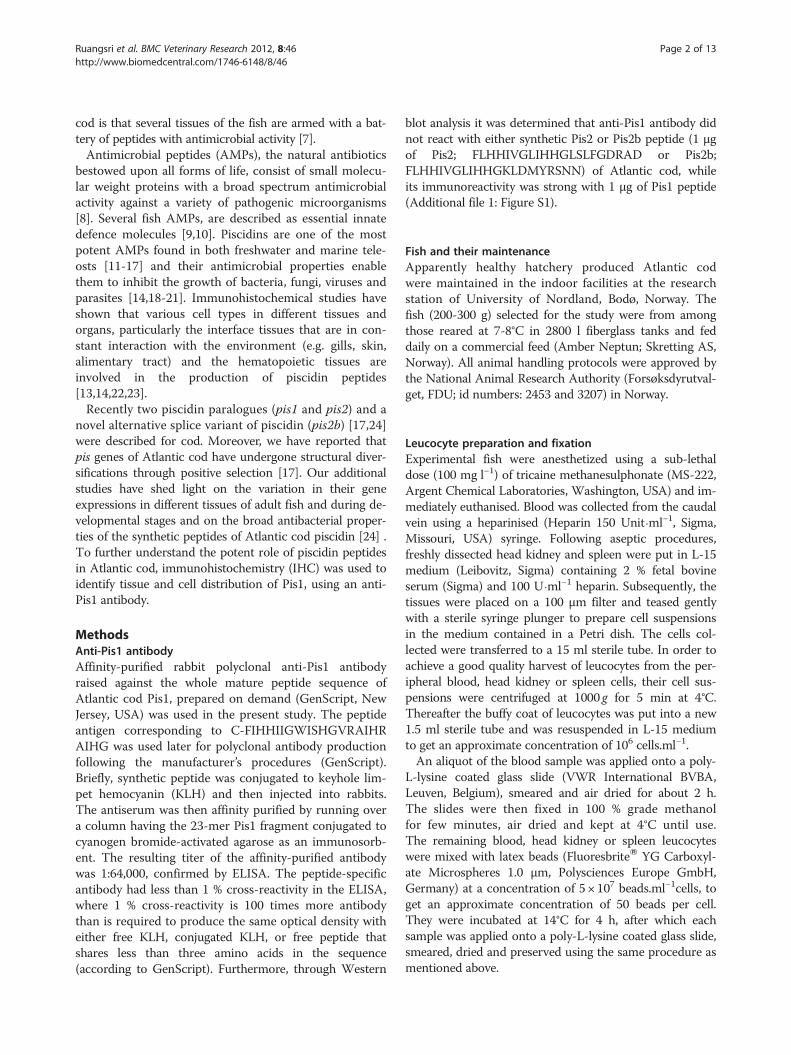
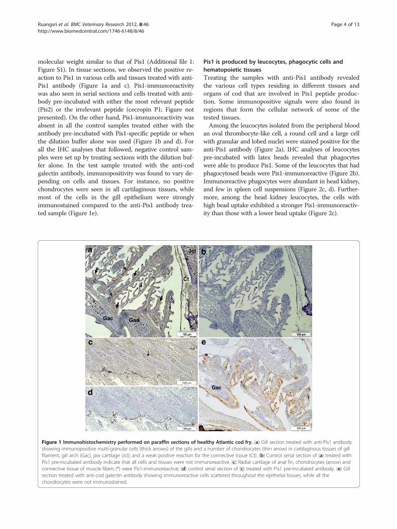
molecular weight similar to that of Pis1 (Additional file 1:Figure S1). In tissue sections, we observed the positive re-action to Pis1 in various cells and tissues treated with anti-Pis1 antibody (Figure 1a and c). Pis1-immunoreactivitywas also seen in serial sections and cells treated with anti-body pre-incubated with either the most relevant peptide(Pis2) or the irrelevant peptide (cecropin P1; Figure notpresented). On the other hand, Pis1-immunoreactivity wasabsent in all the control samples treated either with theantibody pre-incubated with Pis1-specific peptide or whenthe dilution buffer alone was used (Figure 1b and d). Forall the IHC analyses that followed, negative control sam-ples were set up by treating sections with the dilution buf-fer alone. In the test sample treated with the anti-codgalectin antibody, immunopositivity was found to vary de-pending on cells and tissues. For instance, no positivechondrocytes were seen in all cartilaginous tissues, whilemost of the cells in the gill epithelium were stronglyimmunostained compared to the anti-Pis1 antibody trea-ted sample (Figure 1e).
Figure 1 Immunohistochemistry performed on paraffin sections of heshowing immunopositive multi-granular cells (thick arrows) of the gills andfilament, gill arch (Gac), jaw cartilage (Jcl), and a weak positive reaction forPis1 pre-incubated antibody indicate that all cells and tissues were not immconnective tissue of muscle fibers (*) were Pis1-immunoreactive; (d) controsection treated with anti-cod galectin antibody showing immunoreactive cchondrocytes were not immunostained.
Pis1 is produced by leucocytes, phagocytic cells andhematopoietic tissuesTreating the samples with anti-Pis1 antibody revealedthe various cell types residing in different tissues andorgans of cod that are involved in Pis1 peptide produc-tion. Some immunopositive signals were also found inregions that form the cellular network of some of thetested tissues.Among the leucocytes isolated from the peripheral blood
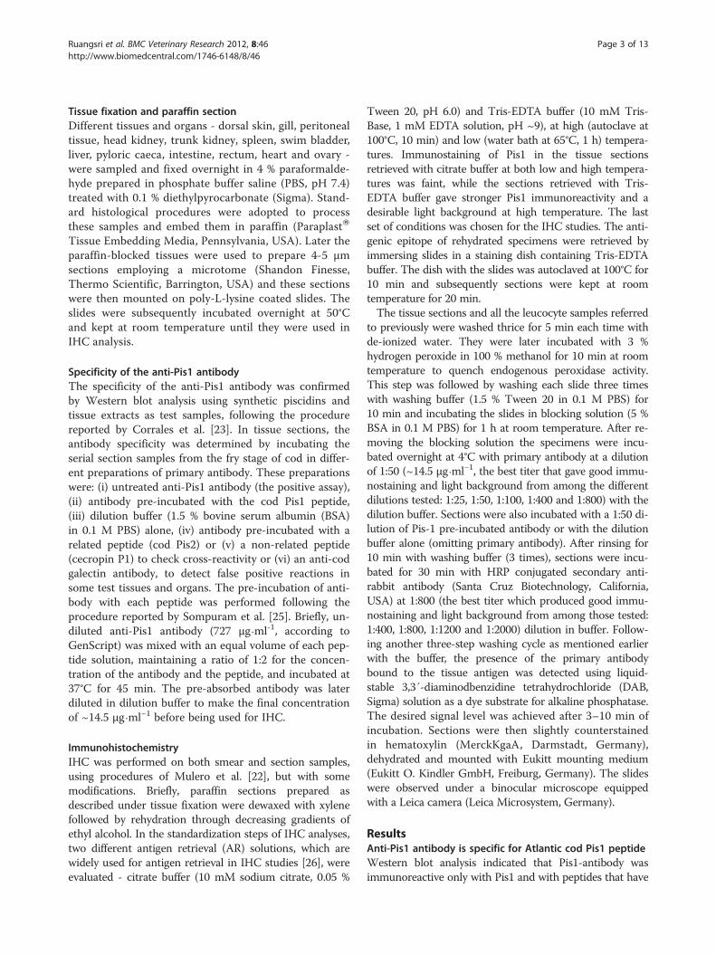
an oval thrombocyte-like cell, a round cell and a large cellwith granular and lobed nuclei were stained positive for theanti-Pis1 antibody (Figure 2a). IHC analyses of leucocytespre-incubated with latex beads revealed that phagocyteswere able to produce Pis1. Some of the leucocytes that hadphagocytosed beads were Pis1-immunoreactive (Figure 2b).Immunoreactive phagocytes were abundant in head kidney,and few in spleen cell suspensions (Figure 2c, d). Further-more, among the head kidney leucocytes, the cells withhigh bead uptake exhibited a stronger Pis1-immunoreactiv-ity than those with a lower bead uptake (Figure 2c).
althy Atlantic cod fry. (a) Gill section treated with anti-Pis1 antibodya number of chondrocytes (thin arrow) in cartilaginous tissues of gillthe connective tissue (Ct). (b) Control serial section of (a) treated withunoreactive. (c) Radial cartilage of anal fin, chondrocytes (arrow) andl serial section of (c) treated with Pis1 pre-incubated antibody. (e) Gillells scattered throughout the epithelial tissues, while all the

Figure 2 Immunohistochemistry on Atlantic cod leucocytes and different hematopoietic tissues. (a) The cytoplasm of different leucocytetypes, including round cells (thin arrows), large cells with granular and lobed nuclei (thick arrows) and thrombocyte-like cells (arrow heads) werePis1-immunoreactive (brown colour). Leucocytes incubated with latex beads (b, c and d). (b) Blood: some bead-containing phagocytes (thickarrows), round cells (thin arrows) and thrombocyte-like cells (arrow heads) were Pis1-immunoreactive. (c) Head kidney: numerous leucocytes,including phagocytes were Pis1-immunoreactive, phagocytes with intense staining had high bead uptake (thick arrows) than those with a lowbead uptake (thin arrows). (d) Spleen: most of the leucocytes including phagocytes (arrows) were Pis1-immunoreactive. (e) Head kidney:numerous vascular tissue (arrows) and some cells in the hematopoietic tissues (*), glomerulus (Gl) and blood vessel (V) were Pis1-immunoreactive;note that glomerulus is surrounded by Bowman’s capsule space (unstained - thin arrow). (f) Trunk kidney: cells in hematopoietic tissue (*),cytoplasm of columnar epithelial cells of collecting tubule (Cb), cells and detached elements in the lumen of renal tubules (arrows) were Pis1-immunoreactive. (g) Spleen: numerous hematopoietic cells were Pis1-immunoreactive (v: blood vessel). Negative control (inset) of the cell samplesand serial sections treated with dilution buffer was Pis1-immunonegative (Larger images are seen in Additional file 1: Figure S2).
Ruangsri et al. BMC Veterinary Research 2012, 8:46 Page 5 of 13http://www.biomedcentral.com/1746-6148/8/46

Ruangsri et al. BMC Veterinary Research 2012, 8:46 Page 6 of 13http://www.biomedcentral.com/1746-6148/8/46
In the head kidney sections, immunopositive signals wereprevalent in the hematopoietic tissue, and also in the cellsof glomerulus and adjacent vascular tissues (Figure 2e). Intrunk kidney many Pis1-immunoreactive cells were alsoobserved in the hematopoietic tissue, and in the cytoplasmof the columnar epithelial cells of collecting tubules and incell debris located in the lumen of renal tubules (Figure 2f).In the spleen, numerous hematopoietic cells were alsoPis1-immunoreactive, particularly those residing close tothe blood vessels (Figure 2g).
Pis1 is produced by mucosal tissuesIn the dorsal skin sections, Pis1-immunoreactivity wasobserved in the epithelial and basal cells of the epidermallayer (Figure 3a). Some skin epidermal tissues, collectedfrom the vicinity of eyes, were also found to be immuno-positive (Figure 3b).In the gill section, respiratory epithelial cells and multi-
granular cells of primary and secondary lamellae werePis1-immunoreactive (Figure 3c). Some multi-granularcells of the adductor muscle were also positive (Figure 3d).Interestingly, Pis1-immunoreactivity of chondrocytes wasstrong in the cartilaginous tissues of both filaments andgill arches (Figure 3e-f).In the gastrointestinal tract, Pis1-immunoreactivity was
weak in the cytoplasm of epithelial cells of the caecal mu-cosal fold, and in some cells and connective tissues sur-rounding the caecae (Figure 4a). In the intestine, epithelialcolumnar cells, multi-granular cells in the lamina propriaand submucosal tissues, and cytoplasm of columnar cellsin compound glands [27] were immunostained, but gobletcells were Pis1-immunonegative (Figure 4b-d).Mucosal epithelial cells of the swim bladder were only
slightly Pis1-immunostained, while the intensity of stainingwas stronger in numerous neural cells of the swim bladderwall (Figure 4e). In addition, a strong positive reaction wasobserved in the fibroblasts, the cellular network of sub-mucosal tissue and in the secretory cells of the gas gland,while the signal was weak in the rete mirabile (Figure 4f-g).
Pis1 is produced by neuronal cells, exocrine andendocrine glandsThe presence of Pis1 peptide was evident in the clusterof nerve cells found in the swim bladder wall as well asin the nervous system of some other organs. A largecluster of immunopositive perikarya was located on thesurface of the head kidney in the parasympathetic ganglia(Figure 5A-a). In the ocular region, cartilaginous tissue,and various cells and tissues of retina - photoreceptorcells, inner nuclear layer, and nerves and nerve tissues -showed Pis1-immunoreaction (Figure 5A-b). In addition,some positive cells were also found in the choroid retemirabile (not shown).
Examination of the sections of intestine and kidneyrevealed that exocrine and endocrine glands are alsoinvolved in the production of Pis1 peptide. In pancreas,most of the exocrine acinar cells exhibited strong Pis1-immunoreactivity (Figure 5B-a). In corpuscles of Stan-nius located on the surface tissue of both head andtrunk kidneys, Pis1-immunoreactive secretory cells werefound (Figure 5B-b).
Pis1-immunoreactivity in other tissues and organsBesides those mentioned above, some other tissues andorgans were Pis1-immunoreactive. In the liver most of thehepatocytes and their cellular network were Pis1-immunostained (Figure 6a). Furthermore, the presence of thismolecule was evident in the epithelial cells of gall bladderand bile ducts (Figure 6b-c). In heart, immunopositivefibroblast cells were observed in the bulbus arteriosus,while such a signal was seen neither in other parts of theheart tissue, nor in the control sample (Figure 6d).In the peritoneum, Pis1-positivity was found in the con-
nective tissues lining a layer of mesothelium (Figure 6e)and in cells and connective tissue of the skin hypodermallayer and around the muscle fibers (myosepta) (Figure 6f).Pis1-immunoreactivity was also noted in many stages of theoocytes; the intensity of immunostaining was stronger inthe early or mid vitellogenic oocyte (Figure 6g). In additionto the gill cartilage chondrocytes mentioned earlier, othercartilaginous tissues in the radial cartilage of dorsal and analfins, hyaline cartilage of the jaws and eyes were also Pis1-immunoreactive (see Figure 1a, c and Figure 5A-b). Further,the notochord of cod larvae was found to be immunoreac-tive as well (not shown).
DiscussionDistribution and localisation of Pis1 peptide, a naturalantibiotic, was studied in different tissues of Atlantic codusing immunohistochemistry. The specificity of the anti-body was confirmed in the serial section samples, byemploying anti-Pis1 antibody that was pre-incubated witheither synthetic Pis1 peptide or other non-related peptides.Through Western blot analysis we have shown that theanti-Pis1 antibody of Atlantic cod does not cross reactwith Pis2 or Pis2b of the same species. On the other hand,similar immunostaining patterns were observed in sectionstreated either with the anti-Pis1antibody or pre-absorbedantibody with Pis2 peptide. The lack of cross-reactivity be-tween anti-Pis1 antibody and Pis2 peptide could be attrib-uted to their structural diversifications and low identity(36 % or 41 %) of Pis1 and Pis2 or Pis2b [24].The specific antibody was used to further investigate
the distribution of Pis1 peptide in fish tissues. There wassound cellular evidence on localization of Pis1 in someof the tissues, corresponding to the observations on pis1gene expression reported previously [24]. Various cell

Figure 3 Immunohistochemical micrographs of different tissues of Atlantic cod. (a) Dorsal skin: numerous epithelial cells were Pis1-immunoreactive, but the basal cells (arrows) exhibited stronger immunoreactivity than the apical cells (Gb: goblet cell; Sc: saccular cell). (b)Epidermal layer of skin surrounding the eye - some Pis1-immunoreactivity was observed in the cytoplasm of epithelial cells (arrows), (N: cells ofneuromast). (c) Gill filament: multi-granular cells of primary lamella (arrows) and epithelial cells of secondary lamella (arrow heads) were Pis1-immunoreactive. (d) Adductor muscle: multi-granular cells adjacent to the muscle fibers (Mf) were Pis1-immunostained. (e) Cartilaginous tissue ofthe primary lamella: all the chondrocytes (*) exhibited very strong Pis1-immunoreactivity. (f) Cartilaginous tissue of the gill filament: onlychondrocytes (open arrows) were Pis1-immunostained (positive). Negative control (inset) of the samples treated in dilution buffer was Pis1-immunonegative (Larger images are seen in Additional file 1: Figure S3).
Ruangsri et al. BMC Veterinary Research 2012, 8:46 Page 7 of 13http://www.biomedcentral.com/1746-6148/8/46
types, such as thrombocyte-like cells [28], multi-granularcells [29], hematopoietic cells [22] and epithelial cellsfrom mucosal tissues [30] reacted positive to Pis1 anti-body. This indicates that the molecule is probably asso-ciated with an immune function as these cells andtissues in fish are known to be involved in a dynamic im-mune defence network [9,29-34]. Furthermore, ourobservations strongly suggest that cod Pis1 activates theelimination of foreign bodies, via different mechanisms.An intracellular killing mechanism is clearly reflectedthrough the presence of Pis1 in phagocytic cells (Figure 2).In addition, the strongly immunostained phagocytes with
high bead uptake indicate that Pis1 peptide is a compo-nent of the host defence mechanism during phagocyt-osis. The extracellular killing mechanism, on the otherhand, could be supported by the detection of peptideoutside the cells of various immune organs. Antimicro-bial piscidins, which are part of the defence system inmany fish species, are not produced by mucosal tissuesalone, but also by cells residing in the spleen and headkidney [13,22,35]. Moreover, Mulero et al. [22] havedemonstrated the ability of piscidins to eliminate invad-ing bacteria through both intracellular and extracellularkilling mechanisms.
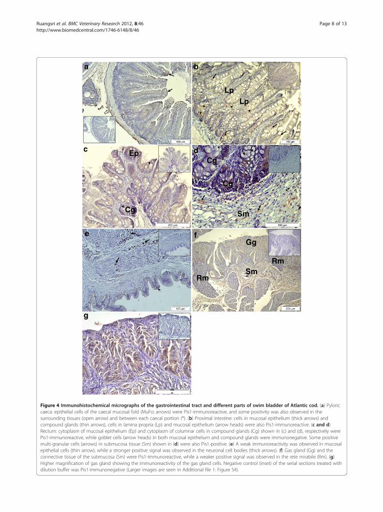
Figure 4 Immunohistochemical micrographs of the gastrointestinal tract and different parts of swim bladder of Atlantic cod. (a) Pyloriccaeca: epithelial cells of the caecal mucosal fold (MuFo; arrows) were Pis1-immunoreactive, and some positivity was also observed in thesurrounding tissues (open arrow) and between each caecal portion (*). (b) Proximal intestine: cells in mucosal epithelium (thick arrows) andcompound glands (thin arrows), cells in lamina propria (Lp) and mucosal epithelium (arrow heads) were also Pis1-immunoreactive. (c and d)Rectum: cytoplasm of mucosal epithelium (Ep) and cytoplasm of columnar cells in compound glands (Cg) shown in (c) and (d), respectively werePis1-immunoreactive, while goblet cells (arrow heads) in both mucosal epithelium and compound glands were immunonegative. Some positivemulti-granular cells (arrows) in submucosa tissue (Sm) shown in (d) were also Pis1-positive. (e) A weak immunoreactivity was observed in mucosalepithelial cells (thin arrow), while a stronger positive signal was observed in the neuronal cell bodies (thick arrows). (f) Gas gland (Gg) and theconnective tissue of the submucosa (Sm) were Pis1-immunoreactive, while a weaker positive signal was observed in the rete mirabile (Rm). (g)Higher magnification of gas gland showing the immunoreactivity of the gas gland cells. Negative control (inset) of the serial sections treated withdilution buffer was Pis1-immunonegative (Larger images are seen in Additional file 1: Figure S4).
Ruangsri et al. BMC Veterinary Research 2012, 8:46 Page 8 of 13http://www.biomedcentral.com/1746-6148/8/46

Figure 5 (5A) Immunohistochemical micrographs of different neural tissues (including eye) of Atlantic cod. (a) Parasympathetic ganglia:various sized neuronal cell bodies (arrows), cells and tissues in axons and dendrites (*) were Pis1-immunoreactive (Hk: head kidney’shematopoietic tissue). (b) Eye: Pis1-immunoreactivity was found in various cell types and tissues –including 1: sensory cells of the retina; 2: cellsand tissues of inner nuclear layer; 3: inner plexiform layer; 4: ganglion cell and nerve fiber layer, and arrowhead shows the inner limitingmembrane. The chondrocytes of the hyaline cartilage (arrows), connective tissues (Ct), and tissue of the lens (*) were also immunoreactive, (m:melanocytes). (5B) Section of exocrine and endocrine glands of Atlantic cod. (a) Exocrine pancreas: the cords of exocrine acinar cells (*) showingstrong intensity of Pis1-immunoreactivity; note the presence of the large hepatic veins (arrows). (b) Corpuscles of Stannius: Pis1-immunoreactivitywas found in the cytoplasm of most secretory cells. Negative control (inset) of the serial sections treated with dilution buffer was Pis1-immunonegative. (Larger images are seen in Additional file 1: Figure S5).
Ruangsri et al. BMC Veterinary Research 2012, 8:46 Page 9 of 13http://www.biomedcentral.com/1746-6148/8/46
Another important observation in the present study isthe discovery of the hitherto unreported information thatpiscidin is produced by chondrocytes of various cartilagin-ous tissues (Figure 1, 3 and 5A). In contrast, immunohisto-chemical studies on other teleost species have shown thatthe chondrocytes did not contain piscidins [13,14,23,35].To our knowledge, more information on teleost chondro-cytes does not exist. Nevertheless, studies on mammalshave shown that this cell type not only produces extracel-lular matrix, but also acts as a central machinery to pro-duce AMPs and other immune factors, thus contributingto host defence mechanisms in non-vascular cartilaginoustissues [36-38]. Therefore, chondrocytes of Atlantic codare also likely to have similar potential function, but moreinvestigations are needed to ascertain it. A strong Pis1-immunoreactivity was also observed in the swim bladder.This organ of zebrafish was found to express several
antimicrobial genes [39] and this seems to be the case aswell for Atlantic cod [24,40]. As neither the genetic profil-ing of swim bladder nor its function in innate immune sys-tem has been described, additional studies need to beundertaken to determine the reason for the presence ofAMPs or the role of AMPs in association with other estab-lished functions such as buoyancy, respiration and com-munication [41]. It should be noted that Silphaduang et al.[13] failed to detect piscidins in the swim bladder of hybridstriped bass, pointing to a species-dependent difference infunctionality.Atlantic cod has a unique immune system architecture
compared to other vertebrates [1] and produce little orhardly any antibody upon vaccination [2,5]. Nevertheless,this fish copes with pathogen invasion [5], and its abilityto protect itself could be linked to the effective innateimmune components [1,3,6]. The data from this study as
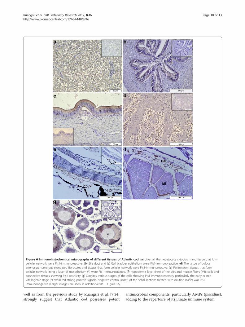
Figure 6 Immunohistochemical micrographs of different tissues of Atlantic cod. (a) Liver: all the hepatocyte cytoplasm and tissue that formcellular network were Pis1-immunoreactive. (b) Bile duct and (c) Gall bladder epithelium were Pis1-immunoreactive. (d) The tissue of bulbusarteriosus: numerous elongated fibrocytes and tissues that form cellular network were Pis1-immunoreactive. (e) Peritoneum: tissues that formcellular network lining a layer of mesothelium (*) were Pis1-immunostained. (f) Hypodermis layer (Hm) of the skin and muscle fibers (Mf): cells andconnective tissues showing Pis1-positivity (g) Oocytes: various stages of the cells showing Pis1-immunoreactivity particularly the early or midvitellogenic stage (*) exhibited strong positive signals. Negative control (inset) of the serial sections treated with dilution buffer was Pis1-immunonegative (Larger images are seen in Additional file 1: Figure S6).
Ruangsri et al. BMC Veterinary Research 2012, 8:46 Page 10 of 13http://www.biomedcentral.com/1746-6148/8/46
well as from the previous study by Ruangsri et al. [7,24]strongly suggest that Atlantic cod possesses potent
antimicrobial components, particularly AMPs (piscidins),adding to the repertoire of its innate immune system.
Ruangsri et al. BMC Veterinary Research 2012, 8:46 Page 11 of 13http://www.biomedcentral.com/1746-6148/8/46
In this study we investigated the presence of peptide inapparently healthy fish. The widespread occurrence ofPis1 suggests other possible functions of this peptide infish, in addition to its roles against microorganisms. Pis1may have a role in mediating cyto-protection as it waspresent in several tissues such as liver, bile duct and gallbladder, which are not anatomically exposed to a highpathogen pressure. Further, the high level of Pis1 peptidedetected in swim bladder may signify its involvement inrepair mechanisms of this tissue. In Atlantic cod, swimbladder damaged by a dramatic change of hydrostaticpressure was found to regain its function, possibly withthe assistance of a membrane that lines its wall (cited by[42]). It has to be emphasized that wound healing proper-ties of various AMPs have been reported in humans [43].Cod Pis1 could also be a neurogenic peptide since its
immunoreactivity was observed in neural tissue of someorgans (Figure 4 and 5A). Little is known on AMPs andtheir function in nervous system of fish, however it hasbeen demonstrated that nerve cells and or nerve tissuesare never located far away from effector organs [30]. Thecrosstalk between nerves and immune system of the ani-mal kingdom has long since been a topic of discussion,and it is now clear that there exists a bidirectional flowof information between the central nervous system andthe immune system [44-47]. Furthermore, it has beenshown that many neuropeptides and peptide hormoneshave similar characteristics, including antimicrobialproperties [47,48]. Moreover, it is evident that togetherwith the nervous system, the endocrine system of all ani-mals including fish, participate in the maintenance of asteady physiologic state, homeostasis [30,47]. As Pis1appeared in several glands (Figure 4 and 5B), this peptidemay have a homeostatic role. Another fish AMP, beta-defensin (previously known as β-defensin) that wasexpressed highly in the pituitary and testis of orangespotted grouper is believed to maintain cellular homeo-stasis [49]. Apart from their antimicrobial properties,most of the hepcidins are known to maintain ironhomeostasis in vertebrates including fish [50].Involvement in osmoregulation and excretion are alter-
native functions which Pis1 may possess, as their presencewas evident in kidney (especially trunk kidney), skin, gills,and corpuscles of Stannius of naïve fish. These organs areknown to have crucial roles in the osmoregulatory and ex-cretory mechanisms of fish besides their immune functions[30]. In addition, osmoregulatory signature of Pis1 peptideis evident from the immunoreactivity found in the retemirabile of the swim bladder or the choroid rete mirabileof the fish eye. These unique parts are known to have keyroles in oxygen secretion in the swim bladder and the eyeof fish [51]. Osmoregulatory functions might also be attrib-uted to peritoneum, hypodermis layer and myosepta wherethe Pis1-immunoreactivity was detected. The presence of
Psoriasin1, an AMP member that belongs to the calcium-binding protein super-family was detected in peritoneal tis-sue of horse [52]. Pis1 peptide was also found in all stagesof the oocytes, their expression being greater in theadvanced developmental stages.
ConclusionIt is evident that various cell types and tissues of Atlanticcod are armed with Pis1 peptide. Its constitutive presencein immune-related tissues of naïve fish suggests a signifi-cant role for this peptide in the innate immune system ofAtlantic cod. In addition, the localisation of Pis1 in some ofthe non-immune tissues and organs confirmed our previ-ous tissue distribution results at the molecular level [24] -suggesting other possible roles of this ubiquitous peptideand its capability to maintain homeostasis in Atlantic cod.
Additional file
Additional file 1: Figure S1. Tests to confirm the mono-specificity ofanti-Pis1 antibody using tissue extracts from Atlantic cod and thesynthetic piscidin peptide. Samples were separated on a 5–16 %acrylamide gel electrophoresis in the presence of sodium dodecyl sulfate(SDS-PAGE) running in Tris-Tricine buffer system, following the procedureof Ruangsri et al. [7]. (A) Results from coomassie blue staining. (B) Westernblot analysis of piscidins and tissue extracts that were transferred onto aPVDF membrane that was blocked for 1 h and incubated overnight at 4°C with anti-Pis1 antibody. Lanes are: 1 - protein marker, 2 - leucocytesfrom head kidney co-incubated with latex beads for 4 h, 3 - head kidney,4 - blood, 5 - muscle, 6 - skin, 7 - ovary, 8 - synthetic Pis2b, 9 - syntheticPis2 and 10 - synthetic Pis1. Anti-Pis antibody was immunoreactive onlywith Pis1 peptide (Lane 10), while moderate to faint immunoreactivitywas detected also in some tissue extracts (Lane 2 and 7) at the positionsthat corresponded to a molecular weight similar to that of Pis1. Figure S2.Immunohistochemical micrographs of different tissues of Atlantic codshowing absence of immunoreactivity in the control samples of (a) blood,(b) blood mixed with latex beads, (c and d) head kidney and spleenleucocytes mixed with latex beads, (e, f and g) sections of head kidney,trunk kidney and spleen treated with dilution buffer. Figure S3.Immunohistochemical micrographs of control sections treated withdilution buffer, showing immunonegative reactions in tissues of Atlanticcod. (a) Dorsal skin, (b) epidermal layer of skin surrounding eye, (c) gillfilament and chondrocytes and (d) adductor muscle. Figure S4.Immunohistochemical micrographs of control sections treated withdilution buffer, showing immunonegative reactions in tissues of Atlanticcod. (a) Pyloric caeca, (b) proximal intestine, (c) mucosal epithelium ofrectum, (d) compound gland and submucosa tissue of rectum, (e) swimbladder wall and mucosal epithelium , (f) gas gland and (g) highermagnification of gas gland. Figure S5. Immunohistochemical micrographsof control sections treated with dilution buffer, showing immunonegativereactions in tissues. (A-a) Parasympathetic ganglia, (A-b) eye, (B-a)exocrine pancreas and (B-b) corpuscles of Stannius. Figure S6.Immunohistochemical micrographs of control sections treated withdilution buffer, showing immunonegative reactions in tissues. (a) Liver, (b)bile duct, (c) gall bladder epithelium, (d) bulbus arteriosus, (e) peritoneumand (f) oocytes.
AcknowledgementsThis work was supported by the Research Council of Norway under theproject ‘Mucosal Immune System of Atlantic Cod - Creating a KnowledgeBase’ (184703). The authors would like to thank Carlos Lanes, for sharing thecod fry stage samples and Divya Kumari Hirkudel Vate for her help inpreparation of some test slides for the study. Hilde Ribe and Ingvild Berg arethanked for their technical support. Jareeporn Ruangsri would like to express
Ruangsri et al. BMC Veterinary Research 2012, 8:46 Page 12 of 13http://www.biomedcentral.com/1746-6148/8/46
her gratitude to Prince of Songkla University (Thailand) for granting the PhDscholarship that enabled her to study at the University of Nordland, Norway.
Author details1Faculty of Biosciences and Aquaculture, University of Nordland, 8049, Bodø,Norway. 2Cell Biology and Immunology Group, Wageningen Institute ofAnimal Sciences, Wageningen University, Marijkeweg 40, 6709 PG,Wageningen, The Netherlands.
Authors’ contributionsJR designed and performed the experiments, analyzed the data and wrotethe manuscript. The experiments were overseen by JMOF, MFB, JHWMR andVK, who were also involved in the revision of the manuscript. All authorshave read and approved the final manuscript.
Received: 8 December 2011 Accepted: 26 April 2012Published: 26 April 2012
References1. Star B, Nederbragt AJ, Jentoft S, Grimholt U, Malmstrom M, Gregers TF,
Rounge TB, Paulsen J, Solbakken MH, Sharma A, et al: The genomesequence of Atlantic cod reveals a unique immune system. Nature 2011,477:207–210.
2. Lund V, Bordal S, Kjellsen O, Mikkelsen H, Schroder MB: Comparison ofantibody responses in Atlantic cod (Gadus morhua L.) to Aeromonassalmonicida and Vibrio anguillarum. Dev Comp Immunol 2006, 30:1145–1155.
3. Magnadóttir B, Jónsdóttir H, Helgason S, Björnsson B, Solem ST, Pilström L:Immune parameters of immunised cod (Gadus morhua L.). Fish ShellfishImmunol 2001, 11:75–89.
4. Espelid S, Rødseth OM, Jørgensen T�: Vaccination experiments and studiesof the humoral immune responses in cod, Gadus morhua L., to four strainsof monoclonal-defined Vibrio anguillarum. J Fish Dis 1991, 14:185–197.
5. Mikkelsen H, Lund V, Larsen R, Seppola M: Vibriosis vaccines based onvarious sero-subgroups of Vibrio anguillarum O2 induce specificprotection in Atlantic cod (Gadus morhua L.) juveniles. Fish ShellfishImmunol 2011, 30:330–339.
6. Persson AC, Stet RJM, Pilstrom L: Characterization of MHC class I and beta(2)-microglobulin sequences in Atlantic cod reveals an unusually highnumber of expressed class I genes. Immunogenetics 1999, 50:49–59.
7. Ruangsri J, Fernandes JMO, Brinchmann M, Kiron V: Antimicrobial activity inthe tissues of Atlantic cod (Gadus morhua L.). Fish Shellfish Immunol 2010,28:879–886.
8. Hancock RE: Peptide antibiotics. Lancet 1997, 349:418–422.9. Smith VJ, Fernandes MOJ: Antimicrobial peptides of the innate immune
system. In Fish Defenses. Edited by Zaccone G, Marchalonis JJ, Schluter SF,Meseguer J, Kapoor BG. USA: Science Publishers; 2009:241–275.
10. Noga EJ, Ullal AJ, Corrales J, Fernandes JMO: Application of antimicrobialpolypeptide host defenses to aquaculture: exploitation of downregulationand upregulation responses. Comp Biochem Physiol D 2011, 6:44–54.
11. Sun BJ, Xie HX, Song Y, Nie P: Gene structure of an antimicrobial peptidefrom mandarin fish, Siniperca chuatsi (Basilewsky), suggests thatmoronecidins and pleurocidins belong in one family: the piscidins. J FishDis 2007, 30:335–343.
12. Salerno G, Parrinello N, Roch P, Cammarata M: cDNA sequence and tissueexpression of an antimicrobial peptide, dicentracin; a new component ofthe moronecidin family isolated from head kidney leukocytes of seabass, Dicentrarchus labrax. Comp Biochem Physiol B 2007, 146:521–529.
13. Silphaduang U, Colorni A, Noga EJ: Evidence for widespread distributionof piscidin antimicrobial peptides in teleost fish. Dis Aquat Org 2006,72:241–252.
14. Silphaduang U, Noga EJ: Peptide antibiotics in mast cells of fish. Nature2001, 414:268–269.
15. Yin ZX, He W, Chen WJ, Yan JH, Yang JN, Chan SM, He JG: Cloning,expression and antimicrobial activity of an antimicrobial peptide,epinecidin-1, from the orange-spotted grouper, Epinephelus coioides.Aquaculture 2006, 253:204–211.
16. Noga EJ, Silphaduang U, Park NG, Seo JK, Stephenson J, Kozowicz S: Piscidin4, a novel member of the piscidin family of antimicrobial peptides. CompBiochem Physiol B 2009, 152:299–305.
17. Fernandes JMO, Ruangsri J, Kiron V: Atlantic cod piscidin and itsdiversification through positive selection. PLoS One 2010, 5:e9501.
18. Lauth X, Shike H, Burns JC, Westerman ME, Ostland VE, Carlberg JM, Van OlstJC, Nizet V, Taylor SW, Shimizu C, et al: Discovery and characterization oftwo isoforms of moronecidin, a novel antimicrobial peptide from hybridstriped bass. J Biol Chem 2002, 277:5030–5039.
19. Chinchar VG, Bryan L, Silphadaung U, Noga E, Wade D, Rollins-Smith L:Inactivation of viruses infecting ectothermic animals by amphibian andpiscine antimicrobial peptides. Virology 2004, 323:268–275.
20. Sung WS, Lee J, Lee DG: Fungicidal effect of piscidin on Candida albicans:pore formation in lipid vesicles and activity in fungal membranes. BiolPharm Bull 2008, 31:1906–1910.
21. Colorni A, Ullal A, Heinisch G, Noga EJ: Activity of the antimicrobialpolypeptide piscidin2 against fish ectoparasites. J Fish Dis 2008, 31:423–432.
22. Mulero I, Noga EJ, Meseguer J, Garcia-Ayala A, Mulero V: The antimicrobialpeptides piscidins are stored in the granules of professional phagocyticgranulocytes of fish and are delivered to the bacteria-containingphagosome upon phagocytosis. Dev Comp Immunol 2008, 32:1531–1538.
23. Corrales J, Mulero I, Mulero V, Noga EJ: Detection of antimicrobial peptidesrelated to piscidin 4 in important aquacultured fish. Dev Comp Immunol2010, 34:331–343.
24. Ruangsri J, Salger SA, Caipang CMA, Kiron V, Fernandes JMO: Differentialexpression and biological activity of two piscidin paralogues and a novelsplice variant in Atlantic cod (Gadus morhua L.). Fish Shellfish Immunol2012, 32:396–406.
25. Sompuram SR, Kodela V, Zhang K, Ramanathan H, Radcliffe G, Falb P, BogenSA: A novel quality control slide for quantitative immunohistochemistrytesting. J Histochem Cytochem 2002, 50:1425–1434.
26. Shi SR, Cote RJ, Taylor CR: Antigen retrieval immunohistochemistry: past,present, and future. J Histochem Cytochem 1997, 45:327–343.
27. Marrison MC: Histology of the Atlantic cod, Gadus morhua: an atlas. PartOne. Digestive Tract and Associated organs. Canada: Can Spec Publ FisAquat Sci; 1987.
28. Claver JA, Quaglia AIE: Comparative morphology, development, andfunction of blood cells in nonmammalian vertebrates. J Exot Pet Med2009, 18:87–97.
29. Reite OB, Evensen �: Inflammatory cells of teleostean fish: a reviewfocusing on mast cells/eosinophilic granule cells and rodlet cells. FishShellfish Immunol 2006, 20:192–208.
30. Genten F, Terwinghe E, Danguy A: Atlas of Fish Histology. USA: SciencePublishers; 2009.
31. Tort L, Balasch J, Mackenzie S: Fish immune system: a crossroads betweeninnate and adaptive responses. Immunologia 2003, 23:277–286.
32. Pakers SGM, Uppalapati S, Meyer W, Maderson P, Sell AF, Kruse C: Paus R:Fish matters: the relevance of fish skin biology to investigativedermatology. Exp Dermatol 2010, 19:313–324.
33. Ellis AE: Innate host defense mechanisms of fish against viruses andbacteria. Dev Comp Immunol 2001, 25:827–839.
34. Rombout JH, Abelli L, Picchietti S, Scapigliati G, Kiron V: Teleost intestinalimmunology. Fish Shellfish Immunol 2011, 31:616–626.
35. Dezfuli BS, Pironi F, Giari L, Noga EJ: Immunocytochemical localization ofpiscidin in mast cells of infected seabass gill. Fish Shellfish Immunol 2010,28:476–482.
36. Varoga D, Pufe T, Mentlein R, Kohrs S, Grohmann S, Tillmann B, HassenpflugJ, Paulsen F: Expression and regulation of antimicrobial peptides inarticular joints. Ann Anat 2005, 187:499–508.
37. Warnke PH, Russo PAJ, Hopfenziz M, Kurz B, Becker ST, Sherry E, Springer I,Sivananthan S: Antimicrobial peptide immunity protects human nasal andauricular cartilage against infection. J Craniofac Surg 2010, 21:198–201.
38. Macrory L, Vaughan-Thomas A, Clegg PD, Innes JF: An exploration of theability of tepoxalin to ameliorate the degradation of articular cartilage ina canine in vitro model. BMC Vet Res 2009, 5:25.
39. Oehlers SH, Flores MV, Chen T, Hall CJ, Crosier KE, Crosier PS: Topographicaldistribution of antimicrobial genes in the zebrafish intestine. Dev CompImmunol 2011, 35:385–391.
40. Ruangsri J, Kiron V, Jeppinamogeru L, Fernandes JMO: A novel betadefensin of Atlantic cod. submitted.
41. Nilsson S: Nervous control of fish swimbladders. Acta Histochem 2009,111:176–184.
42. van der Kooij J, Righton D, Strand E, Michalsen K, Thorsteinsson V, SvedängH, Neat FC, Neuenfeldt S: Life under pressure: insights from electronicdata-storage tags into cod swimbladder function. ICES J Mar Sci 2007,64:1293–1301.
Ruangsri et al. BMC Veterinary Research 2012, 8:46 Page 13 of 13http://www.biomedcentral.com/1746-6148/8/46
43. Steinstraesser L, Koehler T, Jacobsen F, Daigeler A, Goertz O, Langer S,Kesting M, Steinau H, Eriksson E, Hirsch T: Host defense peptides in woundhealing. Mol Med 2008, 14:528–537.
44. Kaplin A, Bartner S: Reciprocal communication between the nervous andimmune systems: crosstalk, back-talk and motivational speeches. Int RevPsychiatr 2006, 17:439–441.
45. Salzet M, Vieau D, Day R: Crosstalk between nervous and immune systemsthrough the animal kingdom: focus on opioids. Trends Neurosci 2000,23:550–555.
46. Zhang N, Oppenheim JJ: Crosstalk between chemokines and neuronalreceptors bridges immune and nervous systems. J Leukocyte Biol 2005,78:1210–1214.
47. Brogden KA, Guthmiller JM, Salzet M, Zasloff M: The nervous system and innateimmunity: the neuropeptide connection. Nat Immunol 2005, 6:558–564.
48. El Karim IA, Linden GJ, Orr DF, Lundy FT: Antimicrobial activity ofneuropeptides against a range of micro-organisms from skin, oral,respiratory and gastrointestinal tract sites. Neuroimmunology 2008,200:11–16.
49. Jin JY, Zhou L, Wang Y, Li Z, Zhao JG, Zhang QY, Gui JF: Antibacterial andantiviral roles of a fish beta-defensin expressed both in pituitary andtestis. PLoS One 2010, 5:e12883.
50. Rodrigues PNS, Vázquez-Dorado S, Neves JV, Wilson JM: Dual function offish hepcidin: Response to experimental iron overload and bacterialinfection in sea bass (Dicentrarchus labrax). Dev Comp Immunol 2006,30:1156–1167.
51. Berenbrink M: Historical reconstructions of evolving physiologicalcomplexity: O2 secretion in the eye and swimbladder of fishes. J Exp Biol2007, 210:1641–1652.
52. Bruhn O, Grotzinger J, Cascorbi I, Jung S: Antimicrobial peptides andproteins of the horse-insights into a well-armed organism. Vet Res 2011,42:98.
doi:10.1186/1746-6148-8-46Cite this article as: Ruangsri et al.: Ubiquitous presence of piscidin-1 inAtlantic cod as evidenced by immunolocalisation. BMC VeterinaryResearch 2012 8:46.
Submit your next manuscript to BioMed Centraland take full advantage of:
• Convenient online submission
• Thorough peer review
• No space constraints or color figure charges
• Immediate publication on acceptance
• Inclusion in PubMed, CAS, Scopus and Google Scholar
• Research which is freely available for redistribution
Submit your manuscript at www.biomedcentral.com/submit
Related Documents