Cell Death & Differentiation (2021) 28:427–438 https://doi.org/10.1038/s41418-020-00648-0 REVIEW ARTICLE Ubiquitin signaling in cell cycle control and tumorigenesis Fabin Dang 1 ● Li Nie 1,2 ● Wenyi Wei 1 Received: 7 July 2020 / Revised: 8 October 2020 / Accepted: 12 October 2020 / Published online: 31 October 2020 © The Author(s) 2020. This article is published with open access Abstract Cell cycle progression is a tightly regulated process by which DNA replicates and cell reproduces. The major driving force underlying cell cycle progression is the sequential activation of cyclin-dependent kinases (CDKs), which is achieved in part by the ubiquitin-mediated proteolysis of their cyclin partners and kinase inhibitors (CKIs). In eukaryotic cells, two families of E3 ubiquitin ligases, anaphase-promoting complex/cyclosome and Skp1-Cul1-F-box protein complex, are responsible for ubiquitination and proteasomal degradation of many of these CDK regulators, ensuring cell cycle progresses in a timely and precisely regulated manner. In the past couple of decades, accumulating evidence have demonstrated that the dysregulated cell cycle transition caused by inefficient proteolytic control leads to uncontrolled cell proliferation and finally results in tumorigenesis. Based upon this notion, targeting the E3 ubiquitin ligases involved in cell cycle regulation is expected to provide novel therapeutic strategies for cancer treatment. Thus, a better understanding of the diversity and complexity of ubiquitin signaling in cell cycle regulation will shed new light on the precise control of the cell cycle progression and guide anticancer drug development. Facts (1) The cell cycle is a tightly orchestrated cellular process that governs the timely DNA replication and cell division events. (2) Sequential activation of cyclin-dependent kinases (CDKs) drives cell cycle progression in a timely and precisely regulated manner. (3) The activity of CDKs is modulated by cyclin partners and CDK inhibitors (CKIs), which are tightly controlled by the ubiquitin–proteasome system. (4) Two important types of E3 ligases, the anaphase- promoting complex or cyclosome (APC/C) and Skp1- Cul1-F-box (SCF) complexes, are dedicated to cell cycle control. (5) Targeting E3 ubiquitin ligases provides effective therapeutic strategies for cancer treatment. Open questions (1) Unlike proteolytic signals, relatively little is known regarding the roles and mechanisms of non-proteolytic signals, such as the ones mediated by K6, K27, and K29 polyubiquitin chain, underlying the cell cycle control. (2) The specificity and diversity of deubiquitinating enzymes (DUBs) in regulating mitosis need to be further investigated. (3) How is the balance of ubiquitination–deubiquitination achieved to ensure accurate cell cycle progression remains elusive. (4) Unlike CDC20, the regulation of the enzymatic activity of CDH1 is not well defined yet. (5) The detailed mechanisms underlying spindle checkpoint imposed various E3 ligase activities of CDC20 toward different substrates need to be further investigated. (6) How those ubiquitination signaling events at the spindle checkpoint are integrated and orchestrated in a space–time-dependent manner remains not fully understood. Edited by F. Pentimalli * Wenyi Wei [email protected] 1 Department of Pathology, Beth Israel Deaconess Medical Center, Harvard Medical School, Boston, MA 02215, USA 2 State Key Laboratory for Quality and Safety of Agro-products, School of Marine Sciences, Ningbo University, Ningbo 315211, China 1234567890();,: 1234567890();,:

Welcome message from author
This document is posted to help you gain knowledge. Please leave a comment to let me know what you think about it! Share it to your friends and learn new things together.
Transcript
Cell Death & Differentiation (2021) 28:427–438https://doi.org/10.1038/s41418-020-00648-0
REVIEW ARTICLE
Ubiquitin signaling in cell cycle control and tumorigenesis
Fabin Dang1● Li Nie1,2 ● Wenyi Wei 1
Received: 7 July 2020 / Revised: 8 October 2020 / Accepted: 12 October 2020 / Published online: 31 October 2020© The Author(s) 2020. This article is published with open access
AbstractCell cycle progression is a tightly regulated process by which DNA replicates and cell reproduces. The major driving forceunderlying cell cycle progression is the sequential activation of cyclin-dependent kinases (CDKs), which is achieved in partby the ubiquitin-mediated proteolysis of their cyclin partners and kinase inhibitors (CKIs). In eukaryotic cells, two familiesof E3 ubiquitin ligases, anaphase-promoting complex/cyclosome and Skp1-Cul1-F-box protein complex, are responsible forubiquitination and proteasomal degradation of many of these CDK regulators, ensuring cell cycle progresses in a timely andprecisely regulated manner. In the past couple of decades, accumulating evidence have demonstrated that the dysregulatedcell cycle transition caused by inefficient proteolytic control leads to uncontrolled cell proliferation and finally results intumorigenesis. Based upon this notion, targeting the E3 ubiquitin ligases involved in cell cycle regulation is expected toprovide novel therapeutic strategies for cancer treatment. Thus, a better understanding of the diversity and complexity ofubiquitin signaling in cell cycle regulation will shed new light on the precise control of the cell cycle progression and guideanticancer drug development.
Facts
(1) The cell cycle is a tightly orchestrated cellular processthat governs the timely DNA replication and celldivision events.
(2) Sequential activation of cyclin-dependent kinases(CDKs) drives cell cycle progression in a timely andprecisely regulated manner.
(3) The activity of CDKs is modulated by cyclin partnersand CDK inhibitors (CKIs), which are tightlycontrolled by the ubiquitin–proteasome system.
(4) Two important types of E3 ligases, the anaphase-promoting complex or cyclosome (APC/C) and Skp1-Cul1-F-box (SCF) complexes, are dedicated to cellcycle control.
(5) Targeting E3 ubiquitin ligases provides effectivetherapeutic strategies for cancer treatment.
Open questions
(1) Unlike proteolytic signals, relatively little is knownregarding the roles and mechanisms of non-proteolyticsignals, such as the ones mediated by K6, K27, and K29polyubiquitin chain, underlying the cell cycle control.
(2) The specificity and diversity of deubiquitinatingenzymes (DUBs) in regulating mitosis need to befurther investigated.
(3) How is the balance of ubiquitination–deubiquitinationachieved to ensure accurate cell cycle progressionremains elusive.
(4) Unlike CDC20, the regulation of the enzymatic activityof CDH1 is not well defined yet.
(5) The detailed mechanisms underlying spindle checkpointimposed various E3 ligase activities of CDC20 towarddifferent substrates need to be further investigated.
(6) How those ubiquitination signaling events at the spindlecheckpoint are integrated and orchestrated in aspace–time-dependent manner remains not fullyunderstood.
Edited by F. Pentimalli
* Wenyi [email protected]
1 Department of Pathology, Beth Israel Deaconess Medical Center,Harvard Medical School, Boston, MA 02215, USA
2 State Key Laboratory for Quality and Safety of Agro-products,School of Marine Sciences, Ningbo University, Ningbo 315211,China
1234
5678
90();,:
1234567890();,:
Introduction
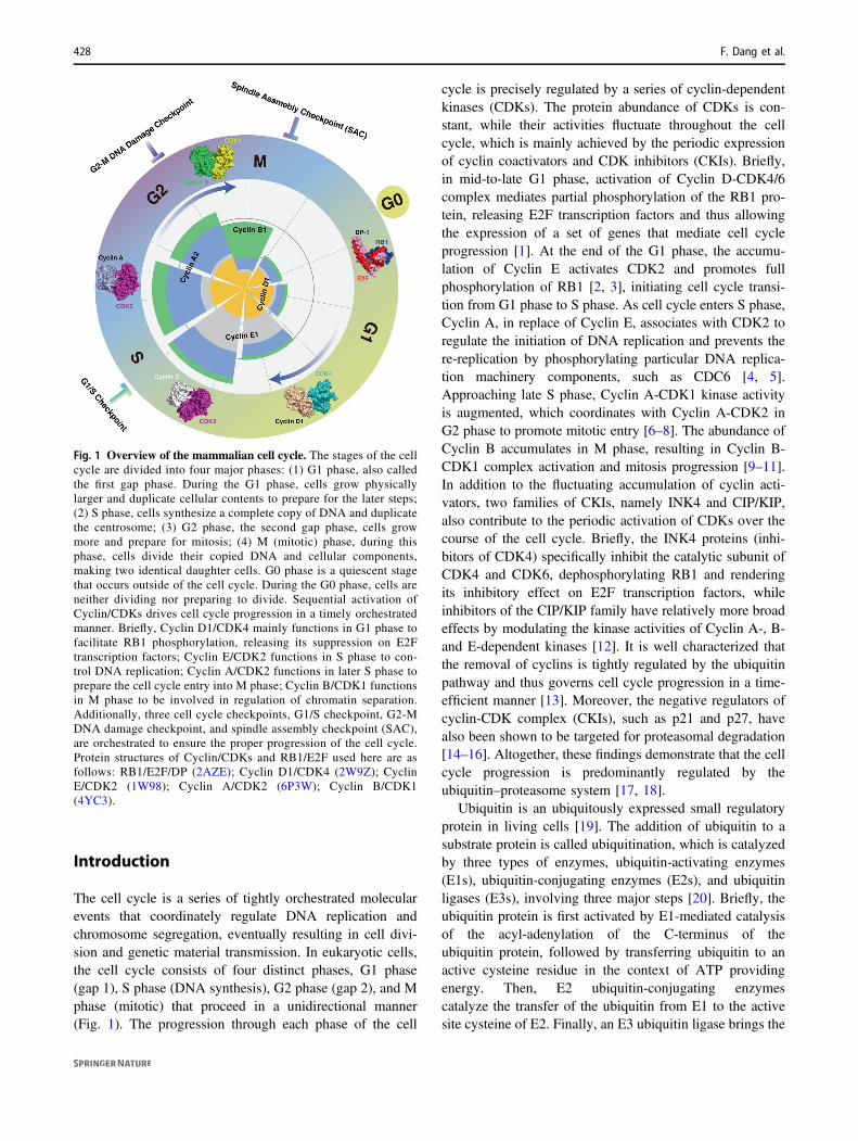
The cell cycle is a series of tightly orchestrated molecularevents that coordinately regulate DNA replication andchromosome segregation, eventually resulting in cell divi-sion and genetic material transmission. In eukaryotic cells,the cell cycle consists of four distinct phases, G1 phase(gap 1), S phase (DNA synthesis), G2 phase (gap 2), and Mphase (mitotic) that proceed in a unidirectional manner(Fig. 1). The progression through each phase of the cell
cycle is precisely regulated by a series of cyclin-dependentkinases (CDKs). The protein abundance of CDKs is con-stant, while their activities fluctuate throughout the cellcycle, which is mainly achieved by the periodic expressionof cyclin coactivators and CDK inhibitors (CKIs). Briefly,in mid-to-late G1 phase, activation of Cyclin D-CDK4/6complex mediates partial phosphorylation of the RB1 pro-tein, releasing E2F transcription factors and thus allowingthe expression of a set of genes that mediate cell cycleprogression [1]. At the end of the G1 phase, the accumu-lation of Cyclin E activates CDK2 and promotes fullphosphorylation of RB1 [2, 3], initiating cell cycle transi-tion from G1 phase to S phase. As cell cycle enters S phase,Cyclin A, in replace of Cyclin E, associates with CDK2 toregulate the initiation of DNA replication and prevents there-replication by phosphorylating particular DNA replica-tion machinery components, such as CDC6 [4, 5].Approaching late S phase, Cyclin A-CDK1 kinase activityis augmented, which coordinates with Cyclin A-CDK2 inG2 phase to promote mitotic entry [6–8]. The abundance ofCyclin B accumulates in M phase, resulting in Cyclin B-CDK1 complex activation and mitosis progression [9–11].In addition to the fluctuating accumulation of cyclin acti-vators, two families of CKIs, namely INK4 and CIP/KIP,also contribute to the periodic activation of CDKs over thecourse of the cell cycle. Briefly, the INK4 proteins (inhi-bitors of CDK4) specifically inhibit the catalytic subunit ofCDK4 and CDK6, dephosphorylating RB1 and renderingits inhibitory effect on E2F transcription factors, whileinhibitors of the CIP/KIP family have relatively more broadeffects by modulating the kinase activities of Cyclin A-, B-and E-dependent kinases [12]. It is well characterized thatthe removal of cyclins is tightly regulated by the ubiquitinpathway and thus governs cell cycle progression in a time-efficient manner [13]. Moreover, the negative regulators ofcyclin-CDK complex (CKIs), such as p21 and p27, havealso been shown to be targeted for proteasomal degradation[14–16]. Altogether, these findings demonstrate that the cellcycle progression is predominantly regulated by theubiquitin–proteasome system [17, 18].
Ubiquitin is an ubiquitously expressed small regulatoryprotein in living cells [19]. The addition of ubiquitin to asubstrate protein is called ubiquitination, which is catalyzedby three types of enzymes, ubiquitin-activating enzymes(E1s), ubiquitin-conjugating enzymes (E2s), and ubiquitinligases (E3s), involving three major steps [20]. Briefly, theubiquitin protein is first activated by E1-mediated catalysisof the acyl-adenylation of the C-terminus of theubiquitin protein, followed by transferring ubiquitin to anactive cysteine residue in the context of ATP providingenergy. Then, E2 ubiquitin-conjugating enzymescatalyze the transfer of the ubiquitin from E1 to the activesite cysteine of E2. Finally, an E3 ubiquitin ligase brings the
Fig. 1 Overview of the mammalian cell cycle. The stages of the cellcycle are divided into four major phases: (1) G1 phase, also calledthe first gap phase. During the G1 phase, cells grow physicallylarger and duplicate cellular contents to prepare for the later steps;(2) S phase, cells synthesize a complete copy of DNA and duplicatethe centrosome; (3) G2 phase, the second gap phase, cells growmore and prepare for mitosis; (4) M (mitotic) phase, during thisphase, cells divide their copied DNA and cellular components,making two identical daughter cells. G0 phase is a quiescent stagethat occurs outside of the cell cycle. During the G0 phase, cells areneither dividing nor preparing to divide. Sequential activation ofCyclin/CDKs drives cell cycle progression in a timely orchestratedmanner. Briefly, Cyclin D1/CDK4 mainly functions in G1 phase tofacilitate RB1 phosphorylation, releasing its suppression on E2Ftranscription factors; Cyclin E/CDK2 functions in S phase to con-trol DNA replication; Cyclin A/CDK2 functions in later S phase toprepare the cell cycle entry into M phase; Cyclin B/CDK1 functionsin M phase to be involved in regulation of chromatin separation.Additionally, three cell cycle checkpoints, G1/S checkpoint, G2-MDNA damage checkpoint, and spindle assembly checkpoint (SAC),are orchestrated to ensure the proper progression of the cell cycle.Protein structures of Cyclin/CDKs and RB1/E2F used here are asfollows: RB1/E2F/DP (2AZE); Cyclin D1/CDK4 (2W9Z); CyclinE/CDK2 (1W98); Cyclin A/CDK2 (6P3W); Cyclin B/CDK1(4YC3).
428 F. Dang et al.
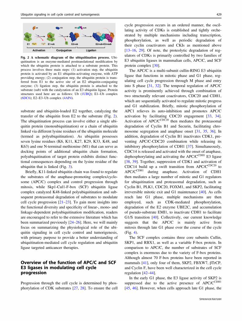
substrate and ubiquitin-loaded E2 together, catalyzing thetransfer of the ubiquitin from E2 to the substrate (Fig. 2).The ubiquitination process can involve either a single ubi-quitin protein (monoubiquitination) or a chain of ubiquitinlinked via different lysine residues of the ubiquitin molecule(termed as polyubiquitination). As ubiquitin possessesseven lysine residues (K6, K11, K27, K29, K33, K48, andK63) and one N-terminal methionine (M1) that can serve asdocking points of additional ubiquitin chain formation,polyubiquitination of target protein exhibits distinct func-tional consequences depending on the lysine residue of theubiquitin that is linked (Fig. 3).
Briefly, K11-linked ubiquitin chain was found to regulatethe substrates of the anaphase-promoting complex/cyclo-some (APC/C) complex and control progression throughmitosis, while Skp1-Cul1-F-box (SCF) ubiquitin ligasecomplex catalyzed K48-linked polyubiquitination and sub-sequent proteasomal degradation of substrates to modulatecell cycle progression [21–23]. To gain more insights intothe functional diversity and specificity of linear-, mono- andlinkage-dependent polyubiquitination modification, readersare encouraged to refer to the extensive literature which hasbeen summarized previously [24–26]. Here, we will mainlyfocus on summarizing the physiological role of the ubi-quitin signaling in cell cycle control and tumorigenesis,with primary purpose to provide a better understanding ofubiquitination-mediated cell cycle regulation and ubiquitinligase targeted anticancer therapies.
Overview of the function of APC/C and SCFE3 ligases in modulating cell cycleprogression
Progression through the cell cycle is determined by phos-phorylation of CDK substrates [27, 28]. To ensure the cell
cycle progression occurs in an ordered manner, the oscil-lating activity of CDKs is established and tightly orche-strated by multiple mechanisms including transcription,phosphorylation, as well as periodic degradation oftheir cyclin coactivators and CKIs as mentioned above[13–16, 29]. Of note, the proteolytic degradation of reg-ulators of CDKs is primarily controlled by two families ofE3 ubiquitin ligases in mammalian cells, APC/C, and SCFprotein complex [30].
The APC/C is a multi-subunit cullin-RING E3 ubiquitinligase that functions in mitotic phase and G1 phase, reg-ulating cell cycle progression through M phase and entryinto S phase [31, 32]. The temporal regulation of APC/Cactivity is prominently achieved through combination oftwo structurally relevant coactivators, CDC20 and CDH1,which are sequentially activated to regulate mitotic progressand G1 stabilization. Briefly, mitotic phosphorylation ofAPC1 relieves its auto-inhibition and promotes APC/Cactivation by facilitating CDC20 engagement [33, 34].Activation of APC/CCDC20 then mediates the proteasomaldegradation of Cyclin B1 and Securin, facilitating chro-mosome segregation and anaphase onset [31, 35, 36]. Inaddition, degradation of Cyclin B1 inactivates CDK1, pre-venting APC/C-CDC20 combination while releasing itsinhibitory phosphorylation of CDH1 [37]. Simultaneously,CDC14 is released and activated with the onset of anaphase,dephosphorylating and activating the APC/CCDH1 E3 ligase[38, 39]. Together, suppression of CDK1 and activation ofCDC14 build up a swift transition from APC/CCDC20 toAPC/CCDH1 during anaphase. Activation of CDH1then mediates a large number of mitotic and G1 regulatorsfor ubiquitination and proteasomal degradation, such asCyclin B1, PLK1, CDC20, FOXM1, and SKP2, facilitatingirreversible mitotic exit and G1 maintenance [40]. As cellsreach late G1 phase, multiple mechanisms are thenemployed, such as CDK-mediated phosphorylation,degradation of the E2 enzyme UBE2C, and accumulationof pseudo-substrate EMI1, to inactivate CDH1 to facilitateG1/S transition [40]. Collectively, our current knowledgesuggests that the APC/C is mainly active frommitosis through late G1 phase over the course of the cycle(Fig. 4).
The SCF complex contains three core subunits Cullin,SKP1, and RBX1, as well as a variable F-box protein. Incomparison to APC/C, the number of substrates of SCFcomplex is enormous due to the variety of F-box proteins.Although almost 70 F-box proteins have been reported inmammals [41], only four of them, SKP2, FBXW7, βTrCP,and Cyclin F, have been well characterized in the cell cycleregulation [42–44].
In the early G1 phase, the E3 ligase activity of SKP2 issuppressed due to the active presence of APC/CCDH1
[45, 46]. However, when cells approach late G1 phase, the
Fig. 2 A schematic diagram of the ubiquitination process. Ubi-quitination is an enzyme-mediated posttranslational modification bywhich the ubiquitin protein is attached to a substrate protein. Thisprocess involves three main steps: (1) activation step, the ubiquitinprotein is activated by an E1 ubiquitin-activating enzyme, with ATPproviding energy; (2) conjugation step, the ubiquitin protein is trans-ferred from E1 to the active site of an E2 ubiquitin-conjugatingenzyme; (3) ligation step, the ubiquitin protein is attached to thesubstrate (sub) with the catalyzation of an E3 ubiquitin ligase. Proteinstructures used here are as follows: Ub (1UBQ); E1–Ub complex(6DC6); E2–E3–Ub complex (4AP4).
Ubiquitin signaling in cell cycle control and tumorigenesis 429

enzymatic activity of CDH1 is diminished and phosphor-ylation of SKP2 by Cyclin E-CDK2 protects it from APC/CCDH1-mediated proteasomal degradation, conferring acti-vation of the SCFSKP2 E3 ligase complex [40, 47]. Mean-while, the activated Cyclin E-CDK2 complex mediatesphosphorylation and ubiquitination of p27 [48, 49]. Sub-sequently, the phosphorylated p27 is recognized and ubi-quitinated by SKP2, leading to its proteasomal degradation[50]. Consequently, degradation of p27 relieves its sup-pression on Cyclin E-CDK2, leading to a positive feedbackloop which contributes to RB1 full phosphorylation and theG1/S transition [2, 3]. In addition to p27, the proteolyticdegradation of the other two CIP/KIP members, p21 andp57, is also controlled by SKP2 [51, 52]. Given theimportance of the CIP/KIP family of CKIs in regulating cellcycle transition [12, 53], it is conceivable that the disruptionof SKP2 E3 ligase activity would cause dysregulation ofcell cycle progression. As mentioned above, Cyclin Aassociation with CDK2, in replace of Cyclin E, is involvedin the regulation of the initiation of DNA synthesis whencells enter S phase [4, 5]. Thus, the timely removal of free
Cyclin E is necessary to ensure cell progresses forwardthrough the cell cycle. In support of this notion, SKP2 wasfound to be capable of ubiquitinating free Cyclin E forproteasomal degradation [54].
When cells entry into G2 phase, Cyclin F ubiquitinatesand restricts the activity of E2Fs, the main and most criticaltranscriptional engines of the cell cycle [55, 56]; mediatesdegradation of SLBP to limit H2A.X accumulation andapoptosis upon genotoxic stress [57]; controls genomeintegrity and centrosome homeostasis by degrading Ribo-nucleotide Reductase M2 (RRM2) and CP110, respectively[58, 59]. Interestingly, the protein stability of CyclinF is modulated by βTrCP to control timely mitotic pro-gression [60].
During the early stage of mitosis, Cyclin A associatesand activates CDK1, driving the initiation of chromosomecondensation [61–63]. Once the activity of APC/CCDC20 isturned on in prometaphase, Cyclin A is ubiquitinated anddegraded by the proteasome [64, 65]. Of note, destructionof Cyclin B, another crucial mitotic cyclin that can be tar-geted by the APC/CCDC20 for proteasomal degradation, is
Fig. 3 Molecular structure of the ubiquitin molecule and linkage-dependent function of ubiquitination. Ubiquitin is a small protein(8.6 kDa) that is expressed in all eukaryotic cells. There are eightamino acids (the N-terminal methionine M1 and seven lysine residues:K6, K11, K27, K29, K33, K48, and K63) that can serve as docking
points for additional ubiquitin addition. The ubiquitination can beeither a single ubiquitin protein (monoubiquitination) or a chain ofubiquitin (polyubiquitination). The variety of different modificationsconfers the diversity of linkage-dependent function of ubiquitination.Structure of ubiquitin is 1UBQ.
430 F. Dang et al.
initiated during the metaphase, and occurs significantly laterthan the destruction of Cyclin A [66, 67]. Investigation ofthis difference of temporal degradation between Cyclin Aand Cyclin B suggests that destruction of Cyclin A is likelyspindle checkpoint independent, while the proteolyticdegradation of Cyclin B1 is largely sensitive to the spindleassembly checkpoint (SAC) [68, 69]. Therefore, degrada-tion of Cyclin B1 by APC/CCDC20 is blocked by MAD2 inprometaphase when chromosomes are not fully attached tothe mitotic spindles [70, 71]. Moreover, protein stability ofCDT2 was found to be regulated by FBXO11 ubiquitinligase [72]. Collectively, the tightly orchestrated sequential
destruction of mitotic regulators contributes to dictate thetiming of events during mitotic exit.
The protein abundance of CDH1 diminished during lateG1 phase (Fig. 5), which is partially mediated by βTrCP-and Cyclin F-directed proteasomal degradation [73, 74].Moreover, βTrCP has been found to play dual roles incontrolling CDK1 activity, turning it on by inducing WEE1and Claspin degradation during G2 phase [75, 76], andturning it off by inducing the degradation of EMI1 andCDC25A in M and S phase, respectively [77, 78]. Activa-tion of CDK1 in G2 phase phosphorylates EMI1, priming itfor recognition and degradation by βTrCP, ensuring the
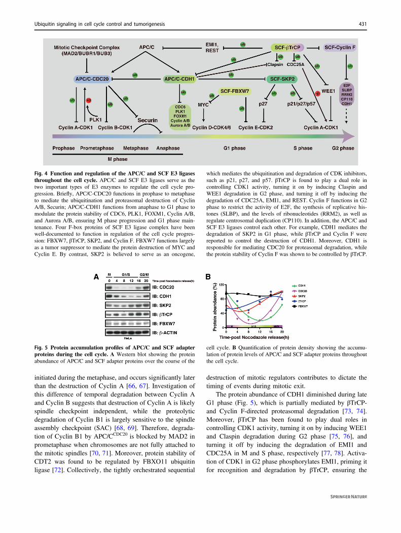
Fig. 4 Function and regulation of the APC/C and SCF E3 ligasesthroughout the cell cycle. APC/C and SCF E3 ligases serve as thetwo important types of E3 enzymes to regulate the cell cycle pro-gression. Briefly, APC/C-CDC20 functions in prophase to metaphaseto mediate the ubiquitination and proteasomal destruction of CyclinA/B, Securin; APC/C-CDH1 functions from anaphase to G1 phase tomodulate the protein stability of CDC6, PLK1, FOXM1, Cyclin A/B,and Aurora A/B, ensuring M phase progression and G1 phase main-tenance. Four F-box proteins of SCF E3 ligase complex have beenwell-documented to function in regulation of the cell cycle progres-sion: FBXW7, βTrCP, SKP2, and Cyclin F. FBXW7 functions largelyas a tumor suppressor to mediate the protein destruction of MYC andCyclin E. By contrast, SKP2 is believed to serve as an oncogene,
which mediates the ubiquitination and degradation of CDK inhibitors,such as p21, p27, and p57. βTrCP is found to play a dual role incontrolling CDK1 activity, turning it on by inducing Claspin andWEE1 degradation in G2 phase, and turning it off by inducing thedegradation of CDC25A, EMI1, and REST. Cyclin F functions in G2phase to restrict the activity of E2F, the synthesis of replicative his-tones (SLBP), and the levels of ribonucleotides (RRM2), as well asregulate centrosomal duplication (CP110). In addition, the APC/C andSCF E3 ligases control each other. For example, CDH1 mediates thedegradation of SKP2 in G1 phase, while βTrCP and Cyclin F werereported to control the destruction of CDH1. Moreover, CDH1 isresponsible for mediating CDC20 for proteasomal degradation, whilethe protein stability of Cyclin F was shown to be controlled by βTrCP.
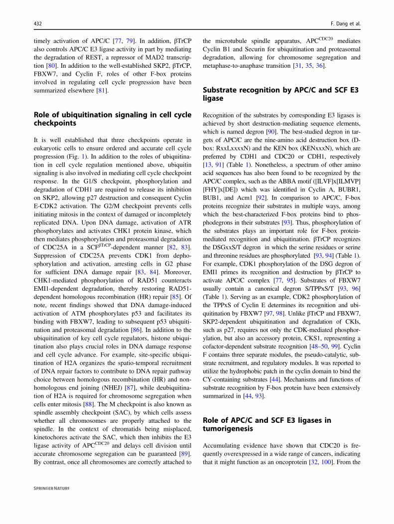
Fig. 5 Protein accumulation profiles of APC/C and SCF adapterproteins during the cell cycle. A Western blot showing the proteinabundance of APC/C and SCF adapter proteins over the course of the
cell cycle. B Quantification of protein density showing the accumu-lation of protein levels of APC/C and SCF adapter proteins throughoutthe cell cycle.
Ubiquitin signaling in cell cycle control and tumorigenesis 431
timely activation of APC/C [77, 79]. In addition, βTrCPalso controls APC/C E3 ligase activity in part by mediatingthe degradation of REST, a repressor of MAD2 transcrip-tion [80]. In addition to the well-established SKP2, βTrCP,FBXW7, and Cyclin F, roles of other F-box proteinsinvolved in regulating cell cycle progression have beensummarized elsewhere [81].
Role of ubiquitination signaling in cell cyclecheckpoints
It is well established that three checkpoints operate ineukaryotic cells to ensure ordered and accurate cell cycleprogression (Fig. 1). In addition to the roles of ubiquitina-tion in cell cycle regulation mentioned above, ubiquitinsignaling is also involved in mediating cell cycle checkpointresponse. In the G1/S checkpoint, phosphorylation anddegradation of CDH1 are required to release its inhibitionon SKP2, allowing p27 destruction and consequent CyclinE-CDK2 activation. The G2/M checkpoint prevents cellsinitiating mitosis in the context of damaged or incompletelyreplicated DNA. Upon DNA damage, activation of ATRphosphorylates and activates CHK1 protein kinase, whichthen mediates phosphorylation and proteasomal degradationof CDC25A in a SCFβTrCP-dependent manner [82, 83].Suppression of CDC25A prevents CDK1 from depho-sphorylation and activation, arresting cells in G2 phasefor sufficient DNA damage repair [83, 84]. Moreover,CHK1-mediated phosphorylation of RAD51 counteractsEMI1-dependent degradation, thereby restoring RAD51-dependent homologous recombination (HR) repair [85]. Ofnote, recent findings showed that DNA damage-inducedactivation of ATM phosphorylates p53 and facilitates itsbinding with FBXW7, leading to subsequent p53 ubiquiti-nation and proteasomal degradation [86]. In addition to theubiquitination of key cell cycle regulators, histone ubiqui-tination also plays crucial roles in DNA damage responseand cell cycle advance. For example, site-specific ubiqui-tination of H2A organizes the spatio-temporal recruitmentof DNA repair factors to contribute to DNA repair pathwaychoice between homologous recombination (HR) and non-homologous end joining (NHEJ) [87], while deubiquitina-tion of H2A is required for chromosome segregation whencells enter mitosis [88]. The M checkpoint is also known asspindle assembly checkpoint (SAC), by which cells assesswhether all chromosomes are properly attached to thespindle. In the context of chromatids being misplaced,kinetochores activate the SAC, which then inhibits the E3ligase activity of APCCDC20 and delays cell division untilaccurate chromosome segregation can be guaranteed [89].By contrast, once all chromosomes are correctly attached to
the microtubule spindle apparatus, APCCDC20 mediatesCyclin B1 and Securin for ubiquitination and proteasomaldegradation, allowing for chromosome segregation andmetaphase-to-anaphase transition [31, 35, 36].
Substrate recognition by APC/C and SCF E3ligase
Recognition of the substrates by corresponding E3 ligases isachieved by short destruction-mediating sequence elements,which is named degron [90]. The best-studied degron in tar-gets of APC/C are the nine-amino acid destruction box (D-box: RxxLxxxxN) and the KEN box (KENxxxN), which arepreferred by CDH1 and CDC20 or CDH1, respectively[13, 91] (Table 1). Nonetheless, a spectrum of other aminoacid sequences has also been found to be recognized by theAPC/C complex, such as the ABBA motif ([ILVF]x[ILMVP][FHY]x[DE]) which was identified in Cyclin A, BUBR1,BUB1, and Acm1 [92]. In comparison to APC/C, F-boxproteins recognize their substrates in multiple ways, amongwhich the best-characterized F-box proteins bind to phos-phodegrons in their substrates [93]. Thus, phosphorylation ofthe substrates plays an important role for F-box protein-mediated recognition and ubiquitination. βTrCP recognizesthe DSGxxS/T degron in which the serine residues or serineand threonine residues are phosphorylated [93, 94] (Table 1).For example, CDK1 phosphorylation of the DSG degron ofEMI1 primes its recognition and destruction by βTrCP toactivate APC/C complex [77, 95]. Substrates of FBXW7usually contain a canonical degron S/TPPxS/T [93, 96](Table 1). Serving as an example, CDK2 phosphorylation ofthe TPPxS of Cyclin E determines its recognition and ubi-quitination by FBXW7 [97, 98]. Unlike βTrCP and FBXW7,SKP2-dependent ubiquitination and degradation of CKIs,such as p27, requires not only the CDK-mediated phosphor-ylation, but also an accessory protein, CKS1, representing acofactor-dependent substrate recognition [48–50, 99]. CyclinF contains three separate modules, the pseudo-catalytic, sub-strate recruitment, and regulatory modules. It was reported toutilize the hydrophobic patch in the cyclin domain to bind theCY-containing substrates [44]. Mechanisms and functions ofsubstrate recognition by F-box protein have been extensivelysummarized in [44, 93].
Role of APC/C and SCF E3 ligases intumorigenesis
Accumulating evidence have shown that CDC20 is fre-quently overexpressed in a wide range of cancers, indicatingthat it might function as an oncoprotein [32, 100]. From the
432 F. Dang et al.

perspective of cell cycle, degradation of Cyclin B andSecurin is required for the onset of anaphase [35, 101]. It isthus conceivable that the loss of CDC20 causes metaphasearrest in mouse embryos [102]. In support of the oncogenicrole, genetic ablation of CDC20 results in efficient tumorregression [103], while the loss of CDC20 inhibition pro-motes tumorigenesis [104], advocating CDC20 as a poten-tial therapeutic target for cancer treatment [105]. In contrast,CDH1 has been found to be downregulated in a largevariety of human cancers [32, 100]. CDH1-deficient cellsproliferate inefficiently and CDH1 heterozygous animalsshow increased susceptibility to spontaneous tumors, lar-gely conferring CDH1 a tumor suppressor role [106]. Inaddition, accumulation of SKP2 due to the loss of CDH1 isconsidered to promote proteasomal degradation of CIP/KIPfamily of CKIs and thus facilitate tumorigenesis.
Regarding the role of SCF E3 complex in cancer devel-opment, emerging evidence suggest that it acts in a F-boxprotein- and context-dependent manner [107–109]. Specifi-cally, SKP2 is a well-defined oncoprotein and was found tobe overexpressed in various human cancers [109, 110]. Tar-gets of SKP2 are mainly tumor suppressor proteins includingp21, p27, p57, p130, and CDT1 [50–52, 111, 112]. Therefore,SKP2 exerts its oncogenic function mainly through degra-dation of its tumor suppressive targets. In support of theoncogenic role of SKP2, pharmacological inhibition of SKP2was found to be able to restrict cancer progression [113]. In
contrast to SKP2, FBXW7 is believed to function mainly as atumor suppressor by targeting various oncogenic proteins fordegradation [96, 107–109]. For example, proteasomaldestruction of Cyclin E through FBXW7-mediated ubiquiti-nation blocks CDK2 activation in late G1 phase and thusdelays G1/S transition, arresting cells in G1 phase[97, 98, 114]. Another well-established oncogenic substrate ofFBXW7 is MYC [115], which serves as a transcription factorinvolved in the genesis of many human cancers [116].Regarding Cyclin F, it is believed to function as a tumorsuppressor by controlling genome integrity and centrosomeduplication by regulating the protein stability of RRM2 andCP110, respectively [44, 58, 59]. Looking at the substrate listof βTrCP, it is obvious that βTrCP plays a dual role in reg-ulating CDK1 activity, turning it on by inducing WEE1 andClaspin destruction [75, 76], while turning it off by targetingEMI1 and CDC25A for proteasomal degradation [77, 78].Importantly, preclinical studies have validated WEE1 inhibi-tion as a viable therapeutic target in treating cancer [117], andCDC25A is also deemed as a suitable therapeutic target forcancer treatment [118], establishing βTrCP as a tumor sup-pressor. On the other hand, βTrCP was found to be involvedin mediating the proteasomal degradation of tumor sup-pressors, such as FOXO3 and DEPTOR [119, 120]. Takingthese results into consideration, βTrCP might be expected tobe oncogenic and exert a tumor suppressive role in a context-dependent manner [107, 109].
Table 1 Examples of key APC/C and SCF substrates involved in cell cycle control.
E3 Adapter Substrate Degron Gene function Role in cancer Refs.
APC/C CDC20 Cyclin A D-box(RxxLxxxxN)
CDK1/2 activation and G1/S, G2/M transition Oncogenic [69]
Cyclin B1 CDK1 activation and mitosis progression Oncogenic [66, 67]
Securin Inhibition of chromosome segregation and p53 activity Oncogene [31]
CDH1 Aurora A D-box(RxxLxxxxN)orKEN-box(KENxxxN)
Regulation of mitosis progression Oncogene [147]
CDC20 Activator of APC/C complex Oncogenic [91]
PLK1 Regulation of mitosis progression Oncogene [148]
SKP2 Substrate recognition component of SCF E3 ligase Oncogene [45, 46]
FOXM1 Transcription factor involved in DNA replication and mitosis Oncogene [149]
SCF SKP2 p21/p27/p57 N/A CDK inhibitor Tumor suppressor [50–52]
p130 Transcription factor regulating cell cycle entry Tumor suppressor [111]
CDT1 Regulator of DNA replication and mitosis Oncogenic [112]
FBXW7 MYC S/TPPxS/T Transcription factor Oncogene [115]
Cyclin E CDK2 activation and G1/S transition Oncogenic [97, 98]
JUN Transcription factor Oncogene [150]
βTrCP FOXO3 DSGxxS/T Transcription factor Tumor suppressor [119]
WEE1 CDK1 inhibition Oncogenic [75]
CDC25A CDK1 activation Oncogenic [78, 82]
EMI1 Regulator of APC activity Oncogenic [77, 79]
Cyclin F SLBP CY motif Histone pre-mRNA processing Not defined [57]
RRM2 Catalyzes the biosynthesis of deoxyribonucleotides Oncogene [58]
CP110 Necessary for centrosome duplication Not defined [59]
Ubiquitin signaling in cell cycle control and tumorigenesis 433
Conclusion and perspective
In the present review, we mainly summarized the proteo-lytic signals involved in the cell cycle control. Moreover,non-proteolytic ubiquitination of the cell cycle regulatorsalso plays crucial roles in controlling cell cycle progression.For example, the endoplasmic reticulum lipid associatedprotein 2 was found to interact and facilitate K63-linkedubiquitination and stabilization of Cyclin B1, facilitatingmitosis exit [121]. Di-ubiquitination of the minichromo-some maintenance protein 10 is required for its interactionwith PCNA to facilitate DNA elongation in S phase [122].Although accumulating evidence supporting critical roles ofnon-proteolytic ubiquitin signals in regulating cell cycleprogression, unlike the functions of proteolytic ubiquitinsignals which have been studied extensively, relatively littleis known regarding the roles and mechanisms underlyingcell cycle control that go beyond proteasomal degradation.In addition, it is well characterized that ubiquitination is areversible process as the ubiquitin can be removed from themodified proteins by an array of deubiquitinating enzymes(DUBs) [123]. Of note, DUBs have been found to playcritical roles in regulation of mitosis [124], and smallmolecular inhibitors against DUBs are expected to offernovel therapeutic opportunities for cancer treatment [125].However, the roles and substrates of DUBs in regulatingcell cycle events remain not well understood. In particular,how is the balance of ubiquitination–deubiquitinationachieved to ensure accurate cell cycle progression remainselusive and needs additional in-depth investigations.
With respect to the well-established APC/C E3 ligasecomplex, activation of CDC20 is required for anaphaseonset, while CDH1 plays a central role in mediating mitosisexit and G1 maintenance. The ordered activation of CDC20and CDH1 is essential for accurate mitosis progression.Mitotic phosphorylation of APC/C relieves its auto-inhibition and facilitates CDC20 engagement [33, 34],while BUB1-mediated phosphorylation of CDC20 uponspindle checkpoint activation inhibits the ubiquitin ligaseactivity of APCCDC20, ensuring the fidelity of chromosomesegregation [126]. Although the protein stability of CDH1has been reported to be modulated by βTrCP and Cyclin F[73, 74], regulation of CDH1 E3 ligase activity is not wellknown yet. A previous study has shown that there are19 serine and threonine residues on CDH1 that can bephosphorylated by multi-kinases in vivo, indicating that thephosphoregulation of CDH1 is much more complex [127].Another intriguing phenomenon is about the timing ofdegradation of proteins controlled by the same substrateadapters. We know that CDC20 functions as an upstreamadapter protein for Cyclin A, Cyclin B, and Securin, med-iating their ubiquitination and proteasomal degradationduring mitosis. Interestingly, degradation of Cyclin A
proceeds before that of Cyclin B and Securin, which isgoverned by the presence of spindle checkpoint signaling[128]. However, the detailed mechanisms underlying thespindle checkpoint imposed various E3 ligase activities ofCDC20 toward different substrates need to be furtherinvestigated.
Overall, the cell cycle progression is tightly regulated toensure the genomic integrity and identity in daughter cells,and ubiquitin signaling involves almost each step of the cellcycle. Dysregulation of the ubiquitination modification ledto uncontrolled cell cycle progression and eventuallyresulted in tumorigenesis [18]. Based upon this notion,targeting the ubiquitin system has provided effective ther-apeutic strategies for cancer treatment [129–146]. At themoment, how these signaling events are integrated andorchestrated in a time–space-dependent manner remains notfully understood. In addition, only a handful of drugs tar-geting the ubiquitin system have been approved by theFDA. Therefore, a better understanding of the ubiquitinsignaling in cell cycle control will expand and diversify therange of anticancer strategies and benefit the clinical treat-ment of cancer patients in the future.
Acknowledgements We apologize for not citing all relevant reportsowing to space limitations. This work was supported in part byfunding from NIH (R01CA200651 to WW).
Compliance with ethical standards
Conflict of interest WW is a co-founder and consultant for theReKindle Therapeutics. Other authors declare no competing financialinterests.
Publisher’s note Springer Nature remains neutral with regard tojurisdictional claims in published maps and institutional affiliations.
Open Access This article is licensed under a Creative CommonsAttribution 4.0 International License, which permits use, sharing,adaptation, distribution and reproduction in any medium or format, aslong as you give appropriate credit to the original author(s) and thesource, provide a link to the Creative Commons license, and indicate ifchanges were made. The images or other third party material in thisarticle are included in the article’s Creative Commons license, unlessindicated otherwise in a credit line to the material. If material is notincluded in the article’s Creative Commons license and your intendeduse is not permitted by statutory regulation or exceeds the permitteduse, you will need to obtain permission directly from the copyrightholder. To view a copy of this license, visit http://creativecommons.org/licenses/by/4.0/.
References
1. Weinberg RA. The Retinoblastoma protein and cell cycle con-trol. Cell. 1995;81:323–30.
2. Zarkowska T, Mittnach S. Differential phosphorylation of theRetinoblastoma protein by G1/S cyclin-dependent kinases. J BiolChem. 1997;272:12738–46.
434 F. Dang et al.
3. Hinds PW, Mittnacht S, Dulic V, Arnold A, Reed SI, WeinbergRA. Regulation of retinoblastoma protein functions by ectopicexpression of human cyclins. Cell. 1992;70:993–1006.
4. Pagano M, Pepperkok R, Verde F, Ansorge W, Draetta G. CyclinA is required at two points in the human cell cycle. EMBO J.1992;11:961–71.
5. Petersen BO, Lukas J, Sørensen CS, Bartek J, Helin K. Phos-phorylation of mammalian CDC6 by cyclin A/CDK2 regulatesits subcellular localization. EMBO J. 1999;18:396–410.
6. Furuno N, Elzen ND, Pines J. Human cyclin A is required formitosis until mid prophase. J Cell Biol. 1999;147:295–306.
7. Boer LD, Oakes V, Beamish H, Giles N, Stevens F, Torres MS,et al. Cyclin A/cdk2 coordinates centrosomal and nuclear mitoticevents. Oncogene. 2008;27:4261–8.
8. Vigneron S, Sundermann L, LabbéJC, Pintard L, Radulescu O,Castro A, et al. Cyclin A-cdk1-dependent phosphorylation ofBora is the triggering factor promoting mitotic entry. Dev Cell.2018;45:637–50.
9. Lindqvist A, Zon WV, Rosenthal CK, Wolthuls RM. Cyclin B1-Cdk1 activation continues after centrosome separation to controlmitotic progression. PLoS Biol. 2007;5:e123. https://doi.org/10.1371/journal.pbio.0050123.
10. Ferrero M, Ferragud J, Orlando L, Valero L, Pino MS, Farràs R,et al. Phosphorylation of AIB1 at mitosis is regulated by CDK1/Cyclin B. PLoS ONE. 2011;6:e28602. https://doi.org/10.1371/journal.pone.0028602.
11. Guo L, Mohd KS, Ren H, Xin G, Jiang Q, Clarke PR, et al.Phosphorylation of importin-alpha1 by CDK1-Cyclin B1 con-trols mitotic spindle assembly. J Cell Sci. 2019;132:jcs232314.https://doi.org/10.1242/jcs.232314.
12. Sherr CJ, Roberts JM. CDK inhibitors: positive and negativeregulators of G1-phase progression. Genes Dev. 1999;13:1501–12.
13. Glotzer M, Murray AW, Kirschner MW. Cyclin is degraded bythe ubiquitin pathway. Nature. 1991;349:132–8.
14. Nakayama K, Nagahama H, Minamishima YA, Miyake S, IshidaN, Hatakeyama S, et al. Skp2-mediated degradation of p27regulates progression into mitosis. Dev Cell. 2004;6:661–72.
15. Tomoda K, Kubota Y, Kato J. Degradation of the cyclin-dependent-kinase inhibitor p27Kip1 is instigated by Jab1. Nature.1999;398:160–5.
16. Bloom J, Amador V, Bartolini F, DeMartino G, Pagano M.Proteasome-mediated degradation of p21 via N-terminal ubi-quitinylation. Cell. 2003;115:71–82.
17. Hershko A. Roles of ubiquitin-mediated proteolysis in cell cyclecontrol. Curr Opin Cell Biol. 1997;9:788–99.
18. Nakayama KI, Nakayama K. Ubiquitin ligases: cell-cycle controland cancer. Nat Rev Cancer. 2006;6:369–81.
19. Goldstein G, Scheid M, Hammerling U, Schlesinger DH, NiallHD, Boyse EA. Isolation of a polypeptide that has lymphocyte-differentiating properties and is probably represented universallyin living cells. Proc Natl Acad Sci USA. 1975;72:11–5.
20. Hershko A, Ciechanover A. The ubiquitin system. Annu RevBiochem. 1998;67:425–79.
21. Matsumoto ML, Wickliffe KE, Dong KC, Yu C, Bosanac I,Bustos D. K11-linked polyubiquitination in cell cycle controlrevealed by a K11 linkage-specific antibody. Mol Cell.2010;39:477–84.
22. Wickliffe KE, Williamson A, Meyer HJ, Kelly A, Rape M. K11-linked ubiquitin chains as novel regulators of cell division.Trends Cell Biol. 2011;21:656–63.
23. Grice GL, Nathan JA. The recognition of ubiquitinated proteinsby the proteasome. Cell Mol Life Sci. 2016;73:3497–506.
24. Emmerich CH, Schmukle AC, Walczak H. The emerging role oflinear ubiquitination in cell signaling. Sci Signal. 2011;4:re5.https://doi.org/10.1126/scisignal.2002187.
25. Akutsu M, Dikic I, Bremm A. Ubiquitin chain diversity at aglance. J Cell Sci. 2016;129:875–80.
26. Swatek KN, Komander D. Ubiquitin modifications. Cell Res.2016;26:399–422.
27. Suryadinata R, Sadowski M, Sarcevic B. Control of cell cycleprogression by phosphorylation of cyclin-dependent kinase(CDK) substrates. Biosci Rep. 2010;30:243–55.
28. Swaffer MP, Jones AW, Flynn HR, Snijders A, Nurse P. CDKsubstrate phosphorylation and ordering the cell cycle. Cell.2016;167:1750–61.
29. Obaya AJ, Sedivy JM. Regulation of cyclin-Cdk activity inmammalian cells. Cell Mol Life Sci. 2002;59:126–42.
30. Vodermaier HC. APC/C and SCF: controlling each other and thecell cycle. Curr Biol. 2004;14:R787–96.
31. Barford D. Structure, function and mechanism of theanaphase promoting complex (APC/C). Q Rev Biophys.2011;44:153–90.
32. Zhang J, Wan L, Dai X, Sun Y, Wei W. Functional character-ization of anaphase promoting complex/cyclosome (APC/C) E3ubiquitin ligases in tumorigenesis. Biochim Biophys Acta.2014;1845:277–93.
33. Qiao R, Weissmann F, Yamaguchi M, Brown NG, VanderLin-den R, Imre R, et al. Mechanism of APC/CCDC20 activation bymitotic phosphorylation. Proc Natl Acad Sci USA. 2016;113:E2570–8.
34. Zhang S, Chang L, Alfieri C, Zhang Z, Yang J, Maslen S, et al.Molecular mechanism of APC/C activation by mitotic phos-phorylation. Nature. 2016;533:260–4.
35. Chang DC, Xu N, Luo KQ. Degradation of cyclin B is requiredfor the onset of anaphase in mammalian cells. J Biol Chem.2003;278:37865–73.
36. Singleton MR, Uhlmann F. Separase-securin complex: a cunningway to control chromosome segregation. Nat Struct Mol Biol.2017;24:337–9.
37. Crasta K, Lim HH, Giddings TH Jr, Winey M, Surana U.Inactivation of Cdh1 by synergistic action of Cdk1 and polokinase is necessary for proper assembly of the mitotic spindle.Nat Cell Biol. 2008;10:665–75.
38. Visintin R, Craig K, Hwang ES, Prinz S, Tyers M, Amon A. Thephosphatase Cdc14 triggers mitotic exit by reversal of Cdk-dependent phosphorylation. Mol Cell. 1998;2:709–18.
39. Sullivan M, Uhlmann F. A non-proteolytic function of separaselinks the onset of anaphase to mitotic exit. Nat Cell Biol.2003;5:249–54.
40. Li M, Zhang P. The function of APC/CCdh1 in cell cycleand beyond. Cell Div. 2009;4. https://doi.org/10.1186/1747-1028-4-2.
41. Jin J, Cardozo T, Lovering RC, Elledge SJ, Pagano M, HarperJW. Systematic analysis and nomenclature of mammalian F-boxproteins. Genes Dev. 2004;18:2573–80.
42. Ang XL, Harper JW. SCF-mediated protein degradation and cellcycle control. Oncogene. 2005;24:2860–70.
43. Nakayama KI, Nakayama K. Regulation of the cell cycle bySCF-type ubiquitin ligases. Semin Cell Dev Biol. 2005;16:323–33.
44. D’Angiolella V, Esencay M, Pagano M. A cyclin without cyclin-dependent kinases: cyclin F controls genome stability throughubiquitin-mediated proteolysis. Trends Cell Biol. 2013;23:135–40.
45. Bashir T, Dorrello NV, Amador V, Guardavaccaro D, Pagano M.Control of the SCFSkp2–Cks1 ubiquitin ligase by the APC/CCdh1
ubiquitin ligase. Nature. 2004;428:190–3.46. Wei W, Ayad NG, Wan Y, Zhang GJ, Kirschner MW, Kaelin
WG Jr. Degradation of the SCF component Skp2 in cell-cyclephase G1 by the anaphase-promoting complex. Nature. 2004;428:194–8.
Ubiquitin signaling in cell cycle control and tumorigenesis 435
47. Geneviève R, Coulombe P, Tanguay PL, Boutonnet C, MelocheS. Phosphorylation of Skp2 regulated by CDK2 and Cdc14Bprotects it from degradation by APCCdh1 in G1 phase. EMBO J.2008;27:679–91.
48. Sheaff RJ, Groudine M, Gordon M, Roberts JM, Clurman BE.Cyclin E-CDK2 is a regulator of p27Kip1. Genes Dev.1997;11:1464–78.
49. Montagnoli A, Fiore F, Eytan E, Carrano AC, Draetta GF,Hershko A, et al. Ubiquitination of p27 is regulated by Cdk-dependent phosphorylation and trimeric complex formation.Genes Dev. 1999;13:1181–9.
50. Carrano AC, Eytan E, Hershko A, Pagano M. SKP2 is requiredfor ubiquitin-mediated degradation of the CDK inhibitor p27.Nat Cell Biol. 1999;1:193–9.
51. Bornstein G, Bloom J, Sitry-Shevah S, Nakayama K, Pagano M,Hershko A. Role of the SCFSkp2 ubiquitin ligase in the degra-dation of p21Cip1 in S phase. J Biol Chem. 2003;278:25752–7.
52. Kamura T, Hara T, Kotoshiba S, Yada M, Ishida N, Imaki H,et al. Degradation of p57Kip2 mediated by SCFSkp2- dependentubiquitylation. Proc Natl Acad Sci USA. 2003;100:10231–6.
53. Denicourt C, Dowdy SF. Cip/Kip proteins: more than just CDKsinhibitors. Genes Dev. 2004;18:851–5.
54. Nakayama K, Nagahama H, Minamishima YA, Matsumoto M,Nakamichi I, Kitagawa K, et al. Targeted disruption of Skp2results in accumulation of cyclin E and p27Kip1, polyploidy andcentrosome overduplication. EMBO J. 2000;19:2069–81.
55. Clijsters L, Hoencamp C, Calis JJ, Marzio A, Handgraaf SM,Cuitino MC, et al. Cyclin F controls cell-cycle transcriptionaloutputs by directing the degradation of the three activator E2Fs.Mol Cell. 2019;74:1264–77.
56. Burdova K, Yang H, Faedda R, Hume S, Chauhan J, Ebner D,et al. E2F1 proteolysis via SCF-cyclin F underlies syntheticlethality between cyclin F loss and Chk1 inhibition. EMBO J.2019;38:e101443. https://doi.org/10.15252/embj.2018101443.
57. Dankert JF, Rona G, Clijsters L, Geter P, Skaar JR, Bermudez-Hernandez K, et al. Cyclin F-mediated degradation of SLBPlimits H2A.X accumulation and apoptosis upon genotoxic stressin G2. Mol Cell. 2016;64:507–19.
58. D’Angiolella V, Donato V, Forrester FM, Jeong YT, Pellacani C,Kudo Y, et al. Cyclin F-mediated degradation of ribonucleotidereductase M2 controls genome integrity and DNA repair. Cell.2012;149:1023–34.
59. D’Angiolella V, Donato V, Vijayakumar S, Saraf A, Florens L,Washburn MP, et al. SCFCyclin F controls centrosome homeostasisand mitotic fidelity through CP110 degradation. Nature. 2010;466:138–42.
60. Mavrommati I, Faedda R, Galasso G, Li J, Burdova K, FischerR, et al. β-TrCP- and casein kinase II-mediated degradation ofCyclin F controls timely mitotic progression. Cell Rep. 2018;24:3404–12.
61. Li C, Vassilev A, DePamphilis ML. Role for Cdk1(Cdc2)/cyclinA in preventing the mammalian origin recognition complex’slargest subunit (Orc1) from binding to chromatin during mitosis.Mol Cell Biol. 2004;24:5875–86.
62. Gong D, Ferrell JE Jr. The roles of cyclin A2, B1, and B2 inearly and late mitotic events. Mol Biol Cell. 2010;21:3149–61.
63. Abe S, Nagasaka K, Hirayama Y, Kozuka-Hata H, Oyama M,Aoyagi Y, et al. The initial phase of chromosome condensationrequires Cdk1-mediated phosphorylation of the CAP-D3 subunitof condensin II. Genes Dev. 2011;25:863–74.
64. Elzen N, Pines J. Cyclin A is destroyed in prometaphase and candelay chromosome alignment and anaphase. J Cell Biol.2001;153:121–36.
65. Geley S, Kramer E, Gieffers C, Gannon J, Peters JM, Hunt T.Anaphase-promoting complex/cyclosome–dependent proteolysisof human cyclin A starts at the beginning of mitosis and is not
subject to the spindle assembly checkpoint. J Cell Biol.2001;153:137–48.
66. Hershko A. Mechanisms and regulation of the degradation ofcyclin B. Philos Trans R Soc Lond B Biol Sci.1999;354:1571–75.
67. Clute P, Pines J. Temporal and spatial control of cyclin B1destruction in metaphase. Nat Cell Biol. 1999;1:82–7.
68. Peters JM. The anaphase-promoting complex: proteolysis inmitosis and beyond. Mol Cell. 2002;9:931–43.
69. Wolthuis R, Clay-Farrace L, Zon W, Yekezare M, Koop L,Ogink J, et al. Cdc20 and Cks direct the spindle checkpoint-independent destruction of cyclin A. Mol Cell.2008;30:290–302.
70. Fang G, Yu H, Kirschner MW. The checkpoint protein MAD2and the mitotic regulator CDC20 form a ternary complex withthe anaphase-promoting complex to control anaphase initiation.Genes Dev. 1998;12:1871–83.
71. Michel L, Diaz-Rodriguez E, Narayan G, Hernando E, Murty V,Benezra R. Complete loss of the tumor suppressor MAD2 causespremature cyclin B degradation and mitotic failure in humansomatic cells. Proc Natl Acad Sci USA. 2004;101:4459–64.
72. Rossi M, Duan S, Jeong YT, Horn M, Saraf A, Florens L, et al.Regulation of the CRL4Cdt2 ubiquitin ligase and cell-cycle exitby the SCFFbxo11 ubiquitin ligase. Mol Cell. 2013;49:1159–66.
73. Fukushima H, Ogura K, Wan L, Lu Y, Li V, Gao D, et al. SCF-mediated Cdh1 degradation defines a negative feedback systemthat coordinates cell-cycle progression. Cell Rep. 2013;4:803–16.
74. Choudhury R, Bonacci T, Arceci A, Lahiri D, Mills CA, KernanJL, et al. APC/C and SCFCyclin F constitute a reciprocal feedbackcircuit controlling S-phase entry. Cell Rep. 2016;16:3359–72.
75. Watanabe N, Arai H, Nishihara Y, Taniguchi M, Watanabe N,Hunter T, et al. M-phase kinases induce phospho-dependentubiquitination of somatic Wee1 by SCFβ-TrCP. Proc Natl Acad SciUSA. 2004;101:4419–24.
76. Peschiaroli A, Dorrello NV, Guardavaccaro D, Venere M,Halazonetis T, Sherman NE, et al. SCFβTrCP-mediated degrada-tion of Claspin regulates recovery from the DNA replicationcheckpoint response. Mol Cell. 2006;23:319–29.
77. Margottin-Goguet F, Hsu JY, Loktev A, Hsieh HM, Reimann J,Jackson PK. Prophase destruction of Emi1 by the SCFβTrCP/Slimb
ubiquitin ligase activates the anaphase promoting complex toallow progression beyond prometaphase. Dev Cell. 2003;4:813–26.
78. Busino L, Donzelli M, Chiesa M, Guardavaccaro D, Ganoth D,Dorrello NV, et al. Degradation of Cdc25A by β-TrCPduring S phase and in response to DNA damage. Nature.2003;426:87–91.
79. Guardavaccaro D, Kudo Y, Boulaire J, Barchi M, Busino L,Donzelli M, et al. Control of meiotic and mitotic progression bythe F box protein β-Trcp1 in vivo. Dev Cell. 2003;4:799–812.
80. Guardavaccaro D, Frescas D, Dorrello NV, Peschiaroli A,Multani AS, Cardozo T, et al. Control of chromosome stabilityby the β-TrCP-REST-Mad2 axis. Nature. 2008;452:365–9.
81. Cardozo T, Pagano M. The SCF ubiquitin ligase: insights into amolecular machine. Nat Rev Mol Cell Biol. 2004;5:739–51.
82. Jin J, Shirogane T, Xu L, Nalepa G, Qin J, Elledge SJ, et al.SCFβ-TRCP links Chk1 signaling to degradation of the Cdc25Aprotein phosphatase. Genes Dev. 2003;17:3062–74.
83. Xiao Z, Chen Z, Gunasekera AH, Sowin TJ, Rosenberg SH,Fesik S, et al. Chk1 mediates S and G2 arrests through Cdc25Adegradation in response to DNA-damaging agents. J Biol Chem.2003;278:21767–73.
84. Busino L, Chiesa M, Draetta GF, Donzelli M. Cdc25Aphosphatase-combinatorial phosphorylation, ubiquitylation andproteolysis. Oncogene. 2004;23:2050–6.
436 F. Dang et al.
85. Marzio A, Puccini J, Kwon Y, Maverakis NK, Arbini A, Sung P,et al. The F-box domain-dependent activity of EMI1 regulatesPARPi sensitivity in triple-negative breast cancers. Mol Cell.2019;73:224–37.
86. Cui D, Xiong X, Shu J, Dai X, Sun Y, Zhao Y. FBXW7 confersradiation survival by targeting p53 for degradation. Cell Rep.2020;30:497–509.
87. Uckelmann M, Sixma TK. Histone ubiquitination in the DNAdamage response. DNA Repair. 2017;56:92–101.
88. Joo HY, Zhai L, Yang C, Nie S, Erdjument-Bromage H, TempstP, et al. Regulation of cell cycle progression and gene expressionby H2A deubiquitination. Nature. 2007;449:1068–72.
89. Lara-Gonzalez P, Westhorpe FG, Taylor SS. The spindleassembly checkpoint. Curr Biol. 2012;22:R966–80.
90. Varshavsky A. Naming a targeting signal. Cell. 1991;64:13–5.91. Pfleger CM, Kirschner MW. The KEN box- an APC recognition
signal distinct from the D box targeted by Cdh1. Genes Dev.2000;14:655–65.
92. Fiore BD, Davey NE, Hagting A, Izawa D, Mansfeld J, GibsonTJ, et al. The ABBA motif binds APC/C activators and is sharedby APC/C substrates and regulators. Dev Cell. 2015;32:358–72.
93. Skaar JR, Pagan JK, Pagano M. Mechanisms and function ofsubstrate recruitment by F-box proteins. Nat Rev Mol Cell Biol.2013;14:369–81.
94. Fuchs SY, Spiegelman VS, Kumar KG. The many faces of β-TrCP E3 ubiquitin ligases: reflections in the magic mirror ofcancer. Oncogene. 2004;23:2028–36.
95. Reimann JD, Freed E, Hsu JY, Kramer ER, Peters JM, JacksonPK. Emi1 is a mitotic regulator that interacts with Cdc20 andinhibits the anaphase promoting complex. Cell. 2001;105:645–55.
96. Cheng Y, Li G. Role of the ubiquitin ligase Fbw7 in cancerprogression. Cancer Metastasis Rev. 2012;31:75–87.
97. Koepp DM, Schaefer LK, Ye X, Keyomarsi K, Chu C, HarperJW, et al. Phosphorylation-dependent ubiquitination of cyclin Eby the SCFFbw7 ubiquitin ligase. Science. 2001;294:173–7.
98. Ye X, Nalepa G, Welcker M, Kessler BM, Spooner E, Qin J,et al. Recognition of phosphodegron motifs in human cyclin E bythe SCFFbw7 ubiquitin ligase. J Biol Chem. 2004;279:50110–9.
99. Hao B, Zheng N, Schulman BA, Wu G, Miller JJ, Pagano M,et al. Structural basis of the Cks1-dependent recognition ofp27Kip1 by the SCFSkp2 ubiquitin ligase. Mol Cell. 2005;20:9–19.
100. Smolders L, Teodoro JG. Targeting the anaphase promotingcomplex: common pathways for viral infection and cancertherapy. Expert Opin Ther Targets. 2011;15:767–80.
101. Hornig N, Knowles PP, McDonald NQ, Uhlmann F. The dualmechanism of separase regulation by securin. Curr Biol.2002;12:973–82.
102. Li M, York JP, Zhang P. Loss of Cdc20 causes a securin-dependent metaphase arrest in two-cell mouse embryos. MolCell Biol. 2007;27:3481–8.
103. Manchado E, Guillamot M, Càrcer G, Eguren M, Trickey M,García-Higuera I, et al. Targeting mitotic exit leads to tumorregression in vivo: modulation by Cdk1, Mastl, and the PP2A/B55α, δ phosphatase. Cancer Cell. 2010;18:641–54.
104. Li M, Fang X, Wei Z, York JP, Zhang P. Loss of spindleassembly checkpoint-mediated inhibition of Cdc20 promotestumorigenesis in mice. J Cell Biol. 2009;185:983–94.
105. Wang Z, Wan L, Zhong J, Inuzuka H, Liu P, Sarkar FH, et al.Cdc20: a potential novel therapeutic target for cancer treatment.Curr Pharm Des. 2013;19:3210–4.
106. García-Higuera I, Manchado E, Dubus P, Cañamero M, MéndezJ, Moreno S, et al. Genomic stability and tumour suppression bythe APC/C cofactor Cdh1. Nat Cell Biol. 2008;10:802–11.
107. Frescas D, Pagano M. Deregulated proteolysis by the F-boxproteins SKP2 and β-TrCP: tipping the scales of cancer. Nat RevCancer. 2008;8:438–49.
108. Welcker M, Clurman BE. FBW7 ubiquitin ligase: a tumoursuppressor at the crossroads of cell division, growth and differ-entiation. Nat Rev Cancer. 2008;8:83–93.
109. Wang Z, Liu P, Inuzuka H, Wei W. Roles of F-box proteins incancer. Nat Rev Cancer. 2014;14:233–47.
110. Gstaiger M, Jordan R, Lim M, Catzavelos C, Mestan J, Sling-erland J, et al. Skp2 is oncogenic and overexpressed in humancancers. Proc Natl Acad Sci USA. 2001;98:5043–8.
111. Bhattacharya S, Garriga J, Calbó J, Yong T, Haines DS, GrañaX. SKP2 associates with p130 and accelerates p130 ubiquityla-tion and degradation in human cells. Oncogene. 2003;22:2443–51.
112. Li X, Zhao Q, Liao R, Sun P, Wu X. The SCFSkp2 ubiquitinligase complex interacts with the human replication licensingfactor Cdt1 and regulates Cdt1 degradation. J Biol Chem.2003;278:30854–8.
113. Chan C, Morrow JK, Li C, Gao Y, Jin G, Moten A, et al.Pharmacological inactivation of Skp2 SCF ubiquitin ligaserestricts cancer stem cell traits and cancer progression. Cell.2013;154:556–68.
114. Minella AC, Welcker M, Clurman BE. Ras activity regulatescyclin E degradation by the Fbw7 pathway. Proc Natl Acad SciUSA. 2005;102:9649–54.
115. Welcker M, Orian A, Jin J, Grim JE, Harper JW, Eisenman RN,et al. The Fbw7 tumor suppressor regulates glycogen synthasekinase 3 phosphorylation-dependent c-Myc protein degradation.Proc Natl Acad Sci USA. 2004;101:9085–90.
116. Dang CV. MYC on the path to cancer. Cell. 2012;149:22–35.117. Do K, Doroshow JH, Kummar S. Wee1 kinase as a target for
cancer therapy. Cell Cycle. 2013;12:3159–64.118. Cangi MG, Cukor B, Soung P, Signoretti S, Moreira G Jr,
Ranashinge M, et al. Role of the Cdc25A phosphatase in humanbreast cancer. J Clin Invest. 2000;106:753–61.
119. Tsai W, Chung YM, Zou Y, Park S, Xu Z, Nakayama K, et al.Inhibition of FOXO3 tumor suppressor function by βTrCP1through ubiquitin-mediated degradation in a tumor mouse mo-del. PLoS ONE. 2010;5:e11171. https://doi.org/10.1371/journal.pone.0011171.
120. Zhao Y, Xiong X, Sun Y. DEPTOR, an mTOR inhibitor, is aphysiological substrate of SCFβTrCP E3 ubiquitin ligase andregulates survival and autophagy. Mol Cell. 2011;44:304–16.
121. Zhang X, Cai J, Zheng Z, Polin L, Lin Z, Dandekar A, et al. Anovel ER-microtubule-binding protein, ERLIN2, stabilizesCyclin B1 and regulates cell cycle progression. Cell Disco.2015;1:15024. https://doi.org/10.1038/celldisc.2015.24.
122. Das-Bradoo S, Ricke RM, Bielinsky AK. Interaction betweenPCNA and diubiquitinated Mcm10 is essential for cell growth inbudding yeast. Mol Cell Biol. 2006;26:4806–17.
123. Amerik AY, Hochstrasser M. Mechanism and function of deu-biquitinating enzymes. Biochim Biophys Acta. 2004;1695:189–207.
124. Park J, Cho J, Kim EE, Song EJ. Deubiquitinating enzymes: acritical regulator of mitosis. Int J Mol Sci. 2019;20:5997. https://doi.org/10.3390/ijms20235997.
125. Pfoh R, Lacdao IK, Saridakis V. Deubiquitinases and the newtherapeutic opportunities offered to cancer. Endocr Relat Cancer.2015;22:T35–54.
126. Tang Z, Shu H, Oncel D, Chen S, Yu H. Phosphorylation ofCdc20 by Bub1 provides a catalytic mechanism for APC/Cinhibition by the spindle checkpoint. Mol Cell. 2004;16:387–97.
127. Hall MC, Warren EN, Borchers CH. Multi kinase phosphor-ylation of the APC/C activator Cdh1 revealed by mass spectro-metry. Cell Cycle. 2004;3:1278–84.
128. Clijsters L, Ogink J, Wolthuis R. The spindle checkpoint, APC/CCdc20, and APC/CCdh1 play distinct roles in connecting mitosisto S phase. J Cell Biol. 2013;201:1013–26.
Ubiquitin signaling in cell cycle control and tumorigenesis 437
129. Hoeller D, Dikic I. Targeting the ubiquitin system in cancertherapy. Nature. 2009;458:438–44.
130. Huang X, Dixit VM. Drugging the undruggables: exploring theubiquitin system for drug development. Cell Res. 2016;26:484–98.
131. Walczak H, Iwai K, Dikic I. Generation and physiological roles oflinear ubiquitin chains. BMC Biol. 2012;10:23. https://doi.org/10.1186/1741-7007-10-23.
132. Wu-Baer F, Ludwig T, Baer R. The UBXN1 protein associateswith autoubiquitinated forms of the BRCA1 tumor suppressor andinhibits its enzymatic function. Mol Cell Biol. 2010;30:2787–98.
133. Durcan TM, Tang MY, Pérusse JR, Dashti EA, Aguileta MA,McLelland GL, et al. USP8 regulates mitophagy by removing K6-linked ubiquitin conjugates from parkin. EMBO J.2014;33:2473–91.
134. Locke M, Toth JI, Petroski MD. Lys11- and Lys48-linked ubi-quitin chains interact with p97 during endoplasmic-reticulum-associated degradation. Biochem J. 2014;459:205–16.
135. Nucifora FC Jr, Nucifora LG, Ng CH, Arbez N, Guo Y, Roby E,et al. Ubiqutination via K27 and K29 chains signals aggregationand neuronal protection of LRRK2 by WSB1. Nat Commun.2016;7:11792. https://doi.org/10.1038/ncomms11792.
136. Palicharla VR, Maddika S. HACE1 mediated K27 ubiquitin link-age leads to YB-1 protein secretion. Cell Signal. 2015;27:2355–62.
137. Chastagner P, Israël A, Brou C. Itch/AIP4 mediates Deltexdegradation through the formation of K29-linked polyubiquitinchains. EMBO Rep. 2006;7:1147–53.
138. Al-Hakim AK, Zagorska A, Chapman L, Deak M, Peggie M,Alessi DR. Control of AMPK-related kinases by USP9X andatypical Lys(29)/Lys(33)-linked polyubiquitin chains. Biochem J.2008;411:249–60.
139. Yuan WC, Li YR, Lin SY, Chang LY, Tan YP, Hung CC, et al.K33-linked polyubiquitination of coronin 7 by Cul3-KLHL20ubiquitin E3 ligase regulates protein trafficking. Mol Cell.2014;54:596–600.
140. Huang H, Jeon MS, Liao L, Yang C, Elly C, Yates JR 3rd, et al.K33-linked polyubiquitination of T cell receptor-zeta regulatesproteolysis-independent T cell signaling. Immunity. 2010;33:60–70.
141. Liu Z, Dong X, Yi HW, Yang J, Gong Z, Wang Y, et al. Structuralbasis for the recognition of K48-linked Ub chain by proteasomalreceptor Rpn13. Cell Discov. 2019;5. https://doi.org/10.1038/s41421-019-0089-7.
142. Zhang L, Xu M, Scotti E, Chen ZJ, Tontonoz P. Both K63 andK48 ubiquitin linkages signal lysosomal degradation of the LDLreceptor. J Lipid Res. 2013;54:1410–20.
143. Ohtake F, Tsuchiya H, Saeki Y, Tanaka K. K63 ubiquitylationtriggers proteasomal degradation by seeding branched ubiquitinchains. Proc Natl Acad Sci USA. 2018;115:E1401–8.
144. Yang WL, Zhang X, Lin HK. Emerging role of Lys-63 ubiquiti-nation in protein kinase and phosphatase activation and cancerdevelopment. Oncogene. 2010;29:4493–503.
145. Erpapazoglou Z, Walker O, Haguenauer-Tsapis R. Versatile roles ofk63-linked ubiquitin chains in trafficking. Cells. 2014;3:1027–88.
146. Lauwers E, Jacob C, André B. K63-linked ubiquitin chains as aspecific signal for protein sorting into the multivesicular bodypathway. J Cell Biol. 2009;185:493–502.
147. Taguchi SI, Honda K, Sugiura K, Yamaguchi A, Furukawa K,Urano T. Degradation of human Aurora-A protein kinase is medi-ated by hCdh1. FEBS Lett. 2002;519:59–65.
148. Lindon C, Pines J. Ordered proteolysis in anaphase inactivates Plk1to contribute to proper mitotic exit in human cells. J Cell Biol.2004;164:233–41.
149. Laoukili J, Alvarez-Fernandez M, Stahl M, Medema RH. FoxM1 isdegraded at mitotic exit in a Cdh1-dependent manner. Cell Cycle.2008;7:2720–6.
150. Wei W, Jin J, Schlisio S, Harper JW, Kaelin WG Jr. The v-Jun pointmutation allows c-Jun to escape GSK3-dependent recognition anddestruction by the Fbw7 ubiquitin ligase. Cancer Cell.2005;8:25–33.
438 F. Dang et al.
Related Documents


![Regulation of Toll-like receptor signaling by NDP52 ... · E3 ubiquitin ligase Peli1 to activate NF-jB signaling [12]. Excessive activation of TLR-mediated responses accumulates pathological](https://static.cupdf.com/doc/110x72/5fb4a6799f261f5b336b0217/regulation-of-toll-like-receptor-signaling-by-ndp52-e3-ubiquitin-ligase-peli1.jpg)