U Tariq MD MBBS, E Wun DDS, J Smith MD MS, W Millar MD MS Geisinger Medical Center Danville, PA. NEUROIMAGING FINDINGS OF UNILATERAL TEMPOROMANDIBULAR JOINT MONOARTHRITIS CAUSED BY CPPD CRYSTAL DEPOSITION DISEASE Control #:1872 Poster #: E-17 GEISINGER Health System

U Tariq MD MBBS, E Wun DDS, J Smith MD MS, W Millar MD MS Geisinger Medical Center Danville, PA. NEUROIMAGING FINDINGS OF UNILATERAL TEMPOROMANDIBULAR.
Dec 27, 2015
Welcome message from author
This document is posted to help you gain knowledge. Please leave a comment to let me know what you think about it! Share it to your friends and learn new things together.
Transcript
U Tariq MD MBBS, E Wun DDS, J Smith MD MS, W Millar MD MS
Geisinger Medical Center
Danville, PA.
NEUROIMAGING FINDINGS OF UNILATERAL TEMPOROMANDIBULAR JOINT MONOARTHRITIS
CAUSED BY CPPD CRYSTAL DEPOSITION DISEASE
Control #:1872Poster #: E-17
GEISINGERHealth System

DISCLOSURES
• The authors of this presentation have no disclosures.
GEISINGERHealth System

PURPOSE
• To describe the clinical presentation and neuroimaging findings of a rare and unusual case of unilateral temporomandibular joint (TMJ) arthritis due to calcium pyrophosphate dihydrate (CPPD) crystal deposition disease.
GEISINGERHealth System

CASE REPORT
• 66 year-old female with severe chronic intermittent pain in left TMJ for multiple years, worse with chewing and jaw activity.
• Physical examination and relative blood work was unremarkable.
GEISINGERHealth System
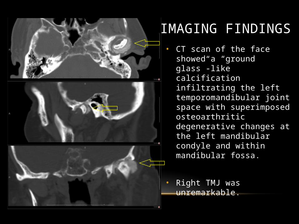
• CT scan of the face showed a “ground glass”-like calcification infiltrating the left temporomandibular joint space with superimposed osteoarthritic degenerative changes at the left mandibular condyle and within mandibular fossa.
• Right TMJ was unremarkable.
IMAGING FINDINGS

Imaging Findings
Subsequent, magnetic resonance imaging (MRI) confirmed the CT findings
Intermediate T1 and mixed T2 signal infiltrates expanding the TMJ joint space

Imaging Findings
Subsequent, magnetic resonance imaging (MRI) confirmed the CT findings
Intermediate T1 and mixed T2 signal infiltrates expanding the TMJ joint space

CASE REPORT
• CPPD crystal deposition disease was suggested as a probable diagnosis.
• Plain films of the bilateral hands and knees performed to evaluate other joints commonly affected by CPPD crystal deposition disease were negative.
GEISINGERHealth System

CASE REPORT
• Subsequently, patient underwent surgical resection and excisional biopsy of left TMJ mass, condylectomy, diskectomy, and total joint arthroplasty.
• The mass mimicked tophaceous gout intraoperatively.
• Pathology confirmed it to be CPPD crystal deposition disease of left TMJ mass, condyle and articular disc.
• Postoperatively patient did well and the left TMJ pain was resolved.
GEISINGERHealth System

GEISINGERHealth System
Intra-operative picture of lesion prior to excision.
PATHOLOGY
Largest portion of the lesion status post excision

GEISINGERHealth System
10X H&E of calcific depositsPathology slides courtesy of Patrick Dorion, MD

GEISINGERHealth System
40X of calcium pyrophosphate crystals with polarized light
Pathology slides courtesy of Patrick Dorion, MD

GEISINGERHealth System
40x of calcium pyrophosphate crystals with compensated polarized light
Pathology slides courtesy of Patrick Dorion, MD
CALCIUM PYROPHOSPHATE DIHYDRATE (CPPD) CRYSTAL DEPOSITION DISEASE
• Metabolic disease where calcium pyrophosphate crystals deposited in synovial fluid result in calcification of articular cartilage, leading to acute arthritis in some patients.
• Predilection for joints with fibrocartilage: Knee, wrist, hip, shoulder, elbow.
• Identification of echogenic foci (crystals) in joint or soft tissues is diagnostic.
1. Löffler C, Sattler H, Peters L, Löffler U, Uppenkamp M, Bergner R. Distinguishing gouty arthritis from calcium pyrophosphate disease and other arthritides. J Rheumatol. 2015 Mar;42(3):513-20. doi: 10.3899/jrheum.140634. Epub 2014 Nov 15. PubMed PMID: 25399385.
2. Naqvi AH, Abraham JL, Kellman RM, Khurana KK. Calcium pyrophosphate dihydrate deposition disease (CPPD)/Pseudogout of the temporomandibular joint – FNA findings and microanalysis. Cytojournal. 2008 Apr 21;5:8. doi:10.1186/1742-6413-5-8. PubMed PMID: 18426573; PubMed Central PMCID: PMC2346483.
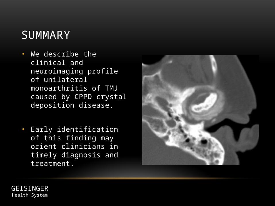
• We describe the clinical and neuroimaging profile of unilateral monoarthritis of TMJ caused by CPPD crystal deposition disease.
• Early identification of this finding may orient clinicians in timely diagnosis and treatment.
SUMMARY
GEISINGERHealth System
Related Documents