1 Type I interferon controls propagation of Long Interspersed Element-1 1 Qiujing Yu, 1 Christopher J. Carbone, 1 Yuliya V. Katlinskaya, 1 Hui Zheng, 1 Ke Zheng, 1 Mengcheng Luo, 1 P. Jeremy Wang, 2 Roger A. Greenberg, 1, @ Serge Y. Fuchs 1 Department of Animal Biology, School of Veterinary Medicine; 2 Department of Cancer Biology, Abramson Family Cancer Research Institute, Basser Research Center for BRCA, Perelman School of Medicine, University of Pennsylvania, Philadelphia, PA 19104, USA. @ Corresponding author: Serge Y. Fuchs, [email protected], tel. (215) 573-6949 fax (215) 746-2295 CAPSULE Background: Type 1 interferons (IFN1) mediate defense against viruses but their role in regulating retrotransposon activities is unknown Results: LINE-1 retrotransposon induces IFN1, which in turn inhibits LINE-1 retrotransposition. Conclusions: IFN1 regulates activities and propagation of LINE-1 Significance: Given that retrotransposons alter the genome, IFN1 plays a role in maintenance of genomic integrity ABSTRACT Type I interferons (IFN) including IFNα and IFNβ are critical for the cellular defense against viruses. Here we report that increased levels of IFNβ were found in testes from mice deficient in Mov10L1, a germ cell- specific RNA helicase that plays a key role in limiting the propagation of retrotransposons including Long Interspersed Element-1 (LINE-1). Additional experiments revealed that activation of LINE-1 retrotransposons increases the expression of IFNβ and of IFN- stimulated genes. Conversely, pre-treatment of cells with IFN suppressed the replication of LINE-1. Furthermore, the efficacy of LINE-1 replication was increased in isogenic cell lines harboring inactivating mutations in diverse elements of the IFN signaling pathway. Knock down of the IFN receptor chain IFNAR1 also stimulated LINE-1 propagation in vitro. Finally, a greater accumulation of LINE-1 was found in mice that lack IFNAR1 compared to wild type mice. We propose that LINE-1-induced IFN plays an important role in restricting LINE-1 propagation and discuss the putative role of IFN in preserving the genome stability. INTRODUCTION Type 1 interferons (IFN, including IFNα and IFNβ) play a major role in anti-viral defenses (1). These cytokines act by interacting with the Type 1 IFN receptor that consists of IFNAR1 and IFNAR2 chains and mediates all cellular effects of IFN (2-4). Association of IFN with a heteromeric receptor leads to activation of Janus kinases TYK2 and JAK1 and phosphorylation-dependent activation of the Signal Transducers and Activators of Transcription (STAT1/2) proteins. These proteins interact with IRF9 to form a potent transcription factor that upregulates the expression of several hundreds of IFN-stimulated genes, whose products both elicit anti-viral effects and contribute to the development of the inflammatory tissue injury (2,3,5-8). Since integration of viruses into host DNA induces genetic changes (9), it may be hypothesized that anti-viral cytokines including IFN can also play a role in maintaining the integrity of the host genome. During evolution, eukaryotic genomes have been undergoing incessant modifications due to diverse events including the activities of mobile genetic elements (10). For example, the Long Interdispersed Element-1 (LINE-1) retrotransposons http://www.jbc.org/cgi/doi/10.1074/jbc.M114.612374 The latest version is at JBC Papers in Press. Published on February 25, 2015 as Manuscript M114.612374 Copyright 2015 by The American Society for Biochemistry and Molecular Biology, Inc. by guest on April 6, 2015 http://www.jbc.org/ Downloaded from

Type I Interferon Controls Propagation of Long Interspersed Element-1
Dec 21, 2015
Type I interferon controls propagation of Long Interspersed Element-1
Welcome message from author
This document is posted to help you gain knowledge. Please leave a comment to let me know what you think about it! Share it to your friends and learn new things together.
Transcript

1
Type I interferon controls propagation of Long Interspersed Element-1 1Qiujing Yu, 1Christopher J. Carbone, 1Yuliya V. Katlinskaya, 1Hui Zheng, 1Ke Zheng,
1Mengcheng Luo, 1P. Jeremy Wang, 2Roger A. Greenberg, 1, @Serge Y. Fuchs 1Department of Animal Biology, School of Veterinary Medicine; 2Department of Cancer
Biology, Abramson Family Cancer Research Institute, Basser Research Center for BRCA, Perelman School of Medicine, University of Pennsylvania, Philadelphia, PA 19104, USA.
@ Corresponding author: Serge Y. Fuchs, [email protected], tel. (215) 573-6949 fax (215) 746-2295
CAPSULE Background: Type 1 interferons (IFN1) mediate defense against viruses but their role in
regulating retrotransposon activities is unknown Results: LINE-1 retrotransposon induces IFN1, which in turn inhibits LINE-1
retrotransposition. Conclusions: IFN1 regulates activities and propagation of LINE-1 Significance: Given that retrotransposons alter the genome, IFN1 plays a role in
maintenance of genomic integrity
ABSTRACT Type I interferons (IFN) including IFNα
and IFNβ are critical for the cellular defense against viruses. Here we report that increased levels of IFNβ were found in testes from mice deficient in Mov10L1, a germ cell-specific RNA helicase that plays a key role in limiting the propagation of retrotransposons including Long Interspersed Element-1 (LINE-1). Additional experiments revealed that activation of LINE-1 retrotransposons increases the expression of IFNβ and of IFN-stimulated genes. Conversely, pre-treatment of cells with IFN suppressed the replication of LINE-1. Furthermore, the efficacy of LINE-1 replication was increased in isogenic cell lines harboring inactivating mutations in diverse elements of the IFN signaling pathway. Knock down of the IFN receptor chain IFNAR1 also stimulated LINE-1 propagation in vitro. Finally, a greater accumulation of LINE-1 was found in mice that lack IFNAR1 compared to wild type mice. We propose that LINE-1-induced IFN plays an important role in restricting LINE-1
propagation and discuss the putative role of IFN in preserving the genome stability.
INTRODUCTION Type 1 interferons (IFN, including IFNα and
IFNβ) play a major role in anti-viral defenses (1). These cytokines act by interacting with the Type 1 IFN receptor that consists of IFNAR1 and IFNAR2 chains and mediates all cellular effects of IFN (2-4). Association of IFN with a heteromeric receptor leads to activation of Janus kinases TYK2 and JAK1 and phosphorylation-dependent activation of the Signal Transducers and Activators of Transcription (STAT1/2) proteins. These proteins interact with IRF9 to form a potent transcription factor that upregulates the expression of several hundreds of IFN-stimulated genes, whose products both elicit anti-viral effects and contribute to the development of the inflammatory tissue injury (2,3,5-8). Since integration of viruses into host DNA induces genetic changes (9), it may be hypothesized that anti-viral cytokines including IFN can also play a role in maintaining the integrity of the host genome.
During evolution, eukaryotic genomes have been undergoing incessant modifications due to diverse events including the activities of mobile genetic elements (10). For example, the Long Interdispersed Element-1 (LINE-1) retrotransposons
http://www.jbc.org/cgi/doi/10.1074/jbc.M114.612374The latest version is at JBC Papers in Press. Published on February 25, 2015 as Manuscript M114.612374
Copyright 2015 by The American Society for Biochemistry and Molecular Biology, Inc.
by guest on April 6, 2015
http://ww
w.jbc.org/
Dow
nloaded from
2
commonly found in many types of mammalian cells (11) have propagated to such extent that they constitute a substantial fraction of genome mass (12). Active propagation of LINE-1 and similar retrotransposons involves near random insertion of these elements into genomic DNA and, accordingly, may lead to gene disruption and an increase in genomic instability (13). Accordingly, germ cells guard their genome by developing a sophisticated and efficient system involving MOV10L1 RNA helicase and Piwi proteins that suppress propagation of LINE-1. Recent analysis of mice that lack Mov10l1 showed that spermatocytes from these animals exhibit an increase in LINE-1 activity, massive DNA damage and post-meiotic proliferation arrest (14).
Here we report that germ cells from the Mov10l1 knockout mice that express highly active LINE-1 also exhibit elevated expression of IFNβ. Using in vitro models of LINE-1 replication in cells we found that LINE-1 stimulates the expression and function of IFN and that the latter functions to suppress LINE-1 propagation. An increased rate of LINE-1 propagation was found in cells and mouse tissues deficient in IFN signaling. These results suggest that IFN produced in response to LINE-1 activities can restrict the very activities of these retrotransposons.
MATERIALS AND METHODS Plasmids, siRNAs and other reagents
The LINE-1-EGFP-puromycin reporter constructs (15,16) pEF06R (which encodes the ORF2 protein with functional endonuclease) and pEF05J (encodes endonuclease-deficient ORF2) were kindly provided by Eline T. Luning Prak (University of Pennsylvania, USA). Human IFNAR2 expression vector pMT2T-hIFNAR2-HA was a generous gift from John Krolewski (University of Rochester Medical Center, USA). The sense strand sequences of siRNAs (Ambion) directed against target molecules were as follows: human RNaseL (5’- GGAAGUCUCUUGUCUGCAAtt -3’), human Mov10 (5’- GACCCUGACUGGAAAGUAUtt -3’), mouse IFNβ (5’- GAAUGAGACUAUUGUUGUAtt -3’), scrambled siRNA (siCon, Ambion Silencer® Negative Control No. 1). Human IFNβ (PBL Inc), and puromycin (Sigma) were purchased.
Cells, cell lines, culture conditions Primary mouse embryonic fibroblasts (MEFs)
were prepared from the embryos of wild-type C57Bl/6J mice as previously described (17). Briefly,
embryos were collected from the pregnant mice on day 14-16 of gestation. Heads and internal organs were removed. Remaining tissue was minced and disassociated with 0.25% trypsin for 5 min. The cells were then plated in DMEM supplemented with 10% FBS (HyClone Laboratories), 100 U/ml penicillin and 0.1 mg/ml streptomycin. Two hour later, the adherent MEFs (P0) were washed twice with phosphate-buffered saline (PBS) and cultured in the complete medium again. Cells were passaged every 2-3 days. Only P2 and P4 MEFs were used in this study. HeLa cells and mouse NIH3T3 cells were obtained from ATCC. Human fibrosarcoma 2fTGH cells and its derivatives (U1A, U3A, and U5A), kindly provided by George Stark, Cleveland Foundation, were maintained in DMEM supplemented with 10% (v/v) FBS, 100 U/ml penicillin and 0.1 mg/ml streptomycin. U1A-derived stable clones expressing either wild type (WT) or kinase deficient (KR) Tyk2 (described in ref. (18,19) – a gift from Sandra Pellegrini, Pasteur Institute, Paris, France) were grown in the same medium with addition of G418 (400µg/ml).
Antibodies and immunofluorescent analysis All the primary and secondary antibodies were
diluted in blocking buffer (5% BSA and 0.1 % Tween-20 in PBS). The following primary antibodies were used: anti- mouse IFNβ was purchased from Millipore. Anti-IRF7 was purchased from Abcam. Secondary antibody used in this study was Alexa Fluor 594 goat anti-rabbit IgG (H+L) (Invitrogen).
NIH3T3 cells were plated in 35mm collagen-coated glass bottom dishes (MatTek Corporation). The next day, the cells were transfected with siRNA (control or against IFNβ) or treated with Abs (control or neutralizing antibody against IFNβ, from Leinco Technologies, both at 10 μg/ml). Twenty-four hours later, the cells were transfected with LINE-1-EGFP-puromycin reporter constructss. After 30 hours, cells were fixed with 4% paraformaldehyde (Affymetrix) in PBS for 10 min at room temperature. The fixed cells were washed three times with PBS and permeabilized with 0.25% Triton X-100 in PBS for 10 min. After another PBS wash, cells were blocked by incubation with 5% BSA and 0.1 % Tween-20 in PBS for 1 h at room temperature. The cells were then incubated with primary antibodies overnight at 4°C. The cells were washed three times with PBS and incubated with
by guest on April 6, 2015
http://ww
w.jbc.org/
Dow
nloaded from

3
secondary antibody for 1h at 37°C in the dark, washed twice more with PBS and treated with DAPI (Sigma) (1 µg/ml) for 2 min. The cells were then washed twice and imaged using the Zeiss LSM710 confocal with a 40×objective lens. All images were processed and quantified using the Fuji software (20). A total of at least 25 fields of cells randomly selected from three independent experiments were scored per group for quantification of percentage of single or double positive cells for GFP, IFNβ or IRF7 proteins in a double blind manner.
Antibodies and immunoblots Antibodies against IFNAR2 (Santa Cruz) and
TYK2 (Cell Signaling) were purchased. Secondary antibodies conjugated to horseradish peroxidase were purchased from Millipore Bioscience Research Reagents. Immunoblotting procedures were described previously (17).
LINE-1 activation assays were carried out by determining the percent of GFP-positive cells (by flow cytometry) indicative of LINE-1 expression and retrotransposition as previously described (15,21) with minor modifications. To determine the role of IFNβ in LINE-1 retrotransposition, Hela cells were plated in a six-well tissue culture plate. The next day, Hela cells were transfected with LINE-1-EGFP-puromycin reporter construct using Lipofectamine 2000 (Invitrogen). Four hours later, the cells were washed and incubated with fresh medium with or without human IFNβ (500 U/ml for 20h). After that, cells were washed twice, incubated with fresh medium without IFN for 24h and then selected in this medium supplemented with puromycin (3µg/ml) for 14 days.
To study the effect of endogenous MOV10 and RNaseL on LINE-1 retrotransposition, Hela cells were transfected with a control siRNA or siRNA against MOV10 or RNaseL using Lipofectamine RNAiMAX Transfection Reagent (Invitrogen). Twenty-four hours later, the cells were transfected with LINE-1-EGFP-puromycin reporter construct using Lipofectamine 2000 (Invitrogen) for four hours, washed and incubated with fresh medium with or without human IFNβ (500 U/ml for 20h). After that, cells were washed and incubated with fresh medium without IFNβ for 24h. An aliquot of the cells without IFNβ treatment was used to analyze the mRNA levels of MOV10, RNaseL and IFNβ by QPCR. The remaining cells were re-plated and then subjected to puromycin selection (3µg/ml for 72h). To study the role of IFNAR1 in LINE-1
retrotransposition, Hela cells were transfected with indicated LINE-1-EGFP-puromycin reporter constructs and shRNA against IFNAR1 or against luciferase (control) using Lipofectamine 2000. After 48h, cells were subjected to 14 days of puromycin selection prior to estimating percent of GFP-positive cells by flow cytometry. In the experiments aimed to detect the effect of endogenous IFNβ signaling on LINE-1 retrotransposition, 2fTGH cells and 2fTGH-derivatives were co-transfected with LINE-1-EGFP-puromycin and mCherry plasmids (to normalize transfection efficiency) for 48 hours and then analyzed by FACS to assess double-positive (GFP+/RFP+) cells. Cells were analyzed using LSRFortessa flow cytometer (BD Biosciences). Results were quantified using FlowJo 7.6 software.
Semi-quantitative RT-PCR and qPCR In experiments to study the expression of LINE-
1 or IFNβ in vivo, the testes collected from male mice (2-month-old Mov10L1+/- and Mov10L1-/- mice or 5-day-old Ifnar1+/+ and Ifnar1-/- mice) were flash-frozen and pulverized in liquid nitrogen, homogenized in Trizol reagent (Ambion, Life Technologies), and extracted with chloroform. In experiments to study the effect of LINE-1 retrotransposition on the expression of IFNβ and its targeted genes in vitro, primary MEFs were plated in 6-well tissue culture plate. The next day, the cells were transfected with LINE-1-EGFP-puromycin reporter constructs using Xfect™ Transfection Reagent (Clontech). After 30 h, total RNA of the cells were prepared with Trizol and chloroform.
Reverse transcription was carried out using Revertaid first strand cDNA synthesis kit (Thermo Scientific) and the cDNA was used for semi-quantitative RT-PCR and quantitative QPCR. Analyses of expression of genes were carried out using the following primers for targeted mouse molecules: Ifnb (FW, 5’ - GTCAGAGTGGAAATCCTAAG - 3’, REV, 5’- ACAGCATCTGCTGGTTGAAG - 3’), Isg15 (FW, 5’ - GGAACGAAAGGGGCCACAGCA - 3’, REV, 5’- CCTCCATGGGCCTTCCCTCGA - 3’), Irf7 (FW, 5’ - CCACACCCCCATCTTCGA - 3’, REV, 5’- CCTCCGAGCCCGAAACTC - 3’), Mov10L1 (FW, 5’- TGTAGCAGTGCAGGACTGTTTTACC - 3’, REV, 5’-CAACAATGGGTTATATGCACCGCAAG - 3’), Mov10 (FW, 5’- GAGGTTCGAGAGTTTTCTGGC - 3’, REV, 5’-
by guest on April 6, 2015
http://ww
w.jbc.org/
Dow
nloaded from
4
GCGATCTTCATTCCATACAGCAT - 3’), Apobec3 (FW, 5’- CAGAGCAGGTACTAAGGTTCCT - 3’, REV, 5’- TTCTGGGTCCCGTATGTTGTA - 3’), LINE-1 (FW, 5’ - GAGAACATCGGCACAACAATC - 3’, REV, 5’- TTTATTGGCGAGTTGAGACCA - 3’), pri-let 7g (FW, 5’- GTACGGTGTGGACCTCATCA - 3’, REV, 5’- TCTTGCTGTGTCCAGGAAAG - 3’), RnaseL (FW, 5’- GTAAACGCCTGTGACAATATGGG - 3’, REV, 5’- AGATGCGTAATAGCCTCCACAT - 3’), β- actin (FW, 5’ - AGAAGAGCTATGAGCTGCCT - 3’, REV, 5’- TCATCGTACTCCTGCTTGCT - 3’). For targeted human molecules: Ifnb (FW, 5’ - AGCTCCAAGAAAGGACGAACAT - 3’, REV, 5’- GCCCTGTAGGTGAGGTTGATCT - 3’), β- actin (FW, 5’ - AGAGCTACGAGCTGCCTGAC - 3’, REV, 5’- CGTGGATGCCACAGGACT - 3’). QPCRs were carried out by using Applied Biosystems 7500 Fast Real-Time PCR system.
Statistical analyses Every shown quantified result represents an
average of at least 3 independent experiments carried out in either triplicate or quadruplicate and calculated as means ±SE. The P values were calculated using the 2-tailed Student t test.
RESULTS LINE-1 activities stimulate IFN expression
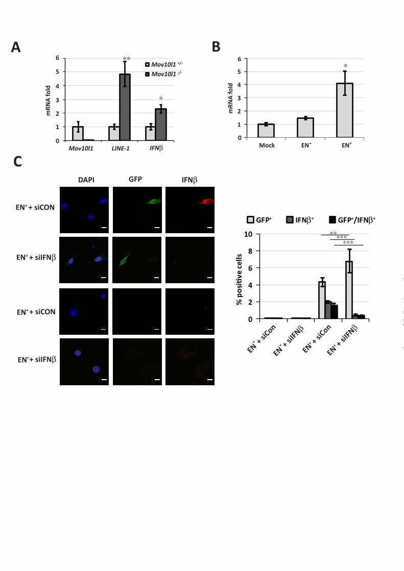
and signaling We have previously reported a high level of
LINE-1 mRNA expression in testes from mice whose spermatocytes lack MOV10L1 (14), RNA helicase, which is essential for silencing retrotransposons in the mouse male germline (14,22,23). Intriguingly, when compared to the testes from heterozygous animals, Mov10L1 knockout tissues expressed noticeably increased mRNA levels of not only LINE-1 but also Ifnb (Figure 1A). LINE-1 encodes ORF1 and ORF2 proteins; the latter harbors endonuclease activity and is capable of inducing double strand breaks (21,24-26). Given the reports that DNA damage-inducing agents (such as ionizing radiation and anti-cancer drugs) can increase IFN expression (27,28), we sought to determine whether such increase can be elicited in response to LINE-1 activation.
We transfected mouse embryonic fibroblast cells with LINE-1-expressing plasmids that enable
detection of LINE-1 retrotransposition by expression of green fluorescent protein (GFP, (21)). Transfection of cells with LINE-1 whose ORF2 was competent in endonuclease activity (EN+) stimulated expression of Ifnb mRNA (Figure 1B). Importantly, delivery of the EN- LINE-1 mutant whose ORF2 lacks the endonuclease function and, as a result, exhibits limited retrotransposition activity (29) led to a significantly lesser induction of Ifnb (Figure 1B). These results suggest that endonuclease-dependent LINE-1 retrotransposition stimulates IFNβ expression.
To corroborate these results we used an immunocytofluorescence assay to assess the levels of IFNβ protein in NIH3T3 cells that received LINE-1 and where its retrotransposition could be monitored by GFP expression (21,30). These studies showed that low yet detectable levels of IFNβ protein were observed predominantly in the GFP-positive cells that received endonuclease-competent LINE-1 (Figure 1C). Targeting Ifnb mRNA with RNAi against this gene robustly decreased the number of IFNβ-positive cells indicating the specificity of IFNβ expression analysis. Together these results suggest that LINE-1 retrotransposons are capable of activating the production of IFNβ. Surprisingly, the overall number of cells that enabled LINE-1 retrotransposition (GFP-positive cells) was increased upon the knockdown of IFNβ (Figure 1C) suggesting that, in turn, IFNβ may control LINE-1 activities.
We next sought to investigate whether LINE-1-induced IFNβ can function as an active cytokine. To this end, we measured mRNA levels of several known IFN-stimulated genes in mouse embryo fibroblasts transfected with LINE-1 plasmids. The expression of Irf7, Isg15, Apobec3 and Mov10 was increased in cells that received endonuclease competent LINE-1 relative to the EN-deficient construct (Figure 2A). Furthermore, the immunofluorescent analyses revealed an increase in IRF7 protein levels in the NIH3T3 cells that received the endonuclease-competent LINE-1 construct and became GFP-positive as a result of LINE-1 retrotransposition (Figure 2B). Very few (if any) IRF7-positive cells were observed in cells receiving the endonuclease-deficient mutant of LINE-1. Importantly, the IRF7 levels in GFP-positive cells could be decreased upon treating cells with IFNβ-neutralizing antibodies (Figure 2B).
by guest on April 6, 2015
http://ww
w.jbc.org/
Dow
nloaded from
5
These results suggest that LINE-1 expression and retrotransposition can activate expression of IRF7 in a manner dependent upon functional IFNβ.
IFN suppresses LINE-1 activity Products of IFN-stimulated genes induced by
IFN, whose expression is triggered by viruses, will in turn limit the spread of these viruses (1). We next sought to determine whether the IFN produced in response to LINE-1 can affect the propagation of this retrotransposon. Quantification of immunofluorescence data showed that inactivation of IFNβ using either RNAi (Figure 1C) or a neutralizing antibody (Figure 2B) increases the number of GFP-positive cells that should have integrated LINE-1. Additional studies examining the effects of anti-IFNβ RNAi oligos and antibodies by immunofluorescence within the same experiment revealed an increase in GFP-positive cells upon IFNβ inactivation (Figure 3A). These data suggest that endogenous IFNβ produced in response to LINE-1 activities may limit the efficacy of LINE-1 retrotransposition.
To further determine the putative role of IFN in LINE-1 control, we used a standard LINE-1 retrotransposition assay in human HeLa cells (Figure 3B and ref. (16,21)). This assay allows to assess LINE-1 retrotransposition by using fluorescence-activated cytometry for measuring the efficacy of the recombination of the LINE-1-EGFP reporter (16,21). We observed that efficacy of LINE-1 retrotransposition in HeLa cells was noticeably decreased after treatment of these cells with recombinant human IFNβ (Figure 3C).
We next sought to investigate the mechanism by which exogenously added IFNβ may inhibit LINE-1 replication. A ubiquitously expressed paralogue of MOV10L1, MOV10 was found among IFN-inducible genes (31) and shown to suppress activities of LINE-1 and other retrotransposons (32). Importantly Mov10 mRNA levels were increased in mouse embryo fibroblasts that received endonuclease-competent LINE-1 (Figure 2A).
The knockdown of Mov10 increased basal levels of LINE-1 retrotransposition and almost completely protected it from suppression by IFNβ (Figures 3D-E). Although knockdown of RNaseL (which was recently implicated in controlling the LINE-1 propagation (33)) increased the basal level of LINE-1 activities, the inhibitory effects of exogenous IFNβ
were still observed in these cells (Figure 3D-E). The efficacy of knockdown of Mov10 and RNaseL were verified by qPCR (Figure 3E). Intriguingly, we found that elevated LINE-1 activities in cells receiving RNAi against Mov10 were found despite the fact that these cells expressed high endogenous levels of Ifnb mRNA (Figure 3E). These results indicate that Mov10 likely acts downstream of IFN signaling to restrict LINE-1 retrotransposition. Overall, these data support the critical role of Mov10 in the control of LINE-1 activities by IFNβ.
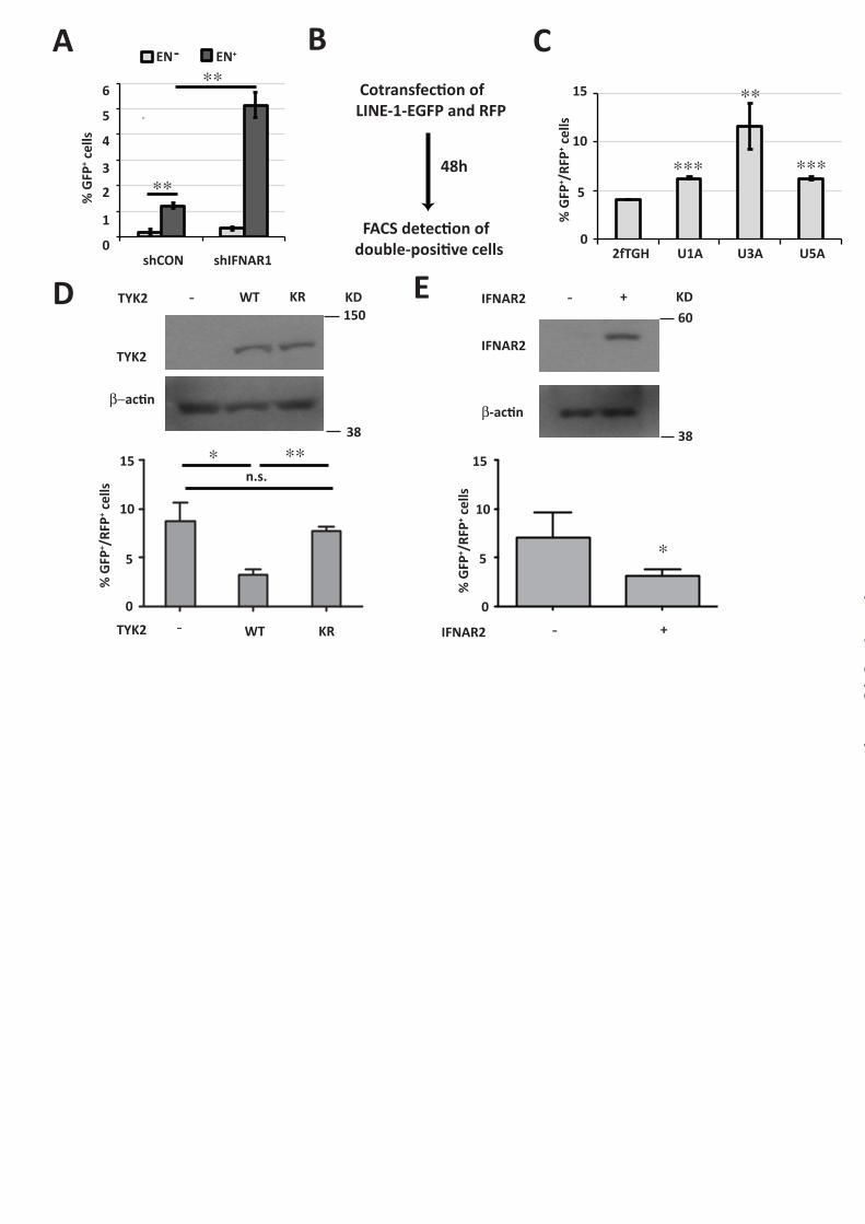
We next investigated the role of proximal mediators of IFN signaling in controlling LINE-1 propagation. The efficacy of retrotransposition of the endonuclease-competent LINE-1 in HeLa cells was robustly increased by knockdown of the IFNAR1 chain of IFN receptor (Figure 4A). While this experiment suggests that IFN signaling may restrict LINE-1 activities, its interpretation is somewhat confounded due to a technical caveat that the assay involves puromycin selection (Figure 3B). Given that both LINE-1 and shRNA (control or against IFNAR1) harbor the puromycin resistance marker, the actual effect of IFNAR1 knockdown on increasing the number of GFP-positive cells can be underestimated. To corroborate these data using an alternative assay that does not involve puromycin selection, we have used co-transfection of LINE-1 plasmid with a plasmid for expression of red fluorescent protein (mCherry, RFP) followed by FACS-based detection of double positive GFP+/RFP+ cells. Furthermore, given that shRNA reagents are capable of inducing IFN response (34), we aimed to avoid possible RNAi-elicited artifacts. To this end, we analyzed retrotransposition of LINE-1 in human isogenic cell lines that differ in status of various elements of the IFN pathway (35). Activation of LINE-1-EGFP was significantly higher in these cell lines that were lacking TYK2 (U1A), STAT1 (U3A) or IFNAR2 (U5A) compared to parental fibrosarcoma 2fTGH cells (Figure 4B-C). Furthermore, U1A cells that were reconstituted with wild type TYK2 (WT) noticeably decreased LINE-1 activities. This effect was not seen in U1A cells expressing the comparable levels of catalytically inactive TYK2 (KR) mutant (Figure 4D). Furthermore, suppression of LINE-1 retrotransposition was also observed in U5A cells upon their reconstitution with IFNAR2 (Figure 4E). These data collectively suggest that endogenous IFN
by guest on April 6, 2015
http://ww
w.jbc.org/
Dow
nloaded from

6
signaling limits propagation of LINE-1 retrotransposon in vitro.
Next we sought to determine whether IFN plays an in vivo role in regulating LINE-1 activities. To this end, we analyzed LINE-1 mRNA levels in testes from wild type mice or animals lacking the IFNAR1 chain of IFN receptor using either semi-quantitative RT-PCR (Figures 5A-B) or quantitative Q-PCR (Figure 5C). As seen from experiments using both approaches, a noticeably greater amount of LINE-1 mRNA was recovered from tissues from Ifnar1-/- mice compared to that from wild type animals. These results collectively suggest that IFN plays a protective role against LINE-1 retrotransoposon activation and propagation.
DISCUSSION The results presented here link IFN production
and activities with the regulation of LINE-1, a major class of retrotransposons , which is common in many types of mammalian cells and which contributes to genome evolution and instability (11). We propose that activation of LINE-1 triggers expression of low levels of IFN, which, in turn, restricts subsequent LINE-1 retrotransposition. This hypothesis is supported by a correlation between LINE-1 and IFNβ mRNA expression and the ability of exogenous LINE-1 to induce IFNβ and several IFN-stimulated genes in vitro (Figures 1-2). Importantly, whereas added IFN suppresses LINE-1 retrotransposition (Figure 3), endogenous IFN signaling appears to restrict the propagation of LINE-1 and the activity of this retrotransposon in cultured cells in a manner that involves engagement of IFNAR1/IFNAR2 and stimulation of the JAK-STAT signaling cascade (Figure 4). Finally, the activities of LINE-1 in vivo are noticeably increased in IFNAR1-deficient mice (Figure 5).
The counteracting relationship between LINE-1 and IFN activities are reminiscent of how virus-induced IFN plays a key role in mounting the anti-viral defenses. It is worth noting that the current paradigm highlights numerous mechanisms by which viruses limit the stimulation of IFN production or inhibit IFN signaling thus allowing propagation of these viruses (1,36). While the nature of similar LINE-1-mediated mechanisms that allow this retrotransposon to ignore IFN signaling is yet to be determined, the putative existence of such mechanisms is supported by the very fact that LINE-
1 and other retrotransposons have been propagating for hundreds of millions of years (37).
It remains to be seen whether IFN can also control other diverse retrotransposons whose activation may also lead to IFN induction. A very robust production of endogenous IFNβ and the ensuing induction of IFN-stimulated genes was observed concurrently with activation of Short Interspersed Nuclear Element (SINE) B1 and B2 in mouse fibroblasts treated with DNA demethylating agent 5-aza-2’-deoxycytidine (38). Intriguingly, these events along with cell death triggered by produced IFNβ were seen only in p53-deficient cells. Given the ability of recombinant IFNβ to induce p53 (39,40) and the role of p53 in IFN-mediated anti-viral effects (41), it is also possible that the modes of negative regulation elicited by IFN may differ between retrotransposons and active viruses.
The mechanisms underlying the inhibitory effects of IFN on the replication of LINE-1 largely remain to be understood. Whereas IFN has been shown to induce the APOBEC3 members of anti-retroviral cytidine deaminases (42,43), and this gene was robustly activated in response to LINE-1 (Figure 2A), the role of these enzymes in suppressing LINE-1 replication remains to be established unequivocally (44-47). However, ubiquitously expressed MOV10 (an APOBEC-interacting paralogue of MOV10L1) was found among IFN-inducible genes (31) and has been shown to suppress activities of LINE-1 and other retrotransposons (32). Our current data demonstrating that Mov10 knockdown relieves suppression of LINE-1 retrotransposition by IFN (Figure 3) indicate that Mov10 plays a critical role in the IFN-mediated control of LINE-1 activities.
It was hypothesized that an increase in activity of LINE-1 and other retrotransposons is associated with development of autoimmune diseases including systemic lupus erythematosus and Sjogren’s syndrome (48,49). Interestingly, these diseases have also been characterized by increased production and activity of IFN (50,51). While these published reports are consistent with our data linking LINE-1 activities and IFN production, the casual relationship between the activity of LINE-1 and IFN signaling in health and diverse diseases and the contribution of these mechanisms to normal and pathogenic processes needs to be further investigated.
by guest on April 6, 2015
http://ww
w.jbc.org/
Dow
nloaded from
7
ACKNOWLEDGMENTS All authors declare no conflict of interest. We
thank George Stark, John Krolewski, Sandra Pellegrini and Eline T. Luning Prak for reagents, Ze’ev Ronai, Eric Lau and the members of Diehl, Koumenis, Greenberg, Witze and Minn labs (at the University of Pennsylvania) for critical suggestions.
This work was supported by National Institutes of Health/National Cancer Institute grants (CA092900 and CA142425 to S.Y.F.). Funding for open access charge: by National Institutes of Health.
References
1. Katze, M. G., He, Y., and Gale, M., Jr. (2002) Viruses and interferon: a fight for supremacy. Nature reviews. Immunology 2, 675-687
2. Fuchs, S. Y. (2013) Hope and fear for interferon: the receptor-centric outlook on the future of interferon therapy. Journal of interferon & cytokine research : the official journal of the International Society for Interferon and Cytokine Research 33, 211-225
3. Fuchs, S. Y. (2012) Ubiquitination-mediated regulation of interferon responses. Growth Factors 30, 141-148
4. Piehler, J., Thomas, C., Garcia, K. C., and Schreiber, G. (2012) Structural and dynamic determinants of type I interferon receptor assembly and their functional interpretation. Immunological reviews 250, 317-334
5. Trinchieri, G. (2010) Type I interferon: friend or foe? The Journal of experimental medicine 207, 2053-2063
6. Platanias, L. C. (2005) Mechanisms of type-I- and type-II-interferon-mediated signalling. Nature reviews. Immunology 5, 375-386
7. Bhattacharya, S., Katlinski, K. V., Reichert, M., Takano, S., Brice, A., Zhao, B., Yu, Q., Zheng, H., Carbone, C. J., Katlinskaya, Y. V., Leu, N. A., McCorkell, K. A., Srinivasan, S., Girondo, M., Rui, H., May, M. J., Avadhani, N. G., Rustgi, A. K., and Fuchs, S. Y. (2014) Triggering ubiquitination of IFNAR1 protects tissues from inflammatory injury. EMBO molecular medicine 6, 384-397
8. Zheng, H., Gupta, V., Patterson-Fortin, J., Bhattacharya, S., Katlinski, K., Wu, J., Varghese, B., Carbone, C. J., Aressy, B., Fuchs, S. Y., and Greenberg, R. A. (2013) A BRISC-SHMT complex deubiquitinates IFNAR1 and regulates interferon responses. Cell reports 5, 180-193
9. Weitzman, M. D., Lilley, C. E., and Chaurushiya, M. S. (2010) Genomes in conflict: maintaining genome integrity during virus infection. Annu Rev Microbiol 64, 61-81
10. Kumar, A., and Bennetzen, J. L. (1999) Plant retrotransposons. Annu Rev Genet 33, 479-532 11. Kazazian, H. H., Jr. (2004) Mobile elements: drivers of genome evolution. Science 303, 1626-
1632 12. Lander, E. S., Linton, L. M., Birren, B., Nusbaum, C., Zody, M. C., Baldwin, J., Devon, K.,
Dewar, K., Doyle, M., FitzHugh, W., Funke, R., Gage, D., Harris, K., Heaford, A., Howland, J., Kann, L., Lehoczky, J., LeVine, R., McEwan, P., McKernan, K., Meldrim, J., Mesirov, J. P., Miranda, C., Morris, W., Naylor, J., Raymond, C., Rosetti, M., Santos, R., Sheridan, A., Sougnez, C., Stange-Thomann, N., Stojanovic, N., Subramanian, A., Wyman, D., Rogers, J., Sulston, J., Ainscough, R., Beck, S., Bentley, D., Burton, J., Clee, C., Carter, N., Coulson, A., Deadman, R., Deloukas, P., Dunham, A., Dunham, I., Durbin, R., French, L., Grafham, D., Gregory, S., Hubbard, T., Humphray, S., Hunt, A., Jones, M., Lloyd, C., McMurray, A., Matthews, L., Mercer, S., Milne, S., Mullikin, J. C., Mungall, A., Plumb, R., Ross, M., Shownkeen, R., Sims, S., Waterston, R. H., Wilson, R. K., Hillier, L. W., McPherson, J. D., Marra, M. A., Mardis, E. R., Fulton, L. A., Chinwalla, A. T., Pepin, K. H., Gish, W. R., Chissoe, S. L., Wendl, M. C., Delehaunty, K. D., Miner, T. L., Delehaunty, A., Kramer, J. B., Cook, L. L., Fulton, R. S., Johnson, D. L., Minx, P. J., Clifton, S. W., Hawkins, T., Branscomb, E., Predki, P., Richardson,
by guest on April 6, 2015
http://ww
w.jbc.org/
Dow
nloaded from
8
P., Wenning, S., Slezak, T., Doggett, N., Cheng, J. F., Olsen, A., Lucas, S., Elkin, C., Uberbacher, E., Frazier, M., Gibbs, R. A., Muzny, D. M., Scherer, S. E., Bouck, J. B., Sodergren, E. J., Worley, K. C., Rives, C. M., Gorrell, J. H., Metzker, M. L., Naylor, S. L., Kucherlapati, R. S., Nelson, D. L., Weinstock, G. M., Sakaki, Y., Fujiyama, A., Hattori, M., Yada, T., Toyoda, A., Itoh, T., Kawagoe, C., Watanabe, H., Totoki, Y., Taylor, T., Weissenbach, J., Heilig, R., Saurin, W., Artiguenave, F., Brottier, P., Bruls, T., Pelletier, E., Robert, C., Wincker, P., Smith, D. R., Doucette-Stamm, L., Rubenfield, M., Weinstock, K., Lee, H. M., Dubois, J., Rosenthal, A., Platzer, M., Nyakatura, G., Taudien, S., Rump, A., Yang, H., Yu, J., Wang, J., Huang, G., Gu, J., Hood, L., Rowen, L., Madan, A., Qin, S., Davis, R. W., Federspiel, N. A., Abola, A. P., Proctor, M. J., Myers, R. M., Schmutz, J., Dickson, M., Grimwood, J., Cox, D. R., Olson, M. V., Kaul, R., Shimizu, N., Kawasaki, K., Minoshima, S., Evans, G. A., Athanasiou, M., Schultz, R., Roe, B. A., Chen, F., Pan, H., Ramser, J., Lehrach, H., Reinhardt, R., McCombie, W. R., de la Bastide, M., Dedhia, N., Blocker, H., Hornischer, K., Nordsiek, G., Agarwala, R., Aravind, L., Bailey, J. A., Bateman, A., Batzoglou, S., Birney, E., Bork, P., Brown, D. G., Burge, C. B., Cerutti, L., Chen, H. C., Church, D., Clamp, M., Copley, R. R., Doerks, T., Eddy, S. R., Eichler, E. E., Furey, T. S., Galagan, J., Gilbert, J. G., Harmon, C., Hayashizaki, Y., Haussler, D., Hermjakob, H., Hokamp, K., Jang, W., Johnson, L. S., Jones, T. A., Kasif, S., Kaspryzk, A., Kennedy, S., Kent, W. J., Kitts, P., Koonin, E. V., Korf, I., Kulp, D., Lancet, D., Lowe, T. M., McLysaght, A., Mikkelsen, T., Moran, J. V., Mulder, N., Pollara, V. J., Ponting, C. P., Schuler, G., Schultz, J., Slater, G., Smit, A. F., Stupka, E., Szustakowski, J., Thierry-Mieg, D., Thierry-Mieg, J., Wagner, L., Wallis, J., Wheeler, R., Williams, A., Wolf, Y. I., Wolfe, K. H., Yang, S. P., Yeh, R. F., Collins, F., Guyer, M. S., Peterson, J., Felsenfeld, A., Wetterstrand, K. A., Patrinos, A., Morgan, M. J., de Jong, P., Catanese, J. J., Osoegawa, K., Shizuya, H., Choi, S., and Chen, Y. J. (2001) Initial sequencing and analysis of the human genome. Nature 409, 860-921
13. Symer, D. E., Connelly, C., Szak, S. T., Caputo, E. M., Cost, G. J., Parmigiani, G., and Boeke, J. D. (2002) Human l1 retrotransposition is associated with genetic instability in vivo. Cell 110, 327-338
14. Zheng, K., and Wang, P. J. (2012) Blockade of pachytene piRNA biogenesis reveals a novel requirement for maintaining post-meiotic germline genome integrity. PLoS Genet 8, e1003038
15. Prak, E. T., Dodson, A. W., Farkash, E. A., and Kazazian, H. H., Jr. (2003) Tracking an embryonic L1 retrotransposition event. Proceedings of the National Academy of Sciences of the United States of America 100, 1832-1837
16. Rangwala, S. H., and Kazazian, H. H., Jr. (2009) The L1 retrotransposition assay: a retrospective and toolkit. Methods 49, 219-226
17. Liu, J., HuangFu, W. C., Kumar, K. G., Qian, J., Casey, J. P., Hamanaka, R. B., Grigoriadou, C., Aldabe, R., Diehl, J. A., and Fuchs, S. Y. (2009) Virus-induced unfolded protein response attenuates antiviral defenses via phosphorylation-dependent degradation of the type I interferon receptor. Cell host & microbe 5, 72-83
18. Gauzzi, M. C., Velazquez, L., McKendry, R., Mogensen, K. E., Fellous, M., and Pellegrini, S. (1996) Interferon-alpha-dependent activation of Tyk2 requires phosphorylation of positive regulatory tyrosines by another kinase. J Biol Chem 271, 20494-20500
19. Marijanovic, Z., Ragimbeau, J., Kumar, K. G., Fuchs, S. Y., and Pellegrini, S. (2006) TYK2 activity promotes ligand-induced IFNAR1 proteolysis. Biochem J 397, 31-38
20. Schindelin, J., Arganda-Carreras, I., Frise, E., Kaynig, V., Longair, M., Pietzsch, T., Preibisch, S., Rueden, C., Saalfeld, S., Schmid, B., Tinevez, J. Y., White, D. J., Hartenstein, V., Eliceiri, K., Tomancak, P., and Cardona, A. (2012) Fiji: an open-source platform for biological-image analysis. Nat Methods 9, 676-682
21. Belgnaoui, S. M., Gosden, R. G., Semmes, O. J., and Haoudi, A. (2006) Human LINE-1 retrotransposon induces DNA damage and apoptosis in cancer cells. Cancer Cell Int 6, 13
22. Frost, R. J., Hamra, F. K., Richardson, J. A., Qi, X., Bassel-Duby, R., and Olson, E. N. (2010) MOV10L1 is necessary for protection of spermatocytes against retrotransposons by Piwi-
by guest on April 6, 2015
http://ww
w.jbc.org/
Dow
nloaded from

9
interacting RNAs. Proceedings of the National Academy of Sciences of the United States of America 107, 11847-11852
23. Zheng, K., Xiol, J., Reuter, M., Eckardt, S., Leu, N. A., McLaughlin, K. J., Stark, A., Sachidanandam, R., Pillai, R. S., and Wang, P. J. (2010) Mouse MOV10L1 associates with Piwi proteins and is an essential component of the Piwi-interacting RNA (piRNA) pathway. Proceedings of the National Academy of Sciences of the United States of America 107, 11841-11846
24. Wallace, N. A., Belancio, V. P., and Deininger, P. L. (2008) L1 mobile element expression causes multiple types of toxicity. Gene 419, 75-81
25. Gilbert, N., Lutz, S., Morrish, T. A., and Moran, J. V. (2005) Multiple fates of L1 retrotransposition intermediates in cultured human cells. Molecular and cellular biology 25, 7780-7795
26. Gasior, S. L., Wakeman, T. P., Xu, B., and Deininger, P. L. (2006) The human LINE-1 retrotransposon creates DNA double-strand breaks. J Mol Biol 357, 1383-1393
27. Kim, T., Kim, T. Y., Song, Y. H., Min, I. M., Yim, J., and Kim, T. K. (1999) Activation of interferon regulatory factor 3 in response to DNA-damaging agents. The Journal of biological chemistry 274, 30686-30689
28. Brzostek-Racine, S., Gordon, C., Van Scoy, S., and Reich, N. C. (2011) The DNA damage response induces IFN. J Immunol 187, 5336-5345
29. Morrish, T. A., Garcia-Perez, J. L., Stamato, T. D., Taccioli, G. E., Sekiguchi, J., and Moran, J. V. (2007) Endonuclease-independent LINE-1 retrotransposition at mammalian telomeres. Nature 446, 208-212
30. Farkash, E. A., Kao, G. D., Horman, S. R., and Prak, E. T. (2006) Gamma radiation increases endonuclease-dependent L1 retrotransposition in a cultured cell assay. Nucleic acids research 34, 1196-1204
31. Schoggins, J. W., Wilson, S. J., Panis, M., Murphy, M. Y., Jones, C. T., Bieniasz, P., and Rice, C. M. (2011) A diverse range of gene products are effectors of the type I interferon antiviral response. Nature 472, 481-485
32. Goodier, J. L., Cheung, L. E., and Kazazian, H. H., Jr. (2012) MOV10 RNA helicase is a potent inhibitor of retrotransposition in cells. PLoS Genet 8, e1002941
33. Zhang, A., Dong, B., Doucet, A. J., Moldovan, J. B., Moran, J. V., and Silverman, R. H. (2014) RNase L restricts the mobility of engineered retrotransposons in cultured human cells. Nucleic acids research 42, 3803-3820
34. Bridge, A. J., Pebernard, S., Ducraux, A., Nicoulaz, A. L., and Iggo, R. (2003) Induction of an interferon response by RNAi vectors in mammalian cells. Nature genetics 34, 263-264
35. McKendry, R., John, J., Flavell, D., Muller, M., Kerr, I. M., and Stark, G. R. (1991) High-frequency mutagenesis of human cells and characterization of a mutant unresponsive to both alpha and gamma interferons. Proceedings of the National Academy of Sciences of the United States of America 88, 11455-11459
36. Galligan, C. L., Murooka, T. T., Rahbar, R., Baig, E., Majchrzak-Kita, B., and Fish, E. N. (2006) Interferons and viruses: signaling for supremacy. Immunologic research 35, 27-40
37. Furano, A. V. (2000) The biological properties and evolutionary dynamics of mammalian LINE-1 retrotransposons. Progress in nucleic acid research and molecular biology 64, 255-294
38. Leonova, K. I., Brodsky, L., Lipchick, B., Pal, M., Novototskaya, L., Chenchik, A. A., Sen, G. C., Komarova, E. A., and Gudkov, A. V. (2013) p53 cooperates with DNA methylation and a suicidal interferon response to maintain epigenetic silencing of repeats and noncoding RNAs. Proceedings of the National Academy of Sciences of the United States of America 110, E89-98
39. Moiseeva, O., Mallette, F. A., Mukhopadhyay, U. K., Moores, A., and Ferbeyre, G. (2006) DNA damage signaling and p53-dependent senescence after prolonged beta-interferon stimulation. Molecular biology of the cell 17, 1583-1592
by guest on April 6, 2015
http://ww
w.jbc.org/
Dow
nloaded from
10
40. Takaoka, A., Hayakawa, S., Yanai, H., Stoiber, D., Negishi, H., Kikuchi, H., Sasaki, S., Imai, K., Shibue, T., Honda, K., and Taniguchi, T. (2003) Integration of interferon-alpha/beta signalling to p53 responses in tumour suppression and antiviral defence. Nature 424, 516-523
41. Munoz-Fontela, C., Macip, S., Martinez-Sobrido, L., Brown, L., Ashour, J., Garcia-Sastre, A., Lee, S. W., and Aaronson, S. A. (2008) Transcriptional role of p53 in interferon-mediated antiviral immunity. The Journal of experimental medicine 205, 1929-1938
42. Bonvin, M., Achermann, F., Greeve, I., Stroka, D., Keogh, A., Inderbitzin, D., Candinas, D., Sommer, P., Wain-Hobson, S., Vartanian, J. P., and Greeve, J. (2006) Interferon-inducible expression of APOBEC3 editing enzymes in human hepatocytes and inhibition of hepatitis B virus replication. Hepatology 43, 1364-1374
43. Tanaka, Y., Marusawa, H., Seno, H., Matsumoto, Y., Ueda, Y., Kodama, Y., Endo, Y., Yamauchi, J., Matsumoto, T., Takaori-Kondo, A., Ikai, I., and Chiba, T. (2006) Anti-viral protein APOBEC3G is induced by interferon-alpha stimulation in human hepatocytes. Biochemical and biophysical research communications 341, 314-319
44. Lovsin, N., and Peterlin, B. M. (2009) APOBEC3 proteins inhibit LINE-1 retrotransposition in the absence of ORF1p binding. Ann N Y Acad Sci 1178, 268-275
45. Niewiadomska, A. M., Tian, C., Tan, L., Wang, T., Sarkis, P. T., and Yu, X. F. (2007) Differential inhibition of long interspersed element 1 by APOBEC3 does not correlate with high-molecular-mass-complex formation or P-body association. J Virol 81, 9577-9583
46. Esnault, C., Heidmann, O., Delebecque, F., Dewannieux, M., Ribet, D., Hance, A. J., Heidmann, T., and Schwartz, O. (2005) APOBEC3G cytidine deaminase inhibits retrotransposition of endogenous retroviruses. Nature 433, 430-433
47. Peng, G., Lei, K. J., Jin, W., Greenwell-Wild, T., and Wahl, S. M. (2006) Induction of APOBEC3 family proteins, a defensive maneuver underlying interferon-induced anti-HIV-1 activity. The Journal of experimental medicine 203, 41-46
48. Nakkuntod, J., Avihingsanon, Y., Mutirangura, A., and Hirankarn, N. (2011) Hypomethylation of LINE-1 but not Alu in lymphocyte subsets of systemic lupus erythematosus patients. Clinica chimica acta; international journal of clinical chemistry 412, 1457-1461
49. Crow, M. K. (2010) Long interspersed nuclear elements (LINE-1): potential triggers of systemic autoimmune disease. Autoimmunity 43, 7-16
50. Bronson, P. G., Chaivorapol, C., Ortmann, W., Behrens, T. W., and Graham, R. R. (2012) The genetics of type I interferon in systemic lupus erythematosus. Curr Opin Immunol 24, 530-537
51. Elkon, K. B., and Wiedeman, A. (2012) Type I IFN system in the development and manifestations of SLE. Curr Opin Rheumatol 24, 499-505
by guest on April 6, 2015
http://ww
w.jbc.org/
Dow
nloaded from
11
Legends to Figures Figure 1: Retrotransposition of LINE-1 induces IFNβ expression. A. Relative mRNA levels of indicated genes in the testes of two month-old Mov10L1 heterozygous
or homozygous knockout mice assessed by qPCR (levels in heterozygous mice taken as 1.0). Average from three independent experiments is shown as Mean±S.D. Here and thereafter: * p<0.05; ** p<0.01; ***p<0.001.
B. Relative IFNβ mRNA levels (compared to Mock taken as 1.0) in mouse embryonic fibroblast cells transfected with LINE-1-expressing vectors that encode functional (EN+) or endonuclease-deficient (EN-) ORF2 protein.
C. Immunofluorescent analysis of IFNβ expression in NIH3T3 cells transfected with indicated RNAi oligos and indicated LINE-1 constructs (left panel; magnification bar: 10 µm). Right panel: quantification of percent of cells single or double positive for GFP and IFNβ proteins in total 25 fields randomly chosen from three independent experiments performed as described in left panel.
Figure 2: LINE-1-induced IFNβ can upregulate IFN-stimulated genes A. Relative mRNA levels of indicated IFN-stimulated genes in mouse embryonic fibroblasts
transfected with indicated LINE-1 plasmids were assessed by qPCR (levels in EN- transfected cells taken as 1.0).
B. Immunofluorescent analysis of IRF7 expression in NIH3T3 cells cultured in the presence of indicated antibodies (control IgG or neutralizing antibody against mouse IFNβ, 10 μg/ml for 30h) and transfected with indicated LINE-1 constructs (left panel; magnification bar: 10 µm). Right panel: quantification of percent of cells single or double positive for GFP and IRF7 proteins in total 30 fields randomly chosen from three independent experiments performed as described in left panel.
Figure 3: Exogenous IFNβ can decrease the efficacy of LINE-1 retrotransposition in a MOV10-
dependent manner A. Appearance of GFP-positive cells NIH3T3 cells that received indicated RNAi (control or against
mouse IFNβ,) or antibodies (control IgG or neutralizing antibody against mouse IFNβ, 10 μg/ml for 30h) prior to being transfected with EN+ LINE-1 construct. Magnification bar: 10 μm.
B. The schematic layout of the experiment described in panel C. C. HeLa cells were transfected with LINE-1-GFP plasmid, incubated for four hours after
transfection and then treated with PBS or IFNβ (500IU/ml for 20hr). After that, cells were washed and incubated with fresh medium without IFN for 24h and then subjected to 14 days of puromycin selection (3µg/ml). Percentage of GFP-positive cells was determined by FACS analysis 24h later.
D. A representative FACS plot diagram (forward scattering versus GFP) of HeLa cells that received indicated RNAi oligos (siCon, siMOV10 or siRNaseL) prior to transfection with LINE-1-GFP plasmid, IFNβ treatment (as indicated), puromycin selection and FACS analysis.
E. Quantification of data from three independent experiments (described in panel D). F. Relative mRNA levels of indicated genes in HeLa cells from experiments described in panels C-
D were assessed by qPCR. Levels in siCON-transfected cells were taken as 1.0. Figure 4: Endogenous IFNβ signaling suppresses the replication of human LINE-1
retrotransposons in vitro A. Effect of IFNAR1 knockdown in HeLa cells on the retrotransposition of endonuclease-positive
(EN+) or negative (EN-) LINE-1 assessed as in A. B. Layout of the experiment described in panel C.
by guest on April 6, 2015
http://ww
w.jbc.org/
Dow
nloaded from

12
C. Parental 2fTGH human fibrosarcoma cells and derivative cell lines lacking various components of IFN signaling including TYK2 (U1A), STAT1 (U3A) and IFNAR2 (U5A) were co-transfected with LINE-1-GFP and RFP-expressing vectors. Retrotransposition of LINE-1 was analyzed as % of double positive cells.
D. U1A cells and its derivative clones harboring wild type TYK2 (WT) or its catalytically inactive mutant (KR) were co-transfected with LINE-1-GFP and RFP-expressing vectors. Retrotransposition of LINE-1 was analyzed as % of double positive cells. Leves of TYK2 and β-actin (used as a loading control) were analyzed by immunoblotting.
E. U5A cells were co-transfected with IFNAR2 expression plasmid or vector, LINE-1-GFP and RFP-expressing vectors. Retrotransposition of LINE-1 was analyzed as % of double positive cells. Leves of IFNAR2 and β-actin (used as a loading control) were analyzed by immunoblotting
Figure 5: IFNβ suppresses the replication of mouse LINE-1 retrotransposons in vivo
A. Expression of endogenous murine LINE-1 mRNA in the testes from indicated 5 days old mice was assessed by RT-PCR. Each lane represents an independent individual mouse. Right panel: quantification of data combined from two independent experiments and presented as Mean ± SD.
B. Expression of endogenous murine LINE-1 mRNA in the testes from indicated mice assessed by qPCR. MicroRNA precursor Pri-let7g mRNA levels were used as a control. Data from three independent experiments have been combined and presented as Mean ± SD.
by guest on April 6, 2015
http://ww
w.jbc.org/
Dow
nloaded from
A
Mock EN EN+0
1
2
3
4
5
6
mRN
A fo
ld
Mov10l1 LINE-1 IFNβ
mRN
A fo
ld
0
1
2
3
4
5
6 Mov10l1 +/-
Mov10l1 -/-
∗∗
∗
∗
+ siCON
EN+ + siCON
+ siIFNβ
EN+ + siIFNβ
DAPI GFP IFNβ
0
2
4
6
8
10%
pos
itive
cells
∗∗∗∗∗∗∗∗
GFP+ IFNβ+ GFP+/IFNβ+
EN + siC
on
EN + siI
FNβ
EN+ + siC
on
EN+ + siI
FNβ
B
C-
EN-
EN-
-
-
by guest on April 6, 2015
http://ww
w.jbc.org/
Dow
nloaded from
Yu et al, Figure 1

A
B
EN + IgG
EN+ + IgG
EN+ + α-mIFNβ
DAPI GFP IRF7
EN + α-mIFNβ
Isg15 Irf7 Apobec3 Mov1001234
15
20
25EN-EN+
mR
NA
fold
mRN
A fo
ld
EN
∗∗∗ ∗∗∗
∗∗∗
∗∗
EN+ Ig
G
EN+ + Ig
G
EN+ α-IF
Nβ
EN+ + α-IF
Nβ
∗
0
2
4
6
8
10
% p
ositi
ve c
ells
GFP+ IRF7+ GFP+/IRF7+
∗∗∗∗∗∗∗∗
-
-
-
--
by guest on April 6, 2015
http://ww
w.jbc.org/
Dow
nloaded from
Yu et al, Figure 2

BPuro
EN∗5’UTR ORF1 ORF2
intron
3’UTR
TranscriptionSplicing out the intron
Reverse transcription Integration
PuroEN∗
3’UTR5’UTR ORF1 ORF2 EGFP
EG FP
A
DAPI
/GFP
IgG α-IFNβ siCON siIFNβ
C
D
siCon siMOV10 siRnaseL
+IFNβ
% G
FP+ ce
lls
0
1
2
3
4
5
∗∗
∗∗
n.s.-IFNβ
0 102 103 104 1050
50K
100K
150K
200K
250K 0.145
0 102 103 104 1050
50K
100K
150K
200K
250K 0.922
0 102 103 104 1050
50K
100K
150K
200K
250K 3.59
0 102 103 104 1050
50K
100K
150K
200K
250K 1.85
0 102 103 104 1050
50K
100K
150K
200K
250K 3.32siMOV10+IFNβ
0 102 103 104 1050
50K
100K
150K
200K
250K 2.84siRNaseL-IFNβ
0 102 103 104 1050
50K
100K
150K
200K
250K 1.41siRNaseL+IFNβ
Untransfected
siCon-IFNβ siCon+IFNβ
siMOV10-IFNβ
EN+
EGFP
FSC
E
0
0.5
1
1.5
2
2.5
3
siCon siMov10 siRnaseL
mRN
A fo
ld
Mov10 RnaseL IFNβ
siCon siMOV10 siRnaseL
***
LINE-1-GFPTransfection
puromycin selection
FACS detection of GFP-positive cells
14 days±IFNβ
0
1
2
3
4
PBS IFNβ
% G
FP+
cells
∗
F
by guest on April 6, 2015
http://ww
w.jbc.org/
Dow
nloaded from
Yu et al, Figure 3
A
shCON shIFNAR10
1
2
3
4
5
6%
GFP
+ cel
ls
EN+ENB
Cotransfection of LINE-1-EGFP and RFP
FACS detection of double-positive cells
48h
C
2fTGH U1A U3A U5A0
5
10
15
% G
FP+ /R
FP+ c
ells
∗∗∗ ∗∗∗
∗∗
∗∗
-
D
TYK2
β−actin
150KD
38
TYK2 WT KR
% G
FP+ /R
FP+ c
ells
E
β-actin
% G
FP+ /R
FP+ c
ells
0
5
10
15
IFNAR2 +
KD60
38
IFNAR2
∗
∗∗∗n.s.
∗∗
TYK2 - WT KR
- -
IFNAR2 - +
0
5
10
15
by guest on April 6, 2015
http://ww
w.jbc.org/
Dow
nloaded from
Yu et al, Figure 4

A B
Actb
LINE-1
Ifnar1 +/+ -/-0
100
200
300
400Ifnar1 +/+ -/-
L1 e
xpre
ssio
n (%
)n=4
n=5∗∗∗
LINE-1 pri-let 7g0
0.5
1
1.5
2
2.5
mRN
A fo
ld
Ifnar1Ifnar1
+/+∗-/-
by guest on April 6, 2015
http://ww
w.jbc.org/
Dow
nloaded from
Yu et al, Figure 5

Greenberg and Serge Y. FuchsMengcheng Luo, P. Jeremy Wang, Roger A.Katlinskaya, Hui Zheng, Ke Zheng, Qiujing Yu, Christopher J. Carbone, Yuliya V. Long Interspersed Element-1Type I interferon controls propagation ofSignal Transduction:
published online February 25, 2015J. Biol. Chem.
10.1074/jbc.M114.612374Access the most updated version of this article at doi:
.JBC Affinity SitesFind articles, minireviews, Reflections and Classics on similar topics on the
Alerts:
When a correction for this article is posted•
When this article is cited•
to choose from all of JBC's e-mail alertsClick here
http://www.jbc.org/content/early/2015/02/25/jbc.M114.612374.full.html#ref-list-1
This article cites 0 references, 0 of which can be accessed free at
by guest on April 6, 2015
http://ww
w.jbc.org/
Dow
nloaded from
Related Documents