Hypersensitiv ity Reaction TYPE I Hirak Jyoti Talukdar and Abhineet Dey

Welcome message from author
This document is posted to help you gain knowledge. Please leave a comment to let me know what you think about it! Share it to your friends and learn new things together.
Transcript

Hypersensitivity ReactionTYPE IHirak Jyoti Talukdar and Abhineet Dey
Hypersensitivity: Immunologically mediated tissue injury Hypersensitivity is defined as a state of exaggerated immune response to an antigen.
Individuals who have been previously exposed to an antigen manifest detectable reactions to that antigen and are therefore said to be “sensitized”.
General Features1. Hypersensitivity reactions can be elicited by
exogenous environmental antigens or endogenous self antigens.
2. Results from failure of normal regulation of immune response.
3. Development of hypersensitivity diseases is often associated with the inheritance of particular susceptibility genes.
Classification1. Immediate or Type I Hypersensitivity Reaction2. Antibody Mediated or Type II Hypersensitivity
Reaction3. Immune Complex mediated or Type III
Hypersensitivity Reaction4. T-Cell Mediated or Type IV Hypersensitivity
Reaction
Immediate or Type I Hypersensitivity ReactionOr better known as, Allergies
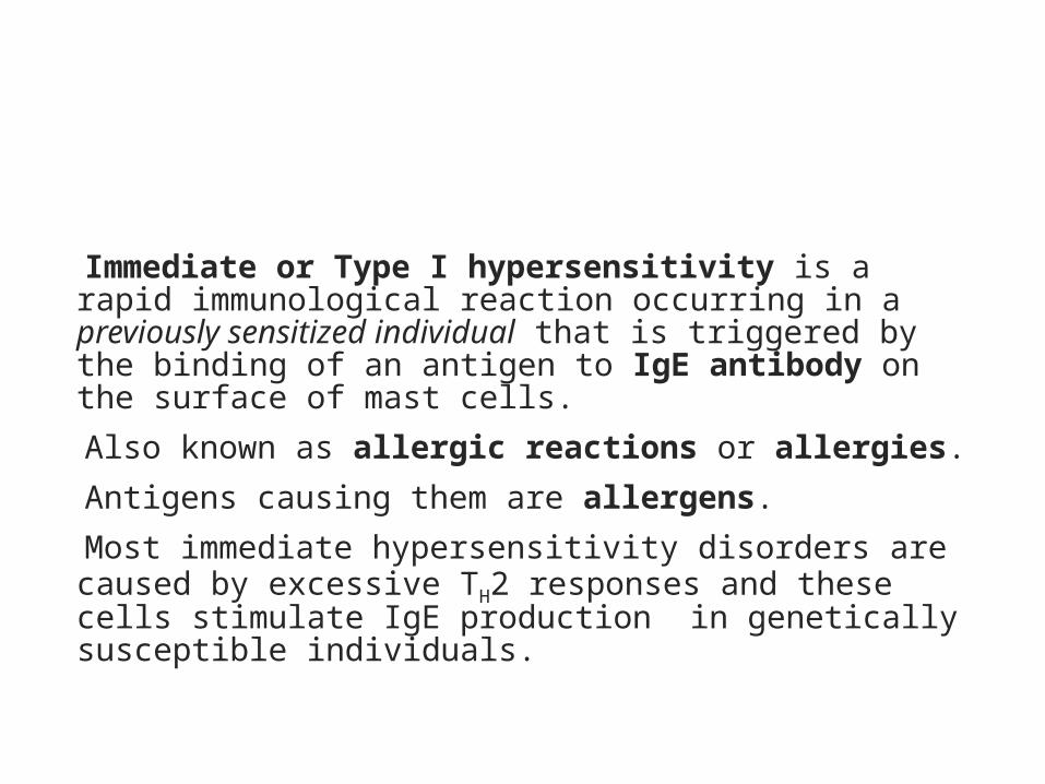
Immediate or Type I hypersensitivity is a rapid immunological reaction occurring in a previously sensitized individual that is triggered by the binding of an antigen to IgE antibody on the surface of mast cells.
Also known as allergic reactions or allergies. Antigens causing them are allergens. Most immediate hypersensitivity disorders are caused by excessive TH2 responses and these cells stimulate IgE production in genetically susceptible individuals.
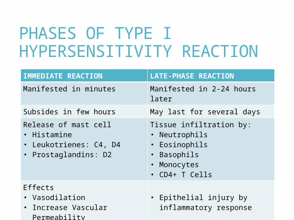
PHASES OF TYPE I HYPERSENSITIVITY REACTIONIMMEDIATE REACTION LATE-PHASE REACTIONManifested in minutes Manifested in 2-24 hours laterSubsides in few hours May last for several daysRelease of mast cell• Histamine• Leukotrienes: C4, D4• Prostaglandins: D2
Tissue infiltration by:• Neutrophils• Eosinophils• Basophils• Monocytes• CD4+ T Cells
Effects• Vasodilation• Increase Vascular
Permeability• Bronchoconstriction• Mucus-secretion
• Epithelial injury by inflammatory response
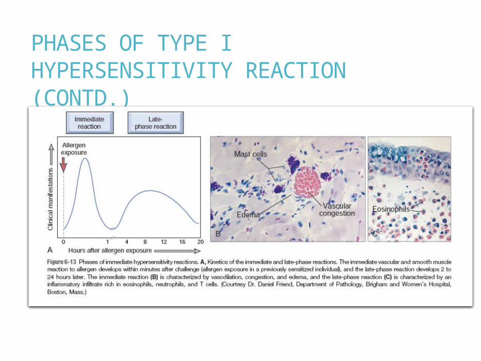
PHASES OF TYPE I HYPERSENSITIVITY REACTION (CONTD.)

Type I hypersensitivity reactionSequence of Events
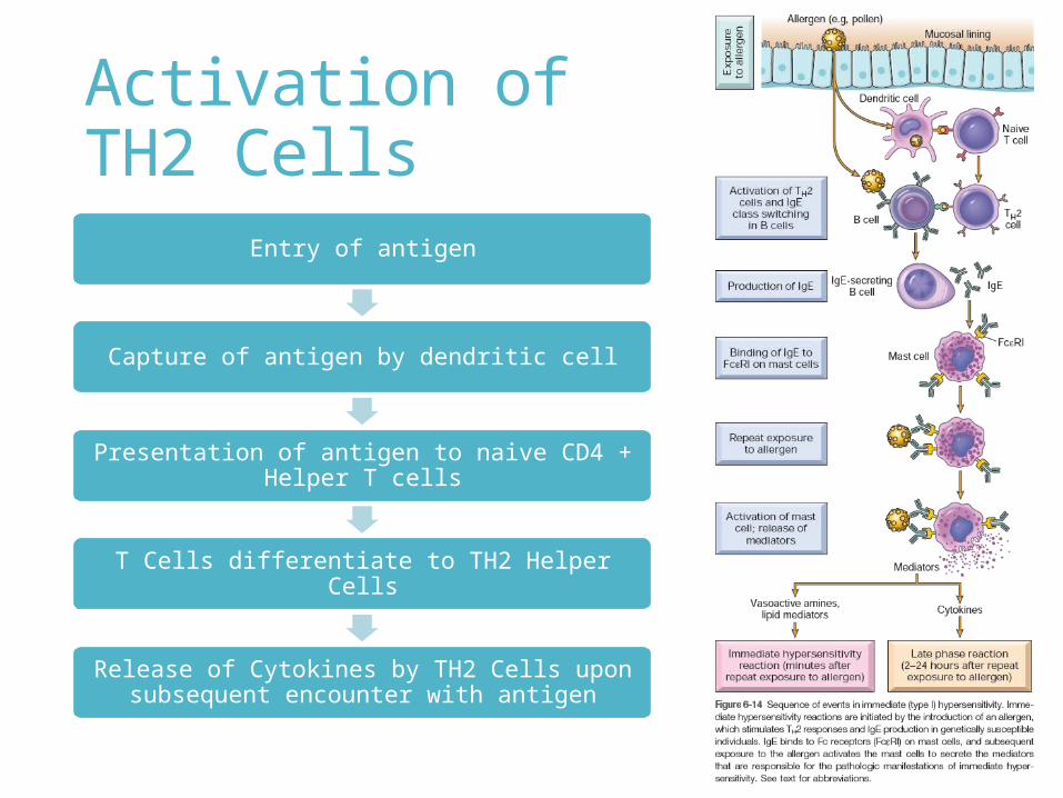
Activation of TH2 Cells
Entry of antigen
Capture of antigen by dendritic cell
Presentation of antigen to naive CD4 + Helper T cells
T Cells differentiate to TH2 Helper Cells
Release of Cytokines by TH2 Cells upon subsequent encounter with antigen
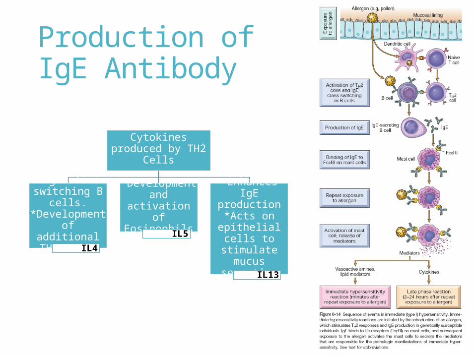
Production of IgE Antibody
Cytokines produced by TH2 Cells
*IgE class switching B cells.*Development of
additional TH2 cells
IL4
*Development and activation of
EosinophilsIL5
*Enhances IgE production
*Acts on epithelial cells to stimulate mucus
secretion
IL13
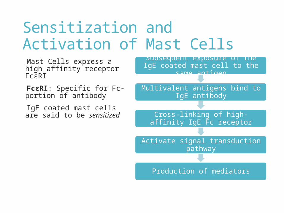
Sensitization and Activation of Mast Cells Mast Cells express a high affinity receptor FcεRI
FcεRI: Specific for Fc-portion of antibody
IgE coated mast cells are said to be sensitized
Subsequent exposure of the IgE coated mast cell to the same antigen
Multivalent antigens bind to IgE antibody
Cross-linking of high-affinity IgE Fc receptor
Activate signal transduction pathway
Production of mediators
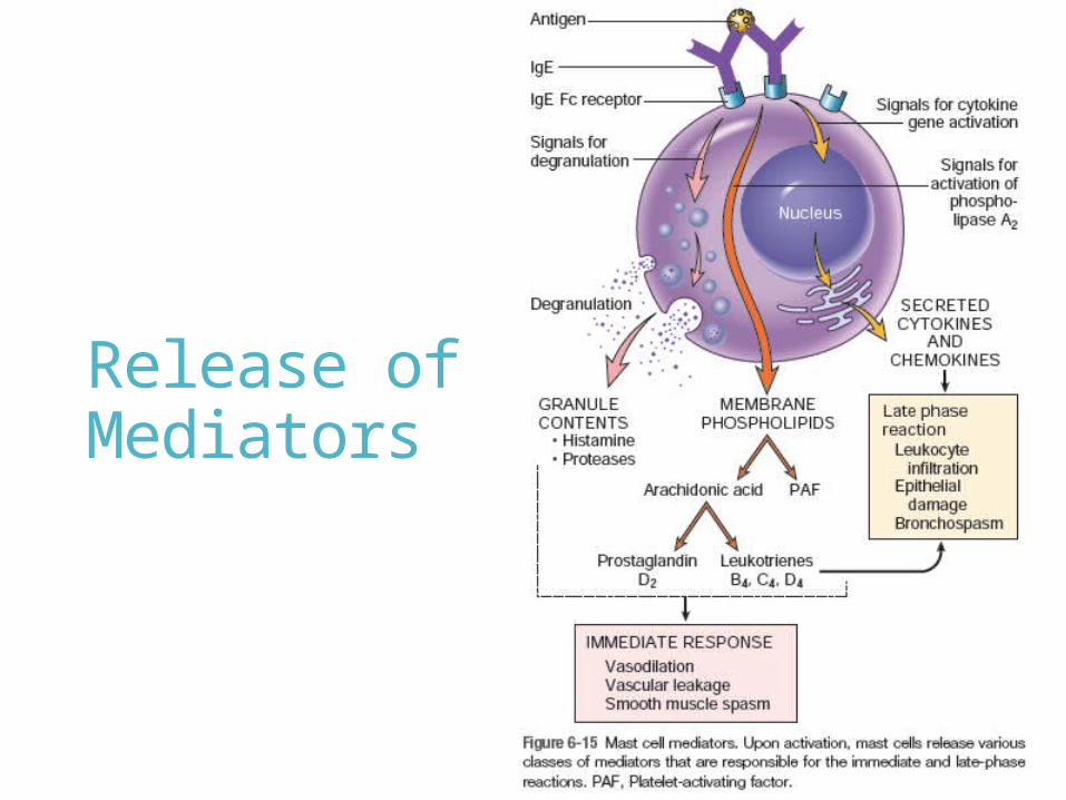
Release of Mediators
Mediators of Type-I Hypersensitivity ReactionMast cell activation leads to degranulation with the discharge of pre-formed or primary mediators that are stored in the granules and de novo synthesis and release of secondary mediators including lipid products and cytokines.
Preformed MediatorsMediators contained within the mast cell granules are the first to be released and can be divided into three categories:1. Vasoactive Amines
Eg. Histamine2. Enzymes
Eg. Neutral proteases (Chymase, Triptase), Acid Hydrolases
3. ProteoglycansEg. Heparin, Chondroitin Sulfate
Lipid Mediators These are Arachidonic Acid derived products. Reactions in the mast cell membrane lead to activation of Phospholipase A2 that converts membrane phospholipids to Arachidonic Acids, from which Leukotrienes and Prostaglandins are produced.1. Leukotrienes
Leukotrienes C4 and D4, Leukotriene B42. Prostaglandins
Prostaglandin D23. Platelet Activating Factor
These are not derived from Arachidonic Acid.
Cytokines These include TNF, Interleukin-1, Chemokines which promote leucocyte recruitment, Interleukin-4 which amplifies TH2 response.
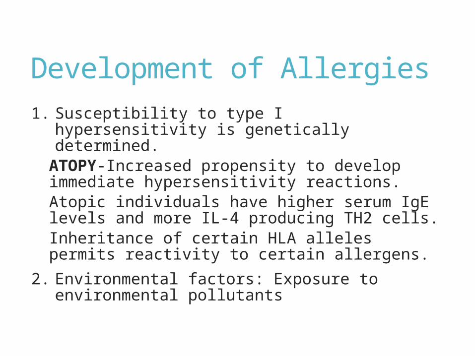
Development of Allergies1. Susceptibility to type I hypersensitivity is
genetically determined. ATOPY-Increased propensity to develop
immediate hypersensitivity reactions. Atopic individuals have higher serum IgE levels
and more IL-4 producing TH2 cells. Inheritance of certain HLA alleles permits
reactivity to certain allergens.2. Environmental factors: Exposure to
environmental pollutants
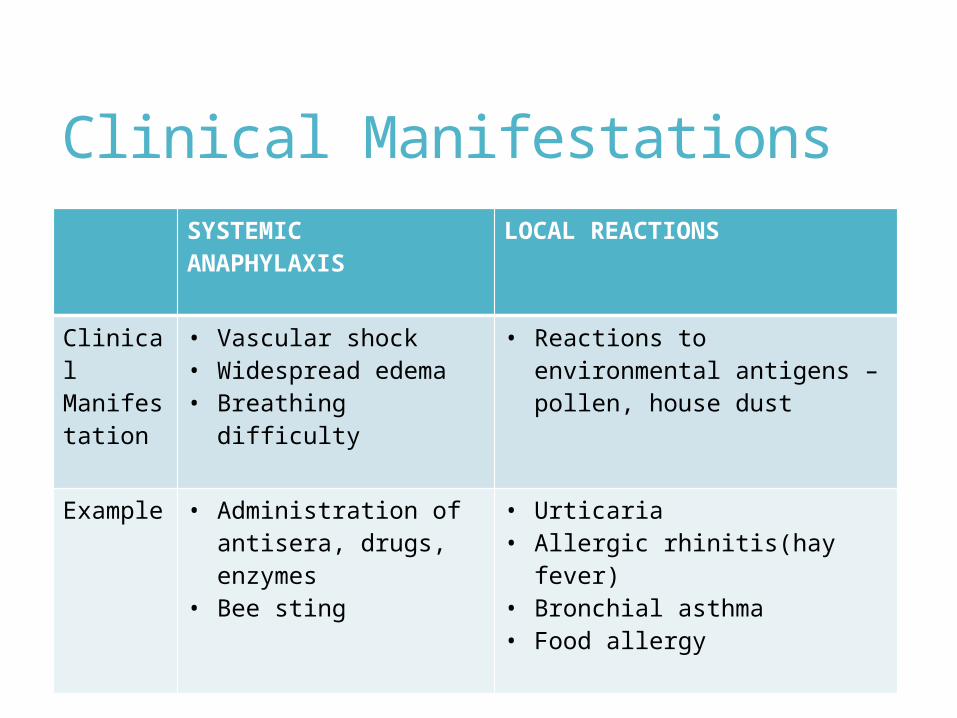
Clinical ManifestationsSYSTEMIC ANAPHYLAXIS
LOCAL REACTIONS
Clinical Manifestation
• Vascular shock• Widespread edema• Breathing difficulty
• Reactions to environmental antigens – pollen, house dust
Example • Administration of antisera, drugs, enzymes
• Bee sting
• Urticaria• Allergic rhinitis(hay fever)• Bronchial asthma• Food allergy

Related Documents


![[PPT]Drug Hypersensitivity Reaction: DRESS (Drug · Web viewDrug Hypersensitivity Reaction: DRESS Syndrome (Drug Reaction with Eosinophilia and Systemic Symptoms) Christopher Caulfield](https://static.cupdf.com/doc/110x72/5aa9eb4b7f8b9a7c188d7291/pptdrug-hypersensitivity-reaction-dress-drug-viewdrug-hypersensitivity-reaction.jpg)