ORIGINAL ARTICLE Two Types of Burst Firing in Gonadotrophin-Releasing Hormone Neurones Z. Chu*, M. Tomaiuolo , R. Bertramà and S. M. Moenter*§– *Department of Molecular and Integrative Physiology, University of Michigan, Ann Arbor, MI, USA. Department of Biological Science and Program in Neuroscience, Florida State University, Tallahassee, FL, USA. àDepartment of Mathematics and Programs in Neuroscience and Molecular Biophysics, Florida State University, Tallahassee, FL, USA. §Department of Internal Medicine, University of Michigan, Ann Arbor, MI, USA. –Department of Obstetrics and Gynecology, University of Michigan, Ann Arbor, MI, USA. Gonadotrophin-releasing hormone (GnRH) neurones form the final common pathway by which the central nervous system regulates fertility. GnRH is released in an episodic manner in males and during most of the female reproductive cycle (1–5). Immortalised GnRH neurones exhibit episodic release, suggesting this patterning may be intrinsic to GnRH neuronal networks (6–8). More recent studies of identified GnRH neurones in brain slices and primary cultures demonstrated episodic activity in both action potential firing and intracellular calcium levels (9–12). These biophysical observations revealed that episodic activity is observed not only at intervals consistent with that of hormone release in vivo (which occurs at intervals from once every several minutes to once every several hours depending on reproductive state) (13,14), but also at much higher frequencies. The highest frequency biophysical activ- ity observed in GnRH neurones thus far is the clustering of action potential firing into bursts. Burst firing is important because it has been shown to increase the efficacy of neuropeptide release and neurotransmission in other systems (15,16). The interval between bursts of firing in GnRH neurones varies within and among cells; peaks in the mean firing rate of individual GnRH neurones occur at intervals that are similar to what would be expected for GnRH release and are associated with bursts being closer together (10,17), suggesting an association between changes in burst interval and hormone secretion. Journal of Neuroendocrinology Correspondence to: Suzanne M. Moenter, 7725 Medical Sciences II, University of Michigan, Ann Arbor, MI 48109-5622, USA (e-mail: [email protected]). Gonadotrophin-releasing hormone (GnRH) neurones fire spontaneous bursts of action potentials, although little is understood about the underlying mechanisms. In the present study, we report evidence for two types of bursting ⁄ oscillation driven by different mechanisms. Properties of these different types are clarified using mathematical modelling and a recently developed active-phase ⁄ silent-phase correlation technique. The first type of GnRH neurone (1–2%) exhibits slow (0.05 Hz) spontaneous oscillations in membrane potential. Action potential bursts are often observed during oscillation depolarisation, although some oscillations were entirely sub- threshold. Oscillations persist after blockade of fast sodium channels with tetrodotoxin (TTX) and blocking receptors for ionotropic fast synaptic transmission, indicating that they are intrinsically generated. In the second type of GnRH neurone, bursts were irregular and TTX caused a stable membrane potential. The two types of bursting cells exhibited distinct active-phase ⁄ silent-phase correlation patterns, which is suggestive of distinct mechanisms underlying the rhythms. Further studies of type 1 oscillating cells revealed that the oscillation period was not affected by current or voltage steps, although amplitude was sometimes damped. Oestradiol, an important feedback regulator of GnRH neuronal activity, acutely and markedly altered oscillations, specifically depo- larising the oscillation nadir and initiating or increasing firing. Blocking calcium-activated potas- sium channels, which are rapidly reduced by oestradiol, had a similar effect on oscillations. Kisspeptin, a potent activator of GnRH neurones, translated the oscillation to more depolarised potentials, without altering period or amplitude. These data show that there are at least two distinct types of GnRH neurone bursting patterns with different underlying mechanisms. Key words: burst, oscillation, hypothalamus, neuroendocrine, parabolic. doi: 10.1111/j.1365-2826.2012.02313.x Journal of Neuroendocrinology, 2012, 24, 1065–1077 ª 2012 The Authors. Journal of Neuroendocrinology ª 2012 Blackwell Publishing Ltd

Welcome message from author
This document is posted to help you gain knowledge. Please leave a comment to let me know what you think about it! Share it to your friends and learn new things together.
Transcript
ORIGINAL ARTICLE
Two Types of Burst Firing in Gonadotrophin-Releasing Hormone NeuronesZ. Chu*, M. Tomaiuolo�, R. Bertram� and S. M. Moenter*§–
*Department of Molecular and Integrative Physiology, University of Michigan, Ann Arbor, MI, USA.
�Department of Biological Science and Program in Neuroscience, Florida State University, Tallahassee, FL, USA.
�Department of Mathematics and Programs in Neuroscience and Molecular Biophysics, Florida State University, Tallahassee, FL, USA.
§Department of Internal Medicine, University of Michigan, Ann Arbor, MI, USA.
–Department of Obstetrics and Gynecology, University of Michigan, Ann Arbor, MI, USA.
Gonadotrophin-releasing hormone (GnRH) neurones form the final
common pathway by which the central nervous system regulates
fertility. GnRH is released in an episodic manner in males and
during most of the female reproductive cycle (1–5). Immortalised
GnRH neurones exhibit episodic release, suggesting this patterning
may be intrinsic to GnRH neuronal networks (6–8). More recent
studies of identified GnRH neurones in brain slices and primary
cultures demonstrated episodic activity in both action potential
firing and intracellular calcium levels (9–12). These biophysical
observations revealed that episodic activity is observed not only at
intervals consistent with that of hormone release in vivo (which
occurs at intervals from once every several minutes to once every
several hours depending on reproductive state) (13,14), but also at
much higher frequencies. The highest frequency biophysical activ-
ity observed in GnRH neurones thus far is the clustering of action
potential firing into bursts. Burst firing is important because it
has been shown to increase the efficacy of neuropeptide release
and neurotransmission in other systems (15,16). The interval
between bursts of firing in GnRH neurones varies within and
among cells; peaks in the mean firing rate of individual GnRH
neurones occur at intervals that are similar to what would be
expected for GnRH release and are associated with bursts being
closer together (10,17), suggesting an association between
changes in burst interval and hormone secretion.
Journal ofNeuroendocrinology
Correspondence to:
Suzanne M. Moenter, 7725 Medical
Sciences II, University of Michigan,
Ann Arbor, MI 48109-5622, USA
(e-mail: [email protected]).
Gonadotrophin-releasing hormone (GnRH) neurones fire spontaneous bursts of action potentials,
although little is understood about the underlying mechanisms. In the present study, we report
evidence for two types of bursting ⁄ oscillation driven by different mechanisms. Properties of
these different types are clarified using mathematical modelling and a recently developed
active-phase ⁄ silent-phase correlation technique. The first type of GnRH neurone (1–2%) exhibits
slow (�0.05 Hz) spontaneous oscillations in membrane potential. Action potential bursts are
often observed during oscillation depolarisation, although some oscillations were entirely sub-
threshold. Oscillations persist after blockade of fast sodium channels with tetrodotoxin (TTX) and
blocking receptors for ionotropic fast synaptic transmission, indicating that they are intrinsically
generated. In the second type of GnRH neurone, bursts were irregular and TTX caused a stable
membrane potential. The two types of bursting cells exhibited distinct active-phase ⁄ silent-phase
correlation patterns, which is suggestive of distinct mechanisms underlying the rhythms. Further
studies of type 1 oscillating cells revealed that the oscillation period was not affected by current
or voltage steps, although amplitude was sometimes damped. Oestradiol, an important feedback
regulator of GnRH neuronal activity, acutely and markedly altered oscillations, specifically depo-
larising the oscillation nadir and initiating or increasing firing. Blocking calcium-activated potas-
sium channels, which are rapidly reduced by oestradiol, had a similar effect on oscillations.
Kisspeptin, a potent activator of GnRH neurones, translated the oscillation to more depolarised
potentials, without altering period or amplitude. These data show that there are at least two
distinct types of GnRH neurone bursting patterns with different underlying mechanisms.
Key words: burst, oscillation, hypothalamus, neuroendocrine, parabolic.
doi: 10.1111/j.1365-2826.2012.02313.x
Journal of Neuroendocrinology, 2012, 24, 1065–1077
ª 2012 The Authors.
Journal of Neuroendocrinology ª 2012 Blackwell Publishing Ltd
Burst firing in neurones and excitable endocrine cells can be dri-
ven by a variety of intrinsic factors, as well as extrinsic or network
factors (18). In the present study, we report evidence for two dis-
tinct types of bursting in GnRH neurones. One type is irregular in
duration and periodicity. The other type is much more regular and
exhibits a parabolic pattern within the interspike interval (Fig. 1).
This latter type of bursting, called ‘parabolic bursting’, has been
observed in other neurones, most notably the R15 neurone of the
mollusk Aplysia, where it was first described and remains a classic
example of parabolic bursting (19,20). A characteristic feature of
this type of bursting is the existence of two or more slow processes,
which combine to generate an underlying periodic slow rhythm. This
rhythm can persist even in the absence of action potentials (21)
and, even if action potentials are present, the parabolic interspike
interval pattern can be difficult to discern (22). Using a well-known
mathematical model of the R15 bursting neurone developed by
Plant (23), we illustrate that this type of bursting oscillation can be
identified by the correlation pattern between active (up state) and
silent (down state) phases, even if impulses are inhibited by block-
ing sodium channels. We show that parabolic oscillations are intrin-
sically generated and rapidly altered by oestradiol, a critical
physiological feedback mediator (24), and by kisspeptin, a strongly
activating neuromodulator of GnRH neurones (25,26).
Materials and methods
Animals
Adult (2–4 months old) transgenic mice in which green fluorescent protein
(GFP) is genetically targeted to GnRH neurones were used (27). Mice were
housed under a 14 : 10 h light ⁄ dark cycle (lights off 16.30 h EST) and fed
Harlan 2916 chow (Indianapolis, IN, USA) and water ad lib. All procedures were
approved by the University of Virginia Animal Care and Use Committee or the
University of Michigan University Committee on the Use and Care of Animals.
Because only a small subpopulation of GnRH neurones exhibit parabolic
oscillations, studies on this type of activity were conducted whenever
encountered in animals that had been prepared for other experiments. A
total of 26 oscillating cells were recorded over approximately 5 years of data
collection. During that time, approximately 1500 cells were recorded in the
whole-cell configuration; the percentage of oscillating GnRH neurones is
thus approximately 2%. The majority of animals were females ovariecto-
mised (OVX) 5–9 days previously (n = 20 animals). OVX females treated with
physiological levels of oestradiol (n = 1), intact dioestrous female (n = 1)
and castrate males (n = 1) were also studied. One (n = 20 mice) or two
(n = 3 mice) cells were studied per animal. The low number of cells in all
but OVX mice precludes statistical comparison among models; the oscillation
period was very similar and data have been combined. All irregularly burst-
ing cells were recorded in slices prepared from OVX mice.
Slice preparation
Reagents were purchased from Sigma Chemical Company (St Louis. MO,
USA) unless noted otherwise. Sagittal brain slices were prepared as reported
previously (28) with slight modifications (29). Briefly, mice were decapitated
and the brain was rapidly removed and placed in ice-cold high-sucrose sal-
ine solution containing (in mM): 250 sucrose, 3.5 KCl, 26 NaHCO3, 10 glu-
cose, 1.25 NaH2PO4, 1.2 MgSO4 and 3.8 MgCl2. Sagittal (300 lm) slices were
cut with a Vibratome 3000 (Ted Pella, Inc., Redding, CA, USA). Slices were
incubated in a 1 : 1 mixture of sucrose saline and artificial-cerebrospinal
fluid (aCSF) containing (in mM): 125 NaCl, 3.5 KCl, 26 NaHCO3, 1.25
NaH2PO4, 2.5 CaCl2, 1.2 MgSO4 and 10 D-glucose (pH 7.4) for 30 min at
31 �C and then transferred to 100% aCSF and incubated for at least an
additional 60 min at room temperature (22–24 �C); all slices were used
within 6 h of preparation.
Recordings
For recording, slices were placed in a chamber on the stage of an Olympus
BX51WI upright fluorescent microscope (Olympus, Tokyo, Japan) and contin-
uously superfused at 5–6 ml ⁄ min with oxygenated aCSF at 30–32 �C. Slices
were stabilised in the chamber for ‡ 5 min before recording. GFP-GnRH
neurones were identified by brief illumination at 470 nm. Recordings were
performed using an EPC 10 double amplifier running PATCHMASTER software
(HEKA Electronics, Lambrecht, Germany). The location of each GnRH neurone
studied was mapped onto figures obtained from a mouse brain atlas (30);
however, no consistent location or gross anatomical feature was noted as
an identifying characteristic for oscillating cells. Basic electrophysiological
characteristics of both oscillating and irregular bursting GnRH neurones
were monitored and compared; no differences were noticed in any parame-
ter monitored between these two types of cells (n = 9–14 cells of the 26
total oscillating cells per parameter; Table 1).
For whole-cell recordings, recording pipettes (2–3 MX) were filled with a
solution containing (in mM): 125 K gluconate, 20 KCl, 10 HEPES, 5 ethylene
glycol tetraacetic acid (EGTA), 4.0 MgATP, 0.4 NaGTP, 1.0 CaCl2 (pH 7.3),
300 mOsm (n = 22 oscillating cells and all irregular bursting cells) or 140
KCl, 10 HEPES, 5 EGTA, 4.0 MgATP, 0.4 NaGTP and 1.0 CaCl2, pH 7.3,
290 mOsm (n = 4 cells). Calculated (31) liquid junction potential of 13 mV
for the K gluconate-based solution and 3 mV for the KCl-based solution
were not corrected.
Drug treatments
Treatments were bath applied and varied among cells; details are in the
results. 20 lM bicuculline methiodide, 20 lM D())2-amino-5-phosphonovaler-
ic acid (APV) and 10 lM 6-cyano-7-nitroquinoxaline were used to block
GABAA, NMDA and AMPA ⁄ KA receptors, respectively. Tetrodotoxin (TTX;
0.5 lM; Calbiochem, San Diego, CA, USA) was used to block fast sodium
channels. Small conductance (SK) calcium-activated potassium current (IKCa)
was blocked with 200 nM apamin; large conductance (BK) IKCa was blocked
with 100 nM iberiotoxin. The G-protein coupled oestradiol receptor GPR30
(32) was activated with the G1 agonist (100–200 lM). The effects of oestra-
diol (10–100 nM) and kisspeptin (10 nM; Phoenix Pharmaceuticals, Inc., Bel-
mont, CA, USA) were also studied.
(A)
(B) (C)
20 mV
10 mV
00
Inte
rspi
kein
terv
al (s
)
10 20Spike position
30
0.1
0.2
–50 mV–70 mV
10 s
1 s
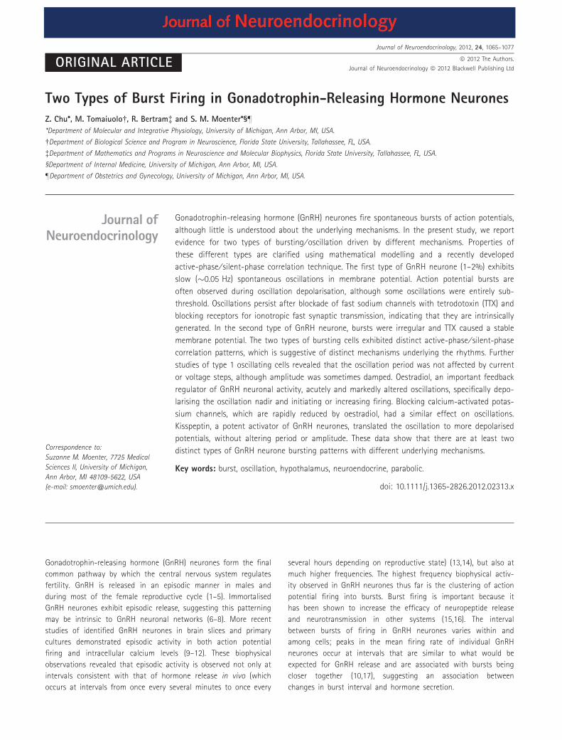
Fig. 1. Parabolic burst firing in a gonadotrophin-releasing hormone (GnRH)
neurone. (A) Example of regular parabolic bursting in a GnRH neurone
recorded in current-clamp mode. (B) Expansion of area in box. (C) Plot of in-
terspike interval versus spike position.
1066 Z. Chu et al.
ª 2012 The Authors. Journal of Neuroendocrinology, 2012, 24, 1065–1077
Journal of Neuroendocrinology ª 2012 Blackwell Publishing Ltd

Correlation analysis
To analyse the relationship between burst active and silent phases for exper-
imental and simulation data, we first had to define these phases (Fig. 2A,D).
After visual inspection of a trace, we define two parameters, Vt (mV) and dt
(ms) (in most cases Vt = )30 mV and dt = (200,1000) ms). When the volt-
age, V, is greater than Vt, a spike is recorded. Once all the spikes in a trace
are recorded, if two adjacent spikes are more that dt apart, they are not
considered part of the same burst. A solitary spike is one that is at least dt
apart from the preceding and following spike. It follows that the shortest
active phase duration is that of spike, whereas the shortest silent phase
duration is that between a burst and an adjacent solitary spike. Because
subthreshold oscillations and those in the presence of tetrodotoxin do not
have action potentials, a different approach was used. We first normalised
each voltage trace to lie between 0 (minimum) and 1 (maximum). We
defined the ‘active phase’ as the time during which the normalised voltage
is > 0.5, and the ‘silent phase’ as the time during which the normalised
voltage is below 0.5. Once active and silent phases were assigned (for burst-
ing or subthreshold oscillations), we then constructed a pair of scatter plots.
In one scatter plot, the duration of a burst active phase is plotted against
the duration of the immediately preceding silent phase (Fig. 2). This is var-
ried out for each burst in the voltage trace, producing a scatter plot of
points. In the other scatter plot, the burst active phase duration is plotted
against the immediately following silent phase duration. Thus each burst is
represented by a point in each of the two scatter plots. We then compute
the Pearson correlation coefficient of the scatter plots. We have shown pre-
viously that the correlations between active and silent phases are indicators
of the dynamical mechanism underlying relaxation oscillations and bursting
(33). That is, different types of bursting patterns exhibit different correlation
patterns. We use this technique in the present study to discriminate
between the parabolic bursting observed in some GnRH neurones and the
irregular (nonparabolic) bursting observed in others.
Mathematical modelling
We used a well-known model for parabolic bursting that was developed for
the R15 neurone of the mollusk Aplysia (23) to illustrate properties that are
characteristic of parabolic bursting. In particular, we use the model to demon-
strate that parabolic bursting oscillations have a negative correlation between
the active and the next silent phase duration, even if spikes are inhibited by
blocking sodium channels (in which case ‘active phase’ means the up state of
the small voltage oscillation that persists when spikes are blocked).
(A) (B) (C)
(D) (E) (F)
Parabolic model
Parabolic GnRH neuron
00
00
3
6
9
12
15
3 6 9
10
15
20
25r = –0.01 P = 0.97
r = –0.27 P = 0.34
r = –0.61 *P = 4.8e–005
20 40 60–80
–40
–20
20
0
20
Priorsilentphase
Priorsilentphase
Nextsilentphase
Nextsilentphase
phaseActive
phaseActive
40 60 80 100Time (s)
20 mV5 s
–61mV
Prior duration (s)
Prior duration (s)
Vm
embr
ane
(mV
)
Act
ive
dura
tion
(s)
Act
ive
dura
tion
(s)
00
3
6
9
12
15
3 6 9
r = –0.90 *P = 4.5e–006
Next duration (s)
Act
ive
dura
tion
(s)
010
15
20
25
20 40 60Next duration (s)
Act
ive
dura
tion
(s)
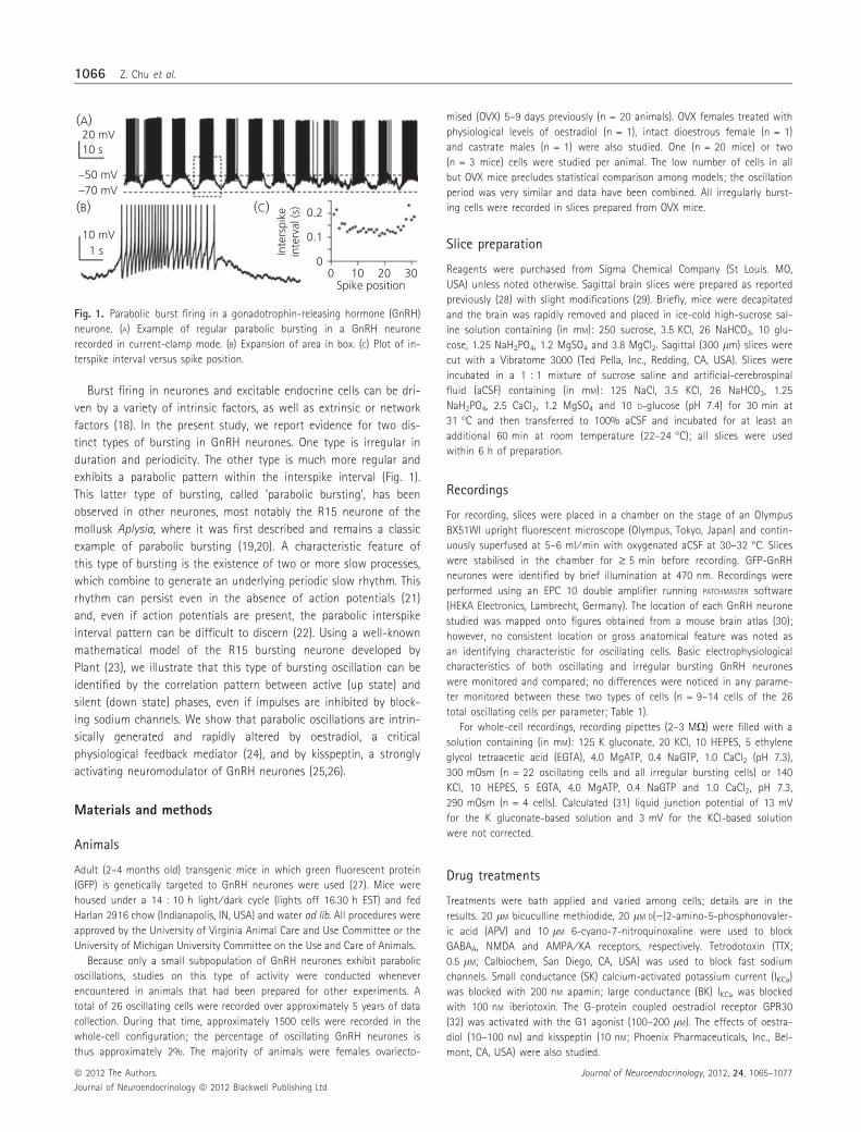
Fig. 2. Active and silent period correlations in a model parabolic neurone and a gonadotrophin-releasing hormone (GnRH) neurone. (A) Plant model for para-
bolic bursting defining prior silent phase, active phase and next silent phase. (B, C) Scatter plots for prior silent duration versus active duration (B) and next
silent duration versus active duration (C) showing significant correlation between next silent duration and active duration. (D) Parabolic bursts in a GnRH neu-
rone illustrating phases. (E, F) Scatter plots for prior silent duration versus active duration (E) and next silent duration versus active duration (F) showing signifi-
cant correlation between next silent duration and active duration.
Table 1. Electrophysiological Characteristics of Oscillating Versus Irregular
Bursting Gonadotrophin-Releasing Hormone Neurones.
Parameter Oscillating
(n = 9–14)
Irregular
(n = 9–13)
Input resistance (MX) 905 � 54 876 � 22
Action potential amplitude (mV) 80.2 � 2.5 78.9 � 1.3
Afterhyperpolarising potential
amplitude (mV)
32.2 � 0.7 31.0 � 0.8
Action potential width at half
maximal amplitude (ms)
1.1 � 0.03 1.1 � 0.04
Action potential threshold (mV) )32.2 � 1.3 )31.9 � 0.6
Resting membrane potential (mV) )61.9 � 1.0 )60.7 � 1.2
Values are the mean � SEM. P > 0.3 for all comparisons via an unpaired
t-test. Action potential amplitude is measured from prespike potential; after-
hyperpolarisation amplitude is measured from the threshold. Note that not
all properties could be monitored in all 26 oscillating cells because some
were quiescent or had action potentials pharmacologically blocked; initial
resting membrane potential (I = 0) could not be determined in cells with
high-amplitude oscillations; and some cells were observed without imposing
voltage steps that are needed to monitor input resistance.
Two types of bursting in GnRH neurones 1067
Journal of Neuroendocrinology, 2012, 24, 1065–1077 ª 2012 The Authors.
Journal of Neuroendocrinology ª 2012 Blackwell Publishing Ltd
In addition to the original publication, the Plant model is described on
the Scholarpedia web site (http://www.scholarpedia.org/Plant_model). It is a
Hodgkin–Huxley-type model with a leak current, spike-producing ionic
currents and two slowly-activating ionic currents that package the spikes
into bursts, CmdVdt¼ � Iin;fast þ Iout;fast þ Iin;slow þ Iout;slow þ IL
� �where Iin,fast
and Iin,slow are fast- and slowly-activating inward currents, respectively (with
Nernst potential of 30 mV), and Iout,fast and Iout,slow are outward K+ currents
(with Nernst potential of )75 mV). The leakage current, IL, has a constant
conductance and a reversal potential of )40 mV. In addition to the voltage
differential equation, there is a differential equation for inactivation of
Iin,fast, one for activation of Iin,slow, and one for activation of Iout,fast. Finally,
there is a differential equation for the free cytosolic Ca2+ concentration,
which activates Iout,slow. Parameter values used are the same as those used
for Fig. 1 (http://www.scholarpedia.org/Plant_model). Computer simulations
were performed with the XPPAUT software package (34) using the Forward
Euler method. The code is available as freeware from http://www.math.f-
su.edu/~bertram/software/neurone.
Analysis
Stored data traces were analysed offline using IGOR PRO (WaveMetrics, Port-
land, OR, USA). Data were transferred to EXCEL (Microsoft Corp., Redmond,
CA, USA), or INSTAT (Graph Pad Software, San Diego, CA, USA) for statistical
analysis. Summary data are shown as the mean � SEM.
Results
Burst firing and membrane oscillations in GnRH neurones
GnRH neurones in slices, after isolation and in primary and im-
mortalised cultures, exhibit burst firing over a range of periods
(9,17,28,35–38). The interval between bursts varies widely in long-
term targeted extracellular recordings (28,39–41). Most GnRH neu-
rones exhibit burst firing without periodic plateaus in the mem-
brane potential. However, some cells (approximately 2%) produce
profound periodic plateaus in the membrane potential (e.g. AMPA,
NMDA and GABAA receptors are blocked; Fig. 1A). Action potential
firing can be observed on the peaks of some of these oscillations,
whereas others are completely subthreshold (e.g. pre-treatment;
Fig. 7A). Figure 1(B) shows an expansion of the oscillation peak, or
upstate, indicated by the dashed box in Fig. 1(A). This cell fires
spikes for almost 4 s, with the interspike interval first declining and
then increasing (Fig. 1C). This parabolic trend in the interspike inter-
val is the defining characteristic of a parabolic bursting pattern that
has been observed in other neurones (42,43).
Although parabolic bursting is often characterised by a clear par-
abolic distribution of interspike intervals as in Fig. 1(C), this feature
may be subtle and not evident (22). Also, the determination of a
parabolic distribution of interspike interval is of no use in cases
when spiking does not occur (e.g. when sodium channels are
blocked). A better way to characterise this form of bursting is in
terms of the underlying dynamics (22,42). Parabolic bursting oscilla-
tions are a result of the interaction of two slow processes (such as
channel activation ⁄ inactivation) that is capable of producing a
rhythm even in the absence of electrical impulses (23). This interac-
tion of slow processes also produces a characteristic pattern
between active (or up state) and silent (or down state) phases of
the bursting (or subthreshold) oscillation, as we demonstrate in
Fig. 2 using a classic mathematical model for parabolic bursting
(23). Figure 2(A) shows two bursts produced by the Plant model,
with a superimposed envelope defining the active or spiking phase
of the burst and the prior and next silent phases. This envelope
extends throughout the voltage trace (n = 36 bursts) and serves to
identify silent versus active phases, allowing computation of their
respective durations. Figure 2(B) shows, for each burst, the active
phase duration plotted against the immediately prior silent phase
duration. Each point in the scatter plot corresponds to a burst and
its prior silent phase (i.e. 36 points corresponding to 36 bursts).
There is no significant correlation between active and prior silent
phases (P = 0.97, n = 36). In the scatter plot of Fig. 2(C), each burst
active phase is plotted against the immediately following silent
phase. In this case, there is a significant negative correlation
between the two phases (r = )0.61, P < 0.01, n = 36). Thus, the
duration of the prior silent phase of the burst does not predict the
length of the burst active phase, although the duration of the
active phase does predict the next silent phase. For example, a long
active phase predicts the silent phase that follows will be short.
This pattern of correlations contrasts with the pattern we have
observed for other types of bursting oscillations (33), such as the
‘square-wave bursting’ that is exhibited by pancreatic islets and
pre-Botzinger neurones.
We next perform the same correlation analysis on a voltage trace
from a GnRH neurone exhibiting parabolic bursting (Fig. 2D, only
two bursts are shown). As with the model neurone, there is no sig-
nificant correlation between active and prior silent phase durations
(P = 0.34, n = 13; Fig. 2E). However, there is a significant negative
correlation between active and next silent phase durations
(r = )0.90, P < 0.01, n = 13; Fig. 2F). This correlation pattern is a
characteristic of parabolic bursting, and serves as a fingerprint for
the slow dynamics underlying this type of spiking pattern.
A second pattern of burst firing is observed in a majority of
GnRH neurones. This consists of irregular bursts of action potentials
with no obvious parabolic pattern in the interspike intervals
(Fig. 3A). These irregular bursts are of varying duration (Fig. 3B) and
there is no parabolic relationship between spike position and inters-
pike interval, and only a very mild spike frequency accommodation
is observed (Fig. 3C). When the same correlation analysis is per-
formed on irregular bursting GnRH neurones, no significant correla-
tion is observed between active phase duration and either prior or
next silent phase duration (Fig. 3D), further illustrating that the
underlying dynamics are different from those of parabolic bursting
GnRH neurones.
GnRH neurone membrane potential oscillations areintrinsically generated
We next examined the effects of blocking fast sodium channels
required for action potential spikes with TTX on each type of
bursting activity. GnRH neurones exhibiting parabolic bursting
(n = 3) were recorded before and after addition of TTX; blockers
for AMPA, NMDA and GABAA receptors were present throughout
the recording to further isolate the cell from fast synaptic
1068 Z. Chu et al.
ª 2012 The Authors. Journal of Neuroendocrinology, 2012, 24, 1065–1077
Journal of Neuroendocrinology ª 2012 Blackwell Publishing Ltd
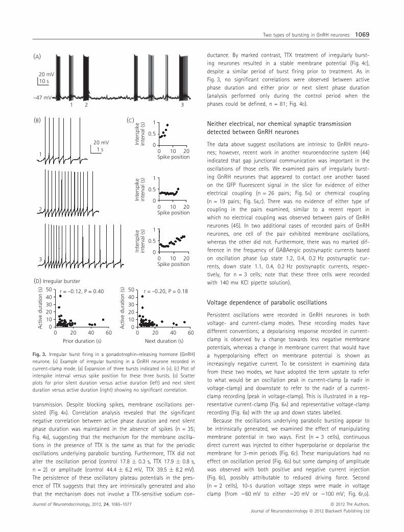
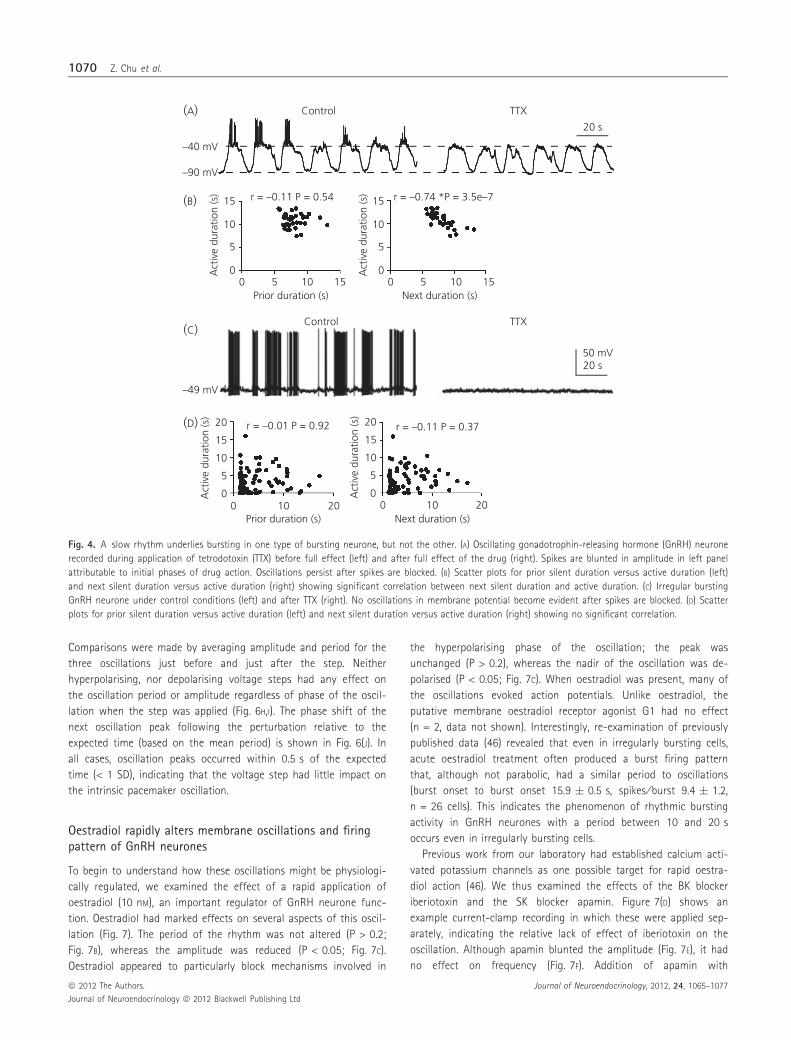
transmission. Despite blocking spikes, membrane oscillations per-
sisted (Fig. 4A). Correlation analysis revealed that the significant
negative correlation between active phase duration and next silent
phase duration was maintained in the absence of spikes (n = 35;
Fig. 4B), suggesting that the mechanism for the membrane oscilla-
tions in the presence of TTX is the same as that for the periodic
oscillations underlying parabolic bursting. Furthermore, TTX did not
alter the oscillation period (control 17.8 � 0.3 s, TTX 17.9 � 0.8 s,
n = 2) or amplitude (control 44.4 � 6.2 mV, TTX 39.5 � 8.2 mV).
The persistence of these oscillatory plateau potentials in the pres-
ence of TTX suggests that they are intrinsically generated and also
that the mechanism does not involve a TTX-sensitive sodium con-
ductance. By marked contrast, TTX treatment of irregularly burst-
ing neurones resulted in a stable membrane potential (Fig. 4C),
despite a similar period of burst firing prior to treatment. As in
Fig. 3, no significant correlations were observed between active
phase duration and either prior or next silent phase duration
(analysis performed only during the control period when the
phases could be defined, n = 81; Fig. 4D).
Neither electrical, nor chemical synaptic transmissiondetected between GnRH neurones
The data above suggest oscillations are intrinsic to GnRH neuro-
nes; however, recent work in another neuroendocrine system (44)
indicated that gap junctional communication was important in the
oscillations of those cells. We examined pairs of irregularly burst-
ing GnRH neurones that appeared to contact one another based
on the GFP fluorescent signal in the slice for evidence of either
electrical coupling (n = 26 pairs; Fig. 5A) or chemical coupling
(n = 19 pairs; Fig. 5B,C). There was no evidence of either type of
coupling in the pairs examined, similar to a recent report in
which no electrical coupling was observed between pairs of GnRH
neurones (45). In two additional cases of recorded pairs of GnRH
neurones, one cell of the pair exhibited membrane oscillations,
whereas the other did not. Furthermore, there was no marked dif-
ference in the frequency of GABAergic postsynaptic currents based
on oscillation phase (up state 1.2, 0.4, 0.2 Hz postsynaptic cur-
rents, down state 1.1, 0.4, 0.2 Hz postsynaptic currents, respec-
tively, for n = 3 cells; note that these three cells were recorded
with 140 mM KCl pipette solution).
Voltage dependence of parabolic oscillations
Persistent oscillations were recorded in GnRH neurones in both
voltage- and current-clamp modes. These recording modes have
different conventions; a depolarising response recorded in current-
clamp is observed by a change towards less negative membrane
potentials, whereas a change in membrane current that would have
a hyperpolarising effect on membrane potential is shown as
increasingly negative current. To be consistent in examining data
from these two modes, we have adopted the term upstate to refer
to what would be an oscillation peak in current-clamp (a nadir in
voltage-clamp) and downstate to refer to the nadir of a current-
clamp recording (peak in voltage-clamp). This is illustrated in a rep-
resentative current-clamp (Fig. 6A) and representative voltage-clamp
recording (Fig. 6B) with the up and down states labelled.
Because the oscillations underlying parabolic bursting appear to
be intrinsically generated, we examined the effect of manipulating
membrane potential in two ways. First (n = 3 cells), continuous
direct current was injected to either hyperpolarise or depolarise the
membrane for 3-min periods (Fig. 6C). These manipulations had no
effect on oscillation period (Fig. 6D) but some damping of amplitude
was observed with both positive and negative current injection
(Fig. 6E), possibly attributable to reduced driving force. Second
(n = 2 cells), 10-s duration voltage steps were made in voltage
clamp (from )60 mV to either )20 mV or )100 mV; Fig. 6F,G).
(A)
20 mV
–47 mV1
1
2
2
3
3
Irregular burster
10 s
20 mV
00In
ters
pike
inte
rval
(s)
0.5
1
10 20Spike position
00In
ters
pike
inte
rval
(s)
0.5
1
10 20Spike position
00
00 20 40
Prior duration (s)
Act
ive
dura
tion
(s)
60
1020304050 r = –0.12, P = 0.40
00 20 40
Next duration (s)
Act
ive
dura
tion
(s)
60
1020304050 r = –0.20, P = 0.18
Inte
rspi
kein
terv
al (s
)
0.5
1
10 20Spike position
1 s
(B)
(D)
(C)
Fig. 3. Irregular burst firing in a gonadotrophin-releasing hormone (GnRH)
neurone. (A) Example of irregular bursting in a GnRH neurone recorded in
current-clamp mode. (B) Expansion of three bursts indicated in (A). (C) Plot of
interspike interval versus spike position for these three bursts. (D) Scatter
plots for prior silent duration versus active duration (left) and next silent
duration versus active duration (right) showing no significant correlation.
Two types of bursting in GnRH neurones 1069
Journal of Neuroendocrinology, 2012, 24, 1065–1077 ª 2012 The Authors.
Journal of Neuroendocrinology ª 2012 Blackwell Publishing Ltd
Comparisons were made by averaging amplitude and period for the
three oscillations just before and just after the step. Neither
hyperpolarising, nor depolarising voltage steps had any effect on
the oscillation period or amplitude regardless of phase of the oscil-
lation when the step was applied (Fig. 6H,I). The phase shift of the
next oscillation peak following the perturbation relative to the
expected time (based on the mean period) is shown in Fig. 6(J). In
all cases, oscillation peaks occurred within 0.5 s of the expected
time (< 1 SD), indicating that the voltage step had little impact on
the intrinsic pacemaker oscillation.
Oestradiol rapidly alters membrane oscillations and firingpattern of GnRH neurones
To begin to understand how these oscillations might be physiologi-
cally regulated, we examined the effect of a rapid application of
oestradiol (10 nM), an important regulator of GnRH neurone func-
tion. Oestradiol had marked effects on several aspects of this oscil-
lation (Fig. 7). The period of the rhythm was not altered (P > 0.2;
Fig. 7B), whereas the amplitude was reduced (P < 0.05; Fig. 7C).
Oestradiol appeared to particularly block mechanisms involved in
the hyperpolarising phase of the oscillation; the peak was
unchanged (P > 0.2), whereas the nadir of the oscillation was de-
polarised (P < 0.05; Fig. 7C). When oestradiol was present, many of
the oscillations evoked action potentials. Unlike oestradiol, the
putative membrane oestradiol receptor agonist G1 had no effect
(n = 2, data not shown). Interestingly, re-examination of previously
published data (46) revealed that even in irregularly bursting cells,
acute oestradiol treatment often produced a burst firing pattern
that, although not parabolic, had a similar period to oscillations
(burst onset to burst onset 15.9 � 0.5 s, spikes ⁄ burst 9.4 � 1.2,
n = 26 cells). This indicates the phenomenon of rhythmic bursting
activity in GnRH neurones with a period between 10 and 20 s
occurs even in irregularly bursting cells.
Previous work from our laboratory had established calcium acti-
vated potassium channels as one possible target for rapid oestra-
diol action (46). We thus examined the effects of the BK blocker
iberiotoxin and the SK blocker apamin. Figure 7(D) shows an
example current-clamp recording in which these were applied sep-
arately, indicating the relative lack of effect of iberiotoxin on the
oscillation. Although apamin blunted the amplitude (Fig. 7E), it had
no effect on frequency (Fig. 7F). Addition of apamin with
(A) Control
Control
TTX
TTX
20 s
–40 mV
–49 mV
50 mV20 s
–90 mV
0
5
10
15
0 5Prior duration (s)
Act
ive
dura
tion
(s) r = –0.11 P = 0.54
10 15
0
5
10
15
20
0Prior duration (s)
Act
ive
dura
tion
(s)
r = –0.01 P = 0.92
10 200
5
10
15
20
0Next duration (s)
Act
ive
dura
tion
(s)
r = –0.11 P = 0.37
10 20
0
5
10
15
0 5Next duration (s)
Act
ive
dura
tion
(s) r = –0.74 *P = 3.5e–7
10 15
(B)
(C)
(D)
Fig. 4. A slow rhythm underlies bursting in one type of bursting neurone, but not the other. (A) Oscillating gonadotrophin-releasing hormone (GnRH) neurone
recorded during application of tetrodotoxin (TTX) before full effect (left) and after full effect of the drug (right). Spikes are blunted in amplitude in left panel
attributable to initial phases of drug action. Oscillations persist after spikes are blocked. (B) Scatter plots for prior silent duration versus active duration (left)
and next silent duration versus active duration (right) showing significant correlation between next silent duration and active duration. (C) Irregular bursting
GnRH neurone under control conditions (left) and after TTX (right). No oscillations in membrane potential become evident after spikes are blocked. (D) Scatter
plots for prior silent duration versus active duration (left) and next silent duration versus active duration (right) showing no significant correlation.
1070 Z. Chu et al.
ª 2012 The Authors. Journal of Neuroendocrinology, 2012, 24, 1065–1077
Journal of Neuroendocrinology ª 2012 Blackwell Publishing Ltd
iberiotoxin depolarises the downstate of the oscillation at the
same time as having minimal effect on the upstate, similar to
oestradiol (Fig. 7G). Taken together, these observations suggest
that one possible mechanism for the rapid action of oestradiol on
the oscillation is reduced conductance via small conductance
potassium channels.
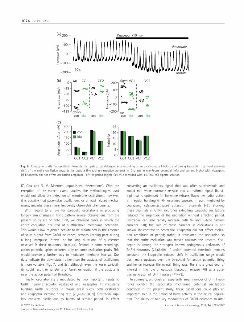
Kisspeptin translates the entire oscillation towards theupstate
Kisspeptin is a strong activator of GnRH neurones (25). We thus
examined its effects on the oscillation. In both current- and volt-
age-clamp (n = 2 each, cell VC2 recorded with 140 mM KCl pipette
solution), kisspeptin translated the oscillation towards the upstate
at the same time as having no effect on amplitude or period
(Fig. 8A–C). These data suggest another possible level of oscillation
in GnRH neurones in which neuromodulators initiate mechanisms
that move upstates in the intrinsic oscillation nearer and further
from the action potential threshold, and thus generate longer term
patterns in the firing rate.
Discussion
In the present study, we provide evidence for two distinct types of
bursting in GnRH neurones with distinct underlying mechanisms. One
type is irregular in duration and periodicity. The other is very regular,
but rare, and exhibits a parabolic interspike interval trajectory.
Parabolic bursting has been observed in other neurones, most
notably the R15 neurone of the mollusk Aplysia, where it was first
described (19,20). One feature of parabolic bursting in R15 is that
blocking action potential firing does not necessarily eliminate the
plateau phases of the membrane potential oscillation (21), a feature
that was explained using a mathematical model (47). In GnRH neu-
rones exhibiting parabolic bursting, we find that similar oscillation
plateaus persist when tetrodotoxin is present, in contrast to irregu-
lar bursters in which membrane potential is very stable after block-
ade of action potential firing. Intriguingly, we find that both the
period of oscillations and the up-phase ⁄ down-phase correlation
patterns in cells not exhibiting action potentials are similar to that
seen in the GnRH neurones exhibiting parabolic bursting. This sug-
gests that currents generating the slow rhythm are responsible for
the parabolic bursting pattern and that this oscillation may have a
pacemaker function.
We introduced a new technique to identify parabolic bursting
and distinguish it from the irregular bursting seen in many other
GnRH neurones. The well-known Plant model was used to demon-
strate that parabolic bursting gives rise to no correlation between
the active phase and the prior silent phase, although a negative
correlation between the active phase and the following silent phase.
This is true (not shown) even for the simpler model of parabolic
bursting developed by Baer et al. (48), and is a property of para-
bolic bursting and not specific to the model itself. This pattern is
unlike that of the irregular bursting neurones and unlike that of
other forms of bursting such as square-wave or elliptic bursting
(33). The correlation pattern thus serves as a ‘fingerprint’ for para-
bolic bursting, and persists even when impulses are blocked.
In oscillating GnRH neurones, the period of the oscillation was
remarkably robust, consistent with pacemaking. Period was not
altered by continuous direct current injection, nor by shorter dura-
tion voltage-steps or current injections, nor by the drug treatments
that were tested. The period of GnRH neurone oscillations is quite
long compared to other central neuroneal oscillations; for example,
theta-frequency oscillations (3–8 Hz) in rodent hippocampus and
entorhinal cortex (49,50) or oscillations associated with burst firing
in neocortical neurones (around 0.5–1 Hz) (51–54). Interestingly,
the period in GnRH neurones is very similar to that of dopaminer-
gic (TIDA) neurones of the arcuate nucleus, which are largely
responsible for the control of prolactin release (44). Despite this
similarity in period, these two neuroendocrine cell types appear to
use different mechanisms as the TIDA neurone rhythm is abolished
by tetrodotoxin, whereas that in GnRH neurones is not. In TIDA
neurones, network properties and interconnections, including
changes in GABAergic transmission from up to down state and gap
junctional communication, were critical in sustaining the oscillation,
whereas, in GnRH neurones, the oscillation appears to be intrinsi-
cally generated because it occurs in the presence of tetrodotoxin
(A)
(B)
(C)
V1
V1
I1 I2
V2
V2
Cell 1 Cell 2
Cell 1 Cell 2
5 mV
–62 mV20 mV
100 ms
100 ms5 mV
100 ms
–60 mV5 mV
100 ms20 mV
100 ms
Fig. 5. No electrical or fast chemical coupling detected between gonadotro-
phin-releasing hormone neurones. (A) Membrane response (top) of cells 1
(V1) and 2 (V2) to +15 to )40 pA current injection (I1, I2, respectively). Bot-
tom, membrane potential of non-injected cell, 100 traces averaged. (B, C)
Elicitation of 1, 2 or 4 spikes by brief current injections (300 pA, 3 ms) into
cell 1 fails to elicit postsynaptic potentials in cell 2; identical injections into
cell 2 fail to elicit postsynaptic potentials in cell 1 (mean of 50 traces).
Two types of bursting in GnRH neurones 1071
Journal of Neuroendocrinology, 2012, 24, 1065–1077 ª 2012 The Authors.
Journal of Neuroendocrinology ª 2012 Blackwell Publishing Ltd

and blockers of fast synaptic transmission. Furthermore, no evi-
dence of chemical or electrical synapses were detected between
GnRH neurone pairs.
The mechanisms underlying parabolic bursting in GnRH neurones
are unresolved; however, there are some mechanisms that can be
ruled out (or in). As mentioned, fast synaptic transmission and spike
generation (and hence tetrodotoxin-sensitive sodium channels) are
not required. The hyperpolarising phase of the oscillation appears
to at least in part involve activation of SK-type calcium-activated
potassium currents, given the effects of blocking these currents on
the oscillation. Other candidate hyperpolarising currents include the
A-type potassium current, which is prominent in these cells (55,56),
and inwardly-rectified potassium currents (56). Candidates for
depolarisation include hyperpolarisation-activated cation currents,
calcium currents and TRPC -mediated currents (35,57–59). Addition-
ally, GnRH neurones maintain high chloride levels in adulthood
(60,61) and hence chloride efflux could also be a mechanism for
the depolarising phase, as in the interstitial cells of Cajal, which
serve as pacemakers within the gastrointestinal tract and have a
similar period (62).
Given the small percentage of GnRH neurones that exhibit para-
bolic bursting or pacemaker oscillations, one might question their
relevance. Rare cells with unique properties can contribute to the
organisation of network activity. For example, GABAergic hub neu-
rones in the hippocampus are rare cells showing high connectivity
but are more likely to induce changes in giant depolarising poten-
tials in the local network (63). Oscillating GnRH neurones had the
same general appearance as irregular bursters, similar electrophysi-
ological characteristics other than oscillations versus irregular firing,
and a similar frequency of ionotropic GABAergic input compared to
(J)up H
up D
rise Hfall H
fall Ddown D
–0.75 0.75–0.25 0.250Seconds
–0.5 0.5
Phase
(A) Current-clamp 20 s
–40 mV
–80 mV
upstate
downstate upstate
downstate(B) Voltage-clamp 20 s
100 pA0 pA
(C) upstate
downstate
30 pA
0 pA
–40 pA
20 mV20 s
(D) (E)
030
Perio
d (s
)
Am
plitu
de (m
V)
141618202224
–40Current (pA)
030
30405060
20–40
Current (pA)
(F)
(G)
upstate
downstate50 pA20 s
–60 mV
–60 mV
–20 mV
–100 mV
(H)
Seco
nds
14161820
upD D D H H H
uprise fall fall downPeriod
(I)
pA80
120160
Amplitude
Fig. 6. Effect of membrane potential and membrane current changes on pacemaker oscillations. (A, B) Current-clamp (A) and voltage-clamp (B) recordings of
oscillating gonadotrophin-releasing hormone neurones defining up and down state (in italics on right of each panel). (C) Current-clamp recording showing that
injecting steady-state DC current to alter membrane potential does not affect oscillation period (D), but does dampen amplitude (E). Different symbols in (D)
and (E) are individual cells. (F, G) Voltage-clamp recording showing that 10 s depolarising (F) or hyperpolarising (G) steps do not affect oscillation period or
amplitude. (H–J) Analysis of the cell in (G) showing that application of the 10-s voltage step in either depolarising (D) or hyperpolarising (H) directions at differ-
ent phases (up, fall, down, rise) of the oscillation does not alter period (H), amplitude (I) or phase of the next peak after the step (J). In (H) and (I), the white
symbol is the mean of three oscillations before the step and the black symbol is the mean of three oscillations after the step. Different step trials are sepa-
rated by vertical dashed lines. In (J), the predicted time of the next oscillation peak is show at 0 s on the x-axis, and the actual time is shown by the black
bars. The grey box shows 1 SD from the mean period. Upstate and downstate for the different recording modes are indicated on right side of each panel in
italics. All recordings were made in the presence of tetrodotoxin and blockers of fast synaptic transmission.
1072 Z. Chu et al.
ª 2012 The Authors. Journal of Neuroendocrinology, 2012, 24, 1065–1077
Journal of Neuroendocrinology ª 2012 Blackwell Publishing Ltd

previous studies (64,65). On the one hand, the different membrane
potential behavior of oscillating cells may point to clear differences
in intrinsic properties that could serve a critical role in network
function. Mechanistically, the periodic hyperpolarisation could
remove inactivation from channels critical for burst firing, and ⁄ or
could periodically reduce responsiveness to synaptic inputs and
thus reduce jitter from ongoing inputs (66). GnRH neurones have
high input resistance (9,36,55) and thus may have a high fidelity of
excitatory input to spike generation (67). In oscillating cells, spike
generation in response to excitatory inputs would undergo a repri-
eve during a hyperpolarising phase, whereas excitatory input would
be more likely to generate spikes during upstates. This would pro-
vide an intrinsic gating of the effectiveness of synaptic input. On
the other hand, oscillations may be an extreme representation of a
periodicity that is more common in GnRH neurones. For example,
oestradiol treatment converted subthreshold oscillations into irregu-
lar bursts with a similar mean period (Fig. 7).
Perhaps the most interesting putative roles for oscillations are
also the most speculative. Oscillations may have a pacemaking role
in burst generation and ⁄ or in altering the timing of bursts to pro-
duce longer-term patterns in overall GnRH neurone firing rate that
occur on the timescale of hormone release. With regard to burst
firing, a pacemaking role for parabolic oscillations is suggested by
their period being tantalisingly close to the interburst interval, as
reported in a recent study of GnRH neurone bursting where cal-
cium-activated potassium currents were implicated as being impor-
tant for generating burst firing in these cells (38). A similar period
was also observed in extracellular recordings of physically isolated
GnRH neurones (9) and in current-clamp recordings of irregularly
bursting GnRH neurones following acute exposure to oestradiol
(A)
(D)
(E) (F) (G)
–40mV
20 mV60 s
20 mV10 s
Control IBtx(100 nM) IBtx(100 nM) + Apa(200 nM)up
down
0*
*
0
Ampl Peak Nadir
10
20
30 Con
Con
Ibtx
Ibtx+apa
apa+Ibtx upstateapa+Ibtx downstate
E
Perio
d (s
)m
V o
r V
mem
bran
e
Vm
embr
ane
(mV
)
–20
–40
–60
–80
–80mV
–40mV
–40 mV
–80 mV
20 mV20 s
0 0–120
–100
–80
–60
–40
–20co
nco
na+
ia+
ico
nco
nIbt
xa+
ia+
iw/o TTX TTX
10Perio
d (s
) 20
30
1 2 3 4
Cell
1 2 3 4
Cell
Am
plitu
de (m
V)
20
40
60
80
–80mV
10 nM oestradiol (B)
(C)
Fig. 7. Oestradiol rapidly alters the oscillation. (A) Gonadotrophin-releasing hormone neurone exhibiting strong subthreshold oscillations begins to spike after
the application of oestradiol. Parts of trace in boxes expanded below. (B) Period is not affected by oestradiol. (C) Amplitude is reduced attributable to a change
in oscillation nadir. (D) Current-clamp recordings showing that Iberiotoxin (Ibtx) did not alter the oscillation when applied alone but in combination (a + i) with
apamin (apa), primarily the downstate of the oscillation is depolarised. (E, F, G) Effect of Iberiotoxin and apamin on oscillation amplitude (E), period (F) and up
and down state membrane potential (G). TTX, tetrodotoxin.
Two types of bursting in GnRH neurones 1073
Journal of Neuroendocrinology, 2012, 24, 1065–1077 ª 2012 The Authors.
Journal of Neuroendocrinology ª 2012 Blackwell Publishing Ltd
(Z. Chu and S. M. Moenter, unpublished observations). With the
exception of the current-clamp studies, the methodologies used
would not allow the detection of membrane oscillations; however,
it is possible that pacemaker oscillations, or at least related mecha-
nisms, underlie these more frequently observable phenomena.
With regard to a role for parabolic oscillations in producing
longer-term changes in firing pattern, several observations from the
present study are of note. First, we observed cases in which the
entire oscillation occurred at subthreshold membrane potentials.
This would allow rhythmic activity to be maintained in the absence
of spike output from GnRH neurones, perhaps keeping pace during
a long interpulse interval or for long durations of quiescence
observed in these neurones (28,40,41). Second, in some recordings,
action potential spikes occurred only on some oscillation peaks. This
would provide a further way to modulate interburst interval. Our
data indicate the downstate, rather than the upstate, of oscillations
is more variable (Figs 7G and 8B), although even the lesser variabil-
ity could result in variability of burst generation if the upstate is
near the action potential threshold.
Finally, oscillations are modulated by two important inputs to
GnRH neurone activity: oestradiol and kisspeptin. In irregularly
bursting GnRH neurones in mouse brain slices, both oestradiol
and kisspeptin increase firing rate (26,46,57,68,69). Oestradiol rap-
idly converts oscillations to bursts of similar period, in effect
converting an oscillatory signal that was often subthreshold and
would not evoke hormone release into a rhythmic signal (burst-
ing) that is optimised for hormone release. Rapid oestradiol action
in irregular bursting GnRH neurones appears, in part, mediated by
decreasing calcium-activated potassium channels (46). Blocking
these channels in GnRH neurones exhibiting parabolic oscillations
reduced the amplitude of the oscillation without affecting period.
Oestradiol can also rapidly increase both N- and R-type calcium
currents (58); the role of these currents in oscillations is not
known. By contrast to oestradiol, kisspeptin did not affect oscilla-
tion amplitude or period; rather, it translated the oscillation so
that the entire oscillation was moved towards the upstate. Kiss-
peptin is among the strongest known endogenous activators of
GnRH neurones (26,68,69). If action potential threshold remains
constant, the kisspeptin-induced shift in oscillation range would
push more upstates over the threshold for action potential firing
and hence increase the overall firing rate. There is a great deal of
interest in the role of episodic kisspeptin release (70) as a puta-
tive generator of GnRH pulses (71–73).
In summary, although an apparently small number of GnRH neu-
rones exhibit the pacemaker membrane potential oscillations
described in the present study, these oscillations could play an
important role in the timing of burst activity in the neural popula-
tion. The ability of two key modulators of GnRH neurones to alter
(A)
(B)
(C)
–20020 s
CC1
CC1
CC2 VC1 VC2
CC2 VC1 VC2 CC1 CC2 VC1 VC2
Kisspeptin (10 nM)
downstate
down
down
upstate
up
up
–100
0
0
0
50
100
150
200
250
0
5
10
15
20
25
–25
–50
–75
–100
–125con
con
kiss
kiss
con kiss con kiss con kiss
I mem
bran
e (p
A)
I mem
bran
e (p
A)
V m
embr
ane
(mV
)A
mpl
itude
(pA
or
mV
)
Perio
d (s
)
100
200
–200
–100
0
100
200
Fig. 8. Kisspeptin shifts the oscillation towards the upstate. (A) Voltage-clamp recording of an oscillating cell before and during kisspeptin treatment showing
shift of the entire oscillation towards the upstate (increasingly negative current). (B) Changes in membrane potential (left) and current (right) with kisspeptin.
(C) Kisspeptin did not affect oscillation amplitude (left) or period (right). Cell VC2 recorded with 140 mM KCl pipette solution.
1074 Z. Chu et al.
ª 2012 The Authors. Journal of Neuroendocrinology, 2012, 24, 1065–1077
Journal of Neuroendocrinology ª 2012 Blackwell Publishing Ltd
the parabolic pacemaker oscillations without changing their period
suggests a physiological significance in maintaining this rhythm at
a relatively constant frequency.
Acknowledgements
We thank Xu-Zhi Xu, Debra Fisher, Laura Burger and Elizabeth Wagenmaker
for expert technical assistance, as well as Kasia Glanowska, Elizabeth Wa-
genmaker, Garrett Gaskins and Kristen Ruka for their helpful editorial com-
ments. This study was supported by National Institute of Health ⁄ Eunice
Kennedy Shriver National Institute of Child Health and Human Development
R01 HD34860 and HD41469 (to S.M.) and National Institutes of Health
grant R01 DK043200 (to R.B.).
Received 4 January 2012,
revised 16 February 2012,
accepted 13 March 2012
References
1 Levine JE, Pau KY, Ramirez VD, Jackson GL. Simultaneous measurement
of luteinizing hormone-releasing hormone and luteinizing hormone
release in unanesthetized, ovariectomized sheep. Endocrinology 1982;
111: 1449–1455.
2 Levine JE, Norman RL, Gliessman PM, Oyama TT, Bangsberg DR, Spies
HG. In vivo gonadotropin-releasing hormone release and serum lutein-
izing hormone measurements in ovariectomized, estrogen-treated rhe-
sus macaques. Endocrinology 1985; 117: 711–721.
3 Clarke IJ, Cummins JT. The temporal relationship between gonadotropin
releasing hormone (GnRH) and luteinizing hormone (LH) secretion in
ovariectomized ewes. Endocrinology 1982; 111: 1737–1739.
4 Hileman SM, Lubbers LS, Petersen SL, Kuehl DE, Scott CJ, Jackson GL.
Influence of testosterone on LHRH release, LHRH mRNA and proopio-
melanocortin mRNA in male sheep. J Neuroendocrinol 1996; 8: 113–
121.
5 Moenter SM, Brand RM, Midgley AR, Karsch FJ. Dynamics of gonado-
tropin-releasing hormone release during a pulse. Endocrinology 1992;
130: 503–510.
6 Pitts GR, Nunemaker CS, Moenter SM. Cycles of transcription and
translation do not comprise the gonadotropin-releasing hormone pulse
generator in GT1 cells. Endocrinology 2001; 142: 1858–1864.
7 Martinez de la Escalera G, Choi AL, Weiner RI. Generation and syn-
chronization of gonadotropin-releasing hormone (GnRH) pulses: intrin-
sic properties of the GT1-1 GnRH neuronal cell line. Proc Natl Acad
Sci USA 1992; 89: 1852–1855.
8 Krsmanovic LZ, Stojilkovic SS, Merelli F, Dufour SM, Virmani MA, Catt
KJ. Calcium signaling and episodic secretion of gonadotropin-releasing
hormone in hypothalamic neurons. Proc Natl Acad Sci USA 1992; 89:
8462–8466.
9 Kuehl-Kovarik MC, Pouliot WA, Halterman GL, Handa RJ, Dudek FE,
Partin KM. Episodic bursting activity and response to excitatory amino
acids in acutely dissociated gonadotropin-releasing hormone neurons
genetically targeted with green fluorescent protein. J Neurosci 2002;
22: 2313–2322.
10 Nunemaker CS, Straume M, DeFazio RA, Moenter SM. Gonadotropin-
releasing hormone neurons generate interacting rhythms in multiple
time domains. Endocrinology 2003; 144: 823–831.
11 Terasawa E, Schanhofer WK, Keen KL, Luchansky L. Intracellular Ca(2+)
oscillations in luteinizing hormone-releasing hormone neurons derived
from the embryonic olfactory placode of the rhesus monkey. J Neuro-
sci 1999; 19: 5898–5909.
12 Jasoni CL, Todman MG, Strumia MM, Herbison AE. Cell type-specific
expression of a genetically encoded calcium indicator reveals intrinsic
calcium oscillations in adult gonadotropin-releasing hormone neurons.
J Neurosci 2007; 27: 860–867.
13 Karsch FJ, Malpaux B, Wayne NL, Robinson JE. Characteristics of the
melatonin signal that provide the photoperiodic code for timing sea-
sonal reproduction in the ewe. Reprod Nutr Dev 1988; 28: 459–472.
14 Moenter SM, Caraty A, Locatelli A, Karsch FJ. Pattern of gonadotropin-
releasing hormone (GnRH) secretion leading up to ovulation in the
ewe: existence of a preovulatory GnRH surge. Endocrinology 1991;
129: 1175–1182.
15 Kim U, McCormick DA. The functional influence of burst and tonic fir-
ing mode on synaptic interactions in the thalamus. J Neurosci 1998;
18: 9500–9516.
16 Dutton A, Dyball RE. Phasic firing enhances vasopressin release from
the rat neurohypophysis. J Physiol 1979; 290: 433–440.
17 Abe H, Terasawa E. Firing pattern and rapid modulation of activity by
estrogen in primate luteinizing hormone releasing hormone-1 neurons.
Endocrinology 2005; 146: 4312–4320.
18 Coombes S, Bressloff PC eds. Bursting: The Genesis of Rhythm in
the Nervous System. Singapore: World Scientific Publishing Co, Ltd,
2005.
19 Frazier WT, Kandel ER, Kupfermann I, Waziri R, Coggeshall RE. Mor-
phological and functional properties of identified neurons in the
abdominal ganglion of Aplysia californica. J Neurophysiol 1967; 30:
1288–1351.
20 Junge D, Stephens CL. Cyclic variation of potassium conductance in a
burst-generating neurone in Aplysia. J Physiol 1973; 235: 155–181.
21 Mathieu PA, Roberge FA. Characteristics of pacemaker oscillations in
Aplysia neurons. Can J Physiol Pharmacol 1971; 49: 787–795.
22 Bertram R, Butte MJ, Kiemel T, Sherman A. Topological and phenome-
nological classification of bursting oscillations. Bull Math Biol 1995;
57: 413–439.
23 Plant RE. Bifurcation and resonance in a model for bursting nerve
cells. J Math Biol 1981; 11: 15–32.
24 Christian CA, Moenter SM. The neurobiology of preovulatory and estra-
diol-induced gonadotropin-releasing hormone surges. Endocr Rev
2010; 31: 544–577.
25 Oakley AE, Clifton DK, Steiner RA. Kisspeptin signaling in the brain. En-
docr Rev 2009; 30: 713–743.
26 Pielecka-Fortuna J, Chu Z, Moenter SM. Kisspeptin acts directly and
indirectly to increase gonadotropin-releasing hormone neuron activity
and its effects are modulated by estradiol. Endocrinology 2008; 149:
1979–1986.
27 Suter KJ, Song WJ, Sampson TL, Wuarin JP, Saunders JT, Dudek FE, Mo-
enter SM. Genetic targeting of green fluorescent protein to gonadotro-
pin-releasing hormone neurons: characterization of whole-cell
electrophysiological properties and morphology. Endocrinology 2000;
141: 412–419.
28 Nunemaker CS, DeFazio RA, Moenter SM. Estradiol-sensitive afferents
modulate long-term episodic firing patterns of GnRH neurons. Endo-
crinology 2002; 143: 2284–2292.
29 Chu Z, Moenter SM. Endogenous activation of metabotropic glutamate
receptors modulates GABAergic transmission to gonadotropin-releasing
hormone neurons and alters their firing rate: a possible local feedback
circuit. J Neurosci 2005; 25: 5740–5749.
30 Paxinos G, Franklin K. The Mouse Brain in Stereotaxic Coordinates, 2nd
edn. New York, NY: Academic Press, 2001.
31 Barry PH. JPCalc, a software package for calculating liquid junction
potential corrections in patch-clamp, intracellular, epithelial and bilayer
measurements and for correcting junction potential measurements. J
Neurosci Meth 1994; 51: 107–116.
Two types of bursting in GnRH neurones 1075
Journal of Neuroendocrinology, 2012, 24, 1065–1077 ª 2012 The Authors.
Journal of Neuroendocrinology ª 2012 Blackwell Publishing Ltd
32 Prossnitz ER, Arterburn JB, Smith HO, Oprea TI, Sklar LA, Hathaway HJ.
Estrogen signaling through the transmembrane G protein-coupled
receptor GPR30. Ann Rev Physiol 2008; 70: 165–190.
33 Tomaiuolo M, Tabak J, Bertram R. Correlation analysis a tool for com-
paring relaxation-type models to experimental data. Methods Enzymol
2009; 467: 1–22.
34 Ermentrout B. Simulating, Analyzing, and Animating Dynamical Sys-
tems: A Guide to XPPAUT for Researchers and Students. Philadelphia,
PA: Society for Industrial and Applied Mathematics, 2002.
35 Chu Z, Takagi H, Moenter SM. Hyperpolarization-activated currents in
gonadotropin-releasing hormone (GnRH) neurons contribute to intrin-
sic excitability and are regulated by gonadal steroid feedback. J Neuro-
sci 2010; 30: 13373–13383.
36 Suter KJ, Wuarin JP, Smith BN, Dudek FE, Moenter SM. Whole-cell
recordings from preoptic ⁄ hypothalamic slices reveal burst firing in
gonadotropin-releasing hormone neurons identified with green fluo-
rescent protein in transgenic mice. Endocrinology 2000; 141: 3731–
3736.
37 Charles AC, Hales TG. Mechanisms of spontaneous calcium oscillations
and action potentials in immortalized hypothalamic (GT1-7) neurons. J
Neurophysiol 1995; 73: 56–64.
38 Lee K, Duan W, Sneyd J, Herbison AE. Two slow calcium-activated af-
terhyperpolarization currents control burst firing dynamics in gonado-
tropin-releasing hormone neurons. J Neurosci 2010; 30: 6214–6224.
39 Nunemaker CS, DeFazio RA, Moenter SM. A targeted extracellular
approach for recording long-term firing patterns of excitable cells: a
practical guide. Biol Proc Online 2003; 5: 53–62.
40 Pielecka J, Moenter SM. Effect of steroid milieu on gonadotropin-
releasing hormone-1 neuron firing pattern and luteinizing hormone
levels in male mice. Biol Reprod 2006; 74: 931–937.
41 Pielecka J, Quaynor SD, Moenter SM. Androgens increase gonadotro-
pin-releasing hormone neuron firing activity in females and interfere
with progesterone negative feedback. Endocrinology 2006; 147: 1474–
1479.
42 Rinzel J, Lee YS. Dissection of a model for neuronal parabolic bursting.
J Math Biol 1987; 25: 653–675.
43 Bertram R. Reduced-system analysis of the effects of serotonin on a
molluscan burster neuron. Biol Cybern 1994; 70: 359–368.
44 Lyons DJ, Horjales-Araujo E, Broberger C. Synchronized network oscilla-
tions in rat tuberoinfundibular dopamine neurons: switch to tonic dis-
charge by thyrotropin-releasing hormone. Neuron 2010; 65: 217–229.
45 Campbell RE, Ducret E, Porteous R, Liu X, Herde MK, Wellerhaus K,
Sonntag S, Willecke K, Herbison AE. Gap junctions between neuronal
inputs but not gonadotropin-releasing hormone neurons control
estrous cycles in the mouse. Endocrinology 2011; 152: 2290–2301.
46 Chu Z, Andrade J, Shupnik MA, Moenter SM. Differential regulation of
gonadotropin-releasing hormone neuron activity and membrane prop-
erties by acutely applied estradiol: dependence on dose and estrogen
receptor subtype. J Neurosci 2009; 29: 5616–5627.
47 Plant RE, Kim M. On the mechanism underlying bursting in the Aplysia
abdominal ganglion R15 cell. Math Biosci 1975; 26: 357–375.
48 Baer SM, Rinzel J, Carrillo H. Analysis of an autonomous phase model
for neuronal parabolic bursting. J Math Biol 1995; 33: 309–333.
49 Canolty RT, Edwards E, Dalal SS, Soltani M, Nagarajan SS, Kirsch HE,
Berger MS, Barbaro NM, Knight RT. High gamma power is phase-
locked to theta oscillations in human neocortex. Science 2006; 313:
1626–1628.
50 Chapman CA, Lacaille J-C. Intrinsic theta-frequency membrane poten-
tial oscillations in hippocampal CA1 interneurons of stratum lacuno-
sum-moleculare. J Neurophysiol 1999; 81: 1296–1307.
51 Steriade M, Contreras D, Curro Dossi R, Nunez A. The slow (< 1 Hz)
oscillation in reticular thalamic and thalamocortical neurons: scenario
of sleep rhythm generation in interacting thalamic and neocortical
networks. J Neurosci 1993; 13: 3284–3299.
52 Steriade M, Amzica F, Nunez A. Cholinergic and noradrenergic modula-
tion of the slow (approximately 0.3 Hz) oscillation in neocortical cells.
J Neurophysiol 1993; 70: 1385–1400.
53 Cowan RL, Wilson CJ. Spontaneous firing patterns and axonal projec-
tions of single corticostriatal neurons in the rat medial agranular cor-
tex. J Neurophysiol 1994; 71: 17–32.
54 Lampl I, Reichova I, Ferster D. Synchronous membrane potential fluctu-
ations in neurons of the cat visual cortex. Neuron 1999; 22: 361–374.
55 DeFazio RA, Moenter SM. Estradiol feedback alters potassium currents
and firing properties of gonadotropin-releasing hormone neurons. Mol
Endocrinol 2002; 16: 2255–2265.
56 Zhang C, Bosch MA, Levine JE, Ronnekleiv OK, Kelly MJ. Gonadotropin-
releasing hormone neurons express K(ATP) channels that are regulated
by estrogen and responsive to glucose and metabolic inhibition. J Neu-
rosci 2007; 27: 10153–10164.
57 Zhang C, Roepke TA, Kelly MJ, Ronnekleiv OK. Kisspeptin depolarizes
gonadotropin-releasing hormone neurons through activation of TRPC-
like cationic channels. J Neurosci 2008; 28: 4423–4434.
58 Sun J, Chu Z, Moenter SM. Diurnal in vivo and rapid in vitro effects of
estradiol on voltage-gated calcium channels in gonadotropin-releasing
hormone neurons. J Neurosci 2010; 30: 3912–3923.
59 Sun J, Moenter SM. Progesterone inhibits and androgen potentiates
voltage-gated calcium currents in gonadotropin-releasing hormone
(GnRH) neurons. Endocrinology 2010; 151: 5349–5358.
60 DeFazio RA, Heger S, Ojeda SR, Moenter SM. Activation of A-type
{gamma}-aminobutyric acid receptors excites gonadotropin-releasing
hormone neurons. Mol Endocrinol 2002; 16: 2872–2891.
61 Herbison AE, Moenter SM. Depolarising and hyperpolarising actions of
GABAA receptor activation on GnRH neurons: towards an emerging
consensus. J Neuroendocrinol 2011; 23: 557–569.
62 McHale N, Hollywood M, Sergeant G, Thornbury K. Origin of spontane-
ous rhythmicity in smooth muscle. J Physiol 2006; 570: 23–28.
63 Bonifazi P, Goldin M, Picardo MA, Jorquera I, Cattani A, Bianconi G,
Represa A, Ben-Ari Y, Cossart R. GABAergic hub neurons orchestrate
synchrony in developing hippocampal networks. Science 2009; 326:
1419–1424.
64 Sullivan SD, DeFazio RA, Moenter SM. Metabolic regulation of fertility
through presynaptic and postsynaptic signaling to gonadotropin-
releasing hormone neurons. J Neurosci 2003; 23: 8578–8585.
65 Christian CA, Moenter SM. Estradiol induces diurnal shifts in GABA
transmission to gonadotropin-releasing hormone neurons to provide a
neural signal for ovulation. J Neurosci 2007; 27: 1913–1921.
66 Schaefer AT, Angelo K, Spors H, Margrie TW. Neuronal oscillations
enhance stimulus discrimination by ensuring action potential precision.
PLoS Biol 2006; 4: e163.
67 Chadderton P, Margrie TW, Hausser M. Integration of quanta in cere-
bellar granule cells during sensory processing. Nature 2004; 428: 856–
860.
68 Han S-K, Gottsch ML, Lee KJ, Popa SM, Smith JT, Jakawich SK, Clifton
DK, Steiner RA, Herbison AE. Activation of gonadotropin-releasing hor-
mone neurons by kisspeptin as a neuroendocrine switch for the onset
of puberty. J Neurosci 2005; 25: 11349–11356.
69 Dumalska I, Wu M, Morozova E, Liu R, van den Pol A, Alreja M. Excit-
atory effects of the puberty-initiating peptide kisspeptin and group I
metabotropic glutamate receptor agonists differentiate two distinct
subpopulations of gonadotropin-releasing hormone neurons. J Neuro-
sci 2008; 28: 8003–8013.
70 Keen KL, Wegner FH, Bloom SR, Ghatei MA, Terasawa E. An increase in
kisspeptin-54 release occurs with the pubertal increase in luteinizing
hormone-releasing hormone-1 release in the stalk-median eminence
1076 Z. Chu et al.
ª 2012 The Authors. Journal of Neuroendocrinology, 2012, 24, 1065–1077
Journal of Neuroendocrinology ª 2012 Blackwell Publishing Ltd
of female Rhesus monkeys in vivo. Endocrinology 2008; 149: 4151–
4157.
71 Lehman MN, Coolen LM, Goodman RL. Minireview: kisspeptin ⁄ neuroki-
nin B ⁄ dynorphin (KNDy) cells of the arcuate nucleus: a central node
in the control of gonadotropin-releasing hormone secretion. Endocri-
nology 2010; 151: 3479–3489.
72 Navarro VM, Castellano JM, McConkey SM, Pineda R, Ruiz-Pino F, Pi-
nilla L, Clifton DK, Tena-Sempere M, Steiner RA. Interactions between
kisspeptin and neurokinin B in the control of GnRH secretion in the
female rat. Am J Physiol Endocrinol Metab 2011; 300: E202–210.
73 Wakabayashi Y, Nakada T, Murata K, Ohkura S, Mogi K, Navarro VM,
Clifton DK, Mori Y, Tsukamura H, Maeda K, Steiner RA, Okamura H.
Neurokinin B and dynorphin A in kisspeptin neurons of the arcuate
nucleus participate in generation of periodic oscillation of neural activ-
ity driving pulsatile gonadotropin-releasing hormone secretion in the
goat. J Neurosci 2010; 30: 3124–3132.
Two types of bursting in GnRH neurones 1077
Journal of Neuroendocrinology, 2012, 24, 1065–1077 ª 2012 The Authors.
Journal of Neuroendocrinology ª 2012 Blackwell Publishing Ltd
Related Documents