Two-photon microscopy-guided femtosecond-laser photoablation of avian cardiogenesis: noninvasive creation of localized heart defects Huseyin C. Yalcin, Akshay Shekhar, Nozomi Nishimura, Ajinkya A. Rane, Chris B. Schaffer, and Jonathan T. Butcher Department of Biomedical Engineering, Cornell University, Ithaca, New York Submitted 21 May 2010; accepted in final form 9 August 2010 Yalcin HC, Shekhar A, Nishimura N, Rane AA, Schaffer CB, Butcher JT. Two-photon microscopy-guided femtosecond-laser photoabla- tion of avian cardiogenesis: noninvasive creation of localized heart defects. Am J Physiol Heart Circ Physiol 299: H1728 –H1735, 2010. First published August 13, 2010; doi:10.1152/ajpheart.00495.2010.—Embryonic heart formation is driven by complex feedback between genetic and hemo- dynamic stimuli. Clinical congenital heart defects (CHD), however, often manifest as localized microtissue malformations with no under- lying genetic mutation, suggesting that altered hemodynamics during embryonic development may play a role. An investigation of this relationship has been impaired by a lack of experimental tools that can create locally targeted cardiac perturbations. Here we have developed noninvasive optical techniques that can modulate avian cardiogenesis to dissect relationships between alterations in mechanical signaling and CHD. We used two-photon excited fluorescence microscopy to monitor cushion and ventricular dynamics and femtosecond pulsed laser photoablation to target micrometer-sized volumes inside the beating chick hearts. We selectively photoablated a small (100 m radius) region of the superior atrioventricular (AV) cushion in Ham- burger-Hamilton 24 chick embryos. We quantified via ultrasound that the disruption causes AV regurgitation, which resulted in a venous pooling of blood and severe arterial constriction. At 48 h postablation, quantitative X-ray microcomputed tomography imaging demonstrated stunted ventricular growth and pronounced left atrial dilation. A histological analysis demonstrated that the laser ablation produced defects localized to the superior AV cushion: a small quasispherical region of cushion tissue was completely obliterated, and the area adjacent to the myocardial wall was less cellularized. Both cushions and myocardium were significantly smaller than sham-operated con- trols. Our results highlight that two-photon excited fluorescence cou- pled with femtosecond pulsed laser photoablation should be consid- ered a powerful tool for studying hemodynamic signaling in cardiac morphogenesis through the creation of localized microscale defects that may mimic clinical CHD. chick embryo; hemodynamics; surgery; valve defects; mechanics; animal model MORPHOGENESIS OF THE embryonic heart is a complex and dy- namic process in which the linear heart tube becomes a multichambered pumping organ. The underlying mechanisms by which this complex remodeling occurs are poorly under- stood. Several studies have cataloged the increasing hemody- namic burden over cardiac morphogenesis (15, 21) during which the heart grows over 100-fold in size (4). As a result of this changing hemodynamic environment, there are alterations in multiple mechanical signals (hydrostatic pressure, strain, fluid shear, etc.) in the heart. Such changes in mechanical stimuli have been shown to drive changes in cell function in adult cardiac cells (2, 5, 19), which suggests that they may influence morphogenesis in vivo. Hove et al. (14) demonstrated that dramatically reducing blood inflow to or outflow from the tubular zebrafish heart resulted in hearts formed with an ab- normal third chamber, diminished looping, and impaired valve formation. Vermot et al. (26) showed that eliminating reversing flows by decreasing blood viscosity in zebrafish embryos resulted in abnormal valvulogenesis. Similarly, the ligation of vitelline veins in embryonic chicks causes perturbations to intracardiac blood flow patterns that lead to a spectrum of cardiac malformations, including ventricular septal defects, valve anomalies, and outflow tract malformations (12). Hall et al. (11) showed that conotruncal banding in chick embryo hearts causes conduction system abnormalities through an interference with Purkinje fibers differentiation. Collectively, these studies support the idea that deviations from normal hemodynamically driven mechanical signaling in embryonic hearts may be a major cause of congenital heart defects (CHD). Well-controlled animal model systems are essential to dis- sect the potentially cyclic relationships between mechanical signaling and cardiac tissue assembly and remodeling. Avian embryos are a useful animal model system to study the pro- gression of CHD because of the structural and functional similarities between avian and human hearts (7). One such well-studied avian heart defect model is the left atrial ligation (LAL), which constricts blood flow to the left ventricle (24). The resulting cardiac remodeling captures many features of hypoplastic left heart syndrome, including the left ventricular atrophy and valve atresia (8, 23). This model enables an in vivo investigation of mechanical perturbations on cardiogenesis. Unfortunately, these microsurgical perturbations do not accu- rately recapitulate what is thought to be the clinical course of this disease in humans, where hypoplastic left heart syndrome is thought to be caused by outflow constrictions and not inflow obstructions (10). While previous models of CHD, like the LAL model, have contributed substantively to our understanding of embryonic heart development and malformation, the clinical translation of this knowledge has been impaired because of the challenges of isolating direct cause and effect relationships. A significant unmet challenge is to develop the means for experimental manipulation of specific tissue locations without causing global or even regional tissue trauma. We have recently determined that changes in cushion valve action, structural organization, and tissue biomechanics both influenced and were strongly influenced by the local hemodynamic environment (3), sug- gesting that there may be a reciprocal feedback between changes in cardiac hemodynamics and changes in valvular structure and function. Connecting such feedback to the un- derlying biological mechanisms of morphogenesis requires Address for reprint requests and other correspondence: J. T. Butcher, Dept. of Biomedical Engineering, 304 Weill Hall, Cornell Univ., Ithaca, NY 14853 (e-mail: [email protected]). Am J Physiol Heart Circ Physiol 299: H1728–H1735, 2010. First published August 13, 2010; doi:10.1152/ajpheart.00495.2010. Innovative Methodology 0363-6135/10 Copyright © 2010 the American Physiological Society http://www.ajpheart.org H1728 on April 4, 2012 ajpheart.physiology.org Downloaded from

Welcome message from author
This document is posted to help you gain knowledge. Please leave a comment to let me know what you think about it! Share it to your friends and learn new things together.
Transcript
Two-photon microscopy-guided femtosecond-laser photoablation of aviancardiogenesis: noninvasive creation of localized heart defects
Huseyin C. Yalcin, Akshay Shekhar, Nozomi Nishimura, Ajinkya A. Rane, Chris B. Schaffer,and Jonathan T. ButcherDepartment of Biomedical Engineering, Cornell University, Ithaca, New York
Submitted 21 May 2010; accepted in final form 9 August 2010
Yalcin HC, Shekhar A, Nishimura N, Rane AA, Schaffer CB,Butcher JT. Two-photon microscopy-guided femtosecond-laser photoabla-tion of avian cardiogenesis: noninvasive creation of localized heart defects.Am J Physiol Heart Circ Physiol 299: H1728–H1735, 2010. First publishedAugust 13, 2010; doi:10.1152/ajpheart.00495.2010.—Embryonic heartformation is driven by complex feedback between genetic and hemo-dynamic stimuli. Clinical congenital heart defects (CHD), however,often manifest as localized microtissue malformations with no under-lying genetic mutation, suggesting that altered hemodynamics duringembryonic development may play a role. An investigation of thisrelationship has been impaired by a lack of experimental tools that cancreate locally targeted cardiac perturbations. Here we have developednoninvasive optical techniques that can modulate avian cardiogenesisto dissect relationships between alterations in mechanical signalingand CHD. We used two-photon excited fluorescence microscopy tomonitor cushion and ventricular dynamics and femtosecond pulsedlaser photoablation to target micrometer-sized volumes inside thebeating chick hearts. We selectively photoablated a small (�100 �mradius) region of the superior atrioventricular (AV) cushion in Ham-burger-Hamilton 24 chick embryos. We quantified via ultrasound thatthe disruption causes AV regurgitation, which resulted in a venouspooling of blood and severe arterial constriction. At 48 h postablation,quantitative X-ray microcomputed tomography imaging demonstratedstunted ventricular growth and pronounced left atrial dilation. Ahistological analysis demonstrated that the laser ablation produceddefects localized to the superior AV cushion: a small quasisphericalregion of cushion tissue was completely obliterated, and the areaadjacent to the myocardial wall was less cellularized. Both cushionsand myocardium were significantly smaller than sham-operated con-trols. Our results highlight that two-photon excited fluorescence cou-pled with femtosecond pulsed laser photoablation should be consid-ered a powerful tool for studying hemodynamic signaling in cardiacmorphogenesis through the creation of localized microscale defectsthat may mimic clinical CHD.
chick embryo; hemodynamics; surgery; valve defects; mechanics;animal model
MORPHOGENESIS OF THE embryonic heart is a complex and dy-namic process in which the linear heart tube becomes amultichambered pumping organ. The underlying mechanismsby which this complex remodeling occurs are poorly under-stood. Several studies have cataloged the increasing hemody-namic burden over cardiac morphogenesis (15, 21) duringwhich the heart grows over 100-fold in size (4). As a result ofthis changing hemodynamic environment, there are alterationsin multiple mechanical signals (hydrostatic pressure, strain,fluid shear, etc.) in the heart. Such changes in mechanicalstimuli have been shown to drive changes in cell function in
adult cardiac cells (2, 5, 19), which suggests that they mayinfluence morphogenesis in vivo. Hove et al. (14) demonstratedthat dramatically reducing blood inflow to or outflow from thetubular zebrafish heart resulted in hearts formed with an ab-normal third chamber, diminished looping, and impaired valveformation. Vermot et al. (26) showed that eliminating reversingflows by decreasing blood viscosity in zebrafish embryosresulted in abnormal valvulogenesis. Similarly, the ligation ofvitelline veins in embryonic chicks causes perturbations tointracardiac blood flow patterns that lead to a spectrum ofcardiac malformations, including ventricular septal defects,valve anomalies, and outflow tract malformations (12). Hallet al. (11) showed that conotruncal banding in chick embryohearts causes conduction system abnormalities through aninterference with Purkinje fibers differentiation. Collectively,these studies support the idea that deviations from normalhemodynamically driven mechanical signaling in embryonichearts may be a major cause of congenital heart defects (CHD).
Well-controlled animal model systems are essential to dis-sect the potentially cyclic relationships between mechanicalsignaling and cardiac tissue assembly and remodeling. Avianembryos are a useful animal model system to study the pro-gression of CHD because of the structural and functionalsimilarities between avian and human hearts (7). One suchwell-studied avian heart defect model is the left atrial ligation(LAL), which constricts blood flow to the left ventricle (24).The resulting cardiac remodeling captures many features ofhypoplastic left heart syndrome, including the left ventricularatrophy and valve atresia (8, 23). This model enables an in vivoinvestigation of mechanical perturbations on cardiogenesis.Unfortunately, these microsurgical perturbations do not accu-rately recapitulate what is thought to be the clinical course ofthis disease in humans, where hypoplastic left heart syndromeis thought to be caused by outflow constrictions and not inflowobstructions (10).
While previous models of CHD, like the LAL model, havecontributed substantively to our understanding of embryonicheart development and malformation, the clinical translation ofthis knowledge has been impaired because of the challenges ofisolating direct cause and effect relationships. A significantunmet challenge is to develop the means for experimentalmanipulation of specific tissue locations without causing globalor even regional tissue trauma. We have recently determinedthat changes in cushion valve action, structural organization,and tissue biomechanics both influenced and were stronglyinfluenced by the local hemodynamic environment (3), sug-gesting that there may be a reciprocal feedback betweenchanges in cardiac hemodynamics and changes in valvularstructure and function. Connecting such feedback to the un-derlying biological mechanisms of morphogenesis requires
Address for reprint requests and other correspondence: J. T. Butcher, Dept.of Biomedical Engineering, 304 Weill Hall, Cornell Univ., Ithaca, NY 14853(e-mail: [email protected]).
Am J Physiol Heart Circ Physiol 299: H1728–H1735, 2010.First published August 13, 2010; doi:10.1152/ajpheart.00495.2010.Innovative Methodology
0363-6135/10 Copyright © 2010 the American Physiological Society http://www.ajpheart.orgH1728
on April 4, 2012
ajpheart.physiology.orgD
ownloaded from
new tools that allow a direct experimental manipulation oftissue on a micrometer scale with minimal collateral effects.
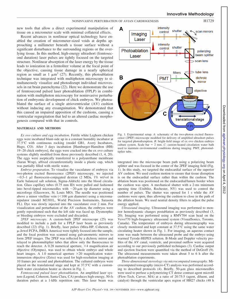
Recent advances in nonlinear optical technology have en-abled the creation of micrometer-sized voids at depths ap-proaching a millimeter beneath a tissue surface without asignificant disturbance to the surrounding regions or the over-lying tissue. In this method, high-energy ultrashort (femtosec-ond duration) laser pulses are tightly focused on the targetedstructure. Nonlinear absorption of the laser energy by the tissueleads to ionization in a femtoliter volume at the focal point ofthe objective, causing tissue damage in a nearly sphericalregion as small as 1 �m3 (27). Recently, this photoablationtechnique was integrated with multiphoton microscopy to si-multaneously visualize and photodisrupt individual microves-sels in rat brain parenchyma (22). Here we demonstrate the useof femtosecond pulsed laser photoablation (FPLP) in combi-nation with multiphoton microscopy for noninvasive perturba-tion of embryonic development of chick embryos. We photoa-blated the surface of a single atrioventricular (AV) cushionwithout inducing any exsanguination. We demonstrated thatthis caused an impaired apposition of the cushions, causing aventricular regurgitation that led to an altered cardiac morpho-genesis compared with that in controls.
MATERIALS AND METHODS
Ex ovo culture and egg incubation. Fertile white Leghorn chickeneggs were incubated blunt-side up in a constant humidity incubator at37.5°C with continuous rocking (model GB1, Avery Incubators,Hugo, CO). After 3 days incubation [Hamburger-Hamilton (HH)19–20 chick embryos], the eggs were cracked into the ex ovo culturesystems slightly modified from those previously described (1, 4, 20).The eggs were aseptically transferred to a polyurethane membrane(Saran Wrap), affixed circumferentially inside a plastic cup, whichwas partially filled with sterile water.
Embryo preparation. To visualize the vasculature of embryos viatwo-photon excited fluorescence (2PEF) microscopy, we injected�0.5–1 �l fluorescein-conjugated dextran (2 MDa, 1% wt/vol inEarle balanced salt solution, Sigma-Aldrich) into the blood circula-tion. Glass capillary tubes (0.75 mm ID) were pulled and fashionedinto bevel-tipped microneedles with �20-�m tip diameter using amicroforge (Glassworx, St. Louis MO). The needle was positionedabove a minor vitelline vein (�100 �m diameter) using a microma-nipulator (model M3301L, World Precision Instruments, SarasotaFL). Dye was slowly injected into the vasculature over 2 min. Forvisualization and perturbation of the AV cushion, the embryos weregently repositioned such that the left side was faced up. Dysmorphicor bleeding embryos were excluded and discarded.
2PEF microscopy. A custom-built 2PEF microscope (25) wasmodified to include a path for a FPLP laser beam as previouslydescribed (22) (Fig. 1). Briefly, laser pulses (Mira-HP, Coherent, or�-Jewel FCPA, IMRA America) were tightly focused into the sample,and the focal position was scanned using galvanometric mirrors toform 2PEF images. The 2PEF was reflected by a dichroic mirror andrelayed to photomultiplier tubes that allow only the fluorescence toreach the detector. A 0.28 numerical aperture, �4 magnification airobjective (Olympus), was used to obtain whole embryo and wholeheart images. A 1.0 numerical aperture �20 magnification water-immersion objective (Zeiss) was used for high-resolution imaging at10 frames per second and photoablation. The cultured embryos wereplaced on the translational stage and kept at 37.5°C with a custom-built water circulation heater as shown in Fig. 1.
Femtosecond pulsed laser photoablation. An amplified laser sys-tem (Legend, Coherent, Santa Clara, CA) produces high-energy, 50-fsduration pulses at a 1-kHz repetition rate. This laser beam was
integrated into the microscope beam path using a polarizing beamsplitter and was focused in the center of the 2PEF imaging field (Fig.1). In this study, we targeted the endocardial surface of the superiorAV cushion. We used cushion motion to ensure that tissue disruptionis on the endocardial surface rather than within the cushion. Theablation beam was positioned on the endocardial/lumen border whenthe cushion was open. A mechanical shutter with a 2-ms minimumopening time (Uniblitz, Rochester, NY) was used to control thenumber of pulses. The shutter was opened for 1 s while the AVcushions were open, thus allowing the cushion to close in the path ofthe ablation beam. We used neutral density filters to adjust the pulseenergy applied.
Ultrasound imaging. Ultrasound imaging was performed to mon-itor hemodynamic changes postablation as previously described (3,20). Imaging was performed using a RMV704 scan head on theVevo770 high-frequency ultrasound system (VisualSonics, Toronto,Canada). The temperature of embryos during ultrasonography wasclosely monitored and kept constant at 37.5°C using the same watercirculating heater shown in Fig. 1. For imaging, an aqueous contactzone was made between the ultrasound probe and the embryo usingwarmed Tyrode-HEPES solution. B-Mode and Doppler velocity pro-files of the AV canal, ventricle, and proximal outflow were acquiredaccording to our previously published techniques (3). Cardiac outputand ejection fraction were quantified via the method of DeGroff (9).The ultrasound measurements were taken about 5 to 6 h after thephotoablation experiments.
Three-dimensional stereology via microcomputed tomography. Mi-crocomputed tomography (micro-CT) analysis was performed accord-ing to described protocols (4). Briefly, 50-�m glass microneedleswere used to perfuse a polymerizing CT dense contrast agent microfil(Flow-Tech, Carver, MA) at a ratio of 20:75:5 (agent, diluent, andcatalyst) through the ventricular apex region of HH27 chicks (48 h
Fig. 1. Experimental setup. A: schematic of the two-photon excited fluores-cence (2PEF) microscope modified for delivery of amplified ultrashort pulsesfor targeted photodisruption. B: bright-field image of ex ovo chicken embryoculture system. Scale bar � 2 mm. C: custom-heated circulation water bathused to maintain environmental conditions during imaging. PMT, photomul-tiplier tube.
Innovative Methodology
H1729NONINVASIVE PERTURBATION OF AVIAN CARDIOGENESIS
AJP-Heart Circ Physiol • VOL 299 • NOVEMBER 2010 • www.ajpheart.org
on April 4, 2012
ajpheart.physiology.orgD
ownloaded from
after photoablation). Perfusion was controlled via gravity (20 cmheight difference), which completely filled the cardiac chamberswithin 10 min. Microfil polymerized into a cast within 15 min, afterwhich the entire embryo was fixed in 4% paraformaldehyde. Embryoswere typically imaged within 48 h after fixation via micro-CT (eXplore CT120, GE Healthcare) at a 25-�m voxel resolution. Datasets wereprocessed using Microview (GE Healthcare) and Osirix (Osirix).Individual chamber volumes were segmented and quantified as pre-viously described (4).
Histology/immunohistochemistry. Additional embryos were fixed48 h after cushion ablation (HH27) with 4% paraformaldehyde,paraffin processed, and cut into 10-�m saggital sections. The sectionswere stained with hematoxylin and eosin to compare the generalmorphology and cellularity between treatments. Draq5 staining (1:1,000, Alexis Biochemicals, San Diego, CA) was applied as a nuclearcounterstain control, whereas fluorescent antibody detection of�-smooth muscle actin (�-SMA, 1:100, Spring Bioscience, Pleasan-ton, CA) was used to identify cells with a mesenchymal phenotypewithin the AV cushions.
Statistics. Student t-test was used to show significance betweengroups, with P � 0.05 denoting significance.
RESULTS
Three-dimensional visualization of deep tissues in chickembryos in vivo via multiphoton microscopy. An exogenousadministration of FITC-dextran via vitelline vein injection
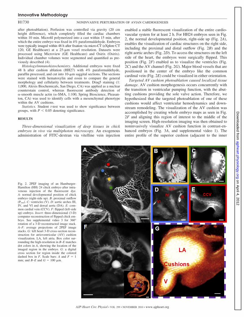
enabled a stable fluorescent visualization of the entire cardio-vascular system for at least 2 h. For HH24 embryos seen in Fig.2, the normal developmental position, right-side up (Fig. 2A),enables the visualization of cardiac structures on the right side,including the proximal and distal outflow (Fig. 2B) and theright aortic arches (Fig. 2D). To access the structures on the leftside of the heart, the embryos were surgically flipped. Thisposition (Fig. 2F) enabled us to visualize the ventricles (Fig.2C) and the AV channel (Fig. 2G). Major blood vessels that arepositioned in the center of the embryo like the commoncardinal vein (Fig. 2E) could be visualized in either orientation.
Targeted AV cushion photoablation caused localized tissuedamage. AV cushion morphogenesis occurs concurrently withthe transition in ventricular pumping function, with the abut-ting cushions providing the sole valve action. Therefore, wehypothesized that the targeted photoablation of one of thesecushions would affect ventricular hemodynamics and down-stream remodeling. The visualization of the AV cushion wasaccomplished by creating whole embryo maps as seen in Fig.2F and aligning this region of interest to the middle of theimaging screen. High-resolution imaging was then obtained tononinvasively visualize AV cushion function in contrast-en-hanced embryos (Fig. 3A, and supplemental video 1). Theentire profile of the superior cushion (adjacent to the inner
Fig. 2. 2PEF imaging of an Hamburger-Hamilton (HH) 24 chick embryo after intra-venous injection of the fluorescent dye.A: normal developmental position of chickembryo (right-side up). B: proximal outflow(Pout). C: ventricles (V). D: aortic arches III,IV, and VI and dorsal aorta (DA). E: com-mon cardial vein (CCV). F: flipped (left-sideup) embryo. Insert: three-dimensional (3-D)computer reconstruction of flipped chick em-bryo. See supplemental video 3 for 360°rotation of a 3-D reconstructed image stack.A–F: average projections of 2PEF imagestacks. G: left heart 3-D cross-section recon-struction for atrioventricular (AV) cushionvisualization. LA, left atria. Box color sur-rounding the high-resolution in B–E matchesdot colors in A, showing the location of theimaged region in the embryo. G: a digitalcross section for region inside the coloreddashed box in F. Scale bars: A and F � 1mm; and B–E and G � 100 �m.
Innovative Methodology
H1730 NONINVASIVE PERTURBATION OF AVIAN CARDIOGENESIS
AJP-Heart Circ Physiol • VOL 299 • NOVEMBER 2010 • www.ajpheart.org
on April 4, 2012
ajpheart.physiology.orgD
ownloaded from
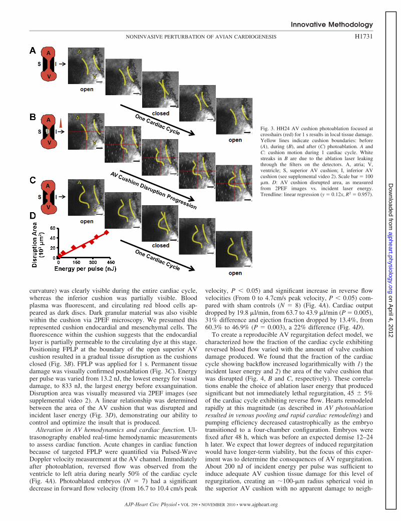
curvature) was clearly visible during the entire cardiac cycle,whereas the inferior cushion was partially visible. Bloodplasma was fluorescent, and circulating red blood cells ap-peared as dark discs. Dark granular material was also visiblewithin the cushion via 2PEF microscopy. We presumed thisrepresented cushion endocardial and mesenchymal cells. Thefluorescence within the cushion suggests that the endocardiallayer is partially permeable to the circulating dye at this stage.Positioning FPLP at the boundary of the open superior AVcushion resulted in a gradual tissue disruption as the cushionsclosed (Fig. 3B). FPLP was applied for 1 s. Permanent tissuedamage was visually confirmed postablation (Fig. 3C). Energyper pulse was varied from 13.2 nJ, the lowest energy for visualdamage, to 833 nJ, the largest energy before exsanguination.Disruption area was visually measured via 2PEF images (seesupplemental video 2). A linear relationship was determinedbetween the area of the AV cushion that was disrupted andincident laser energy (Fig. 3D), demonstrating our ability tocontrol and optimize the insult that is produced.
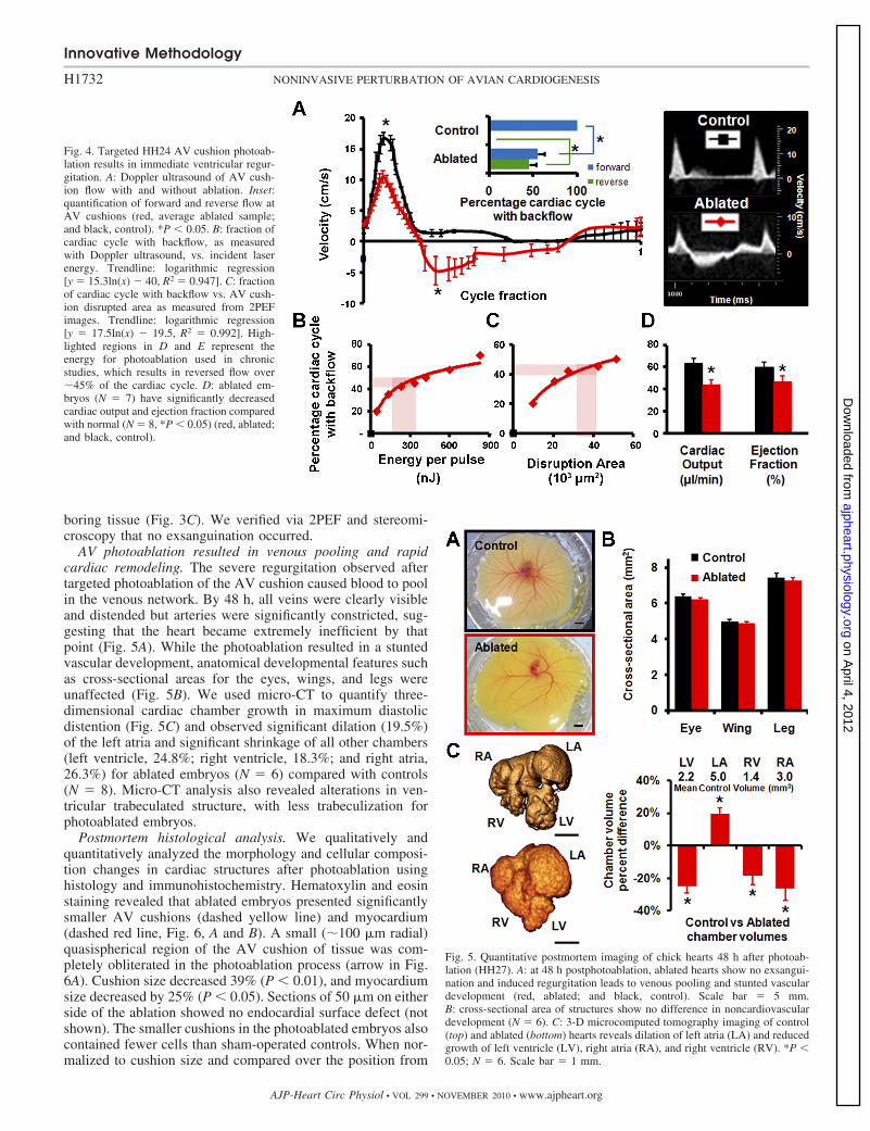
Alteration in AV hemodynamics and cardiac function. Ul-trasonography enabled real-time hemodynamic measurementsto assess cardiac function. Acute changes in cardiac functionbecause of targeted FPLP were quantified via Pulsed-WaveDoppler velocity measurement at the AV channel. Immediatelyafter photoablation, reversed flow was observed from theventricle to left atria during nearly 50% of the cardiac cycle(Fig. 4A). Photoablated embryos (N � 7) had a significantdecrease in forward flow velocity (from 16.7 to 10.4 cm/s peak
velocity, P � 0.05) and significant increase in reverse flowvelocities (From 0 to 4.7cm/s peak velocity, P � 0.05) com-pared with sham controls (N � 8) (Fig. 4A). Cardiac outputdropped by 19.8 �l/min, from 63.7 to 43.9 �l/min (P � 0.005),31% difference and ejection fraction dropped by 13.4%, from60.3% to 46.9% (P � 0.003), a 22% difference (Fig. 4D).
To create a reproducible AV regurgitation defect model, wecharacterized how the fraction of the cardiac cycle exhibitingreversed blood flow varied with the amount of valve cushiondamage produced. We found that the fraction of the cardiaccycle showing backflow increased logarithmically with 1) theincident laser energy and 2) the area of the valve cushion thatwas disrupted (Fig. 4, B and C, respectively). These correla-tions enable the choice of ablation laser energy that producedsignificant but not immediately lethal regurgitation, 45 � 5%of the cardiac cycle exhibiting reverse flow. Hearts remodeledrapidly at this magnitude (as described in AV photoablationresulted in venous pooling and rapid cardiac remodeling) andpumping efficiency decreased catastrophically as the embryotransitioned to a four-chamber configuration. Embryos werefixed after 48 h, which was before an expected demise 12–24h later. We expect that lower degrees of induced regurgitationwould have longer-term viability, but the focus of this exper-iment was to determine the consequences of AV regurgitation.About 200 nJ of incident energy per pulse was sufficient toinduce adequate AV cushion tissue damage for this level ofregurgitation, creating an �100-�m radius spherical void inthe superior AV cushion with no apparent damage to neigh-
Fig. 3. HH24 AV cushion photoablation focused atcrosshairs (red) for 1 s results in local tissue damage.Yellow lines indicate cushion boundaries: before(A), during (B), and after (C) photoablation. A andC: cushion motion during 1 cardiac cycle. Whitestreaks in B are due to the ablation laser leakingthrough the filters on the detectors. A, atria; V,ventricle; S, superior AV cushion; I, inferior AVcushion (see supplemental video 2). Scale bar � 100�m. D: AV cushion disrupted area, as measuredfrom 2PEF images vs. incident laser energy.Trendline: linear regression (y � 0.12x, R2 � 0.957).
Innovative Methodology
H1731NONINVASIVE PERTURBATION OF AVIAN CARDIOGENESIS
AJP-Heart Circ Physiol • VOL 299 • NOVEMBER 2010 • www.ajpheart.org
on April 4, 2012
ajpheart.physiology.orgD
ownloaded from
boring tissue (Fig. 3C). We verified via 2PEF and stereomi-croscopy that no exsanguination occurred.
AV photoablation resulted in venous pooling and rapidcardiac remodeling. The severe regurgitation observed aftertargeted photoablation of the AV cushion caused blood to poolin the venous network. By 48 h, all veins were clearly visibleand distended but arteries were significantly constricted, sug-gesting that the heart became extremely inefficient by thatpoint (Fig. 5A). While the photoablation resulted in a stuntedvascular development, anatomical developmental features suchas cross-sectional areas for the eyes, wings, and legs wereunaffected (Fig. 5B). We used micro-CT to quantify three-dimensional cardiac chamber growth in maximum diastolicdistention (Fig. 5C) and observed significant dilation (19.5%)of the left atria and significant shrinkage of all other chambers(left ventricle, 24.8%; right ventricle, 18.3%; and right atria,26.3%) for ablated embryos (N � 6) compared with controls(N � 8). Micro-CT analysis also revealed alterations in ven-tricular trabeculated structure, with less trabeculization forphotoablated embryos.
Postmortem histological analysis. We qualitatively andquantitatively analyzed the morphology and cellular composi-tion changes in cardiac structures after photoablation usinghistology and immunohistochemistry. Hematoxylin and eosinstaining revealed that ablated embryos presented significantlysmaller AV cushions (dashed yellow line) and myocardium(dashed red line, Fig. 6, A and B). A small (�100 �m radial)quasispherical region of the AV cushion of tissue was com-pletely obliterated in the photoablation process (arrow in Fig.6A). Cushion size decreased 39% (P � 0.01), and myocardiumsize decreased by 25% (P � 0.05). Sections of 50 �m on eitherside of the ablation showed no endocardial surface defect (notshown). The smaller cushions in the photoablated embryos alsocontained fewer cells than sham-operated controls. When nor-malized to cushion size and compared over the position from
Fig. 4. Targeted HH24 AV cushion photoab-lation results in immediate ventricular regur-gitation. A: Doppler ultrasound of AV cush-ion flow with and without ablation. Inset:quantification of forward and reverse flow atAV cushions (red, average ablated sample;and black, control). *P � 0.05. B: fraction ofcardiac cycle with backflow, as measuredwith Doppler ultrasound, vs. incident laserenergy. Trendline: logarithmic regression[y � 15.3ln(x) � 40, R2 � 0.947]. C: fractionof cardiac cycle with backflow vs. AV cush-ion disrupted area as measured from 2PEFimages. Trendline: logarithmic regression[y � 17.5ln(x) � 19.5, R2 � 0.992]. High-lighted regions in D and E represent theenergy for photoablation used in chronicstudies, which results in reversed flow over�45% of the cardiac cycle. D: ablated em-bryos (N � 7) have significantly decreasedcardiac output and ejection fraction comparedwith normal (N � 8, *P � 0.05) (red, ablated;and black, control).
Fig. 5. Quantitative postmortem imaging of chick hearts 48 h after photoab-lation (HH27). A: at 48 h postphotoablation, ablated hearts show no exsangui-nation and induced regurgitation leads to venous pooling and stunted vasculardevelopment (red, ablated; and black, control). Scale bar � 5 mm.B: cross-sectional area of structures show no difference in noncardiovasculardevelopment (N � 6). C: 3-D microcomputed tomography imaging of control(top) and ablated (bottom) hearts reveals dilation of left atria (LA) and reducedgrowth of left ventricle (LV), right atria (RA), and right ventricle (RV). *P �0.05; N � 6. Scale bar � 1 mm.
Innovative Methodology
H1732 NONINVASIVE PERTURBATION OF AVIAN CARDIOGENESIS
AJP-Heart Circ Physiol • VOL 299 • NOVEMBER 2010 • www.ajpheart.org
on April 4, 2012
ajpheart.physiology.orgD
ownloaded from
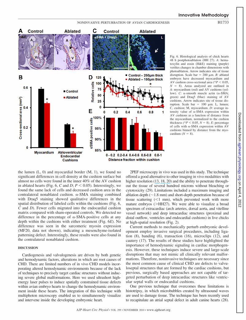
the lumen (L, 0) and myocardial border (M, 1), we found nosignificant differences in cell density at the cushion surface butalmost no cells were found in the inner 40% of the AV cushionin ablated hearts (Fig. 6, C and D; P � 0.05). Interestingly, wefound the same lack of cells and decreased cushion area in thecontralateral nonablated cushion. �-SMA staining combinedwith Draq5 staining showed qualitative differences in thespatial distribution of labeled cells within the cushions (Fig. 6,C and D). Fewer cells migrated into the endocardial cushionmatrix compared with sham-operated controls. We detected nodifference in the percentage of �-SMA-positive cells at anydepth within the cushions with either treatment (Fig. 6E). Nodifference was seen in the sarcomeric myosin expression(MF20, data not shown), indicating a mesenchyme-isolatedpatterning defect. Interestingly, these results were also found inthe contralateral nonablated cushion.
DISCUSSION
Cardiogenesis and valvulogenesis are driven by both geneticand hemodynamic factors, alterations in which are root causes ofCHD. There are limited numbers of in vivo CHD models incor-porating altered hemodynamic environments because of the lackof techniques to precisely target cardiac structures without induc-ing severe global malformations. Here we used ultrashort high-energy laser pulses to induce spatially constrained tissue defectswithin avian embryo hearts to change the hemodynamic environ-ment inside these hearts. The integration of this technique withmultiphoton microscopy enabled us to simultaneously visualizeand intervene inside the developing embryonic heart.
2PEF microscopy in vivo was used in this study. The techniqueoffered a good alternative to other imaging in vivo modalities withhigher resolution (13, 18, 20) and the ability to penetrate through-out the tissue of several hundred microns without bleaching orcytotoxicity (29). Limitations included a maximum imaging andablation depth (�1.8 mm) and short-depth penetration because oftissue scattering (�1 mm), which prevented work with moremature embryos (HH27). We were able to visualize a broadspectrum of extracardiac (arch arteries, dorsal aorta, and vitellinevessel network) and deep intracardiac structures (proximal anddistal outflow, ventricles and endocardial cushions) in live chicksat high-spatial resolution (Fig. 2).
Current methods to mechanically perturb embryonic devel-opment employ invasive surgical procedures, including liga-tion (8), banding (6), transaction (16), microclips (12), andcautery (17). The results of these studies have highlighted theimportance of hemodynamic signaling in cardiac morphogen-esis. However, these techniques induce severe and large-scaledisruptions that may not mimic all clinically relevant malfor-mations. Therefore, noninvasive techniques are necessary sincethe most common cause of clinical CHD are defects to valvu-loseptal structures that are formed by the cardiac cushions, butprevious, surgically based approaches are not capable of tar-geted perturbation of deep intracardiac structures like ventric-ular septal walls or endocardial cushions.
One previous technique that overcomes these limitations ishistotripsy, in which microbubbles created by ultrasound wavesare used to damage tissue. The technique has been recently usedto recapitulate an atrial septal defect in adult canine hearts (28).
Fig. 6. Histological analysis of chick hearts48 h postphotoablation (HH 27). A: hema-toxylin and eosin (H&E) staining (purple)verifies changes in chamber dimensions withphotoablation. Arrow indicates site of tissuedisruption. Scale bar � 200 �m. B: ablatedembryos have decreased myocardium andAV cushion cross-sectional area (*P � 0.05,N � 8). Areas analyzed are outlined inA: myocardium (red) and AV cushions (yel-low). C: �-smooth muscle actin (�-SMA;green) and Draq5 (blue) staining of AVcushions. Arrow indicates site of tissue dis-ruption. Scale bar � 100 �m. L, lumen;C, cushion; M, myocardium. D: average in-tensity value of �-SMA expression withinAV cushions as a function of distance fromthe myocardium, normalized to the cushionthickness (*P � 0.05, N � 8). E: percentageof cells with �-SMA expression within AVcushions binned by distance from the myo-cardium (N � 8).
Innovative Methodology
H1733NONINVASIVE PERTURBATION OF AVIAN CARDIOGENESIS
AJP-Heart Circ Physiol • VOL 299 • NOVEMBER 2010 • www.ajpheart.org
on April 4, 2012
ajpheart.physiology.orgD
ownloaded from
This demonstrates the ability to visualize and disrupt structuresbeneath centimeters of tissue causing millimeter-size holes. Incontrast, our method uses femtosecond duration pulsed laserenergy, which is delivered to a targeted micrometer-sized volumeinside the beating heart to cause localized tissue disruptionthrough nonlinear optical absorption.
To test our approach, we decided to focus on an intracardiacregion in which tissue alterations would lead to immediatehemodynamic changes. We focused femtosecond high-energylaser pulses at the AV cushion surface, which induced alocalized disruption of the cushion, leading to an incompletevalve closure (Fig. 3). This, in turn, led to an immediate andpronounced regurgitation through the AV valve (Fig. 4), caus-ing decreased cardiac function and leading to chamber remod-eling over 48 h (Fig. 5). The degree of severity of the valvecushion damage (Fig. 3D) and therefore the acute changes incardiac blood flow (Fig. 4, B and C) were easily tuned byadjusting the incident laser energy.
The cardiac remodeling quantified in this study was somewhatsimilar to the results from Sedmera et al. (24), who showed thatdecreased ventricular preload via LAL caused stunted left ven-tricular growth and altered myocardial trabeculation. The functionof the cushions/valves in LAL-treated hearts has not been evalu-ated extensively, but the results suggest that these cushions remainthickened but do not regurgitate (8, 24). The permanent constric-tion of the left AV orifice in LAL likely effects more than just AVhemodynamics, including chamber growth and myocardial con-duction patterns. It is unclear how these different effects can beisolated. In contrast to their study, however, we found smaller andless-cellularized AV cushions with targeted photoablation, whichwas a direct effect of both reduced ventricular preload (quantifiedby lower inflow velocity) and AV regurgitation. Collectively,these results suggest that endocardial cells are more sensitive toregurgitation than preload changes and that regurgitation is amuch more serious problem for embryonic heart development.The ability to precisely control the location, amount of cushionablation, and the degree of regurgitation with this techniqueenables a robust experimental analysis of the interactions betweenhemodynamics and biology in valvulogenesis.
In conclusion, 2PEF combined with FPLP is a powerfultechnique to simultaneously visualize and perturb embryonicmorphogenesis. While the current application focused on AVcushion morphogenesis, the technique can be applied to anyembryonic structure. The depth of penetration for both imagingand targeted ablation is ultimately limited by optical scattering,which for the wavelengths used here is about 1 mm in all areasof the embryo that we tested. Deep cardiac manipulation waspossible with the current system up to stage HH27. Tools forlocalized noninvasive manipulation of embryonic developmentare critically needed to complement the significant moleculartechnologies available. A combination of these techniques willdramatically enhance our ability to dissect the complex andinterrelated effects of genetic and hemodynamic signaling thatlikely drives clinical CHD.
ACKNOWLEDGMENTS
We thank Drew Noden, Mark Riccio, and Yung-Nung Chiu for technicalassistance in this study. We thank Coherent and IMRA for the loan of lasers.
Present address of H. C. Yalcin: Mechanical Engineering Dept., DogusUniv., Kadikoy 34722, Istanbul, Turkey.
GRANTS
This work was funded by American Heart Association Scientist Develop-ment Grant 0830384N (to J. T. Butcher), Scientist Development Grant0735644T (to C. B. Schaffer), and Postdoctoral Fellowshhip 09POST2250177(to N. Nishimura) and by a L’Oréal USA Fellowship for Women in Science (toN. Nishimura), a New York State Foundation Centers for Advanced Technol-ogy award (to J. T. Butcher), and the LeDucq foundation (Project MITRAL)(to J. T. Butcher).
DISCLOSURES
No conflicts of interest, financial or otherwise, are declared by the author(s).
REFERENCES
1. Auerbach R, Kubai L, Knighton D, Folkman J. A simple procedure forthe long-term cultivation of chicken embryos. Dev Biol 41: 391–394,1974.
2. Butcher JT, Barrett BC, Nerem RM. Equibiaxial strain stimulatesfibroblastic phenotype shift in smooth muscle cells in an engineered tissuemodel of the aortic wall. Biomaterials 27: 5252–5258, 2006.
3. Butcher JT, McQuinn TC, Sedmera D, Turner D, Markwald RR.Transitions in early embryonic atrioventricular valvular function corre-spond with changes in cushion biomechanics that are predictable by tissuecomposition. Circ Res 100: 1503–1511, 2007.
4. Butcher JT, Sedmera D, Guldberg RE, Markwald RR. Quantitativevolumetric analysis of cardiac morphogenesis assessed through micro-computed tomography. Dev Dyn 236: 802–809, 2007.
5. Butcher JT, Tressel S, Johnson T, Turner D, Sorescu G, Jo H, NeremRM. Transcriptional profiles of valvular and vascular endothelial cellsreveal phenotypic differences: influence of shear stress. ArteriosclerThromb Vasc Biol 26: 69–77, 2006.
6. Clark EB, Hu N, Rosenquist GC. Effect of conotruncal constriction onaortic-mitral valve continuity in the stage 18, 21 and 24 chick embryo. AmJ Cardiol 53: 324–327, 1984.
7. Cruz MD, Markward R. Living Morphogenesis of the Heart: Boston:Birkhauser, 1998.
8. deAlmeida A, McQuinn T, Sedmera D. Increased ventricular preload iscompensated by myocyte proliferation in normal and hypoplastic fetalchick left ventricle. Circ Res 100: 1363–1370, 2007.
9. DeGroff CG. Doppler echocardiography. Pediatr Cardiol 23: 307–333,2002.
10. Gomez-Fifer C. Hypoplastic left heart syndrome in the fetus: Diagnosticfeatures prior to birth and their impact on postnatal outcome. Prog PediatrCardiol 22: 53–60, 2006.
11. Hall CE, Hurtado R, Hewett KW, Shulimovich M, Poma CP, ReckovaM, Justus C, Pennisi DJ, Tobita K, Sedmera D, Gourdie RG, MikawaT. Hemodynamic-dependent patterning of endothelin converting enzyme1 expression and differentiation of impulse-conducting Purkinje fibers inthe embryonic heart. Development 131: 581–592, 2004.
12. Hogers B, DeRuiter MC, Gittenberger-de Groot AC, Poelmann RE.Unilateral vitelline vein ligation alters intracardiac blood flow patterns andmorphogenesis in the chick embryo. Circ Res 80: 473–481, 1997.
13. Hogers B, van der Weerd L, Olofsen H, van der Graaf LM, DeRuiterMC, Gittenberger-de Groot AC, Poelmann RE. Non-invasive trackingof avian development in vivo by MRI NMR in biomedicine. NMR Biomed22: 365–373, 2009.
14. Hove JR, Koster RW, Forouhar AS, Acevedo-Bolton G, Fraser SE,Gharib M. Intracardiac fluid forces are an essential epigenetic factor forembryonic cardiogenesis. Nature 421: 172–177, 2003.
15. Hu N, Clark EB. Hemodynamics of the stage 12 to stage 29 chickembryo. Circ Res 65: 1665–1670, 1989.
16. Icardo JM. Developmental biology of the vertebrate heart. J Exp Zool275: 144–161, 1996.
17. Jaffee OC. Hemodynamic factors in the development of the chick embryoheart. Anat Rec 151: 69–75, 1965.
18. Jenkins MW, Chughtai OQ, Basavanhally AN, Watanabe M, RollinsAM. In vivo gated 4D imaging of the embryonic heart using opticalcoherence tomography. J Biomed Opt 12: 030505, 2007.
19. MacKenna D, Summerour SR, Villarreal FJ. Role of mechanicalfactors in modulating cardiac fibroblast function and extracellular matrixsynthesis. Cardiovasc Res 46: 257–263, 2000.
20. McQuinn TC, Bratoeva M, Dealmeida A, Remond M, Thompson RP,Sedmera D. High-frequency ultrasonographic imaging of avian cardio-vascular development. Dev Dyn 236: 3503–3513, 2007.
Innovative Methodology
H1734 NONINVASIVE PERTURBATION OF AVIAN CARDIOGENESIS
AJP-Heart Circ Physiol • VOL 299 • NOVEMBER 2010 • www.ajpheart.org
on April 4, 2012
ajpheart.physiology.orgD
ownloaded from
21. Nakazawa M, Miyagawa S, Ohno T, Miura S, Takao A. Developmentalhemodynamic changes in rat embryos at 11 to 15 days of gestation: normaldata of blood pressure and the effect of caffeine compared to data fromchick embryo. Pediatr Res 23: 200–205, 1988.
22. Nishimura N, Schaffer CB, Friedman B, Tsai PS, Lyden PD, KleinfeldD. Targeted insult to subsurface cortical blood vessels using ultrashortlaser pulses: three models of stroke. Nat Methods 3: 99–108, 2006.
23. Sedmera D, Cook AC, Shirali G, McQuinn TC. Current issues andperspectives in hypoplasia of the left heart. Cardiol Young 15: 56–72,2005.
24. Sedmera D, Pexieder T, Rychterova V, Hu N, Clark EB. Remodelingof chick embryonic ventricular myoarchitecture under experimentallychanged loading conditions. Anat Rec 254: 238–252, 1999.
25. Tsai PS, Frosting RD. Principles, design and construction of a two-photon laserscanning microscope for in vitro and in vivo brain imaging. In: In Vivo OpticalImaging of Brain Function. Boca Raton: CRC, 2002, p. 113–171.
26. Vermot J, Forouhar AS, Liebling M, Wu D, Plummer D, Gharib M,Fraser SE. Reversing blood flows act through klf2a to ensure normalvalvulogenesis in the developing heart. PLoS Biol 7: e1000246, 2009.
27. Vogel A, Venugopalan V. Mechanisms of pulsed laser ablation ofbiological tissues. Chem Rev 103: 577–644, 2003.
28. Xu Z, Owens G, Gordon D, Cain C, Ludomirsky A. Noninvasivecreation of an atrial septal defect by histotripsy in a canine model.Circulation 121: 742–749, 2010.
29. Zipfel WR, Williams RM, Webb WW. Nonlinear magic: multiphotonmicroscopy in the biosciences. Nat Biotechnol 21: 1369–1377, 2003.
Innovative Methodology
H1735NONINVASIVE PERTURBATION OF AVIAN CARDIOGENESIS
AJP-Heart Circ Physiol • VOL 299 • NOVEMBER 2010 • www.ajpheart.org
on April 4, 2012
ajpheart.physiology.orgD
ownloaded from
Related Documents