B The Author(s), 2010 Published Online: 16 April 2010 DOI: 10.1007/s11307-010-0307-z Mol Imaging Biol (2011) 13:67Y77 RESEARCH ARTICLE Two-Photon Intravital Multicolor Imaging Combined with Inducible Gene Expression to Distinguish Metastatic Behavior of Breast Cancer Cells In Vivo Sylvia E. Le Dévédec, Reshma Lalai, Chantal Pont, Hans de Bont, Bob van de Water Division of Toxicology, Leiden Amsterdam Center for Drug Research (LACDR), Leiden University, Leiden, The Netherlands Abstract Purpose: The aim of this study is to use multicolor intravital imaging together with an inducible cell model to compare metastatic behavior of control and genetically modified breast cancer cell populations within the intact primary tumor of a mouse. Procedure: GFP-MTLn3-ErbB1 cells were generated with doxycycline-regulated conditional transgene expression using lentiviral TREAutoR3-cyan fluorescent protein (CFP). CFP expression together with tumor cell motility is monitored in vitro and in vivo. Results: Effective and tight control of doxycycline-induced CFP expression was observed both in vitro and in vivo. Intravital multiphoton microscopy on intact orthotopic tumors allowed a clear discrimination between GFP-only and (GFP+CFP) cell populations, which enables direct comparison of the motility behavior of two different cell populations in the same microenvironment in vivo. Conclusions: This system is robust and versatile for conditional gene expression and can be used to study the role of individual candidate metastasis genes in vitro and in vivo. This technology will allow investigations of cellular events in cancer metastasis and in particular intravasation within a primary tumor. Key words: Intravital imaging, Two-photon microscopy, Lentiviral inducible system, Metastasis, Breast cancer Introduction D espite many years of laboratory and clinical research, still a large number of women die of breast cancer metastasis formation [1]. Therefore, it is crucial to under- stand the underlying mechanisms of metastasis formation and especially tumor cell intravasation [2, 3]. Improved models are required to study the role of individual candidate metastasis genes involved in this individual steps and in particular in the intravasation steps [4]. So far, constitutive ectopic expression of wild-type genes, their deletion mutants, or shRNA-mediated knockdown is Abbreviations RAG mice, Recombinase activating gene; γc, Common gamma chain; FACS, Fluorescence-activated cell sorter; EGFR, Epidermal growth factor receptor; CFP, Cyan fluorescent protein; EGFP, Enhanced green fluorescent protein; TRE, Tetracycline-responsive promoter; RtTA, Reverse tetracycline-responsive transactivator; Doxy, Doxycycline; ECM, Extracellular matrix; TPLSM, Two-photon laser scanning microscopy Significance: The underlying mechanisms leading to breast cancer metastasis formation are still poorly understood. To enable the analysis of the role of individual candidate metastasis-modifying genes, in this manuscript, we have carefully characterized and determined the feasibility of a methodology that combines the use of an inducible lentiviral expression systems and two-photon intravital multicolor imaging. We anticipate our methodology will have an important impact on the future detailed analysis of tumor cell autonomous mechanisms of metastasis formation, an important area of cancer research. Electronic supplementary material The online version of this article (doi:10.1007/s11307-010-0307-z) contains supplementary material, which is available to authorized users. Correspondence to: Bob van de Water; e-mail: [email protected]

Welcome message from author
This document is posted to help you gain knowledge. Please leave a comment to let me know what you think about it! Share it to your friends and learn new things together.
Transcript
B The Author(s) 2010Published Online 16 April 2010 DOI 101007s11307-010-0307-z
Mol Imaging Biol (2011) 1367Y77
RESEARCH ARTICLE
Two-Photon Intravital Multicolor ImagingCombined with Inducible Gene Expressionto Distinguish Metastatic Behavior of BreastCancer Cells In VivoSylvia E Le Deacuteveacutedec Reshma Lalai Chantal Pont Hans de Bont Bob van de WaterDivision of Toxicology Leiden Amsterdam Center for Drug Research (LACDR) Leiden University Leiden The Netherlands
AbstractPurpose The aim of this study is to use multicolor intravital imaging together with an induciblecell model to compare metastatic behavior of control and genetically modified breast cancer cellpopulations within the intact primary tumor of a mouseProcedure GFP-MTLn3-ErbB1 cells were generated with doxycycline-regulated conditionaltransgene expression using lentiviral TREAutoR3-cyan fluorescent protein (CFP) CFPexpression together with tumor cell motility is monitored in vitro and in vivoResults Effective and tight control of doxycycline-induced CFP expression was observed both invitro and in vivo Intravital multiphoton microscopy on intact orthotopic tumors allowed a cleardiscrimination betweenGFP-only and (GFP+CFP) cell populations which enables direct comparisonof the motility behavior of two different cell populations in the same microenvironment in vivoConclusions This system is robust and versatile for conditional gene expression and can beused to study the role of individual candidate metastasis genes in vitro and in vivo Thistechnology will allow investigations of cellular events in cancer metastasis and in particularintravasation within a primary tumor
Key words Intravital imaging Two-photon microscopy Lentiviral inducible system MetastasisBreast cancer
Introduction
D espite many years of laboratory and clinical researchstill a large number of women die of breast cancer
metastasis formation [1] Therefore it is crucial to under-stand the underlying mechanisms of metastasis formationand especially tumor cell intravasation [2 3] Improvedmodels are required to study the role of individual candidatemetastasis genes involved in this individual steps and inparticular in the intravasation steps [4]
So far constitutive ectopic expression of wild-type genestheir deletion mutants or shRNA-mediated knockdown is
Abbreviations RAG mice Recombinase activating gene γc Common gamma chain FACSFluorescence-activated cell sorter EGFR Epidermal growth factor receptor CFP Cyanfluorescent protein EGFP Enhanced green fluorescent protein TRE Tetracycline-responsivepromoter RtTA Reverse tetracycline-responsive transactivator Doxy Doxycycline ECMExtracellular matrix TPLSM Two-photon laser scanning microscopy
Significance The underlying mechanisms leading to breast cancer metastasisformation are still poorly understood To enable the analysis of the role ofindividual candidate metastasis-modifying genes in this manuscript we havecarefully characterized and determined the feasibility of a methodology thatcombines the use of an inducible lentiviral expression systems and two-photonintravital multicolor imaging We anticipate our methodology will have animportant impact on the future detailed analysis of tumor cell autonomousmechanisms of metastasis formation an important area of cancer research
Electronic supplementary material The online version of this article(doi101007s11307-010-0307-z) contains supplementary material which isavailable to authorized users
Correspondence to Bob van de Water e-mail water_blacdrleidenunivnl
typically used to study the role of candidate metastasisgenes Often this by itself affects already the tumorformation andor is toxic to cells Indeed overexpressionof a deletion mutant or knockdown of a protein may affectcell proliferation or survival and consequently the tumorgrowth in vivo so no conclusion can be drawn on the effectof the candidate gene on tumor cell invasion capacity In thatcase the choice for an experimental metastasis assay is madeto still assess the metastatic capacity of the cells Tocircumvent this problem conditional gene expression strat-egies allowing the expression of target gene only whendesired are invaluable tools for cancer research [5 6] Forexample after orthotopic injection of the transgenic cell lineideally one waits until the tumor reaches a certain size andat a desired moment induces overexpression of a deletionmutant and studies its affect by quantifying the lung burdenAn inducible system is relevant not only for studying theeffect of gene candidates on metastasis formation but alsowhen combined with dual-color imaging for observing at thesame time in the same microenvironment the motilitybehaviors of the control cells and the genetically modifiedcells
Currently available systems often have limited in vivofunctionality because of leakiness insufficient levels ofinduction lack of tissue specificity or complicated designs[7 8] We evaluated the use of a highly versatile lentiviralvector-based tool that allows conditional expression of anycandidate metastasis gene of interest [9] This approachallows one to analyze the behavior of different manipulatedcell populations in the same tumor microenvironment byoptical imaging Two-photon intravital imaging of fluores-cent protein-expressing tumors enables cell motility to beobserved with great detail and at great depth within tissue[10ndash13] In addition to detecting tumor cells two-photonintravital imaging can provide information about cell move-ment and interactions between cells and the microenviron-ment [12 14 15] Our ultimate goal is to compare themetastatic behavior (eg number of tumor cells in blood andnumber and size of lung metastases) of two distinct tumorcell populations (control cells and modified cells withoverexpression or knockdown of a target gene) within thesame primary tumor Since there is a correlation betweenpatterns in gene expression in cells with varying metastaticpotential and differences in cell motility and polarization invivo our aim is to relate the motility cell behavior of the twocell subpopulations and their metastatic capacity For thispurpose we use two-photon intravital microscopy combinedwith fluorescently labeled cells The use of green fluorescentprotein (GFP) and cyan fluorescent protein (CFP) can besimultaneously imaged in a living tumor [12 16] In thisproof-of-concept study we chose this fluorophore combinationto be able to discriminate between control GFP-labeled cellsand GFP-labeled cells that also harbor conditional expressionof CFP alone in the same tumor microenvironment
Here as a concept we generated a GFP-MTLn3-ErbB1cell line that harbors the lentiviral TREAutoR3-CFP con-
struct and established the functionality of this lentiviralvector-based tool that allows conditional expression for bothin vitro and in vivo study We systematically characterizedthe induction kinetics of CFP expression and demonstratedthat doxycycline-induced CFP expression does not influencecell proliferation and motility properties in vitro or in vivoOur data clearly show that two-photon intravital imaging canbe used to simultaneously image cell migration of GFP-onlyand CFP-labeled tumor cells collagen fibers and macro-phages in established tumors in the living animal Thus inthe very same tumor microenvironment we can study theeffect of the expression of a gene candidate on the metastaticbehavior of individual tumor cells since both control andmodified cells can be tracked simultaneously by using two-photon intravital imaging In the context of metastasisformation this model will allow the future evaluation ofcandidate metastasis target genes on tumor cell dissemina-tion programs to the lung
Materials and MethodsReagentsMouse antihuman ErbB1 was purchased from Calbiochem (EMDBiosciences San Diego CA USA) Goat antimouse APC waspurchased from Cedarlane (Ontario CA USA) Alpha modifiedminimal essential medium (αMEM) fetal bovine serum phosphate-buffered saline (PBS) and trypsin were from Life Technologies(Rockville MD USA)
Cloning and Lentivirus ProductionThe lentiviral vector TREAutoR3 was kindly provided by DrJurgen Seppen [9] An AgeIndashPstI digest of TREAutoR3 releasedthe d2eGFP fragment The CFP fragment from Clontech vectorCFP-C1 was cloned into the AgeIndashPstI digest of TREAutoR3 tocreate the TREAutoR3-CFP vector Lentiviral vectors wereprepared as reported previously [17]
Transfection and Stable Cell Line SelectionMTLn3 rat mammary adenocarcinoma cells [18] were culturedas previously described [19] MTLn3-GFP-ErbB1 cell lines werekindly provided by Dr Jeffrey E Segall [20] and weremaintained in αMEM (Life Technologies Inc GaithersburgMD USA) supplemented with 5 fetal bovine serum (LifeTechnologies) TREautoR3 virus containing supernatant wasadded to the medium of the MTLn3 cells After 24-h trans-duction the medium was refreshed and after 48 h the cells werecultured to be frozen at minus80degC and selected for CFP expression afterdoxycycline exposure
ImmunofluorescenceCells were cultured on 12-mm collagen-coated glass coverslips andfixated in 4 (wv) formaldehyde in PBS Subsequently coverslipswere mounted in Aqua PolyMount (Polysciences Warrington PAUSA) Imaging occurred using a Bio-Rad Radiance 2100 MP
68 SE Deacuteveacutedec et al Intravital Imaging of Differential Metastatic Behaviours
confocal laser scanning system with a Nikon Eclipse TE2000-Uinverted fluorescence microscope and a times60 Nikon objective
Cell Proliferation and Motility AssaysGFP-MTLn3-ErbB1-TREAutoR3-CFP cells were cultured in 24-well plates in the absence or presence of doxycycline for 5 daysCells were washed with PBS and subsequently scanned with aTyphoon Imager 9400 (Amersham Biosciences) with both 457 and488 nm laser lines Total GFP fluorescence which is equivalentwith total number of cells was determined with ImageQuant 52software For cell motility assays cells were plated onto collagen-coated 24-well glass-bottom plates (Sensoplate Greiner Bio-One)and after 2 h of serum starvation stimulated with either epidermalgrowth factor (EGF 10 mM) or hepatocyte growth factor (HGF10 ngml) and followed with time lapse microscopy for 2 h Imageacquisition was performed using a Nikon TE2000 combined with aPrior stage and controlled by NIS Element Software Using GFPand CFP filters for epifluorescence microscopy time lapse videoswere recorded using a charge-coupled device (CCD) cameracontrolled by NIS Element Software
In Vivo Tumor Growth and Metastasis FormationSix-week-old Rag2minusminus γcminusminus mice were obtained from in-housebreeding Animals were housed in individually ventilated cagesunder sterile conditions containing three mice per cage Sterilizedfood and water were provided ad libitum To measure spontaneousmetastasis tumor cells were grown to 70 to 85 confluencebefore being harvested for cell counting Cells (5times105) wereinjected into the right thoracic mammary fat pads The cells wereinjected in a volume of 100 μl of PBS with Ca2+ (1 mM) and Mg2+
(2 mM) through a 25-gauge needle Tumor growth rate wasmonitored at weekly intervals after inoculation of tumor cellsHorizontal (h) and vertical (v) diameters were determined andtumor volume (V) was calculated (V=43π(12[radic(htimesv)]3)) After4 weeks the animals were anesthetized with isoflurane and thelungs were excised and rinsed in ice-cold PBS after havingperformed intravital imaging (see below) The right lung was usedto count the tumor burden For rough estimation the right lungswere imaged with the fluorescent imaging unit IVIS (see below)And for detailed quantification the flat side of the right lung wasanalyzed with the immunofluorescence microscope With a times10objective lens (NA 025) we screened the flat surface of the lobeand counted the number of GFP- or CFP-positive metastasesFollowing that step the right lung was cut into two pieces andfixated in 4 paraformaldehyde The left lung was injected withink solution and thereafter destained in water and fixated inFeketersquos (43 (vv) acetic acid 035 (vv) formaldehyde in70 ethanol) Lung tumor burden was also determined by countingthe number of surface metastases The primary tumors and lungsfrom each mouse were used for histologic analysis Samples werefixed in formalin and embedded in paraffin and 5-μm sectionswere stained with hematoxylin and eosin (HampE)
Intravital Imaging by Multiphoton MicroscopyTumor imaging was done as described previously [10 12 16 20]Cells (5times105) were injected into the mammary fat pads of Rag2minusminusγcminusminus
mice as described above and allowed to grow for 4 weeks The animalwas placed under isoflurane anesthesia and the tumor was exposed
using a simple skin flap surgery with as little disruption of thesurrounding vasculature as possible Eventually by injecting 200 microl of20 mgml of Texas Red dextran in PBS 2 h prior to imaging we canalso visualize the blood vessels and the macrophages in the primarytumor [12 15] The animal was then placed in a 30degC temperaturechamber on an inverted Nikon TE2000-U microscope and imagedusing a times20 Nikon objective (Plan Apo NA 075) Briefly a 5-WTsunami laser (Spectra Physics Mountain View CA USA) was usedto run a Radiance 2100 multiphoton system (Bio-Rad Hercules CAUSA) at 860 nm using a 450480-nm filter to image matrix and CFPand a 515530-nm filter to image CFP and GFP Time lapse images ofCFP and GFP-labeled MTLn3-ErbB1 generated tumors were taken at1-min interval for 30 min The images were collected using Bio-RadrsquosLasersharp 2000 software During each 1-min interval a z series of tenimages was taken at a spacing of 10 μm between images beginning atthe periphery of the tumor and moving into the tumor For each tumorthis image acquisition process was repeated for 30 min resulting in atime lapse three-dimensional z series for analysis of tumor cellmotility Image analysis and cell tracking were done with Image ProSoftware
Fluorescent ImagingFluorescent imaging was performed with a highly sensitive cooledCCD camera mounted in a light-tight specimen box (IVIStradeXenogen) Imaging was controlled by the acquisition and analysissoftware Living Imagereg (Xenogen) The light emitted from thebiofluorescent metastases or cells were detected by the IVIStradecamera system integrated digitized and displayed For ex vivoimaging lungs were excised placed into a petri dish and imagedfor 1ndash2 min
Statistical AnalysisStudentrsquos t test was used to determine if there was a significantdifference between two means (pG005) When multiple meanswere compared significance was determined by one-way analysisof variance (pG005) Significant differences are marked in thegraphs
ResultsConditional Transgene Expression In Vitro
To study two different cell populations (with differentialtransgene expression) in the same microenvironment in vivoa model is required that uses two different fluorescentproteins in order to discriminate these populations More-over given the possible effect of transgene expression orknockdown on tumor growth and the microenvironmentsuch a transgene expression should be conditional Here wechose to work with GFP and CFP two fluorescent proteinsthat can be visualized by two-photon intravital imaging andthus in vivo [16] Furthermore to test whether a cell linewith inducible expression of candidate metastasis controllinggenes can be used for in vivo studies we used theestablished GFP-MTLn3-ErbB1 cell line and verified firstthe functionality of the lentiviral vector TREAutoR3 in vitro[9 20 21] We generated a TREAutoR3 lentiviral vector
SE Deacuteveacutedec et al Intravital Imaging of Differential Metastatic Behaviours 69
that expresses CFP after doxycycline-mediated induction tocreate breast cancer cells that either lack or express a transgeneupon treatment with doxycycline (Fig 1a) We transducedGFP-MTLn3-ErbB1 cells and directly after transduction weadded doxycycline to sort the positive cells by FACS analysisfor both CFP and GFP expressions (data not shown) Inaddition similar levels of ErbB1 were observed in both theoriginal cell line (GFP-MTLn3-ErbB1) and the new cell line(GFP-MTLn3-ErbB1-TREAutoR3-CFP) (data not shown)Next we determined whether the expression of the transgenewas tightly and effectively controlled by doxycycline CFP
expression was upregulated after 24 h of doxycycline treatmentwith all concentrations The most optimal concentration was1000 ngml at which over 90 of the cells were CFP positive(Fig 1b) In the absence of doxycycline no CFP expressionwas observed indicating that transgene expression was tightlyand effectively controlled by doxycycline (Fig 1b) We alsochecked the kinetics of the CFP transgene expression overtime After 8 h around 50 of the cells were CFP positivewhile after 24 h already approximately 90 of the cells wereCFP positive (Fig 1c) Together these data show that theautoregulatory lentiviral vector TREautoR3 does not leak at all
Fig 1 In vitro drug-controllable transgene expression a Schematic representation of the constructed lentiviral vector bDoxycycline dosendashresponse of the TRE-autoR3-CFP vector Transduced GFP-MTLn3-ErbB1 cells were cultured in theabsence or presence of doxycycline at different concentrations for 24 h (from 50 until 1000 ngml doxy) and visualized byconfocal microscope In this titration experiment the percentage of CFP-positive cells was calculated c Induction kinetics ofthe CFP expression of GFP-MTLn3 cells transduced with TREautoR3 vector Dual-color imaging of cells exposed to 1000 ngml of doxycycline after different time points The number of CFP-expressing cells after doxycycline induction was countedScale bar for b c=100 microm
70 SE Deacuteveacutedec et al Intravital Imaging of Differential Metastatic Behaviours
in the absence of the drug and that the regulation of thetransgene expression is sensitive fast and tight Moreoverwith this one cell line with inducible expression of CFP wegenerated a versatile tool to study the effect of transgeneexpression compared to control situation in one experimentwhere all conditions are exactly the same
Doxycycline-Mediated Expression of CFPin MTLn3 Cells Does Not Affect Cell Proliferationand Cell Motility
The use of such an inducible system together with CFPexpression might affect cell properties Therefore we care-fully checked whether doxycycline-mediated expression ofCFP alone affects two important aspects in the breast cancerprogression cell proliferation and cell motility To deter-mine the effect of doxycycline-mediated CFP expression oncell proliferation our conditional cell line was cultured in 24wells for 5 days either untreated or treated with doxycyclineto induce CFP expression At indicated time points theincrease in GFP and CFP fluorescence was determined as ameasurement of proliferation using a fluorescent imagerAfter 5 days no significant effect was observed in cellproliferation rate when cells were exposed to doxycycline(Fig 2a) Next we verified whether cell motility remains thesame after doxycycline-induced CFP expression Cellmotility of GFP-only or GFPCFP cells was determinedusing a random cell migration assay with wide fieldfluorescent microscopy No significant difference was
observed in cell motility between GFP and GFPCFP cellsunder control conditions or after stimulation of the cells witheither EGF or HGF (Fig 2b) Thus doxycycline-mediatedexpression of CFP does not affect in vitro cell proliferationand motility
Effective Doxycycline-Induced CFP ExpressionIn Vivo Does Not Affect Tumor Growth and LungMetastasis Formation
We also checked whether doxycycline-mediated expressionof CFP alone affects tumor growth and lung metastasisformation We used inbred Rag2minusminusγcminusminus mice and injectedorthotopically in the fat pad 500000 GFP-MTLn3-ErbB1-TREAutoR3-CFP cells Three groups were defined based onthe doxycycline administration time point The control groupreceived sucrose drinking water without doxycycline Thesecond group received doxycycline sucrose drinking waterstarting from day 1 and the third group starting from day 28which is approximately 48 h before sacrificing the animals(Fig 3a) We measured the tumor progression with a caliperduring the entire length of the experiment and no significantdifference was found in tumor growth rate between the threegroups (data not shown) After sacrifice tumor volume andweight were measured with no differences observed betweendifferent treatments (Fig 3b) Also histological analysis oftumor tissue sections is similar in the three test groups(Fig 3c) Next we quantified with both fluorescenceimaging (FLI Fig 4a) and fluorescence microscopy
Fig 2 Doxycycline-mediated CFP expression does not affect cell proliferation and migration in vitro a Cell proliferation in theabsence or presence of doxycycline (1000 ngml) quantified by measuring the total GFP fluorescence over the time using theTyphoon imager b Cell motility determined in a random cell migration assay after EGF or HGF stimulation in the presence orabsence of doxycycline Cells were followed with epifluorescence for 2 h Scale bar for b=50 microm
SE Deacuteveacutedec et al Intravital Imaging of Differential Metastatic Behaviours 71
(Fig 4b) the lung tumor burden In all the three groups anaverage number of 400 lung metastases were countedshowing no difference in metastasis formation after doxycy-cline-induced CFP expression Histological observations ofthe lungs did not reveal any difference between the threegroups (Fig 4c) In conclusion tumor growth and lungmetastasis formation are not affected by doxycycline-induced CFP expression
Tightly Controlled CFP Expression in Both Tumorand Lung Metastases
Since we want to use this inducible system in vivo it wasessential to evaluate the effectiveness of doxycycline-induced CFP expression With two-photon intravital imag-ing we checked whether doxycycline CFP induction wassuccessful in the tumor and in the lung metastases No CFPexpression at all was detected in the control group yet robustCFP expression was detected in over 90 of the whole tumorwhen doxycycline was added in the drinking water from day 1and in more than 70 of the tumor when doxycycline wasadded 48 h before sacrificing the animals (Fig 5a) Weobserved the same CFP induction when analyzing the lungmetastases with two-photon excitation after sacrifice (Fig 5b)in the control group only GFP-positive and no CFP-positivemetastasis could be observed throughout the entire lung Whendoxycycline was added in the drinking water from day 1almost all the lung metastases were CFP positive A period of48-h doxycycline exposure resulted in approximately 70CFP-positive lungmetastasis Together these data demonstratethat our GFP-MTLn3-ErbB1-TREAutoR3 model allows for
the effective fast and tightly controlled transgene expressionMoreover we can easily discriminate the GFP-only and GFPCFP-positive lung metastases which are a prerequisite toevaluate the effect of transgene expression on lung metastasisformation
Two Distinct Tumor Cell SubpopulationsCan Be Tracked in the Same TumorMicroenvironment and Doxycycline-Induced CFPExpression Does Not Affect Cell Motility In Vivo
Multiphoton imaging technology can be used to get aninsight into the microenvironment and understand its role inthe invasion and intravasation steps of metastasis insideliving mammary tumors [11 12 14 20] Results show thatthe tumor microenvironment is a dynamic place whereinteractions between tumor cells macrophages bloodvessels and extracellular matrix (ECM) fibers define themetastatic phenotype [13 15 22] In our setup we used aBio-Rad Radiance 2100 multiphoton microscope with aninverted Nikon TE2000 connected to a Spectra PhysicsTsunami Ti-Sapphire laser of 5 W As previously shown byE Sahai and colleagues [16] wavelengths around 880 nmwere most suitable for simultaneous imaging of GFP andCFP We used for all experiments 860 nm The selectedemission filters enabled us to visualize at the same time CFP(48030) GFP (51530) and the extracellular matrix (45080)simultaneously (Fig 7 and supplementary movie 1) Collagentype I which is very abundant in the tumor microenvironmentcan be visualized with second harmonic generation generatedby the pulsed laser used in two-photon microscopy [23ndash25] In
Fig 3 Doxycycline-mediated CFP expression does not affect tumor cell growth in vivo a Schedule of in vivo doxycyclinetreatment We defined in total three groups control group (n=5) which gets only sucrose drinking water group 2 (n=5) whichgets doxycycline in the drinking water from day 0 and group 3 (n=5) which gets doxycycline in the drinking water 48 h beforesacrifice b At sacrifice the tumor was isolated and the weight measured c Sectioned tumors were stained with HampE andrepresentative pictures are shown
72 SE Deacuteveacutedec et al Intravital Imaging of Differential Metastatic Behaviours
addition when injecting Texas Red dextran (62060) in the tailvein of the animal 2 h prior to imaging the blood flow and themacrophage in the tumor can be visualized Together thisprovides information about the tumor microenvironment andits host cells (Fig 6a and supplementary movie 2)
To get a better insight into metastasis mechanisms wewant to be able to track two cell subpopulations within a
primary tumor which differentially express a candidatemetastasis target gene by using two-photon intravitalimaging For this purpose we checked firstly whether wewere able to discriminate the GFP-only and GFPCFP cellsand secondly whether the tumor cell motility in vivo was notaffected by doxycycline-induced CFP expression Weperformed two-photon intravital imaging in primary tumors
Fig 4 Doxycycline-mediated CFP expression does not affect the number of lung metastases a Fresh isolated lungs werevisualized with FLI Control group is ldquominus doxyrdquo and group 2 is ldquo+ doxyrdquo b Using epifluorescence we counted the number offluorescent lung metastases only on one side of the small lobe c Part of the isolated lungs was injected with ink and the rest ofthe lungs were stained with HampE and representative pictures are shown
Fig 5 Tightly controlled CFP expression in both tumor and lung metastases Fresh isolated lungs and tumors were imagedwith multiphoton microscopy Control group is ldquominus doxyrdquo and group 2 is ldquo+ doxyrdquo Simultaneous capture of GFP CFP andcollagen second harmonic fluorescence (blue) in the mammary tumors (a) and lung metastases (b) of control group and groupScale bar for a b=100 microm
SE Deacuteveacutedec et al Intravital Imaging of Differential Metastatic Behaviours 73
of both control and doxycycline groups We could easilydetect discriminate and track the two tumor cell subpopu-lations GFP-only and GFPCFP cells (Fig 6b) We alsoobserved equal amount of cell migration events in bothgroups The very few observed migrating cells were solitarywith an ameboid morphology Protrusion crawling and hostcell events were equally observed in the three animal groupsThese cells were often observed moving linearly inassociation with the ECM fibers Figure 6b (and supple-mentary movies 3 to 5) shows time series of both GFP-onlycells (green) and GFPndashCFP (cyan) cells moving alongcollagen fibers as imaged by second harmonic generation(blue) After tracking the cells with image analysis softwarewe could determine the cell speed of both cell types Thisspeed was approximately 75 micrommin which is nearly tentimes faster as normally observed on two-dimensionalsubstrates in vitro (Fig 2b) Doxycycline-induced CFPexpression did not affect average migration speed ofindividual tumor cells (Fig 6c) These data fit with theidentical metastatic spread to the lungs under both con-ditions (Fig 4a b) In summary we can directly comparethe behavior of cells with different phenotype that can betightly controlled by doxycycline induction in the sametumor environment in vivo
DiscussionA better understanding of the mechanisms of increasedtumor cell motility and migration within the primary breasttumor is required Constitutive expression of a transgene orknockdown of a gene in breast cancer cells may affectcritical processes that are required for primary tumor growthTo study the role of specific candidate metastasis genes ontumor cell motility and subsequent metastasis formation amodel is needed in which gene expression can be inducedafter a primary tumor is formed Here we set up anddescribe a breast cancer model in which gene expression canbe induced at any time point during tumor progression usinga doxycycline-dependent conditional gene expression breastcancer cell line combined with two-photon intravital imag-ing This model enabled us to study tumor cell motility andintravasation processes of the control and manipulated breastcancer cell populations within the same primary tumorenvironment
Currently available drug-inducible systems have oftenlimited in vivo functionality because of leakiness andinsufficient levels of induction [7 8] Our results demon-strate a high degree of robustness and versatility in allowingdrug-controllable transgene expression in comparison to
Fig 6 The microenvironment of a mammary tumor can be imaged using multiphoton microscopy a Simultaneous imaging ofmacrophages (red) that are seen outside dextran-labeled vessels (arrow) and the tumor cell mass (green) b Differences inmotility can be determined by imaging cells with different transgene expression in the same tumor The panels show time seriesof images of mammary tumor with cells either expressing GFP only or combined GFPCFP after doxycycline induction cDoxycycline-induced CFP expression does not affect cell motility in vivo The tumor cells move at a rate of around 75 micromminover the course of the 30-min time lapse Scale bar for a b=100 microm
74 SE Deacuteveacutedec et al Intravital Imaging of Differential Metastatic Behaviours
classical systems based on transactivation of syntheticpromoters [7 8] A similar system for conditional transgeneexpression and gene knockdown has been recently describedfor its efficiency and versatility but not yet in the dual-colorimaging model we are aiming for [6 26] Indeed Szulc Jand coworkers designed a lentiviral vector-based thatcontrols conditionally the regulation of endogenous proteinsin vitro and in vivo They were able to perform in vivodoxycycline switches to turn endogenous tp53 expressionoff in a primary tumor at both RNA and protein levels TheGFP marker contained in the same vector was used to trackthe cells with a knockdown Since GFP could be replaced byCFP we also could use this construct to track the cells witha knockdown of a specific metastasis-related protein usingtwo-photon intravital microscopy
Cancer cell migration in primary tumors can be directlyobserved by multiphoton microscopy [11 13] Two-photonlaser scanning microscopy (TPLSM) offers important advan-tages over laser scanning microscopy such as deep tissuepenetration and three-dimensionally localized excitation Herewe used an already well-described pair of fluorescent proteinsin dual-color imaging with TPLSM GFP and CFP Nowa-days EGFP and the monomeric version of DsRed (mRFP1) arewidely used for dual-color imaging experiments using laserscanning confocal microscopy [27ndash35] Nonetheless this
combination of fluorophores is not yet very suitable for two-photon intravital imaging Indeed the high-intensity pulsedinfrared lasers still commonly used for two-photon microscopyproduce light in the 750ndash980-nm range and are unable to exciteRFPs efficiently in deep tissue since the output of most of Ti-Sapphire lasers drops off significantly with the increasingwavelength Thus the combination of fluorophores GFPCFPto track two different subpopulations is still the most suitablefor the common two-photon microscope setups More impor-tantly with this pair of fluorophores there is still a free windowto use fluorescent molecules in the far red spectrum such asTexas-Red-conjugated dextran to detect blood vessels in theprimary tumor which is crucial for visualizing intravasationevents So during image acquisition we are able to visualizeand track tumor cell subpopulations the blood vessels andmacrophages and the extracellular matrix all the differentcomponents indispensable to understanding mechanisms ofmetastasis formation The group of E Sahai is using this dual-color imaging to monitor tumor motility of two cell subpopu-lations in vivo For instance using multiphoton confocalimaging in a mixed tumor population MDA-MB-231 C15-4-GFP parental control cells was found to bemoremotile than theMDA-MB-231 15-20-CFP MRTF-depleted cells [36] Veryrecently they also performed intravital imaging of a ldquomosaicrdquotumor containing MTLn3 CFP-expressing control cells and
Fig 7 Schematic view of the methodology illustrated with an example (conditional expression of a CFP-tagged dominantnegative protein that alters cell migration) Control cells are injected in the mammary fat pad and allowed to grow in a primarytumor without doxycycline exposure In general three groups of animals can be included in the experimental setup (1) a controlgroup to assess the metastatic capacity of the control GFP-MTLn3-ErbB1 cells (lungs full of GFP-positive metastases) (2) aseries of animals where expression of the CFP-tagged dominant negative protein can be induced at different time points duringthe experiment (the earlier the induction the lower the number of GFP-positive lung metastases) (3) a final group to performintravital imaging in which CFP expression is induced half-way through the experiment (not all the cells are CFP positive so thatwe can compare the motility behaviors of the control and the modified cells)
SE Deacuteveacutedec et al Intravital Imaging of Differential Metastatic Behaviours 75
MTLn3 transforming growth factor (TGF)-βR-DN-GFP-expressing cells [37] Analysis of time lapses made in theldquomosaicrdquo tumors revealed that the overexpression of adominant negative of the TGF-β receptor completely inhibitssingle-cell movement while control cells still exhibit bothsingle and cohesive cell migrations Our approach combinesboth a relevant inducible breast cancer model together with asuccessful dual-color imaging In this way we can quantify theeffect of targeting a certain gene in metastasis formation andcorrelate those data with the distinct motility behavior analyzedwith intravital imaging In the case of an overexpression of adominant negative protein (Fig 7) a potential experimentalsetup would include (1) a control group which is not exposed todoxycycline and which cells metastasize to the lungs (2) aseries of treatment groups exposed at different times todoxycycline to evaluate the reduction of lung metastases and(3) a final group exposed only at the end of the experiment todoxycycline so that not all the cells are CFP positive whichallow intravital imaging to determine the motility behavior ofboth cell types and correlate it to their metastatic capacity Weanticipate no problem in discriminating GFP and (GFP + CFP)cells due to the relative expression of the fluorophores since aneven low CFP signal can be easily detected
Our presently described methodology is based on an end-point observation of the tumor It would be more efficient toknow exactly at what time point the angiogenic switchthereby allowing and facilitating intravasation of tumor cellstakes place so that the intravital imaging of the primarytumor would reveal many cell events of intravasation Acombination of monitoring the tumor progression with anoninvasive method (whole-body imaging with fluorescenceor bioluminescence) and TPLSM where tumors can bevisualized at the single-cell resolution would be verypowerful thus predicting the most optimal moment tomonitor a maximum number of intravasation events withinthe primary tumor Still whole-body imaging using fluo-rescence is not yet as optimal as bioluminescence althoughpromising results have described the use of a newfluorescent variant tdTomato [38 39] The latter could alsobe used for TPLSM since its brightness and photostabilityare much better than mRFP or mCherry Recently a newfluorophore pair EGFP-mKeima has been also described asoptimal for two-photon dual-color imaging [40] If mKeimais as performant for whole-body imaging as tdTomato thisnew pair EGFP-mKeima could suit both whole-bodyimaging and TPLSM
To avoid the end-point experiment due to the skin flapsurgery needed to access the primary tumor for visualizationKedrin and coworkers just published a new techniqueperfectly adapted for monitoring tumor cell migration withina mammary primary tumor for prolonged time [41] Theydesign a mammary imaging window so that there is no needany more to perform a skin flap surgery and most of all tosacrifice the mouse after imaging Moreover it is nowpossible to monitor tumor cell migration for more than 24 hand at different time points during tumorigenesis A perfect
model for understanding metastatic behavior of breast cancercells would be the use of this new technique combined withour doxycycline-dependent conditional gene expression cellline
Currently our established multicolor tumor model systemallows visualization of tumor cells with two differentphenotypes that may interact with their microenvironmentsuch as the extracellular matrix and other tumor host such asmacrophages Tumors with distinct cell populations and itsmicroenvironment (macrophages and ECM) are uniquelyidentified by their fluorescence colors The multicolor tumorcellndashenvironment interaction model system allows observa-tions at the single-cell level in fresh tissue affording furtherinsights into the role of a specific protein in relation with thetumor microenvironment in tumor growth and progressionand especially tumor cell motility This is particularly veryimportant for understanding the intravasation process Themodel will now be used to elucidate the role of target geneswithin tumor cells with predicted roles in breast cancerprogression Our model is also excellently suited to evaluatethe effect of targeted therapeutics that should block cellmotility and consequently intravasation and thereby tumorcell dissemination
Acknowledgments We thank J Wyckoff and J Segall for helpful suggestionfor our intravital imaging work and M de Graauw for valuable discussionand critically reading the manuscript This work was financially supportedby grants from the Dutch Cancer Society (UL 2006-3538 and UL 2007-3860) the EU FP7 Health Program Metafight (Grant agreement no201862)the Dutch Organization for Scientific Research (902-21-229 and 911-02-022)and TI Pharma (T3-107)
Open Access This article is distributed under the terms of the CreativeCommons Attribution Noncommercial License which permits anynoncommercial use distribution and reproduction in any mediumprovided the original author(s) and source are credited
References1 Ferlay J Autier P Boniol M et al (2007) Estimates of the cancer
incidence and mortality in Europe in 2006 Ann Oncol 18581ndash5922 Pantel K Brakenhoff RH (2004) Dissecting the metastatic cascade Nat
Rev Cancer 4448ndash4563 Gupta GP Massague J (2006) Cancer metastasis building a framework
Cell 127679ndash6954 Condeelis J Singer RH Segall JE (2005) The great escape when
cancer cells hijack the genes for chemotaxis and motility Annu RevCell Dev Biol 21695ndash718
5 van Nimwegen MJ Verkoeijen S van Buren L Burg D van de WaterB (2005) Requirement for focal adhesion kinase in the early phase ofmammary adenocarcinoma lung metastasis formation Cancer Res654698ndash4706
6 Szulc J Wiznerowicz M Sauvain MO Trono D Aebischer P (2006) Aversatile tool for conditional gene expression and knockdown NatMethods 3109ndash116
7 Kafri T van Praag H Gage FH Verma IM (2000) Lentiviral vectorsregulated gene expression Mol Ther 1516ndash521
8 Lamartina S Silvi L Roscilli G et al (2003) Construction of an rtTA2(s)-m2tts(kid)-based transcription regulatory switch that displays nobasal activity good inducibility and high responsiveness to doxycy-cline in mice and non-human primates Mol Ther 7271ndash280
76 SE Deacuteveacutedec et al Intravital Imaging of Differential Metastatic Behaviours
9 Markusic D Oude-Elferink R Das AT Berkhout B Seppen J (2005)Comparison of single regulated lentiviral vectors with rtTA expressiondriven by an autoregulatory loop or a constitutive promoter NucleicAcids Res 33e63
10 Condeelis JS Wyckoff J Segall JE (2000) Imaging of cancer invasionand metastasis using green fluorescent protein Eur J Cancer 361671ndash1680
11 Condeelis J Segall JE (2003) Intravital imaging of cell movement intumours Nat Rev Cancer 3921ndash930
12 Kedrin D Wyckoff J Sahai E Condeelis J Segall JE (2007) Imagingtumor cell movement in vivo Curr Protoc Cell Biol Chapter 19Unit 197
13 Sahai E (2007) Illuminating the metastatic process Nat Rev Cancer7737ndash749
14 Sidani M Wyckoff J Xue C Segall JE Condeelis J (2006) Probing themicroenvironment of mammary tumors using multiphoton microscopyJ Mammary Gland Biol Neoplasia 11151ndash163
15 Wyckoff JB Wang Y Lin EY et al (2007) Direct visualization ofmacrophage-assisted tumor cell intravasation in mammary tumorsCancer Res 672649ndash2656
16 Sahai E Wyckoff J Philippar U et al (2005) Simultaneous imaging ofGFP CFP and collagen in tumors in vivo using multiphoton micro-scopy BMC Biotechnol 514
17 Seppen J Barry SC Klinkspoor JH et al (2000) Apical gene transferinto quiescent human and canine polarized intestinal epithelial cells bylentivirus vectors J Virol 747642ndash7645
18 Neri A Welch D Kawaguchi T Nicolson GL (1982) Development andbiologic properties of malignant cell sublines and clones of aspontaneously metastasizing rat mammary adenocarcinoma J NatlCancer Inst 68507ndash517
19 Huigsloot M Tijdens IB Mulder GJ van de WB (2002) Differentialregulation of doxorubicin-induced mitochondrial dysfunction andapoptosis by Bcl-2 in mammary adenocarcinoma (MTLn3) cells J BiolChem 27735869ndash35879
20 Xue C Wyckoff J Liang F et al (2006) Epidermal growth factorreceptor overexpression results in increased tumor cell motility in vivocoordinately with enhanced intravasation and metastasis Cancer Res66192ndash197
21 Le Devedec SE van Roosmalen W Maria N et al (2009) An improvedmodel to study tumor cell autonomous metastasis programs usingMTLn3 cells and the Rag2(minusminus) gammac (minusminus) mouse Clin ExpMetastasis
22 Wyckoff J Wang W Lin EY et al (2004) A paracrine loop betweentumor cells and macrophages is required for tumor cell migration inmammary tumors Cancer Res 647022-7029
23 Zoumi A Yeh A Tromberg BJ (2002) Imaging cells and extracellularmatrix in vivo by using second-harmonic generation and two-photonexcited fluorescence Proc Natl Acad Sci U S A 9911014-11019
24 Campagnola PJ Millard AC Terasaki M et al (2002) Three-dimensional high-resolution second-harmonic generation imaging ofendogenous structural proteins in biological tissues Biophys J 82493ndash508
25 Campagnola PJ Loew LM (2003) Second-harmonic imaging micro-scopy for visualizing biomolecular arrays in cells tissues andorganisms Nat Biotechnol 211356ndash1360
26 Shin KJ Wall EA Zavzavadjian JR et al (2006) A single lentiviralvector platform for microRNA-based conditional RNA interference andcoordinated transgene expression Proc Natl Acad Sci U S A10313759ndash13764
27 Amoh Y Bouvet M Li L et al (2006) Visualization of nascent tumorangiogenesis in lung and liver metastasis by differential dual-colorfluorescence imaging in nestin-linked-GFP mice Clin Exp Metastasis23315ndash322
28 Hoffman RM Yang M (2005) Dual-color whole-body imaging inmice Nat Biotechnol 23790
29 Hoffman RM Yang M (2006) Whole-body imaging with fluorescentproteins Nat Protoc 11429ndash1438
30 Yamamoto N Yang M Jiang P et al (2003) Determination of clonalityof metastasis by cell-specific color-coded fluorescent-protein imagingCancer Res 637785ndash7790
31 Yang M Baranov E Jiang P et al (2000) Whole-body optical imagingof green fluorescent protein-expressing tumors and metastases ProcNatl Acad Sci U S A 971206ndash1211
32 Yang M Baranov E Li XM et al (2001) Whole-body and intravitaloptical imaging of angiogenesis in orthotopically implanted tumorsProc Natl Acad Sci U S A 982616ndash2621
33 Yang M Li L Jiang P et al (2003) Dual-color fluorescence imagingdistinguishes tumor cells from induced host angiogenic vessels andstromal cells Proc Natl Acad Sci U S A 10014259ndash14262
34 Yang M Jiang P Yamamoto N et al (2005) Real-time whole-bodyimaging of an orthotopic metastatic prostate cancer model expressingred fluorescent protein Prostate 62374ndash379
35 Yang M Jiang P Hoffman RM (2007) Whole-body subcellularmulticolor imaging of tumor-host interaction and drug response in realtime Cancer Res 675195ndash5200
36 Medjkane S Perez-Sanchez C Gaggioli C Sahai E Treisman R (2009)Myocardin-related transcription factors and SRF are required forcytoskeletal dynamics and experimental metastasis Nat Cell Biol11257ndash268
37 Giampieri S Manning C Hooper S et al (2009) Localized andreversible TGFbeta signalling switches breast cancer cells fromcohesive to single cell motility Nat Cell Biol 111287ndash1296
38 Shaner NC Patterson GH Davidson MW (2007) Advances influorescent protein technology J Cell Sci 1204247ndash4260
39 Winnard PT Jr Kluth JB Raman V (2006) Noninvasive optical trackingof red fluorescent protein-expressing cancer cells in a model ofmetastatic breast cancer Neoplasia 8796ndash806
40 Kawano H Kogure T Abe Y Mizuno H Miyawaki A (2008) Two-photon dual-color imaging using fluorescent proteins Nat Methods5373ndash374
41 Kedrin D Gligorijevic B Wyckoff J et al (2008) Intravital imaging ofmetastatic behavior through a mammary imaging window Nat Methods51019ndash1021
SE Deacuteveacutedec et al Intravital Imaging of Differential Metastatic Behaviours 77
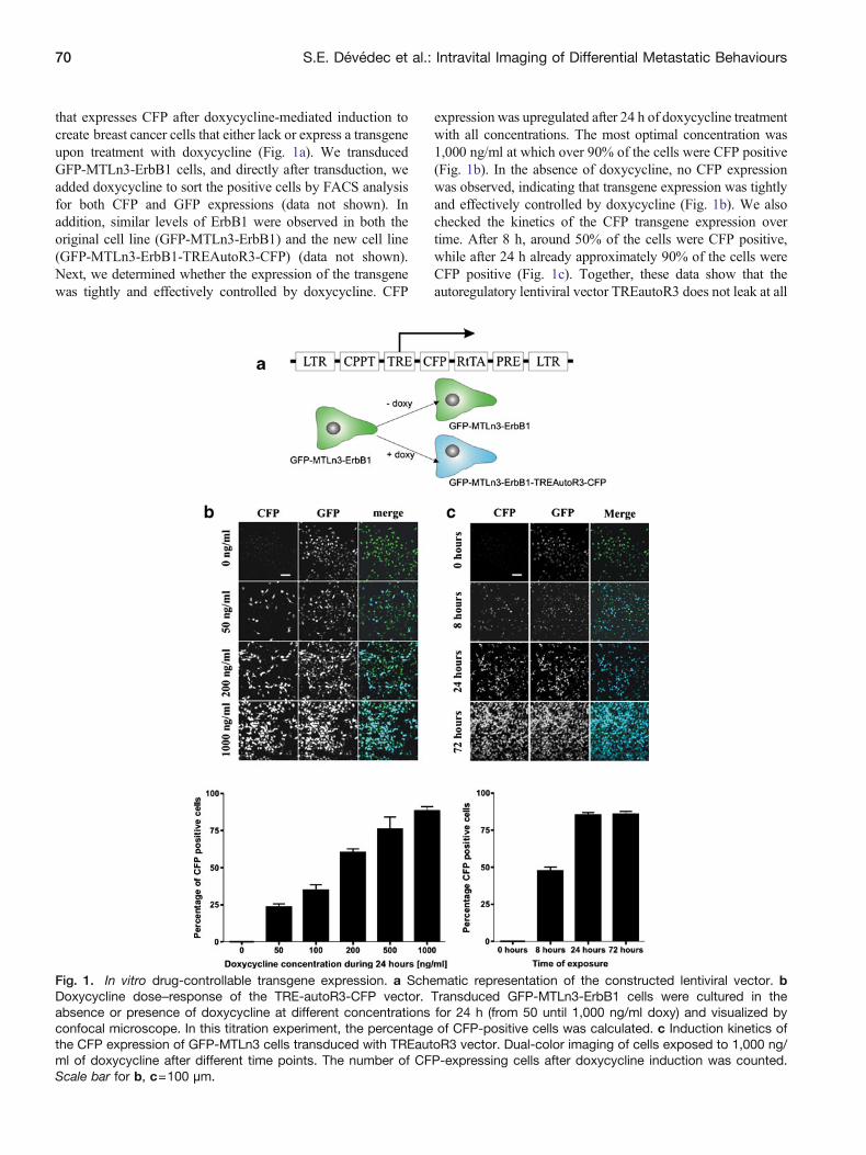
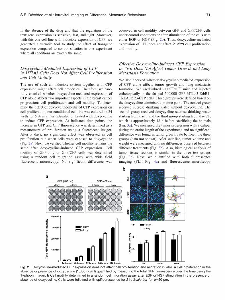
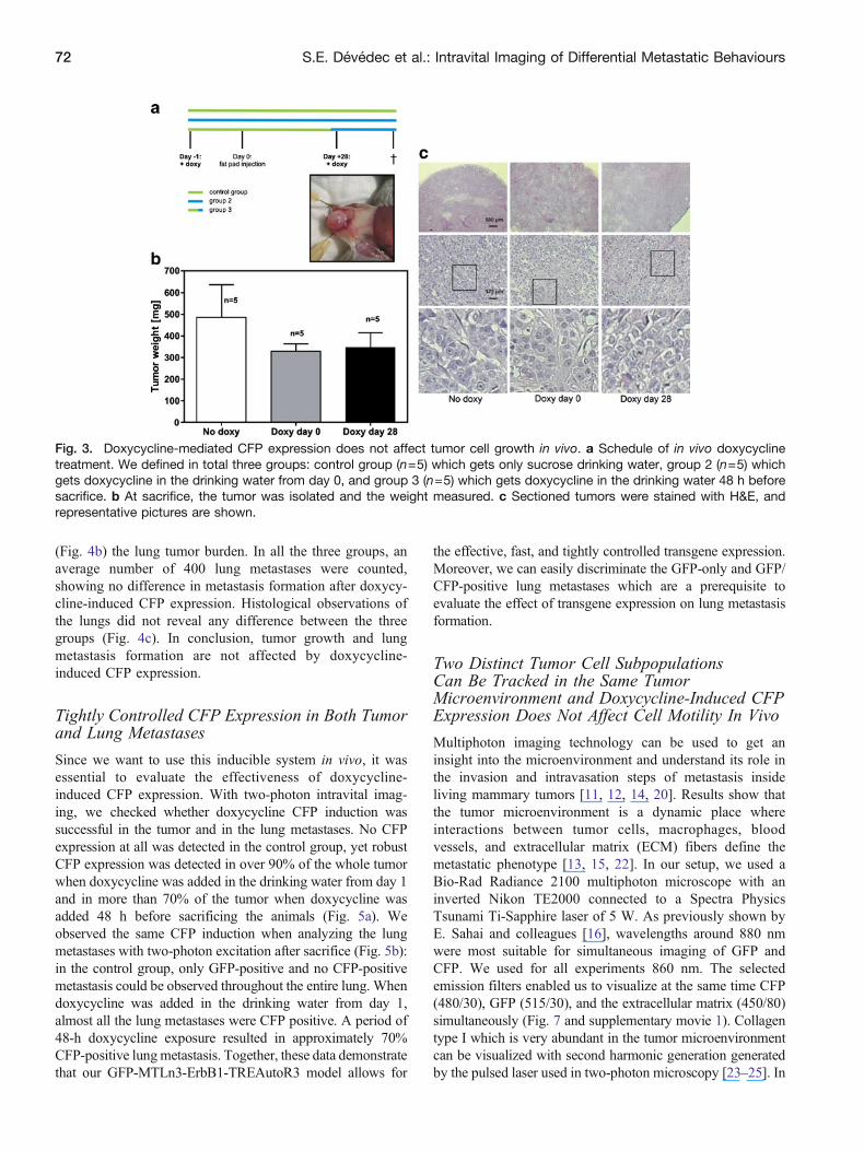
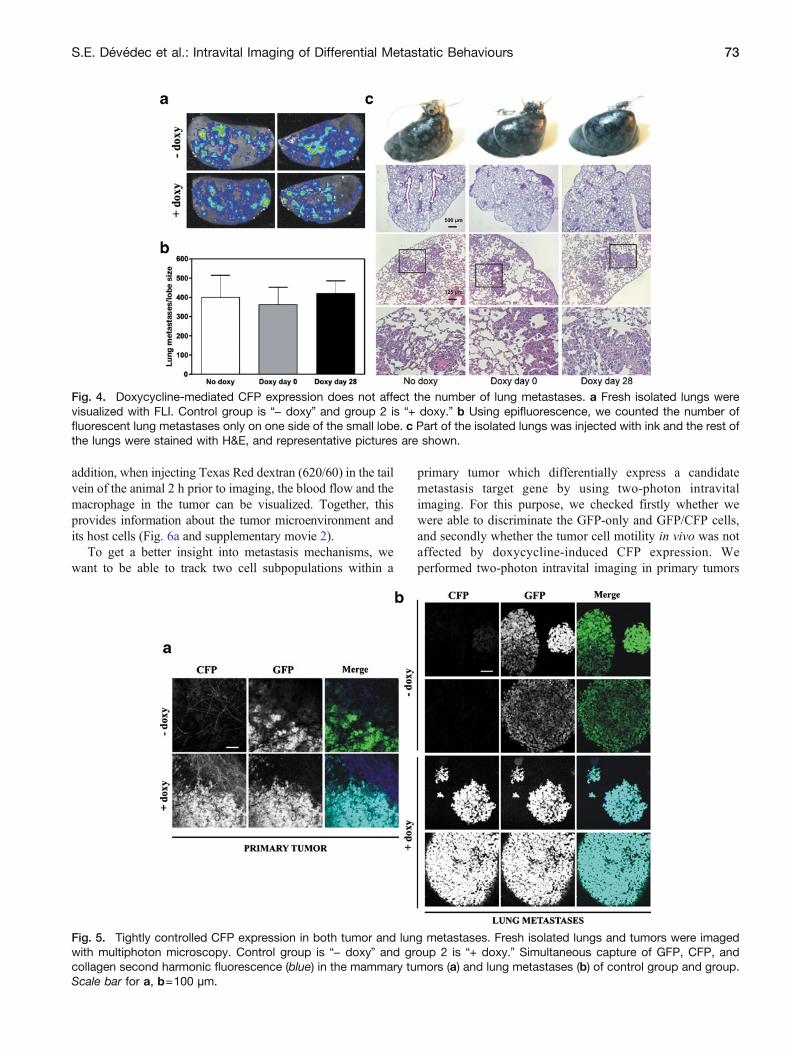
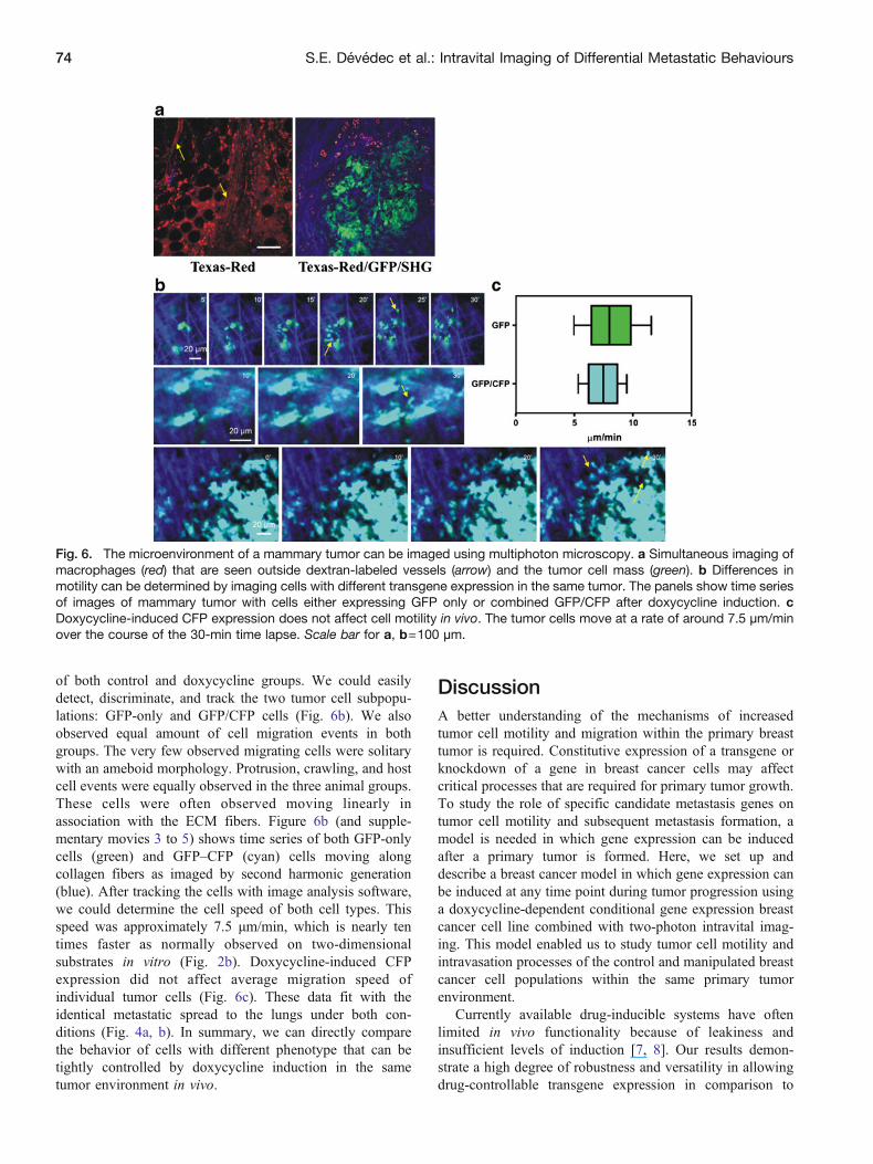
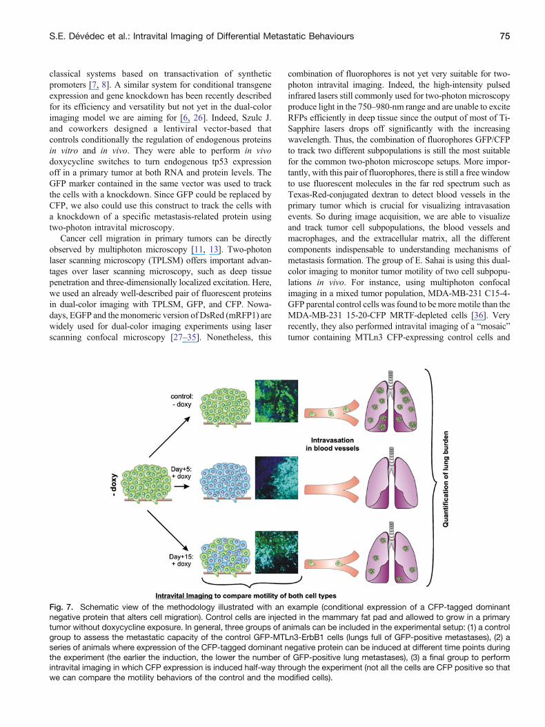
Related Documents