Indicate the patient's last name. Hyphenated names should be recorded with a hyphen. Coding Instructions: Seq. #: 2000 Name: Last Name The value on arrival at this facility Target Value: (none) Selections: (none) Supporting Definitions: Indicate the patient's first name. Coding Instructions: Seq. #: 2010 Name: First Name The value on arrival at this facility Target Value: (none) Selections: (none) Supporting Definitions: Indicate the patient's middle name. Coding Instructions: It is acceptable to specify the patient's middle initial. If the patient does not have a middle name, leave field blank. If the patient has multiple middle names, enter all of the middle names sequentially. Note(s): Seq. #: 2020 Name: Middle Name The value on arrival at this facility Target Value: (none) Selections: (none) Supporting Definitions: Indicate the patient's United States Social Security Number (SSN). Coding Instructions: If the patient does not have a US Social Security Number (SSN), leave blank and check "SSN N/A". Note(s): Seq. #: 2030 Name: SSN The value on arrival at this facility Target Value: (none) Selections: (none) Supporting Definitions: © 2011, American College of Cardiology Foundation 7/1/2014 Page 1 of 161 Effective for Patient Discharges July 01, 2014 Coder's Data Dictionary TVT Registry™ v2.0 A. Demographics

Welcome message from author
This document is posted to help you gain knowledge. Please leave a comment to let me know what you think about it! Share it to your friends and learn new things together.
Transcript

Indicate the patient's last name. Hyphenated names should be recorded with a hyphen.Coding Instructions:
Seq. #: 2000 Name: Last Name
The value on arrival at this facilityTarget Value:
(none)Selections:
(none)Supporting Definitions:
Indicate the patient's first name.Coding Instructions:
Seq. #: 2010 Name: First Name
The value on arrival at this facilityTarget Value:
(none)Selections:
(none)Supporting Definitions:
Indicate the patient's middle name.Coding Instructions:
It is acceptable to specify the patient's middle initial.
If the patient does not have a middle name, leave field blank.
If the patient has multiple middle names, enter all of the middle names sequentially.
Note(s):
Seq. #: 2020 Name: Middle Name
The value on arrival at this facilityTarget Value:
(none)Selections:
(none)Supporting Definitions:
Indicate the patient's United States Social Security Number (SSN).Coding Instructions:
If the patient does not have a US Social Security Number (SSN), leave blank and check "SSN N/A".
Note(s):
Seq. #: 2030 Name: SSN
The value on arrival at this facilityTarget Value:
(none)Selections:
(none)Supporting Definitions:
© 2011, American College of Cardiology Foundation 7/1/2014 Page 1 of 161
Effective for Patient Discharges July 01, 2014
Coder's Data DictionaryTVT Registry™ v2.0
A. Demographics

Indicate if the patient does not have a United States Social Security Number (SSN).Coding Instructions:
Seq. #: 2031 Name: SSN N/A
The value on arrival at this facilityTarget Value:
Selection Text Definition
No
Yes Patient does 'not' have a SSN.
Selections:
(none)Supporting Definitions:
Indicate the number created and automatically inserted by the software that uniquely identifies this patient.Coding Instructions:
Seq. #: 2040 Name: Patient ID
The value on arrival at this facilityTarget Value:
(none)Selections:
(none)Supporting Definitions:
Indicate optional patient identifier, such as medical record number, that can be associated with the patient.Coding Instructions:
Seq. #: 2045 Name: Other ID
N/ATarget Value:
(none)Selections:
(none)Supporting Definitions:
Indicate the patient's date of birth.Coding Instructions:
Seq. #: 2050 Name: Birth Date
The value on arrival at this facilityTarget Value:
(none)Selections:
(none)Supporting Definitions:
© 2011, American College of Cardiology Foundation 7/1/2014 Page 2 of 161
Effective for Patient Discharges July 01, 2014
Coder's Data DictionaryTVT Registry™ v2.0
A. Demographics

Indicate the patient's sex at birth.Coding Instructions:
Seq. #: 2060 Name: Sex
The value on arrival at this facilityTarget Value:
Selection Text Definition
Male
Female
Selections:
(none)Supporting Definitions:
Indicate if the patient is White as determined by the patient/family.Coding Instructions:
If the patient has multiple race origins, specify them using the other race selections in addition to this one.
Note(s):
Seq. #: 2070 Name: Race - White
The value on arrival at this facilityTarget Value:
Selection Text Definition
No
Yes
Selections:
White (race):
Having origins in any of the original peoples of Europe, the Middle East, or North Africa.
Source: U.S. Office of Management and Budget. Classification of Federal Data on Race and Ethnicity
Supporting Definitions:
Indicate if the patient is Black/African American as determined by the patient/family.Coding Instructions:
If the patient has multiple race origins, specify them using the other race selections in addition to this one.
Note(s):
Seq. #: 2071 Name: Race - Black or African American
The value on arrival at this facilityTarget Value:
Selection Text Definition
No
Yes
Selections:
Black/African American (race):
Having origins in any of the black racial groups of Africa. Terms such as "Haitian" or "Negro" can be used in addition to "Black or African American."
Source: U.S. Office of Management and Budget. Classification of Federal Data on Race and Ethnicity
Supporting Definitions:
© 2011, American College of Cardiology Foundation 7/1/2014 Page 3 of 161
Effective for Patient Discharges July 01, 2014
Coder's Data DictionaryTVT Registry™ v2.0
A. Demographics

Indicate if the patient is Asian as determined by the patient/family.Coding Instructions:
If the patient has multiple race origins, specify them using the other race selections in addition to this one.
Note(s):
Seq. #: 2072 Name: Race - Asian
The value on arrival at this facilityTarget Value:
Selection Text Definition
No
Yes
Selections:
Asian (race):
Having origins in any of the original peoples of the Far East, Southeast Asia, or the Indian subcontinent including, for example, Cambodia, China, India, Japan, Korea, Malaysia, Pakistan, the Philippine Islands, Thailand, and Vietnam.
Source: U.S. Office of Management and Budget. Classification of Federal Data on Race and Ethnicity
Supporting Definitions:
Indicate if the patient is American Indian or Alaskan Native as determined by the patient/family.Coding Instructions:
If the patient has multiple race origins, specify them using the other race selections in addition to this one.
Note(s):
Seq. #: 2073 Name: Race - American Indian or Alaskan Native
The value on arrival at this facilityTarget Value:
Selection Text Definition
No
Yes
Selections:
American Indian or Alaskan Native (race):
Having origins in any of the original peoples of North and South America (including Central America), and who maintains tribal affiliation or community attachment.
Source: U.S. Office of Management and Budget. Classification of Federal Data on Race and Ethnicity
Supporting Definitions:
Indicate if the patient is Native Hawaiian/Other Pacific Islander as determined by the patient/family.Coding Instructions:
If the patient has multiple race origins, specify them using the other race selections in addition to this one.
Note(s):
Seq. #: 2074 Name: Race - Native Hawaiian or Pacific Islander
The value on arrival at this facilityTarget Value:
Selection Text Definition
No
Yes
Selections:
Native Hawaiian or Pacific Islander (race):
Having origins in any of the original peoples of Hawaii, Guam, Samoa, or other Pacific Islands.
Source: U.S. Office of Management and Budget. Classification of Federal Data on Race and Ethnicity
Supporting Definitions:
© 2011, American College of Cardiology Foundation 7/1/2014 Page 4 of 161
Effective for Patient Discharges July 01, 2014
Coder's Data DictionaryTVT Registry™ v2.0
A. Demographics

Indicate if the patient is of Hispanic or Latino ethnicity as determined by the patient/family.Coding Instructions:
Seq. #: 2076 Name: Hispanic or Latino Ethnicity
The value on arrival at this facilityTarget Value:
Selection Text Definition
No
Yes
Selections:
Hispanic or Latino Ethnicity:
A person of Cuban, Mexican, Puerto Rican, Cuban, South or Central American, or other Spanish culture or origin, regardless of race. The term, "Spanish origin," can be used in addition to "Hispanic or Latino."
Source: U.S. Office of Management and Budget. Classification of Federal Data on Race and Ethnicity
Supporting Definitions:
Reserved for future use.Coding Instructions:
Seq. #: 2500 Name: Auxiliary 1
N/ATarget Value:
(none)Selections:
(none)Supporting Definitions:
Reserved for future use.Coding Instructions:
Seq. #: 2501 Name: Auxiliary 2
N/ATarget Value:
(none)Selections:
(none)Supporting Definitions:
© 2011, American College of Cardiology Foundation 7/1/2014 Page 5 of 161
Effective for Patient Discharges July 01, 2014
Coder's Data DictionaryTVT Registry™ v2.0
A. Demographics

Indicate the date the patient arrived at your facility.Coding Instructions:
Seq. #: 3000 Name: Arrival Date
N/ATarget Value:
(none)Selections:
(none)Supporting Definitions:
Indicate the time the patient arrived at your facilityCoding Instructions:
Indicate the time (hours:minutes) using the military 24-hour clock, beginning at midnight (00:00 hours).
If the patient came to your facility for an elective or outpatient procedure and the time was not documented, code the scheduled time of arrival.
Note(s):
Seq. #: 3001 Name: Arrival Time
N/ATarget Value:
(none)Selections:
(none)Supporting Definitions:
Indicate the primary residence of the patient prior to arrival. If the primary residence is not available, code not documented.Coding Instructions:
Seq. #: 3003 Name: Residence on Arrival
The value on arrival at this facilityTarget Value:
Selection Text Definition
Home with no health-aid
Home with health aid
Long term care
Other
Not Documented
Selections:
(none)Supporting Definitions:
© 2011, American College of Cardiology Foundation 7/1/2014 Page 6 of 161
Effective for Patient Discharges July 01, 2014
Coder's Data DictionaryTVT Registry™ v2.0
B. Episode of Care

Indicate if the patient's insurance payor(s) included private health insurance.Coding Instructions:
A health maintenance organization (HMO) is considered private health insurance.
Note(s):
Seq. #: 3005 Name: Insurance Payors - Private Health Insurance
The value on arrival at this facilityTarget Value:
Selection Text Definition
No
Yes
Selections:
Private Health Insurance:
Private health insurance is coverage by a health plan provided through an employer or union or purchased by an individual from a private health insurance company.
Source: U.S. Census Bureau
Supporting Definitions:
Indicate if the patient's insurance payor(s) included Medicare.Coding Instructions:
Seq. #: 3006 Name: Insurance Payors - Medicare
The value on arrival at this facilityTarget Value:
Selection Text Definition
No
Yes
Selections:
Medicare:
Medicare is the Federal program which helps pay health care costs for people 65 and older and for certain people under 65 with long-term disabilities.
Source: U.S. Census Bureau
Supporting Definitions:
Indicate if the patient's insurance payor(s) included Medicaid.Coding Instructions:
Seq. #: 3007 Name: Insurance Payors - Medicaid
The value on arrival at this facilityTarget Value:
Selection Text Definition
No
Yes
Selections:
Medicaid:
Medicaid is a program administered at the state level, which provides medical assistance to the needy. Families with dependent children, the aged, blind, and disabled who are in financial need are eligible for Medicaid. It may be known by different names.
Source: U.S. Census Bureau
Supporting Definitions:
© 2011, American College of Cardiology Foundation 7/1/2014 Page 7 of 161
Effective for Patient Discharges July 01, 2014
Coder's Data DictionaryTVT Registry™ v2.0
B. Episode of Care

Indicate if the patient's insurance payor(s) included Military Health Care.Coding Instructions:
Seq. #: 3008 Name: Insurance Payors - Military Health Care
The value on arrival at this facilityTarget Value:
Selection Text Definition
No
Yes
Selections:
Military Health Care:
Military Health care - Military health care includes TRICARE/CHAMPUS (Civilian Health and Medical Program of the Uniformed Services) and CHAMPVA (Civilian Health and Medical Program of the Department of Veterans Affairs), as well as care provided by the Department of Veterans Affairs (VA).
Source: U.S. Census Bureau
Supporting Definitions:
Indicate if the patient's insurance payor(s) included State-Specific Plan (non Medicaid).Coding Instructions:
Seq. #: 3009 Name: Insurance Payors - State-Specific Plan (Non Medicaid)
The value on arrival at this facilityTarget Value:
Selection Text Definition
No
Yes
Selections:
State Specific Plan:
Some states have their own health insurance programs for low-income uninsured individuals. These health plans may be known by different names in different states.
Source: U.S. Census Bureau
Supporting Definitions:
Indicate if the patient's insurance payor(s) included Indian Health Service (IHS).Coding Instructions:
Seq. #: 3010 Name: Insurance Payors - Indian Health Service
The value on arrival at this facilityTarget Value:
Selection Text Definition
No
Yes
Selections:
Indian Health Service:
Indian Health Service (IHS) is a health care program through which the Department of Health and Human Services provides medical assistance to eligible American Indians at IHS facilities. In addition, the IHS helps pay the cost of selected health care services provided at non-IHS facilities.
Source: U.S. Census Bureau
Supporting Definitions:
© 2011, American College of Cardiology Foundation 7/1/2014 Page 8 of 161
Effective for Patient Discharges July 01, 2014
Coder's Data DictionaryTVT Registry™ v2.0
B. Episode of Care

Indicate if the patient's insurance payor(s) included Non-US Insurance.Coding Instructions:
Seq. #: 3011 Name: Insurance Payors - Non-US Insurance
The value on arrival at this facilityTarget Value:
Selection Text Definition
No
Yes
Selections:
Non-US Insurance:
Non-US insurance refers to individuals with a payor that does not originate in the United States.
Source: U.S. Census Bureau
Supporting Definitions:
Indicate if the patient has no insurance payor(s).Coding Instructions:
Seq. #: 3012 Name: Insurance Payors - None
The value on arrival at this facilityTarget Value:
Selection Text Definition
No
Yes The patient has no insurance.
Selections:
None:
'None' refers to individuals with no or limited health insurance. Thus, the individual is the payor regardless of ability to pay.
Source: NCDR
Supporting Definitions:
Indicate the patient's Health Insurance Claim (HIC) number.Coding Instructions:
Seq. #: 3015 Name: Health Insurance Claim Number
The value on arrival at this facilityTarget Value:
(none)Selections:
Health Insurance Claim Number::
The Health Insurance Claim (HIC) number is the unique identifier issued to all Medicare eligible beneficiaries by the Social Security Administration (SSA). The Railroad Retirement Board (RRB) can also assign HIC to those receiving RRB benefits.
Source: Center for Medicare and Medicaid Services
Supporting Definitions:
Reserved for future use.Coding Instructions:
Seq. #: 3020 Name: Auxiliary 3
N/ATarget Value:
(none)Selections:
(none)Supporting Definitions:
© 2011, American College of Cardiology Foundation 7/1/2014 Page 9 of 161
Effective for Patient Discharges July 01, 2014
Coder's Data DictionaryTVT Registry™ v2.0
B. Episode of Care

Reserved for future use.Coding Instructions:
Seq. #: 3025 Name: Auxiliary 4
N/ATarget Value:
(none)Selections:
(none)Supporting Definitions:
Indicate if the patient is enrolled in a research study for the index procedure or the episode of care.Coding Instructions:
Code 'Yes' only for those patients enrolled in an STS/ACC TVT Registry research study. If the patient is in other studies unrelated to the TVT Registry, leave blank or Code 'No'.
Note(s):
Seq. #: 3030 Name: Patient Enrolled in Research Study
N/ATarget Value:
Selection Text Definition
No
Yes
Selections:
(none)Supporting Definitions:
Indicate the research study name as provided by the research study protocol or STS/ACC TVT Registry staff.Coding Instructions:
Study names must follow the format indicated by the research protocol or STS/ACC TVT Registry staff. Deviations may result in an error message.
Note(s):
Seq. #: 3031 Name: Research Study Name
N/ATarget Value:
(none)Selections:
(none)Supporting Definitions:
Indicate the research study patient identification number as assigned by the research protocol or STS/ACC TVT Registry staff.Coding Instructions:
Seq. #: 3032 Name: Research Study Patient ID
N/ATarget Value:
(none)Selections:
(none)Supporting Definitions:
© 2011, American College of Cardiology Foundation 7/1/2014 Page 10 of 161
Effective for Patient Discharges July 01, 2014
Coder's Data DictionaryTVT Registry™ v2.0
B. Episode of Care

Indicate whether the patient has a history of infective endocarditis documented by one of the following:
1. Positive blood cultures2. Vegetation on echocardiography and/or other diagnostic modality3. Documented history of infective endocarditis
Coding Instructions:
Seq. #: 4000 Name: Infective Endocarditis
Any occurrence between birth and the procedureTarget Value:
Selection Text Definition
No
Yes
Selections:
(none)Supporting Definitions:
Indicate the type of endocarditis.Coding Instructions:
Seq. #: 4005 Name: Infective Endocarditis Type
The last value between birth and the procedureTarget Value:
Selection Text Definition
Treated The patient has been treated previously for endocarditis and is not taking antibiotics for the infection (other than prophylactic medications).
Active The patient is currently being treated for endocarditis.
Selections:
(none)Supporting Definitions:
Indicate if the patient has been admitted to the hospital for an inpatient admission with a diagnosis of heart failure within the past year.
Coding Instructions:
Seq. #: 4006 Name: Heart Failure Hospitalization Within Past Year
Any occurrence between one year prior to the procedure and the procedureTarget Value:
Selection Text Definition
No
Yes
Not Documented
Selections:
(none)Supporting Definitions:
© 2011, American College of Cardiology Foundation 7/1/2014 Page 11 of 161
Effective for Patient Discharges July 01, 2014
Coder's Data DictionaryTVT Registry™ v2.0
C. History & Risk Factors

Indicate if the patient currently has a permanent pacemaker or had a permanent pacemaker that was implanted at any time prior to arrival at this facility. This includes patients that had a permanent pacemaker previously, but the device is no longer in place.
Coding Instructions:
Seq. #: 4010 Name: Permanent Pacemaker
Any occurrence between birth and the procedureTarget Value:
Selection Text Definition
No
Yes
Selections:
(none)Supporting Definitions:
Indicate the date the pacemaker was implanted.Coding Instructions:
If the month or day is unknown, enter 01
Note(s):
Seq. #: 4012 Name: Most Recent Pacemaker Date
The last value between birth and the first procedureTarget Value:
(none)Selections:
(none)Supporting Definitions:
Indicate if the pacemaker type includes cardiac resynchronization therapy (CRT). A CRT is a biventricular pacemaker that sends electrical signals to both ventricles that resynchronizes the heart chambers and helps it pump more effectively. It may or may not have an atrial pacing wire.
Coding Instructions:
Seq. #: 4013 Name: Cardiac Resynchronization Therapy
Any occurrence between birth and the procedureTarget Value:
Selection Text Definition
No
Yes
Selections:
(none)Supporting Definitions:
Indicate if the patient had a previous implantable cardioverter defibrillator (ICD). This includes patients that had an ICD previously, but the device is no longer in place.
Coding Instructions:
Seq. #: 4015 Name: Previous ICD
Any occurrence between birth and the procedureTarget Value:
Selection Text Definition
No
Yes
Selections:
(none)Supporting Definitions:
© 2011, American College of Cardiology Foundation 7/1/2014 Page 12 of 161
Effective for Patient Discharges July 01, 2014
Coder's Data DictionaryTVT Registry™ v2.0
C. History & Risk Factors

Indicate if the ICD includes a cardiac resynchronization therapy device. A cardiac resynchronization therapy defibrillator (CRT-D) has dual capabilities. It is a biventricular pacemaker that sends electrical signals to both ventricles as well as a defibrillator. It may or may not have an atrial pacing wire.
Coding Instructions:
Seq. #: 4016 Name: Cardiac Resynchronization Therapy Defibrillator
Any occurrence between birth and the procedureTarget Value:
Selection Text Definition
No
Yes
Selections:
(none)Supporting Definitions:
Indicate if the patient had a previous percutaneous coronary intervention.Coding Instructions:
Seq. #: 4020 Name: Prior PCI
Any occurrence between birth and the procedureTarget Value:
Selection Text Definition
No
Yes
Selections:
Percutaneous Coronary Intervention:
Percutaneous coronary intervention (PCI) is the placement of an angioplasty guide wire, balloon, or other device (e.g. stent, atherectomy, brachytherapy, or thrombectomy catheter) into a native coronary artery or coronary artery bypass graft for the purpose of mechanical coronary revascularization.
Source: NCDR
Supporting Definitions:
Indicate the date of the most recent PCI.Coding Instructions:
If the month or day are unknown, enter 01.
Note(s):
Seq. #: 4025 Name: Most Recent PCI Date
The last value between birth and the procedureTarget Value:
(none)Selections:
(none)Supporting Definitions:
Indicate if the patient had a previous coronary artery bypass graft (CABG) surgery.Coding Instructions:
Seq. #: 4030 Name: Prior CABG
Any occurrence between birth and the procedureTarget Value:
Selection Text Definition
No
Yes
Selections:
(none)Supporting Definitions:
© 2011, American College of Cardiology Foundation 7/1/2014 Page 13 of 161
Effective for Patient Discharges July 01, 2014
Coder's Data DictionaryTVT Registry™ v2.0
C. History & Risk Factors

Indicate the date of the most recent coronay artery bypass graft (CABG).Coding Instructions:
If month or day are unknown, enter 01.
Note(s):
Seq. #: 4035 Name: Most Recent CABG Date
The last value between birth and the procedureTarget Value:
(none)Selections:
(none)Supporting Definitions:
Indicate if the patient had prior other cardiac surgery. Other cardiac surgery includes surgeries not otherwise specified in the cardiac history. It includes, but is not limited to:
1. Previous congenital heart surgery and/or percutaneous procedure (e.g. VSD, ASD, TOF and PFO repair).
2. Previous surgery on the thoracic aorta.
3. Previous intrapericardial or great vessel (e.g., aorta, superior vena cava, inferior vena cava, pulmonary arteries and veins) procedure performed. This may include, but is not limited to LVA, acquired VSD, SVR, TMR, cardiac trauma, pericardial window, pericardiectomy, cardiac tumor, myectomy or heart transplant.
Coding Instructions:
Do not include aortic or non-aortic valve procedures. See Seq Num 4095, Prior Other Non-Aortic Valve Procedure and Seq Num 4060, Prior Aortic Valve Procedure.
Note(s):
Seq. #: 4040 Name: Prior Other Cardiac Surgery
Any occurrence between birth and the procedureTarget Value:
Selection Text Definition
No
Yes
Selections:
(none)Supporting Definitions:
© 2011, American College of Cardiology Foundation 7/1/2014 Page 14 of 161
Effective for Patient Discharges July 01, 2014
Coder's Data DictionaryTVT Registry™ v2.0
C. History & Risk Factors

Indicate the number of open heart cardiac surgeries the patient has had prior to this procedure. This includes open heart coronary artery bypass, or valve replacement/repairs.
Coding Instructions:
Do not include other open chest surgical procedures (not accessing the heart, such as surgery on the thoracic aorta or lung) or other cardiac interventional procedures (such as a PCI, or balloon valvuloplasty).
Note(s):
Seq. #: 4055 Name: Number of Previous Cardiac Surgeries
The total between birth and the procedureTarget Value:
Selection Text Definition
0
1
2
3
>=4
Selections:
(none)Supporting Definitions:
Indicate whether the patient had a previous surgical or interventional replacement and/or repair of the aortic valve.Coding Instructions:
Seq. #: 4060 Name: Prior Aortic Valve Procedure
Any occurrence between birth and the procedureTarget Value:
Selection Text Definition
No
Yes
Selections:
(none)Supporting Definitions:
Indicate the date of the most recent prior aortic valve procedure.Coding Instructions:
If month or day are unknown, enter 01.
Note(s):
Seq. #: 4065 Name: Most Recent Aortic Valve Procedure Date
The last value between birth and the procedureTarget Value:
(none)Selections:
(none)Supporting Definitions:
© 2011, American College of Cardiology Foundation 7/1/2014 Page 15 of 161
Effective for Patient Discharges July 01, 2014
Coder's Data DictionaryTVT Registry™ v2.0
C. History & Risk Factors

Indicate whether a previous procedure included a surgical aortic valve replacement.Coding Instructions:
Seq. #: 4070 Name: Previous Aortic Valve Replacement - Surgical
Any occurrence between birth and the procedureTarget Value:
Selection Text Definition
No
Yes
Selections:
(none)Supporting Definitions:
Indicate the type of aortic valve replacement.Coding Instructions:
Seq. #: 4075 Name: Previous Aortic Valve Procedure Type
The last value between birth and the procedureTarget Value:
Selection Text Definition
Bioprosthetic stented
Bioprosthetic stentless
Not Documented
Selections:
(none)Supporting Definitions:
Indicate the model ID of the prosthetic aortic valve.Coding Instructions:
Seq. #: 4078 Name: Aortic Valve Model ID
The last value between birth and the procedureTarget Value:
(none)Selections:
(none)Supporting Definitions:
Indicate whether a previous procedure included a surgical aortic valve repair.Coding Instructions:
Seq. #: 4080 Name: Previous Aortic Valve Repair - Surgical
Any occurrence between birth and the procedureTarget Value:
Selection Text Definition
No
Yes
Selections:
(none)Supporting Definitions:
© 2011, American College of Cardiology Foundation 7/1/2014 Page 16 of 161
Effective for Patient Discharges July 01, 2014
Coder's Data DictionaryTVT Registry™ v2.0
C. History & Risk Factors

Indicate whether a previous procedure included an aortic balloon valvuloplasty.Coding Instructions:
Seq. #: 4085 Name: Previous Procedure - Aortic Valve Balloon Valvuloplasty
Any occurrence between birth and the procedureTarget Value:
Selection Text Definition
No
Yes
Selections:
(none)Supporting Definitions:
Indicate whether a previous procedure included a transcatheter aortic valve replacement procedure.Coding Instructions:
Seq. #: 4090 Name: Previous Procedure - Aortic Valve Transcatheter Valve Replacement
Any occurrence between birth and the procedureTarget Value:
Selection Text Definition
No
Yes
Selections:
(none)Supporting Definitions:
Indicate whether a previous procedure included a transcatheter aortic valve intervention (such as a procedure that deploys an occluder or plug for aortic regurgitation).
Coding Instructions:
This does not include surgical aortic valve repair/replacements, transcatheter AV replacments or AV balloon valvuloplasties.
Note(s):
Seq. #: 4091 Name: Previous Procedure - Aortic Valve Transcatheter Valve Intervention
Any occurrence between birth and the procedureTarget Value:
Selection Text Definition
No
Yes
Selections:
(none)Supporting Definitions:
Indicate the model ID implanted in the transcatheter aortic valve replacement procedure.Coding Instructions:
Seq. #: 4092 Name: Previous Procedure - AV Transcatheter Valve Model ID
Any occurrence between birth and the procedureTarget Value:
(none)Selections:
(none)Supporting Definitions:
© 2011, American College of Cardiology Foundation 7/1/2014 Page 17 of 161
Effective for Patient Discharges July 01, 2014
Coder's Data DictionaryTVT Registry™ v2.0
C. History & Risk Factors

Indicate whether the patient had a previous surgical or interventional replacement and/or repair of a valve (excluding the aortic valve).
Coding Instructions:
Seq. #: 4095 Name: Prior Non-Aortic Valve Procedure
Any occurrence between birth and the procedureTarget Value:
Selection Text Definition
No
Yes
Selections:
(none)Supporting Definitions:
Indicate the date of the most recent prior mitral valve procedure, if performed.Coding Instructions:
Seq. #: 4097 Name: Prior Mitral Valve Procedure Date
Any occurrence between birth and the procedureTarget Value:
(none)Selections:
(none)Supporting Definitions:
Indicate whether a previous procedure included a surgical mitral valve replacement.Coding Instructions:
Seq. #: 4100 Name: Previous Procedure - Mitral Valve Replacement - Surgical
Any occurrence between birth and the procedureTarget Value:
Selection Text Definition
No
Yes
Selections:
(none)Supporting Definitions:
Indicate the type of mitral valve replacement.Coding Instructions:
Seq. #: 4105 Name: Prior Non-Aortic Valve Procedure - Mitral Valve Type
The last value between birth and the procedureTarget Value:
Selection Text Definition
Mechanical
Bioprosthetic Retired in v2.0
Bioprosthetic stented
Bioprosthetic stentless
Not Documented
Selections:
(none)Supporting Definitions:
© 2011, American College of Cardiology Foundation 7/1/2014 Page 18 of 161
Effective for Patient Discharges July 01, 2014
Coder's Data DictionaryTVT Registry™ v2.0
C. History & Risk Factors

Indicate the model ID of the prosthetic mitral valve.Coding Instructions:
Seq. #: 4106 Name: Previous Procedure - Mitral Valve Replacement Model ID
The last value between birth and the procedureTarget Value:
(none)Selections:
(none)Supporting Definitions:
Indicate whether a previous procedure included a surgical mitral valve repair.Coding Instructions:
Seq. #: 4110 Name: Previous Procedure - Mitral Valve Repair - Surgical
Any occurrence between birth and the procedureTarget Value:
Selection Text Definition
No
Yes
Selections:
(none)Supporting Definitions:
Indicate if the patient had a prior mitral annuloplasty ring implanted surgically.Coding Instructions:
Seq. #: 4111 Name: Prior Mitral Annuloplasty Ring - Surgical
Any occurrence between birth and the procedureTarget Value:
Selection Text Definition
No
Yes - partial
Yes - circumferential
Not documented
Selections:
(none)Supporting Definitions:
Indicate whether a previous procedure included a transcatheter mitral valve intervention.Coding Instructions:
Seq. #: 4112 Name: Prior Mitral Valve Transcatheter Intervention
Any occurrence between birth and the procedureTarget Value:
Selection Text Definition
No
Yes
Selections:
(none)Supporting Definitions:
© 2011, American College of Cardiology Foundation 7/1/2014 Page 19 of 161
Effective for Patient Discharges July 01, 2014
Coder's Data DictionaryTVT Registry™ v2.0
C. History & Risk Factors

Indicate the type of transcatheter mitral valve intervention.Coding Instructions:
Seq. #: 4113 Name: Prior Mitral Transcatheter Type
Any occurrence between birth and the procedureTarget Value:
Selection Text Definition
Leaflet clip
Direct annulopasty intervention
Coronary sinus based intervention
Valve-in-native valve
Valve-in-valve
Other
Selections:
(none)Supporting Definitions:
Indicate the model ID of the prosthetic mitral valve.Coding Instructions:
Seq. #: 4116 Name: Valve or Ring Model
The value between birth and the procedureTarget Value:
(none)Selections:
(none)Supporting Definitions:
Indicate if the patient had a prior tricuspid valve replacement or repair.Coding Instructions:
Seq. #: 4118 Name: Prior Tricuspid Valve Replacement/Repair
Any occurrence between birth and the procedureTarget Value:
Selection Text Definition
No
Yes
Selections:
(none)Supporting Definitions:
Indicate if the patient had a prior pulmonic valve replacement or repair.Coding Instructions:
Seq. #: 4119 Name: Prior Pulmonic Valve Replacement/Repair
Any occurrence between birth and the procedureTarget Value:
Selection Text Definition
No
Yes
Selections:
(none)Supporting Definitions:
© 2011, American College of Cardiology Foundation 7/1/2014 Page 20 of 161
Effective for Patient Discharges July 01, 2014
Coder's Data DictionaryTVT Registry™ v2.0
C. History & Risk Factors

Indicate if the patient has a history of a stroke.Coding Instructions:
Seq. #: 4120 Name: Prior Stroke
Any occurrence between birth and the procedureTarget Value:
Selection Text Definition
No
Yes
Selections:
Stroke:
A stroke is an acute episode of focal or global neurological dysfunction caused by brain, spinal cord, or retinal vascular injury as a result of hemorrhage or infarction.
Source: Standardized Definitions for Cardiovascular Endpoints in Clinical Trials (FDA Draft)
Supporting Definitions:
Indicate the date of the most recent stroke.Coding Instructions:
If the month or day is unknown, enter 01.
Note(s):
Seq. #: 4125 Name: Most Recent Stroke Date
The last value between birth and the procedureTarget Value:
(none)Selections:
(none)Supporting Definitions:
Indicate if the patient has a history of a transient ischemic attack.Coding Instructions:
Seq. #: 4130 Name: Transient Ischemic Attack
Any occurrence between birth and the procedureTarget Value:
Selection Text Definition
No
Yes
Selections:
Transient Ischemic Attack:
Transient ischemic attack (TIA) is defined as a transient episode of focal neurological dysfunction caused by brain, spinal cord, or retinal ischemia, without acute infarction.
Source: Standardized Definitions for Cardiovascular Endpoints in Clinical Trials (FDA Draft)
Supporting Definitions:
© 2011, American College of Cardiology Foundation 7/1/2014 Page 21 of 161
Effective for Patient Discharges July 01, 2014
Coder's Data DictionaryTVT Registry™ v2.0
C. History & Risk Factors

Indicate which carotid artery was determined from any diagnostic test to be greater or equal to 50% stenotic.Coding Instructions:
Seq. #: 4135 Name: Carotid Stenosis Assessment
The highest value between birth and the procedureTarget Value:
Selection Text Definition
None Neither carotid artery is >=50% stenotic.
Right There is >=50% stenosis in the right carotid artery.
Left There is >=50% stenosis in the left carotid artery.
Both There is >=50% stenosis in both the right and left carotid artery.
Not assessed Carotid stenosis was not assessed and is not known.
Selections:
(none)Supporting Definitions:
Indicate whether the patient has a history of previous carotid artery surgery and/or stenting.Coding Instructions:
Seq. #: 4140 Name: Prior Carotid Artery Surgery or Stent
Any occurrence between birth and the procedureTarget Value:
Selection Text Definition
No
Yes
Selections:
(none)Supporting Definitions:
Indicate the best estimate of the most severe percent stenosis in the right carotid artery.Coding Instructions:
Seq. #: 4141 Name: Severity of Stenosis - Right Carotid Artery
The highest value between birth and the procedureTarget Value:
Selection Text Definition
50%-79%
80% to 99%
100 %
Stenosis % not available
Selections:
(none)Supporting Definitions:
© 2011, American College of Cardiology Foundation 7/1/2014 Page 22 of 161
Effective for Patient Discharges July 01, 2014
Coder's Data DictionaryTVT Registry™ v2.0
C. History & Risk Factors

Indicate the best estimate of the most severe percent stenosis in the left carotid artery.Coding Instructions:
Seq. #: 4142 Name: Severity of Stenosis - Left Carotid Artery
The highest value between birth and the procedureTarget Value:
Selection Text Definition
50%-79%
80% to 99%
100%
Stenosis % not available
Selections:
(none)Supporting Definitions:
Indicate if the patient has a history of peripheral arterial disease (PAD) (includes upper and lower extremity, renal, mesenteric, and abdominal aortic systems).
Coding Instructions:
Seq. #: 4145 Name: Peripheral Arterial Disease
Any occurrence between birth and the procedureTarget Value:
Selection Text Definition
No
Yes
Selections:
Peripheral Arterial Disease:
Peripheral arterial disease can include:1. Claudication, either with exertion or at rest.2. Amputation for arterial vascular insufficiency.3. Vascular reconstruction, bypass surgery, or percutaneous intervention to the extremities (excluding dialysis fistulas and vein stripping).4. Documented aortic aneurysm with or without repair.5. Positive non-invasive test (e.g., ankle brachial index <=0.9); ultrasound, magnetic resonance, computed tomography, or angiographic imaging of > 50% diameter stenosis in any peripheral artery (e.g., renal, subclavian, femoral, iliac).
For purposes of the Registry, peripheral arterial disease excludes disease in the carotid and cerebrovascular arteries.
Source: STS and NCDR
Supporting Definitions:
Indicate if the patient has smoked cigarettes anytime during the year prior.Coding Instructions:
Seq. #: 4150 Name: Current/Recent Smoker (w/in 1 year)
Any occurrence between 1 year prior to the procedure and the procedureTarget Value:
Selection Text Definition
No
Yes
Selections:
(none)Supporting Definitions:
© 2011, American College of Cardiology Foundation 7/1/2014 Page 23 of 161
Effective for Patient Discharges July 01, 2014
Coder's Data DictionaryTVT Registry™ v2.0
C. History & Risk Factors

Indicate whether the patient has a diagnosis of hypertension.Coding Instructions:
Seq. #: 4155 Name: Hypertension
Any occurrence between birth and the procedureTarget Value:
Selection Text Definition
No
Yes
Selections:
Hypertension:
Hypertension is defined by any one of the following:1. History of hypertension diagnosed and treated with medication, diet and/or exercise2. Prior documentation of blood pressure greater than 140 mm Hg systolic and/or 90 mm Hg diastolic for patients without diabetes or chronic kidney disease, or prior documentation of blood pressure greater than 130 mm Hg systolic and/or 80 mm Hg diastolic on at least two occasions for patients with diabetes or chronic kidney disease3. Currently on pharmacologic therapy for treatment of hypertension.
Source: ACCF/AHA 2011 Key Data Elements and Definitions of a Base Cardiovascular Vocabulary for Electronic Health Records (JACC 2011;58;202-222)
Supporting Definitions:
Indicate if the patient has a history of diabetes mellitus regardless of duration of disease or need for antidiabetic agents.Coding Instructions:
Seq. #: 4165 Name: Diabetes Mellitus
Any occurrence between birth and the procedureTarget Value:
Selection Text Definition
No
Yes
Selections:
Diabetes:
The American Diabetes Association criteria include documentation of the following:1. A1c >=6.5%; or2. Fasting plasma glucose >=126 mg/dl (7.0 mmol/l); or3. Two-hour plasma glucose >=200 mg/dl (11.1 mmol/l) during an oral glucose tolerance test; or4. In a patient with classic symptoms of hyperglycemia or hyperglycemic crisis, a random plasma glucose >=200 mg/dl (11.1 mmol/l)
This does not include gestational diabetes.
Source: American Diabetes Association
Supporting Definitions:
© 2011, American College of Cardiology Foundation 7/1/2014 Page 24 of 161
Effective for Patient Discharges July 01, 2014
Coder's Data DictionaryTVT Registry™ v2.0
C. History & Risk Factors

Indicate the most aggressive diabetes control therapy.Coding Instructions:
Patients placed on a pre-procedure diabetic pathway of insulin drip after arrival but were not not insulin therapy (treated by diet or oral method) are not coded as insulin treatment.
If a patient had a pancreatic transplant, code "other", since the insulin from the new pancreas is not exogenous insulin.
Do not include "non-insulin" injectables that may improve blood sugar (such as Byetta) as insulin treatment.
Note(s):
Seq. #: 4170 Name: Diabetes Therapy
The last value between birth and prior to the procedureTarget Value:
Selection Text Definition
None No treatment for diabetes.
Diet Diet treatment only.
Oral Oral agent treatment (includes oral agent with/without diet treatment).
Insulin Insulin treatment (includes any combination with insulin).
Other Other adjunctive therapy
Selections:
(none)Supporting Definitions:
Indicate if the patient is currently undergoing either hemodialysis or peritoneal dialysis on an ongoing basis as a result of renal failure.
Coding Instructions:
If the patient is receiving continuous veno-venous hemofiltration (CVVH) as a result of renal failure (and not as treatment to remove fluid for heart failure), code "yes".
Note(s):
Seq. #: 4175 Name: Currently on Dialysis
The value on the procedureTarget Value:
Selection Text Definition
No
Yes
Selections:
(none)Supporting Definitions:
© 2011, American College of Cardiology Foundation 7/1/2014 Page 25 of 161
Effective for Patient Discharges July 01, 2014
Coder's Data DictionaryTVT Registry™ v2.0
C. History & Risk Factors

Indicate if the patient has a history of chronic lung disease, and severity, if present.Coding Instructions:
Seq. #: 4180 Name: Chronic Lung Disease
The value on the procedureTarget Value:
Selection Text Definition
None No documented chronic lung disease.
Mild FEV1 60% to 75% of predicted, and/or on chronic inhaled or oral bronchodilator therapy.
Moderate FEV1 50% to 59% of predicted, and/or on chronic steroid therapy aimed at lung disease.
Severe FEV1 <50% predicted, and/or Room Air pO2 < 60 or Room Air pCO2 > 50.
Selections:
Chronic Lung Disease:
Chronic lung disease can include patients with chronic obstructive pulmonary disease, chronic bronchitis, or emphysema. It can also include a patient who is currently being chronically treated with inhaled or oral pharmacological therapy (e.g., beta-adrenergic agonist, anti-inflammatory agent, leukotriene receptor antagonist, or steroid). Patients with asthma or seasonal allergies are not considered to have chronic lung disease.
Source: NCDR
Supporting Definitions:
Indicate whether patient uses supplemental oxygen at home.Coding Instructions:
Seq. #: 4181 Name: Home Oxygen
The value on arrival at this facilityTarget Value:
Selection Text Definition
No
Yes
Selections:
(none)Supporting Definitions:
© 2011, American College of Cardiology Foundation 7/1/2014 Page 26 of 161
Effective for Patient Discharges July 01, 2014
Coder's Data DictionaryTVT Registry™ v2.0
C. History & Risk Factors

Indicate if the patient has a medical condition that precludes an open chest procedure and that is documented in the medical record. This can include any of the following or other reasons that make redo operation through sternotomy or right anterior thoracotomy prohibitively hazardous:
1. Evidence of abnormal chest wall anatomy due to severe kyphoscoliosis or other skeletal abnormalities (including thoracoplasty, Potts’ disease, sternal bone destruction, evidence of indetectable plane between posterior sternal table and important mediastinal structures )
2. Complications from prior surgery
3. Prior radiation involving the mediastinum/thoracic, or evidence of severe radiation damage (e.g., skin burns, bone destruction, muscle loss, lung fibrosis or esophageal stricture)
4. History of multiple recurrent pleural effusions causing internal adhesions.
5. Chronic, ongoing open skin defects or extremely severe soft tissue atrophy.
6. Complete absence of reconstructive options based on plastic surgeon consult.
Coding Instructions:
Seq. #: 4182 Name: Hostile Chest
The value on the procedureTarget Value:
Selection Text Definition
No
Yes
Selections:
(none)Supporting Definitions:
Indicate whether immunocompromise is present due to immunosuppressive medication therapy within 30 days preceding the operative procedure or existing medical condition. This includes, but is not limited to systemic steroid therapy, inhaled steriod therapy, or preoperative protocol.
Coding Instructions:
Seq. #: 4185 Name: Immunocompromise Present
The value between 30 days prior to the procedure and the procedureTarget Value:
Selection Text Definition
No
Yes
Selections:
(none)Supporting Definitions:
Indicate whether the patient received the medication at home prior to this hospitalization.Coding Instructions:
Seq. #: 4200 Name: Prior Medications ID at Home
The value on arrival at this facilityTarget Value:
(none)Selections:
(none)Supporting Definitions:
© 2011, American College of Cardiology Foundation 7/1/2014 Page 27 of 161
Effective for Patient Discharges July 01, 2014
Coder's Data DictionaryTVT Registry™ v2.0
C. History & Risk Factors

Indicate whether the patient received the medication at home prior to this hospitalization.Coding Instructions:
Seq. #: 4205 Name: Prior Medications Administered at Home
The value on arrival at this facilityTarget Value:
Selection Text Definition
No
Yes
Selections:
(none)Supporting Definitions:
Specify the total daily dose of the medication the patient was taking routinely at home prior to this hospitalization.Coding Instructions:
Seq. #: 4210 Name: Prior Home Medication Dose
The value on arrivalTarget Value:
(none)Selections:
(none)Supporting Definitions:
© 2011, American College of Cardiology Foundation 7/1/2014 Page 28 of 161
Effective for Patient Discharges July 01, 2014
Coder's Data DictionaryTVT Registry™ v2.0
C. History & Risk Factors

Indicate the patient's coronary artery disease (CAD) presentation. Choose the worst status.Coding Instructions:
Seq. #: 5000 Name: CAD Presentation
The highest value between 7 days prior to the procedure and the procedureTarget Value:
© 2011, American College of Cardiology Foundation 7/1/2014 Page 29 of 161
Effective for Patient Discharges July 01, 2014
Coder's Data DictionaryTVT Registry™ v2.0
D. Pre-Procedure Status

Selection Text Definition
No Sxs, no angina No symptoms. No angina.
Sx unlikely to be ischemic
Pain, pressure or discomfort in the chest, neck or arms NOT clearly exertional or NOT otherwise consistent with pain or discomfort of myocardial ischemic origin. This includes patients with non-cardiac pain (e.g.pulmonary embolism, musculoskeletal, or esophageal discomfort), or cardiac pain not caused by myocardial ischemia (e.g., acute pericarditis).
Stable angina Angina without a change in frequency or pattern for the 6 weeks prior to this cath lab visit. Angina is controlled by rest and/or oral or transcutaneous medications.
Unstable angina One of three principal presentations of unstable angina: 1. Rest angina (occurring at rest and prolonged, usually >20 minutes); 2. New-onset angina (within the past 2 months, of at least Canadian Cardiovascular Society Class III severity); or 3. Increasing angina (previously diagnosed angina that has become distinctly more frequent, longer in duration, or increased by 1 or more Canadian Cardiovascular Society class to at least CCS III severity).
Non-STEMI The patient was hospitalized for a non-ST elevation myocardial infarction (STEMI) as documented in the medical record. Non-STEMIs are characterized by the presence of both criteria:a. Cardiac biomarkers (creatinine kinase-myocardial band, Troponin T or I) exceed the upper limit of normal according to the individual hospital's laboratory parameters with a clinical presentation which is consistent or suggestive of ischemia. ECG changes and/or ischemic symptoms may or may not be present.b. Absence of ECG changes diagnostic of a STEMI (see STEMI).
STEMI The patient presented with a ST elevation myocardial infarction (STEMI) or its equivalent as documented in the medical record. STEMIs are characterized by the presence of both criteria:a. ECG evidence of STEMI: New or presumed new ST-segment elevation or new left bundle branch block not documented to be resolved within 20 minutes. ST-segment elevation is defined by new or presumed new sustained ST-segment elevation at theJ-point in two contiguous electrocardiogram (ECG) leads with the cut-off points: >=0.2 mV in men or >= 0.15mV in women in leads V2-V3 and/or >= 0.1 mV in other leads and lasting greater than or equal to 20 minutes. If no exact ST-elevation measurement is recorded in the medical chart, physician's written documentation of ST-elevation or Qwaves is acceptable. If only one ECG is performed, then the assumption that the ST elevation persisted at least the required 20 minutes is acceptable. Left bundle branch block (LBBB) refers to new or presumed new LBBB on the initial ECG.b. Cardiac biomarkers (creatinine kinase-myocardial band, Troponin T or I) exceed the upper limit of normal according to the individual hospital's laboratory parameters a clinical presentation which is consistent or suggestive of ischemia.
Selections:
(none)Supporting Definitions:
© 2011, American College of Cardiology Foundation 7/1/2014 Page 30 of 161
Effective for Patient Discharges July 01, 2014
Coder's Data DictionaryTVT Registry™ v2.0

Indicate if the patient has had at least one documented previous myocardial infarction.Coding Instructions:
Seq. #: 5005 Name: Prior MI
Any occurrence between birth and the procedureTarget Value:
Selection Text Definition
No
Yes
Selections:
Myocardial Infarction:
A myocardial infarction is evidenced by any of the following:
1. A rise and fall of cardiac biomarkers (preferably troponin) with at least one of the values in the abnormal range for that laboratory [typically above the 99th percentile of the upper reference limit (URL) for normal subjects] together with at least one of the following manifestations of myocardial ischemia:a. Ischemic symptoms.b. ECG changes indicative of new ischemia (new ST-T changes, new left bundle branch block, or loss of R wave voltage).c. Development of pathological Q waves in 2 or more contiguous leads in the ECG (or equivalent findings for true posterior MI).d. Imaging evidence of new loss of viable myocardium or new regional wall motion abnormality.e. Documentation in the medical record of the diagnosis of acute myocardial infarction based on the cardiac biomarker pattern in the absence of any items enumerated in a-d due to conditions that may mask their appearance (e.g., peri-operative infarct when the patient cannot report ischemic symptoms; baseline left bundle branch block or ventricular pacing).
2. ECG changes associated with prior myocardial infarction can include the following (with or without prior symptoms):a. Any Q-wave in leads V2-V3 >=0.02 seconds or QS complex in leads V2 and V3.b. Q-wave >=0.03 seconds and >=0.1 mV deep or QS complex in leads I, II, aVL, aVF, or V4-V6 in any two leads of a contiguous lead grouping (I, aVL, V6; V4-V6; II, III, and aVF).c. R-wave >=0.04 seconds in V1-V2 and R/S >=1 with a concordant positive Twave in the absence of a conduction defect.
3. Imaging evidence of a region with new loss of viable myocardium at rest in the absence of a non-ischemic cause. This can be manifest as:a. Echocardiographic, CT, MR, ventriculographic or nuclear imaging evidence of left ventricular thinning or scarring and failure to contract appropriately (i.e., hypokinesis, akinesis, or dyskinesis).b. Fixed (non-reversible) perfusion defects on nuclear radioisotope imaging (e.g., MIBI, thallium).
4. Medical records documentation of prior myocardial infarction.
Source: Joint ESC-ACC-AHA-WHF 2007 Task Force consensus document"Universal Definition of Myocardial Infarction".
Supporting Definitions:
Indicate the timeframe of the myocardial infarction.Coding Instructions:
Seq. #: 5010 Name: Prior MI Timeframe
The last value between birth and the procedureTarget Value:
Selection Text Definition
< 30 Days
>= 30 Days
Selections:
(none)Supporting Definitions:
© 2011, American College of Cardiology Foundation 7/1/2014 Page 31 of 161
Effective for Patient Discharges July 01, 2014
Coder's Data DictionaryTVT Registry™ v2.0
D. Pre-Procedure Status

Indicate if the patient has a history of cardiomyopathy.Coding Instructions:
Seq. #: 5012 Name: Cardiomyopathy
Any occurrence between birth and the procedureTarget Value:
Selection Text Definition
No
Yes - Ischemic
Yes - Non-ischemic
Selections:
(none)Supporting Definitions:
Indicate if there is physician documentation or report that the patient has been in a state of heart failure within the past 2 weeks.Coding Instructions:
Seq. #: 5020 Name: Heart Failure w/in 2 Weeks
Any occurrence between 2 weeks prior to the procedure and the procedureTarget Value:
Selection Text Definition
No
Yes
Selections:
Heart Failure:
Heart failure is defined as physician documentation or a report of any of the following clinical symptoms of heart failure described as unusual dyspnea on light exertion, recurrent dyspnea occurring in the supine position, fluid retention, or the description of rales, jugular venous distention, pulmonary edema on physical examination, or pulmonary edema on chest x- ray presumed to be cardiac dysfunction.
A low ejection fraction alone, without clinical evidence of heart failure, does not qualify as heart failure.
Source: ACCF/AHA 2011 Key Data Elements and Definitions of a Base Cardiovascular Vocabulary for Electronic Health Records (JACC 2011;58;202-222)
Supporting Definitions:
© 2011, American College of Cardiology Foundation 7/1/2014 Page 32 of 161
Effective for Patient Discharges July 01, 2014
Coder's Data DictionaryTVT Registry™ v2.0
D. Pre-Procedure Status

Indicate the patient's functional class, coded as the New York Heart Association (NYHA) classification within the past 2 weeks.Coding Instructions:
Seq. #: 5025 Name: NYHA Class w/in 2 Weeks
The highest value between 2 weeks prior to the procedure and the procedureTarget Value:
Selection Text Definition
Class I Patient has cardiac disease but without resulting limitations of ordinary physical activity. Ordinary physical activity (e.g., walking several blocks or climbing stairs) does not cause undue fatigue, palpitation, dyspnea, or anginal pain. Limiting symptoms may occur with marked exertion.
Class II Patient has cardiac disease resulting in slight limitation of ordinary physical activity. Patient is comfortable at rest. Ordinary physical activity such as walking more than two blocks or climbing more than one flight of stairs results in limiting symptoms (e.g., fatigue, palpitation, dyspnea, or anginal pain).
Class III Patient has cardiac disease resulting in marked limitation of physical activity. Patient is comfortable at rest. Less than ordinary physical activity (e.g., walking one to two level blocks or climbing one flight of stairs) causes fatigue, palpitation, dyspnea, or anginal pain.
Class IV Patient has dyspnea at rest that increases with any physical activity. Patient has cardiac disease resulting in inability to perform any physical activity without discomfort. Symptoms may be present even at rest. If any physical activity is undertaken, discomfort is increased.
Selections:
(none)Supporting Definitions:
Indicate if the patient has been in a state of cardiogenic shock within 24 hrs of procedure.Coding Instructions:
Seq. #: 5030 Name: Cardiogenic Shock w/in 24 Hours
Any occurrence between 24 hours prior to the procedure and up to the procedureTarget Value:
Selection Text Definition
No
Yes
Selections:
Cardiogenic Shock:
Cardiogenic shock is a clinical state of end organ hypoperfusion due to cardiac failure according to the following criteria: persistant hypotension (Systolic BP < 80-90 or mean arterial pressure 30 mmHg lower than baseline) and severe reduction in Cardiac Index (< 1.8 without support or <2.2 with support).
Source: STS
Supporting Definitions:
© 2011, American College of Cardiology Foundation 7/1/2014 Page 33 of 161
Effective for Patient Discharges July 01, 2014
Coder's Data DictionaryTVT Registry™ v2.0
D. Pre-Procedure Status

Indicate if the patient has had an episode of cardiac arrest within 24 hours of the procedure.Coding Instructions:
Seq. #: 5035 Name: Cardiac Arrest w/in 24 Hours
Any occurrence between 24 hours prior to the procedure and the procedureTarget Value:
Selection Text Definition
No
Yes
Selections:
Cardiac Arrest:
"Sudden" cardiac arrest is the sudden cessation of cardiac activity so that the victim becomes unresponsive, with no normal breathing and no signs of circulation. If corrective measures are not taken rapidly, this condition progresses to sudden death. Cardiac arrest should be used to signify an event as described above that is reversed, usually by CPR, and/or defibrillation or cardioversion, or cardiac pacing. Sudden cardiac death should not be used to describe events that are not fatal.
Source: ACCF/AHA 2011 Key Data Elements and Definitions of a Base Cardiovascular Vocabulary for Electronic Health Records (JACC 2011;58;202-222)
Supporting Definitions:
Indicate if the patient has had an interventional, transcatheter or surgical cardiac procedure within 30 days prior to the procedure.Coding Instructions:
Seq. #: 5040 Name: Cardiac Procedure w/in 30 Days
Any occurrence between 30 days prior to the procedure and the procedureTarget Value:
Selection Text Definition
No
Yes
Selections:
(none)Supporting Definitions:
Indicate if the patient has a porcelain aorta as documented by findings on a chest x-ray, CT scan, fluoroscopy at the time of cardiac catheterization or noted during previous cardiothoracic surgery.
Coding Instructions:
Seq. #: 5045 Name: Porcelain Aorta
Any occurrence between birth and the procedureTarget Value:
Selection Text Definition
No
Yes
Selections:
Porcelain Aorta:
A porcelain aorta is defined as "severe atherosclerosis of the aorta, calcification may be severe and diffuse, causing an eggshell appearance seen on chest x-ray or CT".
Source: ACCF/AHA/AATS/ACR/ASA/SCA/SCAI/SIR/STS/SVM Guidelines for the Diagnosis and Management of Patients With Thoracic Aortic Disease (JACC, 2010; 55:27-129)
Supporting Definitions:
© 2011, American College of Cardiology Foundation 7/1/2014 Page 34 of 161
Effective for Patient Discharges July 01, 2014
Coder's Data DictionaryTVT Registry™ v2.0
D. Pre-Procedure Status

Indicate if the patient has a history of atrial fibrillation and/or atrial flutter documented in the medical record.Coding Instructions:
Seq. #: 5050 Name: Atrial Fibrillation/Flutter
Any occurrence between birth and the procedureTarget Value:
Selection Text Definition
No
Yes
Selections:
Atrial Fibrillation and Atrial Flutter:
Atrial Fibrillation is a supraventricular tachyarrhythmia characterized by uncoordinated atrial activity with consequent deterioration of atrial mechanical function. On the electrocardiogram (ECG), atrial fibrillation is characterized by the replacement of consistent P waves with rapid oscillations or fibrillation waves that vary in amplitude, shape and timing, associated with an irregular, frequently rapid ventricular response when atrioventricular conduction is intact.
Atrial Flutter is characterized by a sawtooth pattern of regular atrial activation called flutter waves on the ECG, particularly visible in leads II, III, aVF and v1.
Source: ACC/AHA 2006 Data Standards for Measuring Clinical Managementand Outcomes of Patients with Atrial Fibrillation
Supporting Definitions:
Indicate whether AFib/Aflutter is paroxysmal or continuous/persistent within 30 days prior to the procedureCoding Instructions:
Seq. #: 5052 Name: Atrial Fibrillation Classification
The value between 30 days prior to procedure and procedureTarget Value:
Selection Text Definition
None (No afib/flutter within the past 30 days)
Persistent Persistent atrial fib can also be described as longstanding, permanent or continuous.
Paroxysmal Paroxysmal can sometimes be described as sporadic or intermittent. It can terminate spontaneously without pharmacological therapy or electrical cardioversion.
Selections:
(none)Supporting Definitions:
Indicate if the patient has a conduction defect as evidenced by a right or left bundle branch block, sick sinus syndrome, or 1st, 2nd or 3rd degree heart block on ECG.
Coding Instructions:
Seq. #: 5055 Name: Conduction Defect
Any occurrence on ProcedureTarget Value:
Selection Text Definition
No
Yes
Selections:
(none)Supporting Definitions:
© 2011, American College of Cardiology Foundation 7/1/2014 Page 35 of 161
Effective for Patient Discharges July 01, 2014
Coder's Data DictionaryTVT Registry™ v2.0
D. Pre-Procedure Status

Indicate whether the five meter walk test was performed.Coding Instructions:
Seq. #: 5085 Name: Five Meter Walk Test Performed
The last value between 30 days prior to the procedure and the procedureTarget Value:
Selection Text Definition
Not performed
Yes The five meter walk test was performed.
Unable to walk Five meter walk was not performed because the patient is unable to walk.
Selections:
(none)Supporting Definitions:
Indicate the time in seconds it takes the patient to walk 5 meters for the first of three tests.Coding Instructions:
Seq. #: 5090 Name: Five Meter Walk Time 1
The last value between 30 days prior to the procedure and the procedureTarget Value:
(none)Selections:
(none)Supporting Definitions:
Indicate the time in seconds it takes the patient to walk 5 meters for the second of three tests.Coding Instructions:
Seq. #: 5095 Name: Five Meter Walk Time 2
The last value between 30 days prior to the procedure and the procedureTarget Value:
(none)Selections:
(none)Supporting Definitions:
Indicate the time in seconds it takes the patient to walk 5 meters for the third of three tests.Coding Instructions:
Seq. #: 5100 Name: Five Meter Walk Time 3
The last value between 30 days prior to the procedure and the procedureTarget Value:
(none)Selections:
(none)Supporting Definitions:
© 2011, American College of Cardiology Foundation 7/1/2014 Page 36 of 161
Effective for Patient Discharges July 01, 2014
Coder's Data DictionaryTVT Registry™ v2.0
D. Pre-Procedure Status

Indicate the patient's predicted risk of mortality for surgical aortic valve replacement as determined by the Heart Team and based on the Society for Thoracic Surgeon's risk model.
Coding Instructions:
Seq. #: 5105 Name: Aortic Valve Replacement - STS Risk Score
The value on current procedureTarget Value:
(none)Selections:
(none)Supporting Definitions:
Indicate the patient's predicted risk of mortality for surgical mitral valve replacement as determined by the Heart Team and based on the Society for Thoracic Surgeon's risk model.
Coding Instructions:
Seq. #: 5106 Name: Mitral Valve Replacement - STS Risk Score
The value on current procedureTarget Value:
(none)Selections:
(none)Supporting Definitions:
Indicate the patient's predicted risk of mortality for surgical mitral valve repair as determined by the Heart Team and based on the Society for Thoracic Surgeon's risk model.
Coding Instructions:
Seq. #: 5107 Name: Mitral Valve Repair - STS Risk Score
The value on current procedureTarget Value:
(none)Selections:
(none)Supporting Definitions:
Indicate whether the six minute walk test was performed.Coding Instructions:
Seq. #: 5115 Name: Six Minute Walk Test Performed
The last value between 30 days prior to the procedure and the procedureTarget Value:
Selection Text Definition
Performed
Not Performed - non cardiac reason
Not performed - cardiac reason
Not performed - patient not willing to walk
Not performed by site
Selections:
(none)Supporting Definitions:
© 2011, American College of Cardiology Foundation 7/1/2014 Page 37 of 161
Effective for Patient Discharges July 01, 2014
Coder's Data DictionaryTVT Registry™ v2.0
D. Pre-Procedure Status

Indicate the date the six minute walk test was performed.Coding Instructions:
Seq. #: 5116 Name: Six Minute Walk Test Date
The last value between 30 days prior to the procedure and the procedureTarget Value:
(none)Selections:
(none)Supporting Definitions:
Indicate the total distance, in feet, the patient walked.Coding Instructions:
Seq. #: 5117 Name: Total Distance
The last value between 30 days prior to the procedure and the procedureTarget Value:
(none)Selections:
(none)Supporting Definitions:
Indicate if the baseline Kansas City Cardiomyopathy Questionnaire (KCCQ-12) was performed.Coding Instructions:
Please refer to the separate KCCQ-12 questionnaire for patient instructions. For additional information on scoring, please refer to Seq Num 5182, KCCQ-12 Overall Summary Score.
Note(s):
Seq. #: 5169 Name: KCCQ-12 Patient Questionnaire Performed
Any occurrence on start of procedureTarget Value:
Selection Text Definition
No
Yes
Selections:
(none)Supporting Definitions:
© 2011, American College of Cardiology Foundation 7/1/2014 Page 38 of 161
Effective for Patient Discharges July 01, 2014
Coder's Data DictionaryTVT Registry™ v2.0
D. Pre-Procedure Status

Indicate the patient's response to the Kansas City Cardiomyopathy Questionnaire (KCCQ-12) Question 1a.
Heart Failure Limitation - Showering/bathing
Coding Instructions:
Please refer to the separate KCCQ-12 questionnaire for patient instructions. For additional information on scoring, please refer to Seq Num 5182, KCCQ-12 Overall Summary Score.
Note(s):
Seq. #: 5170 Name: KCCQ-12 Question 1a
The value on start of procedureTarget Value:
Selection Text Definition
Extremely limited
Quite a bit limited
Moderately limited
Slightly limited
Not at all limited
Limited for other reasons or did not do the activity
Selections:
(none)Supporting Definitions:
Indicate the patient's response to the Kansas City Cardiomyopathy Questionnaire (KCCQ-12) Question 1b.
Heart Failure Limitation - Walking 1 block on level ground
Coding Instructions:
Please refer to the separate KCCQ-12 questionnaire for patient instructions. For additional information on scoring, please refer to Seq Num 5182, KCCQ-12 Overall Summary Score.
Note(s):
Seq. #: 5171 Name: KCCQ-12 Question 1b
The value on start of procedureTarget Value:
Selection Text Definition
Extremely limited
Quite a bit limited
Moderately limited
Slightly limited
Not at all limited
Limited for other reasons or did not do the activity
Selections:
(none)Supporting Definitions:
© 2011, American College of Cardiology Foundation 7/1/2014 Page 39 of 161
Effective for Patient Discharges July 01, 2014
Coder's Data DictionaryTVT Registry™ v2.0
D. Pre-Procedure Status

Indicate the patient's response to the Kansas City Cardiomyopathy Questionnaire (KCCQ-12) Question 1c.
Heart Failure Limitation - Hurrying or jogging
Coding Instructions:
Please refer to the separate KCCQ-12 questionnaire for patient instructions. For additional information on scoring, please refer to Seq Num 5182, KCCQ-12 Overall Summary Score.
Note(s):
Seq. #: 5172 Name: KCCQ-12 Question 1c
The value on start of procedureTarget Value:
Selection Text Definition
Extremely limited
Quite a bit limited
Moderately limited
Slightly limited
Not at all limited
Limited for other reasons or did not do the activity
Selections:
(none)Supporting Definitions:
Indicate the patient's response to the Kansas City Cardiomyopathy Questionnaire (KCCQ-12) Question 2.
Symptom Frequency - swelling in legs
Coding Instructions:
Please refer to the separate KCCQ-12 questionnaire for patient instructions. For additional information on scoring, please refer to Seq Num 5182, KCCQ-12 Overall Summary Score.
Note(s):
Seq. #: 5173 Name: KCCQ-12 Question 2
The value on start of procedureTarget Value:
Selection Text Definition
Every morning
3 or more times per week but not every day
1-2 times per week
Less than once a week
Never over the past 2 weeks
Selections:
(none)Supporting Definitions:
© 2011, American College of Cardiology Foundation 7/1/2014 Page 40 of 161
Effective for Patient Discharges July 01, 2014
Coder's Data DictionaryTVT Registry™ v2.0
D. Pre-Procedure Status

Indicate the patient's response to the Kansas City Cardiomyopathy Questionnaire (KCCQ-12) Question 3.
Symptom Frequency - fatigue
Coding Instructions:
Please refer to the separate KCCQ-12 questionnaire for patient instructions. For additional information on scoring, please refer to Seq Num 5182, KCCQ-12 Overall Summary Score.
Note(s):
Seq. #: 5174 Name: KCCQ-12 Question 3
The value on start of procedureTarget Value:
Selection Text Definition
All of the time
Several times per day
At least once a day
3 or more times per week but not every day
1-2 times per week
Less than once a week
Never over the past 2 weeks
Selections:
(none)Supporting Definitions:
Indicate the patient's response to the Kansas City Cardiomyopathy Questionnaire (KCCQ-12) Question 4.
Symptom Frequency - shortness of breath
Coding Instructions:
Please refer to the separate KCCQ-12 questionnaire for patient instructions. For additional information on scoring, please refer to Seq Num 5182, KCCQ-12 Overall Summary Score.
Note(s):
Seq. #: 5175 Name: KCCQ-12 Question 4
The value on start of procedureTarget Value:
Selection Text Definition
All of the time
Several times per day
At least once a day
3 or more times per week but not every day
1-2 times per week
Less than once a week
Never over the past 2 weeks
Selections:
(none)Supporting Definitions:
© 2011, American College of Cardiology Foundation 7/1/2014 Page 41 of 161
Effective for Patient Discharges July 01, 2014
Coder's Data DictionaryTVT Registry™ v2.0
D. Pre-Procedure Status
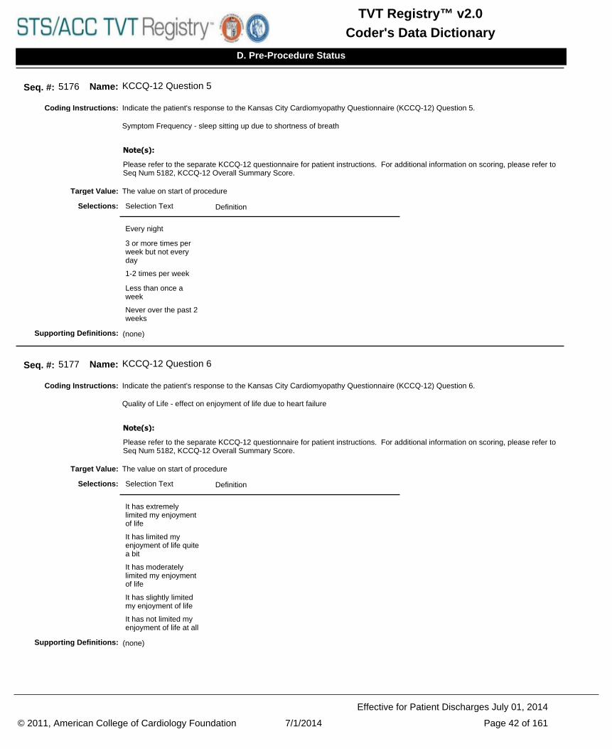
Indicate the patient's response to the Kansas City Cardiomyopathy Questionnaire (KCCQ-12) Question 5.
Symptom Frequency - sleep sitting up due to shortness of breath
Coding Instructions:
Please refer to the separate KCCQ-12 questionnaire for patient instructions. For additional information on scoring, please refer to Seq Num 5182, KCCQ-12 Overall Summary Score.
Note(s):
Seq. #: 5176 Name: KCCQ-12 Question 5
The value on start of procedureTarget Value:
Selection Text Definition
Every night
3 or more times per week but not every day
1-2 times per week
Less than once a week
Never over the past 2 weeks
Selections:
(none)Supporting Definitions:
Indicate the patient's response to the Kansas City Cardiomyopathy Questionnaire (KCCQ-12) Question 6.
Quality of Life - effect on enjoyment of life due to heart failure
Coding Instructions:
Please refer to the separate KCCQ-12 questionnaire for patient instructions. For additional information on scoring, please refer to Seq Num 5182, KCCQ-12 Overall Summary Score.
Note(s):
Seq. #: 5177 Name: KCCQ-12 Question 6
The value on start of procedureTarget Value:
Selection Text Definition
It has extremely limited my enjoyment of life
It has limited my enjoyment of life quite a bit
It has moderately limited my enjoyment of life
It has slightly limited my enjoyment of life
It has not limited my enjoyment of life at all
Selections:
(none)Supporting Definitions:
© 2011, American College of Cardiology Foundation 7/1/2014 Page 42 of 161
Effective for Patient Discharges July 01, 2014
Coder's Data DictionaryTVT Registry™ v2.0
D. Pre-Procedure Status

Indicate the patient's response to the Kansas City Cardiomyopathy Questionnaire (KCCQ-12) Question 7.
Quality of life - remaining life with heart failure
Coding Instructions:
Please refer to the separate KCCQ-12 questionnaire for patient instructions. For additional information on scoring, please refer to Seq Num 5182, KCCQ-12 Overall Summary Score.
Note(s):
Seq. #: 5178 Name: KCCQ-12 Question 7
The value on start of procedureTarget Value:
Selection Text Definition
Not at all satisfied
Mostly dissatisfied
Somewhat satisfied
Mostly satisfied
Completely satisfied
Selections:
(none)Supporting Definitions:
Indicate the patient's response to the Kansas City Cardiomyopathy Questionnaire (KCCQ-12) Question 8a.
Social limitation - hobbies, recreational activities
Coding Instructions:
Please refer to the separate KCCQ-12 questionnaire for patient instructions. For additional information on scoring, please refer to Seq Num 5182, KCCQ-12 Overall Summary Score.
Note(s):
Seq. #: 5179 Name: KCCQ-12 Question 8a
The value on start of procedureTarget Value:
Selection Text Definition
Severely limited
Limited quite a bit
Moderately limited
Slightly limited
Did not limit at all
Does not apply or did not do for other reasons
Selections:
(none)Supporting Definitions:
© 2011, American College of Cardiology Foundation 7/1/2014 Page 43 of 161
Effective for Patient Discharges July 01, 2014
Coder's Data DictionaryTVT Registry™ v2.0
D. Pre-Procedure Status

Indicate the patient's response to the Kansas City Cardiomyopathy Questionnaire (KCCQ-12) Question 8b.
Social limitation - working or doing household chores
Coding Instructions:
Please refer to the separate KCCQ-12 questionnaire for patient instructions. For additional information on scoring, please refer to Seq Num 5182, KCCQ-12 Overall Summary Score.
Note(s):
Seq. #: 5180 Name: KCCQ-12 Question 8b
The value on start of procedureTarget Value:
Selection Text Definition
Severely limited
Limited quite a bit
Moderately limited
Slightly limited
Did not limit at all
Does not apply or did not do for other reasons
Selections:
(none)Supporting Definitions:
Indicate the patient's response to the Kansas City Cardiomyopathy Questionnaire (KCCQ-12) Question 8c.
Social limitation - visiting family or friends
Coding Instructions:
Please refer to the separate KCCQ-12 questionnaire for patient instructions. For additional information on scoring, please refer to Seq Num 5182, KCCQ-12 Overall Summary Score.
Note(s):
Seq. #: 5181 Name: KCCQ-12 Question 8c
The value on start of procedureTarget Value:
Selection Text Definition
Severely limited
Limited quite a bit
Moderately limited
Slightly limited
Did not limit at all
Does not apply or did not do for other reasons
Selections:
(none)Supporting Definitions:
© 2011, American College of Cardiology Foundation 7/1/2014 Page 44 of 161
Effective for Patient Discharges July 01, 2014
Coder's Data DictionaryTVT Registry™ v2.0
D. Pre-Procedure Status

(Auto Calculated) This field is auto-populated by your application.
Kansas City Cardiomyopathy Questionnaire (KCCQ-12) Overall Summary Score.
Coding Instructions:
The 12 patient responses are reduced into four summary scores (Physical Limitation Score, Symptom Frequency Score, Quality of Life Score, Social Limitation Score). The four summary scores are used to calculate the Overall Summary Score.
For more information, please refer to the KCCQ-12 Scoring Instructions document provided by the STS/ACC TVT Registry.
Note(s):
Seq. #: 5182 Name: KCCQ Overall Summary Score
The value on start of procedureTarget Value:
(none)Selections:
(none)Supporting Definitions:
Indicate the patient's height in centimeters.Coding Instructions:
Seq. #: 5200 Name: Height
The first value between arrival at this facility and the procedureTarget Value:
(none)Selections:
(none)Supporting Definitions:
Indicate the patient's weight in kilograms.Coding Instructions:
Seq. #: 5205 Name: Weight
The last value between arrival at this facility and the procedureTarget Value:
(none)Selections:
(none)Supporting Definitions:
Indicate the preprocedure hemoglobin level in g/dL.Coding Instructions:
Seq. #: 5250 Name: Pre-Procedure Hemoglobin
The last value between 30 days prior to procedure and start of the procedureTarget Value:
(none)Selections:
(none)Supporting Definitions:
© 2011, American College of Cardiology Foundation 7/1/2014 Page 45 of 161
Effective for Patient Discharges July 01, 2014
Coder's Data DictionaryTVT Registry™ v2.0
D. Pre-Procedure Status

Indicate if a pre-procedure hemoglobin level was not drawn.Coding Instructions:
Seq. #: 5251 Name: Pre-Procedure Hemoglobin Not Drawn
N/ATarget Value:
Selection Text Definition
No
Yes Code "Yes" if the lab was not drawn.
Selections:
(none)Supporting Definitions:
Indicate the creatinine level closest to the date and time prior to the procedure but prior to anesthetic management (induction area, cath lab or operating room), in mg/dL.
Coding Instructions:
A creatinine level should be collected on all patients, even if they have no prior history. A creatinine value is a high predictor of a patient's outcome and is used in the predicted risk models.
Note(s):
Seq. #: 5255 Name: Pre-Procedure Creatinine
The last value between 30 days prior to procedure and start of the procedureTarget Value:
(none)Selections:
(none)Supporting Definitions:
Indicate if a preprocedure creatinine level was not drawn.Coding Instructions:
Seq. #: 5256 Name: Pre-Procedure Creatinine Not Drawn
N/ATarget Value:
Selection Text Definition
No
Yes Code "Yes" if the lab was not drawn.
Selections:
(none)Supporting Definitions:
Indicate the pre-procedure platelet count in microliters.Coding Instructions:
Seq. #: 5260 Name: Platelet Count
The last value between 30 days prior to procedure and start of the procedureTarget Value:
(none)Selections:
(none)Supporting Definitions:
© 2011, American College of Cardiology Foundation 7/1/2014 Page 46 of 161
Effective for Patient Discharges July 01, 2014
Coder's Data DictionaryTVT Registry™ v2.0
D. Pre-Procedure Status

Indicate if a platelet count was not drawn prior to the procedure.Coding Instructions:
Seq. #: 5261 Name: Platelet Count Not Drawn
N/ATarget Value:
Selection Text Definition
No
Yes Code "Yes" if the lab was not drawn.
Selections:
(none)Supporting Definitions:
Indicate the pre-procedure International Normalized Ratio (INR).Coding Instructions:
Seq. #: 5265 Name: INR
The last value between 30 days prior to procedure and start of the procedureTarget Value:
(none)Selections:
(none)Supporting Definitions:
Indicate if the pre-procedure International Normalized Ratio (INR) was not drawn.Coding Instructions:
Seq. #: 5266 Name: INR Not Drawn
N/ATarget Value:
Selection Text Definition
No
Yes Code "Yes" if the lab was not drawn.
Selections:
(none)Supporting Definitions:
Indicate the total albumin (in g/dL) closest to the date and time prior to the procedure but prior to anesthetic management (induction area, cath lab or operating room).
Coding Instructions:
Seq. #: 5270 Name: Albumin
The last value between 30 days prior to procedure and start of the procedureTarget Value:
(none)Selections:
(none)Supporting Definitions:
© 2011, American College of Cardiology Foundation 7/1/2014 Page 47 of 161
Effective for Patient Discharges July 01, 2014
Coder's Data DictionaryTVT Registry™ v2.0
D. Pre-Procedure Status

Indicate if the total albumin level was not drawn.Coding Instructions:
Seq. #: 5271 Name: Total Albumin Not Drawn
N/ATarget Value:
Selection Text Definition
No
Yes Code "Yes" if the lab was not drawn.
Selections:
(none)Supporting Definitions:
Indicate the total bilirubin (in mg/dL) closest to the date and time prior to the procedure but prior to anesthetic management (induction area, cath lab or operating room).
Coding Instructions:
Seq. #: 5275 Name: Bilirubin
The last value between 30 days prior to procedure and start of the procedureTarget Value:
(none)Selections:
(none)Supporting Definitions:
Indicate if the total bilirubin level was not drawn.Coding Instructions:
Seq. #: 5276 Name: Total Bilirubin Not Drawn
N/ATarget Value:
Selection Text Definition
No
Yes Code "Yes" if the lab was not drawn.
Selections:
(none)Supporting Definitions:
Indicate the patient's brain natriuretic peptide (BNP) level in pg/ml.Coding Instructions:
If BNP was not drawn, leave blank and code 'Yes' for BNP or NT-proBNP or Not Drawn.
Note(s):
Seq. #: 5277 Name: BNP
The last value between 6 months prior to the procedure and start of the procedureTarget Value:
(none)Selections:
(none)Supporting Definitions:
© 2011, American College of Cardiology Foundation 7/1/2014 Page 48 of 161
Effective for Patient Discharges July 01, 2014
Coder's Data DictionaryTVT Registry™ v2.0
D. Pre-Procedure Status

Indicate the patient's NT-pro- brain natriuretic peptide (BNP) level in pg/ml.Coding Instructions:
If NT-proBNP was not drawn, leave blank and code 'Yes' for BNP or NTproBNP Not Drawn.
Note(s):
Seq. #: 5278 Name: NT-proBNP
The last value between 6 months prior to the procedure and start of the procedureTarget Value:
(none)Selections:
(none)Supporting Definitions:
Indicate if a BNP or NT-proBNP level was not drawn.Coding Instructions:
Seq. #: 5279 Name: BNP or NT-proBNP Not Drawn
N/ATarget Value:
Selection Text Definition
No
Yes
Selections:
(none)Supporting Definitions:
Indicate the FEV1 % predicted from the most recent pulmonary function test prior to procedureCoding Instructions:
Seq. #: 5280 Name: Forced Expiratory Volume (FEV1) % Predicted
The last value between 6 months prior to the procedure and start of the procedureTarget Value:
(none)Selections:
(none)Supporting Definitions:
Indicate whether % predicted Forced Expiratory Volume (FEV1) was not performed or the patient did not have a pulmonary function test prior to the procedure.
Coding Instructions:
Seq. #: 5281 Name: Forced Expiratory Volume (FEV1) % Predicted Not Performed
N/ATarget Value:
Selection Text Definition
No
Yes Code "Yes" if the test was not performed.
Selections:
(none)Supporting Definitions:
© 2011, American College of Cardiology Foundation 7/1/2014 Page 49 of 161
Effective for Patient Discharges July 01, 2014
Coder's Data DictionaryTVT Registry™ v2.0
D. Pre-Procedure Status

Indicate the adjusted value of % predicted diffusion capacity of the lung for carbon monoxide (DLCO) value obtained for the patient. This is reported in charts as DLCO/VA% (adjusted value) or D/Vasb (for volume surface body area).
Coding Instructions:
Seq. #: 5285 Name: Adjusted DLCO
The last value between 6 months prior to the procedure and start of the procedureTarget Value:
(none)Selections:
(none)Supporting Definitions:
Indicate if a lung diffusion test (DLCO) was not performedCoding Instructions:
Seq. #: 5286 Name: DLCO Not Performed
N/ATarget Value:
Selection Text Definition
No
Yes Code "Yes" if the test was not performed.
Selections:
(none)Supporting Definitions:
Indicate the duration of the non-ventricular paced or intrinsic QRS complex, in milliseconds, that was derived from the surface electrocardiogram (ECG). Surface ECGs are obtained from the surface of the body and do not include intracardiac ECGs.
Coding Instructions:
Do not code QRS measurements from an intracardiac ECG.
If more than one ECG is available, code the value on the ECG closest to the procedure.
Note(s):
Seq. #: 5290 Name: QRS Duration (Non-Ventricular Paced Complex)
The last value between birth and prior to the first procedure.Target Value:
(none)Selections:
(none)Supporting Definitions:
Indicate if there were only ventricular paced QRS complexes present.Coding Instructions:
If the patient has some intrinsic ventricular complexes present, code 'No'.
Note(s):
Seq. #: 5291 Name: Only Ventricular Paced QRS Complexes Present
The last value between birth and prior to the first procedure.Target Value:
Selection Text Definition
No
Yes
Selections:
(none)Supporting Definitions:
© 2011, American College of Cardiology Foundation 7/1/2014 Page 50 of 161
Effective for Patient Discharges July 01, 2014
Coder's Data DictionaryTVT Registry™ v2.0
D. Pre-Procedure Status

Indicate which medications the patient received 24 hours prior to the procedure.Coding Instructions:
For patients requiring IV inotropic support, indicate positive inotropes only.
To code 'Yes' for aspirin, the minimum dose should be at least 75mg.
Note(s):
Seq. #: 5400 Name: Pre-Procedure Medication
Any occurrence between 24 hours prior to the procedure and start of the procedureTarget Value:
(none)Selections:
(none)Supporting Definitions:
Indicate whether the patient received the medication within 24 hours preceding the procedure.Coding Instructions:
If a medication is contraindicated it needs to be documented in the chart by the physician or other responsible care giver. The reason for contraindication does not need to be documented.
Note(s):
Seq. #: 5405 Name: Pre-Procedure Medication Administered
Any occurrence between 24 hours prior to the procedure and start of the procedureTarget Value:
Selection Text Definition
No
Yes
Contraindicated
Blinded
Selections:
(none)Supporting Definitions:
Indicate whether diagnostic cardiac catheterization was performed.Coding Instructions:
Seq. #: 5500 Name: Diagnostic Catheterization
The last value between 12 months prior to the procedure and start of the procedureTarget Value:
Selection Text Definition
No
Yes
Selections:
(none)Supporting Definitions:
Indicate the date the diagnostic catheterization was performed.Coding Instructions:
Seq. #: 5505 Name: Diagnostic Catheterization Date
The last value between 12 months prior to the procedure and start of the procedureTarget Value:
(none)Selections:
(none)Supporting Definitions:
© 2011, American College of Cardiology Foundation 7/1/2014 Page 51 of 161
Effective for Patient Discharges July 01, 2014
Coder's Data DictionaryTVT Registry™ v2.0
D. Pre-Procedure Status

Indicate the number of diseased major native coronary vessel systems: LAD system, Circumflex system, and/or Right system with >= 50% narrowing of any vessel preoperatively.
Coding Instructions:
Left main disease (>=50%) is counted as TWO vessels (LAD and Circumflex, which may include a Ramus Intermedius). For example, left main and RCA would count as three total.
Note(s):
Seq. #: 5506 Name: Number of Diseased Vessels
The last value between 12 months prior to the procedure and start of the procedureTarget Value:
Selection Text Definition
None No significant coronary obstructive disease
1
2
3
Selections:
(none)Supporting Definitions:
Indicate whether the patient has Left Main Coronary Disease. Left Main Coronary Disease is present when there is >= 50% compromise of vessel diameter preoperatively.
Coding Instructions:
Seq. #: 5507 Name: Left Main >=50%
The last value between 12 months prior to the procedure and start of the procedureTarget Value:
Selection Text Definition
No
Yes
Selections:
(none)Supporting Definitions:
Indicate whether the percent luminal narrowing of the proximal left anterior descending artery at the point of maximal stenosis is greater than or equal to 70%.
Coding Instructions:
Seq. #: 5508 Name: Prox LAD >=70%
The last value between 12 months prior to the procedure and start of the procedureTarget Value:
Selection Text Definition
No
Yes
Selections:
(none)Supporting Definitions:
© 2011, American College of Cardiology Foundation 7/1/2014 Page 52 of 161
Effective for Patient Discharges July 01, 2014
Coder's Data DictionaryTVT Registry™ v2.0
D. Pre-Procedure Status

Indicate the percentage of the blood emptied from the left ventricle at the end of the contraction.Coding Instructions:
Use the most recent determination prior to the surgical intervention documented on a diagnostic report. Enter a percentage in the range of 1 - 99.
If a percentage range is reported, report a whole number using the "mean" (i.e., 50-55%, is reported as 53%).
If only a descriptive value is reported, (i.e., normal), enter the corresponding percentage value from the list below:Normal = 60% Good function = 50% Mildly reduced = 45% Fair function = 40% Moderately reduced = 30% Poor function = 25% Severely reduced = 20%
Note(s):
Seq. #: 5565 Name: Left Ventricle Ejection Fraction
The last value between 12 months prior to the procedure and start of the procedureTarget Value:
(none)Selections:
Left Ventricular Ejection Fraction (LVEF):
The left ventricular ejection fraction is the percentage of the blood emptied from the left ventricle at the end of the contraction.
Source: ACC Clinical Data Standards, The Society of Thoracic Surgeons
Supporting Definitions:
Indicate whether the left ventricular ejection fraction was not assessed or not measured prior to the induction of anesthesia.Coding Instructions:
Seq. #: 5566 Name: Left Ventricle Ejection Fraction Not Assessed
N/ATarget Value:
Selection Text Definition
No
Yes Code "yes" if LVEF was not assessed.
Selections:
(none)Supporting Definitions:
Indicate the cardiac output in mL/min, documented by pre-procedure diagnostic cardiac cath findings.Coding Instructions:
Seq. #: 5567 Name: Cardiac Output
The last value between 12 months prior to the procedure and start of the procedureTarget Value:
(none)Selections:
(none)Supporting Definitions:
© 2011, American College of Cardiology Foundation 7/1/2014 Page 53 of 161
Effective for Patient Discharges July 01, 2014
Coder's Data DictionaryTVT Registry™ v2.0
D. Pre-Procedure Status

Indicate the highest right ventricular systolic pressure in mmHg recorded prior to the start of the procedure. Coding Instructions:
If a value is available from both echo and cardiac cath, code the value from the cardiac cath.
Note(s):
Seq. #: 5568 Name: Right Ventricular Systolic Pressure
The last value between 12 months prior to the procedure and start of the procedureTarget Value:
(none)Selections:
(none)Supporting Definitions:
Indicate whether the cardiac output was not measured pre-procedure.Coding Instructions:
Seq. #: 5569 Name: Cardiac Output Not Performed
N/ATarget Value:
Selection Text Definition
No
Yes
Selections:
(none)Supporting Definitions:
Indicate the pre-procedure pulmonary capillary wedge pressure, in mmHg.Coding Instructions:
If more than one PCWP is available, code the value determined by cardiac catheterization.
Note(s):
Seq. #: 5590 Name: Pulmonary Capillary Wedge Pressure
The last value between 12 months prior to the procedure and start of the procedureTarget Value:
(none)Selections:
(none)Supporting Definitions:
Indicate if the pulmonary capillary wedge pressure was not measured pre-procedure.Coding Instructions:
Seq. #: 5591 Name: Pulmonary Capillary Wedge Pressure Not Measured
N/ATarget Value:
Selection Text Definition
No
Yes
Selections:
(none)Supporting Definitions:
© 2011, American College of Cardiology Foundation 7/1/2014 Page 54 of 161
Effective for Patient Discharges July 01, 2014
Coder's Data DictionaryTVT Registry™ v2.0
D. Pre-Procedure Status

Indicate the pre-procedure pulmonary artery mean pressure, in mmHg.Coding Instructions:
Seq. #: 5593 Name: Pulmonary Artery Pressure (Mean)
The last value between 12 months prior to the procedure and start of the procedureTarget Value:
(none)Selections:
(none)Supporting Definitions:
Indicate if the pre-procedure pulmonary artery mean pressure was not measured pre-procedure.Coding Instructions:
Seq. #: 5594 Name: Pulmonary Artery Pressure (Mean) Not Measured
N/ATarget Value:
Selection Text Definition
No
Yes
Selections:
(none)Supporting Definitions:
Indicate the pre-procedure left ventricular internal systolic dimension in cm.Coding Instructions:
If more than one LV internal systolic diameter is available, code the value determined by echocardiography.
Using a 2D method, it is recommended that LV internal dimensions (LVIDd and LVIDs, respectively) be measured at the level of the LV minor dimension, at the mitral chordae level.
Note(s):
Seq. #: 5595 Name: Left Ventricular Internal Systolic Dimension
The last value between 12 months prior to the procedure and start of the procedureTarget Value:
(none)Selections:
General Principles for Linear and Volumetric LV Measurements:
To obtain accurate linear measurements of interventricular septal wall thickness (SWT), posterior wall thickness (PWT), and LV internal dimension, recordings should be made from the parasternal long-axis acoustic window. It is recommended that LV internal dimensions (LVIDd and LVIDs, respectively) and wall thicknesses be measured at the level of the LV minor axis, approximately at the mitral valve leaflet tips. These linear measurements can be made directly from 2D images or using 2D-targeted M-mode echocardiography.
Source: Recommendations for Chamber Quantification: A Report from the American Society of Echocardiography’s Guidelines and Standards Committee and the Chamber Quantification Writing Group, Developed in Conjunction with the European Association of Echocardiography, a Branch of the European Society of Cardiology, Journal of the American Society of Echocardiography. Volume 18 Number 12
Supporting Definitions:
Indicate if the pre-procedure pulmonary artery systolic pressure, in mmHg.Coding Instructions:
Seq. #: 5596 Name: Pulmonary Artery Pressure (Systolic)
The last value between 12 months prior to the procedure and start of the procedureTarget Value:
(none)Selections:
(none)Supporting Definitions:
© 2011, American College of Cardiology Foundation 7/1/2014 Page 55 of 161
Effective for Patient Discharges July 01, 2014
Coder's Data DictionaryTVT Registry™ v2.0
D. Pre-Procedure Status

Indicate if the pre-procedure pulmonary artery systolic pressure was not measured pre-procedure.Coding Instructions:
Seq. #: 5597 Name: Pulmonary Artery Pressure (Systolic) Not Measured
N/ATarget Value:
Selection Text Definition
No
Yes
Selections:
(none)Supporting Definitions:
Indicate the pre-procedure right atrial pressure or central venous pressure (CVP), in mmHg.Coding Instructions:
Seq. #: 5598 Name: Right Atrial Pressure/CVP (Mean)
The last value between 12 months prior to the procedure and start of the procedureTarget Value:
(none)Selections:
(none)Supporting Definitions:
Indicate if the pre-procedure right atrial pressure or central venous pressure (CVP) was not measured pre-procedure.Coding Instructions:
Seq. #: 5599 Name: Right Atrial Pressure/CVP (Mean) Not Measured
N/ATarget Value:
Selection Text Definition
No
Yes
Selections:
(none)Supporting Definitions:
© 2011, American College of Cardiology Foundation 7/1/2014 Page 56 of 161
Effective for Patient Discharges July 01, 2014
Coder's Data DictionaryTVT Registry™ v2.0
D. Pre-Procedure Status

Indicate the pre-procedure left ventricular internal diastolic dimension in cm. If more than one LV internal diastolic diameter is available, code the value determined by echocardiography.
Coding Instructions:
Using a 2D method, it is recommended that LV internal dimensions (LVIDd and LVIDs, respectively) be measured at the level of the LV minor dimension, at the mitral chordae level.
Note(s):
Seq. #: 5600 Name: Left Ventricular Internal Diastolic Dimension
The last value between 12 months prior to the procedure and start of the procedureTarget Value:
(none)Selections:
General Principles for Linear and Volumetric LV Measurements:
To obtain accurate linear measurements of interventricular septal wall thickness (SWT), posterior wall thickness (PWT), and LV internal dimension, recordings should be made from the parasternal long-axis acoustic window. It is recommended that LV internal dimensions (LVIDd and LVIDs, respectively) and wall thicknesses be measured at the level of the LV minor axis, approximately at the mitral valve leaflet tips. These linear measurements can be made directly from 2D images or using 2D-targeted M-mode echocardiography.
Source: Recommendations for Chamber Quantification: A Report from the American Society of Echocardiography’s Guidelines and Standards Committee and the Chamber Quantification Writing Group, Developed in Conjunction with the European Association of Echocardiography, a Branch of the European Society of Cardiology, Journal of the American Society of Echocardiography. Volume 18 Number 12
Supporting Definitions:
Indicate the left ventricular end systolic volume in ml, documented by pre-procedure echocardiogram.Coding Instructions:
Seq. #: 5601 Name: Left Ventricular End Systolic Volume
The last value between 12 months prior to the procedure and start of the procedureTarget Value:
(none)Selections:
(none)Supporting Definitions:
Indicate if the left ventricular end systolic volume was not measured pre-procedure.Coding Instructions:
Seq. #: 5602 Name: Left Ventricular End Systolic Volume Not Measured
N/ATarget Value:
Selection Text Definition
No
Yes
Selections:
(none)Supporting Definitions:
Indicate the left ventricular end diastolic volume in ml, documented by pre-procedure echocardiogram.Coding Instructions:
Seq. #: 5603 Name: Left Ventricular End Diastolic Volume
The last value between 12 months prior to the procedure and start of the procedureTarget Value:
(none)Selections:
(none)Supporting Definitions:
© 2011, American College of Cardiology Foundation 7/1/2014 Page 57 of 161
Effective for Patient Discharges July 01, 2014
Coder's Data DictionaryTVT Registry™ v2.0
D. Pre-Procedure Status

Indicate if the left ventricular end diastolic volume was not measured pre-procedure.Coding Instructions:
Seq. #: 5604 Name: Left Ventricular End Diastolic Volume Not Measured
N/ATarget Value:
Selection Text Definition
No
Yes
Selections:
(none)Supporting Definitions:
Indicate the pre-procedure septal wall thickness, in cm, measured at end-diastole.Coding Instructions:
If more than one septal wall thickness is available, code the value determined by echocardiography.
Note(s):
Seq. #: 5605 Name: Septal Wall Thickness
The last value between 12 months prior to the procedure and start of the procedureTarget Value:
(none)Selections:
(none)Supporting Definitions:
Indicate the left atrial volume in ml, documented by pre-procedure echocardiogram. If the left atrial volume index is documented, leave this field blank.
Coding Instructions:
Seq. #: 5606 Name: Left Atrial Volume
The last value between 12 months prior to the procedure and start of the procedureTarget Value:
(none)Selections:
(none)Supporting Definitions:
Indicate the left atrial volume index in mL/m2, documented by pre-procedure echocardiogram. If the left atrial volume is documented, leave this field blank.
Coding Instructions:
Seq. #: 5607 Name: Left Atrial Volume Index
The last value between 12 months prior to the procedure and start of the procedureTarget Value:
(none)Selections:
(none)Supporting Definitions:
© 2011, American College of Cardiology Foundation 7/1/2014 Page 58 of 161
Effective for Patient Discharges July 01, 2014
Coder's Data DictionaryTVT Registry™ v2.0
D. Pre-Procedure Status
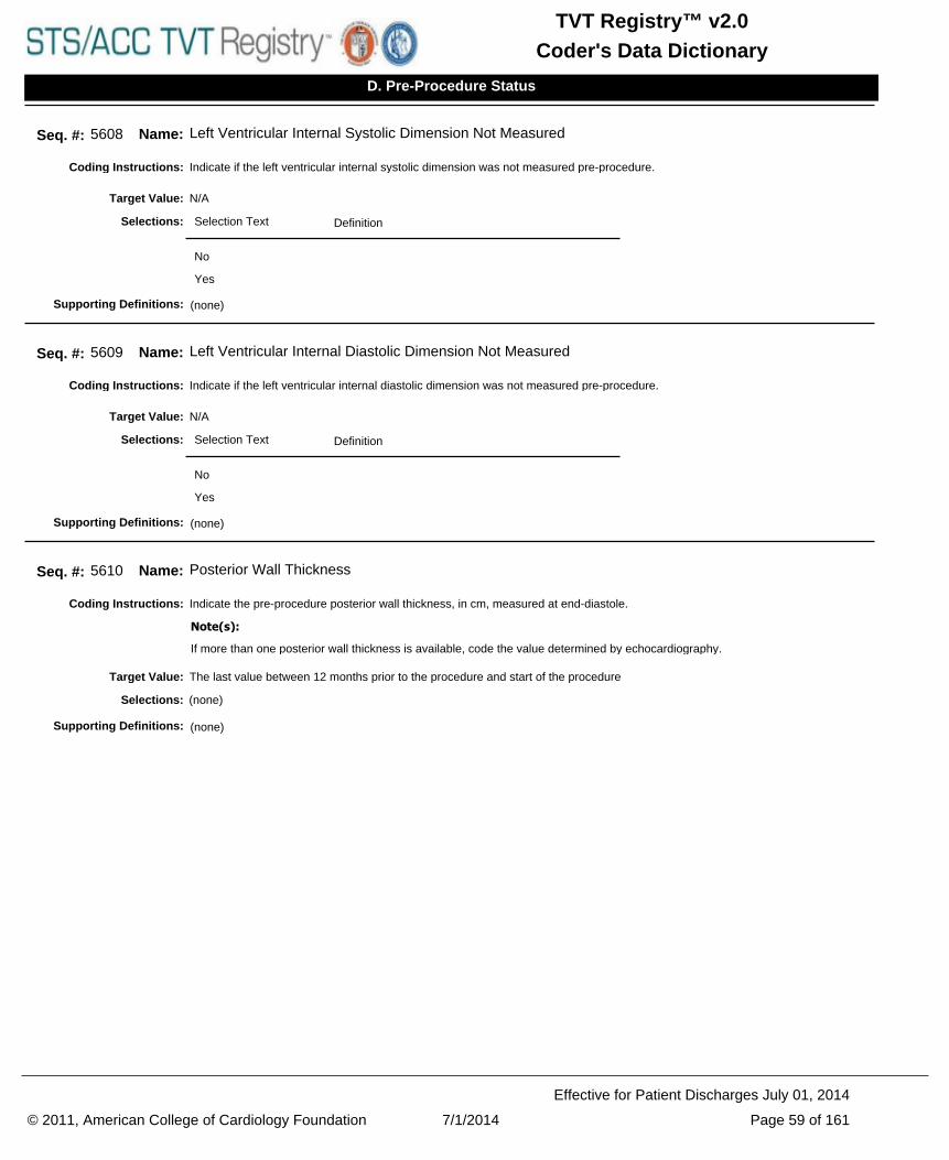
Indicate if the left ventricular internal systolic dimension was not measured pre-procedure.Coding Instructions:
Seq. #: 5608 Name: Left Ventricular Internal Systolic Dimension Not Measured
N/ATarget Value:
Selection Text Definition
No
Yes
Selections:
(none)Supporting Definitions:
Indicate if the left ventricular internal diastolic dimension was not measured pre-procedure.Coding Instructions:
Seq. #: 5609 Name: Left Ventricular Internal Diastolic Dimension Not Measured
N/ATarget Value:
Selection Text Definition
No
Yes
Selections:
(none)Supporting Definitions:
Indicate the pre-procedure posterior wall thickness, in cm, measured at end-diastole.Coding Instructions:
If more than one posterior wall thickness is available, code the value determined by echocardiography.
Note(s):
Seq. #: 5610 Name: Posterior Wall Thickness
The last value between 12 months prior to the procedure and start of the procedureTarget Value:
(none)Selections:
(none)Supporting Definitions:
© 2011, American College of Cardiology Foundation 7/1/2014 Page 59 of 161
Effective for Patient Discharges July 01, 2014
Coder's Data DictionaryTVT Registry™ v2.0
D. Pre-Procedure Status

Indicate primary etiology of aortic valve disease.Coding Instructions:
Seq. #: 5620 Name: Aortic Valve Disease - Disease Etiology
Any occurrence between 12 months prior to the procedure and start of the procedureTarget Value:
Selection Text Definition
Degenerative Degenerative includes calcific, senile, and leaflet prolapse.
Endocarditis
Congenital
Rheumatic fever
Primary aortic disease
LV outflow tract obstruction
Supravalvular aortic stenosis
Tumor
Trauma
Other
Selections:
(none)Supporting Definitions:
© 2011, American College of Cardiology Foundation 7/1/2014 Page 60 of 161
Effective for Patient Discharges July 01, 2014
Coder's Data DictionaryTVT Registry™ v2.0
D. Pre-Procedure Status

Indicate the severity of aortic valve regurgitation.Coding Instructions:
"Moderately severe" should be coded as "Severe".
Reference:Bonow, R.O, et al. 2008 Focused Updated Incorporated into ACC/AHA 2006 Guidelines for the Management of Patients with Valvular Heart Disease: A Report of the American College of Cardiology /American Heart Association Task force on Practice Guidelines. JACC, vol 52, No. 13, 2008, p. e1-e142.
Note(s):
Seq. #: 5630 Name: Aortic Valve Disease - Aortic Regurgitation
The highest value between 12 months prior to the procedure and start of the procedureTarget Value:
Selection Text Definition
None
Trace/Trivial
Mild Mild aortic insufficiency or regurgitation is defined as the following:
Qualitative Measurements:Angiographic grade of 1+; Color Doppler jet width (Central jet) <25% of LVOT; Dopplar vena contracta width <0.3 cm;.
Quantitative Measures (cath or echo)Regurgitant volume <30 ml/beat;Regurgitant fraction <30%;Regurgitant orifice area <0.10 cm(2)
Moderate Moderate aortic insufficiency or regurgitation is defined as the following:
Qualitative Measurements:Angiographic grade of 2+; Color Doppler jet width greater than mild but no signs of severe aortic regurgitation (insufficiency); Dopplar vena contracta width 0.3-0.6 cm;
Quantitative Measures (cath or echo)Regurgitant volume 30-59 ml/beat;Regurgitant fraction 30-49%;Regurgitant orifice area 0.10-0.29 cm(2)
Severe Severe aortic insufficiency or regurgitation is defined as the following:
Qualitative Measurements:Angiographic grade of 3-4+; Color Doppler jet width (Central jet) >65% of LVOT; Dopplar vena contracta width >0.6 cm;.
Quantitative Measures (cath or echo)Regurgitant volume >=60 ml/beat;Regurgitant fraction >=50%;Regurgitant orifice area >=0.30 cm(2)
Additional essential criteria:Left ventricular size is increased
Selections:
(none)Supporting Definitions:
© 2011, American College of Cardiology Foundation 7/1/2014 Page 61 of 161
Effective for Patient Discharges July 01, 2014
Coder's Data DictionaryTVT Registry™ v2.0
D. Pre-Procedure Status

Indicate the morphology of the aortic valve.Coding Instructions:
Seq. #: 5640 Name: Aortic Valve Disease - Valve Morphology
Any occurrence between 12 months prior to the procedure and start of the procedureTarget Value:
Selection Text Definition
Unicuspid
Bicuspid
Tricuspid
Quadracuspid
Uncertain
Selections:
(none)Supporting Definitions:
Indicate if annular calcification is present on the aortic valve.
Code yes if echo reports document calcificaton in the aortic valve leaflets, aorta adjacent to the AV, leaflets or the left ventricular outflow tract (LVOT), or if echo reports document AV calcific degeneration.
Coding Instructions:
Seq. #: 5645 Name: Aortic Valve Disease - Annular Calcification
Any occurrence between 12 months prior to the procedure and start of the procedureTarget Value:
Selection Text Definition
No
Yes
Selections:
(none)Supporting Definitions:
Indicate the aortic valve peak velocity, in meters per second, as determined by continuous wave (CW) spectral velocity recording on echocardiography.
Coding Instructions:
Seq. #: 5650 Name: Aortic Valve Disease - AV Peak Velocity (CW)
The highest value between 12 months prior to the procedure and start of the procedureTarget Value:
(none)Selections:
(none)Supporting Definitions:
Indicate the size, in mm, of the aortic valve annulus.Coding Instructions:
If more than one size is reported, code the mean.
Note(s):
Seq. #: 5655 Name: Aortic Valve Disease - AV Annulus Size
The last value between 12 months prior to the procedure and prior to valve implantTarget Value:
(none)Selections:
(none)Supporting Definitions:
© 2011, American College of Cardiology Foundation 7/1/2014 Page 62 of 161
Effective for Patient Discharges July 01, 2014
Coder's Data DictionaryTVT Registry™ v2.0
D. Pre-Procedure Status

Indicate the method used to assess the aortic valve annulus size.Coding Instructions:
Seq. #: 5660 Name: Aortic Valve Disease - AV Annulus Size Assessment Method
Any occurrence between 12 months prior to the procedure and start of the procedureTarget Value:
Selection Text Definition
TTE Transthoracic Echocardiogram
TEE Transesophageal echocardiogram
CTA computerized tomographic angiography
Angiography coronary angiography
Selections:
(none)Supporting Definitions:
Indicate whether aortic stenosis is present.Coding Instructions:
Seq. #: 5665 Name: Aortic Stenosis
Any occurrence between 12 months prior to the procedure and start of the procedureTarget Value:
Selection Text Definition
No
Yes
Selections:
(none)Supporting Definitions:
Indicate the smallest aortic valve area (in cm squared) obtained from an echocardiogram or cath report.Coding Instructions:
Seq. #: 5670 Name: Aortic Stenosis - AV Area
The lowest value between 12 months prior to the procedure and start of the procedureTarget Value:
(none)Selections:
(none)Supporting Definitions:
Indicate the highest MEAN gradient (in mmHg) across the aortic valve obtained from an echocardiogram or angiogram preoperatively.
Coding Instructions:
Seq. #: 5675 Name: Aortic Stenosis - AV Mean Gradient
The highest value between 12 months prior to the procedure and start of the procedureTarget Value:
(none)Selections:
(none)Supporting Definitions:
© 2011, American College of Cardiology Foundation 7/1/2014 Page 63 of 161
Effective for Patient Discharges July 01, 2014
Coder's Data DictionaryTVT Registry™ v2.0
D. Pre-Procedure Status

Indicate the aortic valve peak gradient in mmHg.Coding Instructions:
Seq. #: 5680 Name: Aortic Stenosis - AV Peak Gradient
The highest value between 12 months prior to the procedure and start of the procedureTarget Value:
(none)Selections:
(none)Supporting Definitions:
Indicate whether mitral valve disease is present.Coding Instructions:
Seq. #: 5685 Name: Mitral Valve Disease
Any occurrence between 12 months prior to the procedure and start of the procedureTarget Value:
Selection Text Definition
No
Yes
Selections:
(none)Supporting Definitions:
Indicate the severity of mitral valve regurgitation according to the American Society of Echocardiography Guidelines integrated approach.
Coding Instructions:
Seq. #: 5695 Name: Mitral Valve Disease - MV Regurgitation
The highest value between 12 months prior to the procedure and start of the procedureTarget Value:
Selection Text Definition
None
Trace/Trivial
Mild 1+/Mild
Moderate 2+/Moderate
Moderate-severe 3+/Moderate-severe
Severe 4+/Severe
Severe Retired TVT 2.00
Selections:
(none)Supporting Definitions:
© 2011, American College of Cardiology Foundation 7/1/2014 Page 64 of 161
Effective for Patient Discharges July 01, 2014
Coder's Data DictionaryTVT Registry™ v2.0
D. Pre-Procedure Status

Indicate the severity of paravalvular mitral regurgitation.Coding Instructions:
Seq. #: 5696 Name: Paravalvular Severity
The highest value between 12 months prior to the procedure and start of the procedureTarget Value:
Selection Text Definition
None
Mild
Moderate
Severe
Not Documented
Selections:
(none)Supporting Definitions:
Indicate the severity of valvular mitral regurgitation.Coding Instructions:
Seq. #: 5697 Name: Valvular Severity
The highest value between 12 months prior to the procedure and start of the procedureTarget Value:
Selection Text Definition
None
Mild
Moderate
Severe
Not Documented
Selections:
(none)Supporting Definitions:
Indicate the effective orifice area (EOA), in cm2.
EOA can also be called effective regurgitant orifice area (EROA).
Coding Instructions:
Seq. #: 5698 Name: Effective Orifice Area (EOA) or EROA
The highest value between 12 months prior to the procedure and start of the procedureTarget Value:
(none)Selections:
(none)Supporting Definitions:
© 2011, American College of Cardiology Foundation 7/1/2014 Page 65 of 161
Effective for Patient Discharges July 01, 2014
Coder's Data DictionaryTVT Registry™ v2.0
D. Pre-Procedure Status

Indicate the method used to assess the effective orifice area. If multiple methods are available, code the 3D planimetry method first, then PISA.
Coding Instructions:
Seq. #: 5699 Name: Method of Assessment
Any occurrence between 12 months prior to the procedure and start of the procedureTarget Value:
Selection Text Definition
3D planimetry Three-dimensional (3D) echocardiography
PISA Proximal isovelocity surface area (PISA)
Quantitative doppler
Other
Selections:
(none)Supporting Definitions:
Indicate whether mitral stenosis is present.Coding Instructions:
Seq. #: 5705 Name: Mitral Valve Disease - Mitral Valve Stenosis
Any occurrence between 12 months prior to the procedure and start of the procedureTarget Value:
Selection Text Definition
No
Yes
Selections:
(none)Supporting Definitions:
Indicate the smallest mitral valve area in centimeters squared.Coding Instructions:
If more than one measurement is available, code the area from the echocardiogram.
Note(s):
Seq. #: 5710 Name: Mitral Valve Stenosis - Mitral Valve Area
The highest value between 12 months prior to the procedure and start of the procedureTarget Value:
(none)Selections:
(none)Supporting Definitions:
Indicate the highest mean gradient (in mm Hg) across the mitral valve.Coding Instructions:
If more than one measurement is available, code the area from the echocardiogram.
Note(s):
Seq. #: 5715 Name: Mitral Valve Mean Gradient
The highest value between 12 months prior to the procedure and start of the procedureTarget Value:
(none)Selections:
(none)Supporting Definitions:
© 2011, American College of Cardiology Foundation 7/1/2014 Page 66 of 161
Effective for Patient Discharges July 01, 2014
Coder's Data DictionaryTVT Registry™ v2.0
D. Pre-Procedure Status

Indicate whether there is evidence of tricuspid valve regurgitation. Enter level of valve function associated with highest risk (i.e., worst performance).
Coding Instructions:
"Moderately severe" should be coded as "Severe".
Note(s):
Seq. #: 5735 Name: Tricuspid Valve Disease - Tricuspid Valve Regurgitation
The highest value between 12 months prior to the procedure and start of the procedureTarget Value:
Selection Text Definition
None
Trace/Trivial
Mild
Moderate
Severe Characterized by vena contracta width greater than 0.7 cm and systolic flow reversal in hepatic veins
Selections:
(none)Supporting Definitions:
Indicate the etiology of the prosthetic mitral valve dysfunction.Coding Instructions:
Seq. #: 5742 Name: Prosthetic Mitral Valve Dysfunction Etiology
Any occurrence between 12 months prior to the procedure and start of the procedureTarget Value:
Selection Text Definition
Primary/degenerative bioprosthetic valve failure
Pannus formation
Thrombus formation
Other
Selections:
(none)Supporting Definitions:
Indicate if the mitral valve disease etiology was functional. Typically the valve structures (i.e.. leaflets and chord tendinae) are normal in functional mitral regurgitation, but a variety of diseases (such as a prior myocardial infarction or cardiomyopathy) compromises the leaflets ability to coapt (i.e. form a tight seal when closed) and results in mitral regurgitation.
Coding Instructions:
Seq. #: 5745 Name: Mitral Valve Disease Etiology - Functional Mitral Regurgitation
Any occurrence between 12 months prior to the procedure and start of the procedureTarget Value:
Selection Text Definition
No
Yes
Selections:
(none)Supporting Definitions:
© 2011, American College of Cardiology Foundation 7/1/2014 Page 67 of 161
Effective for Patient Discharges July 01, 2014
Coder's Data DictionaryTVT Registry™ v2.0
D. Pre-Procedure Status

Indicate if the mitral valve disease etiology was degenerative. Degenerative mitral valve disease is due to multiple conditions that lead to abnormal leaflets and/or chordae that result and mitral regurgitation. The leaflets may prolapse or flail into the left atrium.
Coding Instructions:
Seq. #: 5746 Name: Mitral Valve Disease Etiology - Degenerative Mitral Regurgitation
Any occurrence between 12 months prior to the procedure and start of the procedureTarget Value:
Selection Text Definition
No
Yes
Selections:
(none)Supporting Definitions:
Indicate if the mitral valve disease etiology was post - inflammatory.Coding Instructions:
Seq. #: 5747 Name: Mitral Valve Disease Etiology - Post-Inflammatory
Any occurrence between 12 months prior to the procedure and start of the procedureTarget Value:
Selection Text Definition
No
Yes
Selections:
(none)Supporting Definitions:
Indicate if the mitral valve disease etiology was endocarditis.Coding Instructions:
Seq. #: 5748 Name: Mitral Valve Disease Etiology - Endocarditis
Any occurrence between 12 months prior to the procedure and start of the procedureTarget Value:
Selection Text Definition
No
Yes
Selections:
(none)Supporting Definitions:
Indicate if the mitral valve disease etiology was indeterminant or not otherwise specfied.Coding Instructions:
Seq. #: 5749 Name: Mitral Valve Disease Etiology - Other/Indeterminate
Any occurrence between 12 months prior to the procedure and start of the procedureTarget Value:
Selection Text Definition
No
Yes
Selections:
(none)Supporting Definitions:
© 2011, American College of Cardiology Foundation 7/1/2014 Page 68 of 161
Effective for Patient Discharges July 01, 2014
Coder's Data DictionaryTVT Registry™ v2.0
D. Pre-Procedure Status

Indicate the type of functional mitral regurgitation.Coding Instructions:
Seq. #: 5755 Name: Functional Mitral Regurgitation Type
Any occurrence between 12 months prior to the procedure and start of the procedureTarget Value:
Selection Text Definition
Ischemic-acute, post infarction
Ischemic-chronic
Non-ischemic dilated cardiomyopathy
Restrictive cardiomyopathy
Hypertrophic cardiomyopathy
Pure annular dilation (with normal left ventricular systolic function)
Not Documented
Selections:
(none)Supporting Definitions:
Indicate if there was leaflet prolapse.Coding Instructions:
Seq. #: 5760 Name: Leaflet Prolapse
Any occurrence between 12 months prior to the procedure and start of the procedureTarget Value:
Selection Text Definition
None
Anterior
Posterior
Bi-leaflet
Not Documented
Selections:
(none)Supporting Definitions:
© 2011, American College of Cardiology Foundation 7/1/2014 Page 69 of 161
Effective for Patient Discharges July 01, 2014
Coder's Data DictionaryTVT Registry™ v2.0
D. Pre-Procedure Status

Indicate if there was leaflet flail.Coding Instructions:
Seq. #: 5765 Name: Leaflet Flail
Any occurrence between 12 months prior to the procedure and start of the procedureTarget Value:
Selection Text Definition
None
Anterior
Posterior
Bi-leaflet
Not Documented
Selections:
(none)Supporting Definitions:
Indicate type of inflammatory mitral valve disease.Coding Instructions:
Seq. #: 5770 Name: Inflammatory Type
Any occurrence between 12 months prior to the procedure and start of the procedureTarget Value:
Selection Text Definition
Idiopathic
Prior radiation therapy
Collagen vascular disease
Drug induced
History of rheumatic fever
Not Documented
Selections:
(none)Supporting Definitions:
Indicate if there was leaflet tethering.Coding Instructions:
Seq. #: 5775 Name: Leaflet Tethering
Any occurrence between 12 months prior to the procedure and start of the procedureTarget Value:
Selection Text Definition
None
Anterior
Posterior
Bi-leaflet
Not Documented
Selections:
(none)Supporting Definitions:
© 2011, American College of Cardiology Foundation 7/1/2014 Page 70 of 161
Effective for Patient Discharges July 01, 2014
Coder's Data DictionaryTVT Registry™ v2.0
D. Pre-Procedure Status

Indicate if there was mitral annular calcification.Coding Instructions:
Seq. #: 5800 Name: Mitral Annular Calcification
Any occurrence between 12 months prior to the procedure and start of the procedureTarget Value:
Selection Text Definition
No
Yes
Not Documented
Selections:
(none)Supporting Definitions:
Indicate if there was mitral leaflet calcification.Coding Instructions:
Seq. #: 5810 Name: Mitral Leaflet Calcification
Any occurrence between 12 months prior to the procedure and start of the procedureTarget Value:
Selection Text Definition
No
Yes
Not Documented
Selections:
(none)Supporting Definitions:
Indicate the Carpentier's Functional Class of mitral regurgitation.Coding Instructions:
Seq. #: 5820 Name: Carpentier's Functional Class of Mitral Regurgitation
Any occurrence between 12 months prior to the procedure and start of the procedureTarget Value:
Selection Text Definition
Type I
Type II
Type IIIa
Type IIIb
Not Documented
Selections:
(none)Supporting Definitions:
© 2011, American College of Cardiology Foundation 7/1/2014 Page 71 of 161
Effective for Patient Discharges July 01, 2014
Coder's Data DictionaryTVT Registry™ v2.0
D. Pre-Procedure Status

Indicate if the indication for the leaflet clip procedure was frailty. Frailty must be assessed by an in-person consultation by a cardiac surgeon.
Coding Instructions:
Seq. #: 5900 Name: Leaflet Indication - Frailty
The value on current procedureTarget Value:
Selection Text Definition
No
Yes
Selections:
(none)Supporting Definitions:
Indicate if the indication for the leaflet clip procedure was hostile chest.Coding Instructions:
Seq. #: 5901 Name: Leaflet Indication - Hostile Chest
The value on current procedureTarget Value:
Selection Text Definition
No
Yes
Selections:
(none)Supporting Definitions:
Indicate if the indication for the leaflet clip procedure was severe liver disease, documented by cirrhosis or a “model for end-stage liver disease (MELD) score >12 (which quantifies end stage liver disease).
Coding Instructions:
Seq. #: 5902 Name: Leaflet Indication - Severe Liver Disease (Cirrhosis or MELD score >12)
The value on current procedureTarget Value:
Selection Text Definition
No
Yes
Selections:
(none)Supporting Definitions:
Indicate if the indication for the leaflet clip procedure was porcelain aorta or extensively calcified ascending aorta. Porcelain aorta must be documented by findings on a chest x-ray, CT scan, fluoroscopy at the time of cardiac catheterization or noted during previous cardiothoracic surgery.
Coding Instructions:
Seq. #: 5903 Name: Leaflet Indication - Porcelain Aorta
The value on current procedureTarget Value:
Selection Text Definition
No
Yes
Selections:
(none)Supporting Definitions:
© 2011, American College of Cardiology Foundation 7/1/2014 Page 72 of 161
Effective for Patient Discharges July 01, 2014
Coder's Data DictionaryTVT Registry™ v2.0
D. Pre-Procedure Status

Indicate if the indication for the leaflet clip procedure was a predicted risk of mortality for surgical mitral valve replacement of >=8% as determined by the Heart Team and based on the Society for Thoracic Surgeon's risk model.
Coding Instructions:
Seq. #: 5904 Name: Leaflet Indication - Predicted STS MV Replacement Operative Mortality Risk >=8%
The value on current procedureTarget Value:
Selection Text Definition
No
Yes
Selections:
(none)Supporting Definitions:
Indicate if the indication for the leaflet clip procedure was a predicted risk of mortality for surgical mitral valve repair of >=6% as determined by the Heart Team and based on the Society for Thoracic Surgeon's risk model.
Coding Instructions:
Seq. #: 5905 Name: Leaflet Indication - Predicted STS MV Repair Operative Mortality Risk >=6%
The value on current procedureTarget Value:
Selection Text Definition
No
Yes
Selections:
(none)Supporting Definitions:
Indicate if the indication for the leaflet clip procedure was an unusual extenuating circumstance resulting in prohibitive risk for mitral valve surgery.
Coding Instructions:
Seq. #: 5906 Name: Leaflet Indication - Unusual Extenuating Circumstances
The value on current procedureTarget Value:
Selection Text Definition
No
Yes
Selections:
(none)Supporting Definitions:
Indicate if the unusual extenuating circumstance included right ventricular dysfunction with severe tricupsid regurgitation resulting in prohibitive risk for mitral valve surgery.
Coding Instructions:
Seq. #: 5907 Name: Other Extenuating Circumstance - Right Ventricular Dysfunction with Severe TR
The value on current procedureTarget Value:
Selection Text Definition
No
Yes
Selections:
(none)Supporting Definitions:
© 2011, American College of Cardiology Foundation 7/1/2014 Page 73 of 161
Effective for Patient Discharges July 01, 2014
Coder's Data DictionaryTVT Registry™ v2.0
D. Pre-Procedure Status

Indicate if the unusual extenuating circumstance included chemotherapy for malignancy resulting in prohibitive risk for mitral valve surgery.
Coding Instructions:
Seq. #: 5908 Name: Other Extenuating Circumstance - Chemotherapy for Malignancy
The value on current procedureTarget Value:
Selection Text Definition
No
Yes
Selections:
(none)Supporting Definitions:
Indicate if the unusual extenuating circumstance included major bleeding diathesis resulting in prohibitive risk for mitral valve surgery.
Coding Instructions:
Seq. #: 5909 Name: Other Extenuating Circumstance - Major Bleeding Diathesis
The value on current procedureTarget Value:
Selection Text Definition
No
Yes
Selections:
(none)Supporting Definitions:
Indicate if the unusual extenuating circumstance included immobility resulting in prohibitive risk for mitral valve surgery.Coding Instructions:
Seq. #: 5910 Name: Other Extenuating Circumstance - Immobililty
The value on current procedureTarget Value:
Selection Text Definition
No
Yes
Selections:
(none)Supporting Definitions:
Indicate if the unusual extenuating circumstance included acquired immunodeficiency syndrome (AIDS) resulting in prohibitive risk for mitral valve surgery.
Coding Instructions:
Seq. #: 5911 Name: Other Extenuating Circumstance - AIDS
The value on current procedureTarget Value:
Selection Text Definition
No
Yes
Selections:
(none)Supporting Definitions:
© 2011, American College of Cardiology Foundation 7/1/2014 Page 74 of 161
Effective for Patient Discharges July 01, 2014
Coder's Data DictionaryTVT Registry™ v2.0
D. Pre-Procedure Status

Indicate if the unusual extenuating circumstance included severe dementia resulting in prohibitive risk for mitral valve surgery.Coding Instructions:
Seq. #: 5912 Name: Other Extenuating Circumstance - Severe Dementia
The value on current procedureTarget Value:
Selection Text Definition
No
Yes
Selections:
(none)Supporting Definitions:
Indicate if the unusual extenuating circumstance included that the patient is at high risk for aspiration resulting in prohibitive risk for mitral valve surgery.
Coding Instructions:
Seq. #: 5913 Name: Other Extenuating Circumstance - High Risk of Aspiration
The value on current procedureTarget Value:
Selection Text Definition
No
Yes
Selections:
(none)Supporting Definitions:
Indicate if the unusual extenuating circumstance includes the patient having an internal mammary artery (IMA) at high risk for injury resulting in prohibitive risk for mitral valve surgery.
Coding Instructions:
Seq. #: 5914 Name: Other Extenuating Circumstance - IMA at High Risk of Injury
The value on current procedureTarget Value:
Selection Text Definition
No
Yes
Selections:
(none)Supporting Definitions:
Indicate if the unusual extenuating circumstance includes a reason, not otherwise specified resulting in prohibitive risk for mitral valve surgery.
Coding Instructions:
Seq. #: 5915 Name: Other Extenuating Circumstance - Other
The value on current procedureTarget Value:
Selection Text Definition
No
Yes
Selections:
(none)Supporting Definitions:
© 2011, American College of Cardiology Foundation 7/1/2014 Page 75 of 161
Effective for Patient Discharges July 01, 2014
Coder's Data DictionaryTVT Registry™ v2.0
D. Pre-Procedure Status

Indicate if the patient is having a leaflet clip for an "other indication of unusual extenuating circumstance which was not otherwise specified" describe the reason why the patient is at prohibitive risk.
Coding Instructions:
Seq. #: 5916 Name: Other - Specify Reason Why Patient is Prohibitive Risk
The value on current procedureTarget Value:
(none)Selections:
(none)Supporting Definitions:
Indicate the date the patient arrived into the procedure room.Coding Instructions:
Seq. #: 26060 Name: Procedure Room Arrival Date (Mitral Repair)
The value on current procedureTarget Value:
(none)Selections:
(none)Supporting Definitions:
Indicate the time the patient arrived into the procedure room.Coding Instructions:
Seq. #: 26061 Name: Procedure Room Arrival Time (Mitral Repair)
The value on current procedureTarget Value:
(none)Selections:
(none)Supporting Definitions:
Indicate the time of anesthesia induction.Coding Instructions:
Seq. #: 26070 Name: Anesthesia Induction Time
The value on current procedureTarget Value:
(none)Selections:
(none)Supporting Definitions:
Indicate the time of anesthesia discontinuation.Coding Instructions:
Seq. #: 26071 Name: Anesthesia Discontinuation Time
The value on current procedureTarget Value:
(none)Selections:
(none)Supporting Definitions:
© 2011, American College of Cardiology Foundation 7/1/2014 Page 76 of 161
Effective for Patient Discharges July 01, 2014
Coder's Data DictionaryTVT Registry™ v2.0
E. Procedure Information

Indicate the time of intravascular catheter or transesophageal echocardiogram (TEE) probe insertion (whichever is first)Coding Instructions:
Seq. #: 26075 Name: Procedure Access (Or TEE) Start Time
The value on current procedureTarget Value:
(none)Selections:
(none)Supporting Definitions:
Indicate the time the last catheter, or transesophageal echocardiogram probe was removed (whichever was last).Coding Instructions:
Seq. #: 26076 Name: Procedure Access (Or TEE) Stop Time
The value on current procedureTarget Value:
(none)Selections:
(none)Supporting Definitions:
Indicate the time the septum was accessed.Coding Instructions:
Seq. #: 26080 Name: Transseptal Access Start Time
The value on current procedureTarget Value:
(none)Selections:
(none)Supporting Definitions:
Indicate the time the septum was crossed.Coding Instructions:
Seq. #: 26081 Name: Septum Crossed Time
The value on current procedureTarget Value:
(none)Selections:
(none)Supporting Definitions:
© 2011, American College of Cardiology Foundation 7/1/2014 Page 77 of 161
Effective for Patient Discharges July 01, 2014
Coder's Data DictionaryTVT Registry™ v2.0
E. Procedure Information

Indicate the time the steerable guiding catheter was in the intra-atrial septum.Coding Instructions:
Seq. #: 26086 Name: Steerable Guiding Cath in Intra-Atrial Septum Time
The value on current procedureTarget Value:
(none)Selections:
(none)Supporting Definitions:
Indicate the time the last delivery system was retracted into the steerable guiding catheter.Coding Instructions:
Seq. #: 26091 Name: Delivery System Retracted Time
The value on current procedureTarget Value:
(none)Selections:
(none)Supporting Definitions:
Indicate the time the steerable guiding catheter was retracted from the femoral vein.Coding Instructions:
Seq. #: 26096 Name: Steerable Guiding Cath Device Removal (From Femoral Vein)
The value on current procedureTarget Value:
(none)Selections:
(none)Supporting Definitions:
Indicate if conversion to open heart surgical access was required.Coding Instructions:
Seq. #: 26105 Name: Conversion to Open Heart Surgery (Mitral Repair)
Any occurrence on current procedureTarget Value:
Selection Text Definition
No
Yes
Selections:
(none)Supporting Definitions:
© 2011, American College of Cardiology Foundation 7/1/2014 Page 78 of 161
Effective for Patient Discharges July 01, 2014
Coder's Data DictionaryTVT Registry™ v2.0
E. Procedure Information

Indicate if the patient was placed on a mechanical assist device.Coding Instructions:
Seq. #: 26140 Name: Mechanical Assist Device (Mitral Repair)
The value between arrival and dischargeTarget Value:
Selection Text Definition
No
Yes
Selections:
(none)Supporting Definitions:
Indicate when the mechanical assist device was inserted.Coding Instructions:
Seq. #: 26141 Name: Mechanical Assist Device Timing (Mitral Repair)
The value between arrival and dischargeTarget Value:
Selection Text Definition
Pre-procedure
Intraprocedure
Postprocedure
Selections:
(none)Supporting Definitions:
Indicate the type of mechanical assist device that was inserted.Coding Instructions:
Seq. #: 26142 Name: Mechanical Assist Device Type (Mitral Repair)
N/ATarget Value:
Selection Text Definition
IABP
Catheter-based assist device
Selections:
(none)Supporting Definitions:
Indicate the steerable guide cath model ID utilized during the current procedure.Coding Instructions:
Seq. #: 26180 Name: Steerable Guide Model ID
The value on current procedureTarget Value:
(none)Selections:
(none)Supporting Definitions:
© 2011, American College of Cardiology Foundation 7/1/2014 Page 79 of 161
Effective for Patient Discharges July 01, 2014
Coder's Data DictionaryTVT Registry™ v2.0
E. Procedure Information

Indicate the manufacturer serial number for the steerable guide used during the procedure. Coding Instructions:
Seq. #: 26182 Name: Steerable Guide Cath Serial Number
The value on the current procedureTarget Value:
(none)Selections:
(none)Supporting Definitions:
The leaflet clip counter is used to distinguish between multiple leaflet clips attempted or deployed. Coding Instructions:
The software-assigned leaflet clip counter should start at one and be incremented by one for each clip. The leaflet clip counter is reset back to one for each new Leaflet Clip procedure.
The leaflet clip counter is used to distinguish between multiple clips used during a procedure. At least one clip must be specified for each leaflet clip procedure.
Note(s):
Seq. #: 26240 Name: Leaflet Clip Counter (Mitral Repair)
N/ATarget Value:
(none)Selections:
(none)Supporting Definitions:
Indicate all leaflet clip model IDs utilized during the current procedure. Coding Instructions:
Seq. #: 26245 Name: Leaflet Clip Model ID (Mitral Repair)
The value on current procedureTarget Value:
(none)Selections:
(none)Supporting Definitions:
Indicate the leaflet clip delivery system serial number.Coding Instructions:
Seq. #: 26250 Name: Leaflet Clip Serial Number (Mitral Repair)
The value on current procedureTarget Value:
(none)Selections:
(none)Supporting Definitions:
© 2011, American College of Cardiology Foundation 7/1/2014 Page 80 of 161
Effective for Patient Discharges July 01, 2014
Coder's Data DictionaryTVT Registry™ v2.0
E. Procedure Information

[Reserved for Future Use] Indicate the direct identifier portion of the Unique Device Identifier (UDI) associated with the device used during the procedure. This ID is provided by the device manufacturer, and is either a GTIN or HIBBC number.
Coding Instructions:
The direct identifier portion of the UDI (unique device identifier) is provided by the manufacturer, specified at the unit of use. This is not the package barcode. The value should be mapped from your supply chain or inventory management system into this field. Depending on the device and manufacturer, this number could be between 12 to 25 digits.
- GTIN / GS1 standard: numeric, 12 – 14 characters- HIBCC standard: alphanumeric, 25 characters- ISBT-128: alphanumeric, 25 characters
If a device was not used, leave blank.
Note(s):
Seq. #: 26255 Name: Leaflet Clip UDI Direct Identifier (Mitral Repair)
The value on current procedureTarget Value:
(none)Selections:
UDI:
An identifier that is the main (primary) lookup for a medical device product and meets the requirements to uniquely identify a device through its distribution and use. This value is supplied to the FDA by the manufacturer.
Http://www.fda.gov/MedicalDevices/DeviceRegulationandGuidance/UniqueDeviceIdentification/default.htm
Source: FDA.gov
Supporting Definitions:
[Reserved for Future Use] Indicate the lot number associated with the device used during the leaflet clip procedure. This lot number is provided by the device manufacturer, and should be available within your supply chain or EHR system. Lot numbers indicate the specific manufacturing source or process.
Coding Instructions:
Seq. #: 26260 Name: Leaflet Clip UDI Lot Number (Mitral Repair)
The value on current procedureTarget Value:
(none)Selections:
(none)Supporting Definitions:
[Reserved for Future Use] Indicate the expiration date associated with the leaflet clip device used during the procedure. This expiration date is provided by the device manufacturer, and should be available within your supply chain or EHR system.
Coding Instructions:
Seq. #: 26265 Name: Leaflet Clip UDI Expiration Date (Mitral Repair)
The value on current procedureTarget Value:
(none)Selections:
(none)Supporting Definitions:
© 2011, American College of Cardiology Foundation 7/1/2014 Page 81 of 161
Effective for Patient Discharges July 01, 2014
Coder's Data DictionaryTVT Registry™ v2.0
E. Procedure Information

Indicate the location on the mitral valve where the leaflet clip was attached.Coding Instructions:
Seq. #: 26270 Name: Location (Mitral Repair)
The value on current procedureTarget Value:
Selection Text Definition
A1P1
A2P2
A3P3
Selections:
(none)Supporting Definitions:
Indicate if the leaflet clip was deployed.Coding Instructions:
Seq. #: 26275 Name: Leaflet Clip Deployed (Mitral Repair)
The value on current procedureTarget Value:
Selection Text Definition
No
Yes
Selections:
(none)Supporting Definitions:
Indicate the reason why the leaflet clip was not deployed.Coding Instructions:
Seq. #: 26280 Name: Leaflet Clip Reason Not Deployed (Mitral Repair)
The value on current procedureTarget Value:
Selection Text Definition
Inability to grasp leaflets
Inability to reduce mitral regurgitation
Mitral stenosis
Mitral valve injury
Device malfunction
Adverse event
Other
Selections:
(none)Supporting Definitions:
© 2011, American College of Cardiology Foundation 7/1/2014 Page 82 of 161
Effective for Patient Discharges July 01, 2014
Coder's Data DictionaryTVT Registry™ v2.0
E. Procedure Information

Indicate the severity of mitral valve regurgitation.Coding Instructions:
Seq. #: 26285 Name: Post-Implant Mitral Regurgitation (Mitral Repair)
The value on current procedureTarget Value:
Selection Text Definition
None
Trace/Trivial
Mild 1+/Mild
Moderate 2+/Moderate
Moderate-severe 3+/Moderate-severe
Severe 4+/Severe
Selections:
(none)Supporting Definitions:
Indicate the mitral valve mean gradient, in mm Hg.Coding Instructions:
Seq. #: 26290 Name: Post-Implant MV Mean Gradient (Mitral Repair)
The value on current procedureTarget Value:
(none)Selections:
(none)Supporting Definitions:
Reserved for future use.Coding Instructions:
Seq. #: 26325 Name: Auxiliary 9
N/ATarget Value:
(none)Selections:
(none)Supporting Definitions:
Reserved for future use.Coding Instructions:
Seq. #: 26330 Name: Auxiliary 10
N/ATarget Value:
(none)Selections:
(none)Supporting Definitions:
© 2011, American College of Cardiology Foundation 7/1/2014 Page 83 of 161
Effective for Patient Discharges July 01, 2014
Coder's Data DictionaryTVT Registry™ v2.0
E. Procedure Information

Indicate the operator's reason for the transcatheter valve replacement procedure.Coding Instructions:
If choosing between multiple reasons, choose the "most important" or "highest significant" reason or factor.
Note(s):
Seq. #: 29115 Name: Mitral Replacement - Operator Reason for Procedure
The value on current procedureTarget Value:
Selection Text Definition
Low Risk Low risk includes patients with a predicted <4% risk of 30 day mortality based on the risk model developed by the Society of Thoracic Surgeons.
Intermediate Risk Intermediate risk includes patients with a predicted 4%-7% risk of 30 day mortality based on the risk model developed by the Society of Thoracic Surgeons.
High Risk High risk includes a predicted >=8% risk of mortality within 30 days after the procedure based on the risk model developed by the Society for Thoracic Surgeons.
Inoperable/Extreme Risk
Inoperable/extreme risk includes technically inoperable, co-morbid and debilitated pts.
Selections:
(none)Supporting Definitions:
Indicate whether the current case was canceled or aborted after patient entered the procedure location.Coding Instructions:
Seq. #: 29120 Name: Mitral Replacement - Procedure Aborted
The value on current procedureTarget Value:
Selection Text Definition
No
Yes
Selections:
(none)Supporting Definitions:
© 2011, American College of Cardiology Foundation 7/1/2014 Page 84 of 161
Effective for Patient Discharges July 01, 2014
Coder's Data DictionaryTVT Registry™ v2.0
E. Procedure Information

Indicate the reason why the current aortic procedure was canceled or aborted.Coding Instructions:
Seq. #: 29125 Name: Mitral Replacement - Procedure Aborted Reason
The value on current procedureTarget Value:
Selection Text Definition
Navigation issue after successful access
Other
Access related issue
New clinical findings
Device or delivery system malfunction
Patient status/complication of procedure
Consent issue
System issue
Transseptal access related
Selections:
(none)Supporting Definitions:
Indicate the reason or action take as a result of the aborted aortic procedure.Coding Instructions:
Seq. #: 29127 Name: Mitral Replacement - Procedure Aborted Action
The value on the current procedureTarget Value:
Selection Text Definition
Balloon Valvuloplasty
Rescheduled transcatheter procedure
Conversion to open heart surgery
Converted to medical therapy
Converted to clinical trial
Open heart surgery scheduled
Other
Selections:
(none)Supporting Definitions:
© 2011, American College of Cardiology Foundation 7/1/2014 Page 85 of 161
Effective for Patient Discharges July 01, 2014
Coder's Data DictionaryTVT Registry™ v2.0
E. Procedure Information

Indicate if conversion to open heart surgical access was required.Coding Instructions:
Open heart surgical access is the creation of an incision to open the chest and provide direct access to the heart. It may or may not involve placing the patient on cardiopulmonary bypass.
Note(s):
Seq. #: 29130 Name: Mitral Replacement - Conversion to Open Heart Surgery
Any occurrence on current procedureTarget Value:
Selection Text Definition
No
Yes
Selections:
(none)Supporting Definitions:
Indicate the reason for conversion to open heart surgical access (mitral procedures).Coding Instructions:
Seq. #: 29135 Name: Mitral Replacement - Conversion to Open Heart Surgery Reason
The value on current procedureTarget Value:
Selection Text Definition
Access related problem/injury
Inability to position device
Valve injury
Device embolization
Tamponade/bleeding in the heart
Other
Selections:
(none)Supporting Definitions:
Indicate if the patient was placed on a mechanical assist device.Coding Instructions:
Seq. #: 29140 Name: Mitral Replacement - Mechanical Assist Device
The value between arrival and dischargeTarget Value:
Selection Text Definition
No
Yes
Selections:
(none)Supporting Definitions:
© 2011, American College of Cardiology Foundation 7/1/2014 Page 86 of 161
Effective for Patient Discharges July 01, 2014
Coder's Data DictionaryTVT Registry™ v2.0
E. Procedure Information

Indicate when the mechanical assist device was inserted.Coding Instructions:
Seq. #: 29145 Name: Mitral Replacement - Mechanical Assist Device Timing
The first value between arrival and dischargeTarget Value:
Selection Text Definition
Pre-procedure
Intraprocedure
Postprocedure
Selections:
(none)Supporting Definitions:
Indicate the type of mechanical assist device that was inserted.Coding Instructions:
Seq. #: 29146 Name: Mechanical Assist Device Type
N/ATarget Value:
Selection Text Definition
IABP
Catheter-based assist device
Selections:
(none)Supporting Definitions:
Indicate the access site used to perform the mitral procedure.Coding Instructions:
Seq. #: 29180 Name: Mitral Replacement - Procedure Access Site
The value on current procedureTarget Value:
Selection Text Definition
Transseptal
Transapical
Direct left atrium
Femoral artery
Other
Selections:
(none)Supporting Definitions:
© 2011, American College of Cardiology Foundation 7/1/2014 Page 87 of 161
Effective for Patient Discharges July 01, 2014
Coder's Data DictionaryTVT Registry™ v2.0
E. Procedure Information

Indicate if pre-implant balloon inflation was performed.Coding Instructions:
Seq. #: 29185 Name: Pre-Implant Balloon Inflation Performed
The value on current procedureTarget Value:
Selection Text Definition
No
Yes
Selections:
(none)Supporting Definitions:
Indicate if significant hemodynamic deterioration occurred after inflation.Coding Instructions:
Seq. #: 29190 Name: Significant Hemodynamic Deterioration After Inflation
The value on current procedureTarget Value:
Selection Text Definition
No
Yes
Selections:
(none)Supporting Definitions:
Indicate if post-implant balloon inflation was performed.Coding Instructions:
Seq. #: 29195 Name: Post-Implant Balloon Inflation Performed
The value on current procedureTarget Value:
Selection Text Definition
No
Yes
Selections:
(none)Supporting Definitions:
This is a software-assigned value. The counter will start at one and be incremented by one for each device or system used.Coding Instructions:
Seq. #: 29200 Name: MVR - Device Counter
N/ATarget Value:
(none)Selections:
(none)Supporting Definitions:
© 2011, American College of Cardiology Foundation 7/1/2014 Page 88 of 161
Effective for Patient Discharges July 01, 2014
Coder's Data DictionaryTVT Registry™ v2.0
E. Procedure Information

Indicate all devices (valves, sheaths and delivery systems) utilized during the current procedure. If the valve, sheath and delivery system were separate components, code the manufacturer, model name and number for the sheath and delivery system as well as the manufacturer, model name and number, and serial number for all valves attempted and deployed during the procedure.
Coding Instructions:
Specify the devices in the order they were used.
If a kit is used, do not code the separate components within the kit. Specify the serial number of the valve used from the kit.
If more than one valve is placed (valve-in-valve) during the procedure, specify all devices and corresponding serial numbers (Seq Num 29205).
Note(s):
Seq. #: 29201 Name: MVR - Device Used
The value on current procedureTarget Value:
(none)Selections:
(none)Supporting Definitions:
Indicate the serial number of all valves attempted or implanted into the patient.Coding Instructions:
Seq. #: 29205 Name: MVR - Device Serial Number
The value on current procedureTarget Value:
(none)Selections:
(none)Supporting Definitions:
[Reserved for Future Use] Indicate the direct identifier portion of the Unique Device Identifier (UDI) associated with the device used during the procedure. This ID is provided by the device manufacturer, and is either a GTIN or HIBBC number.
Coding Instructions:
The direct identifier portion of the UDI (unique device identifier) is provided by the manufacturer, specified at the unit of use. This is not the package barcode. The value should be mapped from your supply chain or inventory management system into this field. Depending on the device and manufacturer, this number could be between 12 to 25 digits.
- GTIN / GS1 standard: numeric, 12 – 14 characters- HIBCC standard: alphanumeric, 25 characters- ISBT-128: alphanumeric, 25 characters
If a device was not used, leave blank.
Note(s):
Seq. #: 29210 Name: MVR - Valve Device UDI Direct Identifier
The value on current procedureTarget Value:
(none)Selections:
UDI:
An identifier that is the main (primary) lookup for a medical device product and meets the requirements to uniquely identify a device through its distribution and use. This value is supplied to the FDA by the manufacturer.
Http://www.fda.gov/MedicalDevices/DeviceRegulationandGuidance/UniqueDeviceIdentification/default.htm
Source: FDA.gov
Supporting Definitions:
© 2011, American College of Cardiology Foundation 7/1/2014 Page 89 of 161
Effective for Patient Discharges July 01, 2014
Coder's Data DictionaryTVT Registry™ v2.0
E. Procedure Information

[Reserved for Future Use] Indicate the lot number associated with the device used to close the access site. This lot number is provided by the device manufacturer, and should be available within your supply chain or EHR system. Lot numbers indicate the specific manufacturing source or process.
Coding Instructions:
Seq. #: 29215 Name: MVR - Valve Device UDI Lot Number
The value on current procedureTarget Value:
(none)Selections:
(none)Supporting Definitions:
[Reserved for Future Use] Indicate the expiration date associated with the device used to close the access site. This expiration date is provided by the device manufacturer, and should be available within your supply chain or EHR system.
Coding Instructions:
Seq. #: 29220 Name: MVR - Valve Device UDI Expiration Date
The value on current procedureTarget Value:
(none)Selections:
(none)Supporting Definitions:
Indicate if there is correct positioning of a single prosthetic heart valve in the proper anatomical location.Coding Instructions:
Seq. #: 29225 Name: Mitral Replacement - Device Implanted Successfully
The value on current procedureTarget Value:
Selection Text Definition
No
Yes
Selections:
(none)Supporting Definitions:
Indicate the severity of mitral valve regurgitation.Coding Instructions:
Seq. #: 29285 Name: Mitral Replacement - Post-Implant Mitral Regurgitation
The value on current procedureTarget Value:
Selection Text Definition
None
Trace/Trivial
Mild
Moderate
Moderate-severe
Severe
Selections:
(none)Supporting Definitions:
© 2011, American College of Cardiology Foundation 7/1/2014 Page 90 of 161
Effective for Patient Discharges July 01, 2014
Coder's Data DictionaryTVT Registry™ v2.0
E. Procedure Information

Indicate the mitral valve mean gradient, in mm Hg.Coding Instructions:
Seq. #: 29290 Name: Mitral Replacement - Post-Implant MV Mean Gradient
The value on current procedureTarget Value:
(none)Selections:
(none)Supporting Definitions:
Indicate the volume of contrast (ionic and non-ionic) used in milliliters (ml). The volume recorded should be the total volume for the procedure.
Coding Instructions:
Seq. #: 29295 Name: Mitral Replacement - Contrast Volume
The total between start of the procedure and end of the procedureTarget Value:
(none)Selections:
(none)Supporting Definitions:
Reserved for future use.Coding Instructions:
Seq. #: 29325 Name: Auxiliary 11
N/ATarget Value:
(none)Selections:
(none)Supporting Definitions:
Reserved for future use.Coding Instructions:
Seq. #: 29330 Name: Auxiliary 12
N/ATarget Value:
(none)Selections:
(none)Supporting Definitions:
© 2011, American College of Cardiology Foundation 7/1/2014 Page 91 of 161
Effective for Patient Discharges July 01, 2014
Coder's Data DictionaryTVT Registry™ v2.0
E. Procedure Information

Indicate the last name of TVT implant operator A.Coding Instructions:
At least one operator is required.
Note(s):
Seq. #: 6000 Name: TVT Operator A Last Name
The value on current procedureTarget Value:
(none)Selections:
(none)Supporting Definitions:
Indicate the first name of TVT implant operator A.Coding Instructions:
Seq. #: 6005 Name: TVT Operator A First Name
The value on current procedureTarget Value:
(none)Selections:
(none)Supporting Definitions:
Indicate the middle name of TVT implant operator A.Coding Instructions:
Seq. #: 6010 Name: TVT Operator A Middle Name
The value on current procedureTarget Value:
(none)Selections:
(none)Supporting Definitions:
Indicate the National Provider Identifer (NPI) of TVT implant operator A.Coding Instructions:
Seq. #: 6015 Name: TVT Operator A NPI
The value on current procedureTarget Value:
(none)Selections:
(none)Supporting Definitions:
© 2011, American College of Cardiology Foundation 7/1/2014 Page 92 of 161
Effective for Patient Discharges July 01, 2014
Coder's Data DictionaryTVT Registry™ v2.0
E. Procedure Information

Indicate the last name of TVT implant operator B.Coding Instructions:
Seq. #: 6020 Name: TVT Operator B Last Name
The value on current procedureTarget Value:
(none)Selections:
(none)Supporting Definitions:
Indicate the first name of TVT implant operator B.Coding Instructions:
Seq. #: 6025 Name: TVT Operator B First Name
The value on current procedureTarget Value:
(none)Selections:
(none)Supporting Definitions:
Indicate the middle name of TVT implant operator B.Coding Instructions:
Seq. #: 6030 Name: TVT Operator B Middle Name
The value on current procedureTarget Value:
(none)Selections:
(none)Supporting Definitions:
Indicate the National Provider Idenifier (NPI) of TVT implant operator B.Coding Instructions:
Seq. #: 6035 Name: TVT Operator B NPI
The value on current procedureTarget Value:
(none)Selections:
(none)Supporting Definitions:
© 2011, American College of Cardiology Foundation 7/1/2014 Page 93 of 161
Effective for Patient Discharges July 01, 2014
Coder's Data DictionaryTVT Registry™ v2.0
E. Procedure Information

Indicate the date of the procedure. The index procedure is defined as the initial transcatheter valve procedure of the hospitalization.Coding Instructions:
Seq. #: 6040 Name: Procedure Start Date
The value on current procedureTarget Value:
(none)Selections:
(none)Supporting Definitions:
Indicate the time the procedure started.Coding Instructions:
Seq. #: 6041 Name: Procedure Start Time
N/ATarget Value:
(none)Selections:
Procedure Start Time:
The time the procedure started is defined as the time at which local anesthetic was first administered for vascular access, or the time of the first attempt at vascular access for the procedure (use whichever is earlier).
Code the skin incision time for transapical approaches (or procedure room/OR entry time if skin incision time is not documented). Code the procedure room/OR entry time as the procedure start time for aborted cases that had no local anesthesia or vascular access.
Source: NCDR
Supporting Definitions:
Indicate the date the patient exits the procedure room.Coding Instructions:
Seq. #: 6045 Name: Procedure Stop Date
The last value on current procedureTarget Value:
(none)Selections:
(none)Supporting Definitions:
Indicate the time the patient exits the procedure room.Coding Instructions:
Indicate the time (hours:minutes) using the military 24-hour clock, beginning at midnight (00:00 hours).
Note(s):
Seq. #: 6046 Name: Procedure Stop Time
N/ATarget Value:
(none)Selections:
(none)Supporting Definitions:
© 2011, American College of Cardiology Foundation 7/1/2014 Page 94 of 161
Effective for Patient Discharges July 01, 2014
Coder's Data DictionaryTVT Registry™ v2.0
E. Procedure Information

Indicate the location where the procedure was performed.Coding Instructions:
Seq. #: 6050 Name: Procedure Location
The value on current procedureTarget Value:
Selection Text Definition
Hybrid OR Suite A hybrid room situated in the surgical suite.
Hybrid Cath Lab Suite A hybrid procedure room situated in the catheterization laboratory.
Cath Lab
Other
Selections:
(none)Supporting Definitions:
Indicate the clinical status of the patient prior to the procedure.Coding Instructions:
Seq. #: 6055 Name: Procedure Status
The highest value on current procedureTarget Value:
Selection Text Definition
Elective The patient's cardiac function has been stable in the days or weeks prior to the operation. The procedure could be deferred without increased risk of compromised cardiac outcome.
Urgent Procedure required during same hospitalization in order to minimize chance of further clinical deterioration.
Examples include but are not limited to: Worsening, sudden chest pain, HF, acute myocardial infarction (AMI), anatomy, IABP, unstable angina (USA) with intravenous (IV) nitroglycerin (NTG) or rest angina.
Emergency Patients requiring emergency operations will have ongoing, refractory (difficult, complicated, and/or unmanageable) unrelenting cardiac compromise, with or without hemodynamic instability, and not responsive to any form of therapy except cardiac surgery. An emergency operation is one in which there should be no delay in providing operative intervention.
Salvage The patient is undergoing CPR en route to the procedure or prior to anesthesia induction or has ongoing ECMO to maintain life.
Selections:
(none)Supporting Definitions:
© 2011, American College of Cardiology Foundation 7/1/2014 Page 95 of 161
Effective for Patient Discharges July 01, 2014
Coder's Data DictionaryTVT Registry™ v2.0
E. Procedure Information

Indicate the PRIMARY indication for the procedure. (Choose the most significant if more than one is present.)Coding Instructions:
Seq. #: 6060 Name: Primary Procedure Indication
The value on current procedureTarget Value:
Selection Text Definition
Primary Aortic Stenosis
Primary Aortic Insuffiency
Mixed AS/AI
Failed Bioprosthetic Valve
Selections:
(none)Supporting Definitions:
Indicate if a "valve-in-valve" procedure was performed during the procedure.Coding Instructions:
A "valve-in-valve" procedure implies that the patient has a previously implanted bioprosthetic valve, and the procedure you are documenting is now an additional bioprosthetic valve replacement.
Note(s):
Seq. #: 6065 Name: Valve-in-Valve Procedure
Any occurrence on current procedureTarget Value:
Selection Text Definition
No
Yes
Selections:
(none)Supporting Definitions:
Indicate the status of the valve-in-valve procedure.Coding Instructions:
Seq. #: 6070 Name: Valve-in-Valve Status
The value on current procedureTarget Value:
Selection Text Definition
Elective A planned TAVR typically for degenerated previously surgically implanted bioprosthesis. Elective procedures can also include a previously TAVR valve.
Immediate intraprocedure
An unplanned TAVR usually when a first TAVR has been implanted but immediately it is recognized that there is either acute valve failure or the implant was too high or too low immediately requiring a second TAVR.
Selections:
(none)Supporting Definitions:
© 2011, American College of Cardiology Foundation 7/1/2014 Page 96 of 161
Effective for Patient Discharges July 01, 2014
Coder's Data DictionaryTVT Registry™ v2.0
E. Procedure Information

Indicate the operator's reason for the transcatheter valve replacement procedure. Coding Instructions:
Seq. #: 6071 Name: Operator Reason for Procedure
The value on current procedureTarget Value:
Selection Text Definition
Low Risk Low risk includes patients with a predicted <4% risk of 30 day mortality based on the risk model developed by the Society of Thoracic Surgeons.
Intermediate Risk Intermediate risk includes patients with a predicted 4%-7% risk of 30 day mortality based on the risk model developed by the Society of Thoracic Surgeons.
High risk High risk includes a predicted >=8% risk of mortality within 30 days after the procedure based on the risk model developed by the Society for Thoracic Surgeons.
Inoperable/Extreme Risk
Inoperable/extreme risk includes technically inoperable, co-morbid and debilitated pts.
Patient preference Retired TVT 2.00
Inoperable (technical) Retired TVT 2.00Inoperable (technical) Examples can include situations where the surgeon may decide that the operation cannot be performed successfully because of technical considerations such as prior mediastinal irradiation, porcelain aorta, severe periannular calcification, severe aortic atheromatous disease, prior cardiac operations, internal mammary artery crossing the midline.
Prohibitive risk (debilitated/deconditioned patient)
Retired TVT 2.00Examples can include frailty and related conditions of advanced age, debility and deconditioning that are known to result in the inability to recover from surgical AVR. Prohibitive risk includes a predicted >=50% risk of mortality or irreversible mortality at 30 days based on the STS risk calculator.
Prohibitive risk (co-morbid conditions)
Retired TVT 2.00Examples can include presence of endocarditis, history of previous cardiac surgery, pulmonary hypertension/chronic obstructive pulmonary disease, presence of hepatic dysfunction/ primary liver disease, malignancy, dementia and other conditions that lead to a limited life expectency. Prohibitive risk includes a predicted >=50% risk of mortality or irreversible mortality at 30 days based on the STS risk calculator.
Other Retired TVT 2.00
Selections:
(none)Supporting Definitions:
© 2011, American College of Cardiology Foundation 7/1/2014 Page 97 of 161
Effective for Patient Discharges July 01, 2014
Coder's Data DictionaryTVT Registry™ v2.0
E. Procedure Information

Indicate if two surgeons evaluated the suitability for open heart aortic valve replacement surgery.Coding Instructions:
Seq. #: 6072 Name: Evaluation of Suitability for Open AVR by Two Surgeons
The value on current procedureTarget Value:
Selection Text Definition
No
Yes
Selections:
(none)Supporting Definitions:
Indicate whether the current case was canceled or aborted after patient entered the procedure location.Coding Instructions:
Seq. #: 6075 Name: Procedure Aborted
Any occurrence on current procedureTarget Value:
Selection Text Definition
No
Yes
Selections:
(none)Supporting Definitions:
© 2011, American College of Cardiology Foundation 7/1/2014 Page 98 of 161
Effective for Patient Discharges July 01, 2014
Coder's Data DictionaryTVT Registry™ v2.0
E. Procedure Information
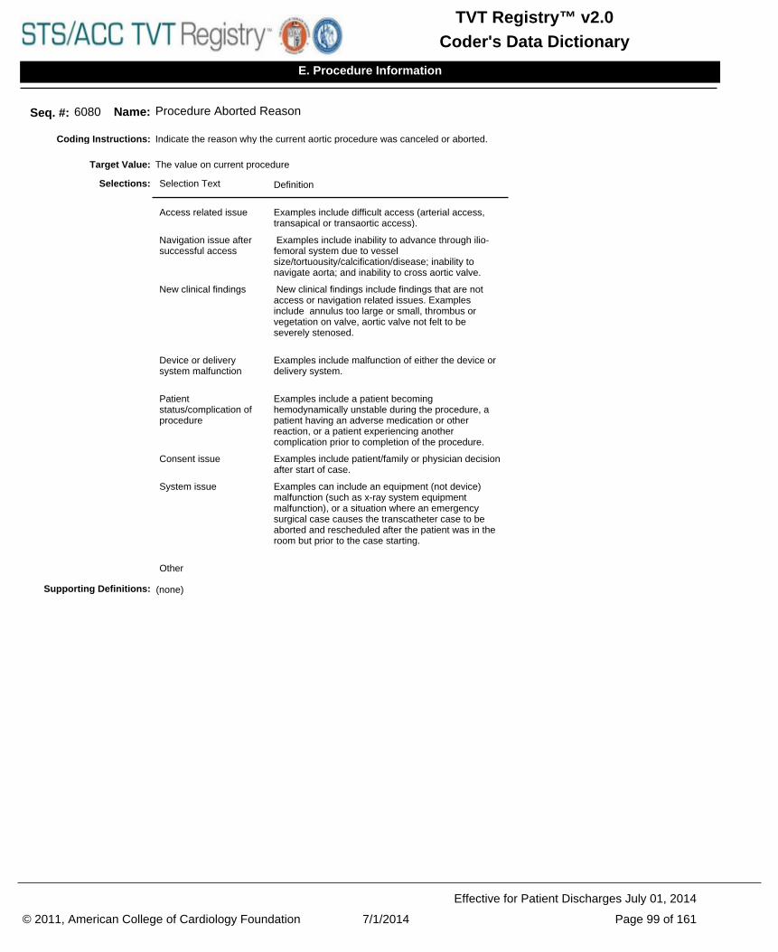
Indicate the reason why the current aortic procedure was canceled or aborted.Coding Instructions:
Seq. #: 6080 Name: Procedure Aborted Reason
The value on current procedureTarget Value:
Selection Text Definition
Access related issue Examples include difficult access (arterial access, transapical or transaortic access).
Navigation issue after successful access
Examples include inability to advance through ilio-femoral system due to vessel size/tortuousity/calcification/disease; inability to navigate aorta; and inability to cross aortic valve.
New clinical findings New clinical findings include findings that are not access or navigation related issues. Examples include annulus too large or small, thrombus or vegetation on valve, aortic valve not felt to be severely stenosed.
Device or delivery system malfunction
Examples include malfunction of either the device or delivery system.
Patient status/complication of procedure
Examples include a patient becoming hemodynamically unstable during the procedure, a patient having an adverse medication or other reaction, or a patient experiencing another complication prior to completion of the procedure.
Consent issue Examples include patient/family or physician decision after start of case.
System issue Examples can include an equipment (not device) malfunction (such as x-ray system equipment malfunction), or a situation where an emergency surgical case causes the transcatheter case to be aborted and rescheduled after the patient was in the room but prior to the case starting.
Other
Selections:
(none)Supporting Definitions:
© 2011, American College of Cardiology Foundation 7/1/2014 Page 99 of 161
Effective for Patient Discharges July 01, 2014
Coder's Data DictionaryTVT Registry™ v2.0
E. Procedure Information

Indicate the reason or action take as a result of the aborted aortic procedure.Coding Instructions:
Seq. #: 6082 Name: Procedure Aborted Action
The value on current procedureTarget Value:
Selection Text Definition
Balloon valvuloplasty
Rescheduled transcatheter procedure
Conversion to open heart surgery
Converted to medical therapy
Converted to clinical trial
Other
Selections:
(none)Supporting Definitions:
Indicate if conversion to open heart surgical access was required. Coding Instructions:
Open heart surgical access is the creation of an incision to open the chest and provide direct access to the heart. It may or may not involve placing the patient on cardiopulmonary bypass.
Note(s):
Seq. #: 6085 Name: Conversion to Open Heart Surgery
Any occurrence on current procedureTarget Value:
Selection Text Definition
No
Yes
Selections:
(none)Supporting Definitions:
© 2011, American College of Cardiology Foundation 7/1/2014 Page 100 of 161
Effective for Patient Discharges July 01, 2014
Coder's Data DictionaryTVT Registry™ v2.0
E. Procedure Information

Indicate the reason for conversion to open heart surgical access (aortic procedures).Coding Instructions:
Seq. #: 6090 Name: Conversion to Open Heart Surgery Reason (Aortic)
The value on current procedureTarget Value:
Selection Text Definition
Valve dislodged to aorta
Valve dislodged to left ventricle
Venticular rupture
Annulus rupture
Aortic dissection
Coronary occlusion
Other
Selections:
(none)Supporting Definitions:
Indicate if that patient had a mechanical assist device in place at the start of the procedure.Coding Instructions:
Seq. #: 6095 Name: Mechanical Assist Device in Place at Start of Procedure
The value on start of procedureTarget Value:
Selection Text Definition
No No mechanical assist device was in place at the start of the procedure.
Yes - IABP Intra-aortic balloon pump in place at the start of the procedure.
Yes - Catheter-based assist device
Catheter based assist device in place at start of the procedure. Examples include Impella and Tandem Heart.
Selections:
(none)Supporting Definitions:
Indicate if cardiopulmonary bypass or coronary perfusion was used during the procedure.Coding Instructions:
Seq. #: 6100 Name: CardioPulmonary Bypass Used
Any occurrence on the procedureTarget Value:
Selection Text Definition
No
Yes
Selections:
(none)Supporting Definitions:
© 2011, American College of Cardiology Foundation 7/1/2014 Page 101 of 161
Effective for Patient Discharges July 01, 2014
Coder's Data DictionaryTVT Registry™ v2.0
E. Procedure Information

Indicate if the use of cardiopulmonary bypass was elective or emergent.Coding Instructions:
Seq. #: 6101 Name: CardioPulmonary Bypass Status
The value on current procedureTarget Value:
Selection Text Definition
Elective Cardiopulmonary bypass was planned as part of the procedure.
Emergent Cardiopulmonary bypass was unplanned, and was initiated during the procedure.
Selections:
(none)Supporting Definitions:
Indicate the total number of minutes that systemic return is diverted into the cardiopulmonary bypass (CPB) circuit and returned to the systemic system. This time period (Cardiopulmonary Bypass Time) includes all periods of cerebral perfusion and sucker bypass. This time period (Cardiopulmonary Bypass Time) excludes any circulatory arrest and modified ultrafiltration periods. If more than one period of CPB is required during the procedure, the sum of all the CPB periods will equal the total number of CPB minutes.
Coding Instructions:
Seq. #: 6105 Name: Cardiopulmonary Bypass Time
The total between start of the procedure and end of the procedureTarget Value:
(none)Selections:
(none)Supporting Definitions:
Indicate the type of anesthesia used for the procedure.Coding Instructions:
Seq. #: 6110 Name: Anesthesia Type
The value on start of procedureTarget Value:
Selection Text Definition
Moderate sedation
General anesthesia
Epidural
Combination
Selections:
Moderate Sedation/Analgesia ("Conscious Sedation"):
Moderate Sedation/Analgesia ("Conscious Sedation") is a drug-induced depression of consciousness during which patients respond purposefully** to verbal commands, either alone or accompanied by light tactile stimulation. No interventions are required to maintain a patent airway, and spontaneous ventilation is adequate. Cardiovascular function is usually maintained.
Source: American Society of Anesthesiologists http://www.asahq.org/publicationsAndServices/standards/20.pdf
General Anesthesia:
General Anesthesia is a drug-induced loss of consciousness during which patients are not arousable, even by painful stimulation. The ability to independently maintain ventilatory function is often impaired. Patients often require assistance in maintaining a patent airway, and positive pressure ventilation may be required because of depressed spontaneous ventilation or drug-induced depression of neuromuscular function. Cardiovascular function may be impaired.
Source: American Society of Anesthesiologists http://www.asahq.org/publicationsAndServices/standards/20.pdf
Supporting Definitions:
© 2011, American College of Cardiology Foundation 7/1/2014 Page 102 of 161
Effective for Patient Discharges July 01, 2014
Coder's Data DictionaryTVT Registry™ v2.0
E. Procedure Information

Indicate which medications the patient received during the procedure.Coding Instructions:
Seq. #: 6120 Name: Intra-Procedure Medication
Any occurrence between start of the procedure and end of the procedureTarget Value:
(none)Selections:
(none)Supporting Definitions:
Indicate whether the patient received the medication during the procedure.Coding Instructions:
If a medication is contraindicated it needs to be documented in the chart by the physician or other responsible care giver. The reason for contraindication does not need to be documented.
Note(s):
Seq. #: 6125 Name: Intra-Procedure Medication Administered
Any occurrence between start of the procedure and end of the procedureTarget Value:
Selection Text Definition
No
Yes
Contraindicated
Blinded
Selections:
(none)Supporting Definitions:
Indicate the access site for the valve sheath.Coding Instructions:
Seq. #: 6200 Name: Valve Sheath Access Site (Aortic)
Any occurrence on current procedureTarget Value:
Selection Text Definition
Femoral
Axillary
Transapical
Transaortic
Subclavian
Transiliac
Transeptal
Transcarotid
Other
Selections:
(none)Supporting Definitions:
© 2011, American College of Cardiology Foundation 7/1/2014 Page 103 of 161
Effective for Patient Discharges July 01, 2014
Coder's Data DictionaryTVT Registry™ v2.0
E. Procedure Information

Indicate the access method used to deliver the valve sheath.Coding Instructions:
Seq. #: 6205 Name: Valve Sheath Access Method
Any occurrence on current procedureTarget Value:
Selection Text Definition
Percutaneous
Cutdown
Mini thoracotomy
Mini sternotomy
Other
Selections:
(none)Supporting Definitions:
Indicate the size, in french, of the valve sheath delivery system.Coding Instructions:
Seq. #: 6210 Name: Valve Sheath Delivery Size
The value on current procedureTarget Value:
(none)Selections:
(none)Supporting Definitions:
Indicate the leaflet clip guiding catheter access site.Coding Instructions:
Seq. #: 6212 Name: Leaflet Clip Guiding Cath Access Site
The value on current procedureTarget Value:
Selection Text Definition
Right femoral vein
Left femoral vein
Jugular vein
Other vein
Selections:
(none)Supporting Definitions:
This is a software-assigned value. The counter will start at one and be incremented by one for each device or system used.Coding Instructions:
Seq. #: 6220 Name: Device Counter
N/ATarget Value:
(none)Selections:
(none)Supporting Definitions:
© 2011, American College of Cardiology Foundation 7/1/2014 Page 104 of 161
Effective for Patient Discharges July 01, 2014
Coder's Data DictionaryTVT Registry™ v2.0
E. Procedure Information

Indicate all devices (valves, sheaths and delivery systems) utilized during the current procedure. If the valve, sheath and delivery system were separate components, code the manufacturer, model name and number for the sheath and delivery system as well as the manufacturer, model name and number, and serial number for all valves attempted and deployed during the procedure.
Coding Instructions:
Specify the devices in the order they were used.
If a kit is used, do not code the separate components within the kit. Specify the serial number of the valve used from the kit.
If more than one valve is placed (valve-in-valve) during the procedure, specify all devices and corresponding serial numbers (Seq Num 6230).
Note(s):
Seq. #: 6225 Name: Device Used
The value on current procedureTarget Value:
(none)Selections:
(none)Supporting Definitions:
Indicate the serial number of all valves attempted or implanted into the patient.Coding Instructions:
Serial numbers are only required for valves. If a kit is used, specify the serial number of the valve used from the kit.
Note(s):
Seq. #: 6230 Name: Device Serial Number
The value on current procedureTarget Value:
(none)Selections:
(none)Supporting Definitions:
Indicate if there is correct positioning of a single prosthetic heart valve in the proper anatomical location.Coding Instructions:
Seq. #: 6232 Name: Device Implanted Successfully
The value on current procedureTarget Value:
Selection Text Definition
No
Yes
Selections:
(none)Supporting Definitions:
© 2011, American College of Cardiology Foundation 7/1/2014 Page 105 of 161
Effective for Patient Discharges July 01, 2014
Coder's Data DictionaryTVT Registry™ v2.0
E. Procedure Information

Indicate if the device deployment was successful, as defined by the Standardized Endpoint Definitions for Transcatheter Aortic Valve Implantation Clinical Trials (JACC, 2011, vol 57, No 3).
Coding Instructions:
Seq. #: 6235 Name: Device Success
The value on current procedureTarget Value:
Selection Text Definition
No
Yes
Selections:
Transcatheter Valve Device Success:
Device Success is defined as all of the following:
1. Successful vascular access, delivery and deployment of the device and successful retrieval of the delivery system,2. Correct position of the device in the proper anatomical location, 3. Intended performance of the prosthetic heart valve (aortic valve area >1.2 cm2 and mean aortic valve gradient <20 mm Hg or peak velocity <3 m/s, without moderate or severe prosthetic valve AR),4. Only one valve implanted in the proper anatomical location.
Source: Standardized Endpoint Definitions for Transcatheter Aortic Valve Implantation Clinical Trials (JACC, 2011, vol 57, No 3)
Supporting Definitions:
[Reserved for Future Use] Indicate the direct identifier portion of the Unique Device Identifier (UDI) associated with the device used during the procedure. This ID is provided by the device manufacturer, and is either a GTIN or HIBBC number.
Coding Instructions:
The direct identifier portion of the UDI (unique device identifier) is provided by the manufacturer, specified at the unit of use. This is not the package barcode. The value should be mapped from your supply chain or inventory management system into this field. Depending on the device and manufacturer, this number could be between 12 to 25 digits.
- GTIN / GS1 standard: numeric, 12 – 14 characters- HIBCC standard: alphanumeric, 25 characters- ISBT-128: alphanumeric, 25 characters
If a device was not used, leave blank.
Note(s):
Seq. #: 6236 Name: Valve Device UDI Direct Identifier
The value on current procedureTarget Value:
(none)Selections:
UDI:
An identifier that is the main (primary) lookup for a medical device product and meets the requirements to uniquely identify a device through its distribution and use. This value is supplied to the FDA by the manufacturer.
Http://www.fda.gov/MedicalDevices/DeviceRegulationandGuidance/UniqueDeviceIdentification/default.htm
Source: FDA.gov
Supporting Definitions:
[Reserved for Future Use] Indicate the lot number associated with the device used to close the access site. This lot number is provided by the device manufacturer, and should be available within your supply chain or EHR system. Lot numbers indicate the specific manufacturing source or process.
Coding Instructions:
Seq. #: 6237 Name: Valve Device UDI Lot Number
The value on current procedureTarget Value:
(none)Selections:
(none)Supporting Definitions:
© 2011, American College of Cardiology Foundation 7/1/2014 Page 106 of 161
Effective for Patient Discharges July 01, 2014
Coder's Data DictionaryTVT Registry™ v2.0
E. Procedure Information

[Reserved for Future Use] Indicate the expiration date associated with the device used to close the access site. This expiration date is provided by the device manufacturer, and should be available within your supply chain or EHR system.
Coding Instructions:
Seq. #: 6238 Name: Valve Device UDI Expiration Date
The value on current procedureTarget Value:
(none)Selections:
(none)Supporting Definitions:
Indicate the post-implant mean aortic valve gradient in mmHg.Coding Instructions:
Seq. #: 6385 Name: Post-Implant Mean Aortic Valve Gradient
The value on current procedureTarget Value:
(none)Selections:
(none)Supporting Definitions:
Indicate the post-implant calculated aortic valve area, in centimeters squared.Coding Instructions:
Seq. #: 6395 Name: Post-Implant Calculated Aortic Valve Area
The value on current procedureTarget Value:
(none)Selections:
(none)Supporting Definitions:
Indicate the volume of contrast (ionic and non-ionic) used in milliliters (ml). The volume recorded should be the total volume for the procedure.
Coding Instructions:
Seq. #: 6450 Name: Contrast Volume
The total between start of the procedure and end of the procedureTarget Value:
(none)Selections:
(none)Supporting Definitions:
© 2011, American College of Cardiology Foundation 7/1/2014 Page 107 of 161
Effective for Patient Discharges July 01, 2014
Coder's Data DictionaryTVT Registry™ v2.0
E. Procedure Information

Indicate the method used to collect the radiation dose.Coding Instructions:
Seq. #: 6455 Name: Radiation Dose Measurement Method
The value on current procedureTarget Value:
Selection Text Definition
Single Plane
Biplane
Selections:
(none)Supporting Definitions:
Indicate the total fluoroscopy time recorded to the nearest 0.1 minute.Coding Instructions:
Please collect Fluoroscopy Time, Reference Air Kerma and Kerma Area Product values, if available.
Note(s):
Seq. #: 6460 Name: Fluoroscopy Time
The total between start of the procedure and end of the procedureTarget Value:
(none)Selections:
(none)Supporting Definitions:
Indicate the total radiation dose (Cumulative Air Kerma, or Reference Air Kerma) recorded to the nearest milligrays (mGy). The value recorded should include the total dose for the lab visit.
Coding Instructions:
Please collect Fluoroscopy Time, Cumulative Air Kerma and Dose Area Product values, if available.
If biplane equipment is used, collect the total dose of both planes and add them together.
Note(s):
Seq. #: 6465 Name: Fluoroscopy Dose - Cumulative Air Kerma
The total between start of the procedure and end of the procedureTarget Value:
(none)Selections:
Cumulative (Reference) Air kerma:
Cumulative air kerma (also known as reference, reference dose, cumulative dose, or cumulative dose at a reference point) is the air kerma accumulated at a specific point in space (the patient entrance reference point) relative to the gantry of the fluoroscopy system.
The quantity, kerma, originated from the acronym, KERMA, for Kinetic Energy Released per unit MAss (of air).
Source: Miller DL, Balter S, Cole PE, et al. Radiation doses in interventional radiology procedures: the RAD-IR study. I. Overall measures of dose. J Vasc Interv Radiol 2003; 14:711–727.
Supporting Definitions:
© 2011, American College of Cardiology Foundation 7/1/2014 Page 108 of 161
Effective for Patient Discharges July 01, 2014
Coder's Data DictionaryTVT Registry™ v2.0
E. Procedure Information
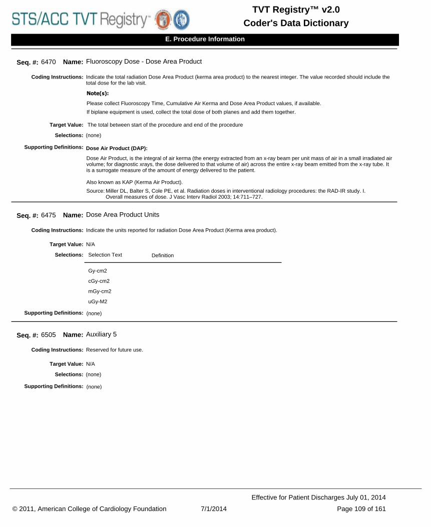
Indicate the total radiation Dose Area Product (kerma area product) to the nearest integer. The value recorded should include the total dose for the lab visit.
Coding Instructions:
Please collect Fluoroscopy Time, Cumulative Air Kerma and Dose Area Product values, if available.
If biplane equipment is used, collect the total dose of both planes and add them together.
Note(s):
Seq. #: 6470 Name: Fluoroscopy Dose - Dose Area Product
The total between start of the procedure and end of the procedureTarget Value:
(none)Selections:
Dose Air Product (DAP):
Dose Air Product, is the integral of air kerma (the energy extracted from an x-ray beam per unit mass of air in a small irradiated air volume; for diagnostic xrays, the dose delivered to that volume of air) across the entire x-ray beam emitted from the x-ray tube. It is a surrogate measure of the amount of energy delivered to the patient.
Also known as KAP (Kerma Air Product).
Source: Miller DL, Balter S, Cole PE, et al. Radiation doses in interventional radiology procedures: the RAD-IR study. I. Overall measures of dose. J Vasc Interv Radiol 2003; 14:711–727.
Supporting Definitions:
Indicate the units reported for radiation Dose Area Product (Kerma area product).Coding Instructions:
Seq. #: 6475 Name: Dose Area Product Units
N/ATarget Value:
Selection Text Definition
Gy-cm2
cGy-cm2
mGy-cm2
uGy-M2
Selections:
(none)Supporting Definitions:
Reserved for future use.Coding Instructions:
Seq. #: 6505 Name: Auxiliary 5
N/ATarget Value:
(none)Selections:
(none)Supporting Definitions:
© 2011, American College of Cardiology Foundation 7/1/2014 Page 109 of 161
Effective for Patient Discharges July 01, 2014
Coder's Data DictionaryTVT Registry™ v2.0
E. Procedure Information

Reserved for future use.Coding Instructions:
Seq. #: 6510 Name: Auxiliary 6
N/ATarget Value:
(none)Selections:
(none)Supporting Definitions:
Indicate if a transcatheter aortic valve replacement procedure was being performed.Coding Instructions:
Seq. #: 6600 Name: Procedure - Transcatheter Aortic Valve Replacement (TAVR)
The value on current procedureTarget Value:
Selection Text Definition
No
Yes
Selections:
(none)Supporting Definitions:
Indicate if a transcatheter mitral valve replacement procedure was being performed.Coding Instructions:
Seq. #: 6601 Name: Procedure - Transcatheter Mitral Valve Replacement
The value on current procedureTarget Value:
Selection Text Definition
No
Yes
Selections:
(none)Supporting Definitions:
Indicate if a mitral leaflet clip procedure was being performed.Coding Instructions:
Seq. #: 6602 Name: Procedure - Mitral Leaflet Clip Procedure
The value on current procedureTarget Value:
Selection Text Definition
No
Yes
Selections:
(none)Supporting Definitions:
© 2011, American College of Cardiology Foundation 7/1/2014 Page 110 of 161
Effective for Patient Discharges July 01, 2014
Coder's Data DictionaryTVT Registry™ v2.0
E. Procedure Information

Indicate if an other procedure was performed concurrently.Coding Instructions:
Seq. #: 6620 Name: Other Procedure Performed Concurrently
The value on current procedureTarget Value:
Selection Text Definition
No
Yes - PCI
Yes - Other
Selections:
(none)Supporting Definitions:
© 2011, American College of Cardiology Foundation 7/1/2014 Page 111 of 161
Effective for Patient Discharges July 01, 2014
Coder's Data DictionaryTVT Registry™ v2.0
E. Procedure Information

Indicate if any intra or post procedure event occurred.Coding Instructions:
Seq. #: 7300 Name: Intra or Post Procedure Event Occurred
Any occurrence between start of the procedure and dischargeTarget Value:
Selection Text Definition
No
Yes
Selections:
(none)Supporting Definitions:
Indicate all adverse events, interventions or surgical procedures that occurred intra or post procedure.Coding Instructions:
If an event occurred more than once, specify each event with its corresponding Intra/Post Procedure Event Date (Seq Number 7302).
Note(s):
Seq. #: 7301 Name: Intra or Post Procedure Event ID
The value between start of procedure and dischargeTarget Value:
(none)Selections:
(none)Supporting Definitions:
Indicate the date of any adverse events, interventions, or surgical procedures that occurred intra or post procedure.Coding Instructions:
If an event occurred more than once, specify each Intra/Post Procedure Event ID (7301) with its corresponding date.
Note(s):
Seq. #: 7302 Name: Intra or Post Procedure Event Date
The value between start of procedure and dischargeTarget Value:
(none)Selections:
(none)Supporting Definitions:
© 2011, American College of Cardiology Foundation 7/1/2014 Page 112 of 161
Effective for Patient Discharges July 01, 2014
Coder's Data DictionaryTVT Registry™ v2.0
F. Adverse Events/Interventions/Surgical Procedures

Indicate the lowest post-procedure hemoglobin level in g/dL.Coding Instructions:
Seq. #: 8040 Name: Post-Procedure Hemoglobin
The lowest value between end of procedure and dischargeTarget Value:
(none)Selections:
(none)Supporting Definitions:
Indicate if a post procedure hemoglobin level was not drawn.Coding Instructions:
Seq. #: 8041 Name: Post-Procedure Hemoglobin Not Drawn
N/ATarget Value:
Selection Text Definition
No
Yes Code "Yes" if the lab was not drawn.
Selections:
(none)Supporting Definitions:
Indicate the highest postoperative creatinine level, in mg/dL. If more than one level is obtained, code the highest level.Coding Instructions:
Seq. #: 8050 Name: Post-Procedure Creatinine Level
The highest value between end of procedure and dischargeTarget Value:
(none)Selections:
(none)Supporting Definitions:
Indicate if a post procedure creatinine level was not drawn.Coding Instructions:
Seq. #: 8051 Name: Post-Procedure Creatinine Level Not Drawn
N/ATarget Value:
Selection Text Definition
No
Yes Code "Yes" if the lab was not drawn.
Selections:
(none)Supporting Definitions:
© 2011, American College of Cardiology Foundation 7/1/2014 Page 113 of 161
Effective for Patient Discharges July 01, 2014
Coder's Data DictionaryTVT Registry™ v2.0
G. Post-Procedure Labs and Tests

Indicate the last post-procedure creatinine level documented in the medical record prior to discharge, in mg/dL.Coding Instructions:
Seq. #: 8055 Name: Discharge Creatinine
The last value between end of procedure and dischargeTarget Value:
(none)Selections:
(none)Supporting Definitions:
Indicate if a discharge creatinine level was not drawn.Coding Instructions:
Seq. #: 8056 Name: Discharge Creatinine Not Drawn
N/ATarget Value:
Selection Text Definition
No
Yes Code "Yes" if the lab was not drawn.
Selections:
(none)Supporting Definitions:
Indicate the post procedure 12 lead ECG findings, if performed. If more than one ECG is performed, document the findings from the ECG closest to discharge.
Coding Instructions:
Seq. #: 8060 Name: Post-Procedure 12 Lead ECG
The last value between end of procedure and dischargeTarget Value:
Selection Text Definition
Not performed
No significant changes
New pathological Q-wave or LBBB
Selections:
(none)Supporting Definitions:
© 2011, American College of Cardiology Foundation 7/1/2014 Page 114 of 161
Effective for Patient Discharges July 01, 2014
Coder's Data DictionaryTVT Registry™ v2.0
G. Post-Procedure Labs and Tests

Indicate whether an echo (and the type of echo) was performed postoperatively prior to discharge.Coding Instructions:
If both types of echos were performed, code "Yes - Tranesophageal Echocardiogram".
Note(s):
Seq. #: 8065 Name: Post-Procedure Echocardiogram
Any occurrence between end of the procedure and dischargeTarget Value:
Selection Text Definition
Not Performed
Yes - TTE Yes - Transthoracic Echocardiogram
Yes - TEE Yes - Tranesophageal Echocardiogram
Selections:
(none)Supporting Definitions:
Indicate the date the echo was performed.Coding Instructions:
Seq. #: 8070 Name: Post-Procedure Echocardiogram Date
The value between end of procedure and dischargeTarget Value:
(none)Selections:
(none)Supporting Definitions:
Indicate the highest level of mitral regurgitation found on echocardiogram prior to discharge.Coding Instructions:
Seq. #: 8075 Name: Post-Procedure Mitral Regurgitation
The highest value between end of procedure and dischargeTarget Value:
Selection Text Definition
None
Trace/Trivial
Mild 1+/Mild
Moderate 2+/Moderate
Moderate-Severe 3+/Moderate-severe
Severe 4+/Severe
Severe Retired TVT 2.00
Selections:
(none)Supporting Definitions:
© 2011, American College of Cardiology Foundation 7/1/2014 Page 115 of 161
Effective for Patient Discharges July 01, 2014
Coder's Data DictionaryTVT Registry™ v2.0
G. Post-Procedure Labs and Tests

Indicate whether aortic stenosis is present.Coding Instructions:
Seq. #: 8080 Name: Post-Procedure Aortic Stenosis
Any occurrence between end of the procedure and dischargeTarget Value:
Selection Text Definition
No
Yes
Selections:
(none)Supporting Definitions:
Indicate the smallest aortic valve area (in cm2) obtained from an echocardiogram.Coding Instructions:
Seq. #: 8085 Name: Post-Procedure Aortic Valve Area
The lowest value between end of procedure and dischargeTarget Value:
(none)Selections:
(none)Supporting Definitions:
Indicate the aortic valve peak velocity in meters per second, as determined by continuous wave (CW) spectral velocity recording on echocardiography.
Coding Instructions:
Seq. #: 8086 Name: Post-Procedure Aortic Valve Peak Velocity
The highest value between end procedure and dischargeTarget Value:
(none)Selections:
(none)Supporting Definitions:
Indicate the aortic valve mean gradient in mmHg obtained from echocardiogram.Coding Instructions:
Seq. #: 8090 Name: Post-Procedure Aortic Valve Mean Gradient
The highest value between end of procedure and dischargeTarget Value:
(none)Selections:
(none)Supporting Definitions:
© 2011, American College of Cardiology Foundation 7/1/2014 Page 116 of 161
Effective for Patient Discharges July 01, 2014
Coder's Data DictionaryTVT Registry™ v2.0
G. Post-Procedure Labs and Tests

Indicate the highest level of aortic regurgitation found on the echocardiogram.Coding Instructions:
Mild-to-moderate should be coded as moderate; moderate to severe should be coded as severe.
Reference:Bonow, R.O, et al. 2008 Focused Updated Incorporated into ACC/AHA 2006 Guidelines for the Management of Patients with Valvular Heart Disease: A Report of the American College of Cardiology /American Heart Association Task force on Practice Guidelines. JACC, vol 52, No. 13, 2008, p. e1-e142.
Note(s):
Seq. #: 8095 Name: Post-Procedure Aortic Regurgitation
The highest value between end of procedure and dischargeTarget Value:
Selection Text Definition
None
Trace/Trivial
Mild Mild aortic insufficiency or regurgitation is defined as the following:
Qualitative Measurements:Angiographic grade of 1+; Color Doppler jet width (Central jet) <25% of LVOT; Dopplar vena contracta width <0.3 cm;.
Quantitative Measures (cath or echo)Regurgitant volume <30 ml/beat;Regurgitant fraction <30%;Regurgitant orifice area <0.10 cm(2)
Moderate Moderate aortic insufficiency or regurgitation is defined as the following:
Qualitative Measurements:Angiographic grade of 2+; Color Doppler jet width greater than mild but no signs of severe aortic regurgitation (insufficiency); Dopplar vena contracta width 0.3-0.6 cm;
Quantitative Measures (cath or echo)Regurgitant volume 30-59 ml/beat;Regurgitant fraction 30-49%;Regurgitant orifice area 0.10-0.29 cm(2)
Severe Severe aortic insufficiency or regurgitation is defined as the following:
Qualitative Measurements:Angiographic grade of 3-4+; Color Doppler jet width (Central jet) >65% of LVOT; Dopplar vena contracta width >0.6 cm;.
Quantitative Measures (cath or echo)Regurgitant volume >=60 ml/beat;Regurgitant fraction >=50%;Regurgitant orifice area >=0.30 cm(2)
Additional essential criteria:Left ventricular size is increased
Selections:
(none)Supporting Definitions:
© 2011, American College of Cardiology Foundation 7/1/2014 Page 117 of 161
Effective for Patient Discharges July 01, 2014
Coder's Data DictionaryTVT Registry™ v2.0
G. Post-Procedure Labs and Tests

Indicate the highest severity of paravalvular aortic insufficiency. Coding Instructions:
Seq. #: 8106 Name: TAVR - Paravalvular Severity
The highest value between end of procedure and dischargeTarget Value:
Selection Text Definition
None
Mild
Moderate
Severe
Not Documented
Selections:
(none)Supporting Definitions:
Indicate the highest severity of central aortic insufficiency. Coding Instructions:
Seq. #: 8107 Name: TAVR - Valvular Severity
The highest value between end of procedure and dischargeTarget Value:
Selection Text Definition
None
Mild
Moderate
Severe
Not Documented
Selections:
(none)Supporting Definitions:
Indicate the highest severity of paravalvular mitral regurgitation.Coding Instructions:
Seq. #: 8112 Name: Mitral Replacement - Paravalvular Severity
The highest value between end of current procedure and dischargeTarget Value:
Selection Text Definition
None
Mild
Moderate
Severe
Not Documented
Selections:
(none)Supporting Definitions:
© 2011, American College of Cardiology Foundation 7/1/2014 Page 118 of 161
Effective for Patient Discharges July 01, 2014
Coder's Data DictionaryTVT Registry™ v2.0
G. Post-Procedure Labs and Tests
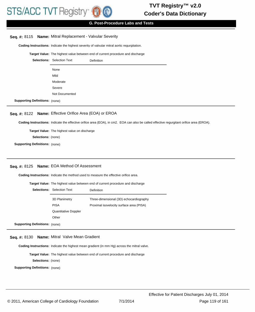
Indicate the highest severity of valvular mitral aortic regurgitation.Coding Instructions:
Seq. #: 8115 Name: Mitral Replacement - Valvular Severity
The highest value between end of current procedure and dischargeTarget Value:
Selection Text Definition
None
Mild
Moderate
Severe
Not Documented
Selections:
(none)Supporting Definitions:
Indicate the effective orifice area (EOA), in cm2. EOA can also be called effective regurgitant orifice area (EROA).Coding Instructions:
Seq. #: 8122 Name: Effective Orifice Area (EOA) or EROA
The highest value on dischargeTarget Value:
(none)Selections:
(none)Supporting Definitions:
Indicate the method used to measure the effective orifice area.Coding Instructions:
Seq. #: 8125 Name: EOA Method Of Assessment
The highest value between end of current procedure and dischargeTarget Value:
Selection Text Definition
3D Planimetry Three-dimensional (3D) echocardiography
PISA Proximal isovelocity surface area (PISA)
Quantitative Doppler
Other
Selections:
(none)Supporting Definitions:
Indicate the highest mean gradient (in mm Hg) across the mitral valve.Coding Instructions:
Seq. #: 8130 Name: Mitral Valve Mean Gradient
The highest value between end of current procedure and dischargeTarget Value:
(none)Selections:
(none)Supporting Definitions:
© 2011, American College of Cardiology Foundation 7/1/2014 Page 119 of 161
Effective for Patient Discharges July 01, 2014
Coder's Data DictionaryTVT Registry™ v2.0
G. Post-Procedure Labs and Tests

Indicate the smallest mitral valve area in centimeters squared.Coding Instructions:
Seq. #: 8135 Name: Mitral Valve Area
(None)Target Value:
(none)Selections:
(none)Supporting Definitions:
Indicate the peak gradient of the left ventricular outflow tract.Coding Instructions:
Seq. #: 8140 Name: Left Ventricular Outflow Tract Gradient (Peak)
The highest value between end of current procedure and dischargeTarget Value:
(none)Selections:
(none)Supporting Definitions:
Indicate if systolic anterior motion was present.Coding Instructions:
Seq. #: 8145 Name: Systolic Anterior Motion Present
Any occurrence between end of current procedure and dischargeTarget Value:
Selection Text Definition
No
Yes
Selections:
(none)Supporting Definitions:
© 2011, American College of Cardiology Foundation 7/1/2014 Page 120 of 161
Effective for Patient Discharges July 01, 2014
Coder's Data DictionaryTVT Registry™ v2.0
G. Post-Procedure Labs and Tests

Indicate if there was a transfusion of either whole blood or packed red blood cells.Coding Instructions:
Seq. #: 9011 Name: RBC/Whole Blood Transfusion
Any occurrence between start of the procedure and dischargeTarget Value:
Selection Text Definition
No
Yes
Selections:
(none)Supporting Definitions:
Indicate the total number of units transfused of either whole blood and/or packed red blood cells. Coding Instructions:
Do not include autologous, cell-saver or chest tube recirculated blood.
Note(s):
Seq. #: 9012 Name: RBC/Whole Blood Transfusion Units Transfused
The total between start of the procedure and dischargeTarget Value:
(none)Selections:
(none)Supporting Definitions:
Indicate the total number of hours spent in the intensive care unit. Do not include hours spent in a telemetry or step-down unit.Coding Instructions:
Seq. #: 9040 Name: Number of Hours in ICU
The total between end of the procedure and dischargeTarget Value:
(none)Selections:
(none)Supporting Definitions:
Indicate the date on which the patient was discharged from your facility.Coding Instructions:
If the deceased is an organ donor, code the Discharge Date as the date of the final organ harvest.
Note(s):
Seq. #: 9045 Name: Discharge Date
The value on dischargeTarget Value:
(none)Selections:
(none)Supporting Definitions:
© 2011, American College of Cardiology Foundation 7/1/2014 Page 121 of 161
Effective for Patient Discharges July 01, 2014
Coder's Data DictionaryTVT Registry™ v2.0
H. Discharge

Indicate whether the patient was alive or deceased at discharge.Coding Instructions:
Seq. #: 9050 Name: Discharge Status
The value on dischargeTarget Value:
Selection Text Definition
Alive
Deceased
Selections:
(none)Supporting Definitions:
Indicate the location to where the patient was discharged.Coding Instructions:
Seq. #: 9055 Name: Discharge Location
The value on dischargeTarget Value:
Selection Text Definition
Home
Extended care/TCU/rehab
Continued “non-acute” care at an extended care facility, transitional care unit, or rehabilitation unit.
Other acute care hospital
Nursing home
Hospice
Other
Left against medical advice
The patient was discharged or eloped against medical advice.
Selections:
(none)Supporting Definitions:
If the patient expired during this hospitalization, indicate if the patient expired in the cath lab, operating room or hybrid suite.Coding Instructions:
Seq. #: 9060 Name: Death in Lab/OR
The value on dischargeTarget Value:
Selection Text Definition
No
Yes
Selections:
(none)Supporting Definitions:
© 2011, American College of Cardiology Foundation 7/1/2014 Page 122 of 161
Effective for Patient Discharges July 01, 2014
Coder's Data DictionaryTVT Registry™ v2.0
H. Discharge

Select the PRIMARY cause of death, i.e. the first significant abnormal event which ultimately led to death.Coding Instructions:
Seq. #: 9065 Name: Primary Cause of Death
The value on dischargeTarget Value:
Selection Text Definition
Cardiac
Neurologic
Renal
Vascular
Infection
Valvular
Pulmonary
Unknown
Other
Selections:
(none)Supporting Definitions:
Indicate which of the following medications were prescribed at discharge.Coding Instructions:
Seq. #: 9100 Name: Discharge Medication
Any occurrence on dischargeTarget Value:
(none)Selections:
(none)Supporting Definitions:
Indicate if the medication was administered, not administered, contraindicated or blinded.Coding Instructions:
If a medication is contraindicated it needs to be documented in the chart by the physician or other responsible care giver. The reason for contraindication does not need to be documented.
Note(s):
Seq. #: 9105 Name: Discharge Medication Administered
The value on dischargeTarget Value:
Selection Text Definition
No
Yes
Contraindicated
Blinded
Selections:
(none)Supporting Definitions:
© 2011, American College of Cardiology Foundation 7/1/2014 Page 123 of 161
Effective for Patient Discharges July 01, 2014
Coder's Data DictionaryTVT Registry™ v2.0
H. Discharge

Specify the total daily dose of the medication prescribed at discharge.Coding Instructions:
Seq. #: 9110 Name: Medication Dose at Discharge
The value on dischargeTarget Value:
(none)Selections:
(none)Supporting Definitions:
© 2011, American College of Cardiology Foundation 7/1/2014 Page 124 of 161
Effective for Patient Discharges July 01, 2014
Coder's Data DictionaryTVT Registry™ v2.0
H. Discharge

Indicate the date the follow-up assessment was performed.Coding Instructions:
Seq. #: 10000 Name: Follow-up Assessment Date
The value on follow-upTarget Value:
(none)Selections:
(none)Supporting Definitions:
Indicate the primary method to determine patient status at follow-up.Coding Instructions:
Seq. #: 10005 Name: Follow-up Assessment Method
Any occurrence on follow-upTarget Value:
Selection Text Definition
Clinic
Medical record
Letter from medical provider
Phone call to patient/family
Social Security Death master File
Other
Selections:
(none)Supporting Definitions:
Indicate the primary residence of the patient during the follow-up period. If the primary residence is not available, code not documented.
Coding Instructions:
Seq. #: 10008 Name: Follow-up Residence
The value on follow-upTarget Value:
Selection Text Definition
Home with no health-aid
Home with health aid
Long term care
Other
Not Documented
Selections:
(none)Supporting Definitions:
© 2011, American College of Cardiology Foundation 7/1/2014 Page 125 of 161
Effective for Patient Discharges July 01, 2014
Coder's Data DictionaryTVT Registry™ v2.0
I. Follow-Up

Indicate whether the patient was alive or deceased at the date the follow-up was performed.Coding Instructions:
Seq. #: 10010 Name: Follow-up Status
Any occurrence on follow-upTarget Value:
Selection Text Definition
Alive
Deceased
Lost to follow-up
Withdrawn
Selections:
(none)Supporting Definitions:
Indicate the PRIMARY cause of death (i.e. the first significant event which ultimately led to death).Coding Instructions:
Seq. #: 10015 Name: Follow-up Primary Cause of Death
Any occurrence on follow-upTarget Value:
Selection Text Definition
Cardiac
Neurologic
Renal
Vascular
Infection
Valvular
Pulmonary
Unknown
Other
Selections:
(none)Supporting Definitions:
Indicate the date the patient was declared dead.Coding Instructions:
Seq. #: 10020 Name: Follow-up Date of Death
The value on follow-upTarget Value:
(none)Selections:
(none)Supporting Definitions:
© 2011, American College of Cardiology Foundation 7/1/2014 Page 126 of 161
Effective for Patient Discharges July 01, 2014
Coder's Data DictionaryTVT Registry™ v2.0
I. Follow-Up

Indicate the hemoglobin value in g/dL collected at follow-up. A hemoglobin level should be collected on all patients to assess for bleeding events as a result of the procedure.
Coding Instructions:
Seq. #: 10085 Name: Follow-up Hemoglobin
The last value on follow-upTarget Value:
(none)Selections:
(none)Supporting Definitions:
Indicate if a hemoglobin level was not drawn during the follow-up period.Coding Instructions:
Seq. #: 10086 Name: Follow-up Hemoglobin Not Drawn
N/ATarget Value:
Selection Text Definition
No
Yes Code "Yes" if the lab was not drawn.
Selections:
(none)Supporting Definitions:
Indicate the creatinine level collected at follow-up, in mg/dL. A creatinine level should be collected on all patients to determine kidney injury as a result of the procedure.
Coding Instructions:
Seq. #: 10090 Name: Follow-up Creatinine
The last value on follow-upTarget Value:
(none)Selections:
(none)Supporting Definitions:
Indicate if a creatinine level was not drawn during the follow-up period.Coding Instructions:
Seq. #: 10091 Name: Follow-up Creatinine Not Drawn
N/ATarget Value:
Selection Text Definition
No
Yes Code "Yes" if the lab was not drawn.
Selections:
(none)Supporting Definitions:
© 2011, American College of Cardiology Foundation 7/1/2014 Page 127 of 161
Effective for Patient Discharges July 01, 2014
Coder's Data DictionaryTVT Registry™ v2.0
I. Follow-Up

Indicate the patient's functional class, coded as the New York Heart Association (NYHA) classification at follow-up.Coding Instructions:
Seq. #: 10100 Name: Follow-up NYHA Classification
The highest value on follow-upTarget Value:
Selection Text Definition
I Patient has cardiac disease but without resulting limitations of ordinary physical activity. Ordinary physical activity (e.g., walking several blocks or climbing stairs) does not cause undue fatigue, palpitation, dyspnea, or anginal pain. Limiting symptoms may occur with marked exertion.
II Patient has cardiac disease resulting in slight limitation of ordinary physical activity. Patient is comfortable at rest. Ordinary physical activity such as walking more than two blocks or climbing more than one flight of stairs results in limiting symptoms (e.g., fatigue, palpitation, dyspnea, or anginal pain).
III Patient has cardiac disease resulting in marked limitation of physical activity. Patient is comfortable at rest. Less than ordinary physical activity (e.g., walking one to two level blocks or climbing one flight of stairs) causes fatigue, palpitation, dyspnea, or anginal pain.
IV Patient has symptoms at rest that increase with any physical activity. Patient has cardiac disease resulting in inability to perform any physical activity without discomfort. Symptoms may be present even at rest. If any physical activity is undertaken, discomfort is increased.
Selections:
(none)Supporting Definitions:
Indicate whether the five meter walk test was performed during the follow-up period.Coding Instructions:
Seq. #: 10135 Name: Follow-up Five Meter Walk Test Performed
Any occurrence on follow-upTarget Value:
Selection Text Definition
Not performed
Yes The five meter walk test was performed.
Unable to walk Five meter walk was not performed because the patient is unable to walk.
Selections:
(none)Supporting Definitions:
Indicate the time in seconds it takes the patient to walk 5 meters for the first of three tests.Coding Instructions:
Seq. #: 10140 Name: Follow-up Five Meter Walk Test Time 1
The value on follow-upTarget Value:
(none)Selections:
(none)Supporting Definitions:
© 2011, American College of Cardiology Foundation 7/1/2014 Page 128 of 161
Effective for Patient Discharges July 01, 2014
Coder's Data DictionaryTVT Registry™ v2.0
I. Follow-Up

Indicate the time in seconds it takes the patient to walk 5 meters for the second of three tests.Coding Instructions:
Seq. #: 10145 Name: Follow-up Five Meter Walk Test Time 2
The value on follow-upTarget Value:
(none)Selections:
(none)Supporting Definitions:
Indicate the time in seconds it takes the patient to walk 5 meters for the third of three tests.Coding Instructions:
Seq. #: 10150 Name: Follow-up Five Meter Walk Test Time 3
The value on follow-upTarget Value:
(none)Selections:
(none)Supporting Definitions:
Indicate the 12 lead ECG findings during the follow-up period.Coding Instructions:
Seq. #: 10155 Name: Follow-up 12-Lead ECG Findings
Any occurrence on follow-upTarget Value:
Selection Text Definition
Not performed
No significant changes
New changes noted
Selections:
(none)Supporting Definitions:
Indicate the ECG changes noted on the follow-up ECG.Coding Instructions:
Seq. #: 10160 Name: Follow-up 12-Lead ECG Changes Noted
Any occurrence on follow-upTarget Value:
Selection Text Definition
Pathological Q-wave or LBBB
New pathologic q-waves and/or new left bundle branch block.
Arrhythmia New onset of atrial or ventricular arrhythmia requiring medication or other therapy.
Both
Selections:
(none)Supporting Definitions:
© 2011, American College of Cardiology Foundation 7/1/2014 Page 129 of 161
Effective for Patient Discharges July 01, 2014
Coder's Data DictionaryTVT Registry™ v2.0
I. Follow-Up

Indicate if an echocardiogram has been performed.Coding Instructions:
Seq. #: 10206 Name: Follow-up Echocardiogram
Any occurrence on follow-upTarget Value:
Selection Text Definition
Not Performed
Yes - TTE
Yes - TEE
Selections:
(none)Supporting Definitions:
Indicate the date the echocardiogram was performed. If more than one echocardiogram has been performed since discharge or the last follow-up period, code the date of the most recent echo.
Coding Instructions:
Seq. #: 10207 Name: Follow-up Date
The last value on follow-upTarget Value:
(none)Selections:
(none)Supporting Definitions:
Indicate the left ventricular ejection fraction (LVEF), or the percentage of the blood emptied from the left ventricle at the end of the contraction.
Use the most recent echocardiogram documented. Enter a percentage in the range of 1 - 99. If a percentage range is reported, report a whole number using the "mean" (i.e., 50-55%, is reported as 53%).If only a descriptive value is reported, (i.e., normal), enter the corresponding percentage value from the list below:
Normal = 60%Good function = 50%Mildly reduced = 45%Fair function = 40%Moderately reduced = 30%Poor function = 25%Severely reduced = 20%
Coding Instructions:
Seq. #: 10210 Name: Follow-up LVEF
The last value on follow-upTarget Value:
(none)Selections:
(none)Supporting Definitions:
© 2011, American College of Cardiology Foundation 7/1/2014 Page 130 of 161
Effective for Patient Discharges July 01, 2014
Coder's Data DictionaryTVT Registry™ v2.0
I. Follow-Up

Indicate whether the left ventricular ejection fraction was not assessed.Coding Instructions:
Seq. #: 10211 Name: Follow-up LVEF Not Assessed
N/ATarget Value:
Selection Text Definition
No
Yes
Selections:
(none)Supporting Definitions:
Indicate the aortic valve mean gradient in mmHg captured on echocardiogram since discharge or the last follow-up period.Coding Instructions:
Seq. #: 10215 Name: Follow-up Aortic Valve Mean Gradient
The last value on follow-upTarget Value:
(none)Selections:
(none)Supporting Definitions:
Indicate the highest level of aortic regurgitation found on echo since discharge or the last follow-up period.Coding Instructions:
Mild-to-moderate should be coded as moderate; moderate to severe should be coded as severe.
Note(s):
Seq. #: 10220 Name: Follow-up Aortic Regurtitation Severity
The highest value on follow-upTarget Value:
Selection Text Definition
None
Trace/Trivial
Mild
Moderate
Severe
Selections:
(none)Supporting Definitions:
© 2011, American College of Cardiology Foundation 7/1/2014 Page 131 of 161
Effective for Patient Discharges July 01, 2014
Coder's Data DictionaryTVT Registry™ v2.0
I. Follow-Up

Indicate the highest severity of perivalvular leak found on echo since discharge or the last follow-up period. Coding Instructions:
Seq. #: 10225 Name: Follow-up Aortic Regurgitation Paravalvular Severity
The highest value on follow-upTarget Value:
Selection Text Definition
None
Mild
Moderate
Severe
Not Documented
Selections:
(none)Supporting Definitions:
Indicate the highest severity of central leak found on echo since discharge or the last follow-up period.Coding Instructions:
Seq. #: 10227 Name: Follow-up Aortic Regurgitation Central Severity
The highest value on follow-upTarget Value:
Selection Text Definition
None
Mild
Moderate
Severe
Not Documented
Selections:
(none)Supporting Definitions:
Indicate if the follow-up Kansas City Cardiomyopathy Questionnaire (KCCQ-12) was performed.Coding Instructions:
Please refer to the separate KCCQ-12 questionnaire for patient instructions. For additional information on scoring, please refer to Seq Num 10243, Follow-Up KCCQ-12 Overall Summary Score.
Note(s):
Seq. #: 10230 Name: Follow-Up KCCQ-12 Patient Questionnaire Performed
Any occurrence on follow-upTarget Value:
Selection Text Definition
No
Yes
Selections:
(none)Supporting Definitions:
© 2011, American College of Cardiology Foundation 7/1/2014 Page 132 of 161
Effective for Patient Discharges July 01, 2014
Coder's Data DictionaryTVT Registry™ v2.0
I. Follow-Up

Indicate the patient's response to the Kansas City Cardiomyopathy Questionnaire (KCCQ-12) Question 1a.
Heart Failure Limitation - Showering/bathing
Coding Instructions:
Please refer to the separate KCCQ-12 questionnaire for patient instructions. For additional information on scoring, please refer to Seq Num 10243, Follow-Up KCCQ-12 Overall Summary Score.
Note(s):
Seq. #: 10231 Name: Follow-Up KCCQ-12 Question 1a
The value on follow-upTarget Value:
Selection Text Definition
Extremely limited
Quite a bit limited
Moderately limited
Slightly limited
Not at all limited
Limited for other reasons or did not do the activity
Selections:
(none)Supporting Definitions:
Indicate the patient's response to the Kansas City Cardiomyopathy Questionnaire (KCCQ-12) Question 1b.
Heart Failure Limitation - Walking 1 block on level ground
Coding Instructions:
Please refer to the separate KCCQ-12 questionnaire for patient instructions. For additional information on scoring, please refer to Seq Num 10243, Follow-Up KCCQ-12 Overall Summary Score.
Note(s):
Seq. #: 10232 Name: Follow-Up KCCQ-12 Question 1b
The value on follow-upTarget Value:
Selection Text Definition
Extremely limited
Quite a bit limited
Moderately limited
Slightly limited
Not at all limited
Limited for other reasons or did not do the activity
Selections:
(none)Supporting Definitions:
© 2011, American College of Cardiology Foundation 7/1/2014 Page 133 of 161
Effective for Patient Discharges July 01, 2014
Coder's Data DictionaryTVT Registry™ v2.0
I. Follow-Up

Indicate the patient's response to the Kansas City Cardiomyopathy Questionnaire (KCCQ-12) Question 1c.
Heart Failure Limitation - Hurrying or jogging
Coding Instructions:
Please refer to the separate KCCQ-12 questionnaire for patient instructions. For additional information on scoring, please refer to Seq Num 10243, Follow-Up KCCQ-12 Overall Summary Score.
Note(s):
Seq. #: 10233 Name: Follow-Up KCCQ-12 Question 1c
The value on follow-upTarget Value:
Selection Text Definition
Extremely limited
Quite a bit limited
Moderately limited
Slightly limited
Not at all limited
Limited for other reasons or did not do the activity
Selections:
(none)Supporting Definitions:
Indicate the patient's response to the Kansas City Cardiomyopathy Questionnaire (KCCQ-12) Question 2.
Symptom Frequency - Swelling in legs
Coding Instructions:
Please refer to the separate KCCQ-12 questionnaire for patient instructions. For additional information on scoring, please refer to Seq Num 10243, Follow-Up KCCQ-12 Overall Summary Score.
Note(s):
Seq. #: 10234 Name: Follow-Up KCCQ-12 Question 2
The value on follow-upTarget Value:
Selection Text Definition
Every morning
3 or more times per week but not every day
1-2 times per week
Less than once a week
Never over the past 2 weeks
Selections:
(none)Supporting Definitions:
© 2011, American College of Cardiology Foundation 7/1/2014 Page 134 of 161
Effective for Patient Discharges July 01, 2014
Coder's Data DictionaryTVT Registry™ v2.0
I. Follow-Up

Indicate the patient's response to the Kansas City Cardiomyopathy Questionnaire (KCCQ-12) Question 3.
Symptom Frequency - Fatigue
Coding Instructions:
Seq. #: 10235 Name: Follow-Up KCCQ-12 Question 3
The value on follow-upTarget Value:
Selection Text Definition
All of the time
Several times per day
At least once a day
3 or more times per week but not every day
1-2 times per week
Less than once a week
Never over the past 2 weeks
Selections:
(none)Supporting Definitions:
Indicate the patient's response to the Kansas City Cardiomyopathy Questionnaire (KCCQ-12) Question 4.
Symptom Frequency - shortness of breath
Coding Instructions:
Please refer to the separate KCCQ-12 questionnaire for patient instructions. For additional information on scoring, please refer to Seq Num 10243, Follow-Up KCCQ-12 Overall Summary Score.
Note(s):
Seq. #: 10236 Name: Follow-Up KCCQ-12 Question 4
The value on follow-upTarget Value:
Selection Text Definition
All of the time
Several times per day
At least once a day
3 or more times per week but not every day
1-2 times per week
Less than once a week
Never over the past 2 weeks
Selections:
(none)Supporting Definitions:
© 2011, American College of Cardiology Foundation 7/1/2014 Page 135 of 161
Effective for Patient Discharges July 01, 2014
Coder's Data DictionaryTVT Registry™ v2.0
I. Follow-Up

Indicate the patient's response to the Kansas City Cardiomyopathy Questionnaire (KCCQ-12) Question 5.
Symptom Frequency - sleep sitting up due to shortness of breath
Coding Instructions:
Please refer to the separate KCCQ-12 questionnaire for patient instructions. For additional information on scoring, please refer to Seq Num 10243, Follow-Up KCCQ-12 Overall Summary Score.
Note(s):
Seq. #: 10237 Name: Follow-Up KCCQ-12 Question 5
The value on follow-upTarget Value:
Selection Text Definition
Every night
3 or more times per week but not every day
1-2 times per week
Less than once a week
Never over the past 2 weeks
Selections:
(none)Supporting Definitions:
Indicate the patient's response to the Kansas City Cardiomyopathy Questionnaire (KCCQ-12) Question 6.
Quality of Life - effect on enjoyment of life due to heart failure
Coding Instructions:
Please refer to the separate KCCQ-12 questionnaire for patient instructions. For additional information on scoring, please refer to Seq Num 10243, Follow-Up KCCQ-12 Overall Summary Score.
Note(s):
Seq. #: 10238 Name: Follow-Up KCCQ-12 Question 6
The value on follow-upTarget Value:
Selection Text Definition
It has extremely limited my enjoyment of life
It has limited my enjoyment of life quite a bit
It has moderately limited my enjoyment of life
It has slightly limited my enjoyment of life
It has not limited my enjoyment of life at all
Selections:
(none)Supporting Definitions:
© 2011, American College of Cardiology Foundation 7/1/2014 Page 136 of 161
Effective for Patient Discharges July 01, 2014
Coder's Data DictionaryTVT Registry™ v2.0
I. Follow-Up

Indicate the patient's response to the Kansas City Cardiomyopathy Questionnaire (KCCQ-12) Question 7.
Quality of life - remaining life with heart failure
Coding Instructions:
Please refer to the separate KCCQ-12 questionnaire for patient instructions. For additional information on scoring, please refer to Seq Num 10243, Follow-Up KCCQ-12 Overall Summary Score.
Note(s):
Seq. #: 10239 Name: Follow-Up KCCQ-12 Question 7
The value on follow-upTarget Value:
Selection Text Definition
Not at all satisfied
Mostly dissatisfied
Somewhat satisfied
Mostly satisfied
Completely satisfied
Selections:
(none)Supporting Definitions:
Indicate the patient's response to the Kansas City Cardiomyopathy Questionnaire (KCCQ-12) Question 8a.
Social limitation - hobbies, recreational activities
Coding Instructions:
Please refer to the separate KCCQ-12 questionnaire for patient instructions. For additional information on scoring, please refer to Seq Num 10243, Follow-Up KCCQ-12 Overall Summary Score.
Note(s):
Seq. #: 10240 Name: Follow-Up KCCQ-12 Question 8a
The value on follow-upTarget Value:
Selection Text Definition
Severely limited
Limited quite a bit
Moderately limited
Slightly limited
Did not limit at all
Does not apply or did not do for other reasons
Selections:
(none)Supporting Definitions:
© 2011, American College of Cardiology Foundation 7/1/2014 Page 137 of 161
Effective for Patient Discharges July 01, 2014
Coder's Data DictionaryTVT Registry™ v2.0
I. Follow-Up

Indicate the patient's response to the Kansas City Cardiomyopathy Questionnaire (KCCQ-12) Question 8b.
Social limitation - working or doing household chores
Coding Instructions:
Please refer to the separate KCCQ-12 questionnaire for patient instructions. For additional information on scoring, please refer to Seq Num 10243, Follow-Up KCCQ-12 Overall Summary Score.
Note(s):
Seq. #: 10241 Name: Follow-Up KCCQ-12 Question 8b
The value on follow-upTarget Value:
Selection Text Definition
Severely limited
Limited quite a bit
Moderately limited
Slightly limited
Did not limit at all
Does not apply or did not do for other reasons
Selections:
(none)Supporting Definitions:
Indicate the patient's response to the Kansas City Cardiomyopathy Questionnaire (KCCQ-12) Question 8c.
Social limitation - visiting family or friends
Coding Instructions:
Please refer to the separate KCCQ-12 questionnaire for patient instructions. For additional information on scoring, please refer to Seq Num 10243, Follow-Up KCCQ-12 Overall Summary Score.
Note(s):
Seq. #: 10242 Name: Follow-Up KCCQ-12 Question 8c
The value on follow-upTarget Value:
Selection Text Definition
Severely limited
Limited quite a bit
Moderately limited
Slightly limited
Did not limit at all
Does not apply or did not do for other reasons
Selections:
(none)Supporting Definitions:
© 2011, American College of Cardiology Foundation 7/1/2014 Page 138 of 161
Effective for Patient Discharges July 01, 2014
Coder's Data DictionaryTVT Registry™ v2.0
I. Follow-Up

(Auto Calculated) This field is auto-populated by your application.
Kansas City Cardiomyopathy Questionnaire (KCCQ-12) Overall Summary Score.
Coding Instructions:
The 12 patient responses are reduced into four summary scores (Physical Limitation Score, Symptom Frequency Score, Quality of Life Score, Social Limitation Score). The four summary scores are used to calculate the Overall Summary Score.
For more information, please refer to the KCCQ-12 Scoring Instructions document provided by the STS/ACC TVT Registry.
Note(s):
Seq. #: 10243 Name: Follow-Up KCCQ Overall Summary Score
The value on follow-upTarget Value:
(none)Selections:
(none)Supporting Definitions:
Indicate if any adverse event, intervention, or surgical procedures occurred between discharge and follow-up, or between follow-up assessment periods.
Coding Instructions:
Seq. #: 10245 Name: Follow-Up Event Occurred
Any occurrence on follow-upTarget Value:
Selection Text Definition
No
Yes
Selections:
(none)Supporting Definitions:
Indicate all adverse events, interventions or surgical procedures that occurred between discharge and 30-day follow-up, or between follow-up assessment periods.
Coding Instructions:
If an event occurred more than once, specify each event with its corresponding Follow-Up Event Date (Seq Number 10247).
Note(s):
Seq. #: 10246 Name: Follow-Up Event ID
The value on follow-upTarget Value:
(none)Selections:
(none)Supporting Definitions:
Indicate the date of any adverse events, interventions, or surgical procedures that occurred between discharge and 30-day follow-up, or between follow-up assessment periods.
Coding Instructions:
If an event occurred more than once, specify each Follow-Up Event ID (10246) with its corresponding date.
If month or day are unknown, enter 01
Note(s):
Seq. #: 10247 Name: Follow-Up Event Date
The value on follow-upTarget Value:
(none)Selections:
(none)Supporting Definitions:
© 2011, American College of Cardiology Foundation 7/1/2014 Page 139 of 161
Effective for Patient Discharges July 01, 2014
Coder's Data DictionaryTVT Registry™ v2.0
I. Follow-Up

Indicate which of the following medications has been prescribed for the patient during the follow-up period.Coding Instructions:
Seq. #: 10250 Name: Follow-up Medication
Any occurrence on follow-upTarget Value:
(none)Selections:
(none)Supporting Definitions:
Indicate if the medication was administered, not administered, contraindicated or blinded when considered during the follow-up period.
Coding Instructions:
If a medication is contraindicated it needs to be documented in the chart by the physician or other responsible care giver. The reason for contraindication does not need to be documented.
Note(s):
Seq. #: 10255 Name: Follow-up Medication Administered
The value on follow-upTarget Value:
Selection Text Definition
No
Yes
Contraindicated
Blinded
Selections:
(none)Supporting Definitions:
Specify the total daily dose of the medication the patient was taking routinely at follow-up for this procedure.Coding Instructions:
Seq. #: 10257 Name: Follow-up Medication Dose
The value on follow-upTarget Value:
(none)Selections:
(none)Supporting Definitions:
Reserved for future use.Coding Instructions:
Seq. #: 10260 Name: Auxiliary 7
N/ATarget Value:
(none)Selections:
(none)Supporting Definitions:
© 2011, American College of Cardiology Foundation 7/1/2014 Page 140 of 161
Effective for Patient Discharges July 01, 2014
Coder's Data DictionaryTVT Registry™ v2.0
I. Follow-Up

Reserved for future use.Coding Instructions:
Seq. #: 10265 Name: Auxiliary 8
N/ATarget Value:
(none)Selections:
(none)Supporting Definitions:
Indicate the level of mitral regurgitation found on echocardiogram in the last follow-up period.Coding Instructions:
Seq. #: 10300 Name: Follow-up Mitral Regurgitation
The highest value on follow-upTarget Value:
Selection Text Definition
None
Trace/Trivial
Mild 1+/Mild
Moderate 2+/Moderate
Moderate-severe 3+/Moderate-severe
Severe 4+/Severe
Severe Retired TVT 2.00
Selections:
(none)Supporting Definitions:
Indicate the highest severity of paravalvular mitral regurgitation.Coding Instructions:
Seq. #: 10305 Name: Follow-up Paravalvular Severity
The highest value on follow-upTarget Value:
Selection Text Definition
None
Mild
Moderate
Severe
Not Documented
Selections:
(none)Supporting Definitions:
© 2011, American College of Cardiology Foundation 7/1/2014 Page 141 of 161
Effective for Patient Discharges July 01, 2014
Coder's Data DictionaryTVT Registry™ v2.0
I. Follow-Up

Indicate the highest severity of valvular mitral regurgitation.Coding Instructions:
Seq. #: 10310 Name: Follow-up Valvular Severity
The highest value on follow-upTarget Value:
Selection Text Definition
None
Mild
Moderate
Severe
Not Documented
Selections:
(none)Supporting Definitions:
Indicate the effective orifice area (EOA), in cm2. EOA can also be called effective regurgitant orifice area (EROA).Coding Instructions:
Seq. #: 10315 Name: Follow-up Effective Orifice Area (EOA) or EROA
The highest value on follow-upTarget Value:
(none)Selections:
(none)Supporting Definitions:
Indicate the method used to measure the effective orifice area.Coding Instructions:
Seq. #: 10320 Name: Follow-up Effective Orifice Area Method of Assessment
The highest value on follow-upTarget Value:
Selection Text Definition
3D Planimetry Three-dimensional (3D) echocardiography
PISA Proximal isovelocity surface area (PISA)
Quantitative Doppler
Other
Selections:
(none)Supporting Definitions:
Indicate the smallest mitral valve area in cm2.Coding Instructions:
Seq. #: 10325 Name: Follow-up Mitral Valve Area
The value on follow-upTarget Value:
(none)Selections:
(none)Supporting Definitions:
© 2011, American College of Cardiology Foundation 7/1/2014 Page 142 of 161
Effective for Patient Discharges July 01, 2014
Coder's Data DictionaryTVT Registry™ v2.0
I. Follow-Up

Indicate the mean gradient (in mm Hg) across the mitral valve.Coding Instructions:
Seq. #: 10330 Name: Follow-up Mean Mitral Gradient
The value on follow-upTarget Value:
(none)Selections:
(none)Supporting Definitions:
Indicate the left atrial volume in ml, documented by echocardiogram. If the left atrial volume index is documented, leave this field blank.
Coding Instructions:
Seq. #: 10335 Name: Follow-up Left Atrial Volume
The value on follow-upTarget Value:
(none)Selections:
(none)Supporting Definitions:
Indicate the left atrial volume index in mL/m2, documented by echocardiogram. If the left atrial volume is documented, leave this field blank.
Coding Instructions:
Seq. #: 10340 Name: Follow-up Left Atrial Volume Index
The value on follow-upTarget Value:
(none)Selections:
(none)Supporting Definitions:
Indicate the left ventricular internal systolic dimension in cm.Coding Instructions:
Seq. #: 10345 Name: Follow-up Left Ventricular Internal Systolic Dimension
The value on follow-upTarget Value:
(none)Selections:
(none)Supporting Definitions:
© 2011, American College of Cardiology Foundation 7/1/2014 Page 143 of 161
Effective for Patient Discharges July 01, 2014
Coder's Data DictionaryTVT Registry™ v2.0
I. Follow-Up

Indicate if the left ventricular internal systolic dimension in cm was not measured at follow-up.Coding Instructions:
Seq. #: 10346 Name: Follow-up Left Ventricular Internal Systolic Dimension Not Measured
N/ATarget Value:
Selection Text Definition
No
Yes
Selections:
(none)Supporting Definitions:
Indicate the left ventricular internal diastolic dimension in cm at follow-up. If more than one LV internal diastolic diameter is available, code the value determined by echocardiography.
Coding Instructions:
Seq. #: 10350 Name: Follow-up Left Ventricular Internal Diastolic Dimension
The value on follow-upTarget Value:
(none)Selections:
(none)Supporting Definitions:
Indicate if the left ventricular internal diastolic dimension in cm was not measured at follow-up.Coding Instructions:
Seq. #: 10351 Name: Follow-up Left Ventricular Internal Diastolic Dimension Not Measured
N/ATarget Value:
Selection Text Definition
No
Yes
Selections:
(none)Supporting Definitions:
Indicate the left ventricular end systolic volume in ml, documented by echocardiogram at follow-up.Coding Instructions:
Seq. #: 10355 Name: Follow-up Left Ventricular End Systolic Volume
The value on follow-upTarget Value:
(none)Selections:
(none)Supporting Definitions:
© 2011, American College of Cardiology Foundation 7/1/2014 Page 144 of 161
Effective for Patient Discharges July 01, 2014
Coder's Data DictionaryTVT Registry™ v2.0
I. Follow-Up

Indicate if the left ventricular end systolic volume in ml was not measured at follow-up.Coding Instructions:
Seq. #: 10356 Name: Follow-up Left Ventricular End Systolic Volume Not Measured
N/ATarget Value:
Selection Text Definition
No
Yes
Selections:
(none)Supporting Definitions:
Indicate the left ventricular end diastolic volume in ml, documented by echocardiogram at follow-up.Coding Instructions:
Seq. #: 10360 Name: Follow-up Left Ventricular End Diastolic Volume
The value on follow-upTarget Value:
(none)Selections:
(none)Supporting Definitions:
Indicate the left ventricular end diastolic volume in ml, documented by echocardiogram was not measured at follow-up.Coding Instructions:
Seq. #: 10361 Name: Follow-up Left Ventricular End Diastolic Volume Not Measured
N/ATarget Value:
Selection Text Definition
No
Yes
Selections:
(none)Supporting Definitions:
Indicate whether there is evidence of tricuspid valve regurgitation. Enter level of valve function associated with highest risk (i.e., worst performance).
Coding Instructions:
Seq. #: 10365 Name: Follow-up Tricuspid Regurgitation
The value on follow-upTarget Value:
Selection Text Definition
None
Trace/Trivial
Mild
Moderate
Severe
Selections:
(none)Supporting Definitions:
© 2011, American College of Cardiology Foundation 7/1/2014 Page 145 of 161
Effective for Patient Discharges July 01, 2014
Coder's Data DictionaryTVT Registry™ v2.0
I. Follow-Up

Indicate the peak gradient of the left ventricular outflow tract at follow-up.Coding Instructions:
Seq. #: 10370 Name: Left Ventricular Outflow Tract Gradient (Peak)
The value on follow-upTarget Value:
(none)Selections:
(none)Supporting Definitions:
Indicate if systolic anterior motion was present at follow-up.Coding Instructions:
Seq. #: 10375 Name: Systolic Anterior Motion Present
The value on follow-upTarget Value:
Selection Text Definition
No
Yes
Selections:
(none)Supporting Definitions:
Indicate whether the six minute walk test was performed at follow-up.Coding Instructions:
Seq. #: 10380 Name: Follow-Up Six Minute Walk Test Performed
Any occurrence on follow-upTarget Value:
Selection Text Definition
Performed
Not performed – unable to walk
Not performed – cardiac reason (SOB)
Not performed – patient not willing to walk
Not performed by site
Selections:
(none)Supporting Definitions:
Indicate the date the six minute walk test was performed at follow-up.Coding Instructions:
Seq. #: 10385 Name: Follow-Up Six Minute Walk Test Date
The value on follow-upTarget Value:
(none)Selections:
(none)Supporting Definitions:
© 2011, American College of Cardiology Foundation 7/1/2014 Page 146 of 161
Effective for Patient Discharges July 01, 2014
Coder's Data DictionaryTVT Registry™ v2.0
I. Follow-Up

Indicate the total distance, in feet, the patient walked at follow-up.Coding Instructions:
Seq. #: 10390 Name: Follow-Up Total Distance
The value on follow-upTarget Value:
(none)Selections:
(none)Supporting Definitions:
© 2011, American College of Cardiology Foundation 7/1/2014 Page 147 of 161
Effective for Patient Discharges July 01, 2014
Coder's Data DictionaryTVT Registry™ v2.0
I. Follow-Up

Indicate the event being adjudicated.Coding Instructions:
Seq. #: 12000 Name: Adjudication Event
N/ATarget Value:
Selection Text Definition
Ischemic Stroke (In-hospital)
Hemorrhagic Stroke (In-hospital)
Undetermined Stroke (In-hospital)
TIA (In-hospital)
Aortic Valve Re-intervention (In-hospital)
Mitral Valve Re-intervention (In-hospital)
Ischemic Stroke (F-U)
Hemorrhagic Stroke (F-U)
Undetermined Stroke (F-U)
TIA (F-U)
Aortic Valve Re-intervention (F-U)
Mitral Valve Re-intervention (F-U)
Readmission - Heart Failure (F-U)
To Be Updated in TVT 1.3
Selections:
(none)Supporting Definitions:
Indicate the clinical event date that occurred during any procedures or during any follow-upsCoding Instructions:
Seq. #: 12005 Name: Event Date
N/ATarget Value:
(none)Selections:
(none)Supporting Definitions:
© 2011, American College of Cardiology Foundation 7/1/2014 Page 148 of 161
Effective for Patient Discharges July 01, 2014
Coder's Data DictionaryTVT Registry™ v2.0
J. Adjudication

Indicate whether the patient was alive or deceased on the date the adjudication was performed.Coding Instructions:
Seq. #: 12010 Name: Adjudication Status
N/ATarget Value:
Selection Text Definition
Alive
Deceased
Selections:
(none)Supporting Definitions:
Indicate the date the patient was declared dead.Coding Instructions:
Seq. #: 12011 Name: Adjudication Date of Death
N/ATarget Value:
(none)Selections:
(none)Supporting Definitions:
Indicate the date of symptom onset of the neurologic deficit.Coding Instructions:
Seq. #: 12015 Name: Date of Symptom Onset
N/ATarget Value:
(none)Selections:
(none)Supporting Definitions:
Indicate if the patient had a sudden onset of a focal or global neurologic deficit (regardless of the duration of symptoms) with at least one of the following present: change in level of consciousness, hemiplegia, hemiparesis, numbness or sensory loss affecting one side of the body, dysphasia or aphasia, hemianopia, amaurosis fugax, other neurological signs or symptoms consistent with a stroke.
Coding Instructions:
Seq. #: 12020 Name: Neurologic Deficit with Rapid Onset
N/ATarget Value:
Selection Text Definition
No
Yes
Selections:
(none)Supporting Definitions:
© 2011, American College of Cardiology Foundation 7/1/2014 Page 149 of 161
Effective for Patient Discharges July 01, 2014
Coder's Data DictionaryTVT Registry™ v2.0
J. Adjudication

Indicate the clinical presentation of the neurologic deficit.Coding Instructions:
Seq. #: 12025 Name: Neurologic Deficit Clinical Presentation
N/ATarget Value:
Selection Text Definition
Stroke/TIA An acute episode of focal or neurological dysfunction caused by presumed brain, spinal cord, or retinal vascular injury as a result of ischemia, hemorrhage or infarction.
Non-Stroke Neurologic deficits due to non-stroke cause such as a brain tumor, trauma, infection, hypoglycemia, peripheral lesion, pharmacological influence.
Selections:
(none)Supporting Definitions:
Indicate if the duration of the neurologic symptoms lasted >= 24 hours.Coding Instructions:
Seq. #: 12030 Name: Neurologic Symptom Duration >= 24 hours
N/ATarget Value:
Selection Text Definition
No
Yes
Selections:
(none)Supporting Definitions:
Indicate if neuroimaging such as CT, MRI, cerebral angiography was performed.Coding Instructions:
Seq. #: 12040 Name: Neuroimaging Performed
N/ATarget Value:
Selection Text Definition
No
Yes
Selections:
(none)Supporting Definitions:
© 2011, American College of Cardiology Foundation 7/1/2014 Page 150 of 161
Effective for Patient Discharges July 01, 2014
Coder's Data DictionaryTVT Registry™ v2.0
J. Adjudication

Indicate the type of deficit found as a result of the neuroimaging study.Coding Instructions:
Seq. #: 12045 Name: Neuroimaging Deficit Type
N/ATarget Value:
Selection Text Definition
No deficit
Infarction
Hemorrhage
Both
Subarachnoid Hemorrhage
Selections:
(none)Supporting Definitions:
Indicate if the diagnosis of stroke was confirmed on formal consultation by a neurologist or neurosurgeon.Coding Instructions:
Seq. #: 12055 Name: Neurologist/Neurosurgeon Confirmation of Diagnosis
N/ATarget Value:
Selection Text Definition
No
Yes
Selections:
(none)Supporting Definitions:
Indicate if the neurologic deficit led to an impairment in the ability to carry out social and or recreational activities (as compared to prior to the event). For example, the patient can no longer play bridge with friends or cannot drive.
Coding Instructions:
Seq. #: 12056 Name: Social/Recreational Activities Impaired
N/ATarget Value:
Selection Text Definition
No
Yes
Selections:
(none)Supporting Definitions:
© 2011, American College of Cardiology Foundation 7/1/2014 Page 151 of 161
Effective for Patient Discharges July 01, 2014
Coder's Data DictionaryTVT Registry™ v2.0
J. Adjudication

Indicate if the neurologic deficit led to an impairment of neurocognitive functions that are essential to the patient and/or their livelihood (as compared to prior to the event). Examples include a pianist who cannot play the piano, accountant who cannot perform mental math, or an individual who now needs help paying bills.
Coding Instructions:
Seq. #: 12057 Name: Neurocognitive Functions Essential to Patient Impaired
N/ATarget Value:
Selection Text Definition
No
Yes
Selections:
(none)Supporting Definitions:
Indicate if the patient required new aids or assistance as a result of the new neurologic event. For example, the patient now needs to use a cane, brace or walker or they need assistance with activities of daily living.
Coding Instructions:
Seq. #: 12058 Name: New Aids or Assistance Required
N/ATarget Value:
Selection Text Definition
No
Yes
Selections:
(none)Supporting Definitions:
Indicate if the neurologic event resulted in death of the patient.Coding Instructions:
Seq. #: 12060 Name: Death as a Result of Neurologic Deficit
N/ATarget Value:
Selection Text Definition
No
Yes
Selections:
(none)Supporting Definitions:
Provide information and details that may assist in assessing this stroke or TIA outcome.Coding Instructions:
Seq. #: 12065 Name: Stroke TIA Clinical Comments
N/ATarget Value:
(none)Selections:
(none)Supporting Definitions:
© 2011, American College of Cardiology Foundation 7/1/2014 Page 152 of 161
Effective for Patient Discharges July 01, 2014
Coder's Data DictionaryTVT Registry™ v2.0
J. Adjudication

Indicate the type of aortic valve re-intervention.Coding Instructions:
Seq. #: 12105 Name: Aortic Valve Re-intervention Type
N/ATarget Value:
Selection Text Definition
Surgical AV Repair/Replacement
Balloon Valvuloplasty
Transcatheter AVR
Other Transcatheter Intervention
Selections:
(none)Supporting Definitions:
Indicate the type of "other" aortic transcatheter intervention. (Such as a procedure that deploys an occluder or plug for aortic regurgitation.) This does not include surgical aortic valve repair/replacements, transcatheter AV replacments or AV balloon valvuloplasties.
Coding Instructions:
Seq. #: 12110 Name: Transcatheter Intervention Type
N/ATarget Value:
(none)Selections:
(none)Supporting Definitions:
Indicate the primary indication for the re-intervention. If more than one indication is present, code the indication the operator feels has the highest significance.
Coding Instructions:
Seq. #: 12115 Name: Aortic Valve Re-intervention Primary Indication
N/ATarget Value:
Selection Text Definition
Aortic insufficiency
Aortic stenosis
Device migration
Device fracture
Endocarditis
Valve thrombosis
Other
Selections:
(none)Supporting Definitions:
© 2011, American College of Cardiology Foundation 7/1/2014 Page 153 of 161
Effective for Patient Discharges July 01, 2014
Coder's Data DictionaryTVT Registry™ v2.0
J. Adjudication
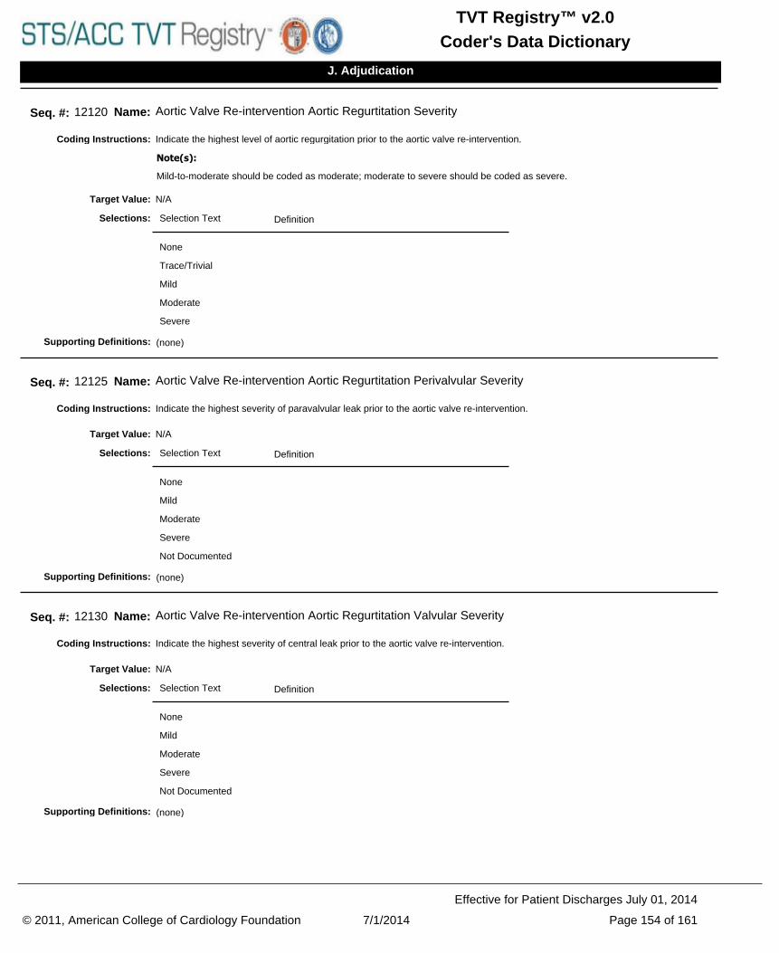
Indicate the highest level of aortic regurgitation prior to the aortic valve re-intervention. Coding Instructions:
Mild-to-moderate should be coded as moderate; moderate to severe should be coded as severe.
Note(s):
Seq. #: 12120 Name: Aortic Valve Re-intervention Aortic Regurtitation Severity
N/ATarget Value:
Selection Text Definition
None
Trace/Trivial
Mild
Moderate
Severe
Selections:
(none)Supporting Definitions:
Indicate the highest severity of paravalvular leak prior to the aortic valve re-intervention. Coding Instructions:
Seq. #: 12125 Name: Aortic Valve Re-intervention Aortic Regurtitation Perivalvular Severity
N/ATarget Value:
Selection Text Definition
None
Mild
Moderate
Severe
Not Documented
Selections:
(none)Supporting Definitions:
Indicate the highest severity of central leak prior to the aortic valve re-intervention.Coding Instructions:
Seq. #: 12130 Name: Aortic Valve Re-intervention Aortic Regurtitation Valvular Severity
N/ATarget Value:
Selection Text Definition
None
Mild
Moderate
Severe
Not Documented
Selections:
(none)Supporting Definitions:
© 2011, American College of Cardiology Foundation 7/1/2014 Page 154 of 161
Effective for Patient Discharges July 01, 2014
Coder's Data DictionaryTVT Registry™ v2.0
J. Adjudication
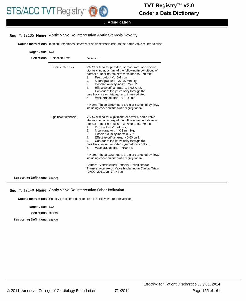
Indicate the highest severity of aortic stenosis prior to the aortic valve re-intervention.Coding Instructions:
Seq. #: 12135 Name: Aortic Valve Re-intervention Aortic Stenosis Severity
N/ATarget Value:
Selection Text Definition
Possible stenosis VARC criteria for possible, or moderate, aortic valve stenosis includes any of the following in conditions of normal or near normal stroke volume (50-70 ml): 1. Peak velocity*: 3-4 m/s;2. Mean gradient*: 20-35 mm Hg; 3. Doppler velocity index 0.29-0.25;4. Effective orifice area: 1.2-0.8 cm2;5. Contour of the jet velocity through the prosthetic valve: triangular to intermediate;6. Acceleration time: 80-100 ms * Note: These parameters are more affected by flow, including concomitant aortic regurgitation.
Significant stenosis VARC criteria for significant, or severe, aortic valve stenosis includes any of the following in conditions of normal or near normal stroke volume (50-70 ml): 1. Peak velocity*: >4 m/s;2. Mean gradient*: >35 mm Hg; 3. Doppler velocity index <0.25;4. Effective orifice area: <0.80 cm2;5. Contour of the jet velocity through the prosthetic valve: rounded symmetrical contour;6. Acceleration time: >100 ms * Note: These parameters are more affected by flow, including concomitant aortic regurgitation.
Source: Standardized Endpoint Definitions for Transcatheter Aortic Valve Implantation Clinical Trials (JACC, 2011, vol 57, No 3)
Selections:
(none)Supporting Definitions:
Specify the other indication for the aortic valve re-intervention.Coding Instructions:
Seq. #: 12140 Name: Aortic Valve Re-intervention Other Indication
N/ATarget Value:
(none)Selections:
(none)Supporting Definitions:
© 2011, American College of Cardiology Foundation 7/1/2014 Page 155 of 161
Effective for Patient Discharges July 01, 2014
Coder's Data DictionaryTVT Registry™ v2.0
J. Adjudication

Provide information and details that may assist in assessing this repeat intervention.Coding Instructions:
Seq. #: 12145 Name: Aortic Valve Re-intervention Clinical Comments
N/ATarget Value:
(none)Selections:
(none)Supporting Definitions:
Indicate the type of mitral valve re-intervention.Coding Instructions:
Seq. #: 12200 Name: Mitral Valve Re-intervention Type
N/ATarget Value:
Selection Text Definition
Surgical mitral valve repair
Surgical mitral valve replacement
Transcatheter mitral valve repair
Transcatheter mitral valve replacement
Leaflet clip procedure
Other transcatheter intervention
Selections:
(none)Supporting Definitions:
Indicate the type of "other" transcatheter mitral valve intervention. This does not include surgical mitral valve repair/replacements, transcatheter MV replacements or MV balloon valvuloplasties.
Coding Instructions:
Seq. #: 12205 Name: Other Type
N/ATarget Value:
(none)Selections:
(none)Supporting Definitions:
© 2011, American College of Cardiology Foundation 7/1/2014 Page 156 of 161
Effective for Patient Discharges July 01, 2014
Coder's Data DictionaryTVT Registry™ v2.0
J. Adjudication

Indicate the primary indication for the re-intervention. If more than one indication is present, code the indication the operator feels has the highest significance.
Coding Instructions:
Seq. #: 12210 Name: Mitral Valve Re-intervention Indication
N/ATarget Value:
Selection Text Definition
Mitral regurgitation
Mitral stensis
Mitral valve injury
Device migration
Device embolization
Device fracture
Endocarditis
Device thrombosis
Other
Selections:
(none)Supporting Definitions:
Specify the other indication for the mitral valve re-intervention.Coding Instructions:
Seq. #: 12215 Name: Mitral Valve Re-intervention Other Indication
N/ATarget Value:
(none)Selections:
(none)Supporting Definitions:
Provide information and details that may assist in assessing this repeat intervention.Coding Instructions:
Seq. #: 12220 Name: Mitral Valve Re-intervention Clinical Comments
N/ATarget Value:
(none)Selections:
(none)Supporting Definitions:
© 2011, American College of Cardiology Foundation 7/1/2014 Page 157 of 161
Effective for Patient Discharges July 01, 2014
Coder's Data DictionaryTVT Registry™ v2.0
J. Adjudication

Indicate if the heart failure readmission required the patient to be hospitalized with treatment in any inpatient unit or ward in the hospital for at least 24 hours, including emergency department stay.
Coding Instructions:
Seq. #: 12225 Name: Hospitalization>=24 Hours
N/ATarget Value:
Selection Text Definition
No
Yes
Information not available
Selections:
(none)Supporting Definitions:
Indicate if the patient had clinical signs and/or symptoms of heart failure, including new or worsening dyspnea, orthopnea, paroxysmal nocturnal dyspnea, increasing fatigue, worsening functional capacity or activity intolerance, or signs and/or symptoms of volume overload.
Coding Instructions:
Seq. #: 12230 Name: Clinical Signs or Sx of Heart Failure
N/ATarget Value:
Selection Text Definition
No
Yes
Information not available
Selections:
(none)Supporting Definitions:
Indicate if the patient had signs and symptoms that resulted in intravenous (e.g, diuretic or vasoactive therapy) or invasive (e.g., ultrafiltration, IABP, mechanical assistance) treatment for heart failure.
Coding Instructions:
Seq. #: 12335 Name: IV or Invasive Treatment Required
N/ATarget Value:
Selection Text Definition
No
Yes
Information not available
Selections:
(none)Supporting Definitions:
© 2011, American College of Cardiology Foundation 7/1/2014 Page 158 of 161
Effective for Patient Discharges July 01, 2014
Coder's Data DictionaryTVT Registry™ v2.0
J. Adjudication

Indicate the participant ID of the submitting facility.Coding Instructions:
Seq. #: 1000 Name: Participant ID
N/ATarget Value:
(none)Selections:
Participant ID:
Participant ID is a unique number assigned to each database participant. A database participant is defined as one entity that signs a Participation Agreement, submits one data submission file to the harvest, and receives one report on their data.
Each participant's data if submitted to harvest must be in one data submission file for a quarter. If one participant keeps their data in more than one file (e.g. at two sites), then the data must be combined into a single data submission to the system to file for the harvest. If two or more participants share a single purchased software, and enter cases into one database, then the data must be exported into different data submission files, one for each participant ID.
Source: NCDR
Supporting Definitions:
Indicate the full name of the facility.Coding Instructions:
Seq. #: 1010 Name: Participant Name
N/ATarget Value:
(none)Selections:
Participant Name:
Indicate the full name of the facility where the procedure was performed. Values should be full, official hospital names with no abbreviations or variations in spelling.
Source: NCDR
Supporting Definitions:
Indicate the time frame of data included in the data submission. Format: YYYYQQ. e.g., 2006Q4Coding Instructions:
Seq. #: 1020 Name: Time Frame of Data Submission
N/ATarget Value:
(none)Selections:
(none)Supporting Definitions:
This is a unique number created, and automatically inserted by the software into extract file. It identifies the number of times the software has created data submission files. The transmission number should be incremented by one every time the data submission files are exported. The transmission number should never be repeated.
Coding Instructions:
Seq. #: 1040 Name: Transmission Number
N/ATarget Value:
(none)Selections:
(none)Supporting Definitions:
© 2011, American College of Cardiology Foundation 7/1/2014 Page 159 of 161
Effective for Patient Discharges July 01, 2014
Coder's Data DictionaryTVT Registry™ v2.0
Z. Administration

Vendor identification (agreed upon by mutual selection between the vendor and the NCDR) to identify software vendor. This is entered into the schema automatically by vendor software. Vendors must use consistent name identification across sites. Changes to vendor name identification must be approved by the NCDR.
Coding Instructions:
Seq. #: 1050 Name: Vendor Identifier
N/ATarget Value:
(none)Selections:
(none)Supporting Definitions:
Vendor's software product name and version number identifying the software which created this record (assigned by vendor). Vendor controls the value in this field. This is entered into the schema automatically by vendor software.
Coding Instructions:
Seq. #: 1060 Name: Vendor Software Version
N/ATarget Value:
(none)Selections:
(none)Supporting Definitions:
The NCDR registry identifier describes the data registry to which these records apply. It is implemented in the software at the time the data is collected and records are created. This is entered into the schema automatically by software.
Coding Instructions:
Seq. #: 1070 Name: Registry Identifier
N/ATarget Value:
(none)Selections:
(none)Supporting Definitions:
Registry version describes the version number of the Data Specifications / Dictionary, to which each record conforms. It identifies which fields should have data, and what are the valid data for each field. It is the version implemented in the software at the time the data is collected and the records are created. This is entered into the schema automatically by software.
Coding Instructions:
Seq. #: 1080 Name: Registry Version
N/ATarget Value:
(none)Selections:
(none)Supporting Definitions:
© 2011, American College of Cardiology Foundation 7/1/2014 Page 160 of 161
Effective for Patient Discharges July 01, 2014
Coder's Data DictionaryTVT Registry™ v2.0
Z. Administration

Indicate if the data contained in the harvest/data file contains either standard patient episode of care records (arrival date to discharge only) or if it contains patient follow-up records
A transmission file with all episode of care records (from Arrival to Discharge only) is considered a "Base Registry Record".
A file with patient follow-up records (any follow-up assessments performed during the quarter selected) is considered a "Follow-Up Record".
Coding Instructions:
Seq. #: 1095 Name: Submission Type
N/ATarget Value:
Selection Text Definition
Episode of Care Records Only
Contains all patient and episode of care records with eligible procedures with a Discharge Date (Seq Num 9045) in the selected timeframe.
An Episode of Care is defined as a patient's admission/arrival to the facility performing the procedure(s), including any symptoms or medical history prior to arrival, ending at discharge or death
Follow-Up Records Only
Contains all patient records with at least one Follow-up Assessment performed (Seq Num 10000) in the selected timeframe.
Selections:
(none)Supporting Definitions:
© 2011, American College of Cardiology Foundation 7/1/2014 Page 161 of 161
Effective for Patient Discharges July 01, 2014
Coder's Data DictionaryTVT Registry™ v2.0
Z. Administration
Related Documents


