E-ISSN: 2548-0030 ISSN:2148-4724 TMSJ TURKISH MEDICAL STUDENT JOURNAL Volume: 9 Issue: 1 February 2022 https://turkmedstudj.com/

Welcome message from author
This document is posted to help you gain knowledge. Please leave a comment to let me know what you think about it! Share it to your friends and learn new things together.
Transcript
E-ISSN: 2548-0030ISSN:2148-4724
TMSJTURKISH MEDICAL STUDENT JOURNAL
Volume: 9 Issue: 1 February 2022
https://turkmedstudj.com/
TMSJTURKISH MEDICAL STUDENT JOURNAL
THE OFFICIAL JOURNAL OFTRAKYA UNIVERSITY SCHOOL OF MEDICINE
VOLUME 9 - ISSUE 1 - FEBRUARY 2022
Free access to the journal's website: https://turkmedstudj.com/
Manuscript Submission: https://tmsj.manuscriptmanager.net/
Published three times a year
Citation Abbreviation: Turk Med Stud J
Printing at: Trakya Üniversitesi MatbaasıEdirne Teknik Bilimler M.Y.O Sarayiçi Yerleşkesi, 22020 Yeni İmaret, Edirne, TurkeyPhone: +90 (284) 224 02 83Printing Date: February 2022ISSN: 2148-4724 E-ISSN: 2548-0030
Editorial OfficeAddress: Trakya Üniversitesi Tıp Fakültesi
22030 Edirne, TurkeyPhone: +90 (284) 235 76 53E-mail: [email protected]
A-I

A-II
Editor-in-ChiefBeliz KOÇYİĞİT
Trakya University School of Medicine, Edirne, [email protected]
https://orcid.org/0000-0001-6056-0219
Deputy Editors-in-ChiefBerfin TAN
Trakya University School of Medicine, Edirne, [email protected]
https://orcid.org/0000-0002-9256-7631
Berkin ERSOYUniversity of Hamburg School of Medicine, Hamburg, Germany
[email protected]://orcid.org/0000-0001-7111-648X
Irmak İrem ÖZYİĞİTTrakya University School of Medicine, Edirne, Turkey
[email protected]://orcid.org/0000-0003-2443-0155
Biostatistics EditorNecdet SÜT, PhD
Department of Biostatistics and Informatics, Trakya University School of Medicine, Edirne, [email protected]
https://orcid.org/0000-0001-6678-482X
Medical Ethics EditorBerna ARDA, MD, PhD
Department of History of Medicine and Medical Ethics, Ankara University School of Medicine, Ankara, [email protected]
https://orcid.org/0000-0003-2043-2444
Language EditorsDefne ERÇELEN
University of California, Los Angeles, [email protected]
https://orcid.org/0000-0001-7480-9011
Web EditorSarper KIZILKAYA
Trakya University School of Medicine, Edirne, [email protected]
https://orcid.org/0000-0002-7868-1585
TMSJTURKISH MEDICAL STUDENT JOURNAL
A-III
Alperen Taha CERTELTrakya University School of Medicine, Edirne, [email protected]://orcid.org/0000-0002-9816-9140
Burak BARDAKÇITrakya University School of Medicine, Edirne, [email protected]://orcid.org/0000-0002-0541-6991
Bengisu ÇIRAYTrakya University School of Medicine, Edirne, [email protected]://orcid.org/0000-0001-6332-7543
Bengisu GÜRİstanbul University İstanbul School of Medicine, İstanbul, [email protected]://orcid.org/0000-0002-4280-3317
Ceren YÜKSELTrakya University School of Medicine, Edirne, [email protected]://orcid.org/0000-0003-2456-7282
Dengiz Koray ŞAHİNTÜRKTrakya University School of Medicine, Edirne, [email protected]://orcid.org/0000-0001-9865-0930
Ege ESKİBOZKURTHarvard Medical School, Boston, [email protected]://orcid.org/0000-0001-6869-9338
Elif CENGİZTrakya University School of Medicine, Edirne, [email protected]://orcid.org/0000-0002-5902-2904
Elif ÇALIŞKANTrakya University School of Medicine, Edirne, [email protected]://orcid.org/0000-0003-4556-8698
Eylül ŞENÖDEYİCİTrakya University School of Medicine, Edirne, [email protected]://orcid.org/0000-0002-4132-1594
Fevzi Oktay ŞİŞMANTrakya University School of Medicine, Edirne, [email protected]://orcid.org/0000-0002-9942-9418
Gizem YILDIRIMBaşkent University School of Medicine, Ankara, [email protected]://orcid.org/0000-0001-5942-2169
Göktuğ Mert ÇİFTCİUniversity Hospital Münster Department of Psychiatry, Münster, Germany
[email protected]://orcid.org/0000-0003-2364-6317
İlayda KARAKOÇNear East University School of Medicine, Nicosia, Northern [email protected]://orcid.org/0000-0002-1118-1260
İsmail Yiğit NAÇARTrakya University School of Medicine, Edirne, [email protected]://orcid.org/0000-0001-7668-8970
Ilgın KILIÇUniversity of Liverpool School of Medicine, Liverpool, [email protected]://orcid.org/0000-0001-7393-7839
Işıl GÜLİstanbul University School of Medicine, İstanbul, [email protected]://orcid.org/0000-0002-4687-6097
Janset ÖZDEMİRTrakya University School of Medicine, Edirne, [email protected]://orcid.org/0000-0001-7774-5068
Mert YÜCELTrakya University School of Medicine, Edirne, [email protected]://orcid.org/0000-0002-4853-1607
Mustafa Alperen KOŞUCUTrakya University School of Medicine, Edirne, [email protected]://orcid.org/0000-0002-2381-5099
Sarper KIZILKAYATrakya University School of Medicine, Edirne, [email protected]://orcid.org/0000-0002-7868-1585
Sebahat ULUSANSüleyman Demirel University School of Medicine, Isparta, [email protected]://orcid.org/0000-0002-4964-6246
Sezin SAYINTrakya University School of Medicine, Edirne, [email protected]://orcid.org/0000-0001-7892-5992
Sıla Ece TİRYAKİTrakya University School of Medicine, Edirne, [email protected]://orcid.org/0000-0002-2318-3140
Zeynep Nihal ERTrakya University School of Medicine, Edirne, [email protected]://orcid.org/0000-0001-6890-6229
TMSJTURKISH MEDICAL STUDENT JOURNAL
Editorial Board

Abdulkadir TURGUT, MDDepartment of Obstetrics and Gynecology, Medeniyet University School of Medicine, İstanbul, Turkey
Ahmet Muzaffer DEMİR, MDDepartment of Hematology, Trakya University School of Medicine, Edirne, Turkey
Ahmet Tolgay AKINCI, MDDepartment of Neurosurgery, Trakya University School of Medicine, Edirne, Turkey
Ahmet ULUGÖL, MDDepartment of Pharmacology, Trakya University School of Medicine, Edirne, Turkey
Ahmet YILMAZ, MDDepartment of Forensic Medicine, Trakya University School of Medicine, Edirne, Turkey
Akif Hakan KURT, MDDepartment of Medical Pharmacology, Bolu Abant İzzet Baysal University School of Medicine, Bolu, Turkey
Ali AYDINLAR, MDDepartment of Cardiology, Bursa Acıbadem Hospital, Bursa, Turkey
Ali İlker FİLİZ, MDDepartment of General Surgery, İstanbul Başkent University Health Practice and Research Center, İstanbul, Turkey
Ali Rıza Cenk ÇELEBİ, MDDepartment of Ophthalmology, Acıbadem University School of Medicine, İstanbul, Turkey
Ali SARIKAYA, MDDepartment of Nuclear Medicine, Trakya University School of Medicine, Edirne, Turkey
Ali Serdar GÖZEN, MDDepartment of Urology, SLK-Kliniken, University of Heidelberg, Heidelberg, Baden-Württemberg, Germany
Alparslan TURAN, MDDepartment of Outcomes Research, Anesthesiology Institute, Cleveland Clinic, Cleveland, OH, USA
Andrea Mario Pompeo ROMANI, MD, PhDDepartment of Physiology and Biophysics, Case Western Reserve University, Cleveland, OH, USA
Atakan SEZER, MDDepartment of General Surgery, Trakya University School of Medicine, Edirne, Turkey
Ayşe ÇAYLAN, MDDepartment of Family Medicine, Trakya University School of Medicine, Edirne, Turkey
Ayşe Gülsen CEYHUN, MDDepartment of Family Medicine, Ankara University School of Medicine, Ankara, Turkey
Babürhan GÜLDİKEN, MDDepartment of Neurology, Trakya University, School of Medicine, Edirne, Turkey
Bahattin TANRIKULU, MDDepartment of Neurosurgery, Acıbadem University School of Medicine, İstanbul, Turkey
Beril GÜLER, MDDepartment of Pathology, Bezmialem University School of Medicine, İstanbul, Turkey
Berna ARDA, MDDepartment of History of Medicine and Ethics, Ankara University School of Medicine, Ankara, Turkey
Berrak ÇAĞLAYAN YEĞEN, MDDepartment of Physiology, Marmara University School of Medicine, İstanbul, Turkey
Birsen ELİBOL, MDDepartment of Medical Biology, Bezmialem University School of Medicine, İstanbul, Turkey
Cem UZUN, MDDepartment of Otorhinolaryngology, Trakya University School of Medicine, Edirne, Turkey
Cenk SAYIN, MDDepartment of Obstetrics and Gynecology, Trakya University School of Medicine, Edirne, Turkey
Cihan İŞLER, MDDepartment of Neurosurgery, İstanbul University-Cerrahpaşa, Cerrahpaşa School of Medicine, İstanbul, Turkey
Dağhan DAĞDELEN, MDDepartment of Plastic, Reconstructive and Aesthetic Surgery, Trakya University School of Medicine, Edirne, Turkey
Dan M. FLISS, MD, PhDDepartment of Maxillofacial and Skullbase Surgery, Tel Aviv Sourasky Medical Center, Tel Aviv, Israel
Demircan ÖZBALCI, MDDepartment of Hematology, Suleyman Demirel University School of Medicine, Isparta,Turkey
Devrim GÜNER, MDDepartment of Medical Pharmacology, TOBB University of Economics and Technology School of Medicine, Ankara, Turkey
Doğan ALBAYRAK, MDDepartment of General Surgery, Trakya University School of Medicine, Edirne, Turkey
Ebru TAŞTEKİN, MDDepartment of Pathology, Trakya University School of Medicine, Edirne, Turkey
Eda ÖZTURAN ÖZER, MDDepartment of Medical Biochemistry, Başkent University School of Medicine, Ankara, Turkey
Emine İkbal ATLI, MDDepartment of Medical Genetics, Trakya University School of Medicine, Edirne, Turkey
Emre DELEN, MDDepartment of Neurosurgery, Trakya University School of Medicine, Edirne, Turkey
A-IV
TMSJTURKISH MEDICAL STUDENT JOURNAL
Editorial Advisory Board

A-V
TMSJTURKISH MEDICAL STUDENT JOURNAL
Erbuğ KESKİN, MDDepartment of Pediatric Urology, İstanbul University, İstanbul School of Medicine, İstanbul, Turkey
Erhan ERTEKİN, MDDepartment of Psychiatry, İstanbul University, İstanbul School of Medicine, İstanbul, Turkey
Erkan KOZANOĞLU, MDDepartment of Physical Therapy and Rehabilitation, Çukurova University School of Medicine, Adana, Turkey
Ersan TATLI, MDDepartment of Cardiology, Sakarya University School of Medicine, Sakarya, Turkey
Ertan ŞAHİN, MDDepartment of Nuclear Medicine, Gaziantep University School of Medicine, Gaziantep, Turkey
Esra SAATÇI, MDDepartment of Family Medicine, Çukurova University School of Medicine, Adana, Turkey
Eugene Nicholas MYERS, MD, FACS, FRCS Edin (Hon)Department of Otorhinolaryngology, Head-Neck Surgery, University of Pittsburgh School of Medicine, Pittsburgh, PA, USA
Fatma Gülsüm ÖNAL, MDDepartment of History of Medicine and Ethics, Trakya University School of Medicine, Edirne, Turkey
Galip EKUKLU, MDDepartment of Public Health, Trakya University School of Medicine, Edirne, Turkey
Gamze ÖZÇÜRÜMEZ, MDDepartment of Psychiatry, Başkent University School of Medicine, Ankara, Turkey
Gamze Varol SARAÇOĞLU, MDDepartment of Public Health, Namık Kemal University School of Medicine, Tekirdağ, Turkey
Geysu KARLIKAYA, MDDepartment of Neurology, Kadıköy Medicana Hospital, İstanbul, Turkey
Gökay TAYLAN, MDDepartment of Cardiology, Trakya University School of Medicine, Edirne, Turkey
Gökhan ÇEVİK, MDDepartment of Urology, Trakya University School of Medicine, Edirne, Turkey
Gülay DURMUŞ ALTUN, MD, PhDDepartment of Nuclear Medicine, Trakya University School of Medicine, Edirne, Turkey
Güldal İnal GÜLTEKİN, MDDepartment of Physiology, Okan University School of Medicine, İstanbul. Turkey
Güldal SÜYEN, MDDepartment of Physiology, Acıbadem University School of Medicine, İstanbul, Turkey
Gülşah GÜMÜŞ, MDDepartment of Ophthalmology, Dr. Ersin Arslan Training and Research Hospital, Gaziantep, Turkey
Hakan AKDERE, MDDepartment of Urology, Trakya University School of Medicine, Edirne, Turkey
Hakan EMMUNGİL, MDDepartment of Rheumatology, Trakya University School of Medicine, Edirne, Turkey
Hakkı Mete ÇEK, MDDepartment of Urology, Trakya University School of Medicine, Edirne, Turkey
Hanefi Yekta GÜRLERTOP, MDDepartment of Cardiology, Trakya University School of Medicine, Edirne, Turkey
Hasan C. ÜMİT, MDDepartment of Gastroenterology, Trakya University School of Medicine, Edirne, Turkey
Hasan YAZICI, MDDepartment of Rheumatology, Academic Hospital, İstanbul, Turkey
Hilmi TOZKIR, MDDepartment of Medical Genetics, Namık Kemal University School of Medicine, Tekirdağ, Turkey
Hüseyin Ahmet TEZEL, MDDepartment of Gastroenterology, Trakya University School of Medicine, Edirne, Turkey
Hüseyin Avni SÖNMEZ, MDDepartment of Medical Biochemistry, İstanbul University-Cerrahpaşa Cerrahpaşa School of Medicine, İstanbul, Turkey
Hüsniye Figen KULOĞLU, MDDepartment of Infectious Diseases, Trakya University School of Medicine, Edirne, Turkey
İlknur ERDEM, MDDepartment of Infectious Diseases, Namık Kemal University School of Medicine, Tekirdağ, Turkey
İsmet KIRPINAR, MDDepartment of Psychiatry, Bezmialem University School of Medicine, İstanbul, Turkey
Jülide Sedef GÖÇMEN, MDDepartment of Medical Microbiology, TOBB University of Economics and Technology School of Medicine, Ankara, Turkey
Kıymet TABAKÇIOĞLU, MDDepartment of Medical Biology, Trakya University School of Medicine, Edirne, Turkey
Levent ÖZTÜRK, MDDepartment of Physiology, Trakya University School of Medicine, Edirne, Turkey
Mehmet ARSLAN, MDDepartment of Psychiatry, Kırklareli University School of Medicine, Kırklareli, Turkey
A-VI
TMSJTURKISH MEDICAL STUDENT JOURNAL
Mehmet Erdal VARDAR, MDDepartment of Psychiatry, Trakya University School of Medicine, Edirne, Turkey
Mehmet ÜNAL, MDDepartment of Ophthalmology, Ankara Dünya Göz Hospital, Ankara, Turkey
Melih Hamdi ÜNAL, MDDepartment of Ophthalmology, Private Practice, İstanbul, Turkey
Mert ÖZCAN, MDDepartment of Orthopedics, Trakya University School of Medicine, Edirne, Turkey
Mesut AYER, MDDepartment of Hematology, İstanbul Haseki Training and Research Hospital, İstanbul, Turkey
Metin ÖZKAN, MDDepartment of Pulmonology, Ankara Memorial Hospital, Ankara, Turkey
Mevlüt Serdar KUYUMCU, MDDepartment of Cardiology, Süleyman Demirel University School of Medicine, Isparta, Turkey
Muhammed KESKİN, MDDepartment of Cardiology, Bahçeşehir University School of Medicine, İstanbul, Turkey
Muhammet Ali KAPLAN, MDDepartment of Medical Oncology, Diyarbakır Memorial Hospital, Diyarbakır, Turkey
Muhittin Emre ALTUNRENDE, MDDepartment of Neurosurgery, Liv Hospital, İstanbul, Turkey
Mustafa Çağrı SAVAŞ, MDDepartment of Pediatric Surgery, Süleyman Demirel University School of Medicine, Isparta, Turkey
Mustafa İNAN, MDDepartment of Pediatric Surgery, Trakya University School of Medicine, Edirne, Turkey
Mustafa Kemal EROL, MDDepartment of Cardiology, Kolan Hospital, İstanbul, Turkey
Mustafa SEVİM, MDDepartment of Physiology, Marmara University School of Medicine, İstanbul, Turkey
Nermin TUNÇBİLEK, MDDepartment of Radiology, Trakya University School of Medicine, Edirne, Turkey
Nihat ŞENGEZE, MDDepartment of Neurology, Süleyman Demirel University School of Medicine, Isparta, Turkey
Nurettin AYDOĞDU, PhDDepartment of Physiology, İnönü University School of Medicine, Malatya, Turkey
Nurija BİLALOVIĆ, MD, PhDDepartment of Pathology, University of Sarajevo, Sarajevo, Bosnia and Herzegovina
Okan ÇALIYURT, MDDepartment of Psychiatry, Trakya University School of Medicine, Edirne, Turkey
Okan ERDOĞAN, MDDepartment of General Surgery, Akdeniz University School of Medicine, Antalya, Turkey
Oktay KAYA, MDDepartment of Physiology, Trakya University School of Medicine, Edirne
Özdal ERSOY, MDDepartment of Gastroenterology, Acıbadem International Hospital, İstanbul, Turkey
Özgür KASAPÇOPUR, MDDepartment of Pediatric Rheumatology, İstanbul University-Cerrahpaşa, Cerrahpaşa School of Medicine, İstanbul, Turkey
Özlem BOYBEYİ, MDDepartment of Pediatric Surgery, Hacettepe University School of Medicine, Ankara, Turkey
Öznur BAYRAKTAR EKMEKÇİGİL, MDDepartment of Medical Biology and Genetics, Okan University School of Medicine, İstanbul, Turkey
Pınar YAMANTÜRK ÇELİK, MDDepartment of Medical Pharmacology, İstanbul University, İstanbul School of Medicine, İstanbul, Turkey
Faik Güntaç UZUNOĞLU, MDDepartment of General, Visceral and Thoracic Surgery, University Medical Center, Hamburg, Hamburg-Eppendorf, Germany
Ranko MLADINA, MD, PhDDepartment of Rhinology, University of Zagreb, Zagreb, Croatia
Ruhan Deniz TOPUZ, MDDepartment of Medical Pharmacology, Trakya University School of Medicine, Edirne, Turkey
Selçuk TUNALI, MDDepartment of Anatomy, TOBB University of Economics and Technology School of Medicine, Ankara, Turkey
Selis Gülseven GÜVEN, MDDepartment of Otorhinolaryngology, Trakya University School of Medicine, Edirne, Turkey
Selma SÜER GÖKMEN, PhDDepartment of Medical Biochemistry, Trakya University School of Medicine, Edirne, Turkey
Semir VRANIĆ, MD, PhDDepartment of Pathology, Qatar University School of Medicine, Doha, Qatar
Semra AYTÜRK, MDDepartment of Endocrinology, Trakya University School of Medicine, Edirne, Turkey
Serdar ÖZTORA, MDDepartment of Family Medicine, Trakya University School of Medicine, Edirne, Turkey

A-VII
TMSJTURKISH MEDICAL STUDENT JOURNAL
Serkan ATICI, MDDepartment of Pediatrics, Okan University School of Medicine, İstanbul, Turkey
Sernaz UZUNOĞLU, MDDepartment of Medical Oncology, Trakya University School of Medicine, Edirne, Turkey
Serpil EROL, MDDepartment of Infectious Diseases and Clinical Microbiology, Haydarpaşa Numune Hospital, İstanbul, Turkey
Sibel GÜLDİKEN, MDDepartment of Endocrinology, Trakya University School of Medicine, Edirne, Turkey
Stanislav YANEV, MD, PhDDepartment of Pharmacology, Bulgarian Academy of Sciences, Sofia, Bulgaria
Suat ERDOĞAN, MDDepartment of Medical Biology, Trakya University School of Medicine, Edirne, Turkey
Süleyman Ayhan ÇALIŞKAN, MD, PhDDepartment of Medical Education, Ege University School of Medicine, İzmir, Turkey
Şaban GÜRCAN, MDDepartment of Infectious Diseases and Clinical Microbiology, Trakya University School of Medicine, Edirne, Turkey
Şebnem BATUR, MDDepartment of Pathology, İstanbul University-Cerrahpaşa, Cerrahpaşa School of Medicine, İstanbul, Turkey
Şerife BAYRAKTAR, MDDepartment of Ophthalmology, İstanbul University, İstanbul School of Medicine, İstanbul, Turkey
Tammam SİPAHİ, PhDDepartment of Biophysics, Trakya University School of Medicine, Edirne, Turkey
Tarkan YETİŞYİĞİT, MDDepartment of Medical Oncology, Namık Kemal University School of Medicine, Tekirdağ, Turkey
Tayfur TOPTAŞ, MDDepartment of Hematology, Marmara University School of Medicine, İstanbul, Turkey
Tolga Turan DÜNDAR, MDDepartment of Neurosurgery, Bezmialem University School of Medicine, İstanbul, Turkey
Ufuk USTA, MDDepartment of Pathology, Trakya University School of Medicine, Edirne, Turkey
Utku AYDİL, MDDepartment of Otorhinolaryngology, Gazi University School of Medicine, Ankara, Turkey
Ülfiye ÇELİKKALP, MDDepartment of Public Health, Trakya University School of Medicine, Edirne, Turkey
Veysel Atilla AYYILDIZ, MDDepartment of Radiology, Süleyman Demirel University School of Medicine, Isparta, Turkey
Volkan İNAL, MDDepartment of Intensive Care, Trakya University School of Medicine, Edirne, Turkey
Volkan YÜKSEL, MDDepartment of Cardiovascular Surgery, Trakya University School of Medicine, Edirne, Turkey
Vuslat GÜRLÜ, MDDepartment of Ophthalmology, Trakya University School of Medicine, Edirne, Turkey
Yekta Altemur KARAMUSTAFAOĞLU, MDDepartment of Thoracic Surgery, Trakya University School of Medicine, Edirne, Turkey
Zafer KOÇAK, MDDepartment of Radiation Oncology, Trakya University School of Medicine, Edirne, Turkey
Zeynep Banu DOĞANLAR, PhDDepartment of Medical Biology, Trakya University School of Medicine, Edirne, Turkey
Zeynep Banu GÜNGÖR, MDDepartment of Medical Biochemistry, İstanbul University-Cerrahpaşa, Cerrahpaşa School of Medicine, İstanbul, Turkey
Zoran GATALICA, MDAnatomical Pathology, University of Oklahoma Health and Sciences Center, Oklahoma City, OK, USA
A-VIII
TMSJTURKISH MEDICAL STUDENT JOURNAL
"Turkish Medical Student Journal (TMSJ) is the first scientific journal in Turkey to be run by medical students and to publish works of medical students only. In that respect, TMSJ encourages and enables all students of medicine to conduct research and to publish their valuable research in all branches of medicine.
Turkish Medical Student Journal publishes researches, interesting case reports and reviews regarding all fields of medicine. The primary aim of the journal is to publish original articles with high scientific and ethical quality and serve as a good example of medical publications for those who plan to build a career in medicine. TMSJ believes that quality of publication will contribute to the progress of medical sciences as well as encourage medical students to think critically and share their hypotheses and research results internationally.
The journal is published every four months. The language of the publication is English.
The Editorial Board of TMSJ and the Publisher follows the principles of the International Council of Medical Journal Editors (ICMJE). Only unpublished papers that are not under review for publication elsewhere can be submitted. The authors are responsible for the scientific content of the material to be published. TMSJ reserves the right to request any research materials on which the paper is based.
Turkish Medical Student Journal does not charge any article submission, article-editorial processing, or publication charges such as page or color charges.TMSJ is a non-profit journal that does not accept any advertisements and financial support from any person or foundation. TMSJ is available as a hard copy. All articles can be downloaded in PDF format from our website, free of charge. There are also no publication fees. Readers are free to share, copy and redistribute the material in any medium or format with the extent of Creative Commons Attribution-NonCommercial-NoDerivatives 4.0 International License (CC BY-NC-ND 4.0).
Turkish Medical Student Journal is a non-profit journal which does not accept any advertisements and financial support from any person or foundation.
MISSIONTurkish Medical Student Journal is an independent, non-profit, peer-reviewed, international, open-access journal; which aims to publish articles of interest to physicians, scientists, and medical students. TMSJ is published three times a year, in February, June, and October by Trakya University. The language of publication is English. Correspondent authors of the articles should be a medical student.
Turkish Medical Student Journal publishes original researches, interesting case reports, and reviews regarding all fields of medicine. All of the published articles are open-access and reachable on our website. The primary aim of the journal is to publish original articles with high scientific and ethical quality and serve as a good example of medical publications for stimulating students, doctors, researchers. Our mission is to feature quality publications that will contribute to the progress of medical sciences as well as encourage medical students to think critically and share their hypotheses and research results internationally.
The Editorial Board and the Publisher adheres to the principles of ICMJE Committee on Publication Ethics (COPE).
PublisherGalenos Publishing House
Address: Molla Gürani Mahallesi Kaçamak Sokak No: 21/1 34093 Fındıkzade - Fatih, İstanbul/Turkey
Phone: +90 (212) 621 99 25
Fax: +90 (212) 621 99 27
E-mail: [email protected]
Responsible ManagerBeliz KOÇYİĞİT
Trakya University School of Medicine, Edirne, Turkey
Indexed inCABI: CAB Abstracts and Global Health
Türk MedlineAsos Indeks
ScilitJ-Gate
OwnerFiliz AKATA, MD
Dean, Trakya University School of Medicine, Edirne, Turkey
Scientific AdvisorZafer KOÇAK, MD
Department of Radiation Oncology, Trakya University School of Medicine, Edirne, Turkey
Aims and Scope
A-IX
TMSJTURKISH MEDICAL STUDENT JOURNAL
OPEN ACCESS STATEMENTTurkish Medical Student Journal is a fully open access journal that provides free and unrestricted online availability to its content immediately upon publication without delay. This means;
1. all content is freely and limitlessly available without charge to the user or her/his institution;
2. everyone has free and unlimited access to the full-text of all articles without subscription;
3. all articles are released immediately in open access format (no embargo period);
4. everyone is allowed to read, download, copy, distribute, print, search, or link to the full texts of these articles, crawl them for indexing, pass them as data to software, or use them for any other lawful purpose, without financial, legal, or technical barriers other than those inseparable from gaining access to the internet itself.
The only constraint on reproduction and distribution, and the only role for copyright in this domain, should be to give authors control over the integrity of their work and the right to be properly acknowledged and cited. The copyright holder of a scholarly work grants usage rights to others using an open license. Turkish Medical Student Journal applies the Creative Commons Attribution (CC BY-NC-ND 4.0) license to its published articles. Authors that submit their papers for publication thereby agree to have the CC BY-NC-ND 4.0 license applied to their works.
The authors retain all patent, and other proprietary rights to the article, including copyright without any restrictions. In addition to non-exclusive rights the publisher has under the CC-BY-NC-ND license, copyright policy also authorizes the publisher to permit commercial use or derivation of the content as well as publication, republication in electronic and print format in the journal, and distribution of the content.
This is in accordance with the Budapest Open Access Initiative which can be assessed at the URL:
http://www.budapestopenaccessinitiative.org/.
PERMISSIONSUnder Open Access license, no special permission is required to copy, distribute or reuse all or part of article published by Turkish Medical Student Journal, including figures and tables, for non-commercial purposes for free as long as the author and original source are properly cited. Reuse of an article does not imply endorsement by the authors or Turkish Medical Student Journal.
EDITORIAL PROCESSAll manuscripts submitted for publication are reviewed for their originality, methodology, importance, quality, ethical nature, and suitability for the journal by the editorial board and briefly revised by the advisory board whose members are respected academicians in their fields. A well-constructed scheme is used for the evaluation process. All manuscripts are reviewed by two different members
of the editorial board, followed by peer revision from at least two professors, belonging to different institutions, who are experts in their areas. The editors assist authors to improve the quality of their papers. The editor-in-chief has full authority over the editorial, scientific content, and the timing of publication.
ETHICSTurkish Medical Student Journal depends on publication ethics to ensure all articles published in TMSJ are acceptable in terms of scientific ethical standards and do not include any kind of plagiarism. TMSJ expects authors and the editorial board to adhere the principles of the COPE. To reach the highest standards, TMSJ has an advisory board member who is a professional in ethics.
All original articles submitted to the TMSJ have to be approved by an ethical committee and include the name of the ethics committee(s) or institutional review board(s), the number/ID of the approval(s). Additionally, informed consent documents obtained from patients involving case reports are required for the submission.
All received manuscripts are screened by plagiarism software (iThenticate). Similarity percentages more than 19 (or more than 5 for one paper) and six consecutive words cited from another published paper in the same order are the causes of immediate rejection.
MATERIAL DISCLAIMERAll opinions, reports, and results within the articles that are published in the TMSJ are the personal opinions of the authors. The Editorial Board, the editorial advisory board, the publisher and the owner of the TMSJ do not accept any responsibility for these articles.
CONFLICT OF INTEREST POLICY
The Turkish Medical Student Journal’s editorial review process pursues the Good Editorial Practice set by international editorial organizations (ICMJE, EASE, WAME, COPE, CSE,…). According to the WAME; a conflict of interest arises when an author, peer-reviewer, or editor in the publication process has an incompatible interest that could unmeritedly influence his or her responsibilities (academic honesty, unbiased conduct, and reporting of research and transparency) in the publication process.If a conflict of interest related to family, personal, financial, political, or religious issues, as well as any competing interest outlined above at the WAME’s definition, exists; TMSJ requires that the author should report the condition to the editorial board and declare at the ICMJE Conflict of Interest form, and specifically define it under a title at the end of the manuscript. The Editorial Board members of the Turkish Medical Student Journal may also submit their own manuscripts to the journal as all of them are active researchers. Nevertheless, they cannot take place at any stage on the editorial evaluation of their manuscripts in order to minimize any possible bias. These manuscripts will be treated like any other author’s, final acceptance of such manuscripts can only be made by at least two positive recommendations of external peer-reviewers.
Turkish Medical Student Journal follows a double-blinded review principle. Authors cannot contact any of the peer-reviewers during
Editorial Policy
A-X
TMSJTURKISH MEDICAL STUDENT JOURNAL
the publication process and vice versa; since any of the peer-reviewers and author’s information are obscured.
For the instructions and further information please visit:
https://publicationethics.org/search/site/conflict
http://icmje.org
http://www.ease.org.uk/publications/science-editors-handbook/
https://www.councilscienceeditors.org
WITHDRAWAL POLICYTurkish Medical Student Journal encourages authors to follow best practice in publication ethics. Therefore, the authors may withdraw their manuscripts in absolutely necessary conditions. If authors want to withdraw their manuscript, they need to submit the ''Manuscript Withdrawal Form''. Authors should state their reason of withdrawal and the form need to be signed by all authors, sent to [email protected].
Turkish Medical Student Journal Editorial Board evaluates the form and if the reason for withdrawal is found as reasonable, the authors will receive a confirmation e-mail. Before getting this confirmation e-mail, the authors should not consider their manuscripts as withdrawn.
COPYRIGHT POLICY AND RIGHTS OF AUTHORSAll articles that are accepted by the Turkish Medical Student Journal, will be licensed under a Creative Commons Attribution-NonCommercial-NoDerivatives 4.0 International License (CC BY-NC-ND 4.0) which allows third parties to freely access, use, and share the material for only non-commercial purposes by giving the appropriate credit to the original work. This also allows others to freely access, copy, and use research provided. Adaptation and modification of the material is not permitted. For further details of the license CC BY-NC-ND 4.0, please visit https://creativecommons.org/licenses/by-nc-nd/4.0/
The authors retain all patent, and other proprietary rights to the article, including copyright without any restrictions. In addition to the non-exclusive rights the publisher has under the CC BY-NC-ND 4.0 license, the copyright policy also authorizes the publisher to permit the commercial use or derivation of the content as well as publication, republication in electronic and print format in the journal, and distribution of the content.
A-XI
TMSJTURKISH MEDICAL STUDENT JOURNAL
CATEGORIES OF ARTICLESThe Journal publishes the following types of articles:
Original Research Articles: Original prospective or retrospective studies of basic or clinical investigations in areas relevant to medicine.
Content:
- Abstract (average 400 words; the structured abstract
contain the following sections: aims, methods, results,
conclusion)
- Introduction
- Material and Methods
- Results
- Discussion
- Conclusion
- References
Review and Mini Review Articles: The authors may be invited to write or may submit a review article. Reviews including the latest medical literature may be prepared on all medical topics.
Content:
- Abstract (average 400 words)
- Titles on related topics
- References
Case Reports: Brief descriptions of a previously undocumented disease process, a unique unreported manifestation or treatment of a known disease process, or unique unreported complications of treatment regimens. They should include an adequate number of photos and figures.
Content:
- Abstract (average 200 words)
- Introduction
- Case Report
- Discussion
- References
Letters to the Editor: These are the letters that include different views, experiments and questions of the readers about the manuscripts that were published in this journal in the recent year and should be no more than 500 words.
Content:
- There is no title and abstract.
- Submitted letters should include a note indicating the attribution to an article (with the number and date) and the name, affiliation and address of the author(s) at the end.
- The answer to the letter is given by the editor or the author(s) of the manuscript and is published in the journal.
MANUSCRIPT PREPARATIONAuthors are encouraged to follow the following principles before submitting their material.
-The article should be written in IBM compatible computers with Microsoft Word.
ABBREVIATIONSAll abbreviations in the text must be defined the first time they are used, and the abbreviations should be displayed in parentheses after the definition. Authors should avoid abbreviations in the title, abstract and at the beginning of the first sentences of the paragraphs.
FIGURES AND TABLES:- All figures and tables should be cited at the end of the relevant sentence. Explanations must be placed at the bottom of figures, whereas at the top of tables.
- Figures and tables must be added to the e-mail as attachments in .jpg or .tiff formats.
- The name of the file should be named as: last name of the first author_Table/Figure_No.TIFF/JPEG. For example: Sancar_Figure_1.JPEG.
- All abbreviations used, must be listed in explanation which will be placed at the bottom of each figures and tables.
- For figures and tables to be reproduced relevant permissions need to be provided. This permission must be mentioned in the explanation.
- Pictures/photographs must be in color, clear and with appropriate contrast to separate details.
TITLE PAGEA concise, informative title, should be provided. All authors should be listed with academic degrees, affiliations, addresses, office and mobile telephone and fax numbers, e-mail and postal addresses, ORCID. If the study was presented in a congress, the author(s) should identify the date/place of the congress of the study presented.
ABSTRACTThe abstracts should be prepared in accordance with the instructions in the “Categories of Articles” and placed in the article file.
KEYWORDS:- They should be minimally three.
- Keywords should be appropriate to “Medical Subject Headings (MESH)” (See: www.nlm.nih.gov/mesh/MBrowser.html).
Acknowledgments: Conflict of interest, financial support, grants, and all other editorial (statistical analysis, language editing) and/or technical assistance if present, must be presented at the end of the text.
REFERENCESReferences should be numbered in the order they are cited. Only published data or manuscripts accepted for publication and recent data should be included. Inaccessible data sources and those not indexed in any database should be omitted. Titles of journals should be abbreviated in accordance with Index Medicus- NLM Style (Patrias
Instructions to Authors
A-XII
TMSJTURKISH MEDICAL STUDENT JOURNAL
K. Citing medicine: the NLM style guide for authors, editors, and publishers [Internet]. 2nd ed. Wendling DL, technical editor. Bethesda (MD): National Library of Medicine (US); 2007 - [updated 2011 Sep 15; cited Year Month Day] (http://www.nlm.nih.gov/citingmedicine). All authors should be listed if an article has three or less authors; first three authors are listed and the rest is represented by “et al.”. Reference format and punctuation should be as in the following examples.
Journal: Muller C, Buttner HJ, Peterson J et al. A randomized comparison of clopidogrel and aspirin versus ticlopidine and aspirin after placement of coronary artery stents. Circulation 2000;101: 590-3.
Book Section: Sherry S. Detection of thrombi. In: Strauss HE, Pitt B, James AE, editors. Cardiovascular Medicine. St Louis: Mosby; 1974.p.273-85.
Books with Single Author: Cohn PF. Silent myocardial ischemia and infarction. 3rd ed. New York: Marcel Dekker; 1993.
Editor(s) as author: Norman IJ, Redfern SJ, editors. Mental health care for elderly people. New York: Churchill Livingstone; 1996.
Conference Proceedings: Bengisson S. Sothemin BG. Enforcement of data protection, privacy and security in medical informatics. In: Lun KC, Degoulet P, Piemme TE, Rienhoff O, editors. MEDINFO 92. Proceedings of the 7th World Congress on Medical Informatics; 1992 Sept 6-10; Geneva, Switzerland. Amsterdam: North-Holland; 1992.p.1561-5.
Scientific or Technical Report: Smith P. Golladay K. Payment for durable medical equipment billed during skilled nursing facility stays. Final report. Dallas (TX) Dept. of Health and Human Services (US). Office of Evaluation and Inspections: 1994 Oct. Report No: HHSIGOE 169200860.
Thesis: Kaplan SI. Post-hospital home health care: the elderly access and utilization (dissertation). St. Louis (MO): Washington Univ. 1995.
Manuscripts accepted for publication, not published yet: Leshner AI. Molecular mechanisms of cocaine addiction. N Engl J Med In press 1997.
Epub ahead of print Articles: Aksu HU, Ertürk M, Gül M et al. Successful treatment of a patient with pulmonary embolism and biatrial thrombus. Anadolu Kardiyol Derg 2012 Dec 26. doi: 10.5152/ akd.2013.062. [Epub ahead of print]
Manuscripts published in electronic format: Morse SS. Factors in the emergence of infectious diseases. Emerg Infect Dis (serial online) l995 Jan-Mar (cited 1996 June 5): 1(1): (24 screens). Available from: URL:http://www.cdc.gov/ncidodlElD/cid.htm.
CONFLICT OF INTEREST STATEMENTConflict of interest is when the author’s primary responsibility to science, ethics and readers is not compatible with author’s private interests such as financial gains or personal rivalry. Credence of the scientific process and the authenticity of articles depend in part on how transparently conflicts of interest are approached. In case of a conflict of interest it should be declared to the editorial board of TMSJ, and clearly written under a particular section at the end of the manuscript. Authors may reach more information and find the instructions for the process from the links below:
https://publicationethics.org/search/site/conflict%20of%20interest
http://www.icmje.org/conflicts-of-interest/
ORCIDIt is recommended that the journals, which are indexed in TR Index or apply to take a part, require ORCID® information from the authors and include this information in the journal/articles. ORCID® is the abbreviation for Open Researcher and Contributor ID. ORCID® is a 16-digit URI compliant with the ISO Standard (ISO 27729), also known as the International Standard Name Identifier (ISNI). The correspondent author who will submit an article to our journal should state ORCID® numbers. Researchers who do not have ORCID® ID can apply for a free registration and get their individual ORCID® number at http://orcid.org.
WITHDRAWAL POLICYTurkish Medical Student Journal encourages authors to follow best practice in publication ethics. Therefore, the authors may withdraw their manuscripts in absolutely necessary conditions. If authors want to withdraw their manuscript, they need to submit the ''Manuscript Withdrawal Form''. Authors should state their reason of withdrawal and the form need to be signed by all authors, sent to [email protected].
Turkish Medical Student Journal Editorial Board evaluates the form and if the reason for withdrawal is found as reasonable, the authors will receive a confirmation e-mail. Before getting this confirmation e-mail, the authors should not consider their manuscripts as withdrawn.
COMPLAINTAll of the complaints regarding the articles should be stated via e-mail to [email protected]. TMSJ Editorial Board evaluates the complaints with the accordance of COPE Guidelines and draws a conclusion after the decision of Editor-in-Chief and an ethics editor.
A-XIII
TMSJTURKISH MEDICAL STUDENT JOURNAL
REVIEWS1 PREVENTION OF CARDIOVASCULAR DISEASE: A NUTRIGENETIC APPROACH Betül Filiz Doğan, Buse Ataçer, Ceyda Kantur, Melis Ocak; Edirne, TURKEY
8 WHAT PHYSICIANS SHOULD KNOW ABOUT COFFEE Ahmed Adel Khalifa; Qena, EGYPT
14 EFFECTS OF MALNUTRITION ON THE PROGNOSIS OF PEDIATRIC ACUTE LYMPHOBLASTIC LEUKEMIA PATIENTS Elif Cengiz, Ilgın Kılıç, Elif Çalışkan, Ceren Yüksel, Fevzi Oktay Şişman, Hakkı Onur Kırkızlar; Edirne, TURKEY, Liverpool, ENGLAND
ORIGINAL ARTICLE20 PARTICIPATION OF CANNABINOID RECEPTORS IN ANTIPRURITIC ACTIVITY INDUCED BY SYSTEMIC DIPYRONE IN MICE Erdem Altan, Najaf Ali Folladwand, Rymejsa Gurmani, Ömür Türkü Özşalap, Kübra Duvan Aydemir, Ahmet Ulugöl; Edirne, TURKEY
CASE REPORTS25 MUCINOUS ADENOCARCINOMA MIMICKING CRYPTOGENIC ORGANIZING PNEUMONIA: A CASE REPORT Barış Altunel, Müçteba Gökalp Özer, Aykut Alkan; Edirne, TURKEY
29 SYNCHRONOUS GASTRIC AND COLON ADENOCARCINOMA WITH SIMULTANEOUS SURGERY: A CASE REPORT Merve Yaren Kayabaş, İbrahim Ethem Cakcak; Edirne, TURKEY
33 CORNEAL ENDOTHELIAL CELL ANALYSIS IN TWO PATIENTS WITH WILSON’S DISEASE AND KAYSER- FLEISCHER RINGS Ege Gürlü, Vuslat Gürlü; İstanbul, Edirne, TURKEY
36 ADULT HEMISPHERIC PILOCYTIC ASTROCYTOMA WITH HEMORRHAGIC COMPONENTS: A CASE REPORT Ömer Polat, Nisanur Ceviz, Ahmet Emir Çelen, Elif Mercan Demirtaş, Barış Chousein, Ahmet Tolgay Akıncı; Edirne, TURKEY
41 MITRAL VALVE INFECTIVE ENDOCARDITIS IN AN INTRAVENOUS DRUG ABUSER Buket Şen, Utku Yartaşı, Servet Altay; Edirne, TURKEY
45 ERRATA
CONTENTS
A-XIV
TMSJTURKISH MEDICAL STUDENT JOURNAL
OPEN ACCESS TO KNOWLEDGE
"Our mission of disseminating knowledge is only half complete if the information is not made widely and readily available to society."
-Berlin Declaration
Open access is defined as a global movement pointed towards giving free and open internet-based access to scholastic data such as scientific publications. A publication is characterized as open access when there is no monetary, legitimate, or specialized hindrance to access, or at least, when anybody can peruse, download, duplicate, appropriate, print, search and search or utilize the data.
Open access is a distributing model for insightful correspondence that offers research data to readers for free, instead of the conventional membership model, where readers can access scientific publications by paying a membership.
The greatest advantage of open access is that it enables the results of scientific research to be spread more quickly and widely. For instance, more people can have access to scientific papers, including those who could not otherwise access this information because they can not afford an expensive journal subscription. New ideas can spread faster and more widely and trigger new research efforts, which serves as a driving force for knowledge.
Turkish Medical Student Journal (TMSJ) encourages and enables all students of medicine and health sciences to research and publish their valuable research in all branches of medicine. TMSJ is also a fully open access journal that provides free and unrestricted online availability to its content immediately upon publication without delay. As the editorial board of TMSJ, we believe that knowledge should be freely available online and the user rights along with the copyright terms should be defined clearly to ensure that this concept works effectively.
Beliz Koçyiğit
Editor-in-Chief, Turkish Medical Student Journal
Trakya University School of Medicine, Edirne, TURKEY
Editorial
REVIEW
www.turkmedstudj.comCopyright@Author(s) - Available online at https://www.turkmedstudj.com/
1
OPEN ACCESS
Turk Med Stud J 2022;9(1):1-7
Cite this article as: Doğan BF, Ataçer B, Kantur C et al. Prevention of cardiovascular disease: a nutrigenetic approach. Turk Med Stud J 2022;9(1):1-7.
Address for Correspondence: Betül Filiz Doğan, Trakya University School of Medicine, Edirne, TURKEY e-mail: [email protected] iDs of the authors: BFD: 0000-0001-5808-0777; BA:0000-0003-3442-0700; CK: 0000-0001-9887-2429; MO: 0000-0002-1020-5590.Received: 13.12.2021 Accepted: 03.01.2022
INTRODUCTION
Nutrigenetics is a branch of genetics that analyzes the relationship between genetic polymorphisms of individuals and their response to a diet (1). Previous research has suggested that nutrigenetics may be associated with many conditions such as obesity, cardiovascular disease (CVD), and hypertension (1). Nutrigenetics can improve the quality of life for many people and allow us to develop preventative measures for metabolic diseases through analyzing their genomes and providing a personalized diet based on their genetic composition (1).
The human genome consists of almost 3 billion base pairs encoding 30,000 genes and 100,000 proteins (2). Although 99.9% of the genome is identical in humans, about 1,500 base pairs contain polymorphisms (2). Previous genetic studies have identified genetic variants related to obesity and concluded that in addition to environmental and lifestyle
factors, genetic factors strongly correlate with CVD, cancer, diabetes, and osteoporosis (2).
Molecular genetics and pharmacogenetics play an important role in the diagnosis, treatment, and prevention of CVDs (3). Genetic testing is used to determine genetic transmission of and familial predisposition to diseases such as hypertrophic cardiomyopathy and familial hypercholesterolemia (4, 5). Nutrigenetics aims to provide another preventative approach through dietary changes based on one’s genome (1).
NUTRIGENETICS IN DISEASE
Nutrigenetic studies have provided insights into many diseases from obesity to bone disease to CVD (1, 6). Genetic factors play a crucial role in obesity and these genetic variants have been useful in the development of nutrition plans in obesity treatment (7). Similarly, 50-80% of bone diseases are associated with genetic polymorphisms. For instance, vitamin D receptor
ABSTRACT
Cardiovascular diseases are the leading cause of death worldwide. Besides genetic factors, environmental factors also contribute to their etiology. Nutrigenetics provides the opportunity to prevent or reduce the incidence of many diseases. In this review article, we investigated the incidence of cardiovascular diseases in relation to nutrigenetics, genotype-specificity, personalized conditions, nutrition, genetic polymorphisms, and environmental factors. We concluded that the genes encoding miRNA, APOB, PCSK-9, SORT1, cytokines, and IFN-γ are strongly associated with development and progression of cardiovascular diseases. Through nutrigenetic testing, we can determine the risk factors, identify preventative strategies, and improve the quality of life through personalized nutrition. Keywords: Cardiovascular disease, nutrigenetics, genotype, diet
1Trakya University School of Medicine, Edirne, TURKEY2Trakya University Faculty of Health Science, Department of Nutrition and Dietetics, Edirne, TURKEY
Betül Filiz Doğan1 , Buse Ataçer2 , Ceyda Kantur2 , Melis Ocak2
Doğan et al. Nutrigenetics and Cardiovascular Diseases
PREVENTION OF CARDIOVASCULAR DISEASE: A NUTRIGENETIC APPROACH
DOI: 10.4274/tmsj.galenos.2022.09.01.01
2Turk Med Stud J 2022;9(1):1-7.Doğan et al. Nutrigenetics and Cardiovascular Diseases
polymorphisms are important in mineral density, calcium, and vitamin D supplementation, while vitamin B and K polymorphisms are not as critically associated (6). Genes related to sodium metabolism have shown that the amount of salt in the diet is associated with hypertension (8).
Cardiovascular Diseases
Cardiovascular diseases are the leading cause of mortality and disability worldwide but can be preventable through lifestyle and dietary changes (9). Genes interact with nutrients to influence cardiovascular regeneration or repair (10). Polymorphisms in certain genes can predispose individuals to myocardial infarction (MI) and stroke, as well as hypertension, diabetes, and inflammation (11). The genetic risk factors of CVDs are not yet fully determined (12, 13). However, it has been observed that individuals with the E4 allele in the apolipoprotein E gene have higher levels of low-density lipoprotein (LDL) in fat intake when compared to other individuals (14). Obesity, sedentary lifestyle, and hyperinsulinemia may also contribute to the risk of CVDs (11).
Hypertension
Hypertension is one of the most prevalent risk factors for CVDs and its treatment has been shown to reduce the incidence of CVDs (15). In particular, the Framingham Heart Study showed a clear relationship between hypertension and the risk of CVDs (15). Its prevalence increases with increasing age such that prevalence is 7.3% in adults aged 18-39 years, 32.2% in adults aged 40-59 years, and 64.9% in adults aged 60 years and over (16).
There are many mechanisms by which hypertension causes atherosclerosis. An increase in blood pressure can cause an increase in vascular stress (14). Especially in areas of non-laminar flow, there is evidence that increased radial forces cause changes in the endothelial layer that make it more susceptible to movement of LDL into the subendothelium as well as making it easier for monocyte attachment (17). Another contributing mechanism is increased oxidative stress. In hypertension, nicotinamide adenine dinucleotide phosphate-oxidase, xanthine oxidase, and cyclo-oxygenase enzyme systems are activated, and the detoxifying enzyme superoxide dismutase is decreased, resulting in increased availability of superoxide anions, which reduces the availability of nitric oxide and generates pro-inflammatory radicals (18).
Smoking
A 2005 INTERHEART study examined the risk factors for heart disease, such as smoking and second-hand smoke, as a prospective case-control study in 52 different countries (19). The INTERHEART study showed that the risk of acute MI increased approximately threefold in smokers and was directly proportional to the number of cigarettes smoked (19).
Smoking increases the risk for atherosclerosis and CVDs through inhibition of nitric oxide production (20). Decreased nitric oxide production impairs vasoconstriction and vasodilation due to endothelial dysfunction and causes LDL oxidation, which is an important factor in the development of atherosclerosis (20). Endothelial damage induces adhesion of monocytes and increases the recruitment and migration of monocytes into the subendothelial space, which further advances atherosclerosis (20). In addition to its role in the development of atherosclerotic plaque, it has been shown that smoking increases fibrinogen levels, which increases thrombosis and therefore may cause subsequent clot formation in the ruptured plaque, which causes acute MI (20).
Obesity
Thirty nine percent of the world’s adult population was overweight and 13% were obese in 2016 (21). Obesity may play a role in the formation and progression of adiposity and other metabolic diseases by affecting energy balance and body weight regulation (21).
There is evidence that obesity triggers atherosclerosis (22). Obesity is an inflammatory disease because adipose tissue is a source of proinflammatory adipokines such as tumor necrosis factor alpha, interleukins, monocyte chemoattractant protein-1, resistin, and leptin. These adipokines have effects such as stimulating vascular reactivity, stimulating inflammation, and even coagulation (22). Therefore, obesity is both a risk factor and a risk marker for CVDs, and its increase over the past 20 years makes it an important target for intervention (11).
Having a sedentary lifestyle increases the risk of CVDs (23, 24). One study considered that decreasing lipoprotein lipase activity during sitting resulted in less catabolism of triglycerides and increased plasma triglyceride levels (23). A sedentary lifestyle also decreases endothelial nitric oxide expression caused by low blood flow in the limbs with prolonged sitting (24).
ROLE OF GENETICS
Role of MicroRNAs in CVDs
MicroRNAs (miRNAs) are part of the non-coding RNA family, which are associated with gene expression and intercellular communication (25). They play a key role in pathogenicity in many diseases such as MI, inflammation, hypertrophy, and atherosclerosis (26). miRNAs in the cardiovascular system control the functions of muscle cells and vascular endothelial cells, and abnormal expressions of these miRNAs are associated with cardiac dysfunction (27-29).
Previous studies demonstrated that miRNA expression in damaged tissues in patients with CVDs is irregular (27, 30).
3Turk Med Stud J 2022;9(1):1-7. Doğan et al. Nutrigenetics and Cardiovascular Diseases
In an experiment in mice, severe developmental defects in the heart and blood vessels demonstrated the importance of miRNAs in heart biology through tissue-specific erasure of genes such as Drosha, DGCR8, Argonaute RISC Catalytic Component 2, or Dicer, which are essential for miRNA biogenesis (23). miRNAs became the new biomarker for the diagnosis of CVDs (31).
Use of Molecular Genetics in the Diagnosis of CVDs
Genetic tests are used to determine the underlying causes of CVDs (32). To better interpret genetic test results in terms of CVD risk, the patient should be evaluated comprehensively (33). Hereditary features of CVDs are of varying degrees. Although most CVD-associated polymorphisms are polygenic, they can also be monogenic. When monogenic CVD markers are not recognized and treated accordingly, they can lead to serious illness, disability, and even death. Familial hypercholesterolemia is a widespread monogenic disease caused by mutations in the LDL receptor, apolipoprotein B (APOB), and proprotein convertase subtilisin/kexin type 9 genes with a frequency of 1/200 (32). Another common familial CVD caused by mutations in approximately 11 genes that encode sarcomere proteins is hypertrophic cardiomyopathy (32).
Our knowledge of CVD risk factors and the genetic background of many non-infectious diseases has increased through Genome with Association Studies (GWAS). GWAS identified that the single nucleotide polymorphisms (SNPs) that are associated strongest with CVDs are located in the p21.3 region of chromosome 9 in humans (32, 34). Another gene identified by GWAS is Sortilin (SORT 1) (32). The most important signals indicating the risk of LDL cholesterol causing CVDs are the SORT1 gene, which also plays a role in determining plasma cholesterol levels, and the CELSR2/PSRC1/SORT1 gene set (32). Although research on SORT1 supported its impact on plasma cholesterol levels and identified it as a risk factor in creating MI, its effects are quite complex and still controversial (35).
There is also a complex relationship between dietary habits and gene expression. Studies show that SNPs in nutrigenetics are associated with plasma lipid rates of nutrients consumed. The relationship of homocysteine with folate intake is thought to be a risk factor for MI and stroke (32). Also, it has been determined that homocysteine concentration is high and methylenetetrahydrofolate reductase polymorphism is more pervasive in regions with low folate intake (36).
Association of Extracellular Vesicles with CVDs
Membranous structures that carry bioactive substances, such as macronutrients, messenger RNA, and miRNA, are called
extracellular vesicles (EVs). Key sources of these vesicles in the heart are endothelial cells, cardiomyocytes, macrophages, and fibroblasts (37). EVs have an important role in the signaling and function of their target organs. Moreover, EV molecules are helpful for transferrin’s protective role in cardiomyocytes (38). EVs enriched with miRNA-30a are released from cardiomyocytes in response to hypoxia-inducible factor1α to protect the myocardium from damage during hypoxia (39). EVs produced in cardiomyocytes express exclusive cellular markers such as flotillin-1 and caveolin 3, as well as sarcomeric and mitochondrial proteins such as myomycin, cardiac-type myosin binding protein C, and tropomyosin (40).
EVs release their cargo to target cells by budding from the plasma membranes (41). With the stimulation of the calcium-dependent mechanism in the plasma membrane, substances inside the EV are poured into the extracellular area (42). Calcium ion affects enzymes such as gelsolin and calpain, which are involved in the disintegration of the cytoskeleton and cause an easy release of microvesicles (37).
Hsa-miRNA-208a was identified as one of the specific miRNAs carried by EVs which increased in blood after MI (43). This research suggested that miRNAs could be useful in the diagnosis of MI and circulating RNAs could be useful biomarkers in the diagnosis of CVDs (43). EVs also protect the heart against MI and arrhythmias (37). Due to the aforementioned features of EVs, they have been proposed to be used as a new diagnostic tool in preventing CVDs (35).
The Effect of the Calcium-Calmodulin Mechanism on CVDs
Intracellular calcium, which plays a key role as a second messenger in heart contraction, and the accompanying calmodulin (CaM) protein are major signaling mechanisms (44). With each heartbeat, calcium is released and then taken back into the cell, and CaM plays a role in the excitation-contraction mechanism (44, 45). CaM is a highly preserved protein that can interact with about 300 different proteins other than calcium (46). CaM binds four calcium molecules and is involved in the regulation of calcium channels (45). CaM is also important in regulating cellular events such as infection, cell death, cell growth, and immunity (44).
Calmodulin-related genes are present in chromosomes 2, 14, and 19 and encode isoforms of CaM which differ at a single nucleotide level (44). To understand the genetics of MI predisposition, it is necessary to identify the polymorphisms formed in the CaM mechanism (44). These polymorphisms have been associated with serious diseases of the heart such as ventricular fibrillation, catecholaminergic polymorphic ventricular tachycardia, and diseases with different pathologies such as osteoarthritis and adolescent idiopathic scoliosis (44, 47-49).
4Turk Med Stud J 2022;9(1):1-7.Doğan et al. Nutrigenetics and Cardiovascular Diseases
It has been reported that polymorphism 34T>A in chromosome 19 affecting the CALM3 transcript is more common in patients with familial hypertrophic cardiomyopathy (FHC) and may therefore be a gene that affects FHC (50). As we learn more about the transcriptional mechanism of the excitation-contraction mechanisms in the heart, we can conclude that genetic factors contribute to the predisposition of individuals to disease CVDs (44).
Role of Cytokines in the Formation of Atherosclerosis
As aforementioned, atherosclerosis is an inflammatory disorder that has a role in the formation of CVDs (10). The atherosclerotic plaque formation causes the vascular lumen to narrow, while the rupture of this plaque causes the vascular lumen to become blocked completely through thrombus formation (51). Such vascular obstruction can lead to MI (10). The severity of atherosclerosis was found to be related to cytokine genes in studies conducted with patients recovering from MI (52, 53). Cytokines regulate the expression of inflammatory molecules that can cause atherosclerotic plaque rupture, so it has been recommended to use inflammatory cytokine levels to track the clinical course of CVDs (54).
Interferon-gamma (IFN-γ), a proinflammatory cytokine, is secreted from macrophages and is associated with the formation of atherosclerosis. IFN-γ stimulates the production of chemokines and cytotoxic molecules from macrophages, and its expression increases in atherosclerosis (10, 55). In the early stages, IFN-γ supports the development of atherosclerosis by stimulating the secretion of adhesion molecules from the endothelium. In the late stages, IFN-γ works to separate atherosclerotic plaques by speeding up apoptosis and extracellular degeneration of macrophages (10, 56).
Countless SNPs have been found in the IFN-γ gene localized at 12q24 (10). One example is the IFN-γ +874 T/A (rs2430561) polymorphism, which affects the formation and development of atherosclerotic plaques, and can therefore be used as a biomarker candidate for early MI detection (10).
TARGETED THERAPIES
Preventative Medicine and Genetics
Health is defined as a state of complete physical, mental, and social well-being (57). Preventive medicine interventions are medical evaluations that reduce the risk of disease and provide early detection and treatment (57). Through genetic diagnostic tests and nutrigenetics, risk factors that may induce diseases can be identified, and preventative measures can be taken with nutrition and lifestyle changes (58). The goals of nutrigenetics include effective individual dietary
strategies to improve quality of life, prevent diseases, and promote wellness (59).
Individualized Nutrition
Both genetic and environmental factors contribute to development of CVDs, and nutrition has special importance in CVDs. Nutritional interventions that are personalized according to the individual’s genetic background may present a new diet-based approach to treat CVDs and improve health outcomes (1). Studies have shown that genetics can be used to identify individuals who are most likely to lose weight, but these findings should be further investigated before wide use (60).
Historically, nutrition interventions were focused on the relationship between nutrient deficiencies and disease but with new studies, there is a potential to improve chronic metabolic disorders through nutrition (61-64). Through nutrigenetics, it is possible to understand the effects of dietary response and nutritional elements on gene expression based on genetic variations (65). However, despite recent advances in nutrigenetics, there are not enough studies about personalized nutrition. Moreover, the cost of genetic testing poses a great challenge to the widespread use of personalized nutrition (63).
Nutrigenetics in Controlling Inflammation and Cardiovascular Risk Factors
Inflammation underlies a wide spectrum of diseases from CVDs to psychiatric disorders (66). There is evidence that genetic variation can predispose one to increased inflammation and can increase the likelihood of disease development through interactions with environmental factors such as diet (67). The Mediterranean diet is suggested for reducing inflammation and reversing inflammatory diseases (68).
Nutrigenetics and the Response to the Mediterranean Diet
As the world population ages, the prevalence of CVDs is also increasing (69). Along with medical treatment, lifestyle changes are also very important in the treatment process (70, 71). Research shows that a diet rich in plant sterols is important in maintaining good health (72).
Previous studies have shown that the Mediterranean diet protects the heart and overall health (73). The traditional Mediterranean diet is characterized by a large consumption of fruits and vegetables, unrefined grains, nuts, fish, legumes, olive oil, moderate consumption of wine, preferably with main meals, and low intake of dairy products and meat (74). The protective effects of the Mediterranean diet are revealed by dietary fiber, unsaturated fatty acids, antioxidants, and bioactive components (75). To get the best results from the Mediterranean diet; cultural, genetic, and socioeconomic factors should be considered (74).
5Turk Med Stud J 2022;9(1):1-7. Doğan et al. Nutrigenetics and Cardiovascular Diseases
Nutrigenetics Challenges
For nutrigenetics to be useful as a public health tool, there is a great need to use statistical and bioinformatics tools to examine the combined effects of multiple gene variants on health outcomes (76). While personalized nutrition based on genetics has great potential, there are many challenges in translating scientific advances into successful strategies for managing dietary intake and dietary health outcomes. These challenges include translating research results into practice, public perception and the likelihood of acceptance, issues of privacy and ethics, commercialization, and the level of evidence needed to enable a beneficial transition from traditional approaches (77).
CONCLUSION
In this review, we explored preventative approaches to CVDs through nutrigenetics. Many genetic variants related to obesity and CVDs have been previously described. In our review, we concluded that genes encoding miRNA, APOB, PCSK-9, SORT1, cytokines, and IFN-γ are strongly associated with CVDs the development and progression of CVDs. Mutations in these genes can cause serious morbidity and mortality. Therefore, genetic testing should be widely implemented, and familial predispositions should be considered when assessing an individual’s health plan. With the help of nutrigenetics, we can determine the risk factors, identify preventative strategies, and improve the quality of life through personalized nutrition.
Ethics Committee Approval: N/A
Informed Consent: N/A
Conflict of Interest: The authors declared no conflict of interest.
Author Contributions: Concept: B.F.D., B.A., C.K., M.O., Design: B.F.D., B.A., C.K., M.O., Supervision: B.F.D., B.A., C.K., M.O., Resources: B.F.D., B.A., C.K., M.O., Materials: B.F.D., B.A., C.K., M.O., Data Collection and/or Processing: B.F.D., B.A., C.K., M.O., Analysis and/or Interpretation: B.F.D., B.A., C.K., M.O., Literature Search: B.F.D., B.A., C.K., M.O., Writing Manuscript: B.F.D., B.A., C.K., M.O., Critical Review: B.F.D., B.A., C.K., M.O.
Financial Disclosure: The authors declared that this study received no financial support.
REFERENCES1. Barrea L, Annunziata G, Bordoni L et al. Nutrigenetics-personalized
nutrition in obesity and cardiovascular diseases. Int J Obes Suppl 2020;10:1-13. [Crossref]
2. McSweeney PLH, McNamara JP, editors. Encyclopedia of dairy sciences. 3rd ed. Academic Press 2022;966-71. [Crossref]
3. Pérez-Castrillon JL, Ruiz-Mambrilla M, Riancho JA. Nutrigenetics of bone health. In: Caterina R, Martinez A, Kohlmeier M, editors. Principles of Nutrigenetics and Nutrigenomics. Elsevier; 2020.p.377-82. [Crossref]
4. Krasi G, Precone V, Paolacci S et al. Genetics and pharmacogenetics in the diagnosis and therapy of cardiovascular diseases. Acta Biomed 2019 ;90(10-S):7-19. [Crossref]
5. Familial hypertrophic cardiomyopathy | Genetic and Rare Diseases Information Center (GARD) – an NCATS Program. Accessed February 6, 2022. https://rarediseases.info.nih.gov/diseases/7229/familial-hypertrophic-cardiomyopathy [Crossref]
6. Genetic Testing for Familial Hypercholesterolemia | CDC. Published April 1, 2020. Accessed February 6, 2022. Available from: URL: https://www. cdc.gov/genomics/disease/fh/testing_FH.htm. [Crossref]
7. Goni L, Milagro FI, Cuervo M et al. Single-nucleotide polymorphisms and DNA methylation markers associated with central obesity and regulation of body weight. Nutr Rev 2014;72:673-90. [Crossref]
8. Luft FC, Weinberger MH. Heterogeneous responses to changes in dietary salt intake: the salt-sensitivity paradigm. Am J Clin Nutr 1997;65(2 Suppl):612S-7S. [Crossref]
9. Massaro M, Scoditti E, Calabriso N et al. Nutrients and gene expression in cardiovascular disease. In: Faintuch J, Faintuch S, editors. Precision Medicine for Investigators, Practitioners and Providers. Academic Press; 2020.p.469-81. [Crossref]
10. Akadam-Teker AB, Teker E, Daglar-Aday A et al. Interactive effects of interferon-gamma functional single nucleotid polymorphism (+874 T/A) with cardiovascular risk factors in coronary heart disease and early myocardial infarction risk. Mol Biol Rep 2020;47:8397-405. [Crossref]
11. Teufel EJ. Risk factors for cardiovascular disease. In: Vasan RS, Sawyer DB, editors. Encyclopedia of Cardiovascular Research and Medicine. Elsevier; 2018. p.307-14. [Crossref]
12. Lusis AJ. Atherosclerosis. Nature 2000;407:233-41. [Crossref]
13. Corella D, Ordovas JM. Nutrigenomics in cardiovascular medicine. Circ Cardiovasc Genet 2009;2:637-51. [Crossref]
14. Chaudhary R, Likidlilid A, Peerapatdit T et al. Apolipoprotein E gene polymorphism: effects on plasma lipids and risk of type 2 diabetes and coronary artery disease. Cardiovasc Diabetol 2012;11:36. [Crossref]
15. Franklin SS, Larson MG, Khan SA et al. Does the relation of blood pressure to coronary heart disease risk change with aging? The Framingham Heart Study. Circulation 2001;103:1245-9. [Crossref]
16. Yoon SS, Carroll MD, Fryar CD. Hypertension Prevalence and Control Among Adults: United States, 2011-2014. NCHS Data Brief 2015;(220): 1-8. [Crossref]
17. Kwak BR, Bäck M, Bochaton-Piallat ML et al. Biomechanical factors in atherosclerosis: mechanisms and clinical implications. Eur Heart J 2014;35:3013-20. [Crossref]
18. Zaheer M, Chrysostomou P, Papademetriou V. Hypertension and atherosclerosis: pathophysiology, mechanisms and benefits of BP control. In: Andreadis EA (ed.) Hypertension and Cardiovascular diseases; 2016. p.201-16. [Crossref]
19. Teo KK, Ounpuu S, Hawken S et al. Tobacco use and risk of myocardial infarction in 52 countries in the INTERHEART study: a case-control study. Lancet 2006;368:647-58. [Crossref]
20. Powell JT. Vascular damage from smoking: disease mechanisms at the arterial wall. Vasc Med 1998;3:21-8. [Crossref]
21. World Health Organization. Obesity and overweight. 2018. Available from: URL: https://www.who.int/news-room/fact-sheets/detail/obesity-andoverweight. [Crossref]
6Turk Med Stud J 2022;9(1):1-7.Doğan et al. Nutrigenetics and Cardiovascular Diseases
22. Mandviwala T, Khalid U, Deswal A. Obesity and Cardiovascular Disease: a Risk Factor or a Risk Marker? Curr Atheroscler Rep 2016;18:21. [Crossref]
23. Zderic TW, Hamilton MT. Physical inactivity amplifies the sensitivity of skeletal muscle to the lipid-induced downregulation of lipoprotein lipase activity. J Appl Physiol (1985) 2006;100:249-57. [Crossref]
24. Stamatakis E, Hamer M, Dunstan DW. Screen-based entertainment time, all-cause mortality, and cardiovascular events: population-based study with ongoing mortality and hospital events follow-up. J Am Coll Cardiol 2011;57:292-9. [Crossref]
25. Wojciechowska A, Braniewska A, Kozar-Kamińska K. MicroRNA in cardiovascular biology and disease. Adv Clin Exp Med 2017;26:865-74.
26. Hata A. Functions of microRNAs in cardiovascular biology and disease. Annu Rev Physiol 2013;75:69-93. [Crossref]
27. Bartel DP. Metazoan MicroRNAs. Cell 2018;173:20-51. [Crossref]
28. Bartel DP. MicroRNAs: target recognition and regulatory functions. Cell 2009;136:215-33. [Crossref]
29. Ambros V. The functions of animal microRNAs. Nature 2004;431:350-5. [Crossref]
30. Wang L, Liu J, Xu B et al. Reduced exosome miR-425 and miR-744 in the plasma represents the progression of fibrosis and heart failure. Kaohsiung J Med Sci 2018;34:626-33. [Crossref]
31. Kalayinia S, Arjmand F, Maleki M et al. MicroRNAs: roles in cardiovascular development and disease. Cardiovasc Pathol 2021;50:107296. [Crossref]
32. Vrablik M, Dlouha D, Todorovova V et al. Genetics of Cardiovascular Disease: How Far Are We from Personalized CVD Risk Prediction and Management? Int J Mol Sci 2021;22:4182. [Crossref]
33. Gersh BJ, Maron BJ, Bonow RO et al. 2011 ACCF/AHA guideline for the diagnosis and treatment of hypertrophic cardiomyopathy: executive summary: a report of the American College of Cardiology Foundation/American Heart Association Task Force on Practice Guidelines. J Am Coll Cardiol 2011;58:2703-38. [Crossref]
34. Palomaki GE, Melillo S, Bradley LA. Association between 9p21 genomic markers and heart disease: a meta-analysis. JAMA 2010;303:648-56.
35. Musunuru K, Strong A, Frank-Kamenetsky M et al. From noncoding variant to phenotype via SORT1 at the 1p13 cholesterol locus. Nature 2010;466:714-9. [Crossref]
36. Holmes MV, Newcombe P, Hubacek JA et al. Effect modification by population dietary folate on the association between MTHFR genotype, homocysteine, and stroke risk: a meta-analysis of genetic studies and randomised trials. Lancet 2011;378:584-94. [Crossref]
37. Femminò S, Penna C, Margarita S et al. Extracellular vesicles and cardiovascular system: Biomarkers and Cardioprotective Effectors. Vascul Pharmacol 2020;135:106790. [Crossref]
38. Giricz Z, Varga ZV, Baranyai T et al. Cardioprotection by remote ischemic preconditioning of the rat heart is mediated by extracellular vesicles. J Mol Cell Cardiol 2014;68:75-8. [Crossref]
39. Matteucci M, Papini G, Ciofini E et al. Epigenetic Regulation of Myocardial Homeostasis, Self-Regeneration and Senescence. Curr Drug Targets 2015;16:827-42. [Crossref]
40. Bang C, Batkai S, Dangwal S et al. Cardiac fibroblast-derived microRNA passenger strand-enriched exosomes mediate cardiomyocyte hypertrophy. J Clin Invest 2014;124:2136-46. [Crossref]
41. Dignat-George F, Boulanger CM. The many faces of endothelial microparticles. Arterioscler Thromb Vasc Biol 2011;31:27-33. [Crossref]
42. Tucher C, Bode K, Schiller P et al. Extracellular Vesicle Subtypes Released From Activated or Apoptotic T-Lymphocytes Carry a
Specific and Stimulus-Dependent Protein Cargo. Front Immunol 2018;9:534. [Crossref]
43. Wang GK, Zhu JQ, Zhang JT et al. Circulating microRNA: a novel potential biomarker for early diagnosis of acute myocardial infarction in humans. Eur Heart J 2010;31:659-66. [Crossref]
44. Beghi S, Cavaliere F, Buschini A. Gene polymorphisms in calcium-calmodulin pathway: Focus on cardiovascular disease. Mutat Res Rev Mutat Res 2020;786:108325. [Crossref]
45. Gambardella J, Trimarco B, Iaccarino G et al. New Insights in Cardiac Calcium Handling and Excitation-Contraction Coupling. Adv Exp Med Biol 2018;1067:373-85. [Crossref]
46. Halling DB, Liebeskind BJ, Hall AW et al. Conserved properties of individual Ca2+-binding sites in calmodulin. Proc Natl Acad Sci U S A 2016;113:E1216-25. [Crossref]
47. Nyegaard M, Overgaard MT, Søndergaard MT et al. Mutations in calmodulin cause ventricular tachycardia and sudden cardiac death. Am J Hum Genet 2012;91:703-12. [Crossref]
48. Makita N, Yagihara N, Crotti L et al. Novel calmodulin mutations associated with congenital arrhythmia susceptibility. Circ Cardiovasc Genet 2014;7:466-74. [Crossref]
49. Zhao D, Qiu GX, Wang YP et al. Association of calmodulin1 gene polymorphisms with susceptibility to adolescent idiopathic scoliosis. Orthop Surg 2009;1:58-65. [Crossref]
50. Friedrich FW, Bausero P, Sun Y et al. A new polymorphism in human calmodulin III gene promoter is a potential modifier gene for familial hypertrophic cardiomyopathy. Eur Heart J 2009;30:1648-55. [Crossref]
51. Roy H, Bhardwaj S, Yla-Herttuala S. Molecular genetics of atherosclerosis. Hum Genet 2009;125:467-91. [Crossref]
52. García-Bermúdez M, López-Mejías R, González-Juanatey C et al. Analysis of the interferon gamma (rs2430561, +874T/A) functional gene variant in relation to the presence of cardiovascular events in rheumatoid arthritis. PLoS One 2012;7:e47166. [Crossref]
53. Heiskanen M, Kähönen M, Hurme M et al. Polymorphism in the IL10 promoter region and early markers of atherosclerosis: the Cardiovascular Risk in Young Finns Study. Atherosclerosis 2010;208:190-6. [Crossref]
54. De Gennaro L, Brunetti ND, Montrone D et al. Subacute inflammatory activation in subjects with acute coronary syndrome and left ventricular dysfunction. Inflammation 2012;35:363-70. [Crossref]
55. Pasqui AL, Di Renzo M, Bova G et al. Pro-inflammatory/anti-inflammatory cytokine imbalance in acute coronary syndromes. Clin Exp Med 2006;6:38-44. [Crossref]
56. Hansson GK. Immune mechanisms in atherosclerosis. Arterioscler Thromb Vasc Biol 2001;21:1876-90. [Crossref]
57. Caradonna F, Consiglio O, Luparello C et al. Science and Healthy Meals in the World: Nutritional Epigenomics and Nutrigenetics of the Mediterranean Diet. Nutrients 2020 ;12:1748. [Crossref]
58. Paladuz S. Preventive medicine and genetics. BAYT Scientific Researches. Proceedings of the 8th National Internal Diseases Congress; Sep 2006 13-17; Antalya, Turkey. Ankara; 2006.p.113-4. [Crossref]
59. Ordovas JM. The quest for cardiovascular health in the genomic era: nutrigenetics and plasma lipoproteins. Proc Nutr Soc 2004;63:145-52.
60. Frankwich KA, Egnatios J, Kenyon ML et al. Differences in Weight Loss Between Persons on Standard Balanced vs Nutrigenetic Diets in a Randomized Controlled Trial. Clin Gastroenterol Hepatol 2015;13:1625-32.e1. [Crossref]
7Turk Med Stud J 2022;9(1):1-7. Doğan et al. Nutrigenetics and Cardiovascular Diseases
61. Coşkun T. Nutritional genomics. Çocuk Sağlığı ve Hastalıkları Dergisi 2007;50:47-66. [Crossref]
62. Gillies PJ. Nutrigenomics: the Rubicon of molecular nutrition. J Am Diet Assoc 2003;103(12 Suppl 2):S50-5. [Crossref]
63. Seçer S, Özden A, Güllüoğlu Ş et al. Genotype-based nutrition: personalizing diet to improve life quality and prevent diet-related diseases. Academic Food Journal 2011;9:51-9. [Crossref]
64. Kussmann M, Fay LB. Nutrigenomics and personalized nutrition: science and concept. Per Med 2008;5:447-55. [Crossref]
65. Simopoulos AP. Genetic variation and dietary response: nutrigenetics/nutrigenomics. Asia Pacific J Clin Nutr 2002;11:117-28. [Crossref]
66. Maydych V. The Interplay Between Stress, Inflammation, and Emotional Attention: Relevance for Depression. Front Neurosci 2019;13:384. [Crossref]
67. Curti ML, Jacob P, Borges MC et al. Studies of gene variants related to inflammation, oxidative stress, dyslipidemia, and obesity: implications for a nutrigenetic approach. J Obes 2011;2011:497401. [Crossref]
68. Widmer RJ, Flammer AJ, Lerman LO et al. The Mediterranean diet, its components, and cardiovascular disease. Am J Med 2015;128:229-38. [Crossref]
69. Çiftçi S, Rakıcıoğlu N. Cardiovascular diseases and nutritional factors in elderly. Bes Diy Derg 2019;47:82-90. [Crossref]
70. National Cholesterol Education Program (NCEP) Expert Panel on Detection, Evaluation, and Treatment of High Blood Cholesterol in Adults
(Adult Treatment Panel III). Third Report of the National Cholesterol Education Program (NCEP) Expert Panel on Detection, Evaluation, and Treatment of High Blood Cholesterol in Adults (Adult Treatment Panel III) final report. Circulation 2002;106:3143-421. [Crossref]
71. Gylling H, Plat J, Turley S et al. Plant sterols and plant stanols in the management of dyslipidaemia and prevention of cardiovascular disease. Atherosclerosis 2014;232:346-60. [Crossref]
72. Çekici H, Yıldıran H. Effects of plant sterols/stanols on atherosclerotic process and cardiovascular diseases. Sakarya Tıp Dergisi 2019;9:218-29. [Crossref]
73. Ordovas JM, Kaput J, Corella D. Nutrition in the genomics era: cardiovascular disease risk and the Mediterranean diet. Mol Nutr Food Res 2007;51:1293-9. [Crossref]
74. Bonaccio M, Iacoviello L, de Gaetano G et al. The Mediterranean diet: the reasons for a success. Thromb Res 2012;129:401-4. [Crossref]
75. Barbaros B, Kabaran S. The Mediterranean diet and its health protective effects. Journal of Nutrition and Dietetics 2014;42:140-7. [Crossref]
76. Rimbach G, Minihane AM. Nutrigenetics and personalised nutrition: how far have we progressed and are we likely to get there? Proc Nutr Soc 2009;68:162-72. [Crossref]
77. Beckett EL, Jones PR, Veysey M et al. Nutrigenetics—personalized nutrition in the genetic age. Explor Res Hypothesis Med 2017;2:109-16. [Crossref]
REVIEW
www.turkmedstudj.comCopyright@Author(s) - Available online at https://www.turkmedstudj.com/
8
OPEN ACCESS
DOI: 10.4274/tmsj.galenos.2022.09.01.02Turk Med Stud J 2022;9(1):8-13
Cite this article as: Khalifa AA. What physicians should know about coffee. Turk Med Stud J 2022;9(1):8-13.
Address for Correspondence: Ahmed Adel Khalifa, South Valley University, Qena Faculty of Medicine and University Hospital, Department of Orthopedic Surgery, Qena, EGYPTe-mail: [email protected] iD of the author: AAK: 0000-0002-0710-6487.Received: 18.12.2021 Accepted: 03.01.2022
INTRODUCTION
The sound of the grinding machine, the coffee aroma smell, and the first sip of the morning cup; most coffee lovers start their day with these rituals; owing to its brain alerting effect, coffee has been thought to be a force in scientific development (1). This review aims to provide some insight related to coffee consumption by discussing factors related to coffee history, consumption, composition, possible beneficial physiological effects, ways of serving, and the suggested daily consumption.
HISTORICAL PERSPECTIVE
The word coffee is believed to originate from an Arabic word, “qahwah,” which refers to a particular type of wine and its dark color (2). For at least more than a thousand years, coffee has occupied a very important place in human society. Origins of consumption of coffee probably start from Northeast Africa, which then spread out to the Middle East in the 15th century and from there to Europe; there is some evidence that it was an established drink in the Yemeni Sufi monasteries,
from where it spread to other areas (3). Leonhart Rauwolff, a German physician, was the first European to remark coffee, around 1582, on his return from Mesopotamia searching for herbal treatments. He described it as “A perfect drink that is as black as ink and very good in illness, especially of the stomach.” (4).
COFFEE CONSUMPTION AMONG VARIOUS NATIONS AND MEDICAL PROFESSIONS
Coffee is considered one of the most used pharmacologically active non-alcoholic beverages, with its consumption becoming part of regular daily practices of modern life worldwide (1). However, coffee consumption varies among nations; Finland and Norway reported the highest consumption with a mean of 9.6 kg and 7.2 kg of coffee consumed per person per year, respectively, while consumption in the United States of America was reported as 3.1 kg per person per year (5). A Canadian Community Health Survey study reported that coffee comes in second place, the first being water, as the most popular drink among Canadian
ABSTRACT
Coffee is one of the most frequently consumed non-alcoholic caffeine-containing beverages. For most physicians, specifically surgeons, morning coffee is considered an integral part of their daily routines. Caffeine is the most studied bioactive constituent of coffee. Coffee and caffeine have beneficial health effects on health, and it has been shown that coffee consumption can assuage the feeling of fatigue, improve alertness, decrease the risk of Alzheimer’s disease, induce ergogenic effects, and may be protective against some types of cancer. On the contrary, too much coffee consumption came with side effects, such as tachycardia, increased blood pressure, and decreased bone density. However, most researchers agreed that coffee’s benefits outweigh its downsides. In this review, several facts related to coffee consumption will be discussed, including its history, composition distribution, possible physiological effects, ways of serving, and the suggested daily consumption. Keywords: Coffee, caffeine, physicians
South Valley University, Qena Faculty of Medicine and University Hospital, Department of Orthopedic Surgery, Qena, EGYPT
Ahmed Adel Khalifa
Ahmed Adel Khalifa. Doctors and Coffee
WHAT PHYSICIANS SHOULD KNOW ABOUT COFFEE
9Turk Med Stud J 2022;9(1):8-13. Ahmed Adel Khalifa. Doctors and Coffee
adults (6). The National Diet and Nutrition Survey in the United Kingdom demonstrated that a British adult consumes a mean of 130 mg of caffeine per day (7). In contrast, a Japanese adult consumes about 260 mg of caffeine per day (8).
For medical professions, it is generally accepted that surgeons consume more coffee than internists; in a Swiss center retrospective study by Giesinger et al. (4) evaluating coffee consumption of physicians at work and comparing the rate among different specialties (n=766 doctors), they found that 84% of physicians purchased coffee at least from one of the hospital canteens. Male physicians purchased significantly more cups of coffee per person each year than females (128±140 versus 86±86, p<0.01). Specialty had a significant effect on yearly coffee purchasing (p<0.01), where orthopedic surgeons reported the highest coffee purchase per person per year (89±136) followed by radiologists (177±191) and general surgeons (167±138), while anesthesiologists reported being the least (39±48). Interestingly, the hierarchical position significantly affected the pattern of purchasing (p=0.04), where senior consultants purchased most cups of coffee per person per year (140±169) while junior doctors purchased the least (95±85) (4). In a study by Do et al. (9) carried out on 271 Scandinavian anesthesiologists, the authors found that 91.3% of anesthesiologists were coffee drinkers, with a median of four cups daily. In a study by Tsiga et al. (10), the authors evaluated and compared the health behaviors between three groups, namely police officers, healthcare workers (doctors and nurses), and ambulance workers in Thessaloniki, Greece. The authors found that doctors reported the highest coffee consumption with a mean of 2.26±1.64 times per day, followed by nurses who reported consuming coffee 2.02±1.33 times per day (10).
THE MAIN COMPONENTS OF COFFEE
Coffee is composed of a mixture of compounds, with caffeine being the most famous constituent (1). However, coffee is rich in various bioactive substances, which possess a variety of physiological effects. Up to 1,000 phytochemicals were described, including chlorogenic acid, lactones, theophylline, cafestol, niacin, vitamin B3, magnesium, and potassium (11). Caffeine was isolated from coffee beans as early as 1820, and since then, it has been the most investigated component of coffee for its pharmacological potentials and possible clinical applications (12). The amount of caffeine in a cup of coffee is determined by several factors, including first the type of coffee bean, and second the method of preparation. The two most widespread plant species that produce coffee beans are Coffea arabica and Coffea canephora. Researchers found that Coffea canephora contains about twice as much caffeine as
Coffea arabica: 68.6-81.6 versus 34.1-38.5 mg of caffeine per kilogram of dry coffee, respectively (13). The preparation method consists of a number of different variables, brewing water temperature, length of contact time, extraction pressure, bean grind size, and coffee bean roasting method (14). Therefore, while a home-prepared filtered coffee cup contains from 30 to 175 mg of caffeine, one espresso shot contains about 200-300 mg caffeine (15).
THE PHARMACOLOGICAL EFFECTS OF COFFEE COMPONENTS
After ingestion of coffee, caffeine is absorbed totally in the small intestine in less than one hour, which then diffuses to other tissues (16). Caffeine has a half-life of approximately four to six hours and is metabolized mainly in the liver. Two to three cups of coffee could raise caffeine plasma levels to 20-40 mol/L; this concentration enables the main action of caffeine as an antagonist of adenosine, which is an endogenous inhibitory neuromodulator leading to drowsiness, fatigue, and sleep (17). As adenosine receptors are present in most tissues, such as the cardiovascular system (CVS), the central nervous system (CNS), liver, muscles, and adipose tissue, what is considered as the physiological effect of coffee is achieved through the blocking action of caffeine on adenosine receptors (14). Another action of coffee is its very potent antioxidant effect exerted through its primary polyphenol content: chlorogenic acid (18).
EFFECTS OF COFFEE ON HEALTH
Generally, coffee, and its most important content caffeine, is the most used stimulant worldwide, owing to its safety and availability, as studies showed that coffee consumption was generally safe, often associated with more benefit than harm (19). Habitual consumption of coffee has several health benefits, which are shown by epidemiological data: it decreases the risk of neurological diseases (e.g., Parkinson’s and Alzheimer’s disease) and the development of certain cancers (e.g., hepatocellular, colorectal, and prostatic), as well as positively affecting liver functions and possibly playing a role in weight loss (its lipolytic effect increases metabolic rate and enhances energy expenditure) (14, 20). Furthermore, in the meta-analysis by Poole et al. (19) analyzing 201 articles to evaluate the relationship between coffee consumption and health outcomes, they found that consuming three to four cups of coffee per day is beneficial. They showed an association between coffee consumption and reduction in mortality, cardiovascular diseases, as well as neurological and metabolic conditions. In the following sections coffee’s effect on various body systems is explained.
10Turk Med Stud J 2022;9(1):8-13.Ahmed Adel Khalifa. Doctors and Coffee
Psychological and Central Nervous System
Caffeine has a CNS stimulant effect as it crosses the blood-brain barrier. It acts as an adenosine receptor antagonist; furthermore, it reduces the loss of dopamine and synergizes with noradrenaline, resulting in fatigue sensation reduction and a stimulated perception (21). Some studies showed that caffeine is an effective stimulant for enhancing simple measures of cognitive performance such as alertness and psycho-motor functions even after prolonged periods of sleep deprivation up to 85 hours (22-24). Franke et al. (25) investigated the use of coffee and its derivatives among surgeons, and after analyzing the responses of 951 surgeons they found that 66.8% reported habitually drinking coffee, and the most common reason for consuming caffeine was to reduce fatigue (54.3%), followed by to stay awake working night shifts (32.2%). A study on surgery trainees by Aggarwal et al. (26) was done using a virtual reality laparoscopic simulator to investigate the effect of caffeine on improving fatigue, as well as technical and neurocognitive skills. Eighteen participants were sleep-deprived and given 150 mg of caffeine on one occasion, and a placebo on another occasion, before performing the simulation laparoscopy. The authors found that after caffeine ingestion, the participants could restore the psychomotor skills to baseline and had a better reaction time compared to the placebo; however, the number of errors between both groups was same.
Coffee consumption was reported to have a protective effect against the development of Parkinson’s disease in men and women (27). Moreover, the risk of Alzheimer’s and Parkinson’s diseases is less in regular coffee consumers (28, 29). Interestingly, a study reported that the relative risk of suicide was decreased by 13% for every cup of coffee consumed daily (29). Some recent studies have concluded a weak protective effect of coffee consumption on the risk of stroke; however, this is still yet to be proven (14).
Endurance Performance and Physical Activity
It has been estimated that about 74% of elite athletes usually use caffeine as an ergogenic aid before or during sports, and endurance sports have the highest prevalence rate for caffeine consumption (30). A study showed that acute caffeine ingestion significantly affects endurance performance in the form of an increase in mean power output and faster time-trial times compared to placebo (31).
In a double-blind, randomized study by Church et al. (32) where the authors examined the ergogenic benefits of Turkish coffee taken an hour before exercise, the authors tested performance in a five km time trial, upper and lower body reaction to visual stimuli after ingestion of either 3.0 mg caffeine per kg body weight or decaffeinated coffee; they
found that the plasma concentration of caffeine, upper body reaction performance, respiratory exchange rate, and systolic blood pressure, were significantly higher with the Turkish coffee compared to the decaffeinated coffee. They concluded that ingestion of Turkish coffee resulted in better performance and improvement in reaction time and an increase in the feeling of energy. A meta-analysis of randomized controlled trials concluded that caffeine intake lowered the incidence of physical exertion during exercise, leading to longer exercise time (33). Furthermore, caffeine ingestion before prolonged cycling helped reduce muscle pain after exercise (34).
Cardiovascular System
There is an ongoing controversy regarding the exact effect on the CVS; some suggest that coffee consumption has some adverse effects on serum cholesterol, blood pressure, and plasma homocysteine, whereas others suggest that the effect of caffeine within the coffee on epinephrine concentrations, hyperglycemia, and blood pressure seems to be weaker compared to caffeine taken in isolation (14, 20). Acute effects of coffee on the CVS presented as tachycardia, increased blood pressure, and occasional arrhythmia could occur directly after coffee intake; however, these are more profound in susceptible individuals (14). In a study by Bodar et al. (35) evaluating the effect of coffee consumption on atrial fibrillation (AF) development risk including 18,960 United States male physicians having a mean age of 66.1 years, the authors found a lower risk to develop AF among physicians who consumed one to three cups per day.
The association between coffee consumption and higher concentrations of total serum cholesterol and low-density lipoprotein cholesterol was attributed to the diterpene content in coffee oil; however, these compounds are usually removed by paper filters, so consuming filtered coffee may slightly increase serum cholesterol (36). The suggested harmful caffeine effect on the CVS may be neutralized by the benefits offered by other compounds in coffee; the 2015-2020 dietary guidelines for Americans associate the consumption of three to five cups of coffee per day with reduced risk for cardiovascular diseases (37, 38).
Digestive System
Coffee consumption has a protective effect on the liver, helping to prevent the development and progression of liver diseases such as liver cirrhosis and hepatocellular carcinoma (39). This was explained by coffee’s ability to lower the levels of liver enzymes (e.g., alanine aminotransferase, aspartate aminotransferase, and gamma-glutamyl transferase) (39). Furthermore, studies have proven no significant relationship between coffee consumption and gastric ulcers, duodenal ulcers, and reflux esophagitis (40).
11Turk Med Stud J 2022;9(1):8-13. Ahmed Adel Khalifa. Doctors and Coffee
Endocrine System
Studies have shown that the risk of developing type 2 diabetes decreases with regular coffee consumption (41). This protective effect was estimated to be dose dependent, with a 7% reduction in risk for each additional cup of coffee (41). In contrast, some studies have shown that caffeine alone can lead to impaired glucose tolerance (14, 20). However, this effect was also found in decaffeinated coffee, leading researchers to think that even if caffeinated coffee causes a hyperglycemic response (14, 20). Other bioactive compounds such as chlorogenic acids and phenolic compounds can neutralize this effect by altering glucose absorption, liver glucose metabolism, and insulin sensitivity (14, 20).
Bone Metabolism
Heavy coffee intake has been associated with increased bone deterioration, lower bone density and the likelihood of fractures (14, 20, 40). This effect is due to the decrease in the efficiency of calcium absorption in the gastrointestinal tract caused by caffeine. This led dietitians to recommend calcium and vitamin D intake along with limiting coffee intake to 2-3 cups per day to reduce the risk of osteoporosis and related fractures, specifically in older age (42).
Risk of Cancer
Generally, coffee consumption was associated with a lower risk of different cancers, including those of the liver and colorectum; however, no clear explanation was offered to explain the mechanism (14). It was suggested that caffeine acts as an anti-cancer compound by inhibiting extracellular signal-regulated kinases and α-serine/threonine kinases and suppressing matrix-2 metalloproteinase and the α-transforming growth factor of cancer-associated fibroblasts (43).
Mortality
Some studies showed a statistically significant inverse association between coffee consumption and mortality after acute myocardial infarction (44). Compared with non-coffee drinkers, light (1-2 cups per day) and heavy (>2 cups per day) coffee drinkers had a decreased risk of mortality, with the maximum benefit being shown with heavy consumption (44).
DOES COFFEE HAVE SIDE EFFECTS?
Although caffeine is considered safe, some side effects with its increased use have been reported with variation among subjects according to their metabolism rate, weight, age, and sex (45). These side effects might include tachycardia, jitteriness, agitation, headache, excitability, insomnia, and gastrointestinal tract symptoms, and symptoms of intoxication were reported to occur at a dose of 1,000 mg of
caffeine; doses of 3,000 to 10,000 mg might be lethal (46).
Furthermore, caffeine is known for its addictive properties, with a persistent desire to consume caffeine-containing foods or drinks and the possibility of developing withdrawal symptoms when caffeine ingestion is abruptly discontinued (20). Although the matter of being an addictive substance is debatable, some reviews showed that some of the symptoms necessary to diagnose dependency, mainly withdrawal symptoms (headache, fatigue, drowsiness), had been reported in heavy caffeine consumers after stopping intake (47). The explanation behind the appearance of some withdrawal symptoms is that with regular caffeine intake, the adenosine receptor numbers in the CNS increase, which makes individuals more sensitive to adenosine, and with subsequent reduction in caffeine consumption this will potentiate the usual physiological effects of adenosine, resulting in withdrawal symptoms (47).
HOW IS COFFEE PREPARED AND BEST SERVED?
Generally, coffee is prepared by infusing hot water through the roasted and ground coffee beans. Various methods could be used, such as filtration, percolation, or pressure (39). It could be prepared through various methods: First-Brewed coffee (filter coffee) can be prepared simply by brewing ground coffee beans in hot water and then passing that through a filter to separate the grounds from the brewed coffee. A cup of 8-oz (237 mL) brewed coffee could contain up to 95 to 330 mg of caffeine (45). Second- Cold brew coffee (different from iced coffee) can be prepared by steeping the coffee grounds in water at room temperature between 8 and 24 hours and then served with ice in it (48). A 12-oz (355 mL) cold brew coffee cup could contain approximately 195 mg of caffeine (49). Third- Instant coffee can be prepared by adding hot water to the granules or coffee powder. It usually has less caffeine compared to freshly brewed coffee. An 8-oz cup of regular instant coffee contains about 30 to 70 mg of caffeine (45). Fourth- Espresso is prepared by forcing hot water or steam through very fine coffee grounds using a machine, stovetop mocha pot, or ready prepared capsules. A single serving of espresso contains about 50 to 150 mg of caffeine (45).
Some research reported that adding sugar to coffee contributes as a risk factor for health problems such as obesity and diabetes, so to keep the health benefits of coffee, adding sugar or sweetener is better avoided (50, 51). Studies showed that people who do not add sugar or sweeteners to their coffee had a lower risk of developing depression or metabolic syndrome (52, 53).
12Turk Med Stud J 2022;9(1):8-13.Ahmed Adel Khalifa. Doctors and Coffee
HOW MUCH COFFEE SHOULD WE DRINK?
Moderate daily caffeine intake (about 2 to 4 coffee cups or 300 mg caffeine per day) is generally safe with less CVS stimulatory effects or behavioral changes in a healthy adult, leading to the natural beneficial effects (54). Dyslipidemic individuals might consume filter coffee, as the filters partly retain the main cholesterol-raising compounds in coffee such as cafestol and kahweol (45). People with known high blood pressure or cardiac arrhythmias are advised to limit caffeine intake; while for women planning on getting pregnant, it is better to limit their intake of caffeine to <300 mg per day (55). Besides, the people at risk of osteoporosis could add two tablespoons of milk to their coffee, which could neutralize the amount of calcium lost per one cup of coffee (55).
CONCLUSION
Coffee is considered the most consumed beverage worldwide, and it contains various bioactive compounds, with caffeine being the most known and studied component. Coffee consumption is various among nations as well as among various medical specialties, with surgeons and senior consultants ranking as the highest consumers. The health benefits of habitual coffee consumption outweigh its harmful effects. Improving fatigue resistance and alertness, having positive effects on psychomotor functions, reducing Parkinson’s risk, protecting against type 2 diabetes, and boosting physical activity were among the health benefits of coffee consumption. However, tachycardia, increase in blood pressure, and withdrawal symptoms were among its side effects. Coffee could be served through various methods: espresso, brewed, instant; and is better served without sugar. It is accepted that two to four cups of coffee per day is generally safe.
Acknowledgments: I would like to sincerely thank Professor Ahmed M. Abdelaal and my friend Dr. Mohamed K. Abdelnasser, who used to buy me a cup of coffee on morning operative lists.
Ethics Committee Approval: N/A
Informed Consent: N/A
Financial Disclosure: The author declared that this study received no financial support.
REFERENCES1. Bae JH, Park JH, Im SS et al. Coffee and health. Integr Med Res
2014;3:189-91. [Crossref]
2. Pendergrast M. Uncommon grounds: The history of coffee and how it transformed our world: Basic Books; 2010. [Crossref]
3. Bonita JS, Mandarano M, Shuta D et al. Coffee and cardiovascular disease: in vitro, cellular, animal, and human studies. Pharmacol Res 2007;55:187-98. [Crossref]
4. Giesinger K, Hamilton DF, Erschbamer M et al. Black medicine: an observational study of doctors’ coffee purchasing patterns at work. BMJ 2015;351:h6446. [Crossref]
5. Temple JL, Bernard C, Lipshultz SE et al. The Safety of Ingested Caffeine: A Comprehensive Review. Front Psychiatry 2017;8:80. [Crossref]
6. Garriguet D. Beverage consumption of Canadian adults. Health Rep 2008;19:23-9. [Crossref]
7. Fitt E, Pell D, Cole D. Assessing caffeine intake in the United Kingdom diet. Food Chem 2013;140:421-6. [Crossref]
8. Yamada M, Sasaki S, Murakami K et al. Estimation of caffeine intake in Japanese adults using 16 d weighed diet records based on a food composition database newly developed for Japanese populations. Public Health Nutr 2010;13:663-72. [Crossref]
9. Do HQ, Granholm A, Ellekjaer KL et al. Coffee consumption and work satisfaction among Scandinavian anaesthesiologists: A survey. Acta Anaesthesiol Scand 2019;63:414-7. [Crossref]
10. Tsiga E, Panagopoulou E, Niakas D. Health promotion across occupational groups: one size does not fit all. Occup Med (Lond) 2015;65:552-7. [Crossref]
11. Spiller GA. Caffeine. CRC Press; 2019. [Crossref]
12. Schudt C, Hatzelmann A, Beume R et al. Phosphodiesterase inhibitors: history of pharmacology. Handb Exp Pharmacol 2011;(204):1-46. [Crossref]
13. Jeszka-Skowron M, Sentkowska A, Pyrzyńska K et al. Chlorogenic acids, caffeine content and antioxidant properties of green coffee extracts: influence of green coffee bean preparation. Eur Food Res Technol 2016;242:1403-9. [Crossref]
14. Cano-Marquina A, Tarín JJ, Cano A. The impact of coffee on health. Maturitas 2013;75:7-21. [Crossref]
15. Crozier TW, Stalmach A, Lean ME et al. Espresso coffees, caffeine and chlorogenic acid intake: potential health implications. Food Funct 2012;3:30-3. [Crossref]
16. Levitt DG. Quantitation of small intestinal permeability during normal human drug absorption. BMC Pharmacol Toxicol 2013;14:34. [Crossref]
17. Fredholm BB, IJzerman AP, Jacobson KA et al International Union of Pharmacology. XXV. Nomenclature and classification of adenosine receptors. Pharmacol Rev 2001;53:527-52. [Crossref]
18. Sato Y, Itagaki S, Kurokawa T et al. In vitro and in vivo antioxidant properties of chlorogenic acid and caffeic acid. Int J Pharm 2011;403:136-8. [Crossref]
19. Poole R, Kennedy OJ, Roderick P et al. Coffee consumption and health: umbrella review of meta-analyses of multiple health outcomes. BMJ 2017;359:j5024. [Crossref]
20. O’Keefe JH, Bhatti SK, Patil HR et al. Effects of habitual coffee consumption on cardiometabolic disease, cardiovascular health, and all-cause mortality. J Am Coll Cardiol 2013;62:1043-51. [Crossref]
21. García-Blanco T, Dávalos A, Visioli F. Tea, cocoa, coffee, and affective disorders: vicious or virtuous cycle? J Affect Disord 2017;224:61-8. [Crossref]
22. Killgore WD, Kahn-Greene ET, Grugle NL et al. Sustaining executive functions during sleep deprivation: A comparison of caffeine, dextroamphetamine, and modafinil. Sleep 2009;32:205-16. [Crossref]
13Turk Med Stud J 2022;9(1):8-13. Ahmed Adel Khalifa. Doctors and Coffee
23. Wesensten NJ, Killgore WD, Balkin TJ. Performance and alertness effects of caffeine, dextroamphetamine, and modafinil during sleep deprivation. J Sleep Res 2005;14:255-66. [Crossref]
24. Benitez PL, Kamimori GH, Balkin TJ et al. Modeling fatigue over sleep deprivation, circadian rhythm, and caffeine with a minimal performance inhibitor model. Methods Enzymol 2009;454:405-21. [Crossref]
25. Franke AG, Bagusat C, McFarlane C et al. The Use of Caffeinated Substances by Surgeons for Cognitive Enhancement. Ann Surg 2015;261:1091-5. [Crossref]
26. Aggarwal R, Mishra A, Crochet P et al. Effect of caffeine and taurine on simulated laparoscopy performed following sleep deprivation. Br J Surg 2011;98:1666-72. [Crossref]
27. Trevitt J, Kawa K, Jalali A et al. Differential effects of adenosine antagonists in two models of parkinsonian tremor. Pharmacol Biochem Behav 2009;94:24-9. [Crossref]
28. de Mendonça A, Cunha RA. Therapeutic opportunities for caffeine in Alzheimer’s disease and other neurodegenerative disorders. J Alzheimers Dis 2010;20 Suppl 1:S1-2. [Crossref]
29. Noyce AJ, Bestwick JP, Silveira-Moriyama L et al. Meta-analysis of early nonmotor features and risk factors for Parkinson disease. Ann Neurol 2012;72:893-901. [Crossref]
30. Del Coso J, Muñoz G, Muñoz-Guerra J. Prevalence of caffeine use in elite athletes following its removal from the World Anti-Doping Agency list of banned substances. Appl Physiol Nutr Metab 2011;36:555-61. [Crossref]
31. Southward K, Rutherfurd-Markwick KJ, Ali A. The Effect of Acute Caffeine Ingestion on Endurance Performance: A Systematic Review and Meta-Analysis. Sports Med 2018;48:1913-28. [Crossref]
32. Church DD, Hoffman JR, LaMonica MB et al. The effect of an acute ingestion of Turkish coffee on reaction time and time trial performance. J Int Soc Sports Nutr 2015;12:37. [Crossref]
33. Doherty M, Smith PM. Effects of caffeine ingestion on rating of perceived exertion during and after exercise: a meta-analysis. Scand J Med Sci Sports 2005;15:69-78. [Crossref]
34. Gliottoni RC, Meyers JR, Arngrimsson SA et al. Effect of caffeine on quadriceps muscle pain during acute cycling exercise in low versus high caffeine consumers. Int J Sport Nutr Exerc Metab 2009;19:150-61. [Crossref]
35. Bodar V, Chen J, Gaziano JM et al. Coffee Consumption and Risk of Atrial Fibrillation in the Physicians’ Health Study. J Am Heart Assoc 2019;8:e011346. [Crossref]
36. Jee SH, He J, Appel LJ et al. Coffee consumption and serum lipids: a meta-analysis of randomized controlled clinical trials. Am J Epidemiol 2001;153:353-62. [Crossref]
37. Chrysant SG. Coffee Consumption and Cardiovascular Health. Am J Cardiol 2015;116:818-21. [Crossref]
38. Voskoboinik A, Koh Y, Kistler PM. Cardiovascular effects of caffeinated beverages. Trends Cardiovasc Med 2019;29:345-50. [Crossref]
39. de Melo Pereira GV, de Carvalho Neto DP, Magalhães Júnior AI et al. Chemical composition and health properties of coffee and coffee by-products. Adv Food Nutr Res 2020;91:65-96. [Crossref]
40. Shimamoto T, Yamamichi N, Kodashima S et al. No association of coffee consumption with gastric ulcer, duodenal ulcer, reflux esophagitis, and non-erosive reflux disease: a cross-sectional study of 8,013 healthy subjects in Japan. PLoS One 2013;8:e65996. [Crossref]
41. Nawrot P, Jordan S, Eastwood J et al. Effects of caffeine on human health. Food Addit Contam 2003;20:1-30. [Crossref]
42. Higdon JV, Frei B. Coffee and health: a review of recent human research. Crit Rev Food Sci Nutr 2006;46:101-23. [Crossref]
43. Niknafs B. Induction of apoptosis and non-apoptosis in human breast cancer cell line (MCF-7) by cisplatin and caffeine. Iran Biomed J 2011;15:130-3. [Crossref]
44. Brown OI, Allgar V, Wong KY. Coffee reduces the risk of death after acute myocardial infarction: a meta-analysis. Coron Artery Dis 2016;27:566-72. [Crossref]
45. Gonzalez de Mejia E, Ramirez-Mares MV. Impact of caffeine and coffee on our health. Trends Endocrinol Metab 2014;25:489-92. [Crossref]
46. Franke AG, Lieb K. Pharmakologisches Neuroenhancement und “Hirndoping” : Chancen und Risiken [Pharmacological neuroenhancement and brain doping : Chances and risks]. Bundesgesundheitsblatt Gesundheitsforschung Gesundheitsschutz 2010;53:853-9. [Crossref]
47. Juliano LM, Griffiths RR. A critical review of caffeine withdrawal: empirical validation of symptoms and signs, incidence, severity, and associated features. Psychopharmacology (Berl) 2004;176:1-29. [Crossref]
48. Fuller M, Rao NZ. The Effect of Time, Roasting Temperature, and Grind Size on Caffeine and Chlorogenic Acid Concentrations in Cold Brew Coffee. Sci Rep 2017;7:17979. [Crossref]
49. Lane S, Palmer J, Christie B et al. Can cold brew coffee be convenient? A pilot study for caffeine content in cold brew coffee concentrate using high performance liquid chromatography. The Arbutus Review 2017;8:15-23. [Crossref]
50. Te Morenga LA, Howatson AJ, Jones RM et al. Dietary sugars and cardiometabolic risk: systematic review and meta-analyses of randomized controlled trials of the effects on blood pressure and lipids. Am J Clin Nutr 2014;100:65-79. [Crossref]
51. O’Connor L, Imamura F, Brage S et al. Intakes and sources of dietary sugars and their association with metabolic and inflammatory markers. Clin Nutr 2018;37:1313-22. [Crossref]
52. Guo X, Park Y, Freedman ND et al. Sweetened beverages, coffee, and tea and depression risk among older US adults. PLoS One 2014;9:e94715.[Crossref]
53. Kim HJ, Cho S, Jacobs DR Jr et al. Instant coffee consumption may be associated with higher risk of metabolic syndrome in Korean adults. Diabetes Res Clin Pract 2014;106:145-53. [Crossref]
54. Mitchell DC, Knight CA, Hockenberry J et al. Beverage caffeine intakes in the U.S. Food Chem Toxicol 2014;63:136-42. [Crossref]
55. Thomas DR, Hodges ID. Dietary Research on Coffee: Improving Adjustment for Confounding. Curr Dev Nutr 2019;4:nzz142. [Crossref]
REVIEW
www.turkmedstudj.com
14
OPEN ACCESS
Copyright@Author(s) - Available online at https://www.turkmedstudj.com/
Cengiz et al. Effects of Malnutrition on Acute Lymphoblastic Leukemia
Turk Med Stud J 2022;9(1):14-9
Cite this article as: Cengiz E, Kılıç I, Çalışkan E et al. Effects of malnutrition on the prognosis of pediatric acute lymphoblastic leukemia patients. Turk Med Stud J 2022;9(1):14-9.
Address for Correspondence: Elif Cengiz, Trakya University School of Medicine, Edirne, TURKEYe-mail: [email protected] iDs of the authors: EC: 0000-0002-5902-2904; IK: 0000-0001-7393-7839; EÇ: 0000-0003-4556-8698; CY: 0000-0003-2456-7282; FOŞ: 0000-0002-9942-9418; HOK: 0000-0001-7523-8599.Received: 05.01.2022 Accepted: 19.01.2022
INTRODUCTION
Acute lymphoblastic leukemia (ALL) is the most common cancer among children and adolescents, with a rate of 34% of all cancers in this population (1, 2). ALL is a malignant transformation and proliferation of lymphoid progenitor cells (3). Chromosomal abnormalities and genetic changes are the prognostic indicators of the disease (3). The incidence of ALL in children between newborn and 14 years of age is 3-4/100,000 and 1/100,000 in patients older than 15 years (2). The peak incidence age for ALL is 2 to 5 years of age (2). The clinical manifestation of ALL is the accumulation of poorly differentiated lymphoid cells in the bone marrow, peripheral blood, and extramedullary regions (3).
Dose-intensification strategies have improved the prognostic outcome in children between 1-14 years with ALL (4).
There have been studies that question the relevance of nutritional status on pediatric ALL patients’ prognoses. The term ‘malnutrition’ generally connotes inadequate
nourishment, although, in clinical practice, it also refers to obesity (5). Both ends of the spectrum of nutritional status, under and over-nutrition, may have consequences on the prognoses. In this review, we aim to illustrate the relationship between malnutrition and the course of ALL.
Diagnosis of malnutrition is mainly based on standard anthropometric indices such as height-for-age or length-for-age Z-score and weight-for-length Z-score, mid-upper arm circumference (MUAC), and edema (6). If a child aged 6-59 months has at least one of either weight-for-height Z-score <-2, MUAC <125 mm or the presence of nutritional edema, he/she can be diagnosed with acute malnutrition (6). Another research aiming to provide consensus-based criteria for the diagnosis of malnutrition suggests two options (7). The first of these options is having a body mass index (BMI) lower than 18.5 kg/m², and the second one is having unintentional weight loss and at least one of either low fat-free mass index or reduced BMI (<18.5 kg/m²) (7).
ABSTRACT
Acute lymphoblastic leukemia is the most common cancer among children and adolescents, with a rate of 34% of all cancers in this population. Studies have been conducted to question the relationship between the prognosis and nutritional status of pediatric acute lymphoblastic leukemia patients. Malnutrition refers to inadequate nourishment and obesity. Both ends of the spectrum of nutritional status might have implications on the prognoses. This review aims to illustrate the relationship between malnutrition and the acute lymphoblastic leukemia prognosis, and to address the bone health of the patients. Keywords: Lymphoblastic leukemia, malnutrition, osteonecrosis
1Trakya University School of Medicine, Edirne, TURKEY2University of Liverpool School of Medicine, Liverpool, ENGLAND3Trakya University School of Medicine, Department of Internal Medicine, Division of Hematology, Edirne, TURKEY
Elif Cengiz1 , Ilgın Kılıç2 , Elif Çalışkan1 , Ceren Yüksel1 , Fevzi Oktay Şişman1 , Hakkı Onur Kırkızlar3
DOI: 10.4274/tmsj.galenos.2022.09.01.03
EFFECTS OF MALNUTRITION ON THE PROGNOSIS OF PEDIATRIC ACUTE LYMPHOBLASTIC LEUKEMIA PATIENTS
15Turk Med Stud J 2022;9(1):14-9. Cengiz et al. Effects of Malnutrition on Acute Lymphoblastic Leukemia
Symptoms of Acute Lymphoblastic Leukemia
Acute lymphoblastic leukemia usually has a variety of nonspecific symptoms (8). Fever caused by leukemia or a secondary infection due to neutropenia is one of these symptoms (2). Shortness of breath due to mediastinum enlargement, bone and joint pain, swelling of lymph nodes, fatigue and lethargy caused by anemia, petechiae and bleeding diathesis connected to thrombocytopenia are also different symptoms of ALL (2, 8).
Relevance of Malnutrition with Clinical Outcomes for Patients with Acute Lymphoblastic Leukemia
Eighty-five percent of all children reside in developing countries, and the well-being of the community is adversely affected by the lack of accessibility to health care, presence of infectious diseases, or undernourishment (9). In developing countries, the prevalence of malnourishment in children under five years old is thought to be between 20% and 71% with an estimate of 43% (9). It is assumed that malnutrition is prevalent in 50% of pediatric cancer patients at diagnosis in the developing world, and in less than 10% in industrialized countries for ALL patients, which rises to 50% for patients with advanced neuroblastoma (9). Despite its value, data relevant to the nutritional aspect of cancer patients’ presentations is lacking, especially in developing countries.
Evaluating the nutritional status of patients with malignancies at the time of diagnosis and tracking it during treatment is crucial due to its potential effect on disease progression and course of survival (9).
Nutritional Morbidity at Diagnosis
In a study conducted with 105 Mexican ALL patients, no evidence of malnutrition was found, as measured by measurements of MUAC and triceps skinfold thickness (TSFT) for three months (10). Another Italian study compared 173 children, newly diagnosed with ALL, to 307 children with non-malignant diseases in terms of anthropometric measures; MUAC and TSFT (11). No statistically significant difference was observed (p=0.077). Studies conducted in Casablanca and Guatemala revealed 20-50% of child patients who were being treated for cancer were malnourished (12, 13). However, a study ran in Northern Mexico pointed out that most pediatric ALL patients were well nourished when diagnosed (14). Another study, by Children’s Cancer Group, that concentrated on obesity presentation in accordance with the patients’ BMI assessed over 4,000 children and youths with ALL (15). They observed that the event-free survival rate for 5-years was worse in obese patients, with a higher possibility of relapse in patients at and over 10 years of age (15).
Due to these contradictory findings, the presence and prevalence of malnutrition at diagnosing ALL are inconclusive. However, most studies indicate that malnutrition may not be a prevalent problem at the time of ALL diagnosis (12, 14). The need for further assessment to determine the patients’ risk of nutritional morbidity perseveres (12, 13).
Nutritional Morbidity During Treatment
Some researchers believe malnourished patients have decreased tolerance to chemotherapy, altered metabolism for antineoplastic drugs, reduced immunity, are more prone to acquiring more persistent and common infections, and present a higher rate of therapy failure (16, 17).
Lobato-Mendizábal et al. (16) reported that malnourished children had a worse 5-year event-free survival rate (26%) than well-nourished children (83%). They also reported malnourished children had lower cumulative doses of maintenance chemotherapy and had more recurrences (16).
Another group reported that malnourished children, based on weight-for-height analysis, had a significantly higher risk of death in earlier phases of treatment compared to their well-nourished counterparts, the study also concluded that malnourished children were 3.5 times more likely to die during maintenance chemotherapy (17).
Cancer treatment may deepen nutrient deficiencies and cause nutritional morbidity, which shows a necessity for nutritional intervention (16, 17).
Nutritional Status and Appetite-regulating Hormones in the Early Treatment of Acute Lymphoblastic Leukemia
The treatment of ALL aims to achieve normal hematopoiesis, to prevent resistant subclone formation and disease progression to other sites in the body, and to prevent relapse (10). Several treatment modalities differ from each other with the drugs involved in the protocol, but the main object of the treatment protocols depends on a basic strategy (10). Induction, consolidation, and maintenance is the most common three-phase treatment plan. Induction therapy aims to re-establish normal blood cell synthesis (10). The consolidation stage involves using agents to eradicate residual abnormal blood cells (10). These two stages can be considered as early treatment, whereas maintenance therapy aims to prevent relapse (18). Figure 1 shows common agents used in the three-phase treatment processes (8).
In addition to these therapeutic agents, environmental factors like diet and adequate nourishment are crucial for ALL treatment, especially during the early treatment stages since inadequate nutrition can decrease patients’ tolerance to chemotherapy and cause reduced immunity, which increases susceptibility to infections and reduces survival
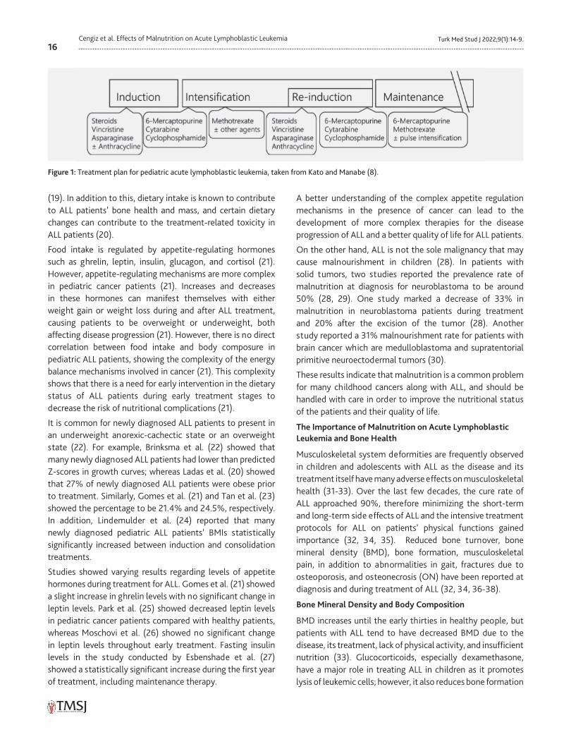
16Turk Med Stud J 2022;9(1):14-9.Cengiz et al. Effects of Malnutrition on Acute Lymphoblastic Leukemia
(19). In addition to this, dietary intake is known to contribute to ALL patients’ bone health and mass, and certain dietary changes can contribute to the treatment-related toxicity in ALL patients (20).
Food intake is regulated by appetite-regulating hormones such as ghrelin, leptin, insulin, glucagon, and cortisol (21). However, appetite-regulating mechanisms are more complex in pediatric cancer patients (21). Increases and decreases in these hormones can manifest themselves with either weight gain or weight loss during and after ALL treatment, causing patients to be overweight or underweight, both affecting disease progression (21). However, there is no direct correlation between food intake and body composure in pediatric ALL patients, showing the complexity of the energy balance mechanisms involved in cancer (21). This complexity shows that there is a need for early intervention in the dietary status of ALL patients during early treatment stages to decrease the risk of nutritional complications (21).
It is common for newly diagnosed ALL patients to present in an underweight anorexic-cachectic state or an overweight state (22). For example, Brinksma et al. (22) showed that many newly diagnosed ALL patients had lower than predicted Z-scores in growth curves; whereas Ladas et al. (20) showed that 27% of newly diagnosed ALL patients were obese prior to treatment. Similarly, Gomes et al. (21) and Tan et al. (23) showed the percentage to be 21.4% and 24.5%, respectively. In addition, Lindemulder et al. (24) reported that many newly diagnosed pediatric ALL patients’ BMIs statistically significantly increased between induction and consolidation treatments.
Studies showed varying results regarding levels of appetite hormones during treatment for ALL. Gomes et al. (21) showed a slight increase in ghrelin levels with no significant change in leptin levels. Park et al. (25) showed decreased leptin levels in pediatric cancer patients compared with healthy patients, whereas Moschovi et al. (26) showed no significant change in leptin levels throughout early treatment. Fasting insulin levels in the study conducted by Esbenshade et al. (27) showed a statistically significant increase during the first year of treatment, including maintenance therapy.
A better understanding of the complex appetite regulation mechanisms in the presence of cancer can lead to the development of more complex therapies for the disease progression of ALL and a better quality of life for ALL patients.
On the other hand, ALL is not the sole malignancy that may cause malnourishment in children (28). In patients with solid tumors, two studies reported the prevalence rate of malnutrition at diagnosis for neuroblastoma to be around 50% (28, 29). One study marked a decrease of 33% in malnutrition in neuroblastoma patients during treatment and 20% after the excision of the tumor (28). Another study reported a 31% malnourishment rate for patients with brain cancer which are medulloblastoma and supratentorial primitive neuroectodermal tumors (30).
These results indicate that malnutrition is a common problem for many childhood cancers along with ALL, and should be handled with care in order to improve the nutritional status of the patients and their quality of life.
The Importance of Malnutrition on Acute Lymphoblastic Leukemia and Bone Health
Musculoskeletal system deformities are frequently observed in children and adolescents with ALL as the disease and its treatment itself have many adverse effects on musculoskeletal health (31-33). Over the last few decades, the cure rate of ALL approached 90%, therefore minimizing the short-term and long-term side effects of ALL and the intensive treatment protocols for ALL on patients’ physical functions gained importance (32, 34, 35). Reduced bone turnover, bone mineral density (BMD), bone formation, musculoskeletal pain, in addition to abnormalities in gait, fractures due to osteoporosis, and osteonecrosis (ON) have been reported at diagnosis and during treatment of ALL (32, 34, 36-38).
Bone Mineral Density and Body Composition
BMD increases until the early thirties in healthy people, but patients with ALL tend to have decreased BMD due to the disease, its treatment, lack of physical activity, and insufficient nutrition (33). Glucocorticoids, especially dexamethasone, have a major role in treating ALL in children as it promotes lysis of leukemic cells; however, it also reduces bone formation
Figure 1: Treatment plan for pediatric acute lymphoblastic leukemia, taken from Kato and Manabe (8).
17Turk Med Stud J 2022;9(1):14-9. Cengiz et al. Effects of Malnutrition on Acute Lymphoblastic Leukemia
by inducing apoptosis of osteoblasts and osteocytes (32, 39). Low BMD in childhood may ensue osteoporotic fractures later in life (39). Research by Alos et al. (40) revealed that 16% of children with ALL had vertebral fractures one year after beginning ALL treatment. The leukemic process has also been linked with low bone turnover as the leukemic cells secrete osteoblast-inhibiting factors and parathyroid hormone-related peptides (39).
In an effort to minimize the effect of low BMD in ALL survivors later in life, patients are often provided nutritional counseling and supplementation, such as vitamin D and calcium. A study conducted by Kaste et al. (41) showed that these treatments did not significantly remediate the low BMD over a two-year period.
Unfortunately, few studies have been conducted on the effects of nutritional deficiencies on bone health in ALL patients and survivors. However, one study suggests that it is possible to prevent the bone health complications of ALL treatment when the patient follows a nutrient-dense diet (42).
Osteonecrosis
Osteonecrosis is one of the most common and significant sequelae of ALL treatment, causing serious pain, decreased mobility, and joint collapse and therefore reduces long-term survivors’ life quality (43). Previous studies show various results on the prevalence (1.6-17.6%) and risk factors of ON presentation during or after the antileukemic treatment (41). A study conducted by Mattano et al. (44) between 1989 and 1995 revealed 9.3% of 1,409 children with ALL experienced ON within 3 years after diagnosis. Another study reported the prevalence of ON observed in patients undergoing intensive chemotherapy for ALL as 1.6% (45). These different outcomes may stem from different types (dexamethasone, prednisone) or dosages of glucocorticoids, adolescent age at diagnosis, or radiation exposure (46). Kadan-Lottick et al. (46) also confirmed that patients treated with dexamethasone were 30% more likely to develop ON than patients treated with prednisone.
CONCLUSION
There have been several studies about the relationship between ALL prognosis and malnutrition. As it can be seen in this review, the results are quite diverse. There are many different methods for evaluating the nutritional status of the patients. The lack of a standardized method could be the reason of these contradictory results. The reason of the discrepancies between findings might also be due to the differences in sample sizes, cohort characteristics, and chemotherapeutic agents used in different studies. Larger
trials examining appetite-regulating hormone levels and ALL disease progression are needed for better comparison. Therefore, it is not correct to compare every study. Nutritional status should still be considered, as ALL requires long-term treatment and has implications for both patients’ quality of life and survival rates.
Adequate nourishment is crucial especially during the early treatment stages since inadequate nutrition could decrease the tolerance to chemotherapy as well as reduced immunity, and has negative effects on hematopoiesis (22, 47). It can lead to impaired production of blood cells, causing leucopenia and anemia. Furthermore, severe protein malnutrition in newborns and infants is related to atrophy in bone marrow and thymus, immature T cell number in the periphery rises and CD4/CD8 ratio decreases (47). These effects of malnutrition can reduce the patient’s immunity, which could make the patient more susceptible to infections affecting survival rate (47).
There is a growing understanding that malnutrition is complex and reflects a range of overlapping comorbidities that are not fully understood.
Patients with ALL have a high recovery rate even with chemotherapy alone, and these patients have a good chance of living a long, healthy life after treatment. Therefore, clinicians should be aware of the importance of nutritional status and focus on patients’ quality of life during treatment.
Ethics Committee Approval: N/A
Informed Consent: N/A
Conflict of Interest: The authors declared no conflict of interest.
Author Contributions: Concept: E.C., I.K., E.Ç., C.Y., F.O.Ş., H.O.K., Design: E.C., I.K., E.Ç., C.Y., F.O.Ş., H.O.K., Supervision: E.C., I.K., E.Ç., C.Y., F.O.Ş., H.O.K., Resources: E.C., I.K., E.Ç., C.Y., F.O.Ş., H.O.K., Materials: E.C., I.K., E.Ç., C.Y., F.O.Ş., H.O.K., Data Collection and/ or Processing: E.C., I.K., E.Ç., C.Y., F.O.Ş., H.O.K., Analysis and/or Interpretation: E.C., I.K., E.Ç., C.Y., F.O.Ş., H.O.K., Literature Search: E.C., I.K., E.Ç., C.Y., F.O.Ş., H.O.K., Writing Manuscript: E.C., I.K., E.Ç., C.Y., F.O.Ş., H.O.K., Critical Review: E.C., I.K., E.Ç., C.Y., F.O.Ş., H.O.K.
Financial Disclosure: The authors declared that this study received no financial support.
REFERENCES1. Khalid A. Evaluation of the nutritional status in children with acute
lymphoblastic leukemia and its effect on the outcome of induction in a developing country. J Clin Oncol 2017;35:e22004. [Crossref]
2. Onciu M. Acute lymphoblastic leukemia. Hematol Oncol Clin North Am 2009;23:655-74. [Crossref]
18Turk Med Stud J 2022;9(1):14-9.Cengiz et al. Effects of Malnutrition on Acute Lymphoblastic Leukemia
3. Terwilliger T, Abdul-Hay M. Acute lymphoblastic leukemia: a comprehensive review and 2017 update. Blood Cancer J 2017;7:e577. [Crossref]
4. Malard F, Mohty M. Acute lymphoblastic leukaemia. Lancet 2020;395:1146-62. [Crossref]
5. Barr RD, Gomez-Almaguer D, Jaime-Perez JC et al. Importance of Nutrition in the Treatment of Leukemia in Children and Adolescents. Arch Med Res 2016;47:585-92. [Crossref]
6. Bhutta ZA, Berkley JA, Bandsma RHJ et al. Severe childhood malnutrition. Nat Rev Dis Primers 2017;3:17067. [Crossref]
7. Cederholm T, Bosaeus I, Barazzoni R et al. Diagnostic criteria for malnutrition - An ESPEN Consensus Statement. Clin Nutr 2015;34:335-40. [Crossref]
8. Kato M, Manabe A. Treatment and biology of pediatric acute lymphoblastic leukemia. Pediatr Int 2018;60:4-12. [Crossref]
9. Sala A, Pencharz P, Barr RD. Children, cancer, and nutrition--A dynamic triangle in review. Cancer 2004;100:677-87. [Crossref]
10. Mejía-Arangure JM, Fajardo-Gutíerrez A, Bernáldez-Ríos R et al. Nutritional state alterations in children with acute lymphoblastic leukemia during induction and consolidation of chemotherapy. Arch Med Res 1997;28:273-9. [Crossref]
11. Uderzo C, Rovelli A, Bonomi M et al. Nutritional status in untreated children with acute leukemia as compared with children without malignancy. J Pediatr Gastroenterol Nutr 1996;23:34-7. [Crossref]
12. Tazi I, Hidane Z, Zafad S et al. Nutritional status at diagnosis of children with malignancies in Casablanca. Pediatr Blood Cancer 2008;51: 495-8. [Crossref]
13. Antillon F, de Maselli T, Garcia T et al. Nutritional status of children during treatment for acute lymphoblastic leukemia in the Central American Pediatric Hematology Oncology Association (AHOPCA): preliminary data from Guatemala. Pediatr Blood Cancer 2008;50(2 Suppl):502-5; discussion 517. [Crossref]
14. Jaime-Pérez JC, González-Llano O, Herrera-Garza JL et al. Assessment of nutritional status in children with acute lymphoblastic leukemia in Northern México: A 5-year experience. Pediatr Blood Cancer 2008;50(2 Suppl):506-8; discussion 517. [Crossref]
15. Butturini AM, Dorey FJ, Lange BJ et al. Obesity and outcome in pediatric acute lymphoblastic leukemia. J Clin Oncol 2007;25:2063-9.[Crossref]
16. Lobato-Mendizábal E, Ruiz-Argüelles GJ, Marín-López A. Leukaemia and nutrition. I: Malnutrition is an adverse prognostic factor in the outcome of treatment of patients with standard-risk acute lymphoblastic leukaemia. Leuk Res 1989;13:899-906. [Crossref]
17. Gómez-Almaguer D, Ruiz-Argüelles GJ, Ponce-de-León S. Nutritional status and socio-economic conditions as prognostic factors in the outcome of therapy in childhood acute lymphoblastic leukemia. Int J Cancer Suppl 1998;11:52-5. [Crossref]
18. Tebeje NB, Bikes GA, Abebe SM et al. Prevalence and major contributors of child malnutrition in developing countries: systematic review and meta-analysis. J Child Obes 2017;2:16. [Crossref]
19. Fayh APT, Bezerra ADL, Friedman R. Appetite hormones in children and adolescents with cancer: a systematic review of observational studies. Nutr Hosp 2018;35:201-10. [Crossref]
20. Ladas EJ, Orjuela M, Stevenson K et al. Dietary intake and childhood leukemia: The Diet and Acute Lymphoblastic Leukemia Treatment (DALLT) cohort study. Nutrition 2016;32:1103-9.e1. [Crossref]
21. Gomes CC, Silva CCGD, Nascimento PRPD et al. Nutritional status and appetite-regulating hormones in early treatment of acute lymphoblastic leukemia among children and adolescents: a cohort study. Sao Paulo Med J 2020;138:118-25. [Crossref]
22. Brinksma A, Roodbol PF, Sulkers E et al. Weight and height in children newly diagnosed with cancer. Pediatr Blood Cancer 2015;62:269-73. [Crossref]
23. Tan SY, Poh BK, Nadrah MH et al. Nutritional status and dietary intake of children with acute leukaemia during induction or consolidation chemotherapy. J Hum Nutr Diet 2013;26 Suppl 1:23-33. [Crossref]
24. Lindemulder SJ, Stork LC, Bostrom B et al. Survivors of standard risk acute lymphoblastic leukemia do not have increased risk for overweight and obesity compared to non-cancer peers: a report from the Children’s Oncology Group. Pediatr Blood Cancer 2015;62:1035-41. [Crossref]
25. Park SH, Jung MH, Chung NG et al. Serum ghrelin and leptin concentrations in children with cancer: comparisons with normal children. Korean J Pediatr 2007;50:905-11. [Crossref]
26. Moschovi M, Trimis G, Vounatsou M et al. Serial plasma concentrations of adiponectin, leptin, and resistin during therapy in children with acute lymphoblastic leukemia. J Pediatr Hematol Oncol 2010;32:e8-13. [Crossref]
27. Esbenshade AJ, Simmons JH, Koyama T et al. Obesity and insulin resistance in pediatric acute lymphoblastic leukemia worsens during maintenance therapy. Pediatr Blood Cancer 2013;60:1287-91. [Crossref]
28. Green GJ, Weitzman SS, Pencharz PB. Resting energy expenditure in children newly diagnosed with stage IV neuroblastoma. Pediatr Res 2008;63:332-6. [Crossref]
29. Rickard KA, Foland BB, Detamore CM et al. Effectiveness of central parenteral nutrition versus peripheral parenteral nutrition plus enteral nutrition in reversing protein-energy malnutrition in children with advanced neuroblastoma and Wilms’ tumor: a prospective randomized study. Am J Clin Nutr 1983;38:445-56. [Crossref]
30. Bakish J, Hargrave D, Tariq N et al. Evaluation of dietetic intervention in children with medulloblastoma or supratentorial primitive neuroectodermal tumors. Cancer 2003;98:1014-20. [Crossref]
31. Mostoufi-Moab S, Halton J. Bone morbidity in childhood leukemia: epidemiology, mechanisms, diagnosis, and treatment. Curr Osteoporos Rep 2014;12:300-12. [Crossref]
32. Ward LM, Ma J, Lang B et al. Bone Morbidity and Recovery in Children With Acute Lymphoblastic Leukemia: Results of a Six-Year Prospective Cohort Study. J Bone Miner Res 2018;33:1435-43. [Crossref]
33. Inaba H, Cao X, Han AQ et al. Bone mineral density in children with acute lymphoblastic leukemia. Cancer 2018;124:1025-35. [Crossref]
34. van der Sluis IM, van den Heuvel-Eibrink MM, Hählen K et al. Altered bone mineral density and body composition, and increased fracture risk in childhood acute lymphoblastic leukemia. J Pediatr 2002;141:204-10. [Crossref]
35. Cox CL, Zhu L, Kaste SC et al. Modifying bone mineral density, physical function, and quality of life in children with acute lymphoblastic leukemia. Pediatr Blood Cancer 2018;65:10.1002/pbc.26929. [Crossref]
36. Halton JM, Atkinson SA, Fraher L et al. Altered mineral metabolism and bone mass in children during treatment for acute lymphoblastic leukemia. J Bone Miner Res 1996;11:1774-83. [Crossref]
37. Marinovic D, Dorgeret S, Lescoeur B et al. Improvement in bone mineral density and body composition in survivors of childhood
19Turk Med Stud J 2022;9(1):14-9. Cengiz et al. Effects of Malnutrition on Acute Lymphoblastic Leukemia
acute lymphoblastic leukemia: a 1-year prospective study. Pediatrics 2005;116:e102-8. [Crossref]
38. Kunstreich M, Kummer S, Laws HJ et al. Osteonecrosis in children with acute lymphoblastic leukemia. Haematologica 2016;101:1295-305. [Crossref]
39. Davies JH, Evans BA, Jenney ME et al. Skeletal morbidity in childhood acute lymphoblastic leukaemia. Clin Endocrinol (Oxf) 2005;63:1-9. [Crossref]
40. Alos N, Grant RM, Ramsay T et al. High incidence of vertebral fractures in children with acute lymphoblastic leukemia 12 months after the initiation of therapy. J Clin Oncol 2012;30:2760-7. [Crossref]
41. Kaste SC, Qi A, Smith K, et al. Calcium and cholecalciferol supplementation provides no added benefit to nutritional counseling to improve bone mineral density in survivors of childhood acute lymphoblastic leukemia (ALL). Pediatr Blood Cancer 2014;61:885-93. [Crossref]
42. Tylavsky FA, Smith K, Surprise H et al. Nutritional intake of long-term survivors of childhood acute lymphoblastic leukemia: evidence for bone
health interventional opportunities. Pediatr Blood Cancer 2010;55: 1362-9. [Crossref]
43. Girard P, Auquier P, Barlogis V et al. Symptomatic osteonecrosis in childhood leukemia survivors: prevalence, risk factors and impact on quality of life in adulthood. Haematologica 2013;98:1089-97. [Crossref]
44. Mattano LA Jr, Sather HN, Trigg ME et al. Osteonecrosis as a complication of treating acute lymphoblastic leukemia in children: a report from the Children’s Cancer Group. J Clin Oncol 2000;18:3262-72. [Crossref]
45. Aricò M, Boccalatte MF, Silvestri D et al. Osteonecrosis: An emerging complication of intensive chemotherapy for childhood acute lymphoblastic leukemia. Haematologica 2003;88:747-53. [Crossref]
46. Kadan-Lottick NS, Dinu I, Wasilewski-Masker K et al. Osteonecrosis in adult survivors of childhood cancer: a report from the childhood cancer survivor study. J Clin Oncol 2008 ;26:3038-45. [Crossref]
47. Getawa S, Getaneh Z, Melku M. Hematological Abnormalities and Associated Factors Among Undernourished Under-Five Children Attending University of Gondar Specialized Referral Hospital, Northwest Ethiopia. J Blood Med 2020;11:465-78. [Crossref]
ORIGINAL ARTICLE
OPEN ACCESS
www.turkmedstudj.comCopyright@Author(s) - Available online at https://www.turkmedstudj.com/
20
Altan et al. Participation of Cannabinoid Receptors in Antipruritic Activity Induced by Systemic Dipyrone in Mice
Cite this article as: Altan E, Folladwand NA, Gurmani R et al. Participation of cannabinoid receptors in antipruritic activity induced by systemic dipyrone in mice. Turk Med Stud J 2022;9(1):20-4.
Address for Correspondence: Erdem Altan, Trakya University School of Medicine, Edirne, TURKEYe-mail: [email protected] iDs of the authors: EA: 0000-0002-4026-5447; NAF: 0000-0001-9542-6650; RG: 0000-0002-2013-0297; ÖTÖ: 0000-0002-9311-4538; KDA: 0000-0001-5573-4915; AU: 0000-0003-4643-1124.Received: 16.11.2021 Accepted: 30.11.2021
1Trakya University School of Medicine, Edirne, TURKEY2Trakya University School of Medicine, Department of Medical Pharmacology, Edirne, TURKEY
ABSTRACT
Aims: The cannabinoid system has been shown to contribute to the antinociceptive effects of nonsteroidal anti-inflammatory drugs. Considering the similar pathophysiological mechanisms underlying pain and itching, we aimed to observe whether dipyrone has an antipruritic effect and whether cannabinoid receptors are involved in this effect.Methods: In this project, we produced scratching behavior in BALB/c mice, intradermally administering the well-known pruritic agent compound 48/80. After observing the anti-scratching effect of dipyrone with increasing doses, we administered AM-251 (1 mg/kg, intraperitoneal) and AM-630 (3 mg/kg, intraperitoneal) to determine whether the endocannabinoid system was associated with this effect of dipyrone. Results: Dipyrone reduced scratching behavior at its highest dose used in this study (600 mg/kg); however, neither AM-251 nor AM-630 changed the antipruritic action of dipyrone. Conclusion: Our findings indicate that dipyrone, at higher doses, attenuates compound 48/80-induced scratching behavior in mice. Cannabinoid receptors have been found not to be involved in the antipruritic effect of dipyrone. Further experiments are required to delineate the mechanisms underlying this high-dose dipyrone effect.Keywords: AM-251, AM-630, dipyrone, pruritus
INTRODUCTION
Cannabinoids have been known to reduce pain for centuries; however, several reasons preclude their use as effective analgesics (1). These chemical compounds include synthetic cannabinoids, those found in the Cannabis plant (phytocannabinoids), and those distributed throughout the body (endocannabinoids) (1). All cannabinoids activate cannabinoid receptors (CB1 and CB2) and take place in many pathophysiological processes (1). CB1 receptors are distributed widely in the central nervous system and appear to play the predominant role in both the therapeutic and the
central adverse effects of these drugs (1). Despite a great deal of research, cannabinoids are only used in limited indications. Synthetic cannabinoids, dronabinol and nabilone, are used in the treatment of chemotherapy-associated emesis, and nabilone is also approved for anorexia associated with acquired immunodeficiency syndrome (1). Nabiximol (Δ9-tetrahydrocannabinol + cannabidiol) is recommended for use in neuropathy and spasticity associated with multiple sclerosis and cancer pain (2, 3). These cannabinoids are still alternatives in aforementioned indications and are approved only in some countries; however, numerous ongoing clinical
Erdem Altan1 , Najaf Ali Folladwand1 , Rymejsa Gurmani1 , Ömür Türkü Özşalap1 , Kübra Duvan Aydemir2 , Ahmet Ulugöl2
PARTICIPATION OF CANNABINOID RECEPTORS IN ANTIPRURITIC ACTIVITY INDUCED BY SYSTEMIC DIPYRONE IN MICE
DOI: 10.4274/tmsj.galenos.2021.09.01.04Turk Med Stud J 2022;9(1):20-4
21Turk Med Stud J 2022;9(1):20-4. Altan et al. Participation of Cannabinoid Receptors in Antipruritic Activity Induced by Systemic Dipyrone in Mice
trials seem to enhance their usage in different pathological conditions in addition to its use for pain reduction (2, 3).
Nonsteroidal anti-inflammatory drugs (NSAIDs) are a group of medicines used worldwide to reduce pain, fever, and inflammation (4). They block the cyclooxygenase enzymes (COX-1, COX-2) and inhibit the production of prostaglandins (4). Distinct from traditional NSAIDs, paracetamol and dipyrone appear not to act on peripheral COX enzymes; a recent research indicates that they do not have a direct significant effect on endocannabinoid levels in a rat’s brain and spinal cord (4). Yet, there are previous studies indicating that both increases in endocannabinoid tonus and cannabinoid receptors are associated with the antinociceptive effects of paracetamol and dipyrone (5-7). The cannabinoid system has also been proposed to participate in antipyretic, anxiolytic, and anticonvulsive effects of these drugs, although there are contradictory data (7-10).
Pruritus, also known as itching, is an uncomfortable, irritating sensation that provokes the urge to scratch (11). It is the predominant symptom of many diseases and can affect the quality of life (11). Overall, pruritus and pain are two sensations showing many similarities, especially in pathological and chronic conditions (11, 12). Accordingly, similar to pain, involvement of the cannabinoid system in the development of pruritus and the probable therapeutic effect of cannabinoids in pruritic states have been suggested (13, 14). Cannabinoid agonists have been shown to attenuate histamine-induced responses, while rimonabant, CB1 receptor antagonist, induced scratching behavior (13, 14). Similarly, we presented that activation of cannabinoid receptors using the synthetic cannabinoid agonist WIN 55,212-2 and enhancing endocannabinoid tonus via blockade of endocannabinoid degrading enzymes, fatty acid amide hydrolase, and monoacylglycerol lipase exhibit dose-dependent antipruritic activity (15-17).
This study aims to examine whether dipyrone reduces itching behavior induced by compound 48/80 (C 48/80) and to observe whether cannabinoid receptors mediate dipyrone’s effects in case of any antipruritic action.
MATERIAL AND METHODS
Animals & Ethics
In this study, male BALB/c mice (2-3 months) weighing 20-30 g were used (Center of the Laboratory Animals, Trakya University). All animals were housed in a light (12/12 h day/night cycles) and temperature-controlled (21±2 °C) room where food and water were available ad libitum. A total of 48 mice were used in this study. Separate groups, each involving 6 mice were used for each set of experiments. The experiments
were conducted after approval of the local “Animal Care Ethics Committee” (protocol code: TÜHADYEK-2019/27, date: 25.10.2019). Moreover, during all procedures, the Ethical Committee of the International Association for the Study of Pain guidelines were followed strictly (18).
Study Design
Itching behavior was evoked by intradermal injection of 100 µg/50 µL of C 48/80 into the rostral part of the back of each mouse. Several scratches per second were accepted as one bout of scratching. These scratches were videotaped and counted for 30 minutes, beginning just after administering C 48/80. To examine the antipruritic effect of the drug, different doses of dipyrone [150, 300, 600 mg/kg, intraperitoneal (i.p.)] were tested. Subsequently, effects of the cannabinoid CB1 receptor antagonist AM-251 (1 mg/kg, i.p.) and the cannabinoid CB2 receptor antagonist AM-630 (3 mg/kg, i.p.) on the antipruritic activity of dipyrone were analyzed. Dipyrone was injected 30 minutes before C 48/80 administration, and cannabinoid receptor antagonists were administered 10 minutes before dipyrone.
Drugs
Compounds 48/80 and AM-630 were supplied from Sigma-Aldrich (St Louis, MO, USA); AM-251 was obtained from Tocris (UK); and dipyrone was purchased from Cayman (Ann Arbor, MI, USA). C 48/80 and dipyrone were dissolved in physiological saline, while AM-251 and AM-630 were administered in 20% dimethyl sulfoxide, 1% Tween 80, 1% ethanol, and 78% saline. Doses of each drug and treatment schedules were chosen from our previous studies (19-24).
Statistical Analysis
Graphpad Prism 8.4 was used for statistical analysis and to plot the graphs. To assess the significance of any difference between groups, the Kruskal-Wallis test, then Dunn’s Multiple Comparisons test were performed. In all analyses, p<0.05 was considered statistically significant.
RESULTS
Reduction of Compound 48/80-induced Scratching Behavior by Dipyrone
Dipyrone (150, 300, 600 mg/kg) administration attenuated C 48/80-induced scratches at its highest dose (30 mg/kg; *p<0.001, compared to vehicle); but treatment with lower dipyrone doses had no effect on scratching behavior (Figure 1).
Influence of Cannabinoid Receptor Antagonism on Dipyrone-induced Antipruritic Activity
Neither AM-251 (1 mg/kg) nor AM-630 (3 mg/kg) had any influence on the antipruritic activity of dipyrone (Figure 2).
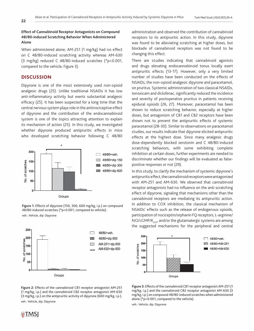
22Turk Med Stud J 2022;9(1):20-4.Altan et al. Participation of Cannabinoid Receptors in Antipruritic Activity Induced by Systemic Dipyrone in Mice
Effect of Cannabinoid Receptor Antagonists on Compound 48/80-induced Scratching Behavior When Administered Alone
When administered alone, AM-251 (1 mg/kg) had no effect on C 48/80-induced scratching activity whereas AM-630 (3 mg/kg) reduced C 48/80-induced scratches (*p<0.001, compared to the vehicle; Figure 3).
DISCUSSION
Dipyrone is one of the most extensively used non-opioid analgesic drugs (25). Unlike traditional NSAIDs it has low anti-inflammatory activity but exerts substantial analgesic efficacy (25). It has been suspected for a long time that the central nervous system plays role in the antinociceptive effect of dipyrone and the contribution of the endocannabinoid system is one of the topics attracting attention to explain its mechanism of action (25). In this study, we investigated whether dipyrone produced antipruritic effects in mice who developed scratching behavior following C 48/80
administration and observed the contribution of cannabinoid receptors to its antipruritic action. In this study, dipyrone was found to be alleviating scratching at higher doses, but blockade of cannabinoid receptors was not found to be changing this effect.
There are studies indicating that cannabinoid agonists and drugs elevating endocannabinoid tonus locally exert antipruritic effects (13-17). However, only a very limited number of studies have been conducted on the effects of NSAIDs, the non-opioid analgesic dipyrone and paracetamol, on pruritus. Systemic administration of two classical NSAIDs, tenoxicam and diclofenac, significantly reduced the incidence and severity of postoperative pruritus in patients receiving epidural opioids (26, 27). Moreover, paracetamol has been shown to reduce scratching behavior, especially at higher doses, but antagonism of CB1 and CB2 receptors have been shown not to prevent the antipruritic effects of systemic paracetamol (28-30). Similar to observations on paracetamol studies, our results indicate that dipyrone elicited antipruritic effects at the highest dose. Since many analgesic drugs dose-dependently blocked serotonin and C 48/80-induced scratching behaviors, with some exhibiting complete inhibition at certain doses, further experiments are needed to discriminate whether our findings will be evaluated as false-positive responses or not (29).
In this study, to clarify the mechanism of systemic dipyrone’s antipruritic effect, the cannabinoid receptors were antagonized with AM-251 and AM-630. We observed that cannabinoid receptor antagonists had no influence on the anti-scratching effect of dipyrone, signaling that mechanisms other than the cannabinoid receptors are mediating its antipruritic action. In addition to COX inhibition, the classical mechanism of NSAIDs’ effects such as the release of endogenous opioids, participation of nociceptin/orphanin FQ receptors, L-arginine/NO/cGMP/KATP, and/or the glutamatergic systems are among the suggested mechanisms for the peripheral and central
Figure 1: Effects of dipyrone (150, 300, 600 mg/kg, i.p.) on compound 48/80-induced scratches (*p<0.001, compared to vehicle).
veh.: Vehicle, dip: Dipyrone
Figure 2: Effects of the cannabinoid CB1 receptor antagonist AM-251 (1 mg/kg, i.p.) and the cannabinoid CB2 receptor antagonist AM-630 (3 mg/kg, i.p.) on the antipruritic activity of dipyrone (600 mg/kg, i.p.).
veh.: Vehicle, dip: Dipyrone
Figure 3: Effects of the cannabinoid CB1 receptor antagonist AM-251 (1 mg/kg, i.p.) and the cannabinoid CB2 receptor antagonist AM-630 (3 mg/kg, i.p.) on compound 48/80-induced scratches when administered alone (*p<0.001, compared to the vehicle).
veh.: Vehicle, dip: Dipyrone
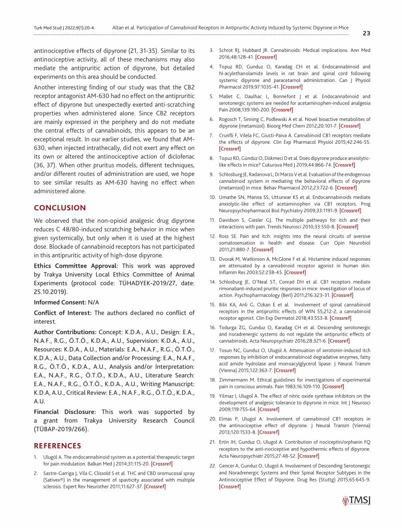
23Turk Med Stud J 2022;9(1):20-4. Altan et al. Participation of Cannabinoid Receptors in Antipruritic Activity Induced by Systemic Dipyrone in Mice
antinociceptive effects of dipyrone (21, 31-35). Similar to its antinociceptive activity, all of these mechanisms may also mediate the antipruritic action of dipyrone, but detailed experiments on this area should be conducted.
Another interesting finding of our study was that the CB2 receptor antagonist AM-630 had no effect on the antipruritic effect of dipyrone but unexpectedly exerted anti-scratching properties when administered alone. Since CB2 receptors are mainly expressed in the periphery and do not mediate the central effects of cannabinoids, this appears to be an exceptional result. In our earlier studies, we found that AM-630, when injected intrathecally, did not exert any effect on its own or altered the antinociceptive action of diclofenac (36, 37). When other pruritus models, different techniques, and/or different routes of administration are used, we hope to see similar results as AM-630 having no effect when administered alone.
CONCLUSION
We observed that the non-opioid analgesic drug dipyrone reduces C 48/80-induced scratching behavior in mice when given systemically, but only when it is used at the highest dose. Blockade of cannabinoid receptors has not participated in this antipruritic activity of high-dose dipyrone.
Ethics Committee Approval: This work was approved by Trakya University Local Ethics Committee of Animal Experiments (protocol code: TÜHADYEK-2019/27, date: 25.10.2019).
Informed Consent: N/A
Conflict of Interest: The authors declared no conflict of interest.
Author Contributions: Concept: K.D.A., A.U., Design: E.A., N.A.F., R.G., Ö.T.Ö., K.D.A., A.U., Supervision: K.D.A., A.U., Resources: K.D.A., A.U., Materials: E.A., N.A.F., R.G., Ö.T.Ö., K.D.A., A.U., Data Collection and/or Processing: E.A., N.A.F., R.G., Ö.T.Ö., K.D.A., A.U., Analysis and/or Interpretation: E.A., N.A.F., R.G., Ö.T.Ö., K.D.A., A.U., Literature Search: E.A., N.A.F., R.G., Ö.T.Ö., K.D.A., A.U., Writing Manuscript: K.D.A, A.U., Critical Review: E.A., N.A.F., R.G., Ö.T.Ö., K.D.A., A.U.
Financial Disclosure: This work was supported by a grant from Trakya University Research Council (TÜBAP-2019/266).
REFERENCES1. Ulugöl A. The endocannabinoid system as a potential therapeutic target
for pain modulation. Balkan Med J 2014;31:115-20. [Crossref]
2. Sastre-Garriga J, Vila C, Clissold S et al. THC and CBD oromucosal spray (Sativex®) in the management of spasticity associated with multiple sclerosis. Expert Rev Neurother 2011;11:627-37. [Crossref]
3. Schrot RJ, Hubbard JR. Cannabinoids: Medical implications. Ann Med 2016;48:128-41. [Crossref]
4. Topuz RD, Gunduz O, Karadag CH et al. Endocannabinoid and N-acylethanolamide levels in rat brain and spinal cord following systemic dipyrone and paracetamol administration. Can J Physiol Pharmacol 2019;97:1035-41. [Crossref]
5. Mallet C, Daulhac L, Bonnefont J et al. Endocannabinoid and serotonergic systems are needed for acetaminophen-induced analgesia Pain 2008;139:190-200. [Crossref]
6. Rogosch T, Sinning C, Podlewski A et al. Novel bioactive metabolites of dipyrone (metamizol). Bioorg Med Chem 2012;20:101-7. [Crossref]
7. Crunfli F, Vilela FC, Giusti-Paiva A. Cannabinoid CB1 receptors mediate the effects of dipyrone. Clin Exp Pharmacol Physiol 2015;42:246-55. [Crossref]
8. Topuz RD, Gündüz O, Dökmeci D et al. Does dipyrone produce anxiolytic-like effects in mice? Cukurova Med J 2019;44:866-74. [Crossref]
9. Schlosburg JE, Radanova L, Di Marzo V et al. Evaluation of the endogenous cannabinoid system in mediating the behavioral effects of dipyrone (metamizol) in mice. Behav Pharmacol 2012;23:722-6. [Crossref]
10. Umathe SN, Manna SS, Utturwar KS et al. Endocannabinoids mediate anxiolytic-like effect of acetaminophen via CB1 receptors. Prog Neuropsychopharmacol Biol Psychiatry 2009;33:1191-9. [Crossref]
11. Davidson S, Giesler GJ. The multiple pathways for itch and their interactions with pain. Trends Neurosci 2010;33:550-8. [Crossref]
12. Ross SE. Pain and itch: insights into the neural circuits of aversive somatosensation in health and disease. Curr Opin Neurobiol 2011;21:880-7. [Crossref]
13. Dvorak M, Watkinson A, McGlone F et al. Histamine induced responses are attenuated by a cannabinoid receptor agonist in human skin. Inflamm Res 2003;52:238-45. [Crossref]
14. Schlosburg JE, O’Neal ST, Conrad DH et al. CB1 receptors mediate rimonabant-induced pruritic responses in mice: investigation of locus of action. Psychopharmacology (Berl) 2011;216:323-31. [Crossref]
15. Bilir KA, Anli G, Ozkan E et al. Involvement of spinal cannabinoid receptors in the antipruritic effects of WIN 55,212-2, a cannabinoid receptor agonist. Clin Exp Dermatol 2018;43:553-8. [Crossref]
16. Todurga ZG, Gunduz O, Karadag CH et al. Descending serotonergic and noradrenergic systems do not regulate the antipruritic effects of cannabinoids. Acta Neuropsychiatr 2016;28:321-6. [Crossref]
17. Tosun NC, Gunduz O, Ulugol A. Attenuation of serotonin-induced itch responses by inhibition of endocannabinoid degradative enzymes, fatty acid amide hydrolase and monoacylglycerol lipase. J Neural Transm (Vienna) 2015;122:363-7. [Crossref]
18. Zimmermann M. Ethical guidelines for investigations of experimental pain in conscious animals. Pain 1983;16:109-110. [Crossref]
19. Yilmaz I, Ulugol A. The effect of nitric oxide synthase inhibitors on the development of analgesic tolerance to dipyrone in mice. Int J Neurosci 2009;119:755-64. [Crossref]
20. Elmas P, Ulugol A. Involvement of cannabinoid CB1 receptors in the antinociceptive effect of dipyrone. J Neural Transm (Vienna) 2013;120:1533-8. [Crossref]
21. Ertin IH, Gunduz O, Ulugol A. Contribution of nociceptin/orphanin FQ receptors to the anti-nociceptive and hypothermic effects of dipyrone. Acta Neuropsychiatr 2015;27:48-52. [Crossref]
22. Gencer A, Gunduz O, Ulugol A. Involvement of Descending Serotonergic and Noradrenergic Systems and their Spinal Receptor Subtypes in the Antinociceptive Effect of Dipyrone. Drug Res (Stuttg) 2015;65:645-9. [Crossref]
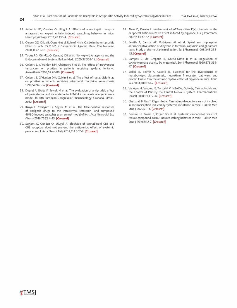
24Turk Med Stud J 2022;9(1):20-4.Altan et al. Participation of Cannabinoid Receptors in Antipruritic Activity Induced by Systemic Dipyrone in Mice
23. Aydemir KD, Gunduz O, Ulugol A. Effects of a nociceptin receptor antagonist on experimentally induced scratching behavior in mice. Neurophysiology 2017;49:130-4. [Crossref]
24. Gercek OZ, Oflaz B, Oguz N et al. Role of Nitric Oxide in the Antipruritic Effect of WIN 55,212-2, a Cannabinoid Agonist. Basic Clin Neurosci 2020;11:473-80. [Crossref]
25. Topuz RD, Gündüz Ö, Karadağ ÇH et al. Non-opioid Analgesics and the Endocannabinoid System. Balkan Med J 2020;37:309-15. [Crossref]
26. Colbert S, O’Hanlon DM, Chambers F et al. The effect of intravenous tenoxicam on pruritus in patients receiving epidural fentanyl. Anaesthesia 1999;54:76-80. [Crossref]
27. Colbert S, O’Hanlon DM, Galvin S et al. The effect of rectal diclofenac on pruritus in patients receiving intrathecal morphine. Anaesthesia 1999;54:948-52.[Crossref]
28. Dogrul A, Ilkaya F, Seyrek M et al. The evaluation of antipruritic effect of paracetamol and its metabolite AM404 in an acute allergenic mice model. In. 6th European Congress of Pharmacology: Granada, SPAIN. 2012. [Crossref]
29. Ilkaya F, Yesilyurt O, Seyrek M et al. The false-positive responses of analgesic drugs to the intradermal serotonin- and compound 48/80-induced scratches as an animal model of itch. Acta Neurobiol Exp (Wars) 2016;76:234-43. [Crossref]
30. Saglam G, Gunduz O, Ulugol A. Blockade of cannabinoid CB1 and CB2 receptors does not prevent the antipruritic effect of systemic paracetamol. Acta Neurol Belg 2014;114:307-9. [Crossref]
31. Alves D, Duarte I. Involvement of ATP-sensitive K(+) channels in the peripheral antinociceptive effect induced by dipyrone. Eur J Pharmacol 2002;444:47-52. [Crossref]
32. Beirith A, Santos AR, Rodrigues AL et al. Spinal and supraspinal antinociceptive action of dipyrone in formalin, capsaicin and glutamate tests. Study of the mechanism of action. Eur J Pharmacol 1998;345:233-45. [Crossref]
33. Campos C, de Gregorio R, García-Nieto R et al. Regulation of cyclooxygenase activity by metamizol. Eur J Pharmacol 1999;378:339-47. [Crossref]
34. Siebel JS, Beirith A, Calixto JB. Evidence for the involvement of metabotropic glutamatergic, neurokinin 1 receptor pathways and protein kinase C in the antinociceptive effect of dipyrone in mice. Brain Res 2004;1003:61-7. [Crossref]
35. Vanegas H, Vazquez E, Tortorici V. NSAIDs, Opioids, Cannabinoids and the Control of Pain by the Central Nervous System. Pharmaceuticals (Basel) 2010;3:1335-47. [Crossref]
36. Chatzisali B, Gas T, Kilgin H et al. Cannabinoid receptors are not involved in antinociception induced by systemic diclofenac in mice. Turkish Med Stud J 2020;7:1-4. [Crossref]
37. Demirel H, Baksin E, Ozgur EO et al. Systemic cannabidiol does not reduce compound 48/80-induced itching behavior in mice. Turkish Med Stud J 2019;6:12-7. [Crossref]
CASE REPORT
www.turkmedstudj.comCopyright@Author(s) - Available online at https://www.turkmedstudj.com/
25
OPEN ACCESS
Turk Med Stud J 2022;9(1):25-8
Cite this article as: Altunel B, Özer MG, Alkan A. Mucinous adenocarcinoma mimicking cryptogenic organizing pneumonia: A case report. Turk Med Stud J 2022;9(1):25-8.
Address for Correspondence: Barış Altunel, Trakya University School of Medicine, Edirne, TURKEYe-mail: [email protected] iDs of the authors: BA: 0000-0002-4962-8865; MGÖ: 0000-0002-4592-5491; AA: 0000-0001-6903-5314. Received: 04.08.2021 Accepted: 07.09.2021
INTRODUCTION
Lung cancer is one of the world’s leading causes of death (1). Lung cancers are mostly divided into two main groups: small cell lung cancers in about 15% of all lung neoplasms and non-small cell lung cancers (NSCLCs) in about 85% of all lung neoplasms (2). Lung adenocarcinoma is the most common form among non-small cell cancers at about 50% of all NSCLCs (3). Adenocarcinoma is a malignancy that originates from glands or gland-like structures and can occur in many organs. Pneumonia is the inflammation of the distal lung structures. If the pneumonia is not resolved, it can lead to the organization of inflammatory exudate, which leads to fibrosis in the lung tissue, called organizing pneumonia. Organizing pneumonia is a prominent inflammatory lung disease due to easily reversible intra-alveolar fibrosis (4, 5).
Exclusion of any other etiology and a histopathologic examination of the lung tissue leads to the diagnosis of
“cryptogenic” organizing pneumonia (COP) while organizing pneumonia secondary to other conditions such as infectious agents, drugs (5-aminosalicylic acid, acebutolol, amiodarone, amphotericin B, bleomycin, busulfan, carbamazepine, cephalosporin, fluvastatin, gold salts, methotrexate, etc.), radiation, or malignancy termed as “secondary” organizing pneumonia (5). Lung adenocarcinoma is known to mimic different types of pneumonia, including organizing pneumonia, and can be confused on computed tomography (CT) imaging (6).
In this study, we aimed to present a patient whose pneumonia mimicked COP, who was later diagnosed with mucinous adenocarcinoma after his condition worsened. As it can be difficult to distinguish between these two pathologies, we believe that this study will raise awareness about the differential diagnosis of these conditions.
ABSTRACT
Mucinous adenocarcinoma of the lung is a type of malignancy that originates from glands or gland-like structures in the lung and can mimic pneumonia forms on radiological images. Our case report aims to raise awareness about this radiological pattern. A 72-year-old male patient was applied to an external center with the complaint of cough with sputum and back pain. Computed tomography revealed a kind of consolidation in the lower right lung zone of the patient. The patient, whose consolidation progressed in the lower right zone of the lung, was diagnosed with cryptogenic organizing pneumonia, and then corticosteroids were started. His condition worsened and a biopsy was requested for further investigation. The biopsy revealed mucinous adenocarcinoma of the lung in the patient. Mucinous adenocarcinoma is a malignancy that can be misdiagnosed as organizing pneumonia due to the similarities in patterns between the radiological images. Therefore, differential diagnosis of adenocarcinoma should be considered in cases of organizing pneumonia. Keywords: Neoplasms, pneumonia, mucinous adenocarcinoma
1Trakya University School of Medicine, Edirne, TURKEY2Trakya University School of Medicine, Department of Radiology, Edirne, TURKEY
Barış Altunel1 , Müçteba Gökalp Özer1 , Aykut Alkan2
Altunel et al. A Case of Adenocarcinoma Mimicking Pneumonia
MUCINOUS ADENOCARCINOMA MIMICKING CRYPTOGENIC ORGANIZING PNEUMONIA: A CASE REPORT
DOI: 10.4274/tmsj.galenos.2021.09.01.05
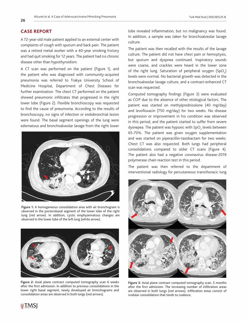
26Turk Med Stud J 2022;9(1):25-8.Altunel et al. A Case of Adenocarcinoma Mimicking Pneumonia
CASE REPORT
A 72-year-old male patient applied to an external center with complaints of cough with sputum and back pain. The patient was a retired metal worker with a 40-year smoking history and had quit smoking for 12 years. The patient had no chronic disease other than hypothyroidism.
A CT scan was performed on the patient (Figure 1), and the patient who was diagnosed with community-acquired pneumonia was referred to Trakya University School of Medicine Hospital, Department of Chest Diseases for further examination. The chest CT performed on the patient showed pneumonic infiltrates that progressed in the right lower lobe (Figure 2). Flexible bronchoscopy was requested to find the cause of pneumonia. According to the results of bronchoscopy, no signs of infection or endobronchial lesion were found. The basal segment openings of the lung were edematous and bronchoalveolar lavage from the right lower
lobe revealed inflammation, but no malignancy was found. In addition, a sample was taken for bronchoalveolar lavage culture.
The patient was then recalled with the results of the lavage culture. The patient did not have chest pain or hemoptysis, but sputum and dyspnea continued. Inspiratory sounds were coarse, and crackles were heard in the lower zone of the right lung. Saturation of peripheral oxygen (SpO2) levels were normal. No bacterial growth was detected in the bronchoalveolar lavage culture, and a contrast-enhanced CT scan was requested.
Computed tomography findings (Figure 3) were evaluated as COP due to the absence of other etiological factors. The patient was started on methylprednisolone (40 mg/day) and levofloxacin (750 mg/day) for two weeks. No disease progression or improvement in his condition was observed in this period, and the patient started to suffer from severe dysnepea. The patient was hypoxic with SpO2 levels between 65-70%. The patient was given oxygen supplementation and was started on piperacillin-tazobactam for two weeks. Chest CT was also requested. Both lungs had peripheral consolidations compared to older CT scans (Figure 4). The patient also had a negative coronavirus disease-2019 polymerase chain reaction test in this period.
The patient was then referred to the department of interventional radiology for percutaneous transthoracic lung
Figure 3: Axial plane contrast computed tomography scan, 5 months after the first admission. The increasing number of infiltration areas are observed in both lungs (red arrows). Infiltration areas consist of nodular consolidation that tends to coalesce.
Figure 1: A homogeneous consolidation area with air bronchogram is observed in the posterobasal segment of the lower lobe of the right lung (red arrow). In addition, cystic emphysematous changes are observed in the lower lobe of the left lung (white arrow).
Figure 2: Axial plane contrast computed tomography scan 6 weeks after the first admission. In addition to previous consolidations in the lower right basal segment, newly developed air bronchograms and consolidation areas are observed in both lungs (red arrows).
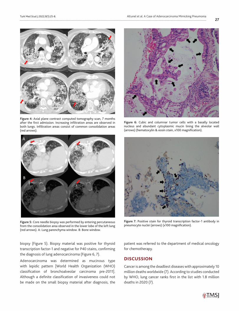
27Turk Med Stud J 2022;9(1):25-8. Altunel et al. A Case of Adenocarcinoma Mimicking Pneumonia
biopsy (Figure 5). Biopsy material was positive for thyroid transcription factor-1 and negative for P40 stains, confirming the diagnosis of lung adenocarcinoma (Figure 6, 7).
Adenocarcinoma was determined as mucinous type with lepidic pattern [World Health Organization (WHO) classification of bronchoalveolar carcinoma pre-2011]. Although a definite classification of invasiveness could not be made on the small biopsy material after diagnosis, the
patient was referred to the department of medical oncology for chemotherapy.
DISCUSSION
Cancer is among the deadliest diseases with approximately 10 million deaths worldwide (7). According to studies conducted by WHO, lung cancer ranks first in the list with 1.8 million deaths in 2020 (7).
Figure 4: Axial plane contrast computed tomography scan, 7 months after the first admission. Increasing infiltration areas are observed in both lungs. Infiltration areas consist of common consolidation areas (red arrows).
Figure 5: Core needle biopsy was performed by entering percutaneous from the consolidation area observed in the lower lobe of the left lung (red arrows). A: Lung parenchyma window. B: Bone window.
Figure 6: Cubic and columnar tumor cells with a basally located nucleus and abundant cytoplasmic mucin lining the alveolar wall (arrows) (hematoxylin & eosin stain, x100 magnification).
Figure 7: Positive stain for thyroid transcription factor-1 antibody in pneumocyte nuclei (arrows) (x100 magnification).
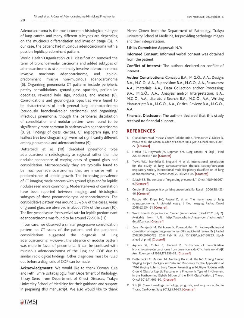
28Turk Med Stud J 2022;9(1):25-8.Altunel et al. A Case of Adenocarcinoma Mimicking Pneumonia
Adenocarcinoma is the most common histological subtype of lung cancer, and many different subtypes are depending on the mucinous differentiation and invasion stage (3). In our case, the patient had mucinous adenocarcinoma with a possible lepidic predominant pattern.
World Health Organization 2011 classification removed the term of bronchoalveolar carcinoma and added subtypes of adenocarcinoma in situ, minimally invasive adenocarcinoma, invasive mucinous adenocarcinoma, and lepidic-predominant invasive non-mucinous adenocarcinoma (6). Organizing pneumonia CT patterns include peripheric patchy consolidations, ground-glass opacities, perilobular opacities, reversed halo sign, nodules, and masses (8). Consolidations and ground-glass opacities were found to be characteristics of both general lung adenocarcinoma (previously bronchoalveolar carcinoma) and organizing/infectious pneumonia, though the peripheral distribution of consolidation and nodular pattern were found to be significantly more common in patients with adenocarcinoma (8, 9). Findings of cysts, cavities, CT angiogram sign, and leafless tree bronchogram sign were not significantly different among pneumonia and adenocarcinoma (9).
Detterbeck et al. (10) described pneumonic type adenocarcinoma radiologically as regional rather than the nodular appearance of varying areas of ground glass and consolidation. Microscopically they are typically found to be mucinous adenocarcinomas that are invasive with a predominance of lepidic growth. The increasing prevalence of CT imaging made cancers with ground glass and/or lepidic nodules seen more commonly. Moderate levels of correlation have been reported between imaging and histological subtypes of these pneumonic-type adenocarcinomas. The consolidative pattern was around 33-75% of the cases. Areas of ground glass are observed in about 75% of the cases (10). The five-year disease-free survival rate for lepidic predominant adenocarcinoma was found to be around 72-90% (11).
In our case, we observed a similar progressive consolidation pattern on CT scans of the patient, and the peripheral consolidations suggested the diagnosis of lung adenocarcinoma. However, the absence of nodular pattern was more in favor of pneumonia. It can be confused with mucinous adenocarcinoma of the lung and COP due to similar radiological findings. Other diagnoses must be ruled out before a diagnosis of COP can be made.
Acknowledgments: We would like to thank Osman Kula and Fethi Emre Ustabaşıoğlu from Department of Radiology, Bilkay Serez from Department of Chest Diseases, Trakya University School of Medicine for their guidance and support in preparing this manuscript. We also would like to thank
Merve Çimen from the Department of Pathology, Trakya University School of Medicine, for providing pathology images and their interpretation.
Ethics Committee Approval: N/A
Informed Consent: Informed verbal consent was obtained from the patient.
Conflict of Interest: The authors declared no conflict of interest.
Author Contributions: Concept: B.A., M.G.Ö., A.A., Design: B.A., M.G.Ö., A.A., Supervision: B.A., M.G.Ö., A.A., Resources: A.A., Materials: A.A., Data Collection and/or Processing: B.A., M.G.Ö., A.A., Analysis and/or Interpretation: B.A., M.G.Ö., A.A., Literature Search: B.A., M.G.Ö., A.A., Writing Manuscript: B.A., M.G.Ö., A.A., Critical Review: B.A., M.G.Ö., A.A.
Financial Disclosure: The authors declared that this study received no financial support.
REFERENCES1. Global Burden of Disease Cancer Collaboration, Fitzmaurice C, Dicker D,
Pain A et al. The Global Burden of Cancer 2013. JAMA Oncol 2015;1:505-27. [Crossref]
2. Herbst RS, Heymach JV, Lippman SM. Lung cancer. N Engl J Med 2008;359:1367-80. [Crossref]
3. Travis WD, Brambilla E, Noguchi M et al. International association for the study of lung cancer/american thoracic society/european respiratory society international multidisciplinary classification of lung adenocarcinoma. J Thorac Oncol 2011;6:244-85. [Crossref]
4. Sulavik SB. The concept of “organizing pneumonia”. Chest 1989;96:967-9. [Crossref]
5. Cordier JF. Cryptogenic organising pneumonia. Eur Respir J 2006;28:422-46. [Crossref]
6. Pascoe HM, Knipe HC, Pascoe D, et al. The many faces of lung adenocarcinoma: A pictorial essay. J Med Imaging Radiat Oncol 2018;62:654-61. [Crossref]
7. World Health Organization. Cancer (serial online) (cited 2021 July 7). Available from: URL: http://www.who.int/news-room/fact-sheets/detail/cancer. [Crossref]
8. Zare Mehrjardi M, Kahkouee S, Pourabdollah M. Radio-pathological correlation of organizing pneumonia (OP): a pictorial review. Br J Radiol 2017;90:20160723. 2017 Feb 17. doi: 10.1259/bjr.20160723. [Epub ahead of print] [Crossref]
9. Aquino SL, Chiles C, Halford P. Distinction of consolidative bronchioloalveolar carcinoma from pneumonia: do CT criteria work? AJR Am J Roentgenol 1998;171:359-63. [Crossref]
10. Detterbeck FC, Marom EM, Arenberg DA et al. The IASLC Lung Cancer Staging Project: Background Data and Proposals for the Application of TNM Staging Rules to Lung Cancer Presenting as Multiple Nodules with Ground Glass or Lepidic Features or a Pneumonic Type of Involvement in the Forthcoming Eighth Edition of the TNM Classification. J Thorac Oncol 2016;11:666-80. [Crossref]
11. Suh JH. Current readings: pathology, prognosis, and lung cancer. Semin Thorac Cardiovasc Surg 2013;25:14-21. [Crossref]
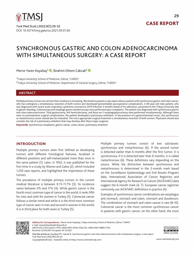
CASE REPORT
www.turkmedstudj.comCopyright@Author(s) - Available online at https://www.turkmedstudj.com/
29
OPEN ACCESSCite this article as: Kayabaş MY, Cakcak İE. Synchronous gastric and colon adenocarcinoma with simultaneous surgery: a case report. Turk Med Stud J 2022;9(1):29-32.
Address for Correspondence: Merve Yaren Kayabaş, Trakya University School of Medicine, Edirne, TURKEYe-mail: [email protected] iDs of the authors: MYK: 0000-0001-8136-2326; İEC: 0000-0001-8885-514X.Received: 23.09.2021 Accepted: 05.10.2021
INTRODUCTION
Multiple primary tumors were first defined as developing tumors with different histological features, localized in different positions and self-metastasized more than once in the same patient (1). Later, in 1932, it was published for the first time in a study by Warren and Gates (2), which included 1,259 case reports, and highlighted the importance of these tumors.
The prevalence of multiple primary tumors in the current medical literature is between 0.73-11.7% (3). Its incidence varies between 2% and 17% (4). While gastric cancer is the fourth most common type of cancer in the world; it ranks fifth for men and sixth for women in Turkey (5). Colorectal cancer follows a similar trend and while it is the third most common type of cancer seen in men and second in women in the world; it is in third place for both sexes in Turkey (5).
Multiple primary tumors consist of two subclasses: synchronous and metachronous (6). If the second tumor is detected earlier than 6 months after the first tumor, it is synchronous; if it is detected later than 6 months, it is called metachronous (6). These definitions vary depending on the source. While the distinction between synchronous and metachronous is determined in the 2-month mark based on the Surveillance Epidemiology and End Results Program data, International Association of Cancer Registries and International Agency for Research on Cancer (IACR/IARC) data suggest the 6-month mark (4, 7). European cancer registries commonly use IACR/IARC definitions in practice (4).
Examples of synchronous cancer combinations are esophagus and stomach, stomach and colon, stomach and duodenum. The combination of stomach and colon cancer is rare (8-10). Colorectal cancer is the most common synchronous cancer in patients with gastric cancer; on the other hand, the most
ABSTRACT
Multiple primary tumors are rare but their incidence is increasing. We aimed to present a case report about a patient with synchronous gastric and colon cancer, who has undergone a simultaneous resection of both tumors and developed (preventable) postoperative complications. A 69-year-old male patient, who was diagnosed with severe acute respiratory syndrome coronavirus-2019 infection 3 months ahead of his admission, presented to the Trakya University due to gastric bleeding. Colonoscopy and esophago gastro duodenoscopy were performed upon complaints. The patient was diagnosed with synchronous gastric and colon adenocarcinoma. Total gastrectomy, left hemicolectomy, and Roux-en-Y esophagojejunostomy were performed simultaneously. Although there were no postoperative surgical complications, the patient developed a pulmonary embolism. In the presence of a gastrointestinal tumor, the synchronous or metachronous tumor should also be evaluated. The most appropriate surgical treatment is simultaneous resection of both tumors. Physicians should also consider the risk of a pulmonary embolism that may develop after these major surgeries. Keywords: Synchronous neoplasms, gastric cancer, colon cancer, pulmonary embolism
Kayabaş and Cakcak. Synchronous Gastric and Colon Adenocarcinoma
1Trakya University School of Medicine, Edirne, TURKEY2Trakya University School of Medicine, Department of General Surgery, Edirne, TURKEY
Merve Yaren Kayabaş1 , İbrahim Ethem Cakcak2
SYNCHRONOUS GASTRIC AND COLON ADENOCARCINOMA WITH SIMULTANEOUS SURGERY: A CASE REPORT
DOI: 10.4274/tmsj.galenos.2021.09.01.06Turk Med Stud J 2022;9(1):29-32
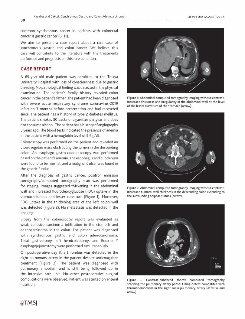
30Turk Med Stud J 2022;9(1):29-32.Kayabaş and Cakcak. Synchronous Gastric and Colon Adenocarcinoma
common synchronous cancer in patients with colorectal cancer is gastric cancer (6, 11).
We aim to present a case report about a rare case of synchronous gastric and colon cancer. We believe this case will contribute to the literature with the treatments performed and prognosis on this rare condition.
CASE REPORT
A 69-year-old male patient was admitted to the Trakya University Hospital with loss of consciousness due to gastric bleeding. No pathological finding was detected in the physical examination. The patient’s family history revealed colon cancer in the patient’s father. The patient had been diagnosed with severe acute respiratory syndrome coronavirus-2019 infection 3 months before presentation and had recovered since. The patient has a history of type 2 diabetes mellitus. The patient smokes 30 packs of cigarettes per year and does not consume alcohol. The patient has a history of angiography 3 years ago. The blood tests indicated the presence of anemia in the patient with a hemoglobin level of 9.6 g/dL.
Colonoscopy was performed on the patient and revealed an ulcerovegetan mass obstructing the lumen in the descending colon. An esophago-gastro-duodenoscopy was performed based on the patient’s anemia. The esophagus and duodenum were found to be normal, and a malignant ulcer was found in the gastric fundus.
After the diagnosis of gastric cancer, positron emission tomography/computed tomography scan was performed for staging. Images suggested thickening in the abdominal wall and increased fluorodeoxyglucose (FDG) uptake in the stomach fundus and lesser curvature (Figure 1). Moreover, FDG uptake in the thickening area of the left colon wall was detected (Figure 2). No metastasis was detected in the imaging.
Biopsy from the colonoscopy report was evaluated as weak cohesive carcinoma infiltration in the stomach and adenocarcinoma in the colon. The patient was diagnosed with synchronous gastric and colon adenocarcinoma. Total gastrectomy, left hemicolectomy, and Roux-en-Y esophagojejunostomy were performed simultaneously.
On postoperative day 3, a thrombus was detected in the right pulmonary artery in the patient despite anticoagulant treatment (Figure 3). The patient was diagnosed with pulmonary embolism and is still being followed up in the intensive care unit. No other postoperative surgical complications were observed. Patient was started on enteral nutrition.
Figure 1: Abdominal computed tomography imaging without contrast. Increased thickness and irregularity in the abdominal wall at the level of the lesser curvature of the stomach (arrow).
Figure 2: Abdominal computed tomography imaging without contrast. Increased tumoral wall thickness in the descending colon extending to the surrounding adipose tissues (arrow).
Figure 3: Contrast-enhanced thorax computed tomography scanning the pulmonary artery phase. Filling defect compatible with thromboembolism in the right main pulmonary artery (asterisk and arrow).
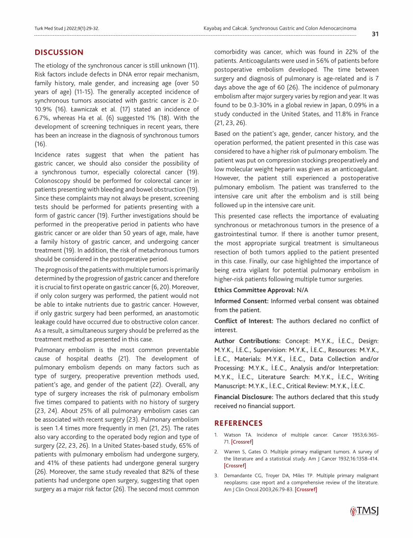
31Turk Med Stud J 2022;9(1):29-32. Kayabaş and Cakcak. Synchronous Gastric and Colon Adenocarcinoma
DISCUSSION
The etiology of the synchronous cancer is still unknown (11). Risk factors include defects in DNA error repair mechanism, family history, male gender, and increasing age (over 50 years of age) (11-15). The generally accepted incidence of synchronous tumors associated with gastric cancer is 2.0-10.9% (16). Ławniczak et al. (17) stated an incidence of 6.7%, whereas Ha et al. (6) suggested 1% (18). With the development of screening techniques in recent years, there has been an increase in the diagnosis of synchronous tumors (16).
Incidence rates suggest that when the patient has gastric cancer, we should also consider the possibility of a synchronous tumor, especially colorectal cancer (19). Colonoscopy should be performed for colorectal cancer in patients presenting with bleeding and bowel obstruction (19). Since these complaints may not always be present, screening tests should be performed for patients presenting with a form of gastric cancer (19). Further investigations should be performed in the preoperative period in patients who have gastric cancer or are older than 50 years of age, male, have a family history of gastric cancer, and undergoing cancer treatment (19). In addition, the risk of metachronous tumors should be considered in the postoperative period.
The prognosis of the patients with multiple tumors is primarily determined by the progression of gastric cancer and therefore it is crucial to first operate on gastric cancer (6, 20). Moreover, if only colon surgery was performed, the patient would not be able to intake nutrients due to gastric cancer. However, if only gastric surgery had been performed, an anastomotic leakage could have occurred due to obstructive colon cancer. As a result, a simultaneous surgery should be preferred as the treatment method as presented in this case.
Pulmonary embolism is the most common preventable cause of hospital deaths (21). The development of pulmonary embolism depends on many factors such as type of surgery, preoperative prevention methods used, patient’s age, and gender of the patient (22). Overall, any type of surgery increases the risk of pulmonary embolism five times compared to patients with no history of surgery (23, 24). About 25% of all pulmonary embolism cases can be associated with recent surgery (23). Pulmonary embolism is seen 1.4 times more frequently in men (21, 25). The rates also vary according to the operated body region and type of surgery (22, 23, 26). In a United States-based study, 65% of patients with pulmonary embolism had undergone surgery, and 41% of these patients had undergone general surgery (26). Moreover, the same study revealed that 82% of these patients had undergone open surgery, suggesting that open surgery as a major risk factor (26). The second most common
comorbidity was cancer, which was found in 22% of the patients. Anticoagulants were used in 56% of patients before postoperative embolism developed. The time between surgery and diagnosis of pulmonary is age-related and is 7 days above the age of 60 (26). The incidence of pulmonary embolism after major surgery varies by region and year. It was found to be 0.3-30% in a global review in Japan, 0.09% in a study conducted in the United States, and 11.8% in France (21, 23, 26).
Based on the patient’s age, gender, cancer history, and the operation performed, the patient presented in this case was considered to have a higher risk of pulmonary embolism. The patient was put on compression stockings preoperatively and low molecular weight heparin was given as an anticoagulant. However, the patient still experienced a postoperative pulmonary embolism. The patient was transferred to the intensive care unit after the embolism and is still being followed up in the intensive care unit.
This presented case reflects the importance of evaluating synchronous or metachronous tumors in the presence of a gastrointestinal tumor. If there is another tumor present, the most appropriate surgical treatment is simultaneous resection of both tumors applied to the patient presented in this case. Finally, our case highlighted the importance of being extra vigilant for potential pulmonary embolism in higher-risk patients following multiple tumor surgeries.
Ethics Committee Approval: N/A
Informed Consent: Informed verbal consent was obtained from the patient.
Conflict of Interest: The authors declared no conflict of interest.
Author Contributions: Concept: M.Y.K., İ.E.C., Design: M.Y.K., İ.E.C., Supervision: M.Y.K., İ.E.C., Resources: M.Y.K., İ.E.C., Materials: M.Y.K., İ.E.C., Data Collection and/or Processing: M.Y.K., İ.E.C., Analysis and/or Interpretation: M.Y.K., İ.E.C., Literature Search: M.Y.K., İ.E.C., Writing Manuscript: M.Y.K., İ.E.C., Critical Review: M.Y.K., İ.E.C.
Financial Disclosure: The authors declared that this study received no financial support.
REFERENCES1. Watson TA. Incidence of multiple cancer. Cancer 1953;6:365-
71. [Crossref]
2. Warren S, Gates O. Multiple primary malignant tumors. A survey of the literature and a statistical study. Am J Cancer 1932;16:1358-414. [Crossref]
3. Demandante CG, Troyer DA, Miles TP. Multiple primary malignant neoplasms: case report and a comprehensive review of the literature. Am J Clin Oncol 2003;26:79-83. [Crossref]
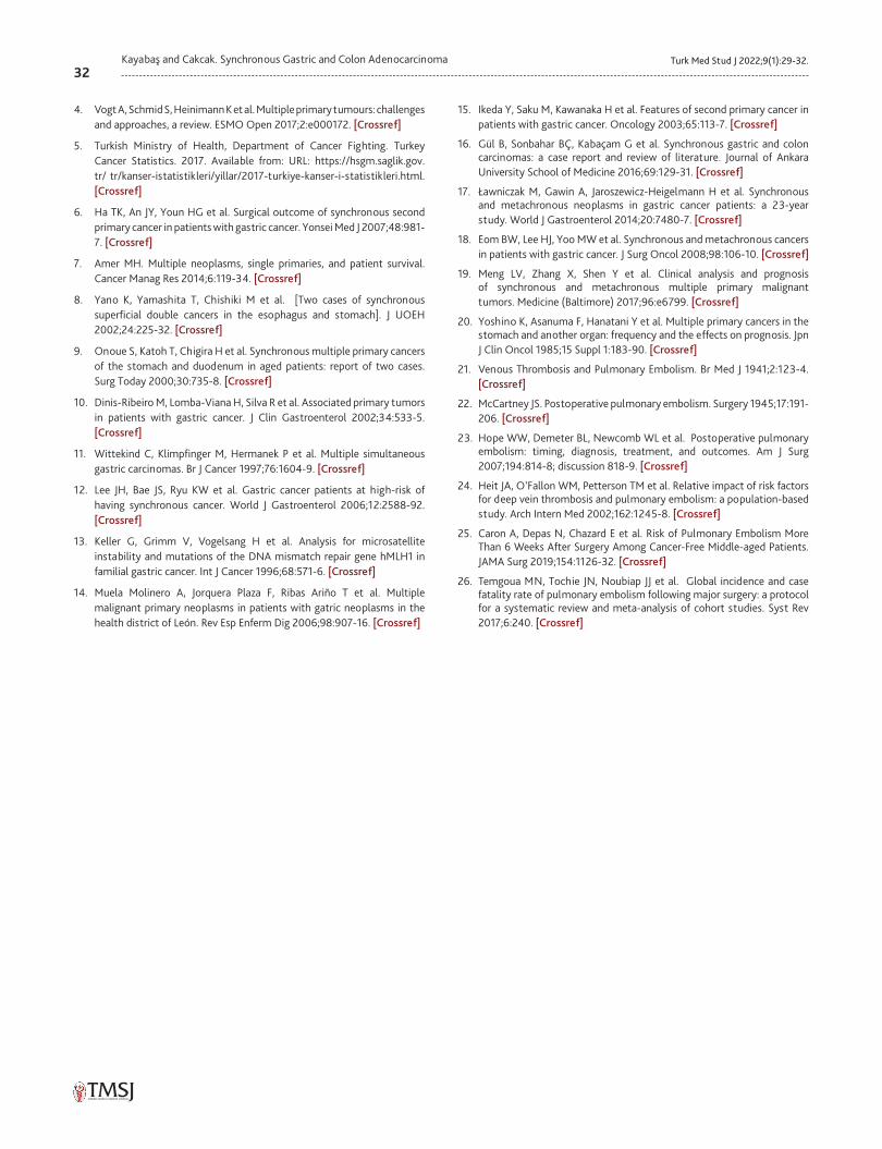
32Turk Med Stud J 2022;9(1):29-32.Kayabaş and Cakcak. Synchronous Gastric and Colon Adenocarcinoma
4. Vogt A, Schmid S, Heinimann K et al. Multiple primary tumours: challenges and approaches, a review. ESMO Open 2017;2:e000172. [Crossref]
5. Turkish Ministry of Health, Department of Cancer Fighting. Turkey Cancer Statistics. 2017. Available from: URL: https://hsgm.saglik.gov.tr/ tr/kanser-istatistikleri/yillar/2017-turkiye-kanser-i-statistikleri.html. [Crossref]
6. Ha TK, An JY, Youn HG et al. Surgical outcome of synchronous second primary cancer in patients with gastric cancer. Yonsei Med J 2007;48:981-7. [Crossref]
7. Amer MH. Multiple neoplasms, single primaries, and patient survival. Cancer Manag Res 2014;6:119-34. [Crossref]
8. Yano K, Yamashita T, Chishiki M et al. [Two cases of synchronous superficial double cancers in the esophagus and stomach]. J UOEH 2002;24:225-32. [Crossref]
9. Onoue S, Katoh T, Chigira H et al. Synchronous multiple primary cancers of the stomach and duodenum in aged patients: report of two cases. Surg Today 2000;30:735-8. [Crossref]
10. Dinis-Ribeiro M, Lomba-Viana H, Silva R et al. Associated primary tumors in patients with gastric cancer. J Clin Gastroenterol 2002;34:533-5. [Crossref]
11. Wittekind C, Klimpfinger M, Hermanek P et al. Multiple simultaneous gastric carcinomas. Br J Cancer 1997;76:1604-9. [Crossref]
12. Lee JH, Bae JS, Ryu KW et al. Gastric cancer patients at high-risk of having synchronous cancer. World J Gastroenterol 2006;12:2588-92. [Crossref]
13. Keller G, Grimm V, Vogelsang H et al. Analysis for microsatellite instability and mutations of the DNA mismatch repair gene hMLH1 in familial gastric cancer. Int J Cancer 1996;68:571-6. [Crossref]
14. Muela Molinero A, Jorquera Plaza F, Ribas Ariño T et al. Multiple malignant primary neoplasms in patients with gatric neoplasms in the health district of León. Rev Esp Enferm Dig 2006;98:907-16. [Crossref]
15. Ikeda Y, Saku M, Kawanaka H et al. Features of second primary cancer in patients with gastric cancer. Oncology 2003;65:113-7. [Crossref]
16. Gül B, Sonbahar BÇ, Kabaçam G et al. Synchronous gastric and colon carcinomas: a case report and review of literature. Journal of Ankara University School of Medicine 2016;69:129-31. [Crossref]
17. Ławniczak M, Gawin A, Jaroszewicz-Heigelmann H et al. Synchronous and metachronous neoplasms in gastric cancer patients: a 23-year study. World J Gastroenterol 2014;20:7480-7. [Crossref]
18. Eom BW, Lee HJ, Yoo MW et al. Synchronous and metachronous cancers in patients with gastric cancer. J Surg Oncol 2008;98:106-10. [Crossref]
19. Meng LV, Zhang X, Shen Y et al. Clinical analysis and prognosis of synchronous and metachronous multiple primary malignant tumors. Medicine (Baltimore) 2017;96:e6799. [Crossref]
20. Yoshino K, Asanuma F, Hanatani Y et al. Multiple primary cancers in the stomach and another organ: frequency and the effects on prognosis. Jpn J Clin Oncol 1985;15 Suppl 1:183-90. [Crossref]
21. Venous Thrombosis and Pulmonary Embolism. Br Med J 1941;2:123-4. [Crossref]
22. McCartney JS. Postoperative pulmonary embolism. Surgery 1945;17:191-206. [Crossref]
23. Hope WW, Demeter BL, Newcomb WL et al. Postoperative pulmonary embolism: timing, diagnosis, treatment, and outcomes. Am J Surg 2007;194:814-8; discussion 818-9. [Crossref]
24. Heit JA, O’Fallon WM, Petterson TM et al. Relative impact of risk factors for deep vein thrombosis and pulmonary embolism: a population-based study. Arch Intern Med 2002;162:1245-8. [Crossref]
25. Caron A, Depas N, Chazard E et al. Risk of Pulmonary Embolism More Than 6 Weeks After Surgery Among Cancer-Free Middle-aged Patients. JAMA Surg 2019;154:1126-32. [Crossref]
26. Temgoua MN, Tochie JN, Noubiap JJ et al. Global incidence and case fatality rate of pulmonary embolism following major surgery: a protocol for a systematic review and meta-analysis of cohort studies. Syst Rev 2017;6:240. [Crossref]
CASE REPORT
www.turkmedstudj.comCopyright@Author(s) - Available online at https://www.turkmedstudj.com/
33
OPEN ACCESS
Turk Med Stud J 2022;9(1):33-5
Cite this article as: Gürlü E, Gürlü V. Corneal endothelial cell analysis in two patients with wilson’s disease and kayser-fleischer rings. Turk Med Stud J 2022;9(1):33-5.
Address for Correspondence: Ege Gürlü, Yeditepe University School of Medicine, İstanbul, TURKEY e-mail: [email protected] iDs of the authors: EG: 0000-0003-4569-9082; VG: 0000-0001-5650-2075.Received: 12.12.2021 Accepted: 26.12.2021
INTRODUCTION
Wilson’s disease is an autosomal recessive disorder of copper metabolism that results in the pathological accumulation of copper in many tissues, primarily the liver, brain, and cornea (1-3). This accumulation results in liver disease such as genetic hemochromatosis and iron overload disorders, and a-1 antitrypsin deficiency, as well as neurological symptoms, and Kayser-Fleischer (KF) rings (1). Although a KF ring is considered the pathognomonic sign of Wilson’s disease, it is not confined to this disease. A KF ring can also rarely be found in the presence of chronic cholestatic disorders, such as primary biliary cholangitis and neonatal cholestasis (2, 3). A KF ring appears as golden-brown pigment deposits at the level of the Descemet’s membrane of the corneal periphery (2, 3). It has been reported that KF rings may exist in the asymptomatic phase of the disease, furthermore, they may be a sign of disease severity, and their regression is possible with treatment (2-4). However, the effects of this copper
accumulation on the morphology of the endothelial cells are not known.
In this study, a noncontact specular microscope Topcon SP3000P by Topcon Cooperation in Tokyo, Japan was used for endothelial cell analysis. With this method, images of the corneal endothelial cells are taken by a photomicroscope and sent to a computer, which is used with a specular microscope, in order to perform image analysis by software.
The following parameters can be calculated with a specular image, using the instruments’ built-in software: endothelial cell density, hexagonality, and coefficient of variances of the endothelial cell layer.
Endothelial cell density is the number of cells in the corneal endothelium per mm2. The mean value for adults is 2400 cells/mm2 (1500-3500 cells/mm2). Endothelial cell density may decrease with age, in the presence of corneal diseases, after surgical procedures or trauma.
ABSTRACT
CORNEAL ENDOTHELIAL CELL ANALYSIS IN TWO PATIENTS WITH WILSON’S DISEASE AND KAYSER-FLEISCHER RINGS
We aimed to present two patients who were suffering from Wilson’s disease along with the Kayser-Fleischer rings. Patient 1: A 15-year-old female patient was admitted to Trakya University School of Medicine Hospital with worsening consciousness, slurred speech, and cognitive impairment. The neurological examination was consistent with Wilson’s disease. In the ophthalmologic examination, Kayser-Fleischer rings were revealed. Right corneal cell density was found to be 3270 cell/mm2 and the left corneal endothelial cell density was found to be 3177 cell/mm2. Patient 2: A 12-year-old female patient was admitted to the Trakya University School of Medicine Hospital with complaints of difficulty in speaking, involuntary movements of her limbs, and inability to stand or sit. Additional symptoms were microphonia, episodic abnormal posture, and rigidity of the limbs. Corneal endothelial cell density was found to be 3287 cell/mm2 in the right eye and 3720 cell/mm2 in the left eye. The corneal copper deposition forming a Kayser-Fleischer ring caused no changes in endothelial cell morphology.Keywords: Corneal endothelium, Kayser-Fleischer ring, microscopy, Wilson’s disease
1Yeditepe University School of Medicine, İstanbul, TURKEY2Trakya University School of Medicine, Department of Ophthalmology, Edirne, TURKEY
Ege Gürlü1 , Vuslat Gürlü2
Gürlü and Gürlü. Corneal Endothelium in Wilson’s Disease
DOI: 10.4274/tmsj.galenos.2021.09.01.07
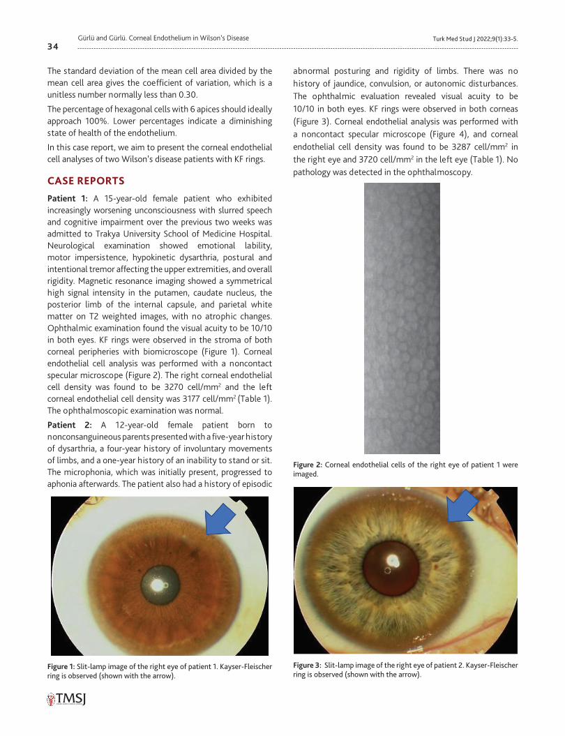
34Turk Med Stud J 2022;9(1):33-5.Gürlü and Gürlü. Corneal Endothelium in Wilson’s Disease
The standard deviation of the mean cell area divided by the mean cell area gives the coefficient of variation, which is a unitless number normally less than 0.30.
The percentage of hexagonal cells with 6 apices should ideally approach 100%. Lower percentages indicate a diminishing state of health of the endothelium.
In this case report, we aim to present the corneal endothelial cell analyses of two Wilson’s disease patients with KF rings.
CASE REPORTS
Patient 1: A 15-year-old female patient who exhibited increasingly worsening unconsciousness with slurred speech and cognitive impairment over the previous two weeks was admitted to Trakya University School of Medicine Hospital. Neurological examination showed emotional lability, motor impersistence, hypokinetic dysarthria, postural and intentional tremor affecting the upper extremities, and overall rigidity. Magnetic resonance imaging showed a symmetrical high signal intensity in the putamen, caudate nucleus, the posterior limb of the internal capsule, and parietal white matter on T2 weighted images, with no atrophic changes. Ophthalmic examination found the visual acuity to be 10/10 in both eyes. KF rings were observed in the stroma of both corneal peripheries with biomicroscope (Figure 1). Corneal endothelial cell analysis was performed with a noncontact specular microscope (Figure 2). The right corneal endothelial cell density was found to be 3270 cell/mm2 and the left corneal endothelial cell density was 3177 cell/mm2 (Table 1). The ophthalmoscopic examination was normal.
Patient 2: A 12-year-old female patient born to nonconsanguineous parents presented with a five-year history of dysarthria, a four-year history of involuntary movements of limbs, and a one-year history of an inability to stand or sit. The microphonia, which was initially present, progressed to aphonia afterwards. The patient also had a history of episodic
abnormal posturing and rigidity of limbs. There was no history of jaundice, convulsion, or autonomic disturbances. The ophthalmic evaluation revealed visual acuity to be 10/10 in both eyes. KF rings were observed in both corneas (Figure 3). Corneal endothelial analysis was performed with a noncontact specular microscope (Figure 4), and corneal endothelial cell density was found to be 3287 cell/mm2 in the right eye and 3720 cell/mm2 in the left eye (Table 1). No pathology was detected in the ophthalmoscopy.
Figure 1: Slit-lamp image of the right eye of patient 1. Kayser-Fleischer ring is observed (shown with the arrow).
Figure 2: Corneal endothelial cells of the right eye of patient 1 were imaged.
Figure 3: Slit-lamp image of the right eye of patient 2. Kayser-Fleischer ring is observed (shown with the arrow).
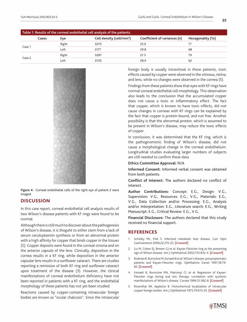
35Turk Med Stud J 2022;9(1):33-5. Gürlü and Gürlü. Corneal Endothelium in Wilson’s Disease
DISCUSSION
In this case report, corneal endothelial cell analysis results of two Wilson’s disease patients with KF rings were found to be normal.
Although there is still much to discover about the pathogenesis of Wilson’s disease, it is thoguht to either stem from a lack of serum ceruloplasmin synthesis or from an abnormal protein with a high affinity for copper that binds copper in the tissues (5). Copper deposits were found in the corneal stroma and on the anterior capsule of the lens. Clinically, deposition in the cornea results in a KF ring, while deposition in the anterior capsular lens results in a sunflower cataract. There are studies reporting a remission of both KF ring and sunflower cataract upon treatment of the disease (3). However, the clinical manifestations of corneal endothelium deficiency have not been reported in patients with a KF ring, and the endothelial morphology of these patients has not yet been studied.
Reactions caused by copper-containing intraocular foreign bodies are known as “ocular chalcosis”. Since the intraocular
foreign body is usually intravitreal in these patients, toxic effects caused by copper were observed in the vitreous, retina, and lens, while no changes were observed in the cornea (5).
Findings from these patients show that eyes with KF rings have normal corneal endothelial cell morphology. This observation also leads to the conclusion that the accumulated copper does not cause a toxic or inflammatory effect. The fact that copper, which is known to have toxic effects, did not cause changes in corneas with KF rings can be explained by the fact that copper is protein-bound, and not free. Another possibility is that the abnormal protein, which is assumed to be present in Wilson’s disease, may reduce the toxic effects of copper.
In conclusion, it was determined that the KF ring, which is the pathognomonic finding of Wilson’s disease, did not cause a morphological change in the corneal endothelium. Longitudinal studies evaluating larger numbers of subjects are still needed to confirm these data.
Ethics Committee Approval: N/A
Informed Consent: Informed verbal consent was obtained from both patients.
Conflict of Interest: The authors declared no conflict of interest.
Author Contributions: Concept: E.G., Design: V.G., Supervision: V.G., Resources: E.G., V.G., Materials: E.G., V.G., Data Collection and/or Processing: E.G., Analysis and/or Interpretation: E.G., Literature search: E.G., Writing Manuscript: E.G., Critical Review: E.G., V.G.
Financial Disclosure: The authors declared that this study received no financial support.
REFERENCES1. Schilsky ML, Fink S. Inherited metabolic liver disease. Curr Opin
Gastroenterol 2006;22:215-22. [Crossref]
2. Liu M, Cohen EJ, Brewer GJ et al. Kayser-Fleischer ring as the presenting sign of Wilson disease. Am J Ophthalmol 2002;133:832-4. [Crossref]
3. Rodman R, Burnstine M, Esmaeli B et al. Wilson’s disease: presymptomatic patients and Kayser-Fleischer rings. Ophthalmic Genet 1997;18:79-85. [Crossref]
4. Esmaeli B, Burnstine MA, Martonyi CL et al. Regression of Kayser-Fleischer rings during oral zinc therapy: correlation with systemic manifestations of Wilson’s disease. Cornea 1996;15:582-8. [Crossref]
5. Rosenthal AR, Appleton B. Histochemical localization of intraocular copper foreign bodies. Am J Ophthalmol 1975;79:613-25. [Crossref]
Table 1: Results of the corneal endothelial cell analysis of the patients.
Cases Eye Cell density (cell/mm2) Coefficient of variances (n) Hexagonality (%)
Case 1Right 3270 25.6 77
Left 3177 29.8 68
Case 2Right 3287 27.5 79
Left 3720 28.9 62
Figure 4: Corneal endothelial cells of the right eye of patient 2 were imaged.
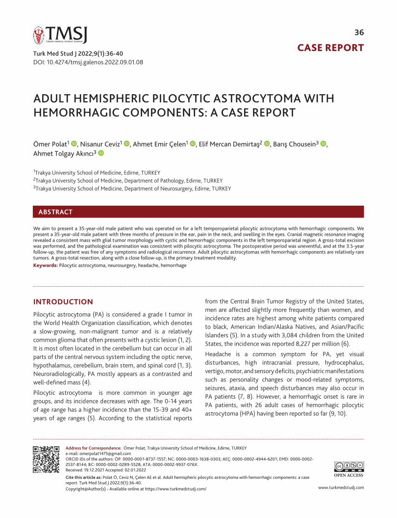
CASE REPORT
www.turkmedstudj.comCopyright@Author(s) - Available online at https://www.turkmedstudj.com/
36
OPEN ACCESS
Polat et al. Adult Hemispheric Pilocytic Astrocytoma
Turk Med Stud J 2022;9(1):36-40
Cite this article as: Polat Ö, Ceviz N, Çelen AE et al. Adult hemispheric pilocytic astrocytoma with hemorrhagic components: a case report. Turk Med Stud J 2022;9(1):36-40.
Address for Correspondence: Ömer Polat, Trakya University School of Medicine, Edirne, TURKEYe-mail: [email protected] iDs of the authors: ÖP: 0000-0001-8737-1557; NC: 0000-0003-1638-0303; AEÇ: 0000-0002-4944-6201; EMD: 0000-0002-2537-8144; BC: 0000-0002-0289-5528; ATA: 0000-0002-9937-076X.Received: 19.12.2021 Accepted: 02.01.2022
INTRODUCTION
Pilocytic astrocytoma (PA) is considered a grade I tumor in the World Health Organization classification, which denotes a slow-growing, non-malignant tumor and is a relatively common glioma that often presents with a cystic lesion (1, 2). It is most often located in the cerebellum but can occur in all parts of the central nervous system including the optic nerve, hypothalamus, cerebellum, brain stem, and spinal cord (1, 3). Neuroradiologically, PA mostly appears as a contrasted and well-defined mass (4).
Pilocytic astrocytoma is more common in younger age groups, and its incidence decreases with age. The 0-14 years of age range has a higher incidence than the 15-39 and 40+ years of age ranges (5). According to the statistical reports
from the Central Brain Tumor Registry of the United States, men are affected slightly more frequently than women, and incidence rates are highest among white patients compared to black, American Indian/Alaska Natives, and Asian/Pacific Islanders (5). In a study with 3,084 children from the United States, the incidence was reported 8,227 per million (6).
Headache is a common symptom for PA, yet visual disturbances, high intracranial pressure, hydrocephalus, vertigo, motor, and sensory deficits, psychiatric manifestations such as personality changes or mood-related symptoms, seizures, ataxia, and speech disturbances may also occur in PA patients (7, 8). However, a hemorrhagic onset is rare in PA patients, with 26 adult cases of hemorrhagic pilocytic astrocytoma (HPA) having been reported so far (9, 10).
Ömer Polat1 , Nisanur Ceviz1 , Ahmet Emir Çelen1 , Elif Mercan Demirtaş2 , Barış Chousein3 , Ahmet Tolgay Akıncı3
1Trakya University School of Medicine, Edirne, TURKEY2Trakya University School of Medicine, Department of Pathology, Edirne, TURKEY3Trakya University School of Medicine, Department of Neurosurgery, Edirne, TURKEY
ABSTRACT
We aim to present a 35-year-old male patient who was operated on for a left temporoparietal pilocytic astrocytoma with hemorrhagic components. We present a 35-year-old male patient with three months of pressure in the ear, pain in the neck, and swelling in the eyes. Cranial magnetic resonance imaging revealed a consistent mass with glial tumor morphology with cystic and hemorrhagic components in the left temporoparietal region. A gross-total excision was performed, and the pathological examination was consistent with pilocytic astrocytoma. The postoperative period was uneventful, and at the 3.5-year follow-up, the patient was free of any symptoms and radiological recurrence. Adult pilocytic astrocytomas with hemorrhagic components are relatively rare tumors. A gross-total resection, along with a close follow-up, is the primary treatment modality. Keywords: Pilocytic astrocytoma, neurosurgery, headache, hemorrhage
ADULT HEMISPHERIC PILOCYTIC ASTROCYTOMA WITH HEMORRHAGIC COMPONENTS: A CASE REPORT
DOI: 10.4274/tmsj.galenos.2022.09.01.08
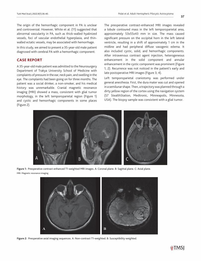
37Turk Med Stud J 2022;9(1):36-40. Polat et al. Adult Hemispheric Pilocytic Astrocytoma
The origin of the hemorrhagic component in PA is unclear and controversial. However, White et al. (11) suggested that abnormal vascularity in PA, such as thick-walled hyalinized vessels, foci of vascular endothelial hyperplasia, and thin-walled ectatic vessels, may be associated with hemorrhage.
In this study, we aimed to present a 35-year-old male patient diagnosed with cerebral PA with a hemorrhagic component.
CASE REPORT
A 35-year-old male patient was admitted to the Neurosurgery Department of Trakya University School of Medicine with complaints of pressure in the ear, neck pain, and swelling in the eye. The complaints had been going on for three months. The patient was a social drinker, a non-smoker, and his medical history was unremarkable. Cranial magnetic resonance imaging (MRI) showed a mass, consistent with glial tumor morphology, in the left temporoparietal region (Figure 1) and cystic and hemorrhagic components in some places (Figure 2).
The preoperative contrast-enhanced MRI images revealed a lobule contoured mass in the left temporoparietal area, approximately 53x55x43 mm in size. The mass caused significant pressure on the occipital horn in the left lateral ventricle, resulting in a shift of approximately 1 cm in the midline and had peripheral diffuse vasogenic edema. It also included cystic, solid, and hemorrhagic components. After intravenous contrast agent injection, heterogeneous enhancement in the solid component and annular enhancement in the cystic component was prominent (Figure 1, 2). Recurrence was not noticed in the patient’s early and late postoperative MRI images (Figure 3, 4).
Left temporoparietal craniotomy was performed under general anesthesia. First, the dura mater was cut and opened in a semilunar shape. Then, a trajectory was planned through a dirty yellow region of the cortex using the navigation system (S7 StealthStation, Medtronic, Minneapolis, Minnesota, USA). The biopsy sample was consistent with a glial tumor.
Figure 1: Preoperative contrast-enhanced T1-weighted MRI images. A: Coronal plane. B: Sagittal plane. C: Axial plane.
MRI: Magnetic resonance imaging
Figure 2: Preoperative axial imaging sequences. A: Non-contrast T1-weighted. B: Susceptibility weighted.
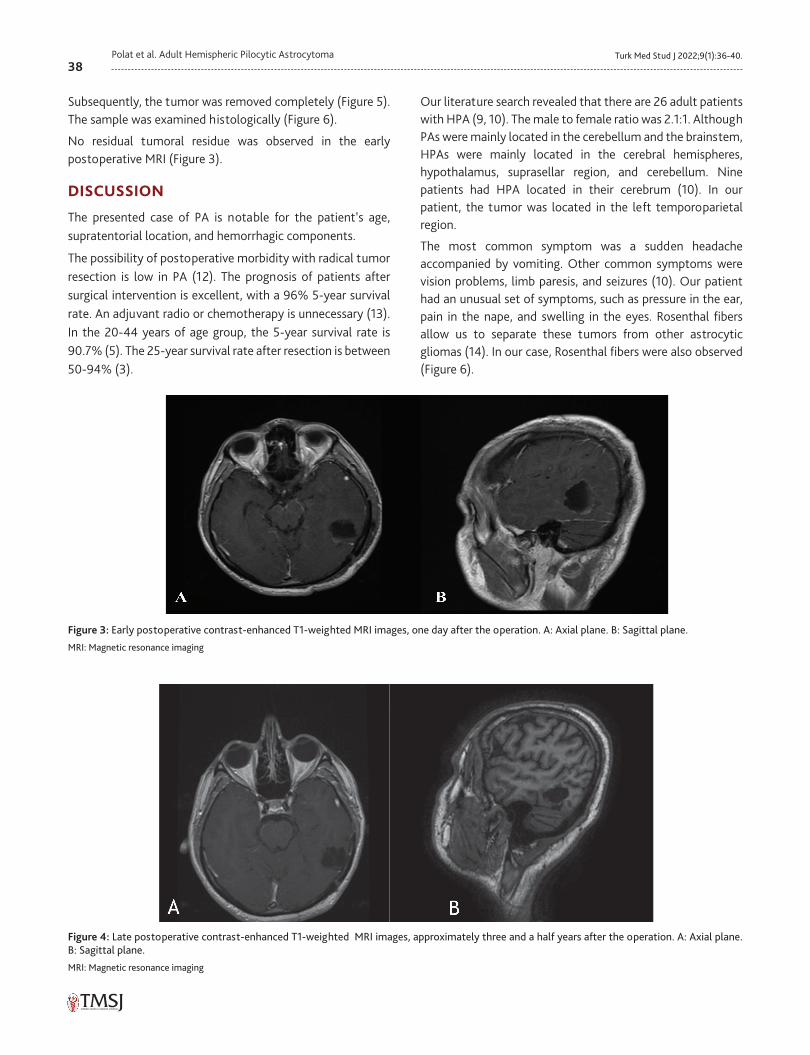
38Turk Med Stud J 2022;9(1):36-40.Polat et al. Adult Hemispheric Pilocytic Astrocytoma
Subsequently, the tumor was removed completely (Figure 5). The sample was examined histologically (Figure 6).
No residual tumoral residue was observed in the early postoperative MRI (Figure 3).
DISCUSSION
The presented case of PA is notable for the patient’s age, supratentorial location, and hemorrhagic components.
The possibility of postoperative morbidity with radical tumor resection is low in PA (12). The prognosis of patients after surgical intervention is excellent, with a 96% 5-year survival rate. An adjuvant radio or chemotherapy is unnecessary (13). In the 20-44 years of age group, the 5-year survival rate is 90.7% (5). The 25-year survival rate after resection is between 50-94% (3).
Our literature search revealed that there are 26 adult patients with HPA (9, 10). The male to female ratio was 2.1:1. Although PAs were mainly located in the cerebellum and the brainstem, HPAs were mainly located in the cerebral hemispheres, hypothalamus, suprasellar region, and cerebellum. Nine patients had HPA located in their cerebrum (10). In our patient, the tumor was located in the left temporoparietal region.
The most common symptom was a sudden headache accompanied by vomiting. Other common symptoms were vision problems, limb paresis, and seizures (10). Our patient had an unusual set of symptoms, such as pressure in the ear, pain in the nape, and swelling in the eyes. Rosenthal fibers allow us to separate these tumors from other astrocytic gliomas (14). In our case, Rosenthal fibers were also observed (Figure 6).
Figure 3: Early postoperative contrast-enhanced T1-weighted MRI images, one day after the operation. A: Axial plane. B: Sagittal plane.
MRI: Magnetic resonance imaging
Figure 4: Late postoperative contrast-enhanced T1-weighted MRI images, approximately three and a half years after the operation. A: Axial plane. B: Sagittal plane.
MRI: Magnetic resonance imaging
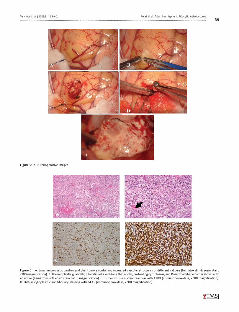
39Turk Med Stud J 2022;9(1):36-40. Polat et al. Adult Hemispheric Pilocytic Astrocytoma
Figure 6: A: Small microcystic cavities and glial tumors containing increased vascular structures of different calibers (hematoxylin & eosin stain, x100 magnification). B: The neoplastic glial cells, pilocytic cells with long thin nuclei, protruding cytoplasms, and Rosenthal fiber which is shown with an arrow (hematoxylin & eosin stain, x200 magnification). C: Tumor diffuse nuclear reaction with ATRX (immunoperoxidase, x200 magnification). D: Diffuse cytoplasmic and fibrillary staining with GFAP (immunoperoxidase, x200 magnification).
Figure 5: A-E: Perioperative images.
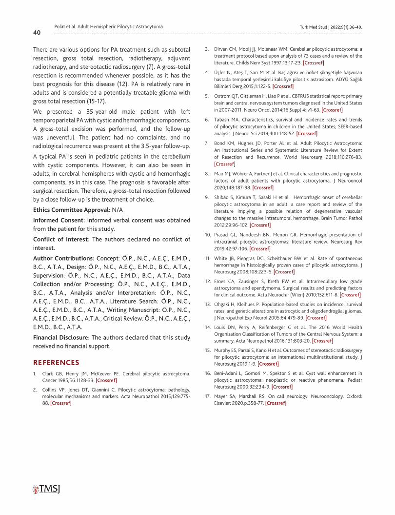
40Turk Med Stud J 2022;9(1):36-40.Polat et al. Adult Hemispheric Pilocytic Astrocytoma
There are various options for PA treatment such as subtotal resection, gross total resection, radiotherapy, adjuvant radiotherapy, and stereotactic radiosurgery (7). A gross-total resection is recommended whenever possible, as it has the best prognosis for this disease (12). PA is relatively rare in adults and is considered a potentially treatable glioma with gross total resection (15-17).
We presented a 35-year-old male patient with left temporoparietal PA with cystic and hemorrhagic components. A gross-total excision was performed, and the follow-up was uneventful. The patient had no complaints, and no radiological recurrence was present at the 3.5-year follow-up.
A typical PA is seen in pediatric patients in the cerebellum with cystic components. However, it can also be seen in adults, in cerebral hemispheres with cystic and hemorrhagic components, as in this case. The prognosis is favorable after surgical resection. Therefore, a gross-total resection followed by a close follow-up is the treatment of choice.
Ethics Committee Approval: N/A
Informed Consent: Informed verbal consent was obtained from the patient for this study.
Conflict of Interest: The authors declared no conflict of interest.
Author Contributions: Concept: Ö.P., N.C., A.E.Ç., E.M.D., B.C., A.T.A., Design: Ö.P., N.C., A.E.Ç., E.M.D., B.C., A.T.A., Supervision: Ö.P., N.C., A.E.Ç., E.M.D., B.C., A.T.A., Data Collection and/or Processing: Ö.P., N.C., A.E.Ç., E.M.D., B.C., A.T.A., Analysis and/or Interpretation: Ö.P., N.C., A.E.Ç., E.M.D., B.C., A.T.A., Literature Search: Ö.P., N.C., A.E.Ç., E.M.D., B.C., A.T.A., Writing Manuscript: Ö.P., N.C., A.E.Ç., E.M.D., B.C., A.T.A., Critical Review: Ö.P., N.C., A.E.Ç., E.M.D., B.C., A.T.A.
Financial Disclosure: The authors declared that this study received no financial support.
REFERENCES1. Clark GB, Henry JM, McKeever PE. Cerebral pilocytic astrocytoma.
Cancer 1985;56:1128-33. [Crossref]
2. Collins VP, Jones DT, Giannini C. Pilocytic astrocytoma: pathology, molecular mechanisms and markers. Acta Neuropathol 2015;129:775-88. [Crossref]
3. Dirven CM, Mooij JJ, Molenaar WM. Cerebellar pilocytic astrocytoma: a treatment protocol based upon analysis of 73 cases and a review of the literature. Childs Nerv Syst 1997;13:17-23. [Crossref]
4. Üçler N, Ateş T, Sarı M et al. Baş ağrısı ve nöbet şikayetiyle başvuran hastada temporal yerleşimli kalsifiye pilositik astrositom. ADYÜ Sağlık Bilimleri Derg 2015;1:122-5. [Crossref]
5. Ostrom QT, Gittleman H, Liao P et al. CBTRUS statistical report: primary brain and central nervous system tumors diagnosed in the United States in 2007-2011. Neuro Oncol 2014;16 Suppl 4:iv1-63. [Crossref]
6. Tabash MA. Characteristics, survival and incidence rates and trends of pilocytic astrocytoma in children in the United States; SEER-based analysis. J Neurol Sci 2019;400:148-52. [Crossref]
7. Bond KM, Hughes JD, Porter AL et al. Adult Pilocytic Astrocytoma: An Institutional Series and Systematic Literature Review for Extent of Resection and Recurrence. World Neurosurg 2018;110:276-83. [Crossref]
8. Mair MJ, Wöhrer A, Furtner J et al. Clinical characteristics and prognostic factors of adult patients with pilocytic astrocytoma. J Neurooncol 2020;148:187-98. [Crossref]
9. Shibao S, Kimura T, Sasaki H et al. Hemorrhagic onset of cerebellar pilocytic astrocytoma in an adult: a case report and review of the literature implying a possible relation of degenerative vascular changes to the massive intratumoral hemorrhage. Brain Tumor Pathol 2012;29:96-102. [Crossref]
10. Prasad GL, Nandeesh BN, Menon GR. Hemorrhagic presentation of intracranial pilocytic astrocytomas: literature review. Neurosurg Rev 2019;42:97-106. [Crossref]
11. White JB, Piepgras DG, Scheithauer BW et al. Rate of spontaneous hemorrhage in histologically proven cases of pilocytic astrocytoma. J Neurosurg 2008;108:223-6. [Crossref]
12. Eroes CA, Zausinger S, Kreth FW et al. Intramedullary low grade astrocytoma and ependymoma. Surgical results and predicting factors for clinical outcome. Acta Neurochir (Wien) 2010;152:611-8. [Crossref]
13. Ohgaki H, Kleihues P. Population-based studies on incidence, survival rates, and genetic alterations in astrocytic and oligodendroglial gliomas. J Neuropathol Exp Neurol 2005;64:479-89. [Crossref]
14. Louis DN, Perry A, Reifenberger G et al. The 2016 World Health Organization Classification of Tumors of the Central Nervous System: a summary. Acta Neuropathol 2016;131:803-20. [Crossref]
15. Murphy ES, Parsai S, Kano H et al. Outcomes of stereotactic radiosurgery for pilocytic astrocytoma: an international multiinstitutional study. J Neurosurg 2019:1-9. [Crossref]
16. Beni-Adani L, Gomori M, Spektor S et al. Cyst wall enhancement in pilocytic astrocytoma: neoplastic or reactive phenomena. Pediatr Neurosurg 2000;32:234-9. [Crossref]
17. Mayer SA, Marshall RS. On call neurology. Neurooncology. Oxford: Elsevier; 2020.p.358-77. [Crossref]
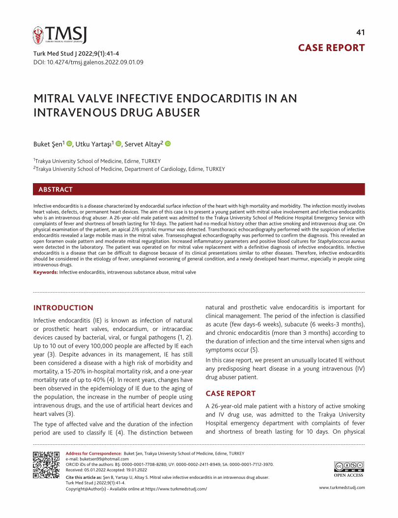
CASE REPORT
www.turkmedstudj.comCopyright@Author(s) - Available online at https://www.turkmedstudj.com/
41
OPEN ACCESS
ABSTRACT
Turk Med Stud J 2022;9(1):41-4
Cite this article as: Şen B, Yartaşı U, Altay S. Mitral valve infective endocarditis in an intravenous drug abuser. Turk Med Stud J 2022;9(1):41-4.
Address for Correspondence: Buket Şen, Trakya University School of Medicine, Edirne, TURKEYe-mail: [email protected] iDs of the authors: BŞ: 0000-0001-7708-8280; UY: 0000-0002-2411-8949; SA: 0000-0001-7112-3970.Received: 05.01.2022 Accepted: 19.01.2022
INTRODUCTION
Infective endocarditis (IE) is known as infection of natural or prosthetic heart valves, endocardium, or intracardiac devices caused by bacterial, viral, or fungal pathogens (1, 2). Up to 10 out of every 100,000 people are affected by IE each year (3). Despite advances in its management, IE has still been considered a disease with a high risk of morbidity and mortality, a 15-20% in-hospital mortality risk, and a one-year mortality rate of up to 40% (4). In recent years, changes have been observed in the epidemiology of IE due to the aging of the population, the increase in the number of people using intravenous drugs, and the use of artificial heart devices and heart valves (3).
The type of affected valve and the duration of the infection period are used to classify IE (4). The distinction between
natural and prosthetic valve endocarditis is important for clinical management. The period of the infection is classified as acute (few days-6 weeks), subacute (6 weeks-3 months), and chronic endocarditis (more than 3 months) according to the duration of infection and the time interval when signs and symptoms occur (5).
In this case report, we present an unusually located IE without any predisposing heart disease in a young intravenous (IV) drug abuser patient.
CASE REPORT
A 26-year-old male patient with a history of active smoking and IV drug use, was admitted to the Trakya University Hospital emergency department with complaints of fever and shortness of breath lasting for 10 days. On physical
Infective endocarditis is a disease characterized by endocardial surface infection of the heart with high mortality and morbidity. The infection mostly involves heart valves, defects, or permanent heart devices. The aim of this case is to present a young patient with mitral valve involvement and infective endocarditis who is an intravenous drug abuser. A 26-year-old male patient was admitted to the Trakya University School of Medicine Hospital Emergency Service with complaints of fever and shortness of breath lasting for 10 days. The patient had no medical history other than active smoking and intravenous drug use. On physical examination of the patient, an apical 2/6 systolic murmur was detected. Transthoracic echocardiography performed with the suspicion of infective endocarditis revealed a large mobile mass in the mitral valve. Transesophageal echocardiography was performed to confirm the diagnosis. This revealed an open foramen ovale pattern and moderate mitral regurgitation. Increased inflammatory parameters and positive blood cultures for Staphylococcus aureus were detected in the laboratory. The patient was operated on for mitral valve replacement with a definitive diagnosis of infective endocarditis. Infective endocarditis is a disease that can be difficult to diagnose because of its clinical presentations similar to other diseases. Therefore, infective endocarditis should be considered in the etiology of fever, unexplained worsening of general condition, and a newly developed heart murmur, especially in people using intravenous drugs. Keywords: Infective endocarditis, intravenous substance abuse, mitral valve
1Trakya University School of Medicine, Edirne, TURKEY2Trakya University School of Medicine, Department of Cardiology, Edirne, TURKEY
Buket Şen1 , Utku Yartaşı1 , Servet Altay2
MITRAL VALVE INFECTIVE ENDOCARDITIS IN AN INTRAVENOUS DRUG ABUSER
Şen et al. Infective Endocarditis in an Intravenous Drug Abuser
DOI: 10.4274/tmsj.galenos.2022.09.01.09
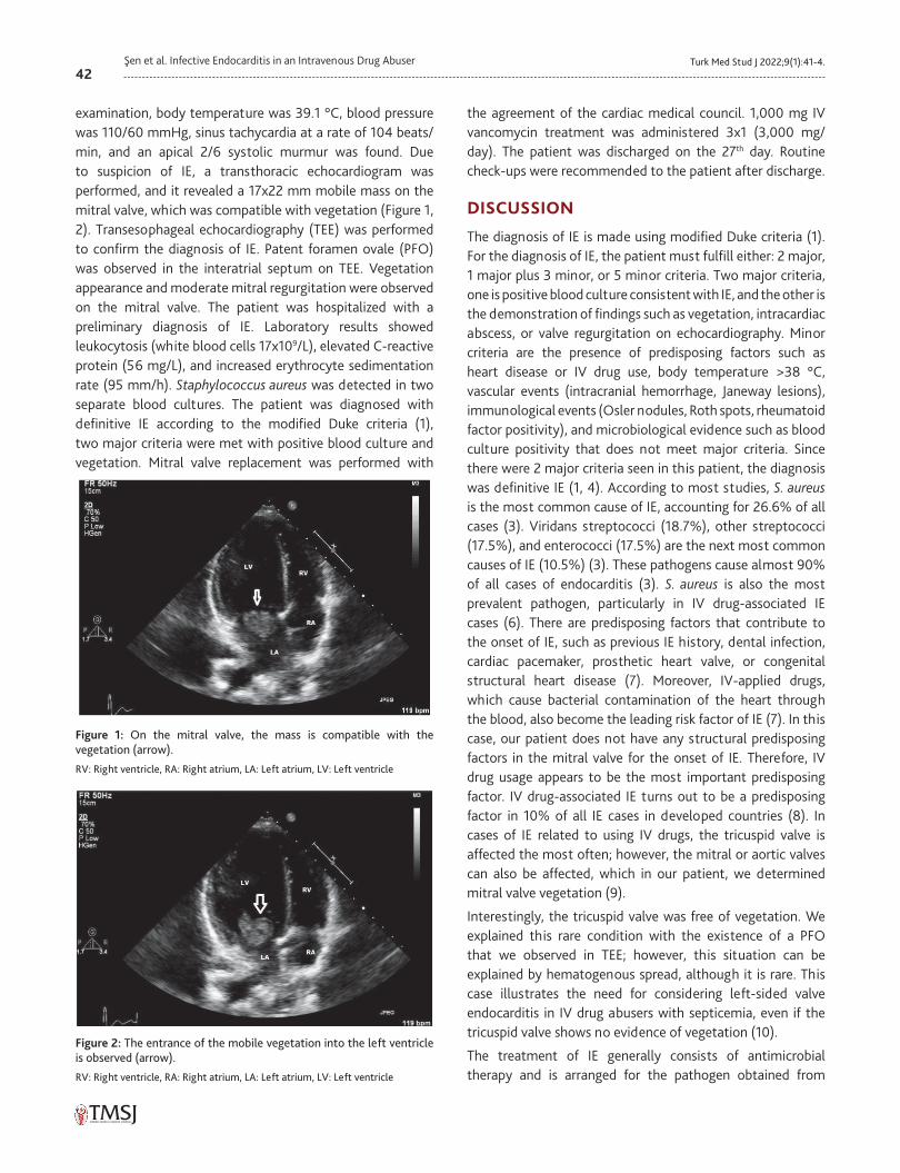
42Turk Med Stud J 2022;9(1):41-4.Şen et al. Infective Endocarditis in an Intravenous Drug Abuser
examination, body temperature was 39.1 °C, blood pressure was 110/60 mmHg, sinus tachycardia at a rate of 104 beats/min, and an apical 2/6 systolic murmur was found. Due to suspicion of IE, a transthoracic echocardiogram was performed, and it revealed a 17x22 mm mobile mass on the mitral valve, which was compatible with vegetation (Figure 1, 2). Transesophageal echocardiography (TEE) was performed to confirm the diagnosis of IE. Patent foramen ovale (PFO) was observed in the interatrial septum on TEE. Vegetation appearance and moderate mitral regurgitation were observed on the mitral valve. The patient was hospitalized with a preliminary diagnosis of IE. Laboratory results showed leukocytosis (white blood cells 17x109/L), elevated C-reactive protein (56 mg/L), and increased erythrocyte sedimentation rate (95 mm/h). Staphylococcus aureus was detected in two separate blood cultures. The patient was diagnosed with definitive IE according to the modified Duke criteria (1), two major criteria were met with positive blood culture and vegetation. Mitral valve replacement was performed with
the agreement of the cardiac medical council. 1,000 mg IV vancomycin treatment was administered 3x1 (3,000 mg/day). The patient was discharged on the 27th day. Routine check-ups were recommended to the patient after discharge.
DISCUSSION
The diagnosis of IE is made using modified Duke criteria (1). For the diagnosis of IE, the patient must fulfill either: 2 major, 1 major plus 3 minor, or 5 minor criteria. Two major criteria, one is positive blood culture consistent with IE, and the other is the demonstration of findings such as vegetation, intracardiac abscess, or valve regurgitation on echocardiography. Minor criteria are the presence of predisposing factors such as heart disease or IV drug use, body temperature >38 °C, vascular events (intracranial hemorrhage, Janeway lesions), immunological events (Osler nodules, Roth spots, rheumatoid factor positivity), and microbiological evidence such as blood culture positivity that does not meet major criteria. Since there were 2 major criteria seen in this patient, the diagnosis was definitive IE (1, 4). According to most studies, S. aureus is the most common cause of IE, accounting for 26.6% of all cases (3). Viridans streptococci (18.7%), other streptococci (17.5%), and enterococci (17.5%) are the next most common causes of IE (10.5%) (3). These pathogens cause almost 90% of all cases of endocarditis (3). S. aureus is also the most prevalent pathogen, particularly in IV drug-associated IE cases (6). There are predisposing factors that contribute to the onset of IE, such as previous IE history, dental infection, cardiac pacemaker, prosthetic heart valve, or congenital structural heart disease (7). Moreover, IV-applied drugs, which cause bacterial contamination of the heart through the blood, also become the leading risk factor of IE (7). In this case, our patient does not have any structural predisposing factors in the mitral valve for the onset of IE. Therefore, IV drug usage appears to be the most important predisposing factor. IV drug-associated IE turns out to be a predisposing factor in 10% of all IE cases in developed countries (8). In cases of IE related to using IV drugs, the tricuspid valve is affected the most often; however, the mitral or aortic valves can also be affected, which in our patient, we determined mitral valve vegetation (9).
Interestingly, the tricuspid valve was free of vegetation. We explained this rare condition with the existence of a PFO that we observed in TEE; however, this situation can be explained by hematogenous spread, although it is rare. This case illustrates the need for considering left-sided valve endocarditis in IV drug abusers with septicemia, even if the tricuspid valve shows no evidence of vegetation (10).
The treatment of IE generally consists of antimicrobial therapy and is arranged for the pathogen obtained from
Figure 1: On the mitral valve, the mass is compatible with the vegetation (arrow).
RV: Right ventricle, RA: Right atrium, LA: Left atrium, LV: Left ventricle
Figure 2: The entrance of the mobile vegetation into the left ventricle is observed (arrow).
RV: Right ventricle, RA: Right atrium, LA: Left atrium, LV: Left ventricle
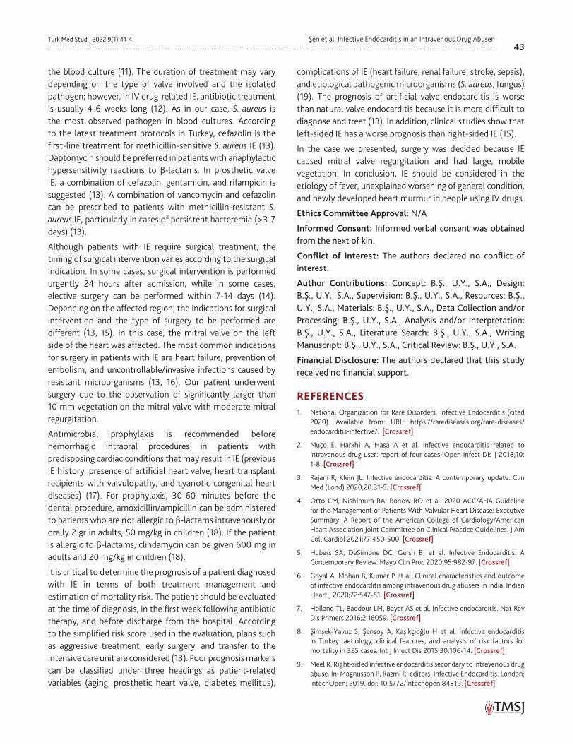
43Turk Med Stud J 2022;9(1):41-4. Şen et al. Infective Endocarditis in an Intravenous Drug Abuser
the blood culture (11). The duration of treatment may vary depending on the type of valve involved and the isolated pathogen; however, in IV drug-related IE, antibiotic treatment is usually 4-6 weeks long (12). As in our case, S. aureus is the most observed pathogen in blood cultures. According to the latest treatment protocols in Turkey, cefazolin is the first-line treatment for methicillin-sensitive S. aureus IE (13). Daptomycin should be preferred in patients with anaphylactic hypersensitivity reactions to β-lactams. In prosthetic valve IE, a combination of cefazolin, gentamicin, and rifampicin is suggested (13). A combination of vancomycin and cefazolin can be prescribed to patients with methicillin-resistant S. aureus IE, particularly in cases of persistent bacteremia (>3-7 days) (13).
Although patients with IE require surgical treatment, the timing of surgical intervention varies according to the surgical indication. In some cases, surgical intervention is performed urgently 24 hours after admission, while in some cases, elective surgery can be performed within 7-14 days (14). Depending on the affected region, the indications for surgical intervention and the type of surgery to be performed are different (13, 15). In this case, the mitral valve on the left side of the heart was affected. The most common indications for surgery in patients with IE are heart failure, prevention of embolism, and uncontrollable/invasive infections caused by resistant microorganisms (13, 16). Our patient underwent surgery due to the observation of significantly larger than 10 mm vegetation on the mitral valve with moderate mitral regurgitation.
Antimicrobial prophylaxis is recommended before hemorrhagic intraoral procedures in patients with predisposing cardiac conditions that may result in IE (previous IE history, presence of artificial heart valve, heart transplant recipients with valvulopathy, and cyanotic congenital heart diseases) (17). For prophylaxis, 30-60 minutes before the dental procedure, amoxicillin/ampicillin can be administered to patients who are not allergic to β-lactams intravenously or orally 2 gr in adults, 50 mg/kg in children (18). If the patient is allergic to β-lactams, clindamycin can be given 600 mg in adults and 20 mg/kg in children (18).
It is critical to determine the prognosis of a patient diagnosed with IE in terms of both treatment management and estimation of mortality risk. The patient should be evaluated at the time of diagnosis, in the first week following antibiotic therapy, and before discharge from the hospital. According to the simplified risk score used in the evaluation, plans such as aggressive treatment, early surgery, and transfer to the intensive care unit are considered (13). Poor prognosis markers can be classified under three headings as patient-related variables (aging, prosthetic heart valve, diabetes mellitus),
complications of IE (heart failure, renal failure, stroke, sepsis), and etiological pathogenic microorganisms (S. aureus, fungus) (19). The prognosis of artificial valve endocarditis is worse than natural valve endocarditis because it is more difficult to diagnose and treat (13). In addition, clinical studies show that left-sided IE has a worse prognosis than right-sided IE (15).
In the case we presented, surgery was decided because IE caused mitral valve regurgitation and had large, mobile vegetation. In conclusion, IE should be considered in the etiology of fever, unexplained worsening of general condition, and newly developed heart murmur in people using IV drugs.
Ethics Committee Approval: N/A
Informed Consent: Informed verbal consent was obtained from the next of kin.
Conflict of Interest: The authors declared no conflict of interest.
Author Contributions: Concept: B.Ş., U.Y., S.A., Design: B.Ş., U.Y., S.A., Supervision: B.Ş., U.Y., S.A., Resources: B.Ş., U.Y., S.A., Materials: B.Ş., U.Y., S.A., Data Collection and/or Processing: B.Ş., U.Y., S.A., Analysis and/or Interpretation: B.Ş., U.Y., S.A., Literature Search: B.Ş., U.Y., S.A., Writing Manuscript: B.Ş., U.Y., S.A., Critical Review: B.Ş., U.Y., S.A.
Financial Disclosure: The authors declared that this study received no financial support.
REFERENCES1. National Organization for Rare Disorders. Infective Endocarditis (cited
2020). Available from: URL: https://rarediseases.org/rare-diseases/ endocarditis-infective/. [Crossref]
2. Muço E, Harxhi A, Hasa A et al. Infective endocarditis related to intravenous drug user: report of four cases. Open Infect Dis J 2018;10: 1-8. [Crossref]
3. Rajani R, Klein JL. Infective endocarditis: A contemporary update. Clin Med (Lond) 2020;20:31-5. [Crossref]
4. Otto CM, Nishimura RA, Bonow RO et al. 2020 ACC/AHA Guideline for the Management of Patients With Valvular Heart Disease: Executive Summary: A Report of the American College of Cardiology/American Heart Association Joint Committee on Clinical Practice Guidelines. J Am Coll Cardiol 2021;77:450-500. [Crossref]
5. Hubers SA, DeSimone DC, Gersh BJ et al. Infective Endocarditis: A Contemporary Review. Mayo Clin Proc 2020;95:982-97. [Crossref]
6. Goyal A, Mohan B, Kumar P et al. Clinical characteristics and outcome of infective endocarditis among intravenous drug abusers in India. Indian Heart J 2020;72:547-51. [Crossref]
7. Holland TL, Baddour LM, Bayer AS et al. Infective endocarditis. Nat Rev Dis Primers 2016;2:16059. [Crossref]
8. Şimşek-Yavuz S, Şensoy A, Kaşıkçıoğlu H et al. Infective endocarditis in Turkey: aetiology, clinical features, and analysis of risk factors for mortality in 325 cases. Int J Infect Dis 2015;30:106-14. [Crossref]
9. Meel R. Right-sided infective endocarditis secondary to intravenous drug abuse. In: Magnusson P, Razmi R, editors. Infective Endocarditis. London: IntechOpen; 2019. doi: 10.5772/intechopen.84319. [Crossref]
44Turk Med Stud J 2022;9(1):41-4.Şen et al. Infective Endocarditis in an Intravenous Drug Abuser
10. Seghatol F, Grinberg I. Left-sided endocarditis in intravenous drug users: a case report and review of the literature. Echocardiography 2002;19:509-11. [Crossref]
11. Martí-Carvajal AJ, Dayer M, Conterno LO et al. A comparison of different antibiotic regimens for the treatment of infective endocarditis. Cochrane Database Syst Rev 2016;4:CD009880.[Crossref]
12. Ji Y, Kujtan L, Kershner D. Acute endocarditis in intravenous drug users: a case report and literature review. J Community Hosp Intern Med Perspect 2012;2. [Crossref]
13. Şimşek-Yavuz S, Akar AR, Aydoğdu S et al. İnfektif endokarditin tanısı, tedavisi ve önlenmesi: Ulusal uzlaşı raporu. Klimik Derg 2019;32 (Suppl 1):2-116. [Crossref]
14. Morjan M, Van Hemelrijk M, Maisano F et al. Contemporary surgery in infective endocarditis. EJCM 2019;7:166-71. [Crossref]
15. Moldovan H, Adrian M, Victor C et al. Infective endocarditis in intravenous drug users: surgical treatment. In: Magnusson P, Razmi R,
editors. Infective Endocarditis. London: IntechOpen; 2019. doi: 10.5772/intechopen.84708. [Crossref]
16. Prendergast BD, Tornos P. Surgery for infective endocarditis: who and when? Circulation 2010;121:1141-52. [Crossref]
17. Allen U. Infective endocarditis: Updated guidelines. Can J Infect Dis Med Microbiol 2010;21:74-7. [Crossref]
18. Habib G, Lancellotti P, Antunes MJ et al. 2015 ESC Guidelines for the management of infective endocarditis: The Task Force for the Management of Infective Endocarditis of the European Society of Cardiology (ESC). Endorsed by: European Association for Cardio-Thoracic Surgery (EACTS), the European Association of Nuclear Medicine (EANM). Eur Heart J 2015;36:3075-128. [Crossref]
19. Gálvez-Acebal J, Rodríguez-Baño J, Martínez-Marcos FJ et al. Prognostic factors in left-sided endocarditis: results from the Andalusian multicenter cohort. BMC Infect Dis 2010;10:17. [Crossref]
Turk Med Stud J 2022;9(1):45-7ERRATA
45
DOI: 10.4274/tmsj.galenos.2022.09.01.e001
DOI: 10.4274/tmsj.galenos.2019.06.01.01
In the article by Özkan et al., entitled “An Investigation on the Anticancer Effect of Spider Web in Human Cervical Cell Line” that was published in the February 2019 issue of Turkish Medical Student Journal, citation information was wrongly written due to a publishing software program error. The Editorial Board reviewed the case and “Turkish Med. Stud. J. 2018; 5: 50-3” is corrected as “Turkish Med Stud J 2019;6(1):1-11”.
DOI: 10.4274/tmsj.galenos.2022.09.01.e002
DOI: 10.4274/tmsj.galenos.2019.06.01.02
In the article by Demirel et al., entitled “Systemic Cannabidiol Does Not Reduce Compound 48/80-Induced Itching Behavior in Mice” that was published in the February 2019 issue of Turkish Medical Student Journal, citation information was wrongly written due to a publishing software program error. The Editorial Board reviewed the case and “Turkish Med. Stud. J. 2018; 5: 54-7” is corrected as “Turkish Med Stud J 2019;6(1):12-7”.
DOI: 10.4274/tmsj.galenos.2022.09.01.e003
DOI: 10.4274/tmsj.galenos.2019.06.01.03
In the article by Özyiğit et al., entitled “Evaluation of Malnutrition Statuses in Systolic Heart Failure Patients” that was published in the February 2019 issue of Turkish Medical Student Journal, citation information was wrongly written due to a publishing software program error. The Editorial Board reviewed the case and “Turkish Med. Stud. J. 2018; 5: 58-63” is corrected as “Turkish Med Stud J 2019;6(1):18-24”.
DOI: 10.4274/tmsj.galenos.2022.09.01.e004
DOI: 10.4274/tmsj.galenos.2019.06.01.04
In the article by Kolotylo et al., entitled “The Influence of Intercurrent Diseases on the Course of HIV in Association with Active Tuberculosis” that was published in the February 2019 issue of Turkish Medical Student Journal, citation information was wrongly written due to a publishing software program error. The Editorial Board reviewed the case and “Turkish Med. Stud. J. 2018; 5: 64-8” is corrected as “Turkish Med Stud J 2019;6(1):25-30”.
DOI: 10.4274/tmsj.galenos.2022.09.01.e005
DOI:10.4274/tmsj.galenos.2019.06.01.05
In the article by Avul et al., entitled “A Case Report: The Role of Prostate-Specific Membrane Antigen Labeled Theranostic Agents in the Diagnosis and Treatment of Prostate Cancer” that was published in the February 2019 issue of Turkish Medical Student Journal, citation information was wrongly written due to a publishing software program error. The Editorial Board reviewed the case and “Turkish Med. Stud. J. 2018; 5: 69-75” is corrected as “Turkish Med Stud J 2019;6(1):31-6”.
DOI: 10.4274/tmsj.galenos.2022.09.01.e006
DOI: 10.4274/tmsj.galenos.2019.06.01.06
In the article by Kılıççalan et al., entitled “Caenorhabditis Elegans and Angiogenesis” that was published in the February 2019 issue of Turkish Medical Student Journal, citation information was wrongly written due to a publishing software program error. The Editorial Board reviewed the case and “Turkish Med. Stud. J. 2018; 5: 79-84” is corrected as “Turkish Med Stud J 2019;6(1):37-41”.
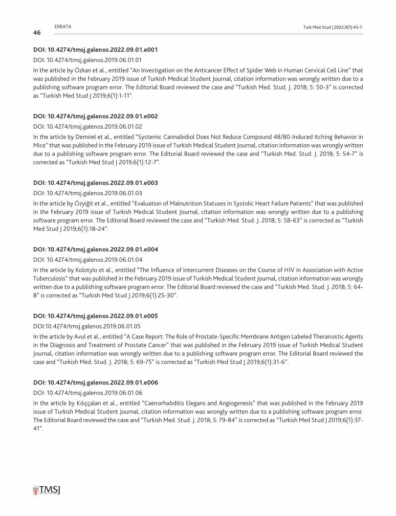
46Turk Med Stud J 2022;9(1):45-7.ERRATA
DOI: 10.4274/tmsj.galenos.2022.09.01.e001
DOI: 10.4274/tmsj.galenos.2019.06.01.01
In the article by Özkan et al., entitled “An Investigation on the Anticancer Effect of Spider Web in Human Cervical Cell Line” that was published in the February 2019 issue of Turkish Medical Student Journal, citation information was wrongly written due to a publishing software program error. The Editorial Board reviewed the case and “Turkish Med. Stud. J. 2018; 5: 50-3” is corrected as “Turkish Med Stud J 2019;6(1):1-11”.
DOI: 10.4274/tmsj.galenos.2022.09.01.e002
DOI: 10.4274/tmsj.galenos.2019.06.01.02
In the article by Demirel et al., entitled “Systemic Cannabidiol Does Not Reduce Compound 48/80-Induced Itching Behavior in Mice” that was published in the February 2019 issue of Turkish Medical Student Journal, citation information was wrongly written due to a publishing software program error. The Editorial Board reviewed the case and “Turkish Med. Stud. J. 2018; 5: 54-7” is corrected as “Turkish Med Stud J 2019;6(1):12-7”.
DOI: 10.4274/tmsj.galenos.2022.09.01.e003
DOI: 10.4274/tmsj.galenos.2019.06.01.03
In the article by Özyiğit et al., entitled “Evaluation of Malnutrition Statuses in Systolic Heart Failure Patients” that was published in the February 2019 issue of Turkish Medical Student Journal, citation information was wrongly written due to a publishing software program error. The Editorial Board reviewed the case and “Turkish Med. Stud. J. 2018; 5: 58-63” is corrected as “Turkish Med Stud J 2019;6(1):18-24”.
DOI: 10.4274/tmsj.galenos.2022.09.01.e004
DOI: 10.4274/tmsj.galenos.2019.06.01.04
In the article by Kolotylo et al., entitled “The Influence of Intercurrent Diseases on the Course of HIV in Association with Active Tuberculosis” that was published in the February 2019 issue of Turkish Medical Student Journal, citation information was wrongly written due to a publishing software program error. The Editorial Board reviewed the case and “Turkish Med. Stud. J. 2018; 5: 64-8” is corrected as “Turkish Med Stud J 2019;6(1):25-30”.
DOI: 10.4274/tmsj.galenos.2022.09.01.e005
DOI:10.4274/tmsj.galenos.2019.06.01.05
In the article by Avul et al., entitled “A Case Report: The Role of Prostate-Specific Membrane Antigen Labeled Theranostic Agents in the Diagnosis and Treatment of Prostate Cancer” that was published in the February 2019 issue of Turkish Medical Student Journal, citation information was wrongly written due to a publishing software program error. The Editorial Board reviewed the case and “Turkish Med. Stud. J. 2018; 5: 69-75” is corrected as “Turkish Med Stud J 2019;6(1):31-6”.
DOI: 10.4274/tmsj.galenos.2022.09.01.e006
DOI: 10.4274/tmsj.galenos.2019.06.01.06
In the article by Kılıççalan et al., entitled “Caenorhabditis Elegans and Angiogenesis” that was published in the February 2019 issue of Turkish Medical Student Journal, citation information was wrongly written due to a publishing software program error. The Editorial Board reviewed the case and “Turkish Med. Stud. J. 2018; 5: 79-84” is corrected as “Turkish Med Stud J 2019;6(1):37-41”.
47Turk Med Stud J 2022;9(1):45-7. ERRATA
DOI: 10.4274/tmsj.galenos.2022.09.01.e007
DOI: 10.4274/tmsj.galenos.2019.06.01.07
In the article by Tsagkaris, entitled “Letter to the Editor” that was published in the February 2019 issue of Turkish Medical Student Journal, citation information was wrongly written due to a publishing software program error. The Editorial Board reviewed the case and “Turkish Med. Stud. J. 2018; 5: 69-75” is corrected as “Turkish Med Stud J 2019;6(1):42”.
DOI: 10.4274/tmsj.galenos.2022.09.01.e008
DOI: 10.4274/tmsj.galenos.2019.06.02.04
In the article by Mutlu et al., entitled “A Newborn with Esophageal Atresia, Tracheoesophageal Fistula and Feeding Problems” (Turkish Med Stud J 2019:6(2);60-3.) Figure 1 was reflected on the y axis by mistake and caused a misunderstand like the patient had situs inversus. The figure is corrected as follows:
DOI: 10.4274/tmsj.galenos.2022.09.01.e009
In the article by Özkan et al., entitled “Evaluating Orthorexia Tendency Among Trakya University Medical School Students” that was published in the January 2015 issue of Turkish Medical Student Journal, the name of an author was wrongly written. The Editorial Board reviewed the case and “Atila Ülkücü” is corrected as “Attila Ülkücü”.
DOI: 10.4274/tmsj.galenos.2022.09.01.e010
DOI: 10.4274/tmsj.galenos.2020.07.01.06
In the article by Göztepe et al., entitled “Arrow Cause of Angina Pectoris: Single Coronary Artery in Elderly Patient” that was published in the February 2020 issue of Turkish Medical Student Journal, the name of the article was wrongly written. The Editorial Board reviewed the case and “Arrow Cause of Angina Pectoris: Single Coronary Artery in Elderly Patient” is corrected as “A Case Report: Single Coronary Artery in Elderly Patient”.
DOI: 10.4274/tmsj.galenos.2022.09.01.e011
DOI: 10.4274/tmsj.galenos.2021.08.01.04
In the article by Tan et al., entitled “Fear of COVID-19 Among Medical Students and Associated Factors” that was published in the February 2021 issue of Turkish Medical Student Journal, protocol code was wrongly written. The Editorial Board reviewed the case and “Protocol Code: TÜTF-BAEK 2020/440” is corrected as “Protocol Code: TÜTF-BAEK 2020/448”.
Related Documents