Visual system 2 and accessory visual structures Organum visuale et structurae oculi accessoriae David Kachlík

Welcome message from author
This document is posted to help you gain knowledge. Please leave a comment to let me know what you think about it! Share it to your friends and learn new things together.
Transcript
Visual system 2
and accessory visual
structures
Organum visuale et
structurae oculi
accessoriae
David Kachlík
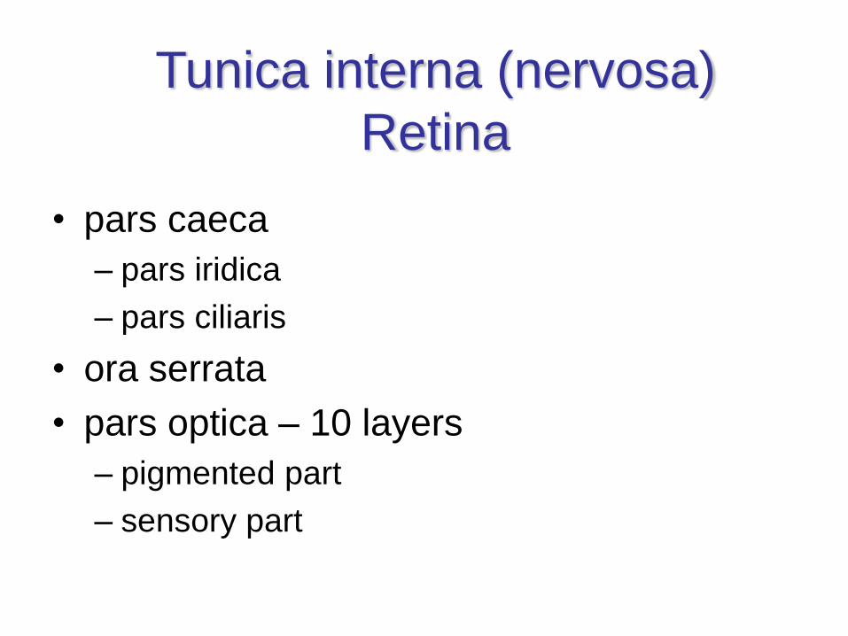
Tunica interna (nervosa)
Retina
• pars caeca
– pars iridica
– pars ciliaris
• ora serrata
• pars optica – 10 layers
– pigmented part
– sensory part
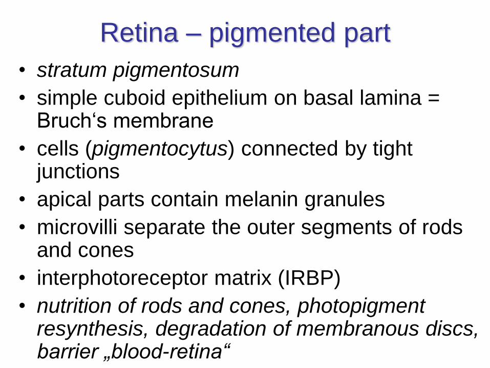
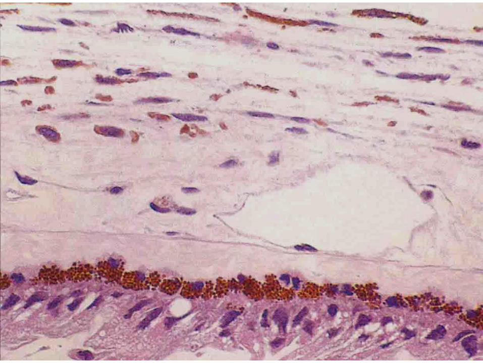
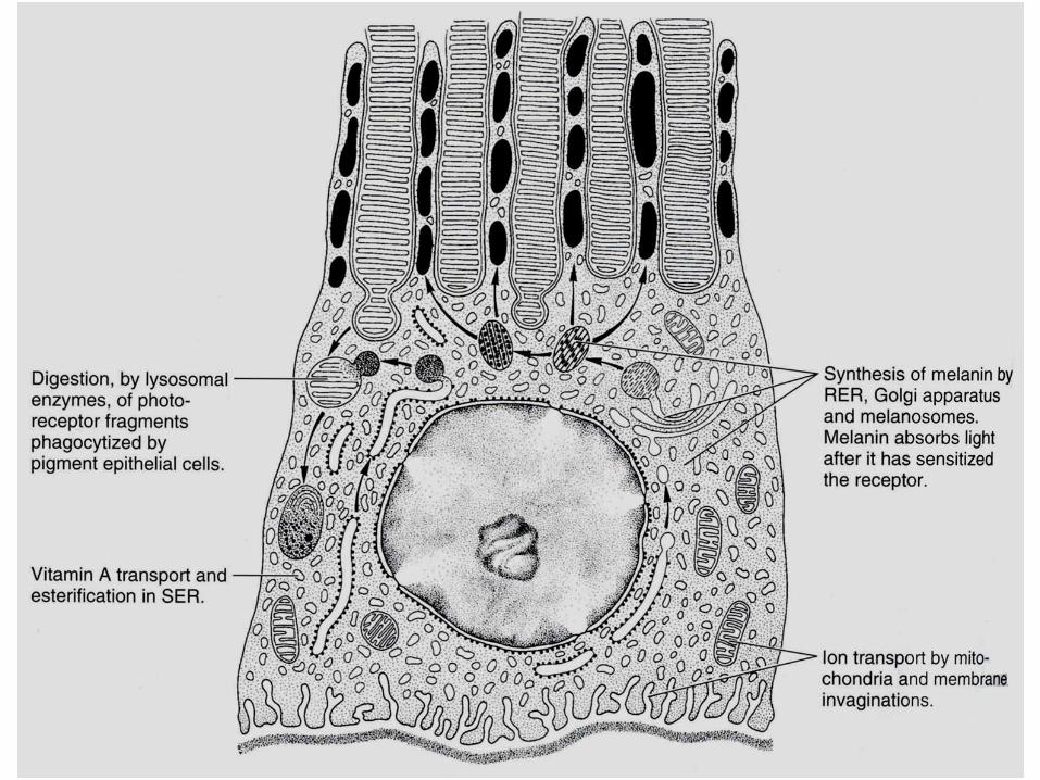
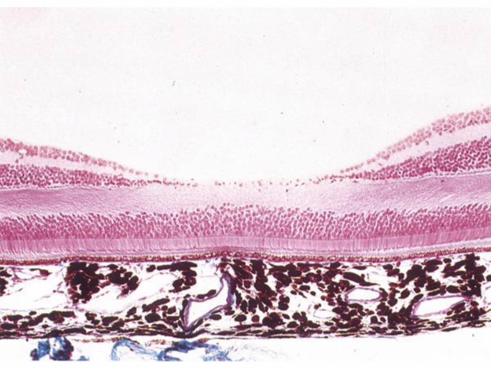
Retina – pigmented part
• stratum pigmentosum
• simple cuboid epithelium on basal lamina = Bruch‘s membrane
• cells (pigmentocytus) connected by tight junctions
• apical parts contain melanin granules
• microvilli separate the outer segments of rods and cones
• interphotoreceptor matrix (IRBP)
• nutrition of rods and cones, photopigmentresynthesis, degradation of membranous discs, barrier „blood-retina“
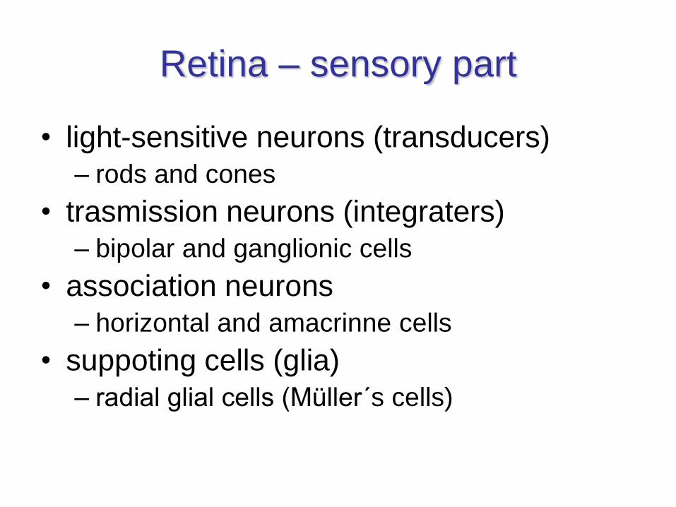
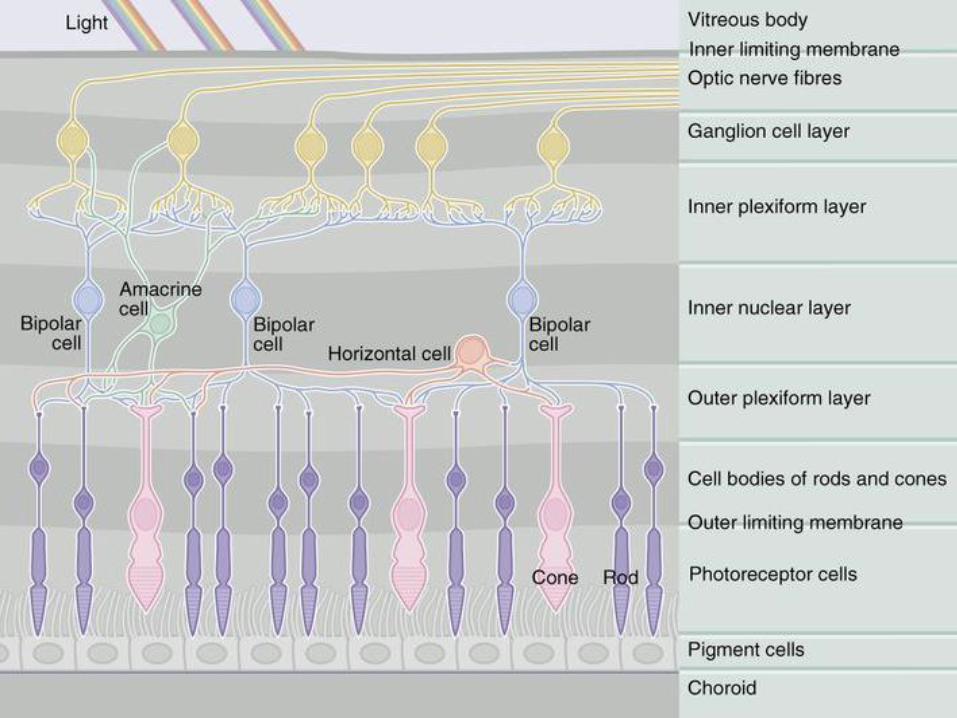
Retina – sensory part
• light-sensitive neurons (transducers)
– rods and cones
• trasmission neurons (integraters)
– bipolar and ganglionic cells
• association neurons
– horizontal and amacrinne cells
• suppoting cells (glia)
– radial glial cells (Müller´s cells)
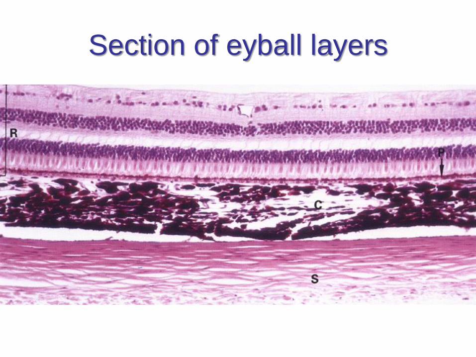
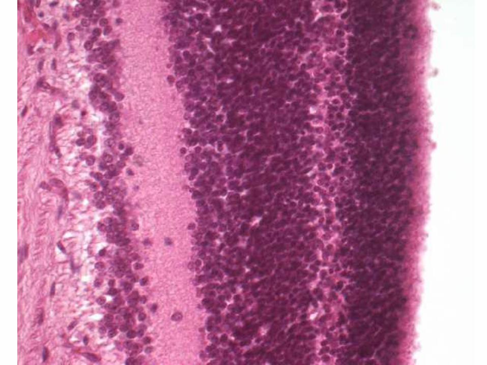
Section of eyball layers
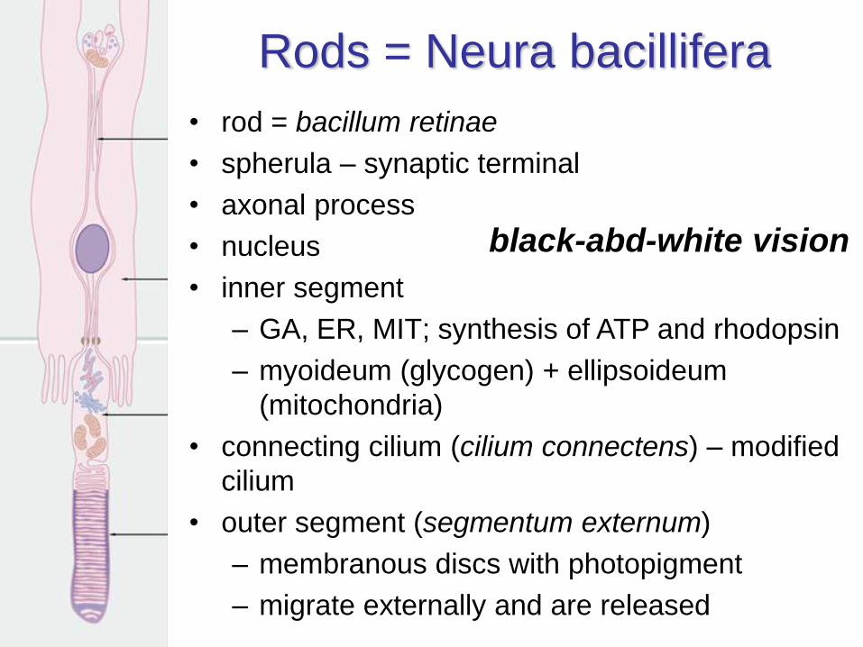
Rods = Neura bacillifera
• rod = bacillum retinae
• spherula – synaptic terminal
• axonal process
• nucleus
• inner segment
– GA, ER, MIT; synthesis of ATP and rhodopsin
– myoideum (glycogen) + ellipsoideum
(mitochondria)
• connecting cilium (cilium connectens) – modified
cilium
• outer segment (segmentum externum)
– membranous discs with photopigment
– migrate externally and are released
black-abd-white vision
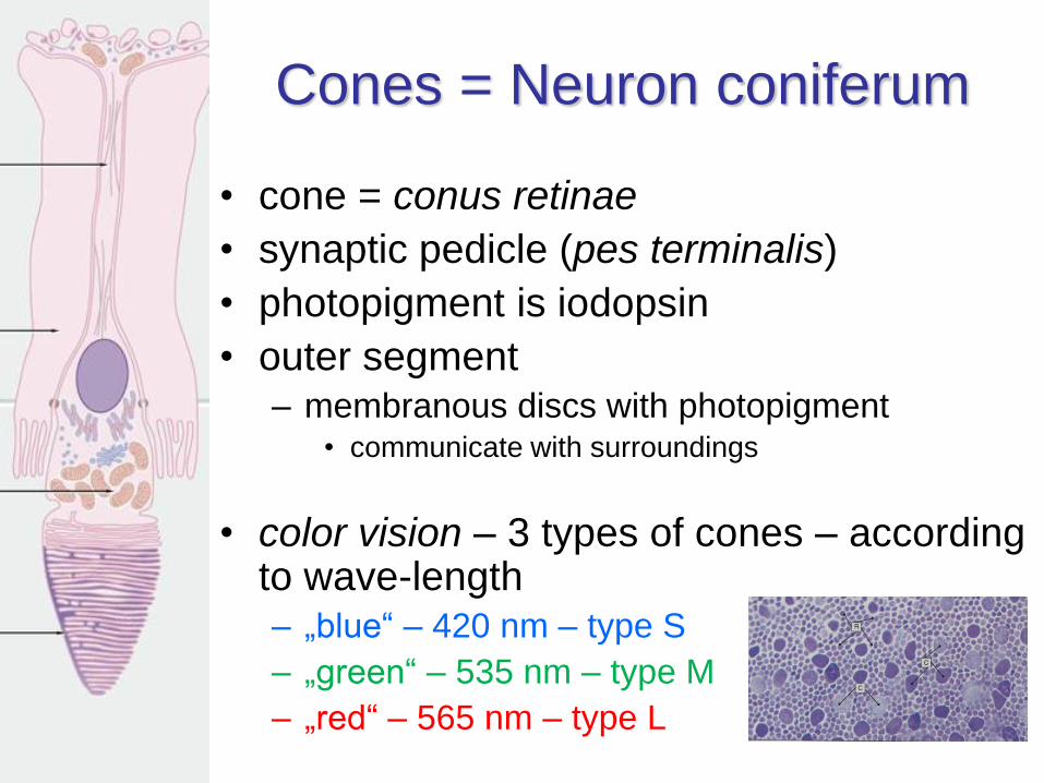
Cones = Neuron coniferum
• cone = conus retinae
• synaptic pedicle (pes terminalis)
• photopigment is iodopsin
• outer segment– membranous discs with photopigment
• communicate with surroundings
• color vision – 3 types of cones – according to wave-length– „blue“ – 420 nm – type S
– „green“ – 535 nm – type M
– „red“ – 565 nm – type L
Retina – Transmission neurons
• Bipolar neurons (Neuron bipolare)
– rod bipolar neurons (n.b. bacillotopicum)
– cone bipolar neurons (n.b. conotopicum)
• midget (n.b.c. nanum) – macula lutea (no convergence = 1 : 1 : 1)
• diffuse (n.b.c. diffusum) – convergence of signal
– contact with retinal ganglion cells
• Retinal ganglion cells (N. ganglionare multipolare)
– diffuse (n.g.m. umbelliforme) – connect more bipolar
neurons
– midget (n.g.m. nanum) – connect with midget bipolar
neurons
– their axons form the nervus opticus
Retina – Association neurons
signal modification and synchronization
• Horizontal neurons (N. horizontale)
– connections with axons of rods and cones and
with dendrites of bipolar neurons
– integrate rods and ocnes of adjacent areas
– supress signals from less lit areas
• amacrine cells (N. amacrinum)
– no axon!
– connections with axons of bipolar cells and with
dendrites of retina ganglion cells
– remove noise
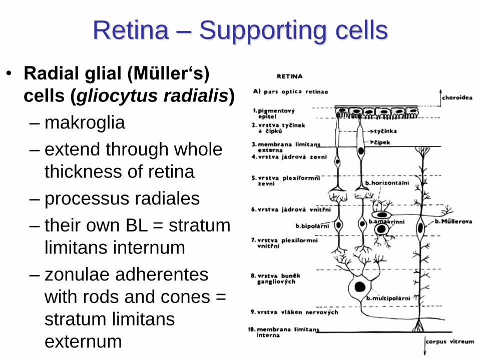
Retina – Supporting cells
• Radial glial (Müller‘s)
cells (gliocytus radialis)
– makroglia
– extend through whole
thickness of retina
– processus radiales
– their own BL = stratum
limitans internum
– zonulae adherentes
with rods and cones =
stratum limitans
externum
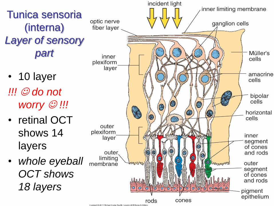
Tunica sensoria
(interna)
Layer of sensory
part
• 10 layer
!!! do not
worry !!!
• retinal OCT
shows 14
layers
• whole eyeball
OCT shows
18 layers
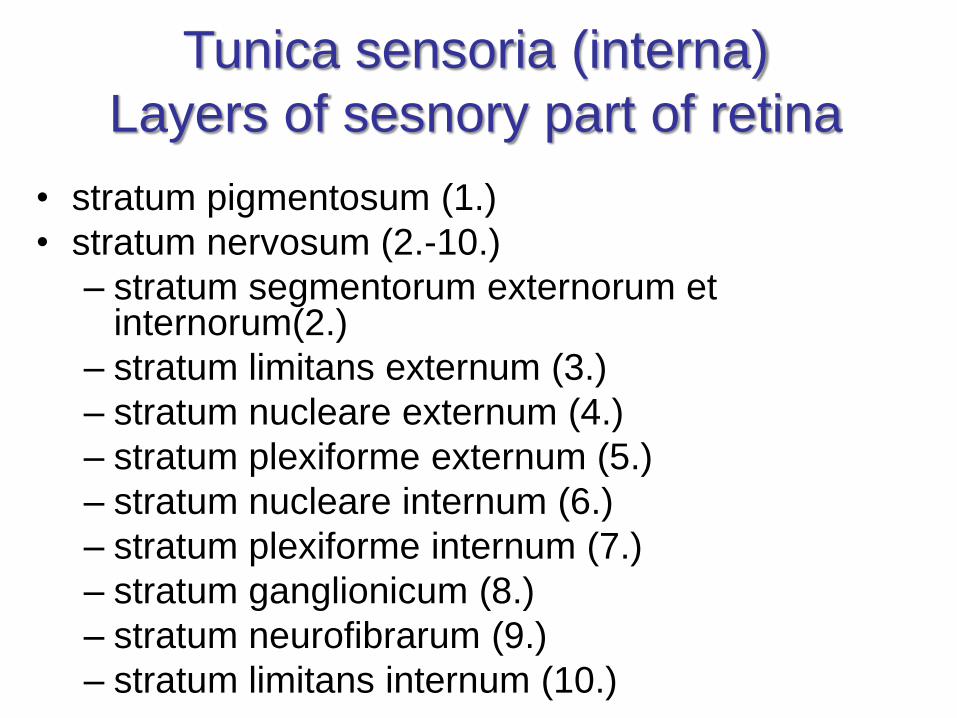
Tunica sensoria (interna)
Layers of sesnory part of retina
• stratum pigmentosum (1.)
• stratum nervosum (2.-10.)
– stratum segmentorum externorum et internorum(2.)
– stratum limitans externum (3.)
– stratum nucleare externum (4.)
– stratum plexiforme externum (5.)
– stratum nucleare internum (6.)
– stratum plexiforme internum (7.)
– stratum ganglionicum (8.)
– stratum neurofibrarum (9.)
– stratum limitans internum (10.)
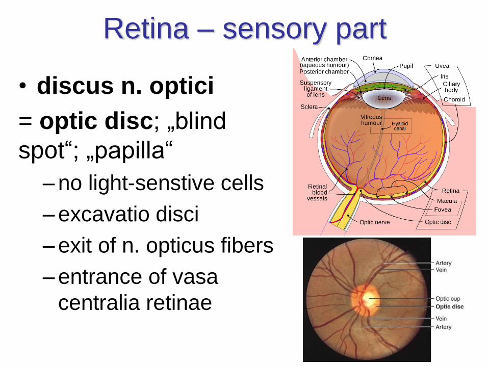
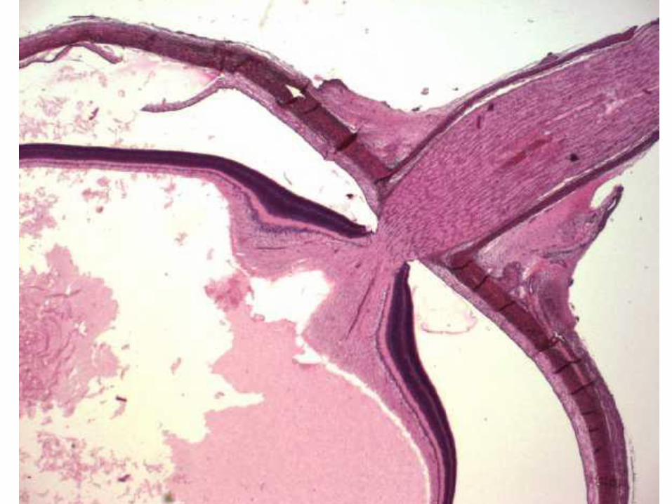
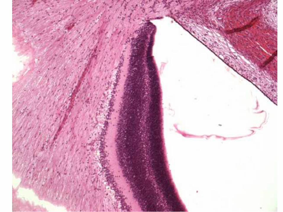
Retina – sensory part
• discus n. optici
= optic disc; „blind
spot“; „papilla“
– no light-senstive cells
– excavatio disci
– exit of n. opticus fibers
– entrance of vasa
centralia retinae
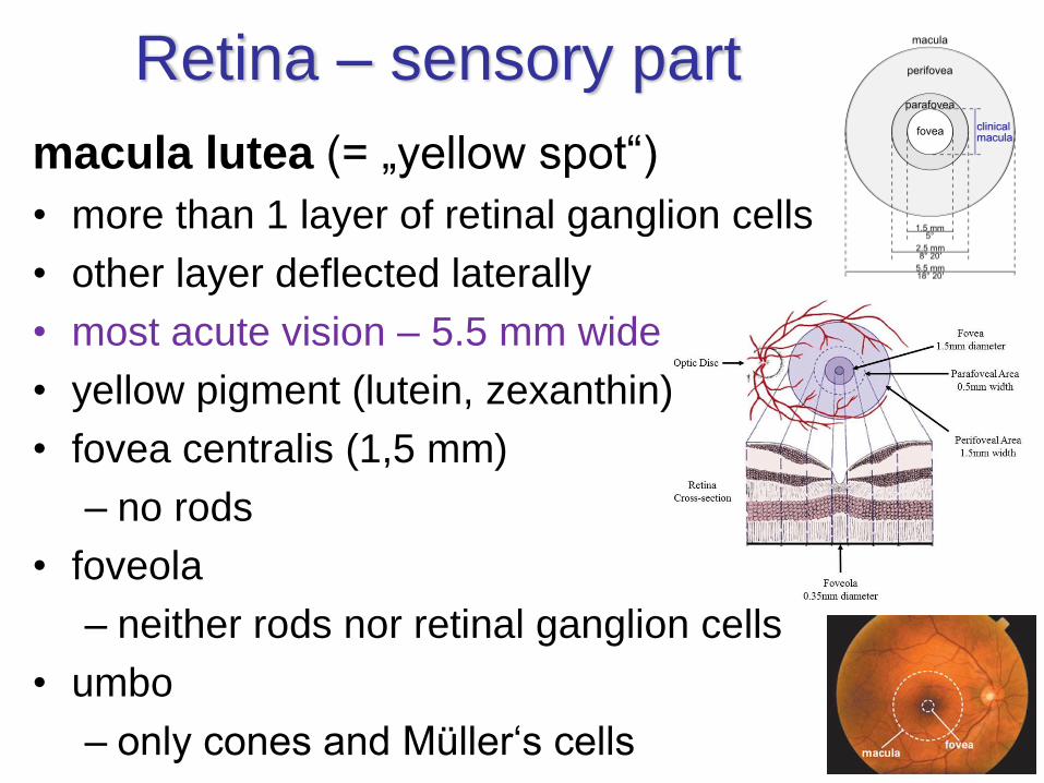
macula lutea (= „yellow spot“)
• more than 1 layer of retinal ganglion cells
• other layer deflected laterally
• most acute vision – 5.5 mm wide
• yellow pigment (lutein, zexanthin)
• fovea centralis (1,5 mm)
– no rods
• foveola
– neither rods nor retinal ganglion cells
• umbo
– only cones and Müller‘s cells
Retina – sensory part
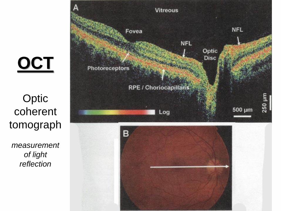
OCT
Optic
coherent
tomograph
measurement
of light
reflection
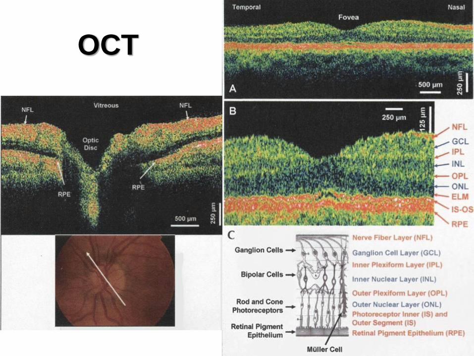
OCT
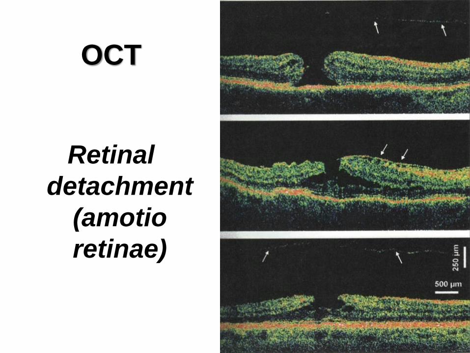
OCT
Retinal
detachment
(amotio
retinae)
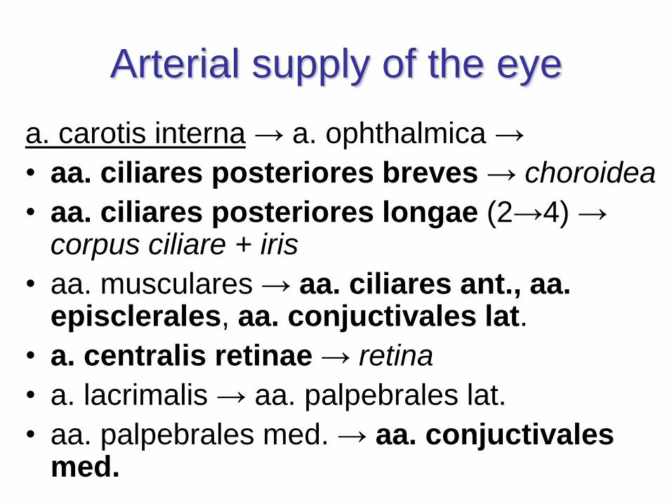
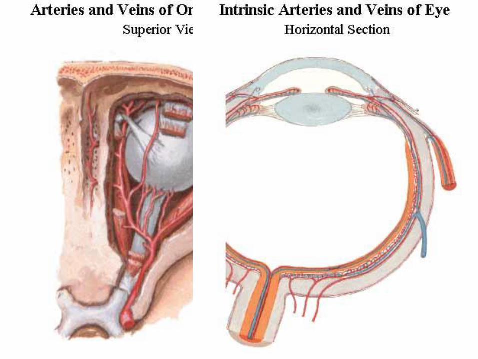
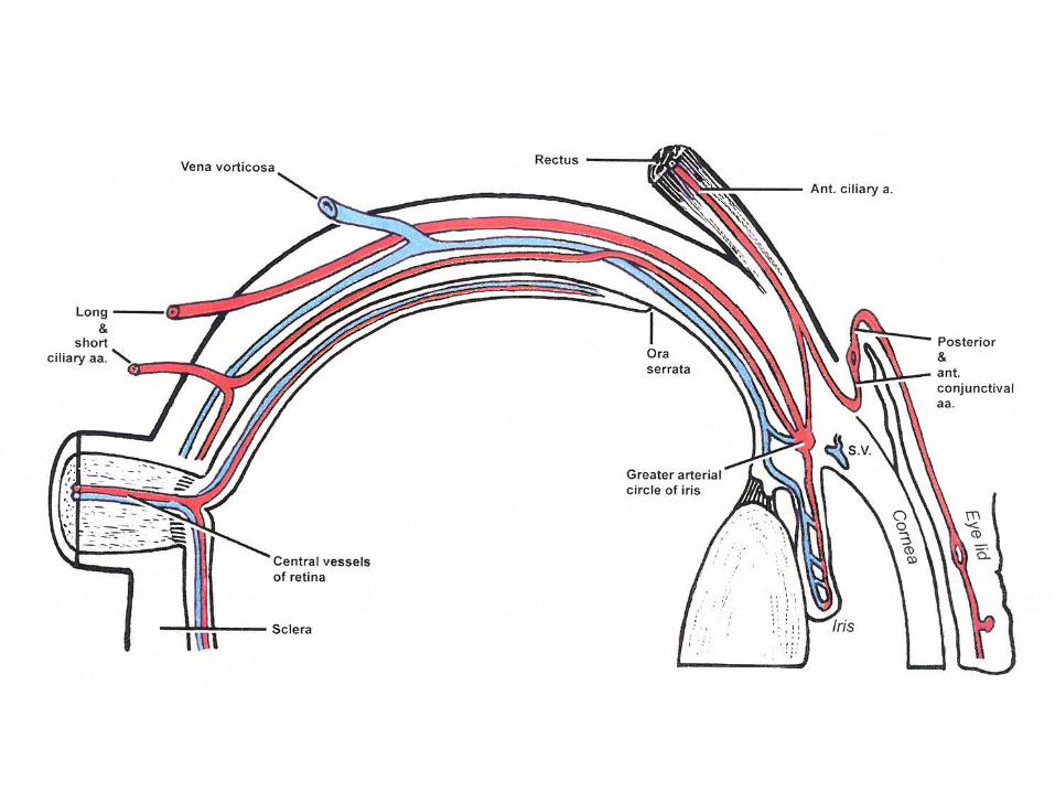
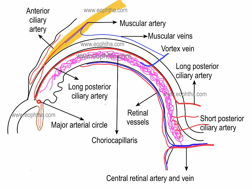
Arterial supply of the eye
a. carotis interna → a. ophthalmica →
• aa. ciliares posteriores breves → choroidea
• aa. ciliares posteriores longae (2→4) →corpus ciliare + iris
• aa. musculares → aa. ciliares ant., aa. episclerales, aa. conjuctivales lat.
• a. centralis retinae → retina
• a. lacrimalis → aa. palpebrales lat.
• aa. palpebrales med. → aa. conjuctivalesmed.
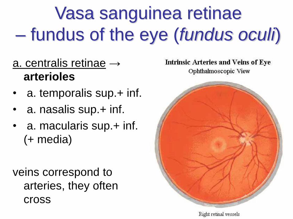
Vasa sanguinea retinae
– fundus of the eye (fundus oculi)
a. centralis retinae →
arterioles
• a. temporalis sup.+ inf.
• a. nasalis sup.+ inf.
• a. macularis sup.+ inf.
(+ media)
veins correspond to
arteries, they often
cross
Arteria cilioretinalis
• present in 10-33% of eyes
• branches from a. ciliaris posterior brevis
• exits the discus n.II separately from a. centralisretinae
• additional supply to macula lutea from choroidal circulation
• provide a small amount of blood supply to the retina when a. centralis retinae is occluded
• 90% located temporally x 10% nasally
• closure of a. cilioretinalis → central scotoma
• closure of a. centralis retinae → non-affectedmacula lutea and spared central vision
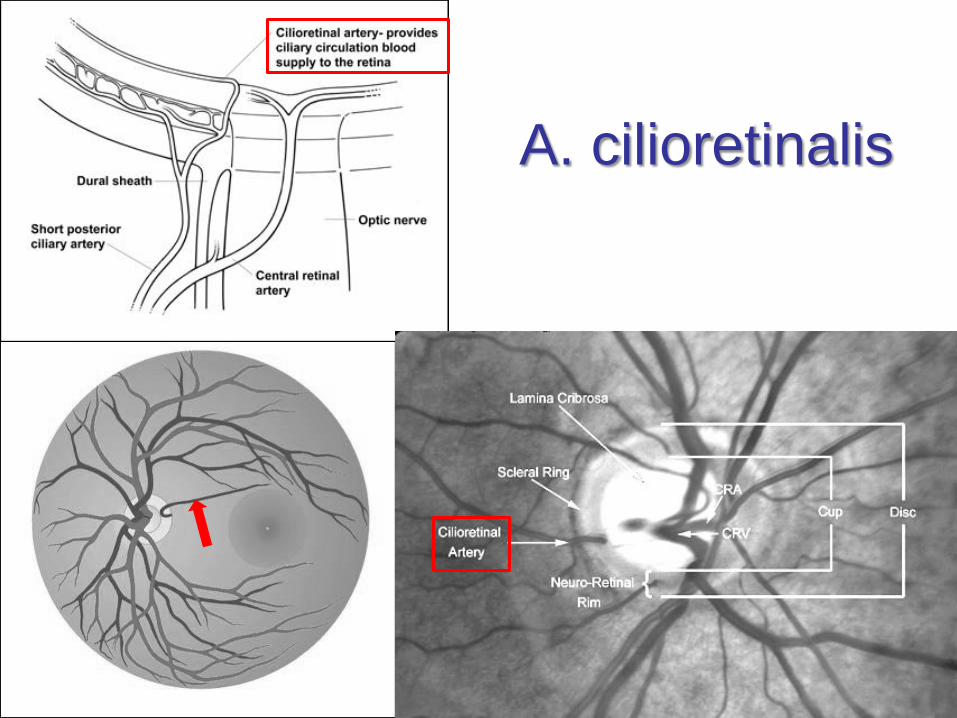
A. cilioretinalis
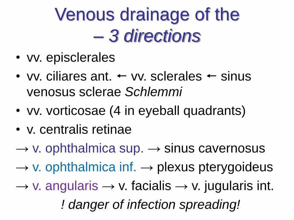
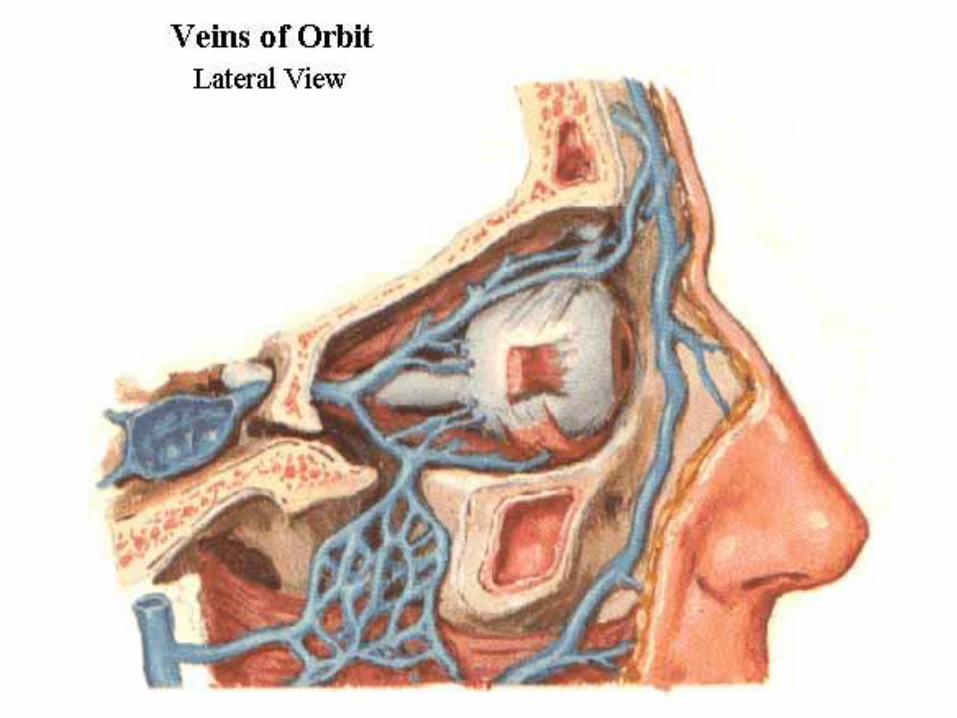
Venous drainage of the
– 3 directions• vv. episclerales
• vv. ciliares ant. vv. sclerales sinus
venosus sclerae Schlemmi
• vv. vorticosae (4 in eyeball quadrants)
• v. centralis retinae
→ v. ophthalmica sup. → sinus cavernosus
→ v. ophthalmica inf. → plexus pterygoideus
→ v. angularis → v. facialis → v. jugularis int.
! danger of infection spreading!
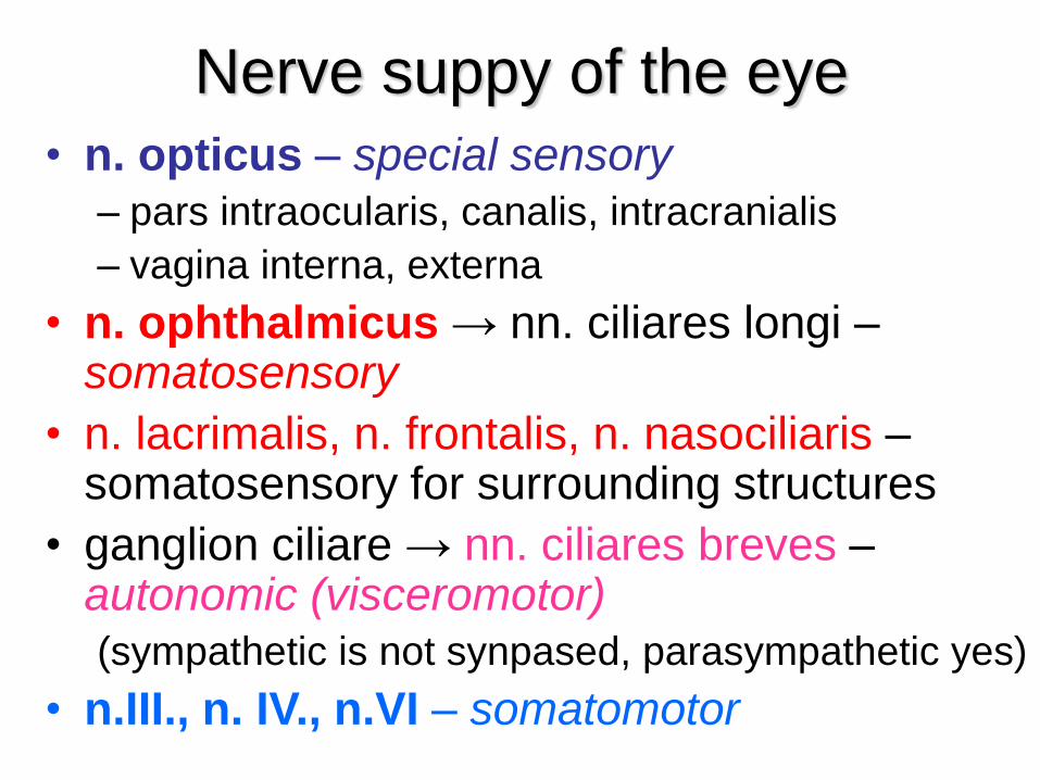
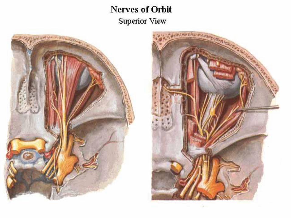
Nerve suppy of the eye
• n. opticus – special sensory
– pars intraocularis, canalis, intracranialis
– vagina interna, externa
• n. ophthalmicus → nn. ciliares longi –somatosensory
• n. lacrimalis, n. frontalis, n. nasociliaris –somatosensory for surrounding structures
• ganglion ciliare → nn. ciliares breves –autonomic (visceromotor)
(sympathetic is not synpased, parasympathetic yes)
• n.III., n. IV., n.VI – somatomotor
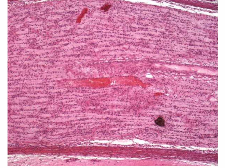
Nervus opticus
• čistě senzorický
• výchlipka diencefala (thalamus opticus)
• axony odděleny endoneuriem
• na povrchu jsou analogy mozkových obalů
– vagina externa = pachymeninx
– vagina interna = leptomeninx
• nervem probíhá a. et v. centralis retinae
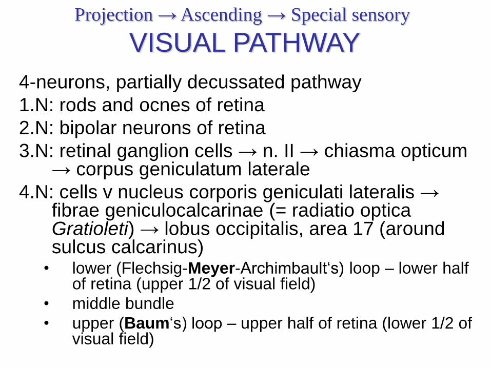
Projection → Ascending → Special sensory
VISUAL PATHWAY
4-neurons, partially decussated pathway
1.N: rods and ocnes of retina
2.N: bipolar neurons of retina
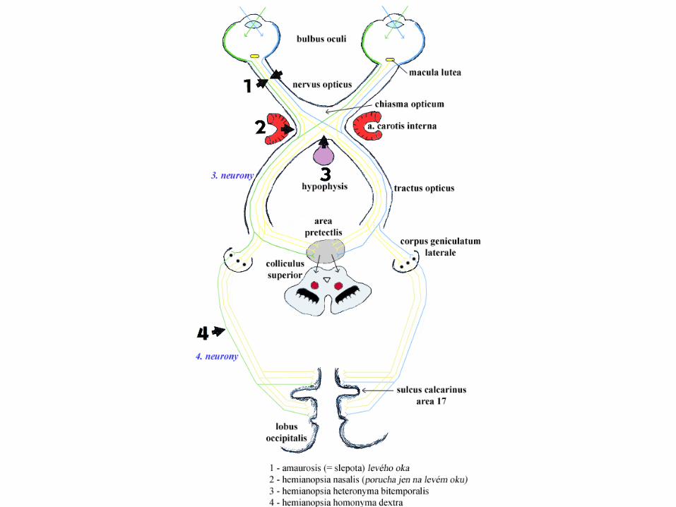
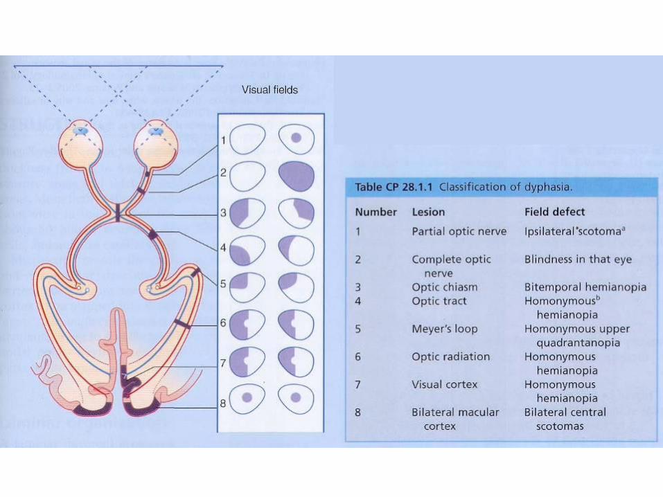
3.N: retinal ganglion cells → n. II → chiasma opticum→ corpus geniculatum laterale
4.N: cells v nucleus corporis geniculati lateralis → fibrae geniculocalcarinae (= radiatio opticaGratioleti) → lobus occipitalis, area 17 (around sulcus calcarinus)
• lower (Flechsig-Meyer-Archimbault‘s) loop – lower half of retina (upper 1/2 of visual field)
• middle bundle
• upper (Baum‘s) loop – upper half of retina (lower 1/2 of visual field)
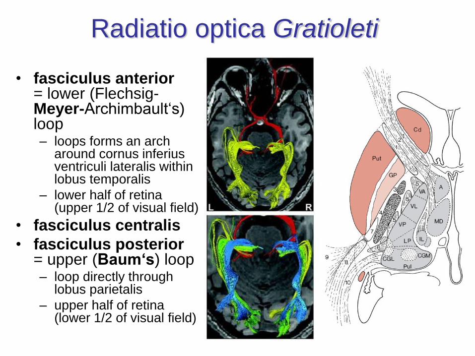
Radiatio optica Gratioleti
• fasciculus anterior= lower (Flechsig-Meyer-Archimbault‘s) loop– loops forms an arch
around cornus inferiusventriculi lateralis within lobus temporalis
– lower half of retina (upper 1/2 of visual field)
• fasciculus centralis
• fasciculus posterior= upper (Baum‘s) loop– loop directly through
lobus parietalis
– upper half of retina (lower 1/2 of visual field)
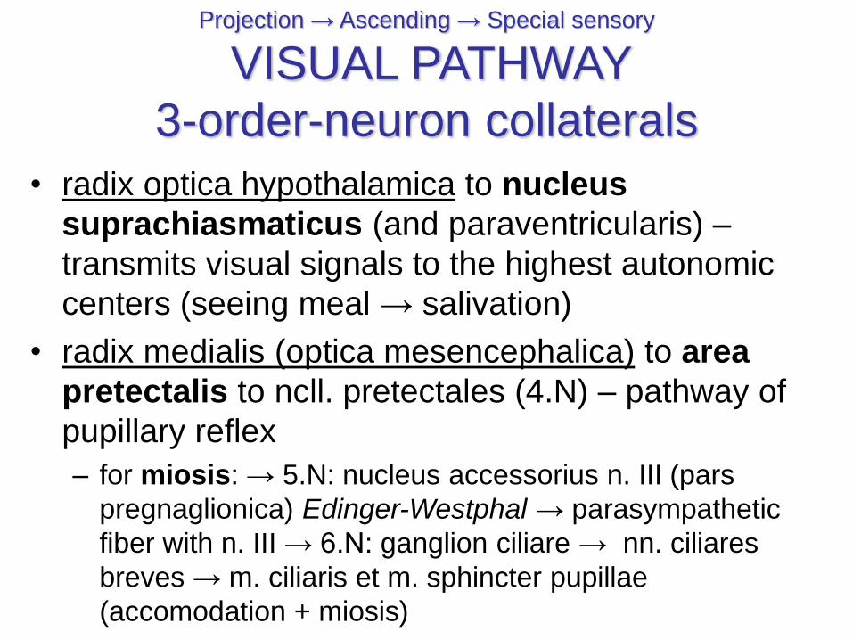
Projection → Ascending → Special sensory
VISUAL PATHWAY
3-order-neuron collaterals
• radix optica hypothalamica to nucleus
suprachiasmaticus (and paraventricularis) –
transmits visual signals to the highest autonomic
centers (seeing meal → salivation)
• radix medialis (optica mesencephalica) to area
pretectalis to ncll. pretectales (4.N) – pathway of
pupillary reflex
– for miosis: → 5.N: nucleus accessorius n. III (pars
pregnaglionica) Edinger-Westphal → parasympathetic
fiber with n. III → 6.N: ganglion ciliare → nn. ciliares
breves → m. ciliaris et m. sphincter pupillae
(accomodation + miosis)
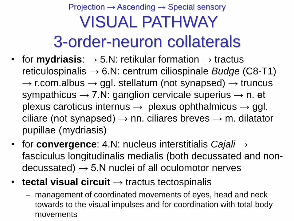
• for mydriasis: → 5.N: retikular formation → tractus
reticulospinalis → 6.N: centrum ciliospinale Budge (C8-T1)
→ r.com.albus → ggl. stellatum (not synapsed) → truncus
sympathicus → 7.N: ganglion cervicale superius → n. et
plexus caroticus internus → plexus ophthalmicus → ggl.
ciliare (not synapsed) → nn. ciliares breves → m. dilatator
pupillae (mydriasis)
• for convergence: 4.N: nucleus interstitialis Cajali →
fasciculus longitudinalis medialis (both decussated and non-
decussated) → 5.N nuclei of all oculomotor nerves
• tectal visual circuit → tractus tectospinalis
– management of coordinated movements of eyes, head and neck
towards to the visual impulses and for coordination with total body
movements
Projection → Ascending → Special sensory
VISUAL PATHWAY
3-order-neuron collaterals
Structurae oculi accessoriae
Accessory visual organs
• ligamentous apparatus = apparatus
ligamentosus
• eyelids = palpebrae
• conjunctiva = tunica conjunctiva
• lacrimal apparatus = apparatus lacrimalis
• muscular apparatus (extraocular muscles) =
apparatus muscularis (mm. externi bulbi oculi)
• supercilium
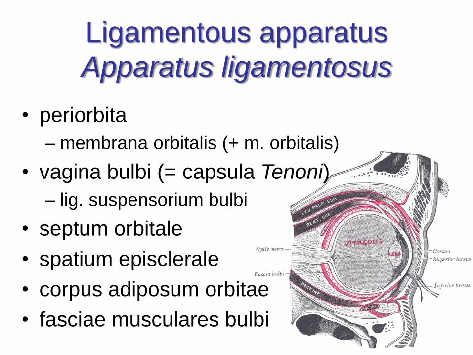
Ligamentous apparatus
Apparatus ligamentosus
• periorbita
– membrana orbitalis (+ m. orbitalis)
• vagina bulbi (= capsula Tenoni)
– lig. suspensorium bulbi
• septum orbitale
• spatium episclerale
• corpus adiposum orbitae
• fasciae musculares bulbi
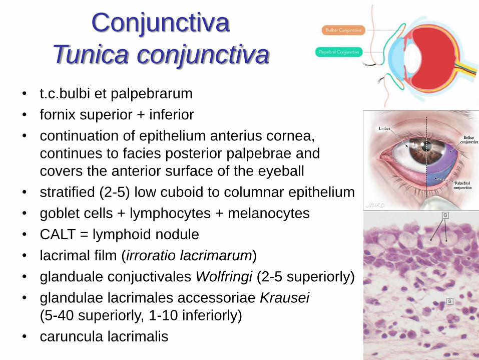
Conjunctiva
Tunica conjunctiva
• t.c.bulbi et palpebrarum
• fornix superior + inferior
• continuation of epithelium anterius cornea,
continues to facies posterior palpebrae and
covers the anterior surface of the eyeball
• stratified (2-5) low cuboid to columnar epithelium
• goblet cells + lymphocytes + melanocytes
• CALT = lymphoid nodule
• lacrimal film (irroratio lacrimarum)
• glanduale conjuctivales Wolfringi (2-5 superiorly)
• glandulae lacrimales accessoriae Krausei
(5-40 superiorly, 1-10 inferiorly)
• caruncula lacrimalis
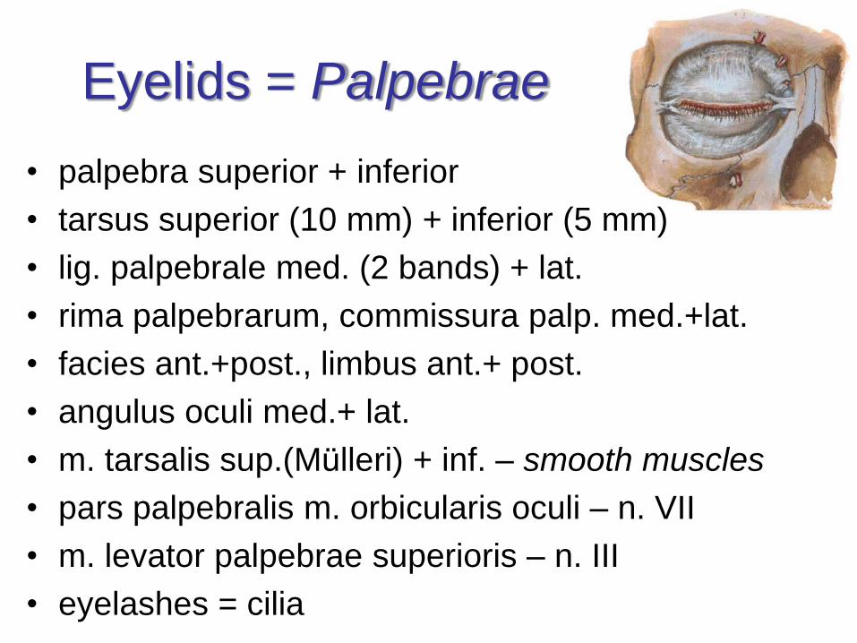
Eyelids = Palpebrae
• palpebra superior + inferior
• tarsus superior (10 mm) + inferior (5 mm)
• lig. palpebrale med. (2 bands) + lat.
• rima palpebrarum, commissura palp. med.+lat.
• facies ant.+post., limbus ant.+ post.
• angulus oculi med.+ lat.
• m. tarsalis sup.(Mülleri) + inf. – smooth muscles
• pars palpebralis m. orbicularis oculi – n. VII
• m. levator palpebrae superioris – n. III
• eyelashes = cilia
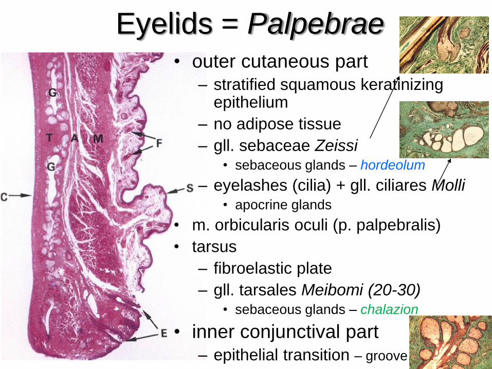
Eyelids = Palpebrae• outer cutaneous part
– stratified squamous keratinizingepithelium
– no adipose tissue
– gll. sebaceae Zeissi• sebaceous glands – hordeolum
– eyelashes (cilia) + gll. ciliares Molli• apocrine glands
• m. orbicularis oculi (p. palpebralis)
• tarsus
– fibroelastic plate
– gll. tarsales Meibomi (20-30)• sebaceous glands – chalazion
• inner conjunctival part– epithelial transition – groove
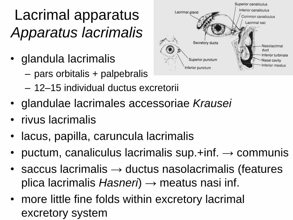
Lacrimal apparatus
Apparatus lacrimalis
• glandula lacrimalis
– pars orbitalis + palpebralis
– 12–15 individual ductus excretorii
• glandulae lacrimales accessoriae Krausei
• rivus lacrimalis
• lacus, papilla, caruncula lacrimalis
• puctum, canaliculus lacrimalis sup.+inf. → communis
• saccus lacrimalis → ductus nasolacrimalis (features
plica lacrimalis Hasneri) → meatus nasi inf.
• more little fine folds within excretory lacrimal
excretory system
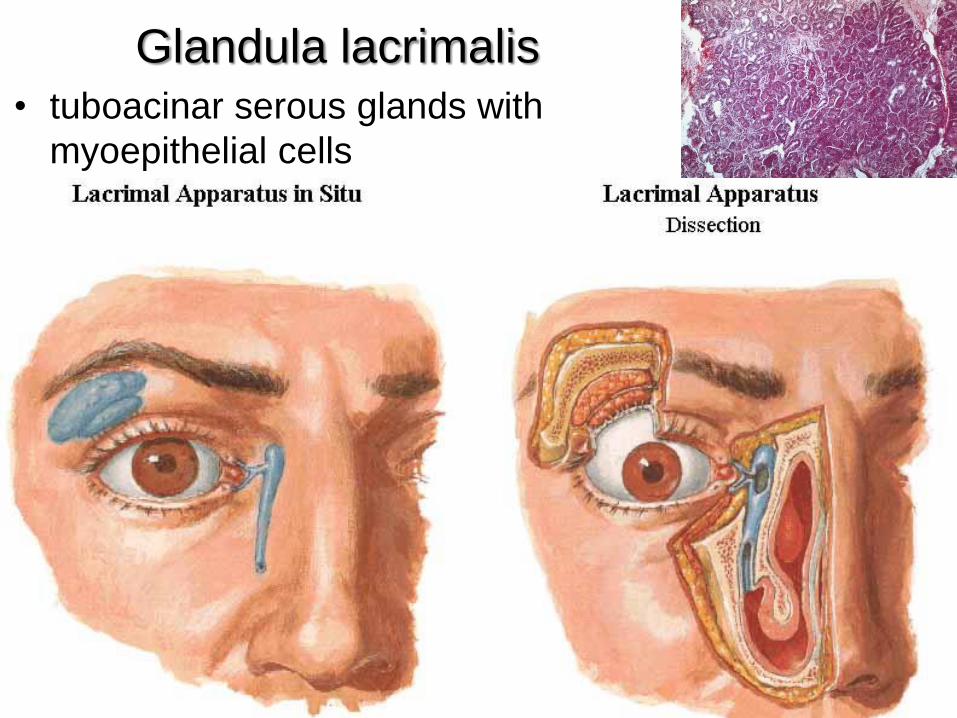
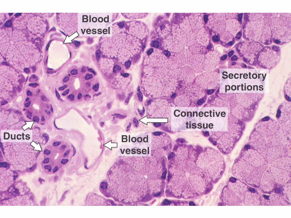
Glandula lacrimalis• tuboacinar serous glands with
myoepithelial cells
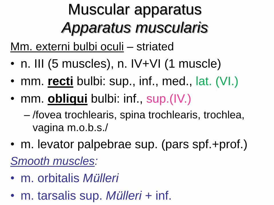
Muscular apparatus
Apparatus muscularisMm. externi bulbi oculi – striated
• n. III (5 muscles), n. IV+VI (1 muscle)
• mm. recti bulbi: sup., inf., med., lat. (VI.)
• mm. obliqui bulbi: inf., sup.(IV.)
– /fovea trochlearis, spina trochlearis, trochlea,
vagina m.o.b.s./
• m. levator palpebrae sup. (pars spf.+prof.)
Smooth muscles:
• m. orbitalis Mülleri
• m. tarsalis sup. Mülleri + inf.
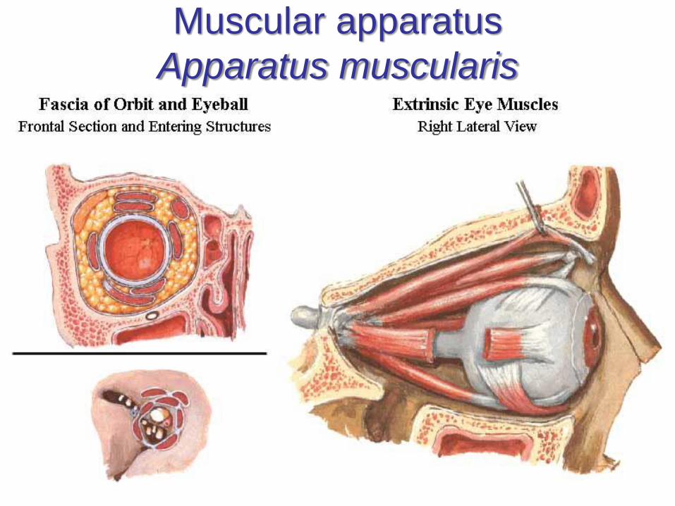
Muscular apparatus
Apparatus muscularis
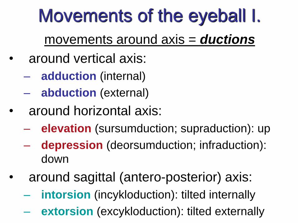
Movements of the eyeball I.
movements around axis = ductions
• around vertical axis:
– adduction (internal)
– abduction (external)
• around horizontal axis:
– elevation (sursumduction; supraduction): up
– depression (deorsumduction; infraduction):
down
• around sagittal (antero-posterior) axis:
– intorsion (incykloduction): tilted internally
– extorsion (excykloduction): tilted externally

Movements of the eye-ball II.paired movements (both eyes working together)
• simultaneous movement of both eyes in the same direction = version (conjugate movements)– dextroversion (to the right) + levoversion (to the left)
– supraversion (sursumversion) + infra/deorsumversion (up + down)
– dextro/levoelevation + dextro/levodepression (up/down and to side)
– dextro/levocykloversion (rotation to the right/left)
• simultaneous movement of both eyes in opposite directions = vergence (disconjugate movements), convergence = both eyes moving nasally or inward , divergence = both eyes moving temporally or outward
• strabismus; heterotropia; squint = one eye constantly is turned inward (“crossed-eye”), outward (“wall-eye”), upward, or downward.
http://www.tedmontgomery.com/the_eye/eom.html
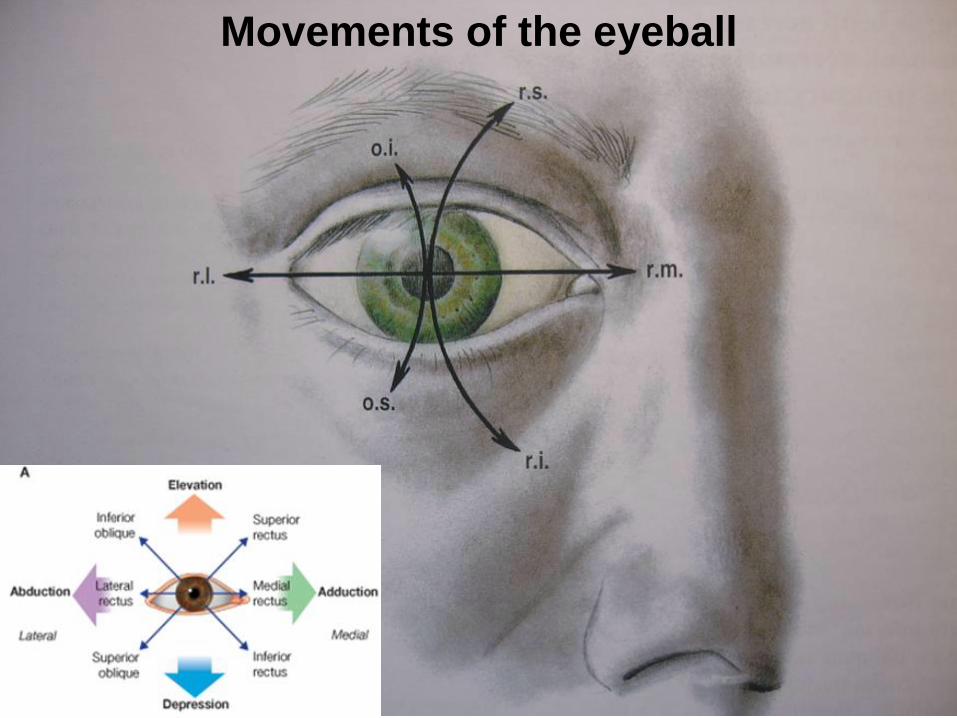
Movements of the eyeball
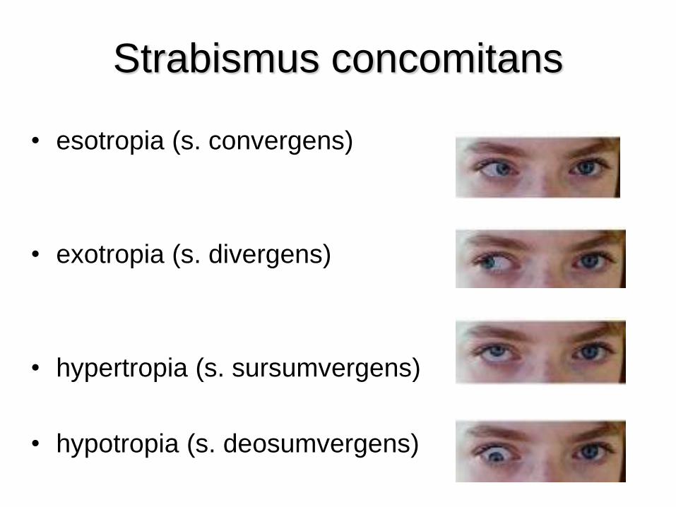
Strabismus concomitans
• esotropia (s. convergens)
• exotropia (s. divergens)
• hypertropia (s. sursumvergens)
• hypotropia (s. deosumvergens)
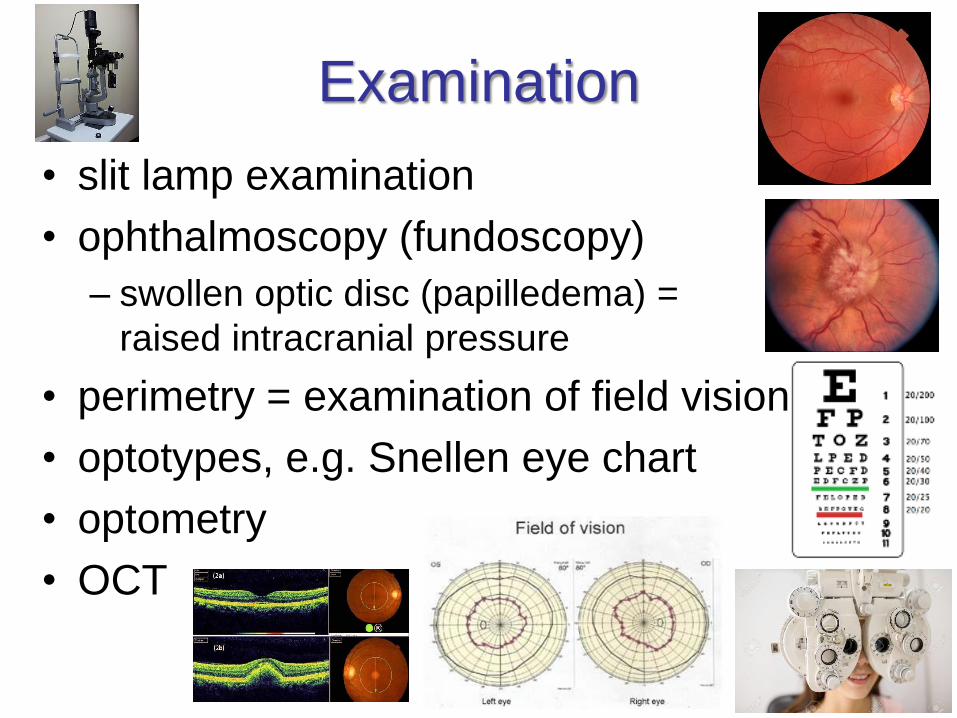
Examination
• slit lamp examination
• ophthalmoscopy (fundoscopy)
– swollen optic disc (papilledema) =
raised intracranial pressure
• perimetry = examination of field vision
• optotypes, e.g. Snellen eye chart
• optometry
• OCT
Symptoms and defects
• epiphora (excessive lacrimation)
• myopy – hypermetropy
• hypermetry (cerebellar disorder!)
• presbyopy
• hemeralopy
• amblyopy – functional disorder (e.g. in
squint)
• daltonism
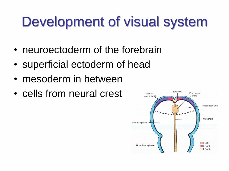
Development of visual system
• neuroectoderm of the forebrain
• superficial ectoderm of head
• mesoderm in between
• cells from neural crest
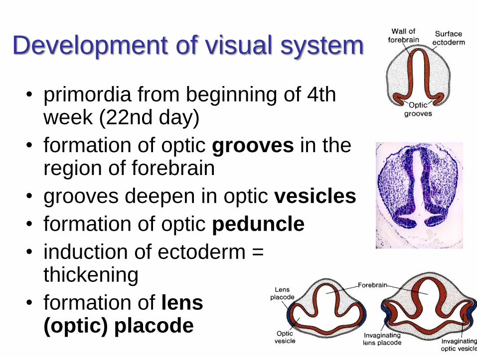
Development of visual system
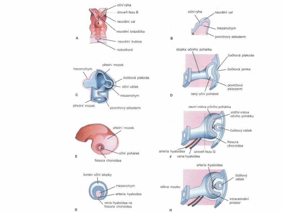
• primordia from beginning of 4th week (22nd day)
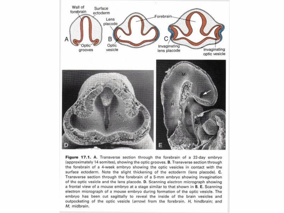
• formation of optic grooves in the region of forebrain
• grooves deepen in optic vesicles
• formation of optic peduncle
• induction of ectoderm = thickening
• formation of lens (optic) placode
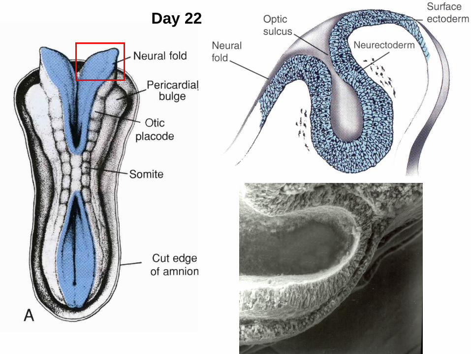
Day 22
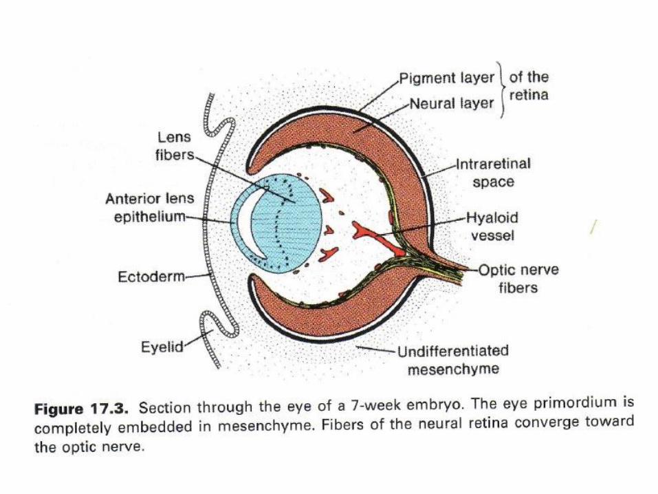
Development of visual system
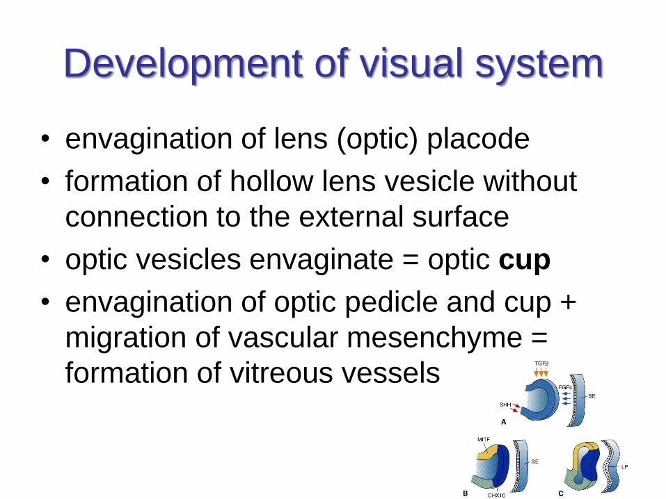
• envagination of lens (optic) placode
• formation of hollow lens vesicle without
connection to the external surface
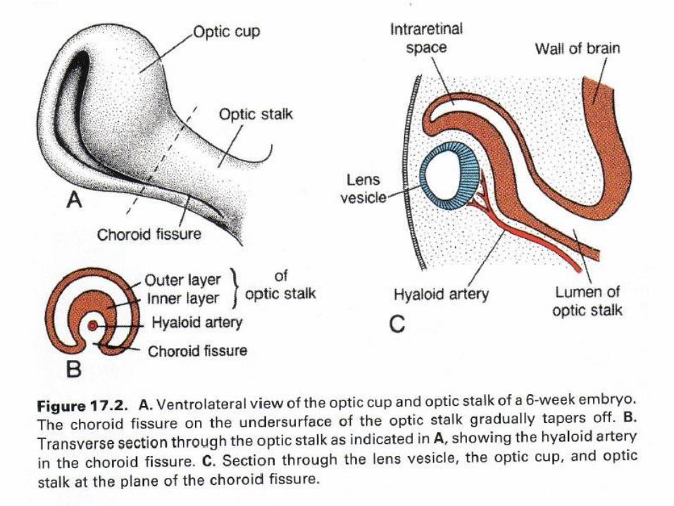
• optic vesicles envaginate = optic cup
• envagination of optic pedicle and cup +
migration of vascular mesenchyme =
formation of vitreous vessels
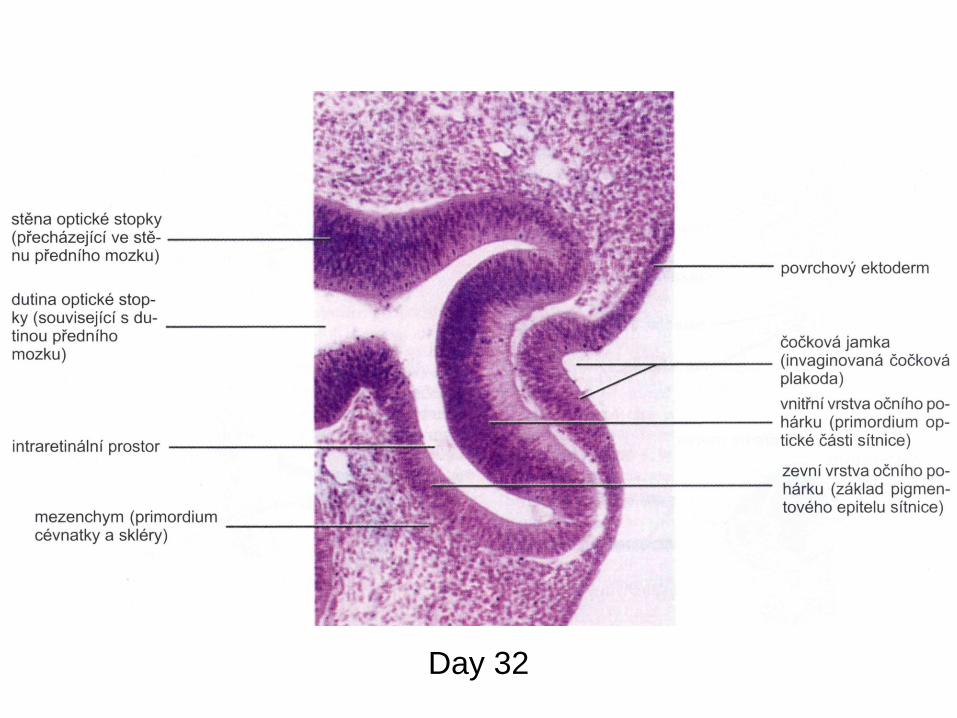
Day 32
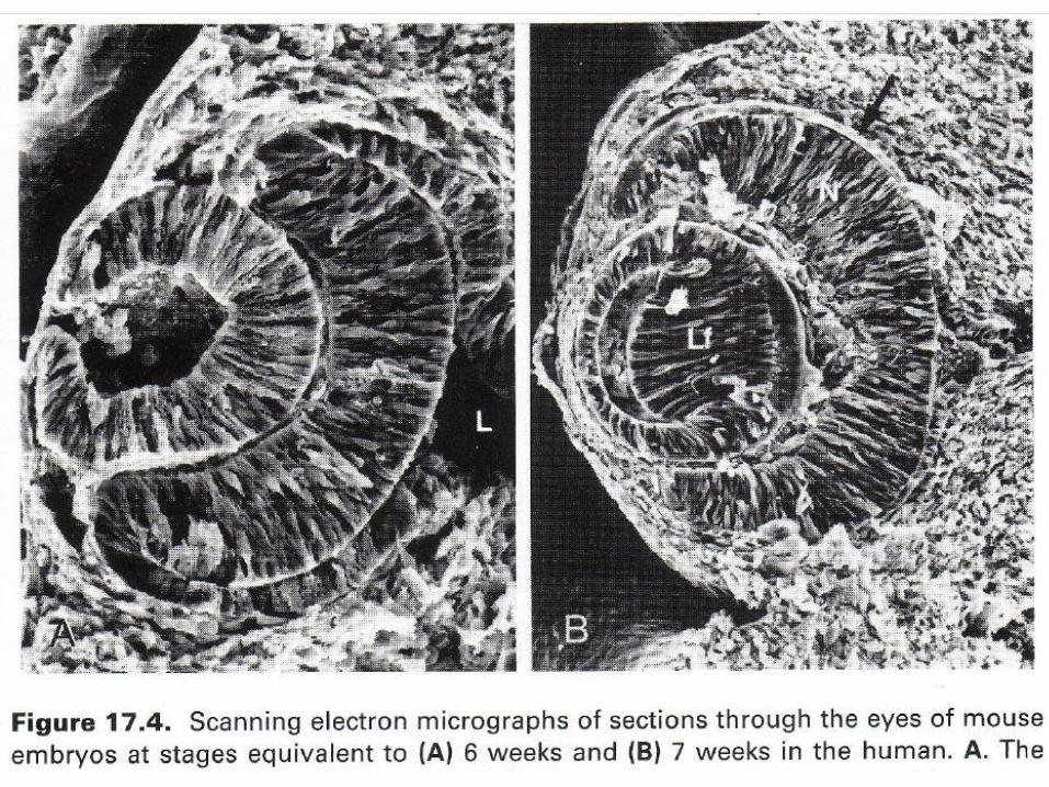
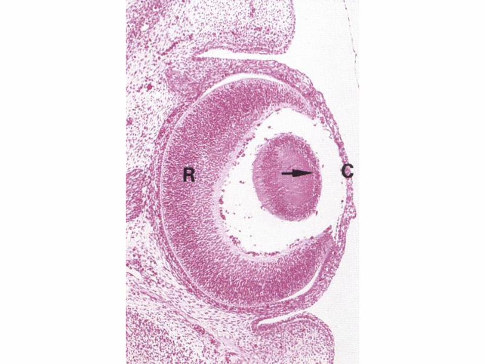
Development of retina
• origin from optic cup
• external layer – pigment epithelium
• internal layer – proliferates into pars
nervosa
• intraretinal space – successively fades out
• inversion of retina
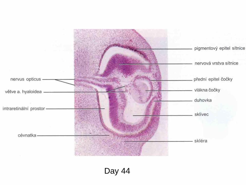
Day 44
Development of nervus opticus
• fibres from ganglionic cells growth through
the optic pedicle
• cavity of the optic pedicle fades out
• fissure (evagination) fades out
Development of visual systém
in motion
• https://www.youtube.com/watch?v=ghHDF
WlfpoQ
• https://www.youtube.com/watch?v=Xme8P
A6xv-M
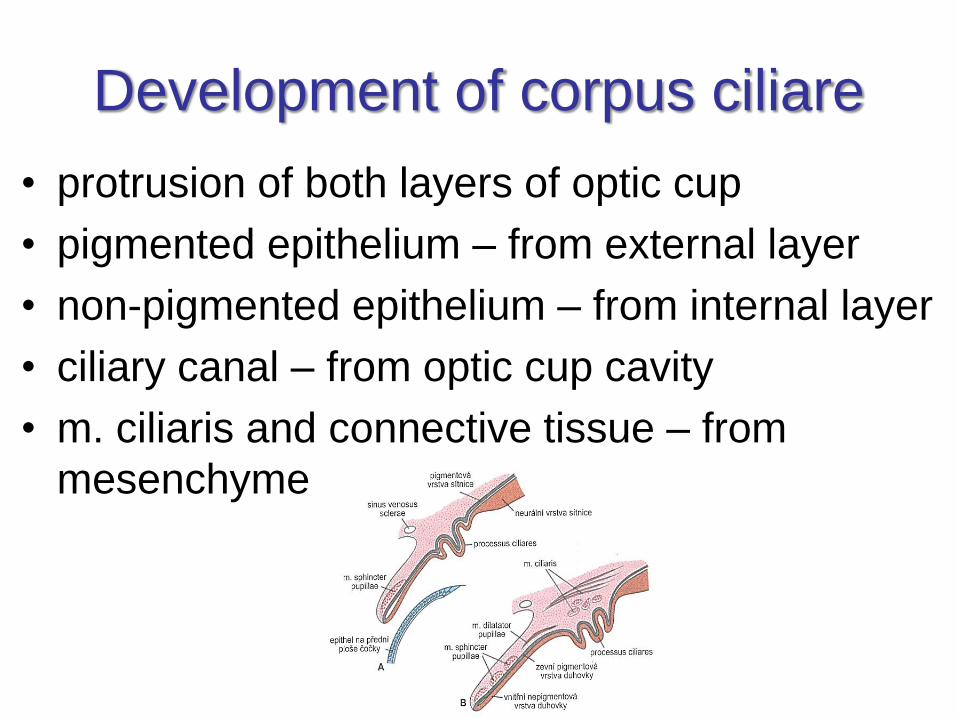
Development of corpus ciliare
• protrusion of both layers of optic cup
• pigmented epithelium – from external layer
• non-pigmented epithelium – from internal layer
• ciliary canal – from optic cup cavity
• m. ciliaris and connective tissue – from
mesenchyme
Development of iris
• margin of eye cup
• external layer → smooth muscles
• internal layer → pigmented epithelium
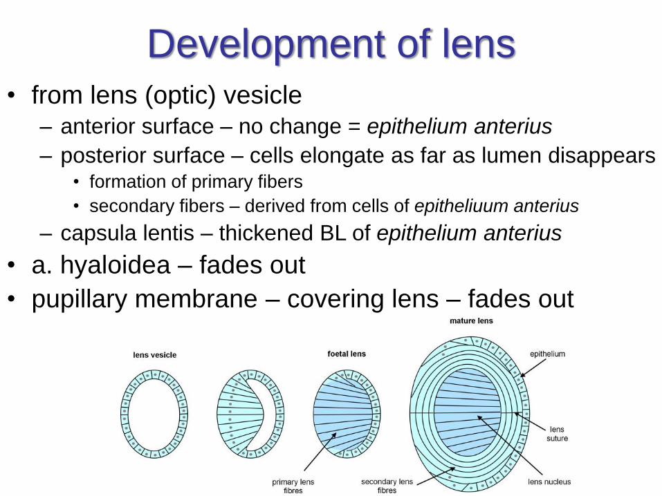
Development of lens• from lens (optic) vesicle
– anterior surface – no change = epithelium anterius
– posterior surface – cells elongate as far as lumen disappears• formation of primary fibers
• secondary fibers – derived from cells of epitheliuum anterius
– capsula lentis – thickened BL of epithelium anterius
• a. hyaloidea – fades out
• pupillary membrane – covering lens – fades out
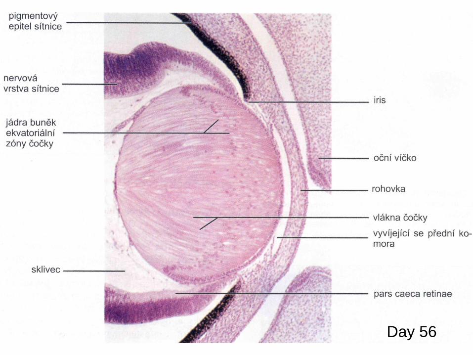
Day 56
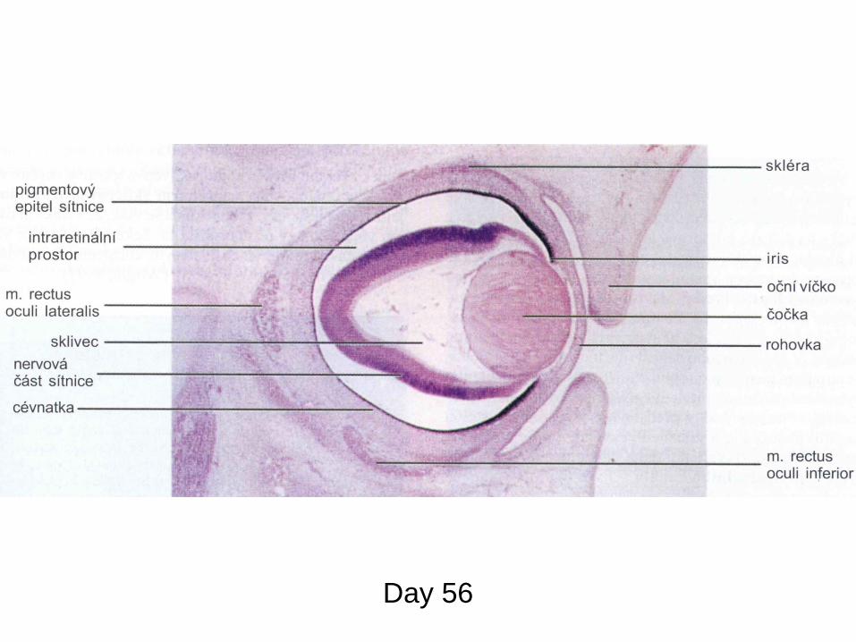
Day 56
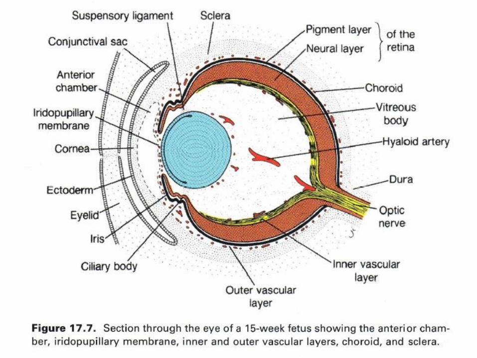
Development of camerae bulbi
• camera anterior
– fissure between primordial lens and cornea
• camera posterior
– fissure in optic cup on sides of lens vesicle
Development of cornea,
choroidea and sclera
• cornea
– superficial ectoderm
– mesenchyme
– cells of neural crest
• choroidea and sclera
– surrounding mesenchyme
Development of palpebrae
• 6th week: cutaneous folds across cornea
• 10th week: both folds fuse
• 28th week: folds separate again
• inbetween: conjunctiva adheres internally
• muscles: 2nd pharyngeal arch
• tarsus and glands from mesenchyme
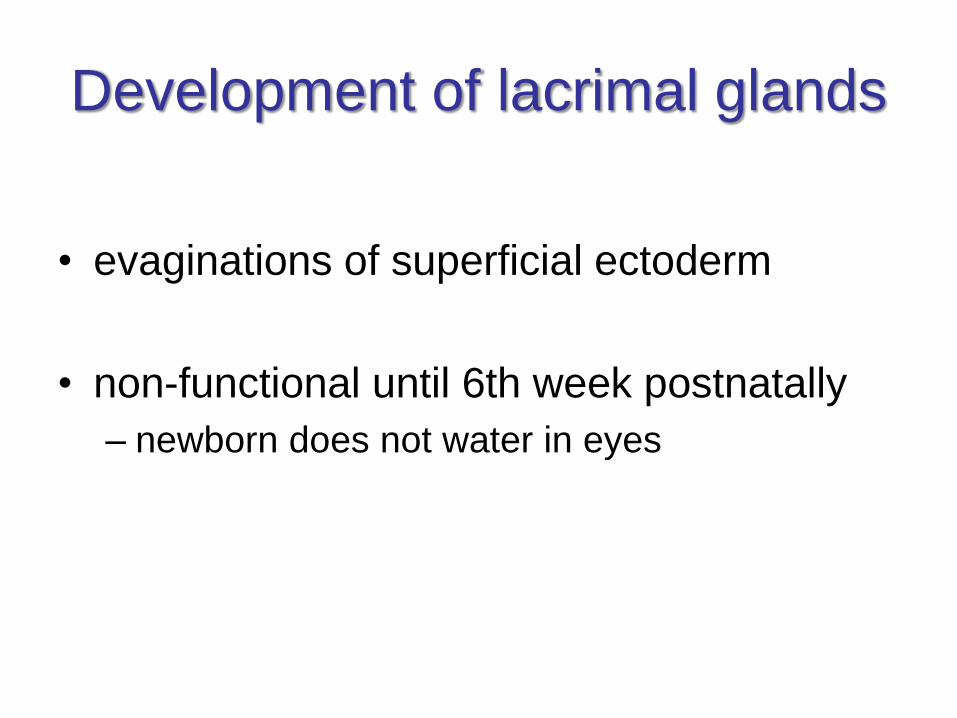
Development of lacrimal glands
• evaginations of superficial ectoderm
• non-functional until 6th week postnatally
– newborn does not water in eyes
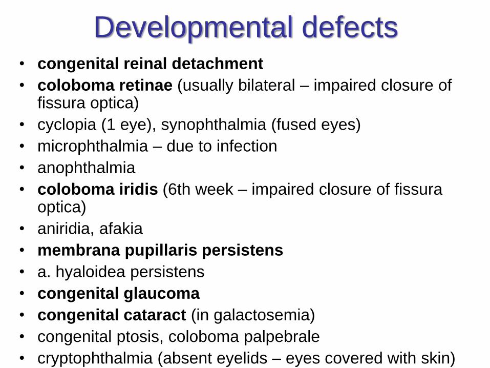
Developmental defects• congenital reinal detachment
• coloboma retinae (usually bilateral – impaired closure of fissura optica)
• cyclopia (1 eye), synophthalmia (fused eyes)
• microphthalmia – due to infection
• anophthalmia
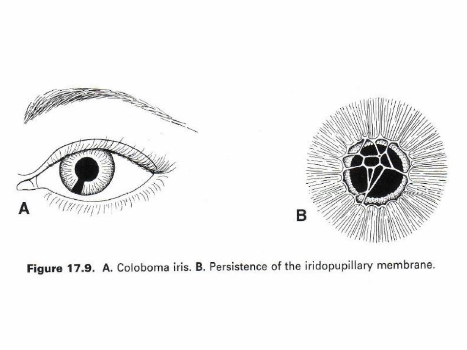
• coloboma iridis (6th week – impaired closure of fissuraoptica)
• aniridia, afakia
• membrana pupillaris persistens
• a. hyaloidea persistens
• congenital glaucoma
• congenital cataract (in galactosemia)
• congenital ptosis, coloboma palpebrale
• cryptophthalmia (absent eyelids – eyes covered with skin)

END
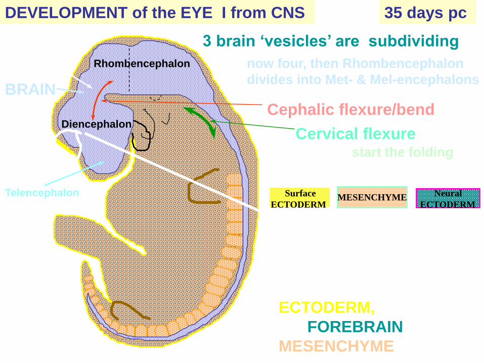
BRAIN
3 brain ‘vesicles’ are subdividing
Cephalic flexure/bend
35 days pcDEVELOPMENT of the EYE I from CNS
Telencephalon
Diencephalon
Mesencephalon
now four, then Rhombencephalon
divides into Met- & Mel-encephalons
Cervical flexure
Rhombencephalon
start the folding
Already before 35d pc, on
each side of the ‘head’,
interactions have started
between surface
ECTODERM, a bulge of
the FOREBRAIN & the
MESENCHYME
Surface
ECTODERMMESENCHYME RETINA
Neural
ECTODERM
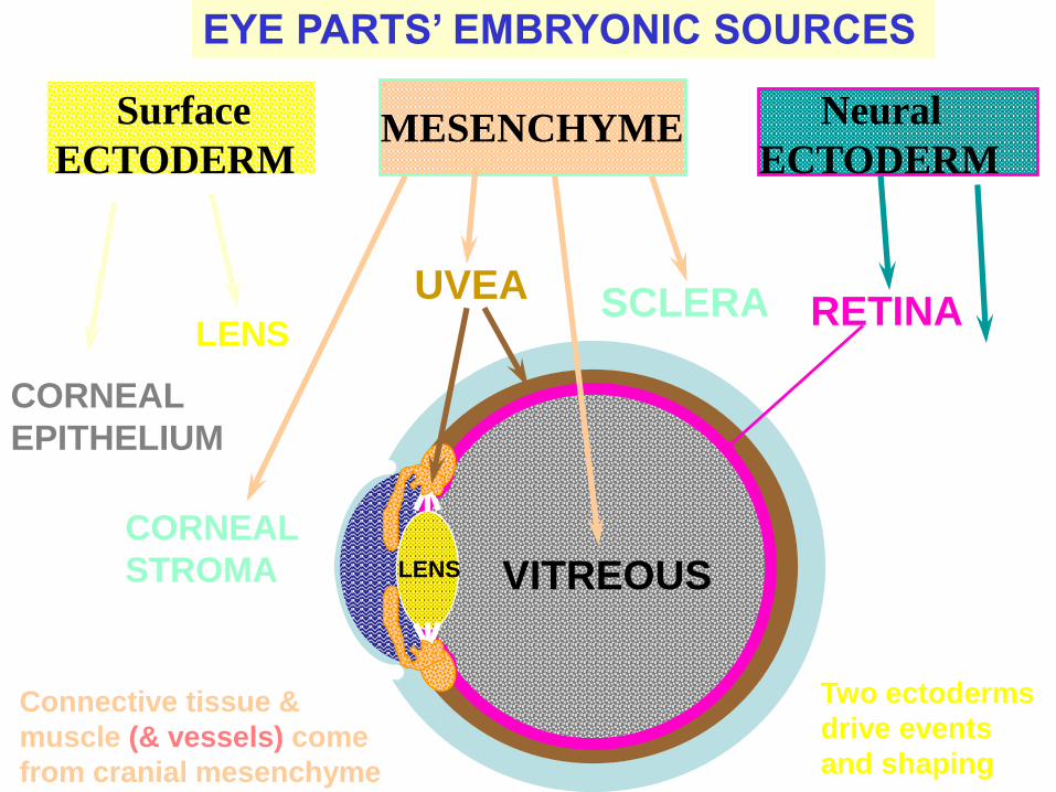
CORNEAL
EPITHELIUM
EYE PARTS’ EMBRYONIC SOURCES
UVEA
LENS
Connective tissue &
muscle (& vessels) come
from cranial mesenchyme
Surface
ECTODERMMESENCHYME RETINA
Neural
ECTODERM
SCLERALENS
CORNEAL
STROMA VITREOUS
RETINA
OPTIC
NERVE
Two ectoderms
drive events
and shaping
CORNEAL
EPITHELIUM
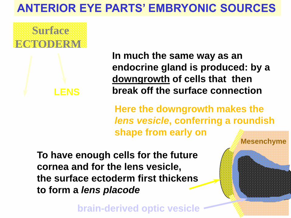
ANTERIOR EYE PARTS’ EMBRYONIC SOURCES
Surface
ECTODERM
LENS
How does a surface layer produce
two separate structures?
In much the same way as an
endocrine gland is produced: by a
downgrowth of cells that then
break off the surface connection
Here the downgrowth makes the
lens vesicle, conferring a roundish
shape from early on
To have enough cells for the future
cornea and for the lens vesicle,
the surface ectoderm first thickens
to form a lens placode
over the brain-derived optic vesicle
Mesenchyme
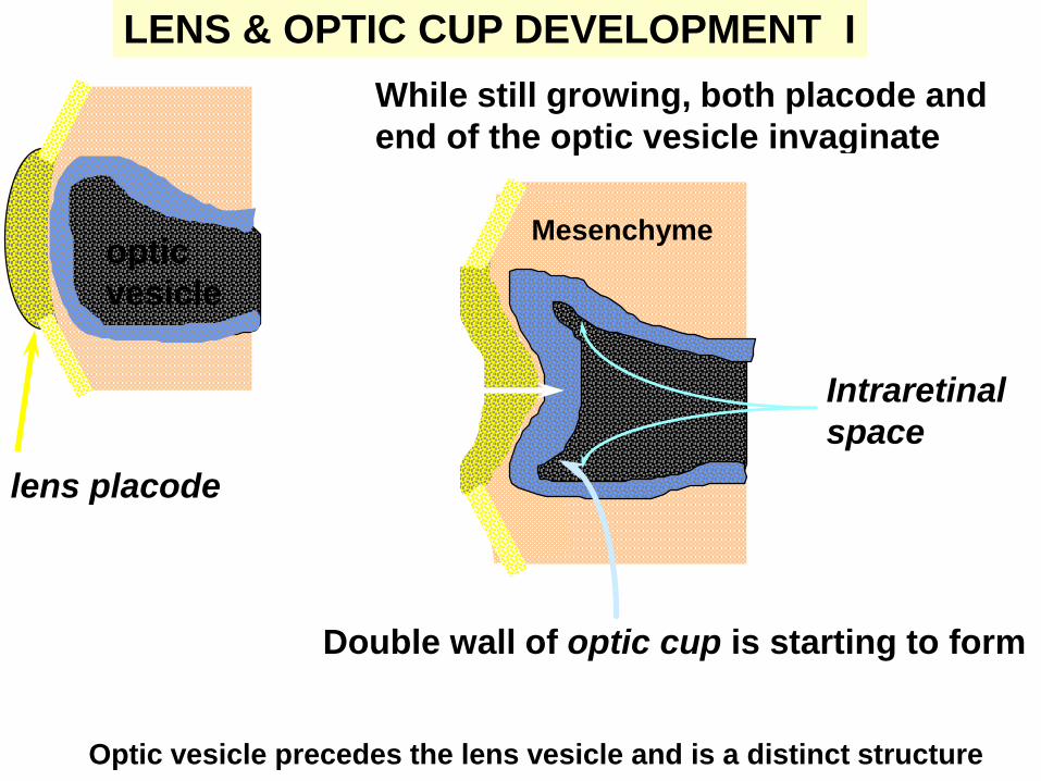
LENS & OPTIC CUP DEVELOPMENT I
While still growing, both placode and
end of the optic vesicle invaginate
lens placode
optic
vesicle
Double wall of optic cup is starting to form
Intraretinal
space
Mesenchyme
Optic vesicle precedes the lens vesicle and is a distinct structure
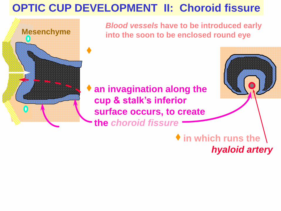
OPTIC CUP DEVELOPMENT II: Choroid fissure
Together with the invagination
centrally at the end of the optic
cup,
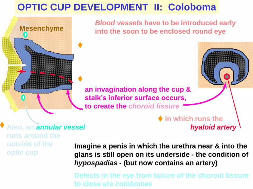
MesenchymeBlood vessels have to be introduced early
into the soon to be enclosed round eye
an invagination along the
cup & stalk’s inferior
surface occurs, to create
the choroid fissure
in which runs the
hyaloid artery
OPTIC CUP DEVELOPMENT II: Coloboma
Together with the invagination
centrally at the end of the optic
cup,
Blood vessels have to be introduced early
into the soon to be enclosed round eye
an invagination along the cup &
stalk’s inferior surface occurs,
to create the choroid fissure
in which runs the
hyaloid arteryAlso, an annular vessel
runs around the
outside of the
optic cup
Mesenchyme
Imagine a penis in which the urethra near & into the
glans is still open on its underside - the condition of
hypospadias - (but now contains an artery)
Defects in the eye from failure of the choroid fissure
to close are colobomas
Mesenchyme
Mesenchyme
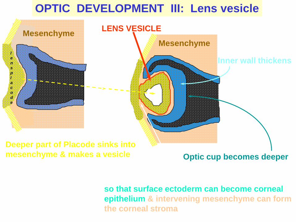
OPTIC DEVELOPMENT III: Lens vesicle
Deeper part of Placode sinks into
mesenchyme & makes a vesicle
Attachment to surface ectoderm will be broken
LENS VESICLE
Optic cup becomes deeper
Inner wall thickensl
e
n
s
p
l
a
c
o
d
e
so that surface ectoderm can become corneal
epithelium & intervening mesenchyme can form
the corneal stroma
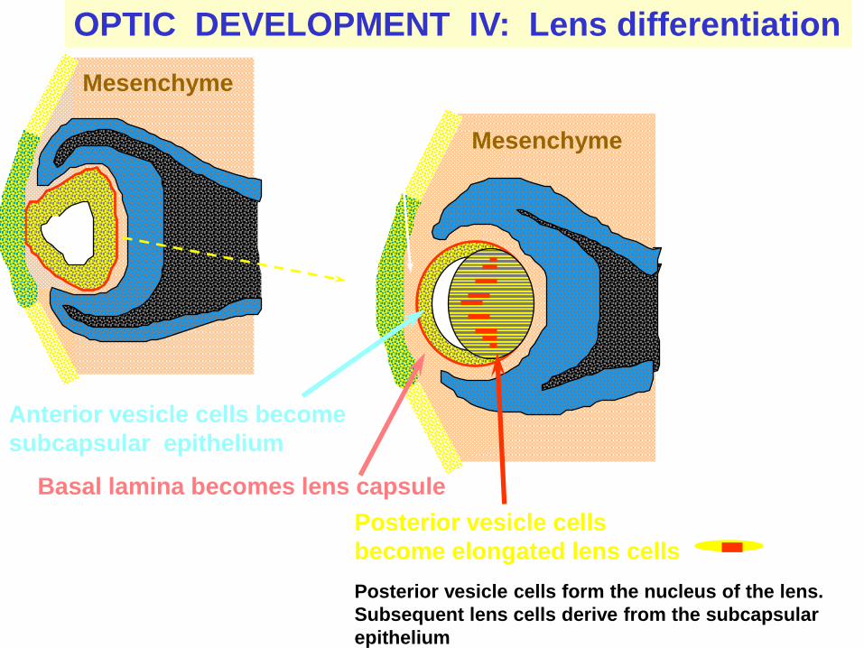
OPTIC DEVELOPMENT IV: Lens differentiation
Posterior vesicle cells
become elongated lens cells
Attachment to surface ectoderm lostMesenchyme
Anterior vesicle cells become
subcapsular epithelium
Mesenchyme
Basal lamina becomes lens capsule
Posterior vesicle cells form the nucleus of the lens.
Subsequent lens cells derive from the subcapsular
epithelium
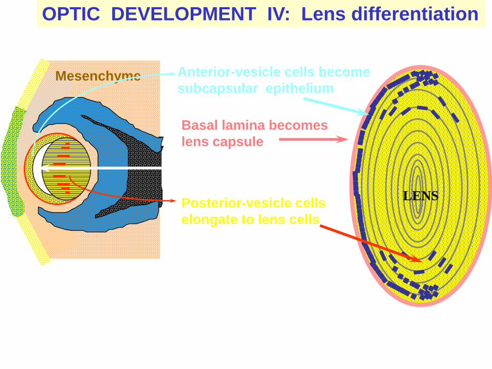
OPTIC DEVELOPMENT IV: Lens differentiation
Posterior-vesicle cells
elongate to lens cells
Anterior-vesicle cells become
subcapsular epitheliumMesenchyme
Basal lamina becomes
lens capsule
Lumen obliterated
LENS
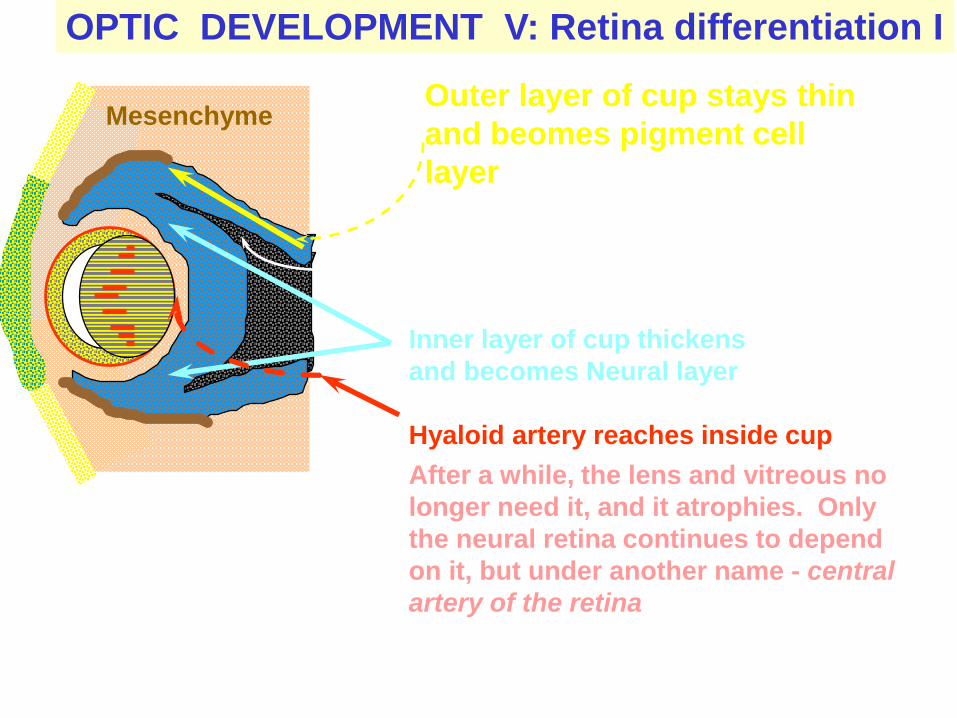
OPTIC DEVELOPMENT V: Retina differentiation I
Outer layer of cup stays thin
and beomes pigment cell
layer
Intra-retinal space occluded
Mesenchyme
Inner layer of cup thickens
and becomes Neural layer
Hyaloid artery reaches inside cup
After a while, the lens and vitreous no
longer need it, and it atrophies. Only
the neural retina continues to depend
on it, but under another name - central
artery of the retina
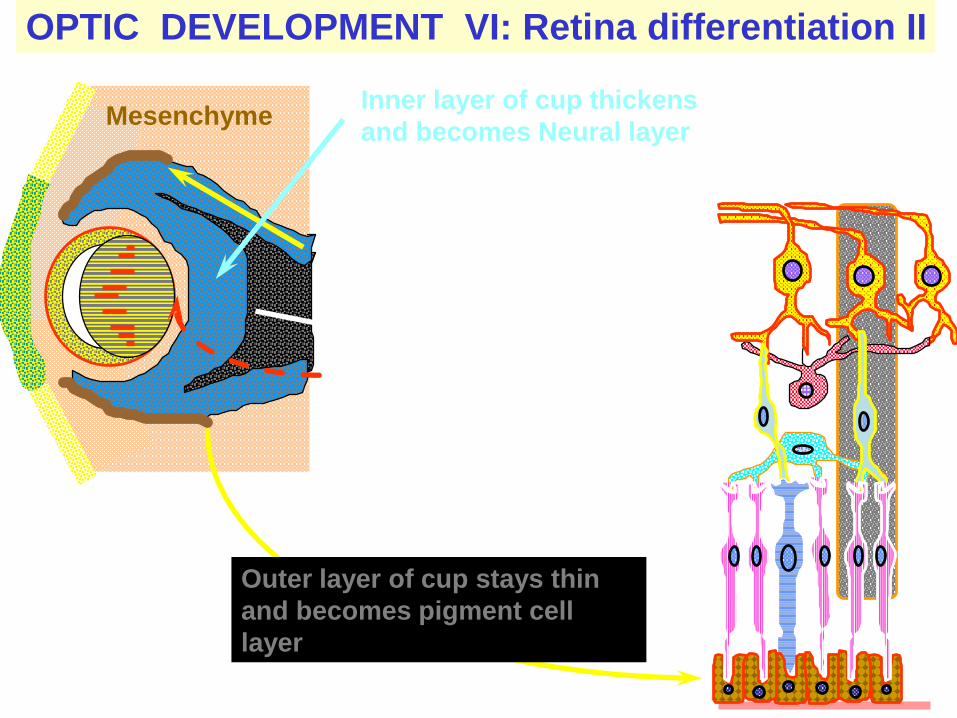
OPTIC DEVELOPMENT VI: Retina differentiation II
MesenchymeInner layer of cup thickens
and becomes Neural layer
Where cells multiply, form
layers and differentiate to
the several cell types of the
neural retina
Outer layer of cup stays thin
and becomes pigment cell
layer
Related Documents