Position Paper Tumour markers in colorectal cancer: European Group on Tumour Markers (EGTM) guidelines for clinical use M.J. Duffy a,b, *, A. van Dalen c , C. Haglund d , L. Hansson e , E. Holinski-Feder f , R. Klapdor g , R. Lamerz h , P. Peltomaki i , C. Sturgeon j , O. Topolcan k a Department of Pathology and Laboratory Medicine, Nuclear Medicine Laboratory, St Vincent’s University Hospital, Elm Park, Dublin 4, Ireland b School of Medicine and Medical Science, Conway Institute of Biomolecular and Biomedical Research, University College, Dublin 4, Ireland c Institute of Tumour Marker Oncology, Van Strijenstraat 44, 2801 TG Gouda, The Netherlands d Department of Surgery, Helsinki University Central Hospital, Helsinki, Finland e Department of Clinical Chemistry and Pharmacology, Akademiska Hospital, Uppsala, Sweden f Department of Medical Genetics, Ludwig Maxmilians University, Munich, Germany g Centre for Clinical and Experimental Tumour Diagnosis and Therapy, Hamburg, Germany h Klinikum Großhadern, Med. Klinik II, Ludwig Maximilians Universita ¨ t, Mu ¨ nchen, Germany i Department of Medical Genetics, University of Helsinki, Helsinki, Finland j Department of Clinical Biochemistry, Royal Infirmary of Edinburgh, Edinburgh, UK k Second Department of Internal Medicine, University Hospital, Pilsen, Czech Republic ARTICLE INFO Article history: Received 2 November 2006 Received in revised form 15 March 2007 Accepted 27 March 2007 Available online 18 May 2007 Keywords: Colorectal cancer CEA Guidelines Tumour markers EGTM ABSTRACT The aim of this article is to present updated guidelines for the use of serum, tissue and fae- cal markers in colorectal cancer (CRC). Lack of specificity and sensitivity preclude the use of all existing serum markers for the early detection of CRC. For patients with stage II or stage III CRC who may be candidates for either liver resection or systemic treatment should recurrence develop, CEA should be measured every 2–3 months for at least 3 years after diagnosis. Insufficient evidence exists to recommend routine use of tissue factors such as thymidylate synthase, microsatellite instability (MSI), p53, K-ras and deleted in colon cancer (DCC) for either determining prognosis or predicting response to therapy in patients with CRC. Microsatellite instability, however, may be used as a pre-screen for patients with suspected hereditary non-polyposis colorectal cancer. Faecal occult blood testing but not faecal DNA markers may be used to screen asymptomatic subjects 50 years or older for early CRC. Ó 2007 Elsevier Ltd. All rights reserved. 1. Introduction Colorectal cancer (CRC) is the third most common cancer worldwide with an estimated 1 million new cases and a half million deaths each year. 1 It is now clear that CRC results from the cumulative effects of sequential genetic alterations in proto-oncogenes, tumour suppressor genes and DNA repair genes (for review, see Ref. [2]). In sporadic CRC, these altera- 0959-8049/$ - see front matter Ó 2007 Elsevier Ltd. All rights reserved. doi:10.1016/j.ejca.2007.03.021 * Corresponding author: Address: Department of Pathology and Laboratory Medicine, Nuclear Medicine Laboratory, St Vincent’s University Hospital, Elm Park, Dublin 4, Ireland. Tel.: +353 1 2094378; fax: +353 1 2696018. E-mail address: [email protected] (M.J. Duffy). EUROPEAN JOURNAL OF CANCER 43 (2007) 1348 – 1360 available at www.sciencedirect.com journal homepage: www.ejconline.com

Welcome message from author
This document is posted to help you gain knowledge. Please leave a comment to let me know what you think about it! Share it to your friends and learn new things together.
Transcript
E U R O P E A N J O U R N A L O F C A N C E R 4 3 ( 2 0 0 7 ) 1 3 4 8 – 1 3 6 0
. sc iencedi rec t .com
ava i lab le at wwwjournal homepage: www.ejconl ine.com
Position Paper
Tumour markers in colorectal cancer: European Group onTumour Markers (EGTM) guidelines for clinical use
M.J. Duffya,b,*, A. van Dalenc, C. Haglundd, L. Hanssone, E. Holinski-Federf, R. Klapdorg,R. Lamerzh, P. Peltomakii, C. Sturgeonj, O. Topolcank
aDepartment of Pathology and Laboratory Medicine, Nuclear Medicine Laboratory, St Vincent’s University Hospital, Elm Park, Dublin 4,
IrelandbSchool of Medicine and Medical Science, Conway Institute of Biomolecular and Biomedical Research, University College, Dublin 4, IrelandcInstitute of Tumour Marker Oncology, Van Strijenstraat 44, 2801 TG Gouda, The NetherlandsdDepartment of Surgery, Helsinki University Central Hospital, Helsinki, FinlandeDepartment of Clinical Chemistry and Pharmacology, Akademiska Hospital, Uppsala, SwedenfDepartment of Medical Genetics, Ludwig Maxmilians University, Munich, GermanygCentre for Clinical and Experimental Tumour Diagnosis and Therapy, Hamburg, GermanyhKlinikum Großhadern, Med. Klinik II, Ludwig Maximilians Universitat, Munchen, GermanyiDepartment of Medical Genetics, University of Helsinki, Helsinki, FinlandjDepartment of Clinical Biochemistry, Royal Infirmary of Edinburgh, Edinburgh, UKkSecond Department of Internal Medicine, University Hospital, Pilsen, Czech Republic
A R T I C L E I N F O
Article history:
Received 2 November 2006
Received in revised form 15 March
2007
Accepted 27 March 2007
Available online 18 May 2007
Keywords:
Colorectal cancer
CEA
Guidelines
Tumour markers
EGTM
0959-8049/$ - see front matter � 2007 Elsevidoi:10.1016/j.ejca.2007.03.021
* Corresponding author: Address: DepartmUniversity Hospital, Elm Park, Dublin 4, Irela
E-mail address: [email protected] (M
A B S T R A C T
The aim of this article is to present updated guidelines for the use of serum, tissue and fae-
cal markers in colorectal cancer (CRC). Lack of specificity and sensitivity preclude the use of
all existing serum markers for the early detection of CRC. For patients with stage II or stage
III CRC who may be candidates for either liver resection or systemic treatment should
recurrence develop, CEA should be measured every 2–3 months for at least 3 years after
diagnosis. Insufficient evidence exists to recommend routine use of tissue factors such
as thymidylate synthase, microsatellite instability (MSI), p53, K-ras and deleted in colon
cancer (DCC) for either determining prognosis or predicting response to therapy in patients
with CRC. Microsatellite instability, however, may be used as a pre-screen for patients with
suspected hereditary non-polyposis colorectal cancer. Faecal occult blood testing but not
faecal DNA markers may be used to screen asymptomatic subjects 50 years or older for
early CRC.
� 2007 Elsevier Ltd. All rights reserved.
1. Introduction
Colorectal cancer (CRC) is the third most common cancer
worldwide with an estimated 1 million new cases and a half
er Ltd. All rights reserved
ent of Pathology and Lnd. Tel.: +353 1 2094378;.J. Duffy).
million deaths each year.1 It is now clear that CRC results
from the cumulative effects of sequential genetic alterations
in proto-oncogenes, tumour suppressor genes and DNA repair
genes (for review, see Ref. [2]). In sporadic CRC, these altera-
.
aboratory Medicine, Nuclear Medicine Laboratory, St Vincent’sfax: +353 1 2696018.
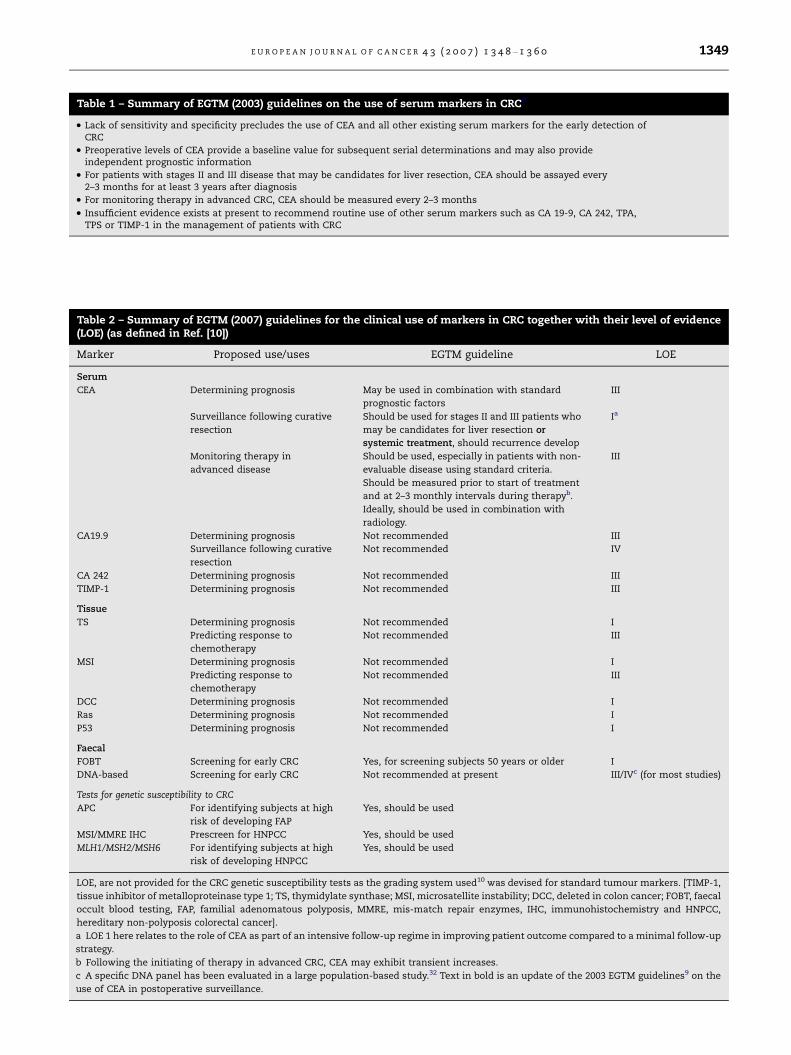
Table 1 – Summary of EGTM (2003) guidelines on the use of serum markers in CRC9
• Lack of sensitivity and specificity precludes the use of CEA and all other existing serum markers for the early detection ofCRC
• Preoperative levels of CEA provide a baseline value for subsequent serial determinations and may also provideindependent prognostic information
• For patients with stages II and III disease that may be candidates for liver resection, CEA should be assayed every2–3 months for at least 3 years after diagnosis
• For monitoring therapy in advanced CRC, CEA should be measured every 2–3 months
• Insufficient evidence exists at present to recommend routine use of other serum markers such as CA 19-9, CA 242, TPA,TPS or TIMP-1 in the management of patients with CRC
Table 2 – Summary of EGTM (2007) guidelines for the clinical use of markers in CRC together with their level of evidence(LOE) (as defined in Ref. [10])
Marker Proposed use/uses EGTM guideline LOE
Serum
CEA Determining prognosis May be used in combination with standard
prognostic factors
III
Surveillance following curative
resection
Should be used for stages II and III patients who
may be candidates for liver resection or
systemic treatment, should recurrence develop
Ia
Monitoring therapy in
advanced disease
Should be used, especially in patients with non-
evaluable disease using standard criteria.
Should be measured prior to start of treatment
and at 2–3 monthly intervals during therapyb.
Ideally, should be used in combination with
radiology.
III
CA19.9 Determining prognosis Not recommended III
Surveillance following curative
resection
Not recommended IV
CA 242 Determining prognosis Not recommended III
TIMP-1 Determining prognosis Not recommended III
Tissue
TS Determining prognosis Not recommended I
Predicting response to
chemotherapy
Not recommended III
MSI Determining prognosis Not recommended I
Predicting response to
chemotherapy
Not recommended III
DCC Determining prognosis Not recommended I
Ras Determining prognosis Not recommended I
P53 Determining prognosis Not recommended I
Faecal
FOBT Screening for early CRC Yes, for screening subjects 50 years or older I
DNA-based Screening for early CRC Not recommended at present III/IVc (for most studies)
Tests for genetic susceptibility to CRC
APC For identifying subjects at high
risk of developing FAP
Yes, should be used
MSI/MMRE IHC Prescreen for HNPCC Yes, should be used
MLH1/MSH2/MSH6 For identifying subjects at high
risk of developing HNPCC
Yes, should be used
LOE, are not provided for the CRC genetic susceptibility tests as the grading system used10 was devised for standard tumour markers. [TIMP-1,
tissue inhibitor of metalloproteinase type 1; TS, thymidylate synthase; MSI, microsatellite instability; DCC, deleted in colon cancer; FOBT, faecal
occult blood testing, FAP, familial adenomatous polyposis, MMRE, mis-match repair enzymes, IHC, immunohistochemistry and HNPCC,
hereditary non-polyposis colorectal cancer].
a LOE 1 here relates to the role of CEA as part of an intensive follow-up regime in improving patient outcome compared to a minimal follow-up
strategy.
b Following the initiating of therapy in advanced CRC, CEA may exhibit transient increases.
c A specific DNA panel has been evaluated in a large population-based study.32 Text in bold is an update of the 2003 EGTM guidelines9 on the
use of CEA in postoperative surveillance.
E U R O P E A N J O U R N A L O F C A N C E R 4 3 ( 2 0 0 7 ) 1 3 4 8 – 1 3 6 0 1349
1350 E U R O P E A N J O U R N A L O F C A N C E R 4 3 ( 2 0 0 7 ) 1 3 4 8 – 1 3 6 0
tions are acquired, and are likely to be caused by exogenous
and endogenous carcinogens. In contrast, in cancer syn-
dromes such as familial adenomatous polyposis (FAP) and
hereditary non-polyposis CRC (HNPCC), critical genetic altera-
tions that predispose to malignancy are inherited.3 For exam-
ple in FAP, a germline mutation in the APC gene which occurs
in every cell predisposes to adenomatous polyps, while in
HNPCC, mutations in DNA repair genes result in a more rapid
accumulation of genetic alterations which increases the risk
of polyp formation.
In recent years a multiplicity of markers have been pro-
posed for CRC (for review, see Refs. [4–8]). These markers
can be measured in serum, tissue or stools. In 2003, the Euro-
pean Group on Tumour Markers (EGTM) published guidelines
on the use of tumour markers in CRC9 (see Table 1 for a sum-
mary). These guidelines focussed almost exclusively on ser-
um markers, especially CEA. The aims of this article are to
present guidelines on tissue and faecal markers as well as
to update the previous EGTM guidelines on serum markers.
We also summarise existing published guidelines on genetic
testing for inherited susceptibility to CRC. A summary of the
updated EGTM guidelines together with the level of evi-
dence10 for their clinical application is outlined in Table 2.
These guidelines should be particularly helpful to sur-
geons, physicians and nurses involved in the management
of patients with CRC and to laboratory personnel undertaking
measurement of tumour markers. Their adoption is of course
voluntary and the ultimate decision regarding use of any mar-
ker should be made by the treating clinician, i.e. the guide-
lines are intended to aid rather than replace clinical
judgement.
2. Serum markers
2.1. CEA in postoperative surveillance
Although the oldest, CEA remains the most widely used ser-
um marker in patients with CRC. EGTM guidelines for the
use of CEA in CRC were previously published in 2003 and
are summarised in Table 1. The main use of CEA in CRC is
in surveillance following curative resection for primary can-
cer. Five independent meta-analyses have compared outcome
in patients undergoing intensive follow-up versus minimal or
no follow-up.11–15 The first 2 of these 2 studies11,12 included
both non-randomised and randomised trials, whereas the
other 313–15 included only randomised trials. Although the fre-
quency and modalities of screening used in the individual
studies varied, all five meta-analyses concluded that use of
an intensive follow-up regime resulted in a modest but statis-
tically significant improvement in outcome compared with a
minimal follow-up strategy.
The Cochrane Review15 concluded that there was an overall
survival benefit at 5 years of follow-up for patients undergoing
more intensive surveillance (OR = 0.67, 95% confidence inter-
val, 0.53–0.84). The absolute numbers of recurrences however
were similar in the 2 groups. No data were presented on quality
of life, harm or cost-effectiveness of the intensive follow-up.
Two of these meta-analyses investigated the specific con-
tribution of CEA to the improved outcome. In the first of
these, Bruinvels et al.11 concluded that intensive follow-up
was associated with an improved outcome, but only if regular
CEA determinations were carried out. Similarly, Figueredo
et al.14 found that trials using serial CEA measurements had
a significant impact on survival, whereas those not using
CEA failed to impact on outcome. It should be stated that
compared to other follow-up tests for CRC patients (e.g. radi-
ology and endoscopy), measurement of CEA is relatively inex-
pensive and causes minimal inconvenience for patients.
2.2. Early detection of metastatic disease in candidates forliver resection
One of the main aims of intensive follow-up is the early
detection of resectable recurrences and metastases, espe-
cially liver metastasis. Twenty five to 50% of patients undergo-
ing resection for primary CRC develop liver metastasis within
5 years of diagnosis.15 Hepatic resection offers the only poten-
tial curative therapy for these patients. Curative resection,
however, is possible in less than 25% of patients with metas-
tasis confined to liver. Nevertheless, the 5-year survival for
these patients is approximately 30% with about two-third of
these being disease-free.16 This 5-year survival rate is in strik-
ing contrast to historical cases of unresected patients in
which the median survival was 6–9 months with few surviv-
ing for 5 years.17
Because of the relative success of surgery for treating CRC
metastases to liver, previous guidelines recommended that
for stages II and III CRC patients who are suitable candidates
for liver resection, CEA should be determined every 2–3
months for at least the first 3 years after diagnosis.9,18
2.3. Early detection of metastatic disease in candidates forchemotherapy
While surgical resection offers the best prospect of long-term
survival in patients with liver metastases from CRC, major
progress is being made in the treatment of metastatic CRC
with chemotherapy. Until relatively recently, the main form
of systemic therapy for advanced CRC was 5-fluorouracil (5-
FU) and leucovorin. This treatment reduced tumour size by
at least 50% in about 20% of patients and prolonged median
survival from about 6 to 11 months.19 The addition of either
irinotecan or oxaliplatin to 5-FU and leucovorin increased
median survival to about 15 months.20 Median survival in-
creased to about 20 months if all three cytotoxic agents were
used at some point in treatment or if combined cytotoxic and
antibody (e.g. bevacizumab or cetuximab) therapy was
administered.20 Thus, patients with advanced CRC treated
with systemic therapy may now survive approximately twice
as long as they did 10 years ago.20
As a result of these developments with systemic therapy,
the EGTM Panel has updated its guidelines on the use of
CEA in postoperative surveillance. The new guidelines state
that for stages II and III CRC patients, CEA should be mea-
sured every 2–3 months for at least 3 years, not only for pa-
tients who are suitable candidates for liver resection, but
also for patients who are candidates for receiving systemic
therapy. This inclusion of systemic therapy is in line with
the recently updated guidelines from the American Society
of Clinical Oncology.21 The updated ASCO guidelines also
E U R O P E A N J O U R N A L O F C A N C E R 4 3 ( 2 0 0 7 ) 1 3 4 8 – 1 3 6 0 1351
state that an elevated CEA, if confirmed following retesting,
requires further evaluation for the presence of metastasis,
but does not justify initiation of adjuvant therapy or systemic
treatment for presumed metastatic disease. The EGTM Panel
supports this statement. Because of the low risk of recurrence
in patients with stage 1 CRC, regular surveillance following
surgery may not be necessary.
2.4. Other serum markers in CRC
EGTM guidelines on the use of other serum markers in CRC
are as previously published.9
3. Stool-based markers
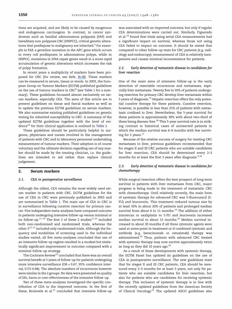
3.1. Faecal occult blood testing and screening for CRC
Faecal occult blood testing (FOBT) is the most widely used
screening modality for CRC.22 Two main types of FOBT exist,
i.e. the guaiac test which is based on the peroxidase-like activ-
ity (i.e. pseudoperoxidase) of haem in haemoglobin and the
immunochemical test which detects the globin moiety in hae-
moglobin. Of these 2, the guaiac test has been the more widely
Table 3 – Some advantages and disadvantages of FOBT inscreening for CRC
Advantages
• Potentially examines the entire colorectal tract
• Non-invasive
• Requires no patient preparation (unlikeendoscopic investigations)
• Simple and affordable
• Can be carried out in privacy of home
• Most extensively validated screening test for CRC
Disadvantages
• Low sensitivity for both adenomas (�10%) and CRCs(40–85%)
• Low specificity for both CRCs and adenomas (90–98%)
• Ingestion of certain foods (red meats, fruits and vegetables)and medicines (non-steroidal anti-inflammatory drugs)can yield false-positive results with the guaiac-based assaya
• Multiple stool samples are necessary
• Must be performed annually to increase chances of detectingintermittent bleeding
a These limitations do not apply to the immunochemical FOBT.
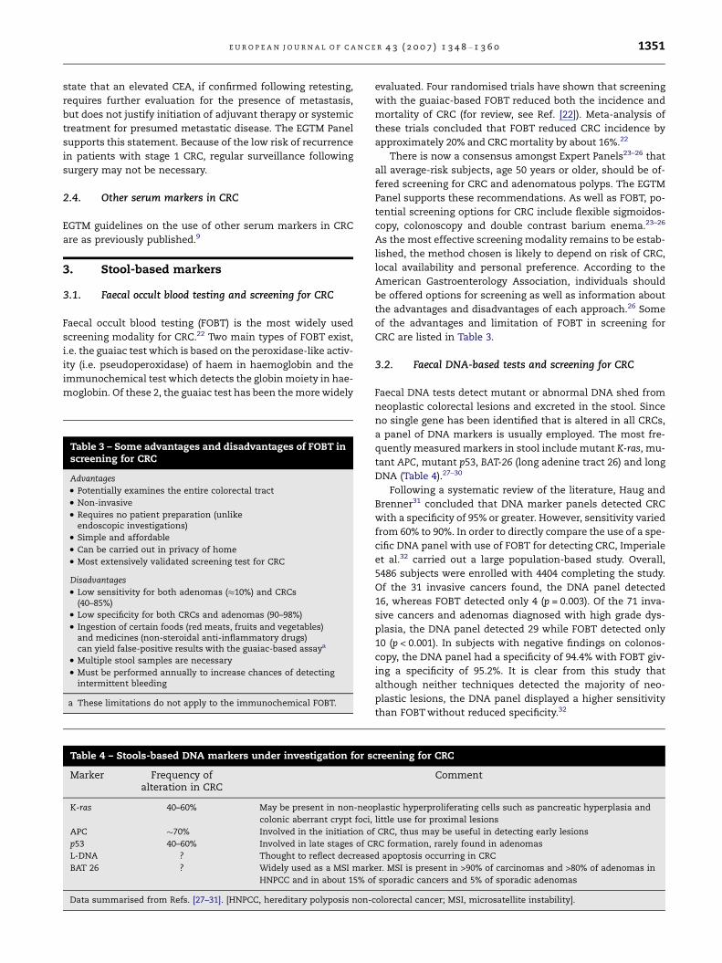
Table 4 – Stools-based DNA markers under investigation for s
Marker Frequency ofalteration in CRC
K-ras 40–60% May be present in non-neo
colonic aberrant crypt foci,
APC �70% Involved in the initiation o
p53 40–60% Involved in late stages of C
L-DNA ? Thought to reflect decrease
BAT 26 ? Widely used as a MSI mark
HNPCC and in about 15% o
Data summarised from Refs. [27–31]. [HNPCC, hereditary polyposis non-
evaluated. Four randomised trials have shown that screening
with the guaiac-based FOBT reduced both the incidence and
mortality of CRC (for review, see Ref. [22]). Meta-analysis of
these trials concluded that FOBT reduced CRC incidence by
approximately 20% and CRC mortality by about 16%.22
There is now a consensus amongst Expert Panels23–26 that
all average-risk subjects, age 50 years or older, should be of-
fered screening for CRC and adenomatous polyps. The EGTM
Panel supports these recommendations. As well as FOBT, po-
tential screening options for CRC include flexible sigmoidos-
copy, colonoscopy and double contrast barium enema.23–26
As the most effective screening modality remains to be estab-
lished, the method chosen is likely to depend on risk of CRC,
local availability and personal preference. According to the
American Gastroenterology Association, individuals should
be offered options for screening as well as information about
the advantages and disadvantages of each approach.26 Some
of the advantages and limitation of FOBT in screening for
CRC are listed in Table 3.
3.2. Faecal DNA-based tests and screening for CRC
Faecal DNA tests detect mutant or abnormal DNA shed from
neoplastic colorectal lesions and excreted in the stool. Since
no single gene has been identified that is altered in all CRCs,
a panel of DNA markers is usually employed. The most fre-
quently measured markers in stool include mutant K-ras, mu-
tant APC, mutant p53, BAT-26 (long adenine tract 26) and long
DNA (Table 4).27–30
Following a systematic review of the literature, Haug and
Brenner31 concluded that DNA marker panels detected CRC
with a specificity of 95% or greater. However, sensitivity varied
from 60% to 90%. In order to directly compare the use of a spe-
cific DNA panel with use of FOBT for detecting CRC, Imperiale
et al.32 carried out a large population-based study. Overall,
5486 subjects were enrolled with 4404 completing the study.
Of the 31 invasive cancers found, the DNA panel detected
16, whereas FOBT detected only 4 (p = 0.003). Of the 71 inva-
sive cancers and adenomas diagnosed with high grade dys-
plasia, the DNA panel detected 29 while FOBT detected only
10 (p < 0.001). In subjects with negative findings on colonos-
copy, the DNA panel had a specificity of 94.4% with FOBT giv-
ing a specificity of 95.2%. It is clear from this study that
although neither techniques detected the majority of neo-
plastic lesions, the DNA panel displayed a higher sensitivity
than FOBT without reduced specificity.32
creening for CRC
Comment
plastic hyperproliferating cells such as pancreatic hyperplasia and
little use for proximal lesions
f CRC, thus may be useful in detecting early lesions
RC formation, rarely found in adenomas
d apoptosis occurring in CRC
er. MSI is present in >90% of carcinomas and >80% of adenomas in
f sporadic cancers and 5% of sporadic adenomas
colorectal cancer; MSI, microsatellite instability].
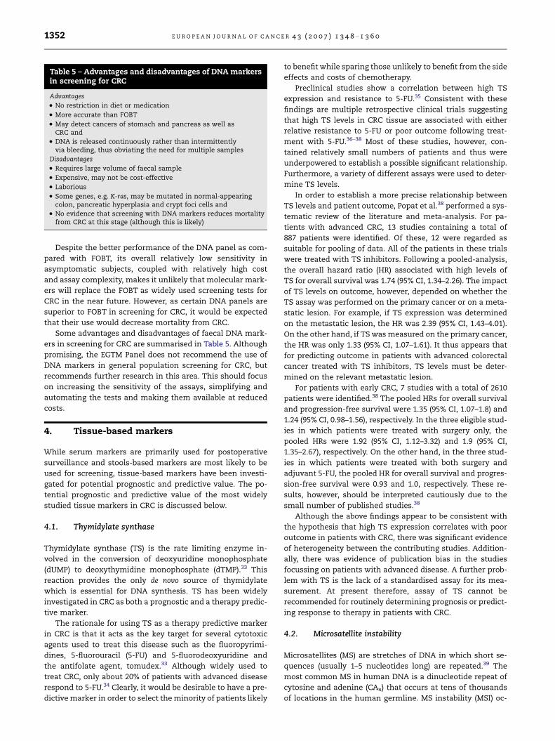
Table 5 – Advantages and disadvantages of DNA markersin screening for CRC
Advantages
• No restriction in diet or medication
• More accurate than FOBT
• May detect cancers of stomach and pancreas as well asCRC and
• DNA is released continuously rather than intermittentlyvia bleeding, thus obviating the need for multiple samples
Disadvantages
• Requires large volume of faecal sample
• Expensive, may not be cost-effective
• Laborious
• Some genes, e.g. K-ras, may be mutated in normal-appearingcolon, pancreatic hyperplasia and crypt foci cells and
• No evidence that screening with DNA markers reduces mortalityfrom CRC at this stage (although this is likely)
1352 E U R O P E A N J O U R N A L O F C A N C E R 4 3 ( 2 0 0 7 ) 1 3 4 8 – 1 3 6 0
Despite the better performance of the DNA panel as com-
pared with FOBT, its overall relatively low sensitivity in
asymptomatic subjects, coupled with relatively high cost
and assay complexity, makes it unlikely that molecular mark-
ers will replace the FOBT as widely used screening tests for
CRC in the near future. However, as certain DNA panels are
superior to FOBT in screening for CRC, it would be expected
that their use would decrease mortality from CRC.
Some advantages and disadvantages of faecal DNA mark-
ers in screening for CRC are summarised in Table 5. Although
promising, the EGTM Panel does not recommend the use of
DNA markers in general population screening for CRC, but
recommends further research in this area. This should focus
on increasing the sensitivity of the assays, simplifying and
automating the tests and making them available at reduced
costs.
4. Tissue-based markers
While serum markers are primarily used for postoperative
surveillance and stools-based markers are most likely to be
used for screening, tissue-based markers have been investi-
gated for potential prognostic and predictive value. The po-
tential prognostic and predictive value of the most widely
studied tissue markers in CRC is discussed below.
4.1. Thymidylate synthase
Thymidylate synthase (TS) is the rate limiting enzyme in-
volved in the conversion of deoxyuridine monophosphate
(dUMP) to deoxythymidine monophosphate (dTMP).33 This
reaction provides the only de novo source of thymidylate
which is essential for DNA synthesis. TS has been widely
investigated in CRC as both a prognostic and a therapy predic-
tive marker.
The rationale for using TS as a therapy predictive marker
in CRC is that it acts as the key target for several cytotoxic
agents used to treat this disease such as the fluoropyrimi-
dines, 5-fluorouracil (5-FU) and 5-fluorodeoxyuridine and
the antifolate agent, tomudex.33 Although widely used to
treat CRC, only about 20% of patients with advanced disease
respond to 5-FU.34 Clearly, it would be desirable to have a pre-
dictive marker in order to select the minority of patients likely
to benefit while sparing those unlikely to benefit from the side
effects and costs of chemotherapy.
Preclinical studies show a correlation between high TS
expression and resistance to 5-FU.35 Consistent with these
findings are multiple retrospective clinical trials suggesting
that high TS levels in CRC tissue are associated with either
relative resistance to 5-FU or poor outcome following treat-
ment with 5-FU.36–38 Most of these studies, however, con-
tained relatively small numbers of patients and thus were
underpowered to establish a possible significant relationship.
Furthermore, a variety of different assays were used to deter-
mine TS levels.
In order to establish a more precise relationship between
TS levels and patient outcome, Popat et al.38 performed a sys-
tematic review of the literature and meta-analysis. For pa-
tients with advanced CRC, 13 studies containing a total of
887 patients were identified. Of these, 12 were regarded as
suitable for pooling of data. All of the patients in these trials
were treated with TS inhibitors. Following a pooled-analysis,
the overall hazard ratio (HR) associated with high levels of
TS for overall survival was 1.74 (95% CI, 1.34–2.26). The impact
of TS levels on outcome, however, depended on whether the
TS assay was performed on the primary cancer or on a meta-
static lesion. For example, if TS expression was determined
on the metastatic lesion, the HR was 2.39 (95% CI, 1.43–4.01).
On the other hand, if TS was measured on the primary cancer,
the HR was only 1.33 (95% CI, 1.07–1.61). It thus appears that
for predicting outcome in patients with advanced colorectal
cancer treated with TS inhibitors, TS levels must be deter-
mined on the relevant metastatic lesion.
For patients with early CRC, 7 studies with a total of 2610
patients were identified.38 The pooled HRs for overall survival
and progression-free survival were 1.35 (95% CI, 1.07–1.8) and
1.24 (95% CI, 0.98–1.56), respectively. In the three eligible stud-
ies in which patients were treated with surgery only, the
pooled HRs were 1.92 (95% CI, 1.12–3.32) and 1.9 (95% CI,
1.35–2.67), respectively. On the other hand, in the three stud-
ies in which patients were treated with both surgery and
adjuvant 5-FU, the pooled HR for overall survival and progres-
sion-free survival were 0.93 and 1.0, respectively. These re-
sults, however, should be interpreted cautiously due to the
small number of published studies.38
Although the above findings appear to be consistent with
the hypothesis that high TS expression correlates with poor
outcome in patients with CRC, there was significant evidence
of heterogeneity between the contributing studies. Addition-
ally, there was evidence of publication bias in the studies
focussing on patients with advanced disease. A further prob-
lem with TS is the lack of a standardised assay for its mea-
surement. At present therefore, assay of TS cannot be
recommended for routinely determining prognosis or predict-
ing response to therapy in patients with CRC.
4.2. Microsatellite instability
Microsatellites (MS) are stretches of DNA in which short se-
quences (usually 1–5 nucleotides long) are repeated.39 The
most common MS in human DNA is a dinucleotide repeat of
cytosine and adenine (CAn) that occurs at tens of thousands
of locations in the human germline. MS instability (MSI) oc-
E U R O P E A N J O U R N A L O F C A N C E R 4 3 ( 2 0 0 7 ) 1 3 4 8 – 1 3 6 0 1353
curs when a germline MS allele gains or loses a repeat unit.
This gain or loss of MS alleles results from the absence of a
functional mismatch repair (MMR) enzyme, i.e. an enzyme
that repairs errors occurring during DNA replication.
Testing for MSI has a number of potential applications in
CRC, including
• use as a surrogate marker for hereditary non-polyposis
colorectal cancer (HNPCC) [see later discussion on genetic
testing],
• determining prognosis in patients with sporadic CRC and
• predicting response to adjuvant chemotherapy in patients
with sporadic CRC.
Popat et al.40 carried out a systematic review and pooled
analysis of published studies relating MSI to prognosis in pa-
tients with CRC. Of the 43 studies identified, 32 were found to
be eligible for analysis, giving a total of 7642 patients includ-
ing 1277 with MSI. Following a pooled analysis, patients with
MSI had a 15% better outcome compared to those without
MSI. The benefit of MSI was seen in patients with both locally
advanced (stages II and III disease) and advanced CRC (stage
IV). The reason for the association between MSI and favour-
able prognosis may be related to a protective role provided
by functionally active lymphocytes which infiltrate MSI-posi-
tive CRCs.41
Although available data suggest that testing for MSI may
differentiate between indolent and aggressive CRCs, Popat
et al.40 caution that prior to introduction of routine MSI test-
ing as an aid to determine prognosis, validation in a prospec-
tive trial is essential. The EGTM Panel supports this, and does
not currently recommend routine MSI testing for determining
prognosis in patients with CRC.
In vitro studies show that colorectal cancer cells displaying
MSI are less responsive than MS-stable (MS-S) cells to 5-fluo-
rouracil (5-FU).42,43 Consistent with these findings, Ribic
et al.44 reported that adjuvant 5-FU plus levamisole was of
no significant benefit in stages II and III patients with MSI-po-
sitive tumours (defined as instability at 30% or more of loci
screened). In contrast, chemotherapy improved overall sur-
vival in patients lacking MSI (hazard ratio, 0.72, p = 0.04). Care-
thers et al.45 also found that the benefit of 5-FU was different
in patients with and without MSI. Thus, patients with non-
MSI tumours who received this drug had better survival than
those not treated. Conversely, patients with MSI-positive tu-
mours who were treated with 5-FU had a similar survival pat-
tern to those without treatment. In contrast to these findings,
other investigators found that the presence of MSI was a sig-
nificant predictor of a survival benefit from adjuvant 5-FU-
based chemotherapy.46,47 Because of these conflicting findings,
MSI testing cannot be recommended at present for predicting
response to 5-FU in the adjuvant treatment of CRC.
4.3. p53
The p53 tumour suppressorgene encodes a transcription factor
that regulates the expression of genes involved in apoptosis,
angiogenesis, cell cycle and genome maintenance.48,49 Muta-
tions in the p53 gene are found in approximately 50% of CRCs,
with most mutations occurring in exons 5–8. These mutations
appear to be formed relatively late in the genesis of CRC, i.e.
during the conversion of dysplastic adenomas to invasive
carcinomas.
p53 has been widely investigated both as a prognostic and
as a therapy predictive marker in CRC. As with the tissue
markers discussed above, most of these studies are retrospec-
tive and insufficiently powered to establish a meaningful rela-
tionship with patient outcome. Furthermore, a multiplicity of
methods was used to determine p53 abnormalities. These
methods have generally employed either immunohistochem-
istry to detect p53 protein or DNA sequence analysis to detect
gene mutations.
Munro et al.50 carried out a systematic review of published
studies that investigated the relationship between p53 abnor-
mality and outcome in patients with CRC. In total, 168 eligible
studies comprising survival data on 18,766 patients were
identified. The key findings were as follows:
• Patients with abnormal p53 whether detected by immuno-
histochemistry (IHC) or DNA sequence analysis had an
increased risk of death. The relative risk (RR) with IHC
was 1.32 (95% CI, 1.23–1.42) and with sequence analysis
was 1.31 (95% CI, 1.19–1.90).
• The adverse impact of p53 abnormality was stronger in
patients likely to have a good outcome compared to those
likely to have poor outcome.
• p53 had no impact on outcome in patients treated with
chemotherapy.
• Abnormal p53 correlated with failure of response to radio-
therapy in patients with rectal cancer (RR, 1.49, 95% CI,
1.25–1.77).
In a similar study, Russo et al.51 pooled data from 25 differ-
ent groups (n = 3583) invited to participate in a multicenter
study aimed at evaluating the potential prognostic and pre-
dictive value of p53 mutations in CRC. Unlike the analysis of
Munro et al.50 mentioned above, only gene mutations were
investigated in this study. The main findings were
• in patients with proximal tumours, mutations in exon 5 of
p53 were associated with adverse outcome,
• in patients with distal colon tumours, gene deletions
resulting in loss of amino acids were associated with poor
survival,
• patients with wild-type p53 gene displayed a better out-
come when treated with chemotherapy compared to those
treated with surgery alone, and
• patients with rectal tumours containing wild-type p53
derived a significant survival benefit from 5-fluorouracil
(5-FU), irrespective of whether or not radiotherapy was
administered.
Although the above studies suggest that altered p53 may
have an impact on outcome in patients with CRC, this rela-
tionship is at best only modest. Consequently, at present, rou-
tine determination of p53 status cannot be recommended for
either assessing prognosis or predicting response to therapy
in CRC. Furthermore, as with other tissue-based markers in
CRC, standardised assays are not available for determining
p53 abnormalities.
1354 E U R O P E A N J O U R N A L O F C A N C E R 4 3 ( 2 0 0 7 ) 1 3 4 8 – 1 3 6 0
4.4. K-ras
K-ras is one of the most frequently mutated c-oncogenes in
human cancer. It functions as a guanine nucleotide binding
protein involved in signal transduction.52 This signalling
may result in increased cell proliferation, enhanced cell sur-
vival or induction of apoptosis.
Mutant K-ras is present in approximately 50% of CRCs and
appears to play a role in the relatively early stages of CRC car-
cinogenesis.53 Conflicting data exist on the relationship be-
tween the presence of mutant K-ras and prognosis in
patients with CRC.53 Based on an early meta-analysis of pub-
lished data, Andreyev et al.54 concluded that K-ras mutations
correlated with poor outcome in patients with CRC. A more
recent meta-analysis involving 4268 patients from 42 differ-
ent institutions however, concluded that only a specific type
of K-ras mutation predicted poor outcome, i.e. only patients
with a G–T transversion at codon 12 had an adverse out-
come.55 This specific mutation was detected in less than
10% of tumours and was prognostic in Dukes’ C but not in
Dukes’ B patients.
Based on the available data, K-ras cannot be recommended
for determining prognosis or for predicting response to che-
motherapy in patients with CRC.
5. Tests for susceptibility to CRC
Approximately 15% of CRC are thought to be due to an inher-
ited or familial predisposition.3 The most common hereditary
conditions giving rise to an increased risk of CRC are heredi-
tary non-polyposis colorectal cancer (HNPCC) and familial
adenomatous polyposis (FAP).
5.1. Familial adenomatous polyposis
FAP is an autosomal dominant condition characterised by
hundreds to thousands of adenomas in the colon and rectum.
It has an incidence of approximately 1 per 8000 to 1 per 14,000
of the population and accounts for about 0.5% of all CRCs.56
CRC is an almost inevitable consequence of classical FAP, if
untreated, with an average of onset of about 39 years of age.
FAP may be associated with extra-colonic manifestations
such as congenital hypertrophy of retinal pigment epithelium
(CHRPE), desmoid tumours, osteomas, teeth abnormalities,
thyroid cancer and hepatoblastoma.
An attenuated form of the syndrome (AFAP) is character-
ised by fewer adenomas (<100). Subjects with this attenuated
form of FAP also have a high risk of developing CRC, i.e.
approximately 80% by the age of 70 years. For patients with
this form of APC, CRC is diagnosed approximately 12 years la-
ter than in classical FAP.56
A further variant of the FAP syndrome results from biall-
elic inherited mutations in the BER (base excision repair)
gene, known as MutYH or MYH (for review, see Ref. [57]). This
syndrome, which is now referred to as MAP or MYH-associ-
ated polyposis, is often indistinguishable in its clinical mani-
festation from classic or attenuated forms of FAP. In one
study, biallelic mutations in the MYH gene accounted for
approximately 30% of families with multiple adenomas (15–
100) who failed to exhibit an autosomal pattern of inheritance
or a germline mutation in the APC gene.58
Approximately 70–80% of patients with classical APC har-
bour germline mutations in the APC gene. Although at least
800 different mutations in the APC gene have been found to
cause FAP, the vast majority of these mutations are nonsense
or frameshift that gives rise to a truncated protein. Approxi-
mately one third of the so far identified FAP-causing muta-
tions occur at codons 1061 and 1309.59 Other mutations are
spread between codons 200 and 1600. The attenuated form
of FAP usually results from mutations at the extreme 5 0 or 3 0
ends of the APC gene or from an alternatively spliced region
of exon 9.60
Screening for FAP should commence with a detailed family
history. For individuals with suspected FAP, genetic testing
can be used both to confirm diagnosis in a suspected proband
and to assess risk in pre-symptomatic family members. Pro-
vided the mutation responsible for FAP within a family is
known, testing for APC mutations can be considered for at-
risk family members.23 Most expert panels recommend that
for families with classic FAP, APC gene testing should be con-
sidered at 10–12 years of age.23,61–63
While it is generally recommended that APC testing should
start at 10–12 years of age, some authors suggest that screen-
ing for hepatoblastoma should be carried out in children of
FAP patients.64–67 Hepatoblastoma is a rare malignant embry-
onal tumour of the liver that occurs in children usually be-
tween 6 months and 3 years of age. The risk of
hepatoblastoma is considerably increased in children of pa-
tients with FAP.65,68 The best available screening tests for
hepatoblastoma are serum AFP levels and liver ultra-
sound.65,68 Interpretation of AFP levels in newborn infants,
however, may be difficult as serum levels are increased in
the first few months of life. These high levels quickly decline
with adult concentrations being reached after about 10
months.
It is important to state that genetic testing for cancer sus-
ceptibility syndromes such as FAP should only be performed
following appropriate counselling.69 The counselling should
involve discussion of possible risks and benefits of the early
detection of cancer as well as prevention modalities.69 Indi-
viduals found not to carry the specific APC gene mutation
responsible for FAP in that family can be spared intensive sur-
veillance but should still undergo similar screening to that of
an average risk individual.23,61,62 On the other hand, if an APC
gene mutation is identified, prophylactic surgery should be
performed once adenomas are found. The main surgical op-
tions include colectomy and ileorectal anastomosis, proctoco-
lectomy with ileostomy and proctocolectomy with ileal
pouch-anal anastomosis23,61,62 The EGTM Panel supports
these recommendations for the detection and management
of patients with FAP.
Approximately 20–30% of families with classic FAP are
APC mutation-negative using existing testing methods.
The NCCN guidelines state that if an APC mutation consis-
tent with recessive inheritance cannot be found in a family,
testing for MYH should be performed.23 As heterozygote
MAP carriers appear to have an increased risk of developing
CRC, regular surveillance should be carried out on these
subjects.
E U R O P E A N J O U R N A L O F C A N C E R 4 3 ( 2 0 0 7 ) 1 3 4 8 – 1 3 6 0 1355
For patients with the attenuated form of APC, treatment
depends on patient age and number of adenomas. According
to the NCCN23, for subjects 21 years or younger with a low
number of adenomas, colonoscopy and polypectomy should
be performed every 1–2 years. On the other hand, for individ-
uals >40 years of age and for those with large numbers of pol-
yps that cannot be easily managed by polypectomy, a
colectomy and ileorectal anastomosis should be performed.
It should be noted that a potential benefit from genetic
testing in FAP is presumed and is not based on evidence from
high-level studies such as prospective randomised trials.
5.2. Hereditary non-polyposis colorectal cancer
HNPCC is clinically defined by the fulfillment of the Amster-
dam Criteria (Table 6). HNPCC includes affected families with
disease causing mutations in DNA mis-match repair (MMR)
genes displaying an MSI-H phenotype in their corresponding
tumours (a subgroup also called Lynch syndrome) and fami-
lies with MSS tumours and no mutations in DNA MMR genes.
The genetic pathogenesis of the latter group is currently
unclear.
Lynch syndrome is an autosomal dominant disorder char-
acterised by the early onset of CRC in the setting of relatively
few polyps. The syndrome has an incidence of approximately
1:1000 in the general population and accounts for 1–5% of
CRCs.70,71 It is characterised by a 70–80% lifetime risk of devel-
oping CRC and, for women, a 40–60% lifetime risk of develop-
ing endometrial cancer. Carriers also have an increased risk
of developing ovarian, upper gastrointestinal, urological and
central nervous system cancers but this is usually less than
15%.70–72
The genes primarily responsible for Lynch syndrome are
involved in MMR and include MSH2, MLH1 and MSH6.39,70,71
Mutations in MSH2 and MLH1 are thought to be responsible
for at least 80% of the reported mutations in Lynch syndrome
that involve defective mismatch repair.70,71 Over 90% of pa-
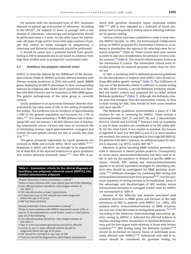
Table 6 – Amsterdam criteria for the clinical diagnosis ofhereditary non-polyposis colorectal cancer (HNPCC), FAP,familial adenomatous polyposis
Original Amsterdam Criteria (Amsterdam I criteria)73
Three or more relatives with colon cancer plus all of the following:
• One affected patient should be a first degree relative ofthe other 2
• CRC should involve at least 2 generations
• At least one case of CRC should have been diagnosedbefore the age of 50 years
• FAP has been excluded.
Revised Amsterdam criteria (Amsterdam II criteria)74
Three or more relatives with HNPCC-associated cancer (CRC or
cancer of the endometrium, small bowel, ureter or renal pelvis)
plus all of the following
• One affected patient should be a first-degree relative ofthe other 2
• Two or more successive generations should be affected
• Cancer in one or more affected relative should bediagnosed before the age of 50 years
• FAP should be excluded in any case of CRC
• Tumours should be diagnosed by pathological examination
tients with germline mismatch repair mutations exhibit
MSI.70,71 MSI is thus regarded as a hallmark of Lynch syn-
drome and consequently is widely used in selecting individu-
als for genetic testing.
Various criteria have been published in order to help iden-
tify HNPCC families. In 1991, the International Collaborative
Group on HNPCC proposed the Amsterdam I criteria in an at-
tempt to standardise the approach for selecting cases for re-
search purposes73 (Table 6). These criteria were later revised
in order to take into consideration the presence of extra-colo-
nic tumours74 (Table 6). The revised criteria became known as
the Amsterdam II criteria. The Amsterdam criteria were in-
tended primarily for research purposes rather than for clini-
cal use.
In 1997, a workshop held in Bethesda produced guidelines
for the identification of subjects with HNPCC who should un-
dergo MSI and/or genetic testing75 (Table 7). The fulfillment of
any one of the Bethesda criteria is sufficient to justify testing
for MSI. More recently, a second Bethesda workshop simpli-
fied the earlier criteria and proposed the so-called revised
Bethesda guidelines76 (Table 7). Although the Bethesda crite-
ria were less stringent than the Amsterdam criteria, as they
include testing for MSI, they should be both more sensitive
and more specific.77
The Bethesda guidelines recommended a panel of 5 MS
markers for use in screening for HNPCC. These include 2
mononucleotides (BAT 25 and BAT 26) and 3 dinucleotides
(D2S123, D5S346 and D17S250).78 Tumours with no instability
in any of these markers are considered to be MS stable (MS-
S). On the other hand, if one marker is mutated, the tumour
is regarded to have low MSI (MSI-L) and if 2 or more markers
are mutated, the tumour is regarded to have high MSI (MSI-H).
For patients with MSI-L tumours, an additional panel of mark-
ers is required, e.g. MYCL and/or BAT 40.76
Mutation in genes encoding MMR enzymes generally re-
sults in abnormal or absent protein products. Consequently,
in recent years, immunohistochemistry has been used in or-
der to test for the presence or absence of specific MMR en-
zymes. Overall, MSI testing and immunohistochemistry
appear to be almost equivalent strategies for identifying sub-
jects who should be investigated for MMR germline muta-
tions.79–83 Different strategies for combining MSI testing and
immunohistochemistry have been proposed84–87, but the opti-
mum sequence of testing remains to be established. Some of
the advantages and disadvantages of MSI analysis versus
immunohistochemistry as surrogate marker tests for HNPCC
are summarised in Table 8.
Because of the difficulty and expense associated with
mutation detection in MMR genes and because of the high
prevalence of MSI in patients with HNPCC (i.e. >90%), MSI
analysis and/or immunohistochemistry of MMR enzymes,
are now one of the first steps in testing these patients.23,61,62,87
According to the American Gastroenterology Association, ge-
netic testing in HNPCC is indicated for affected subjects in
families meeting either Amsterdam or modified Bethesda cri-
teria and for first-degree adult relatives of those with known
mutation.61,62 MSI testing using the Bethesda markers76,78
should be performed on tumour tissue of individuals puta-
tively affected with HNPCC.61,62 Individuals with MSI-H tu-
mours should be considered for germline testing for
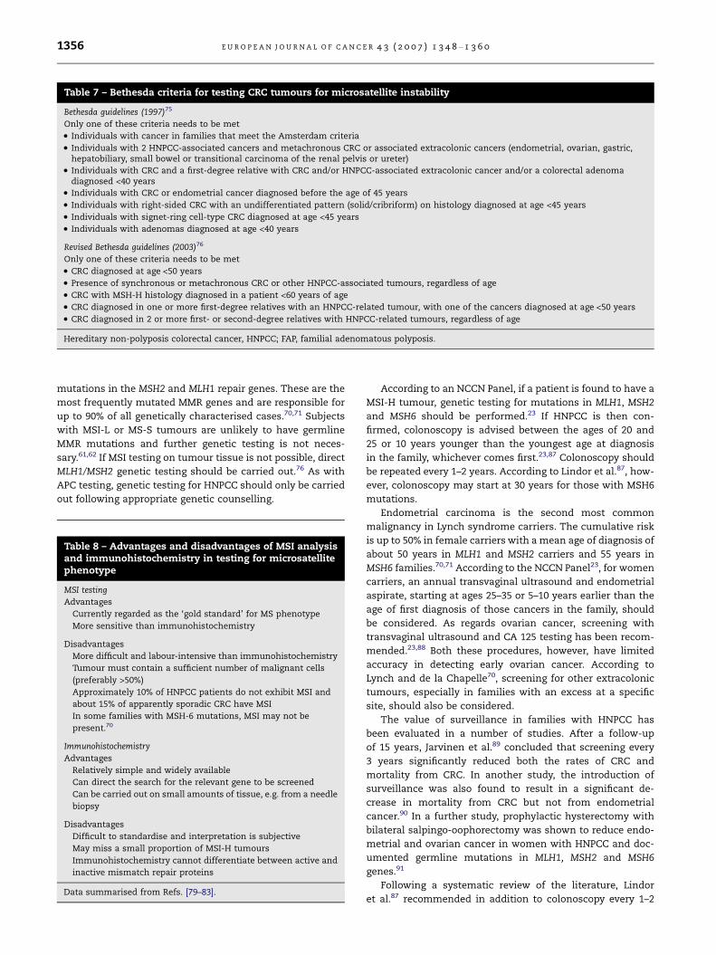
Table 7 – Bethesda criteria for testing CRC tumours for microsatellite instability
Bethesda guidelines (1997)75
Only one of these criteria needs to be met
• Individuals with cancer in families that meet the Amsterdam criteria
• Individuals with 2 HNPCC-associated cancers and metachronous CRC or associated extracolonic cancers (endometrial, ovarian, gastric,hepatobiliary, small bowel or transitional carcinoma of the renal pelvis or ureter)
• Individuals with CRC and a first-degree relative with CRC and/or HNPCC-associated extracolonic cancer and/or a colorectal adenomadiagnosed <40 years
• Individuals with CRC or endometrial cancer diagnosed before the age of 45 years
• Individuals with right-sided CRC with an undifferentiated pattern (solid/cribriform) on histology diagnosed at age <45 years
• Individuals with signet-ring cell-type CRC diagnosed at age <45 years
• Individuals with adenomas diagnosed at age <40 years
Revised Bethesda guidelines (2003)76
Only one of these criteria needs to be met
• CRC diagnosed at age <50 years
• Presence of synchronous or metachronous CRC or other HNPCC-associated tumours, regardless of age
• CRC with MSH-H histology diagnosed in a patient <60 years of age
• CRC diagnosed in one or more first-degree relatives with an HNPCC-related tumour, with one of the cancers diagnosed at age <50 years
• CRC diagnosed in 2 or more first- or second-degree relatives with HNPCC-related tumours, regardless of age
Hereditary non-polyposis colorectal cancer, HNPCC; FAP, familial adenomatous polyposis.
1356 E U R O P E A N J O U R N A L O F C A N C E R 4 3 ( 2 0 0 7 ) 1 3 4 8 – 1 3 6 0
mutations in the MSH2 and MLH1 repair genes. These are the
most frequently mutated MMR genes and are responsible for
up to 90% of all genetically characterised cases.70,71 Subjects
with MSI-L or MS-S tumours are unlikely to have germline
MMR mutations and further genetic testing is not neces-
sary.61,62 If MSI testing on tumour tissue is not possible, direct
MLH1/MSH2 genetic testing should be carried out.76 As with
APC testing, genetic testing for HNPCC should only be carried
out following appropriate genetic counselling.
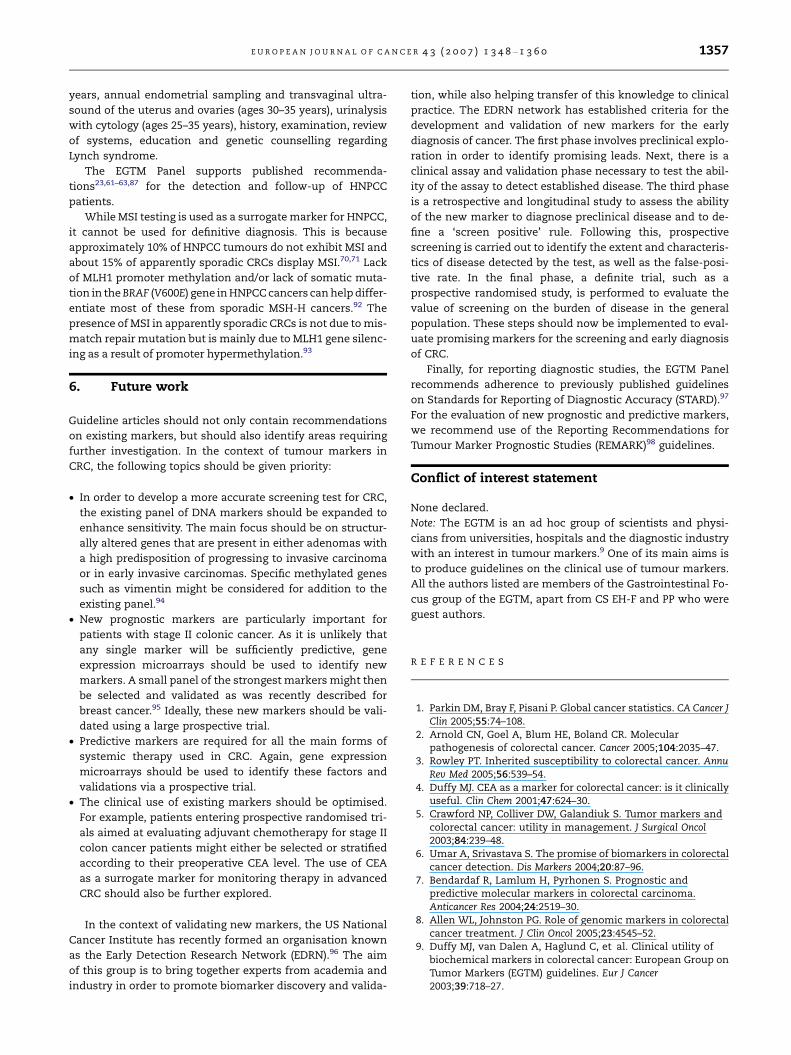
Table 8 – Advantages and disadvantages of MSI analysisand immunohistochemistry in testing for microsatellitephenotype
MSI testing
Advantages
Currently regarded as the ‘gold standard’ for MS phenotype
More sensitive than immunohistochemistry
Disadvantages
More difficult and labour-intensive than immunohistochemistry
Tumour must contain a sufficient number of malignant cells
(preferably >50%)
Approximately 10% of HNPCC patients do not exhibit MSI and
about 15% of apparently sporadic CRC have MSI
In some families with MSH-6 mutations, MSI may not be
present.70
Immunohistochemistry
Advantages
Relatively simple and widely available
Can direct the search for the relevant gene to be screened
Can be carried out on small amounts of tissue, e.g. from a needle
biopsy
Disadvantages
Difficult to standardise and interpretation is subjective
May miss a small proportion of MSI-H tumours
Immunohistochemistry cannot differentiate between active and
inactive mismatch repair proteins
Data summarised from Refs. [79–83].
According to an NCCN Panel, if a patient is found to have a
MSI-H tumour, genetic testing for mutations in MLH1, MSH2
and MSH6 should be performed.23 If HNPCC is then con-
firmed, colonoscopy is advised between the ages of 20 and
25 or 10 years younger than the youngest age at diagnosis
in the family, whichever comes first.23,87 Colonoscopy should
be repeated every 1–2 years. According to Lindor et al.87, how-
ever, colonoscopy may start at 30 years for those with MSH6
mutations.
Endometrial carcinoma is the second most common
malignancy in Lynch syndrome carriers. The cumulative risk
is up to 50% in female carriers with a mean age of diagnosis of
about 50 years in MLH1 and MSH2 carriers and 55 years in
MSH6 families.70,71 According to the NCCN Panel23, for women
carriers, an annual transvaginal ultrasound and endometrial
aspirate, starting at ages 25–35 or 5–10 years earlier than the
age of first diagnosis of those cancers in the family, should
be considered. As regards ovarian cancer, screening with
transvaginal ultrasound and CA 125 testing has been recom-
mended.23,88 Both these procedures, however, have limited
accuracy in detecting early ovarian cancer. According to
Lynch and de la Chapelle70, screening for other extracolonic
tumours, especially in families with an excess at a specific
site, should also be considered.
The value of surveillance in families with HNPCC has
been evaluated in a number of studies. After a follow-up
of 15 years, Jarvinen et al.89 concluded that screening every
3 years significantly reduced both the rates of CRC and
mortality from CRC. In another study, the introduction of
surveillance was also found to result in a significant de-
crease in mortality from CRC but not from endometrial
cancer.90 In a further study, prophylactic hysterectomy with
bilateral salpingo-oophorectomy was shown to reduce endo-
metrial and ovarian cancer in women with HNPCC and doc-
umented germline mutations in MLH1, MSH2 and MSH6
genes.91
Following a systematic review of the literature, Lindor
et al.87 recommended in addition to colonoscopy every 1–2
E U R O P E A N J O U R N A L O F C A N C E R 4 3 ( 2 0 0 7 ) 1 3 4 8 – 1 3 6 0 1357
years, annual endometrial sampling and transvaginal ultra-
sound of the uterus and ovaries (ages 30–35 years), urinalysis
with cytology (ages 25–35 years), history, examination, review
of systems, education and genetic counselling regarding
Lynch syndrome.
The EGTM Panel supports published recommenda-
tions23,61–63,87 for the detection and follow-up of HNPCC
patients.
While MSI testing is used as a surrogate marker for HNPCC,
it cannot be used for definitive diagnosis. This is because
approximately 10% of HNPCC tumours do not exhibit MSI and
about 15% of apparently sporadic CRCs display MSI.70,71 Lack
of MLH1 promoter methylation and/or lack of somatic muta-
tion in the BRAF (V600E) gene in HNPCC cancers can help differ-
entiate most of these from sporadic MSH-H cancers.92 The
presence of MSI in apparently sporadic CRCs is not due to mis-
match repair mutation but is mainly due to MLH1 gene silenc-
ing as a result of promoter hypermethylation.93
6. Future work
Guideline articles should not only contain recommendations
on existing markers, but should also identify areas requiring
further investigation. In the context of tumour markers in
CRC, the following topics should be given priority:
• In order to develop a more accurate screening test for CRC,
the existing panel of DNA markers should be expanded to
enhance sensitivity. The main focus should be on structur-
ally altered genes that are present in either adenomas with
a high predisposition of progressing to invasive carcinoma
or in early invasive carcinomas. Specific methylated genes
such as vimentin might be considered for addition to the
existing panel.94
• New prognostic markers are particularly important for
patients with stage II colonic cancer. As it is unlikely that
any single marker will be sufficiently predictive, gene
expression microarrays should be used to identify new
markers. A small panel of the strongest markers might then
be selected and validated as was recently described for
breast cancer.95 Ideally, these new markers should be vali-
dated using a large prospective trial.
• Predictive markers are required for all the main forms of
systemic therapy used in CRC. Again, gene expression
microarrays should be used to identify these factors and
validations via a prospective trial.
• The clinical use of existing markers should be optimised.
For example, patients entering prospective randomised tri-
als aimed at evaluating adjuvant chemotherapy for stage II
colon cancer patients might either be selected or stratified
according to their preoperative CEA level. The use of CEA
as a surrogate marker for monitoring therapy in advanced
CRC should also be further explored.
In the context of validating new markers, the US National
Cancer Institute has recently formed an organisation known
as the Early Detection Research Network (EDRN).96 The aim
of this group is to bring together experts from academia and
industry in order to promote biomarker discovery and valida-
tion, while also helping transfer of this knowledge to clinical
practice. The EDRN network has established criteria for the
development and validation of new markers for the early
diagnosis of cancer. The first phase involves preclinical explo-
ration in order to identify promising leads. Next, there is a
clinical assay and validation phase necessary to test the abil-
ity of the assay to detect established disease. The third phase
is a retrospective and longitudinal study to assess the ability
of the new marker to diagnose preclinical disease and to de-
fine a ‘screen positive’ rule. Following this, prospective
screening is carried out to identify the extent and characteris-
tics of disease detected by the test, as well as the false-posi-
tive rate. In the final phase, a definite trial, such as a
prospective randomised study, is performed to evaluate the
value of screening on the burden of disease in the general
population. These steps should now be implemented to eval-
uate promising markers for the screening and early diagnosis
of CRC.
Finally, for reporting diagnostic studies, the EGTM Panel
recommends adherence to previously published guidelines
on Standards for Reporting of Diagnostic Accuracy (STARD).97
For the evaluation of new prognostic and predictive markers,
we recommend use of the Reporting Recommendations for
Tumour Marker Prognostic Studies (REMARK)98 guidelines.
Conflict of interest statement
None declared.
Note: The EGTM is an ad hoc group of scientists and physi-
cians from universities, hospitals and the diagnostic industry
with an interest in tumour markers.9 One of its main aims is
to produce guidelines on the clinical use of tumour markers.
All the authors listed are members of the Gastrointestinal Fo-
cus group of the EGTM, apart from CS EH-F and PP who were
guest authors.
R E F E R E N C E S
1. Parkin DM, Bray F, Pisani P. Global cancer statistics. CA Cancer JClin 2005;55:74–108.
2. Arnold CN, Goel A, Blum HE, Boland CR. Molecularpathogenesis of colorectal cancer. Cancer 2005;104:2035–47.
3. Rowley PT. Inherited susceptibility to colorectal cancer. AnnuRev Med 2005;56:539–54.
4. Duffy MJ. CEA as a marker for colorectal cancer: is it clinicallyuseful. Clin Chem 2001;47:624–30.
5. Crawford NP, Colliver DW, Galandiuk S. Tumor markers andcolorectal cancer: utility in management. J Surgical Oncol2003;84:239–48.
6. Umar A, Srivastava S. The promise of biomarkers in colorectalcancer detection. Dis Markers 2004;20:87–96.
7. Bendardaf R, Lamlum H, Pyrhonen S. Prognostic andpredictive molecular markers in colorectal carcinoma.Anticancer Res 2004;24:2519–30.
8. Allen WL, Johnston PG. Role of genomic markers in colorectalcancer treatment. J Clin Oncol 2005;23:4545–52.
9. Duffy MJ, van Dalen A, Haglund C, et al. Clinical utility ofbiochemical markers in colorectal cancer: European Group onTumor Markers (EGTM) guidelines. Eur J Cancer2003;39:718–27.
1358 E U R O P E A N J O U R N A L O F C A N C E R 4 3 ( 2 0 0 7 ) 1 3 4 8 – 1 3 6 0
10. Hayes DF, Bast R, Desch CE, et al. A tumor marker utilitygrading system (TMUGS): a framework to evaluate clinicalutility of tumor markers. J Natl Cancer Inst 1996;88:1456–66.
11. Bruinvels DJ, Stiggelbout AM, Kievit J, van Houwelingen HC,Habbema DF, van de Velde CH. Follow up of colorectal cancer:a meta-analysis. Ann Surg 1994;219:174–82.
12. Rosen M, Chan L, Beart RW, Vukasin P, Anthone G. Follow-upof colorectal cancer: a meta analysis. Dis Colon Rectum1998;41:1116–26.
13. Renehan AG, Egger M, Saunders MP, O’Dwyer ST. Impact onsurvival of intensive follow up after curative resection forcolorectal cancer: systematic review and meta-analysis ofrandomised trials. Br Med J 2002;324:813–6.
14. Figueredo A, Rumble RB, Maroun J, et al. Follow-up of patientswith curatively resected colorectal cancer: a practiceguideline. BMC Cancer 2003;3:26–39.
15. Jeffery GM, Hickey BE, Hider P. Follow-up strategies forpatients treated for nonmetastatic colorectal cancer(Cochrane Review). In: The Cochrane Library Issue 2, 2004,Chichester (UK): John Wiley & Sons, Ltd.
16. Simmonds PC, Primrose JN, Colquitt JL, Garden OJ, Poston GJ,Rees M. Surgical resection of hepatic metastases fromcolorectal cancer: a systematic review of published studies. BrJ Cancer 2006;94:982–99.
17. Cromheecke M, de Jong KP, Hoekstra HJ. Current treatment forcolorectal cancer metastatic to the liver. Eur J Surg Oncol1999;25:451–63.
18. Anonymous. Clinical practice guidelines for the use of tumormarkers in breast and colorectal cancer. J Clin Oncol1996;14:2843–77.
19. Thirion P, Michiels S, Pignon JP, et al. Modulation offluorouracil by leucovorin in patients with advancedcolorectal cancer: an updated meta-analyis. J Clin Oncol2004;22:3766–75.
20. Meyerhardt JA, Mayer RJ. Drug therapy: systematic therapy forcolorectal cancer. N Engl J Med 2005;352:476–87.
21. Locker GY, Hamilton S, Harris J, et al. ASCO 2006. Update ofrecommendations for the use of tumor markers ingastrointestinal cancer. J Clin Oncol 2006;24:5313–27.
22. Huang CS, Lal SK, Farraye FA. Colorectal cancer screening inaverage risk individuals. Cancer Causes Control 2005;16:171–88.
23. National Comprehensive Cancer Network Clinical PracticeGuidelines in Oncology, Colorectal Cancer Screening. Version1.2006. <http://www.nccn.org/professionals/physician_gls/PDF/colorectal_screening.pdf> (accessed 10.10.2006).
24. Smith RA, Cokkinides V, Eyre HJ. American Cancer SocietyGuidelines for the Early Detection of Cancer. CA Cancer J Clin2006;56:11–25.
25. US Preventive Services Task Force. Screening for colorectalcancer: recommendations and rationale. Ann Int Med2002;137:132–41.
26. Winawer S, Fletcher R, Rex D, et al. Colorectal cancerscreening and surveillance: clinical guidelines and rationale,update based on new evidence. Gastroenterology2003;124:544–60.
27. Anderson WF, Guyton KZ, Hiatt RA, Vernon SW, Levin B, HawkE. Colorectal cancer screening for persons at average risk. JNatl Cancer Instit 2002;94:1126–33.
28. Sidransky D, Tokino T, Hamilton SR, et al. Identification of rasoncogene mutations in the stools of patients with curablecolorectal tumors. Science 1992;256:102–5.
29. Ahlquist DA, Skoletsky JE, Boynton KA, et al. Colorectalcancer screening by detection of altered human DNA in stool:feasibility of a multitarget assay panel. Gastroenterology2000;119:1219–27.
30. Dong SM, Traverso G, Johnson C, et al. Detecting colorectalcancer in stool with use of multiple genetic targets. J NatlCancer Inst 2001;93:858–65.
31. Haug U, Brenner H. New stool test for colorectal cancerscreening: a systematic review focusing on performancecharacteristics and practicalness. Int J Cancer 2005;117:169–76.
32. Imperiale T, Ransohoff D, Itzkowitz SH, Turnbull BA, Ross ME.Faecal DNA versus faecal occult blood for colorectal-cancerscreening in an average risk population. N Engl J Med2004;351:2704–14.
33. Longley DB, Harkin P, Johnston PG. 5-Fluorouracil: mechanismof action and clinical strategies. Nat Rev Cancer 2003;3:330–8.
34. Machover D. A comprehensive review of 5-fluorouracil andleucovorin in patients with metastatic colorectal carcinoma.Cancer 1997;80:1179–87.
35. van Triest B, Pinedo HM, van Hensbergen Y, et al.Thymidylate synthase level as the main predictive parameterfor sensitivity to 5-fluorouracil, but not for folate-basedthymidylate synthase inhibitors, in 13 nonselected coloncancer cell lines. Clin Cancer Res 1999;5:643–54.
36. Banerjee D, Mayer-Kuckuk P, Capiaux G, Budak-Alpdogan T,Gorl R, Bertino JR. Novel aspects of resistance to drugstargeted to dihydrofolate reductase and thymidylatesynthase. Biochim Biophys Acta 2002;1587:164–73.
37. Bertino JR, Banerjee D. Is the measurement of thymidylatesynthase to determine suitability for treatment with5-fluoropyrimidines ready for prime time? Clin Cancer Res2003;9:1235–9.
38. Popat S, Matakidou A, Houlston RS. Thymidylate synthaseexpression and prognosis in colorectal cancer: a systematicreview and meta-analysis. J Clin Oncol 2004;22:529–35.
39. de la Chapelle A. Microsatellite instability. N Engl J Med2003;349:209–10.
40. Popat S, Hubner R, Houlston RS. Systematic review ofmicrosatellite instability and colorectal cancer prognosis.J Clin Oncol 2005;23:609–18.
41. Buckowitz A, Knaebel H-P, Benner A, et al. Microsatelliteinstability in colorectal cancer is associated with locallymphocyte infiltration and low frequency of distantmetastases. Br J Cancer 2005;92:1746–53.
42. Carethers JM, Chauhan DP, Fink D, et al. Mismatch repairproficiency and in vitro response to 5-fluorouracil.Gastroenterology 1999;117:123–31.
43. Arnold CN, Goel A, Boland CR. Role of hMLH1 promoterhypermethylation in drug resistance to 5-fluorouracil incolorectal cancer cell lines. Int J Cancer 2003;106:66–73.
44. Ribic CM, Sargent DJ, Moore MJ, et al. Tumor microsatelliteinstability status as a predictor of benefit from fluorouracil-based adjuvant chemotherapy for colon cancer. N Engl J Med2003;349:247–57.
45. Carethers JM, Smith EJ, Behling CA, et al. Use of 5-fluorouraciland survival in patients with microsatellite unstablecolorectal cancer. Gastroenterology 2004;126:394–401.
46. Elsaleh H, Powell B, McCaul K, et al. P53 alterations andmicrosatellite instability have predictive value for survivalbenefit from chemotherapy in stage III colorectal cancer. ClinCancer Res 2001;7:1343–9.
47. Watanabe T, Wu TT, Catalano PJ, et al. Molecular predictors ofsurvival after adjuvant chemotherapy for colon cancer. N EnglJ Med 2001;344:1196–206.
48. Mills AA. P53: links to the past, bridge to the future. Genes Dev2005;19:2091–9.
49. Vogelstein B, Lane D, Levine AJ. Surfing the p53 network.Nature 2000;408:307–10.
50. Munro AJ, Lain S, Lane DP. P53 abnormalities in colorectalcancer: a systematic review. Br J Cancer 2005;92:434–44.
51. Russo A, Bazan V, Iacopetta B, et al. The TP53 ColorectalCollaborative Study on the prognostic and predictivesignificance of p53 mutations: influence of tumor site, type ofmutation and adjuvant treatment. J Clin Oncol2005;23:7518–28.
E U R O P E A N J O U R N A L O F C A N C E R 4 3 ( 2 0 0 7 ) 1 3 4 8 – 1 3 6 0 1359
52. Castagnola P, Giaretti W. Mutant KRAS, chromosomalinstability and prognosis in colorectal cancer. Biochim BiophysActa 2005;1756:115–25.
53. Fearon ER, Vogelstein B. A genetic model for colorectaltumorigenesis. Cell 1990;61:759–67.
54. Andreyev HJ, Norman AR, Cunningham JR, et al. Kirsten rasmutations in patients with colorectal cancer: the multicenter‘‘RASCAL’’ study. J Natl Cancer Inst 1998;90:675–84.
55. Andreyev HJ, Norman AR, Cunningham D, et al. Kirsten rasmutations in patients with colorectal cancer: the ‘‘RASCAL II’’study. Br J Cancer 2001;85:692–6.
56. Fearnhead NS, Wilding JL, Bodmer WF. Genetic of colorectalcancer: hereditary aspects and overview of colorectaltumorigenesis. Br Med Bull 2002;64:27–43.
57. Sampson JR, Jones S, Dolwani S, Cheadle JP. MutYH (MYH) andcolorectal cancer. Biochem Trans Soc 2005;33:679–83.
58. Sieber OM, Lipton L, Crabtree M, et al. Multiple colorectaladenomas, classic adenomatous polyposis and germ-linemutations in MYH. N Engl J Med 2003;348:791–9.
59. Sieber OM, Tomilinson IP, Lamium H. The adenomatouspolyposis coli (APC) tumor suppressor: genetics, function anddisease. Mol Med Today 2000;6:462–9.
60. Giardiello FM, Brensinger JD, Luce MC, et al. Phenotypeexpression in adenomatous families with mutation in the 5 0
region of the adenomatous gene. Ann Int Med 1997;126:514–9.61. Giardiello FM, Brensinger JD, Petersen GM. AGA technical
review on hereditary colorectal cancer and genetic testing.Gastroenterology 2001;121:198–213.
62. American Gastroenterological Association. AmericanGastroenterological Association medical position statement:hereditary colorectal cancer and genetic testing.Gastroenterology 2001;121:195–7.
63. Guillem JG, Wood WC, Moley JF, et al. ASCO/SSO review ofcurrent role of risk-reducing surgery in common hereditarycancer syndromes. J Clin Oncol 2006;24:4642–60.
64. Giardiello FM, Petersen GM, Brensinger JD, et al.Hepatoblastoma and APC gene mutation in familialadenomatous polyposis. Gut 1996;39:867–9.
65. Giardiello FM, Offerhaus GJA, Krush AJ, et al. The risk ofhepatoblastoma in familial polyposis. J Pediatr 1991;119:766–8.
66. Aretz S, Koch A, Uhlhaas S. Should children at risk of familialpolyposis be screened for hepatoblastoma and children withapparently sporadic hepatoblastoma be screened for APCgermline mutations? Pediatr Blood Cancer 2006;47:811–5.
67. Sanders RP, Furman WL. Familial adenomatous polyposis in 2brothers with hepatoblastoma: implications for diagnosis andscreening. Pediatr Blood Cancer 2006;47:851–4.
68. Galiatsatos P, Foulkes WD. Familial adenomatous polyposis.Am J Gastroenterol 2006;101:385–98.
69. American Society of Clinical Oncology policy statementupdate: genetic testing for cancer susceptibility. J Clin Oncol2003;21:1–10.
70. Lynch HT, de la Chapelle A. Hereditary colorectal cancer. NEngl J Med 2003;348:919–32.
71. Hendriks YMC, de Jong AE, Morreau H, et al. Diagnosticapproach and management of Lynch syndrome (Hereditarynonplyposis colorectal cancer): a guide for clinicians. CACancer J Clin 2006;56:213–25.
72. Aarnio M, Sankila R, Pukkala E, et al. Cancer risk mutationcarriers of DNA-mismatch-repair genes. Int J Cancer1999;81:214–8.
73. Vasen HF, Mecklin JP, Khan P, et al. The InternationalCollaborative Group on hereditary non-polyposis colorectalcancer (ICG-HNPCC). Dis Colon Rectum 1991;34:424–5.
74. Vasen HF, Watson P, Mecklin JP, Lynch HT. New clinical criteriafor hereditary polyposis colorectal cancer (HNPCC, Lynchsyndrome) proposed by the International Group on HNPCC.Gastroenterology 1999;116:1453–6.
75. Rodriguez-Bigas MA, Boland CR, Hamilton SR, et al. ANational Cancer Institute workshop on hereditarynonpolyposis colorectal cancer syndrome: meeting highlightsand Bethesda guidelines. J Natl Cancer Inst 1997;89:1758–62.
76. Umar A, Boland CR, Terdiman JP, et al. Revised Bethesdaguidelines for hereditary nonpolyposis colorectal cancer(Lynch syndrome) and microsatellite instability. J Natl CancerInst 2004;96:261–8.
77. Jass JR. What’s new in hereditary colorectal cancer. Arch PatholLab Med 2005;129:1380–4.
78. Boland CR, Thibodeau SN, Hamilton SR, et al. A NationalCancer Institute Workshop on microsatellite instability forcancer detection and familial predisposition: developmentof international criteria for the determination ofmicrosatellite instability in colorectal cancer. Cancer Res1998;58:5248–57.
79. Thibodeau SN, French AJ, Roche PC, et al. Altered expressionof hMSH2 and hMLH1 in tumors with mismatch repair genes.Cancer Res 1996;56:4836–40.
80. Terdiman JP, Gum JR, Conrad PG, et al. Efficient detection ofhereditary non-polyposis colorectal cancer gene carriers byscreening for microsatellite instability before germlinegenetic testing. Gastroenterology 2001;120:21–30.
81. Pinol V, Castells A, Andreu M, et al. Accuracy of revisedBethesda guidelines, microsatellite instability andimmunohistochemistry for the identification of patients withhereditary nonpolyposis colorectal cancer. JAMA2005;293:1986–94.
82. Lindor NM, Burgart LJ, Leontovich O, et al.Immunohistochemistry versus microsatellite instabilitytesting in phenotyping colorectal tumors. J Clin Oncol2005;20:1043–8.
83. Southey MC, Jenkins MA, Mead L, et al. Use of moleculartumor characteristics to prioritise mismatch repair genetesting in early-onset colorectal cancer. J Clin Oncol2005;23:6524–32.
84. Debniak A, Kurawski G, Gorski B, et al. Value of pedigree/clinical data, immunohistochemistry and microsatelliteinstability analyses in reducing the costs of determininghMLH1 and MSH2 gene mutation s in patients with colorectalcancer. Eur J Cancer 2000;36:49–54.
85. Ponz de Leon M, Benatti P, Di Gregorio C, et al. Genetic testingamong high-risk individuals in families with hereditarynonpolyposis colorectal cancer. Br J Cancer 2004;90:882–7.
86. Engel C, Forberg J, Holinski-Feder E, et al. Novel strategy foroptimal sequential application of clinical criteria,immunohistochemistry and microsatellite analysis in thediagnosis of hereditary nonpolyposis colorectal cancer. Int JCancer 2006;118:112–5.
87. Lindor NM, Petersen GM, Hadley DW, et al.Recommendations for the care of individuals with aninherited predisposition to Lynch syndrome, a systematicreview. JAMA 2006;296:1507–17.
88. Mecklin J-P, Jarvinen HJ. Surveillance in Lynch syndrome.Familial Cancer 2005;4:267–71.
89. Jarvinen HJ, Aarnio M, Mustonen H, et al. Controlled 15-yeartrial on screening for colorectal cancer in families withhereditary non-polyposis colorectal cancer. Gastroenterology2000;118:829–34.
90. de Jong AE, Hendriks YMC, Kleibeuker JH, et al. Decrease inmortality in Lynch syndrome families because ofsurveillance. Gastroenterology 2006;130:665–71.
91. Schmeler KM, Henry HT, Chen L-M, et al. Prophylacticsurgery to reduce the risk of gynecologic cancers in the Lynchsyndrome. N Engl J Med 2006;354:261–9.
92. Domingo E, Laiho P, Ollikainen M, et al. BRAF screening as alow-cost effective strategy for simplifying HNPCC genetictesting. J Med Gen 2004;41:664–8.

1360 E U R O P E A N J O U R N A L O F C A N C E R 4 3 ( 2 0 0 7 ) 1 3 4 8 – 1 3 6 0
93. Veigl ML, Kasturi L, Olechnowicz J, et al. Biallelic inactivationof hMLH1 by epigenetic gene silencing, a novel mechanismcausing human MSI cancers. Proc Natl Acad Sci USA1998;95:8698–9702.
94. Chen W-D, Han J, Skoletsky J, et al. Detection of faecal DNA ofcolon cancer specific methylation of the non-expressedvimentin gene. J Natl Cancer Inst 2005;97:1124–32.
95. Paik S, Shak S, Tang T, et al. A multi-gene assay to predictrecurrence of tamoxifen-treated node-negative breast cancer.N Engl J Med 2005;351:2817–26.
96. Pepe MS, Etzioni R, Feng Z, et al. Phases of biomarkerdevelopment for early detection of cancer. J Natl Cancer Inst2001;93:1054–6.
97. Bossuyt PM, Reitsma JB, Bruns DE, et al. Towards completeand accurate reporting of studies of diagnostic accuracy: theSTARD initiative. Standards for reporting of diagnosticaccuracy. Clin Chem 2003;49:1–6.
98. McShane LM, Altman DG, Sauerbrei W, et al. Reportingrecommendations for tumor marker prognostic studies(REMARK). J Natl Cancer Inst 2005;97:1180–4.
Related Documents


