Tumors and Tumor-Like Lesions of Infancy and Childhood Emad Raddaoui,MD, FCAP, FASC

Tumors and Tumor-Like Lesions of Infancy and Childhood
Feb 09, 2016
Tumors and Tumor-Like Lesions of Infancy and Childhood. Emad Raddaoui,MD , FCAP, FASC. Only 2% of all malignant tumors occur in infancy and childhood. cancer (including leukemia) is a leading cause of death from disease in the United States in children over age 4 and up to age 14. - PowerPoint PPT Presentation
Welcome message from author
This document is posted to help you gain knowledge. Please leave a comment to let me know what you think about it! Share it to your friends and learn new things together.
Transcript

Tumors and Tumor-Like Lesions of Infancy and
ChildhoodEmad Raddaoui,MD, FCAP, FASC
Only 2% of all malignant tumors occur in infancy and childhood.
cancer (including leukemia) is a leading cause of death from disease in the United States in children over age 4 and up to age 14.
Benign tumors are even more common than cancers.
It is sometimes difficult to segregate, on morphologic grounds, true tumors or neoplasms from tumor-like lesions in the infant and child.
two special categories of tumor-like lesions should be distinguished from true tumors
heterotopia (or choristoma) is applied to microscopically normal cells or tissues that are present in abnormal locations.
Examples of heterotopias include a rest of pancreatic tissue found in the wall of the stomach or small intestine or a small mass of adrenal cells found in the kidney, lungs, ovaries, or elsewhere
Hamartoma refers to an excessive but focal overgrowth of cells and tissues native to the organ in which it occurs.
Hemangiomas, lymphangiomas, rhabdomyomas of the heart, adenomas of the liver, and developmental cysts within the kidneys, lungs, or pancreas are interpreted by some as hamartomas and by others as true neoplasms.

Virtually any tumor may be encountered in children
most common neoplasms of childhood are so-called soft tissue tumors, with a mesenchymal derivation. This contrasts with adults, where the most common tumors, benign or malignant, have an epithelial origin.
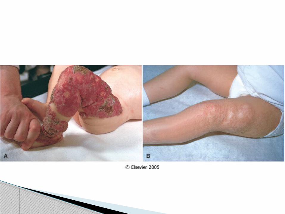
BENIGN TUMORS AND TUMOR-LIKE LESIONS

the most common tumors of infancy. Architecturally, they do not differ from those
encountered in adults. Both cavernous and capillary hemangiomas
may be encountered most are located in the skin, particularly on the
face and scalp, where they produce flat-to-elevated, irregular, red-blue masses; some of the flat, larger lesions (considered by some to represent vascular ectasias) are referred to as port-wine stains
Hemangioma.
Hemangiomas may enlarge along with the growth of the child, but in many instances they spontaneously regress
they can represent one facet of the hereditary disorder von Hippel-Lindau disease
Hemangioma

They may occur as: benign, well-differentiated cystic lesions
(mature teratomas), as lesions of indeterminate potential
(immature teratomas), or as unequivocally malignant teratomas
(usually admixed with another germ cell tumor component such as endodermal sinus tumor)
Teratomas
They exhibit two peaks in incidence: the first at approximately 2 years of age and the second in late adolescence or early
adulthood. The first peak represents congenital
neoplasms; the later-occurring lesions may also be of
prenatal origin but are more slowly growing
Teratomas
the most common teratomas of childhood, accounting for 40% or more of cases
They occur with a frequency of 1 in 20,000 to 40,000 live births,
four times more commonly in girls than in boys. approximately 10% of sacrococcygeal
teratomas are associated with congenital anomalies, primarily defects of the hindgut and cloacal region and other midline defects (e.g., meningocele, spina bifida) not believed to result from local effects of the tumor.
Sacrococcygeal teratomas

Approximately 75% of these tumors are mature teratomas, and
about 12% are unequivocally malignant and lethal.
The remainder are immature teratomas, and their malignant potential correlates with the amount of immature tissue, usually immature neuroepithelial elements, present.
Sacrococcygeal teratomas
Most of the benign teratomas are encountered in younger infants (<4 months), whereas children with malignant lesions tend to be somewhat older.
Other sites for teratomas in childhood include the testis ,ovaries ,and various midline locations, such as the mediastinum, retroperitoneum, and head and neck.
Sacrococcygeal teratomas


Cancers of infancy and childhood differ biologically and histologically from their counterparts occurring later in life
MALIGNANT TUMORS

Incidence and Types : The most frequent childhood cancers arise
in the hematopoietic system, nervous tissue (including the central and sympathetic nervous system, adrenal medulla, and retina), soft tissues, bone, and kidney.
This is in sharp contrast to adults, in whom the skin, lung, breast, prostate, and colon are the most common sites of tumors.

Leukemia Retinoblastoma Neuroblastoma Wilms tumor Hepatoblastoma Soft tissue sarcoma (especially
rhabdomyosarcoma Teratomas Central nervous system tumors
0 to 4 Years

Leukemia Retinoblastoma Neuroblastoma Hepatocarcinoma Soft tissue sarcoma Central nervous system tumors Ewing sarcoma Lymphoma
5 to 9 Years
Hepatocarcinoma Soft tissue sarcoma Osteogenic sarcoma Thyroid carcinoma Hodgkin disease
10 to 14 Years
many childhood tumors have been collectively referred to as small round blue cell tumors
SRCT

includes tumors of the sympathetic ganglia and adrenal medulla that are derived from primordial neural crest cells populating these sites
Neuroblastic tumor

neuroblastic tumors demonstrate certain characteristic features such as: spontaneous or therapy-induced differentiation of primitive neuroblasts into mature elements, spontaneous tumor regression, and a wide range of clinical behavior and prognosis, which often mirror the extent of histologic differentiation
Neuroblastic tumor

Neuroblastoma is the most important member of this family.
It is the second most common solid malignancy of childhood after brain tumors,
accounting for 7% to 10% of all pediatric neoplasms, and
as many as 50% of malignancies diagnosed in infancy
Neuroblastoma

The median age at diagnosis is 22 months; a little more than a third of the cases are diagnosed in infancy.
There is a higher incidence of neuroblastoma in Caucasian as compared to African American populations, and males are at a marginally greater risk than females.
Alone, it accounts for at least 15% of all childhood cancer deaths
Neuroblastoma

Most occur sporadically, but a few are familial with autosomal dominant transmission, and in such cases the neoplasms may involve both of the adrenals or multiple primary autonomic sites.
Neuroblastoma

40% of neuroblastomas arise in the adrenal medulla.
The remainder occur anywhere along the sympathetic chain, with the most common locations being the paravertebral region of the abdomen (25%) and posterior mediastinum (15%).
Tumors may arise in numerous other sites, including the pelvis and neck and within the brain (cerebral neuroblastomas).
Neuroblastoma
Clinical elevation in level of urinary catecholamines
Neurosecretory granules by electron microscopy
Neuron-specific enolase expression
Neuroblastoma , Useful Markers

Histologically, classic neuroblastomas are composed of small, primitive-appearing cells with dark nuclei, scant cytoplasm
small, blue, round cell tumors Some neoplasms show signs of maturation that
can be spontaneous or therapy-induced. Larger cells having more abundant cytoplasm with large vesicular nuclei and a prominent nucleolus, representing ganglion cells in various stages of maturation, may be found in tumors admixed with primitive neuroblasts (ganglioneuroblastoma)
Neuroblastoma

Metastases, when they develop, appear early and widely. In addition to local infiltration and lymph node spread, there is a pronounced tendency to spread through the bloodstream to involve the liver, lungs, bone marrow, and bones
Neuroblastoma
In young children, under age 2 years, neuroblastomas generally present with large abdominal masses, fever, and possibly weight loss.
In older children, they may not come to attention until metastases produce manifestations, such as bone pain, respiratory symptoms, or gastrointestinal complaints.
Neuroblastomas may metastasize widely through the hematogenous and lymphatic systems, particularly to liver, lungs, and bones, in addition to the bone marrow
Clinical Course and Prognostic Features
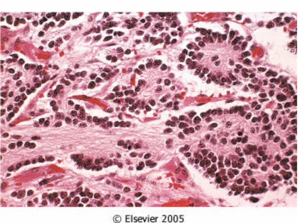
Ganglioneuromas, unlike their malignant counterparts, tend to produce either asymptomatic mass lesions or symptoms related to compression.
Ganglioneuromas

The course of neuroblastomas is extremely variable. Several clinical, histopathologic, molecular, and biochemical factors have been identified in neuroblastomas that have a bearing on prognosis
neuroblastomas


Related Documents