TUMOR REGRESSIONS OBSERVED IN PATIENTS WITH METASTATIC MELANOMA TREATED WITH AN ANTIGENIC PEPTIDE ENCODED BY GENE MAGE-3 AND PRESENTED BY HLA-A1 Marie MARCHAND 1,2,3 *, Nicolas VAN BAREN 1,2 , Patrick WEYNANTS 1,2,4 , Vincent BRICHARD 1,2,3 , Brigitte DRE ´ NO 5 , Marie-He ´le `ne T ESSIER 5 , Elaine RANKIN 6 , Giorgio PARMIANI 7 , Flavio ARIENTI 7 , Yves HUMBLET 3 , Andre ´BOURLOND 3 , Romain V ANWIJCK 3 , Danielle LIE ´ NARD 8 , Marc BEAUDUIN 9 , Pierre-Yves DIETRICH 10 , Vincenzo RUSSO 11 , Joseph KERGER 12 , Giuseppe MASUCCI 13 , Elke JA ¨ GER 14 , Jacques DE GREVE 15 , Jens ATZPODIEN 16 , Francis BRASSEUR 1,2 , Pierre G. COULIE 2 , Pierre VAN DER BRUGGEN 1,2 and Thierry BOON 1,2 1 Ludwig Institute for Cancer Research, Brussels Branch, Brussels, Belgium 2 Cellular Genetics Unit, Universite ´ Catholique de Louvain, Brussels, Belgium 3 Department of Oncology, Cliniques Universitaires Saint-Luc, Universite ´ Catholique de Louvain, Brussels, Belgium 4 Department of Pneumology, Cliniques Universitaires de Mont-Godinne, Universite ´ Catholique de Louvain, Yvoir, Belgium 5 Department of Dermatology, Ho ˆtel Dieu, Centre Hospitalier Universitaire, Nantes, France 6 Department of Oncology, Antoni van Leeuwenhoek Huis, The Netherlands Cancer Institute, Amsterdam, The Netherlands 7 Division of Experimental Oncology D, Instituto Nazionale per lo Studio e la Cura dei Tumori, Milan, Italy 8 Ludwig Institute for Cancer Research, Lausanne Branch, and Centre Pluridisciplinaire d’Oncologie, Centre Hospitalier Universitaire Vaudois, Lausanne, Switzerland 9 Department of Oncology and Nuclear Medicine, Ho ˆ pital de Jolimont, Haine-Saint-Paul, Belgium 10 Department of Oncology, Ho ˆpital Cantonal Universitaire de Gene `ve, Geneva, Switzerland 11 Gene Therapy Unit, H. San Raffaele, Milan, Italy 12 Department of Oncology, Institut Jules Bordet, Universite ´ Libre de Bruxelles, Brussels, Belgium 13 Department of Oncology, Karolinska Institutet Radiumhemmet, Stockholm, Sweden 14 Department of Hematology and Oncology, II. Medizinische Klinik, Krankenhaus Nordwest, Frankfurt am Main, Germany 15 Department of Oncology, Akademisch Ziekenhuis, Vrije Universiteit Brussel, Brussels, Belgium 16 Department of Hematology and Oncology, Medizinische Hochschule Hannover, Hannover, Germany Thirty-nine tumor-bearing patients with metastatic mela- noma were treated with 3 subcutaneous injections of the MAGE-3.A1 peptide at monthly intervals. No significant toxicity was observed. Of the 25 patients who received the complete treatment, 7 displayed significant tumor regres- sions. All but one of these regressions involved cutaneous metastases. Three regressions were complete and 2 of these led to a disease-free state, which persisted for more than 2 years after the beginning of treatment. No evidence for a cytolytic T lymphocyte (CTL) response was found in the blood of the 4 patients who were analyzed, including 2 who displayed complete tumor regression. Our results suggest that injection of the MAGE-3.A1 peptide induced tumor regression in a significant number of the patients, even though no massive CTL response was produced. Int. J. Cancer 80:219–230, 1999. r 1999 Wiley-Liss, Inc. Human melanoma cells bear antigens that can be recognized by autologous cytolytic T lymphocytes (CTL) (Boon et al., 1994). Some of these antigens are encoded by genes of the MAGE family (van der Bruggen et al., 1991). The MAGE genes are expressed in a significant proportion of tumors of various histological origins. They are not expressed in normal cells, except male germ line cells, which are devoid of major histocompatibility complex (MHC) molecules and therefore do not present MAGE antigens (Takahashi et al., 1995; Tomita et al., 1993; Uyttenhove et al., 1997). The MAGE-encoded antigens are therefore tumor-specific shared anti- gens. Gene MAGE-3 codes for an antigenic nonapeptide that is recognized by cytolytic T cells on the human leukocyte antigen (HLA)-A1 molecule (Gaugler et al., 1994). Analysis by reverse transcription-polymerase chain reaction (RT-PCR) amplification of a large number of melanoma samples indicated that MAGE-3 was expressed in 76% of metastatic melanomas and in 36% of primary melanomas (Brasseur et al., 1995). In Europe, about 28% of the population carry the HLA-A1 allele. Accordingly, about 21% of the metastatic melanomas are expected to present the MAGE-3.A1 antigen. We examined whether immunization against the MAGE-3.A1 antigen can be of therapeutic benefit to cancer patients. The objectives of our study were to define the safety and toxicity of the MAGE-3.A1 peptide, to observe the clinical evolution of tumor- bearing patients immunized with this antigenic peptide and to find out whether tumor regressions were linked to strong CTL re- sponses. In 1995, we published a preliminary report describing 3 tumor regressions observed among 6 melanoma patients treated with subcutaneous (s.c.) injections of the MAGE-3.A1 peptide given 3 times at monthly intervals (Marchand et al., 1995). To confirm these results, our study was extended to a larger group of patients. We report here the observations made on 39 melanoma tumor-bearing patients. SUBJECTS AND METHODS Eligibility criteria From March 1994 to July 1997, HLA-A1-positive patients whose tumor expressed the MAGE-3 gene were included in our study (LB 94-001) if they were older than 18 years of age and had a good performance status (WHO scale 0 or 1). The study was opened to melanoma patients with histologically proven tumors of stages III and IV, according to the American Joint Committee on Cancer staging system (AJCC, 1992). The patients described here were classified in one of the following subgroups according to the Grant sponsors: Inter-University Poles of Attraction Program, Belgium; La fondation du patrimoine de l’Universite ´ Catholique de Louvain (UCL), Belgium; Association contre le Cancer, Brussels, Belgium; the BIOMED-2 program of the European Community; Fonds J. Maisin, Belgium; CGER- Assurances, VIVA, Brussels, Belgium; La Ligue Contre le Cancer, Paris, France; Fonds National de la Recherche Scientifique, Brussels, Belgium. *Correspondence to: Ludwig Institute for Cancer Research, Avenue Hippocrate 74, UCL 74.59, B-1200 Brussels, Belgium. Fax: (32) 2-764-65- 65. E-mail: [email protected] Received 11 May 1998; Revised 29 July 1998 Int. J. Cancer: 80, 219–230 (1999) r 1999 Wiley-Liss, Inc. Publication of the International Union Against Cancer Publication de l’Union Internationale Contre le Cancer

Welcome message from author
This document is posted to help you gain knowledge. Please leave a comment to let me know what you think about it! Share it to your friends and learn new things together.
Transcript
TUMOR REGRESSIONS OBSERVED IN PATIENTS WITH METASTATICMELANOMA TREATED WITH AN ANTIGENIC PEPTIDE ENCODEDBY GENE MAGE-3AND PRESENTED BY HLA-A1Marie MARCHAND1,2,3*, NicolasVAN BAREN1,2, Patrick WEYNANTS1,2,4, Vincent BRICHARD1,2,3, Brigitte DRENO5, Marie-Helene TESSIER5,Elaine RANKIN 6, Giorgio PARMIANI 7, Flavio ARIENTI7, Yves HUMBLET3, AndreBOURLOND3, Romain VANWIJCK3, Danielle LIENARD8,Marc BEAUDUIN9, Pierre-Yves DIETRICH10, Vincenzo RUSSO11, Joseph KERGER12, Giuseppe MASUCCI13, Elke JAGER14,Jacques DE GREVE15, Jens ATZPODIEN16, Francis BRASSEUR1,2, Pierre G. COULIE2, PierreVAN DER BRUGGEN1,2 and Thierry BOON1,2
1Ludwig Institute for Cancer Research, Brussels Branch, Brussels, Belgium2Cellular Genetics Unit, Universite´ Catholique de Louvain, Brussels, Belgium3Department of Oncology, Cliniques Universitaires Saint-Luc, Universite´ Catholique de Louvain, Brussels, Belgium4Department of Pneumology, Cliniques Universitaires de Mont-Godinne, Universite´ Catholique de Louvain, Yvoir, Belgium5Department of Dermatology, Hoˆtel Dieu, Centre Hospitalier Universitaire, Nantes, France6Department of Oncology, Antoni van Leeuwenhoek Huis, The Netherlands Cancer Institute, Amsterdam, The Netherlands7Division of Experimental Oncology D, Instituto Nazionale per lo Studio e la Cura dei Tumori, Milan, Italy8Ludwig Institute for Cancer Research, Lausanne Branch, and Centre Pluridisciplinaire d’Oncologie, Centre Hospitalier UniversitaireVaudois, Lausanne, Switzerland9Department of Oncology and Nuclear Medicine, Hoˆpital de Jolimont, Haine-Saint-Paul, Belgium10Department of Oncology, Hoˆpital Cantonal Universitaire de Gene`ve, Geneva, Switzerland11Gene Therapy Unit, H. San Raffaele, Milan, Italy12Department of Oncology, Institut Jules Bordet, Universite´ Libre de Bruxelles, Brussels, Belgium13Department of Oncology, Karolinska Institutet Radiumhemmet, Stockholm, Sweden14Department of Hematology and Oncology, II. Medizinische Klinik, Krankenhaus Nordwest, Frankfurt am Main, Germany15Department of Oncology, Akademisch Ziekenhuis, Vrije Universiteit Brussel, Brussels, Belgium16Department of Hematology and Oncology, Medizinische Hochschule Hannover, Hannover, Germany
Thirty-nine tumor-bearing patients with metastatic mela-noma were treated with 3 subcutaneous injections of theMAGE-3.A1 peptide at monthly intervals. No significanttoxicity was observed. Of the 25 patients who received thecomplete treatment, 7 displayed significant tumor regres-sions. All but one of these regressions involved cutaneousmetastases. Three regressions were complete and 2 of theseled to a disease-free state, which persisted for more than 2years after the beginning of treatment. No evidence for acytolytic T lymphocyte (CTL) response was found in theblood of the 4 patients who were analyzed, including 2 whodisplayed complete tumor regression. Our results suggestthat injection of the MAGE-3.A1 peptide induced tumorregression in a significant number of the patients, eventhough no massive CTL response was produced. Int. J. Cancer80:219–230, 1999.r 1999 Wiley-Liss, Inc.
Human melanoma cells bear antigens that can be recognized byautologous cytolytic T lymphocytes (CTL) (Boonet al., 1994).Some of these antigens are encoded by genes of theMAGE family(van der Bruggenet al.,1991). TheMAGEgenes are expressed in asignificant proportion of tumors of various histological origins.They are not expressed in normal cells, except male germ line cells,which are devoid of major histocompatibility complex (MHC)molecules and therefore do not present MAGE antigens (Takahashiet al., 1995; Tomitaet al., 1993; Uyttenhoveet al., 1997). TheMAGE-encoded antigens are therefore tumor-specific shared anti-gens.
Gene MAGE-3 codes for an antigenic nonapeptide that isrecognized by cytolytic T cells on the human leukocyte antigen(HLA)-A1 molecule (Gaugleret al., 1994). Analysis by reversetranscription-polymerase chain reaction (RT-PCR) amplification ofa large number of melanoma samples indicated thatMAGE-3 wasexpressed in 76% of metastatic melanomas and in 36% of primarymelanomas (Brasseuret al., 1995). In Europe, about 28% of thepopulation carry the HLA-A1 allele. Accordingly, about 21% of themetastatic melanomas are expected to present the MAGE-3.A1antigen.
We examined whether immunization against the MAGE-3.A1antigen can be of therapeutic benefit to cancer patients. Theobjectives of our study were to define the safety and toxicity of theMAGE-3.A1 peptide, to observe the clinical evolution of tumor-bearing patients immunized with this antigenic peptide and to findout whether tumor regressions were linked to strong CTL re-sponses. In 1995, we published a preliminary report describing 3tumor regressions observed among 6 melanoma patients treatedwith subcutaneous (s.c.) injections of the MAGE-3.A1 peptidegiven 3 times at monthly intervals (Marchandet al., 1995). Toconfirm these results, our study was extended to a larger group ofpatients. We report here the observations made on 39 melanomatumor-bearing patients.
SUBJECTS AND METHODS
Eligibility criteria
From March 1994 to July 1997, HLA-A1-positive patientswhose tumor expressed theMAGE-3 gene were included in ourstudy (LB 94-001) if they were older than 18 years of age and had agood performance status (WHO scale 0 or 1). The study wasopened to melanoma patients with histologically proven tumors ofstages III and IV, according to the American Joint Committee onCancer staging system (AJCC, 1992). The patients described herewere classified in one of the following subgroups according to the
Grant sponsors: Inter-University Poles of Attraction Program, Belgium;La fondation du patrimoine de l’Universite´ Catholique de Louvain (UCL),Belgium; Association contre le Cancer, Brussels, Belgium; the BIOMED-2program of the European Community; Fonds J. Maisin, Belgium; CGER-Assurances, VIVA, Brussels, Belgium; La Ligue Contre le Cancer, Paris,France; Fonds National de la Recherche Scientifique, Brussels, Belgium.
*Correspondence to: Ludwig Institute for Cancer Research, AvenueHippocrate 74, UCL 74.59, B-1200 Brussels, Belgium. Fax: (32) 2-764-65-65. E-mail: [email protected]
Received 11 May 1998; Revised 29 July 1998
Int. J. Cancer:80,219–230 (1999)
r 1999 Wiley-Liss, Inc.
Publication of the International Union Against CancerPublication de l’Union Internationale Contre le Cancer

extent of their disease: regional disease (stage III)N2a,metastasisof more than 3 cm in greatest dimension in any regional lymphnodes;N2b, in-transit metastasis;N2c,bothN2aandN2b.Distantmetastasis (stage IV)M1a,metastasis in skin or s.c. tissue or lymphnode(s) beyond the regional lymph nodes, andM1b, visceralmetastasis. Patients with brain metastasis, as assessed by braincomputed tomography (CT) scan, were excluded. HLA class Ityping was performed on blood lymphocytes by classical serologi-cal methods. The expression of geneMAGE-3 was assessed byRT-PCR on a small frozen tumor sample as described previously(Weynantset al., 1994). No other anti-tumor treatment was givenduring the study. All participants gave written informed consentbefore entering the study and were treated as outpatients.
Thirty-nine tumor-bearing melanoma patients entered the studyand their evolution is described in this report. In addition, 6tumor-free melanoma patients and 4 tumor-bearing patients withtumors other than melanoma received the same treatment. Noregression or significant stabilization was observed in any of the 4non-melanoma tumor-bearing patients, whose evolution is notdescribed in the present report.
VaccinationThe MAGE-3.A1 nonapeptide EVDPIGHLY (acetate) was syn-
thesized by U.C.B-Bioproducts (Braine-l’Alleud, Belgium; UCBSF124, batch 1S1, issued on 10/03/1994). The lyophilized peptidewas diluted in sterile, endotoxin-free phosphate-buffered saline(PBS), pH 7.4 (Boehringer Ingelheim Bioproducts) at a concentra-tion of 100 or 300 µg/ml, and distributed in 1.5-ml vials for singleinjections. Vials were stored at280°C and thawed just beforeinjection.
The treatment consisted of 3 vaccinations at 1-month intervals.For each vaccination, the peptide was injected at a total dose ofeither 100 or 300 µg. The vaccine was divided between 2 s.c. sitesand 2 intradermal (i.d.) sites distant from the tumor (4⁄10 of the dosewas injected at each s.c. site, and1⁄10 at each i.d. site). No injectionswere given into limbs where the draining lymph nodes had beensurgically removed or irradiated. When possible, the injection siteswere changed for each vaccination.
Some tumor-bearing patients who displayed tumor regressionfollowing 3 immunizations received additional vaccinations withthe MAGE-3.A1 peptide. In such cases, the same dose and route ofadministration were used.
Follow-up of patientsToxicity was evaluated according to the National Cancer Insti-
tute of Canada common toxicity criteria. Tumor staging was carriedout at study entry and in the 2-week period following the thirdinjection, by physical examination and CT scans of brain, chest andabdomen. For patients who displayed tumor stabilization orregression, additional tumor staging was performed at regularintervals.
Because our report was aimed at assessing all possible evidencefor an effect of the treatment on tumor evolution, we describe herethe tumor regressions in detail. In future studies, for which the aimwill be the quantitative evaluation of the efficacy of the treatment,tumor responses will be reported according to the synthetic WHOcriteria.
Analysis of the CTL responseThe presence of anti-MAGE-3 CTL was evaluated as follows.Method A. Autologous peripheral blood mononuclear cells
(PBMC) were thawed, resuspended in serum-free X-VIVO 10medium (Whittaker, Walkersville, MD), and incubated over 40 minat 4°C with magnetic microbeads coated with an anti-CD8antibody (MACS; Miltenyi, Bergisch Gladbach, Germany). TheCD81 cells were sorted with a magnet, washed, resuspended instimulation medium consisting of Dulbecco’s modified Eagle’smedium (DMEM) (GIBCO, Eggenstein, Germany) supplementedwith 5% human serum,L-arginine (116 mg/L),L-asparagine (36
mg/L), L-glutamine (216 mg/L) and 2-mercaptoethanol (53 1025
M), and seeded at 105 cells/well in 1-ml wells (Falcon 3078, BectonDickinson, Mountain View, CA). Cells (53 105 CD81) wereseeded for patient 1 (LB-945) and 106 CD81 cells for patient 7(AVL-3). The CD82 cells were washed, resuspended in X-VIVO10 medium containing MAGE-3.A1 peptide EVDPIGHLY orinfluenza.A1 peptide CTELKLSDY (100 µM), and incubated atroom temperature for 60 min, during which time the cells wereirradiated (30 Gy from a137Cs source), washed and resuspended instimulation medium. These stimulator cells were added (106
cells/well) to the CD81 lymphocytes, and human rIL-2 (Proleukin,Chiron, Amsterdam, The Netherlands) was added at 30 U/ml (1U/ml is the concentration that gives half-maximal proliferation ofCTLL-2 cells). On day 7, the responder lymphocytes of each wellwere washed, resuspended in medium containing the same concen-tration of rIL-2 in 2-ml wells, restimulated with 106 autologousPBMC pulsed with the relevant peptide and irradiated as on day 0.On day 14, restimulation was performed under the same condi-tions, except that peptide-pulsed C1R cells (Storkuset al., 1987)transfected with an HLA-A1 construct were used as stimulators.On day 21, aliquots of the cultures were tested in a chromium-release assay against Epstein-Barr virus (EBV)-transformed B cellsBM21, in the presence or absence of the relevant antigenicpeptides, and in the presence of a 50-fold excess of unlabelledK562 cells to inhibit the activity of natural killer (NK)-like lyticeffectors.
Method B.Another approach involving a large number of CD81
lymphocytes (more than 23 107) was carried out as describedelsewhere (Chauxet al.,1998). In brief, responder CD81 T cells ofpatient 7 (AVL-3) were distributed among 96 microcultures(226,000 cells/well). Autologous monocyte-derived dendritic cellswere irradiated, incubated with 50 µM of the MAGE-3.A1 peptideEVDPIGHLY, and distributed in the microcultures (85,000 cells/well) to stimulate the autologous CD81 T cells. IL-6 (1,000 U/ml)and IL-12 (10 ng/ml) were added during the first week, followed byIL-2 (10 U/ml) and IL-7 (5 ng/ml) from day 7. The microcultureswere restimulated each week with autologous phytohemagglutinin(PHA)-activated T cells (150,000 cells/well), incubated with thepeptide and irradiated. On day 25, aliquots of the cultures weretested in a chromium-release assay against the HLA-A1 EBV-transformed B cell line BM21 in the presence or absence of theMAGE-3.A1 peptide, and in the presence of a 50-fold excess ofunlabelled K562 cells to inhibit the activity of NK-like lyticeffectors.
RESULTS
We treated 39 HLA-A1 patients having a melanoma thatexpressed geneMAGE-3.All patients had detectable metastases.The treatment consisted of 3 vaccinations with the syntheticMAGE-3.A1 peptide at monthly intervals. For each vaccination, 7patients received a total dose of 100 µg, 32 patients received 300µg. Peptide was injected in 2 s.c. and 2 i.d. injection sites distantfrom the tumor.
ToxicityNo significant toxicity or side effects were observed in any of the
patients. Six patients had a mild inflammatory reaction at the site ofsuperficial metastases (grade 1 toxicity). No delayed-type hypersen-sitivity reactions or other inflammatory reactions were observed atthe sites of i.d. injection.
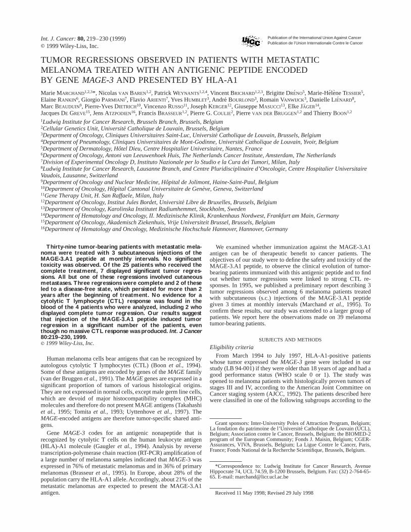
Tumor evolutionOf the 39 patients, 14 were withdrawn after the first or the
second injection because of rapid progression of the diseasenecessitating other forms of treatment. Of these 14 patients, 3 hadregional disease and 11 had distant metastases at study entry(Fig. 1). Three patients died of tumor progression in the 4-weekperiod following the last injection.
220 MARCHAND ET AL.
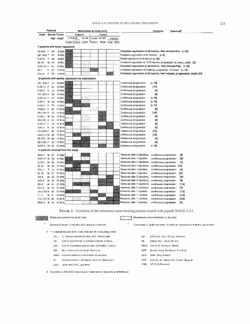
FIGURE 1 – Evolution of the melanoma tumor-bearing patients treated with peptide MAGE-3.A1.
221MAGE-3.A1 PEPTIDE IN MELANOMA TREATMENT
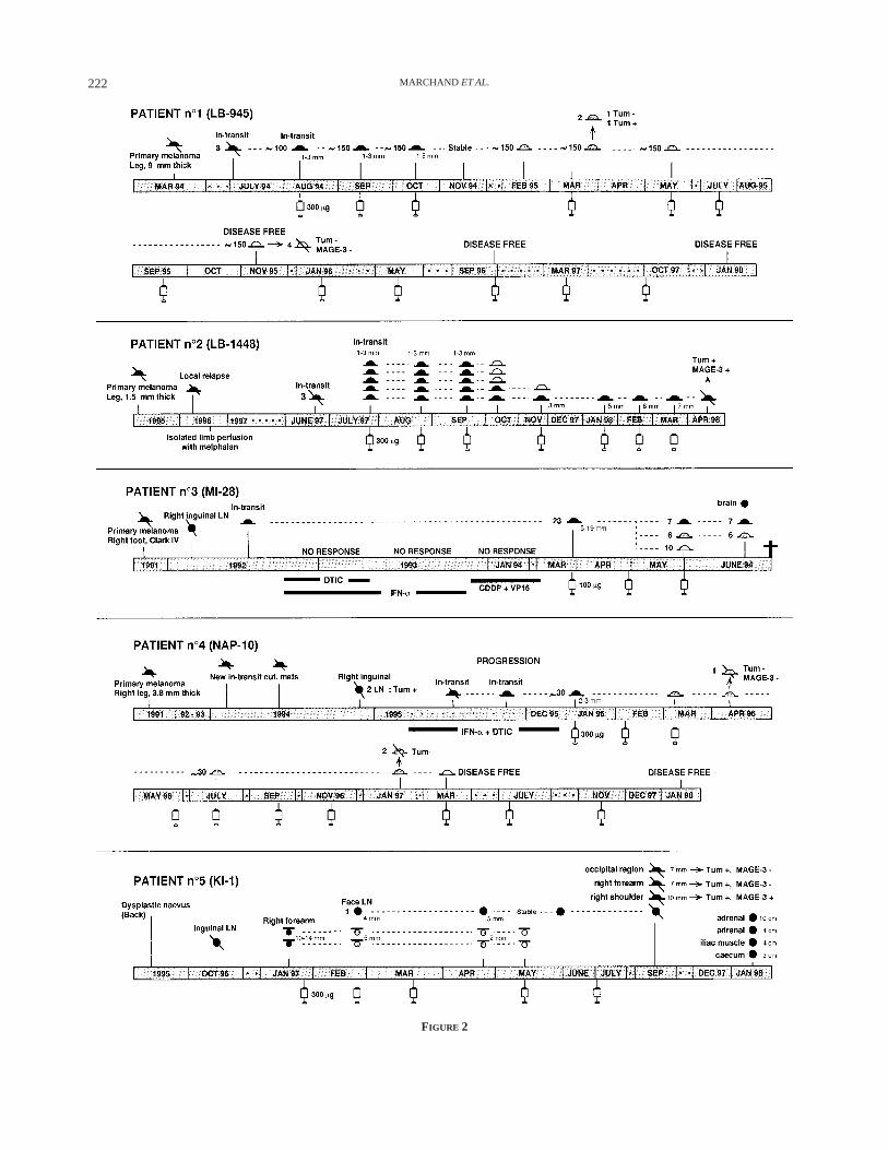
FIGURE 2
222 MARCHAND ET AL.

Among the 25 tumor-bearing patients who received the plannedtreatment of 3 vaccinations, neither regression nor stabilization wasobserved with 18 patients who all showed continuous progression.Seven of these patients had regional disease, whereas 11 had distantmetastases (Fig. 1).
Tumor regressions were observed in 7 of the 25 patients whoreceived the complete treatment (Fig. 1). Five of these patients hadregional disease only and 2 had distant metastases. Their evolutionis described below and summarized in Figures 2 and 3.
Patient 1 (LB-945, female, 49 years) had about 100 smallcutaneous in-transit metastases on the left leg at study entry(Fig. 2). The patient had received no treatment other than surgery.The MAGE-3.A1 peptide (300 µg) was injected in the thighs and inthe arms. By the second injection, the cutaneous nodules hadbecome more numerous, but no further progression was observedat the time of the third injection. The stabilization was confirmed 1month later. Four months after the third injection, all the nodulesappeared flattened and dry (Fig. 4a). One month later, 2 nodulesites were biopsied and found to contain a large number ofmacrophages containing melanin. A few remaining tumor cellswere observed in one site and none in the other. Because of thefavorable clinical evolution, the patient received 4 additionalinjections of the same peptide at 2-month intervals. No othertreatment was given. Fifteen months after the first injection, thepatient appeared to be completely free of disease: all the skinlesions had become impalpable even though melanin tattooingremained visible at the sites of the former nodules. Biopsies ofseveral sites revealed no tumor cells and noMAGE-3expression asassessed by RT-PCR. Physical examinations and CT scans showedno evidence of disease. The patient received additional injections atincreasingly longer intervals. Forty-one months after the onset oftreatment, she was still disease-free and the melanin tattooing hadcompletely disappeared (Fig. 4a).
Patient 2 (LB-1448, female, 67 years) had undergone in 1996surgical excision of a cutaneous local relapse on the right leg,followed by adjuvant isolated perfusion with melphalan (Fig. 2). InJune 1997, 3 new in-transit cutaneous metastases appeared andwere removed surgically. One nodule was tested forMAGE-3expression and found to be positive. One month later, the patientdeveloped 5 new small cutaneous in-transit metastases on the rightleg. Vaccination was initiated with 300 µg of MAGE-3.A1 peptide.Two weeks after the third injection, 3 of the 5 in-transit metastaseshad completely disappeared, the 2 remaining ones appeared to bestable, and no new metastases had appeared. Six weeks later, only 1nodule of 3 mm diameter remained present. Two additionalvaccinations were carried out at 2-month intervals. The completeregression of 4 of the 5 nodules continued 9 months after studyentry, but, at that time, the 5th nodule had progressed from 3 to 7mm diameter. It was excised and found to express geneMAGE-3.
Patient 3 (MI-28, male, 59 years) had melanoma localized to theright leg with 23 cutaneous or subcutaneous in-transit metastaticnodules ranging from 5 to 19 mm diameter at study entry (Fig. 2).These nodules had not responded to several cycles of chemother-apy involving dacarbazine (DTIC), interferon-a (IFN-a) and acombination of cisplatin and etoposide. The patient was vaccinatedwith 100 µg of peptide, starting 4 weeks after cessation of
chemotherapy. By the third injection, 10 of the 23 nodules,including the largest one, had regressed completely. Six othernodules had regressed partially. Six nodules were stable and onewas progressing. Three weeks later, a brain metastasis wasdiscovered and the patient died soon thereafter.
Patient 4 (NAP-10, female, 65 years) had a primary melanoma ofthe right leg removed in 1991 (Fig. 2). In 1994, in-transitmetastases and 2 invaded regional lymph nodes were removed. In1995, despite adjuvant treatment associating DTIC and IFN-a, newin-transit metastases appeared and were removed, and this treat-ment was interrupted by the end of the year. In January 1996, whenvaccination with 300 µg of peptide was started, the patientdisplayed about 30 small cutaneous in-transit metastases dissemi-nated on the right leg and thigh (Fig. 4b). By the third vaccination,partial flattening of all the lesions was observed. One month later,one regressing lesion was biopsied and neitherMAGE-3expressionnor tumor cells were found. By September 1996, all the lesionswere almost completely flat (Fig. 4b). The patient receivedadditional injections with the same amount of peptide, first at2-month and then at 4-month intervals. Fourteen months after theonset of treatment, all the lesions were impalpable. After 24months, the patient remained free of disease.
Patient 5 (KI-1, female, 42 years) had a suspect lesion of theback removed in 1995 (Fig. 2). In October 1996, an inguinal lymphnode was removed and was found to contain melanoma cellsexpressingMAGE-3. In January 1997, 2 new s.c. metastasesmeasuring 10 and 14 mm diameter, respectively, appeared on theright upper arm. Vaccination with 300 µg of peptide was initiated.By the second injection, the 2 s.c. metastases had reduced to 5 mmdiameter, but a small metastatic maxillary lymph node hadappeared. Two months after the third injection of peptide, the 2 s.c.lesions of the right arm had totally disappeared and the lymph nodewas stable. Two additional injections were given. Three monthslater, during which the lymph node had remained stable, 3 newcutaneous metastases had appeared. All the lesions were removedsurgically and were found to contain tumor cells. GeneMAGE-3was expressed in only 1 of the 3 cutaneous lesions. Because of thistumor progression, the injections were stopped. The patient thenremained disease-free for a period of 4 months, after which sherelapsed with several visceral metastases.
Patient 6 (NAP-8, female, 48 years) had a primary melanoma ofthe left leg in 1989 (Fig. 3). Between 1990 and 1995, in-transitmetastases appeared and displayed continuous slow progression,despite repeated treatments with DTIC, vindesine and IFN-a. Thepatient was vaccinated with 300 µg of peptide, starting in October1995. At that time, she had about 40 small cutaneous in-transitmetastases, the largest one measuring 11 mm diameter. At the timeof the third injection, all the cutaneous nodules displayed someflattening. The patient received additional injections of peptide. Inthe following months, a few cutaneous nodules disappeared and theother lesions were either moderately regressing or stable. In May1996, small doses of IFN-a were injected in an attempt to favor theputative immune rejection of the tumor by increasing its expressionof HLA class I molecules. The patient received 106 U IFN-a s.c., 3times per week for 4 weeks. No acceleration of the regressionprocess was observed. Vaccination with the MAGE-3 peptide was
FIGURE 2 – Clinical evolution of patients with tumor regression.
223MAGE-3.A1 PEPTIDE IN MELANOMA TREATMENT
FIGURE 3
224 MARCHAND ET AL.

pursued. Between May 1996 and December 1997, the cutaneousmetastases continued to regress slowly, but significantly. However,a small s.c. nodule appeared on the left thigh in March 1997.
In addition to the melanoma lesions of the leg, this patientdisplayed a suspect lung nodule of 15 mm diameter at the onset oftreatment. This nodule remained stable throughout 1996, butincreased gradually in size throughout 1997 to reach 25 mmdiameter. In February 1998, it was excised and diagnosed as aperipheral lung adenocarcinoma. This second tumor was negativefor the expression of theMAGE-3gene.
Patient 7 (AVL-3, female, 50 years) had undergone removal ofregional lymph node metastases in October 1994 (Fig. 3). She hadnot received any form of treatment other than surgery. In April1995, she presented with 3 lung metastases and vaccination with300 µg of peptide was started. At the time of the third injection, aCT scan revealed an increase in the size of the known lung nodulesand 2 new lung metastases. In addition, an enlarged mediastinallymph node was visible. Two months later, 2 of the 5 lung noduleshad disappeared and the 3 remaining nodules had shrunk signifi-cantly. The mediastinal lymph node was stable. Because of thisfavorable evolution, the patient received an additional peptideinjection, and no other form of treatment was given. One monthlater, 1 of the 3 metastases had regressed further and the 2 othershad disappeared. Two months later, in November 1995, all themetastatic nodules had disappeared and the mediastinal lymphnode had returned to normal size (Fig. 5). The patient displayed noother evidence of disease. She received 3 additional injections withthe same amount of peptide, at 2-month intervals, and remainedfree of disease for an additional 4 months. In March 1996, a newmetastatic mediastinal lymph node was observed. Injections werediscontinued and local radiotherapy was given to this lesion, whichpartially regressed and then remained stable. Ten months later, as.c. and an adrenal metastasis appeared. Because the s.c. metastasisexpressed both theMAGE-3andMAGE-1genes, the patient wasincluded in another study involving vaccination with both theMAGE-3.A1 peptide and aMAGE-1-encoded peptide that is alsopresented by HLA-A1 (Traversariet al., 1992). However, thetumor continued to progress during vaccination. Subsequent chemo-therapy with DTIC caused a transient partial response. The patientdied from tumor progression, 33 months after entering the treat-ment with the MAGE-3.A1 peptide. It is noteworthy that during the22 months following her first relapse the patient acquired progres-sive lesions in several sites but none in the 5 lung sites or in themediastinal site that had regressed, suggesting that the regressionsat these sites had removed all the tumoral cells, although smallermetastases had escaped the process that caused the regressions.
Some features of the regressions that were observed deserve tobe mentioned. In some patients, the regressions appeared to havestarted at a late time: the first signs of regression were observed 4months after the third immunization in patient 1 and 2 months inpatient 7. Late occurrence was not the mark of weak regressions asboth patients displayed complete regression. Moreover, once theyhad started, most regressions proceeded very slowly. It took 9months for the regressions of patients 1 and 4 to become complete.Patient 6 constitutes a remarkable case with cutaneous lesions thatwere still decreasing very slowly but continuously 22 months afterthe first signs of regression were noticed.
In patients 1, 4 and 6, all the cutaneous lesions, which werespread over a wide area, regressed more or less concurrently.However, in patients 2, 3 and 5, the regressions were nothomogeneous: some lesions disappeared completely, while othersremained stable or progressed.
The occurrence of the slow and incomplete regressions as seenwith patients 2, 5 and 6, and that of the relapse after completeregression observed with patient 7, raises the issue of whether themaintenance of a more frequent injection schedule pursued evenafter complete regression might not have improved the fate of someof these patients. Possibly relevant to this issue are the followingobservations made with 1 patient who did not regress afterimmunization with the MAGE-3.A1 peptide. Patient LB-1348(female, 73 years) had a primary melanoma of the left leg, and alocal relapsed lesion removed in 1994 (Figs. 3, 4). A new in-transitmetastasis was excised in January 1996 and found to expressMAGE-3 and notMAGE-1.Although the patient was treated bylimb perfusion with melphalan, new cutaneous in-transit nodulesappeared 3 months later. In July 1996, the patient displayed 9 smallcutaneous nodules on the left leg, and vaccination with 300 µg ofpeptide was initiated. By November, the number of the nodules hadincreased to 28, their size had increased and s.c. lesions hadappeared. But a few nodules displayed a necrotic aspect and tofavor this putative regressive process, 4 additional injections weregiven at 2-week intervals. By February 1997, 40 cutaneous and 12s.c. nodules were spread over the left leg. However, 1 small nodulehad become completely flat, while an adjacent 1 showed clear signsof regression. Because a cutaneous nodule removed in December1996 had been found to express not onlyMAGE-3 but alsoMAGE-1,unlike the nodule tested in January 1996, the patient wasincluded in the study involving immunization with both theMAGE-3.A1 and the MAGE-1.A1 peptides. Between Februaryand April 1997, a few additional small cutaneous nodules becamecompletely flat but new cutaneous and s.c. nodules appeared andothers enlarged (Fig. 4c). The treatment was pursued. By August1997, there was widespread regression: many cutaneous and s.c.nodules had completely disappeared, while many others hadbecome flatter. However, 2 metastases necessitated local radio-therapy because of progression and ulceration. Following electronbeam irradiation limited to an area surrounding the 2 lesions (Fig.4c), both lesions flattened and became necrotic. During thefollowing months, many cutaneous and s.c. nodules located faraway from the irradiated region regressed. Additional immuniza-tions were performed. By February 1998, almost all the cutaneousnodules had completely regressed and the others were clearlyregressing (Fig. 4c). All the s.c. nodules had completely disap-peared. No distant metastases were detected. We cannot excludethat the combination of 2 peptides produced a regression that theMAGE-3.A1 peptide would not have produced alone. It is alsopossible that a similar result could have been achieved by pursuingfrequent injections with the single MAGE-3.A1 peptide throughout1997.
Our results provide no significant evidence that the higher doseof peptide was more effective in inducing regressions than thelower dose. The lower dose was administered to1⁄7 patients whoshowed tumor regression, to5⁄14 who were removed from the trialand to1⁄18 who showed continuous tumor progression.
FIGURE 3 – Clinical evolution of patients with tumor regression (followed).
225MAGE-3.A1 PEPTIDE IN MELANOMA TREATMENT

FIGURE 4 – Evolution of cutaneous and subcutaneous in-transit melanoma metastases. For patient LB-1348, the 2 nodules that were irradiatedare indicated by arrows; the irradiated field is surrounded by the black line.
226 MARCHAND ET AL.

Analysis of the CTL responseWe analyzed 53 105 CD81 T lymphocytes in a blood sample
collected from patient 1 (LB-945) during the tumor regression(March 1995). These lymphocytes were stimulated 3 timesin vitrowith cells pulsed with the MAGE-3.A1 peptide (see Method A inthe Subjects and Methods section). No CTL activity was observedagainst targets expressing the MAGE-3.A1 antigen. A similaranalysis was performed with 106 lymphocytes from patient 7(AVL-3) collected when regression was ongoing (August 1995) orcomplete (January 1996). The results were also negative. Thelymphocytes of patients 1 and 7 produced significant CTL activityupon stimulation with an influenza peptide, indicating that the lympho-cytes of these patients remained capable of responding to antigenicstimulation. The lymphocytes of patients LB-1045 and LB-278, forwhom neither regression nor stabilization was observed, were alsorestimulated with the MAGE-3.A1 peptide and no CTL activity specificfor targets expressing the MAGE-3.A1 antigen was detected.
Stimulation of large numbers of CD81 T lymphocytes (morethan 107) by peptide-pulsed cells has made it possible to evaluatethe frequency of anti-MAGE-3.A1 CTL precursors in cancer-freeindividuals (Chauxet al., 1998). This frequency was estimated at2 3 1027. The presence of anti-MAGE-3.A1 CTL precursors wastested using this method in 23 107 blood CD81 T lymphocytes of
patient 7, 2 months after documentation of a complete regression(see Method B in the Subjects and Methods section). The observedfrequency was 1.33 1027.
Thus, the complete regressions observed in patients 1 and 7 werenot paralleled by a strong increase in circulating CTL precursorsdirected against the MAGE-3.A1 antigen.
DISCUSSION
Of a total of 39 melanoma tumor-bearing patients who receivedthe MAGE-3.A1 peptide, 14 were removed after only 1 or 2injections because of fast progression, and 18 showed continuousprogression without any regression or significant stabilization, but7 patients showed significant regressions. Three regressions werecomplete and 2 of these have led to a disease-free state maintainedfor more than 2 years after the beginning of treatment. Theseregressions are very likely to be linked to the treatment, given thevery low frequency of spontaneous complete regressions reportedfor metastatic melanoma,i.e.,0.4% (Baldoet al.,1991).
Fourteen patients were withdrawn after 1 or 2 injections becauseother treatments were deemed necessary to cope with the progres-sion of the disease. Most of these patients had distant metastases
FIGURE 5 – Evolution of distant melanoma metastases.
227MAGE-3.A1 PEPTIDE IN MELANOMA TREATMENT

and had failed to respond to multiple previous treatments. Becausethe clinical course of melanoma is unpredictable, it is impossible toexclude that our treatment accelerated the course of the disease insome patients, but we feel that there is no significant evidence tosupport this conclusion.
Should we consider that the patients withdrawn from the studyhave failed to respond to the treatment? Insofar as all but 1 patientwho displayed regressions did so only from the time of the thirdinjection or later, it is tempting to consider that early withdrawalprevented the observation of the full potential of the treatment. Thisis, however, a secondary issue, as the aim of our limited study wasnot to evaluate the efficacy with any degree of precision, but ratherto find out whether regressions could be observed. Interestingly, theproportion of withdrawn patients decreased as the study proceeded,as it was realized that regressions occurred late in the process.
Although the regressions appear to be linked to the treatment, wehave failed to obtain any evidence supporting the anticipatedmechanism of anti-tumor response, namely the development of astrong anti-MAGE-3 CTL response capable of destroying thetumor cells. No evidence was found for any significant increase ofanti-MAGE-3 CTL precursors in the blood following repeatedimmunizations. Nevertheless, it is possible that a weak CTLresponse was produced in the regressor patients, but that theseCTLs migrated immediately to the vicinity of the tumor so that theycould not be found in the blood. In agreement with this notion, aT-cell receptor (TCR) repertoire study performed on a lesionexcised after immunization of patient 3, who showed cutaneousregressions, indicated that expansion of a T-cell clone, expressingTCRBV20/TCRAV27, occurred. This T-cell clone was also found,but at low frequency, in a different pre-vaccination metastasis butnot in peripheral blood lymphocytes either before or after vaccina-tion (data not shown). It is also possible that the putative responderT lymphocytes were located almost exclusively in lymph nodes.Nevertheless, we believe that a massive CTL response would havebeen observed with our tests. Our preferred interpretation is that thepeptide injections produced a weak CTL response in some of thepatients, including the patients in whom tumor regressions wereobserved. This interpretation is compatible with the slow pace ofthe tumor regressions.
If one accepts the tentative conclusion that the regressions arelinked to the immunizations, it is worth considering why about30% of the patients who received the full treatment showedsignificant regressions whereas 70% did not show any regression.Among the possible factors, we will consider the extent of thedisease, the amplitude of the T lymphocyte response to the vaccineand various properties of the tumor cells.
Our limited data do not demonstrate a strict correlation betweenthe stage of the disease and the occurrence of regressions.However, as might have been expected, there is a definite trendtoward better results with less advanced forms of disease. Six of the7 regressions occurred in cutaneous metastases, 5 regional and 1distant. Cutaneous melanoma metastases are also those whichrespond better to chemotherapy, radiotherapy and cytokine treat-ment than more advanced metastases (Balch, 1992).
It is clear that the tumor mass present at the onset of treatmentdoes not determine the outcome. In addition to the tumor-bearingmelanoma patients, some of whom had regressions, 6 patients whohad been rendered tumor-free by surgery received the vaccine. Yetthey all relapsed. The set of patients, who had only in-transitcutaneous metastases, will probably be most suitable for the studyof the other factors that influence the outcome of the treatment, assome of these patients displayed complete and long-term regres-sions, whereas others failed to show any regression.
A determining factor may have been the intensity of the immuneresponse to the vaccine. Our results could be explained by theoccurrence of a modest immune response in the regressing patientsand a complete lack of response in the others. This finding could be
the result of a stochastic process whereby each peptide injectionwould have a probability of, say, 10% of triggering a CTL response.After 3 immunizations, about one-third of the patients would haveinitiated a response, and all of these CTL responses would haveproduced noticeable regressions. If this interpretation is correct, itfollows that increasing the number and the frequency of injectionsought to improve the results. A clinical study based on thishypothesis has been initiated. On the other hand, the variouspatients may differ in their ability to be immunized, due forinstance to different frequencies of anti-MAGE-3.A1 CTL precur-sors. Possibly, the patients with a lower response potential requirebetter modes of immunizations, such as MAGE protein combinedwith adjuvant. This procedure is presently being tested in a clinicalstudy involving the MAGE-3 protein. Defective recombinantpoxviruses or adenoviruses carryingMAGEsequence may also bemore effective immunogens than peptide. A recombinant adenovi-rus has been found to be effective in producing CTL responses inmice against tumor antigen P815AB (Warnieret al.,1996).
In addition to the difficulty in eliciting anti-tumor immuneresponse, it is possible that even when these responses can beinduced, they remain weak and rapidly subside, so that even inthose patients in whom tumor regression is initiated, vaccinationmust be pursued to boost the immune response and render itcapable of bringing the regression to completion. The observationmade with patient LB-1348 suggests that results might be im-proved by numerous and frequent vaccinations. Likewise, thosepatients who display a complete regression, may have to beimmunized regularly for several years to maintain a sufficient levelof immune protection against emerging metastases.
Many properties of the tumor cells could affect the outcome oftreatment. A first set relates directly to the presence of the antigen.Tumor cells vary considerably in their extent of expression of theMAGE-3gene. They can also vary in their general ability to presentantigens on class I MHC molecules. This variability depends on thelevel of expression of HLA genes and on the integrity of theantigen-processing mechanism, which involves the Transporterassociated with antigen processing (TAP) transporter proteins andproteasomes. These properties are known to differ between tumorsof different individuals and between individual metastases of thesame patient. They may even be the object of microheterogeneitywithin the same metastasis. Rare cells having lost the antigen couldbe selected by the anti-tumoral process and cause subsequentrelapse.
The level ofMAGE-3expression measured in a tumor sampleprior to treatment was not clearly correlated with the occurrence ofregressions. The tumor samples of regressing patients NAP-10 andLB-1448 had only low levels of MAGE-3 mRNA. But the degreeof expression ofMAGE genes in tumor samples is subject tounderestimation due to the presence of contaminating normal cells.Conversely, high expression was not sufficient to ensure success.Several patients, including LB-1045 and NAP-13, who had onlyregional disease, had tumors containing very high levels of MAGE-3mRNAand nevertheless did not show any sign of regression.
Heterogeneity ofMAGEexpression among different metastasesmight explain why some metastases regressed while others re-mained stable, progressed, or emerged, as seen in patients LB-1448, MI-28, KI-1, NAP-8 and AVL-3. Unfortunately, it has notbeen possible to analyzeMAGE-3 expression of most of thesemetastases. As seen with patients LB-1448 and AVL-3, theprogressing lesions were oftenMAGE-3positive. In patient KI-1, 2progressing lesions did not expressMAGE-3, and the third wasMAGE-3positive. We conclude that failure of some metastases toexpressMAGE-3is not the sole explanation for the heterogeneity inthe regression process. A new metastasis, which occurred andprogressed in patient AVL-3 after her complete response, wasMAGE-3positive. This new metastasis was cultured and found tobe lysed by anti-MAGE-3 CTL, indicating that this progressioncould not be explained by failure to present the antigen.
228 MARCHAND ET AL.

Local escape by selection of rare cells that fail to express theMAGE-3.A1 antigen has not been noticed, because we have notseen any recurrence on sites that had completely regressed. Thisobservation was particularly striking with patient AVL-3, whoselungs remained clear even when she had numerous progressingvisceral metastases in other locations. Perhaps escape will notappear as a critical issue until such time as long-term completeregression of large tumor masses are obtained.
Other properties than those affecting the presence of the antigenon the tumor might affect the outcome of therapeutic vaccination.Some relate to the accessibility of the tumor to the CTL. Tumorshave been reported to be fed by microvessels with an abnormalendothelium, which prevents proper migration of T lymphocytesinto the tumor (Jainet al.,1996; Pialiet al.,1995). This abnormalendothelium might explain why in some patients some lesionsregress completely while others progress: the initial access oflymphocytes to a lesion would be a rare occurrence, but once asmall number of CTL had entered a lesion, they could initiate apositive feedback process attracting a large number of other CTLs,leading to the complete regression of that lesion. Once T lympho-cytes have gained access to the tumor, their activation may beinhibited by tumor growth factor (TGF)-b, or they could be killedby FAS ligands expressed by the tumor cells (Hahneet al., 1996;Ingeet al.,1992). Finally, it is possible that the individual makeupof the tumors, including oncogenes and anti-oncogenes, mightaffect their sensitivity to immunotherapy.
A major limitation of the effectiveness of the anti-tumorrejection process could be that the CTL elicited by the MAGE-3.A1peptides that reach the tumor fail to be restimulated by the tumorcells, so that massive proliferation and activation does not occur atthe tumor site. A similar situation has been observed in a transgenicmouse model, in which a tumor carrying a lymphocytic choriomen-ingitis virus (LCMV) antigen not only failed to induce a CTLresponse against this antigen, but also failed to maintain a responseinduced with LCMV (Speiseret al., 1997). Perhaps only simulta-neous vaccination against tumor-specific antigens presented byclass I and class II HLA molecules will improve this situation: as afew CD81 CTL would start destroying a few tumor cells in a lesion,tumor-specific proteins of these cells could be taken up andpresented to CD41 T helper cells that have been stimulated by theclass II antigens of the vaccine. These T helper cells could thensecrete cytokines that would co-stimulate the CTL and produce a
positive feedback loop leading to fast and complete elimination ofall the tumor cells. A class II determinant has recently beenidentified for MAGE-3 (data not shown).
Several tumor regressions have been observed in melanomapatients immunized with antigenic peptides, dendritic cells pulsedwith antigenic peptides or antigenic peptides combined withhigh-dosage IL-2 (Jaegeret al., 1996; Nestleet al., 1998; Rosen-berget al., 1998). We are increasingly convinced that immuniza-tion with tumor-specific defined antigens can produce some tumorregressions, and that this regression occurs in the absence of anytoxicity. Considerable further progress is needed, however, beforeimmunization with tumor-specific antigens recognized by T cellsbecomes an effective and generally applicable cancer therapy. Ourresults provide some orientation for future work and suggest thatsuccess may be achievable.
ACKNOWLEDGEMENTS
We gratefully acknowledge Dr. A. Spatz, Pathology Department,Institut Gustave-Roussy, Villejuif, France, for reviewing the slidesof patient LB-945. We are grateful to Drs. G. Andry, F. Belli, J.-L.Canon, N. Cascinelli, R. Deraemaecker, J. Hansson, A. Knuth, C.Laurent, M. Mousseau, J.-M. Naeyaert, R. Plagne, M. Probst, F.Sales, C. Traversari and F. Wittke who contributed to the treatmentor to the follow-up of some of the patients included in this study.We thank Drs. J.C. Renaud and B. Van den Eynde for criticalreading of the manuscript, Mrs. M. Swinarska for excellenttechnical assistance, Mrs. N. Maes and Mrs. I. Desaunois for theirparticipation in the data management and Mrs. S. Khaoulali andMr. S. Mapp for their help in the preparation of the manuscript.This work was partially supported by the Belgian program onInter-University Poles of Attraction initiated by the Belgian state,Prime Minister’s Office, Office for Science, Technology andCulture, La foundation du patrimoine de l’Universite´ Catholique deLouvain (UCL), Association contre le Cancer, Brussels, Belgium,the BIOMED-2 program of the European Community, Fonds J.Maisin, Belgium, CGER-Assurances, VIVA, Brussels, Belgiumand La Ligue Contre le Cancer, Paris, France. NVB is supported bya TELEVIE grant from the Fonds National de la RechercheScientifique Brussels, Belgium.
REFERENCES
AMERICAN JOINT COMMITTEE ON CANCER. Manual for staging of cancer.O.H. BEAHRS, D.E. HENSON, R.V.P. HUTTER and B.J. KENNEDY (eds.), pp.143–148, J.B. Lippincott, Philadelphia (1992).
BALCH, C.M., HOUGHTON, A.N., MILTON, G.W., SOBER, A.J. and SOONG,S.-J. (eds.)Cutaneous melanoma.pp. 499, 516, 524, J.B. Lippincott,Philadelphia (1992).
BALDO, M., SCHIAVON, M., CICOGNA, P.A., BOCCATO, P. and MAZZOLENI, F.,Spontaneous regression of subcutaneous metastasis of cutaneous mela-noma.Plast. Reconstr. Surg.,90,1073–1076 (1991).
BOON, T., CEROTTINI, J.-C., VAN DEN EYNDE, B., VAN DER BRUGGEN, P. andVAN PEL, A., Tumor antigens recognized by T lymphocytes.Ann. Rev.Immunol.,12,337–365 (1994).
BRASSEUR, F. and 29OTHERS, Expression ofMAGE genes in primary andmetastatic cutaneous melanoma.Int. J. Cancer,63,375–380 (1995).
CHAUX, P., VANTOMME, V., COULIE, P., BOON, T. andVAN DER BRUGGEN, P.,Estimation of the frequencies of anti-MAGE-3 cytolytic T lymphocyteprecursors in blood from individuals without cancer.Int. J. Cancer,77,538–542 (1998).
GAUGLER, B., VAN DEN EYNDE, B., VAN DER BRUGGEN, P., ROMERO, P.,GAFORIO, J.J., DE PLAEN, E., LETHE, B., BRASSEUR, F. and BOON, T., Humangene MAGE-3 codes for an antigen recognized on a melanoma byautologous cytolytic T lymphocytes.J. exp. Med.,179,921–930 (1994).
HAHNE, M., RIMOLDI , D., SCHROTER, M., ROMERO, P., SCHREIER, M.,FRENCH, L.E., SCHNEIDER, P., BORNAND, T., FONTANA, A., LIENARD, D.,CEROTTINI, J.-C. and TSCHOPP, J., Melanoma cell expression of Fas
(Apo-1/CD95) ligand: implications for tumor immune escape.Science,274,1363–1366 (1996).
INGE, T.H., HOOVER, S.K., SUSSKIND, B.M., BARRETT, S.K. and BEAR, H.D.,Inhibition of tumor-specific cytotoxic T-lymphocyte responses by transform-ing growth factor beta 1.Cancer Res.,52,1386–1392 (1992).
JAEGER, E., BERNHARD, H., ROMERO, P., RINGHOFFER, M., ARAND, M.,KARBACH, J., ILSEMANN, C., HAGEDORN, M. and KNUTH, A., Generation ofcytotoxic T-cell responses with synthetic melanoma-associated peptidesinvivo: implications for tumor vaccines with melanoma-associated antigens.Int. J. Cancer,66,162–169 (1996).
JAIN, R.K., KOENIG, G.C., DELLIAN , M., FUKUMURA, J.D., MUN, L.L. andMELDER, R.J., Leukocyte-endothelial adhesion and angiogenesis in tumors.Cancer Metastasis Rev.,15,195–204 (1996).
MARCHAND, M. and 20OTHERS, Tumor regression responses in melanomapatients treated with a peptide encoded by geneMAGE-3. Int. J. Cancer,63,883–885 (1995).
NESTLE, F.O., ALIJAGIC, S., GILLIET , M., SUN, Y., GRABBE, S., DUMMER, R.,BURG, G. and SCHADENDORF, D., Vaccination of melanoma patients withpeptide- or tumor lysate-pulsed dendritic cells.Nature(Med.), 4, 328–332(1998).
PIALI , L., FICHTEL, A., TERPE, H.-J., IMHOF, B.A. and GISLER, R.H.,Endothelial vascular cell adhesion molecule 1 expression is suppressed bymelanoma and carcinoma.J. exp. Med.,181,811–816 (1995).
ROSENBERG, S.A. and 15OTHERS, Immunologic and therapeutic evaluation
229MAGE-3.A1 PEPTIDE IN MELANOMA TREATMENT

of a synthetic peptide vaccine for the treatment of patients with metastaticmelanoma.Nature(Med.), 4, 321–327 (1998).SPEISER, D.E., MIRANDA, R., ZAKARIAN , A., BACHMANN, M.F., MCKALL -FAIENZA, K., ODERMATT, B., HANAHAN , D., ZINKERNAGEL, R.M. andOHASHI, P.S., Self antigens expressed by solid tumors do not efficientlystimulate naive or activated T cells: implications for immunotherapy.J. exp.Med.,186,645–653 (1997).STORKUS, W.J., HOWELL, D.N., SALTER, R.D., DAWSON, J.R. and CRESS-WELL, P., NK susceptibility varies inversely with target cell class I HLAantigen expression.J. Immunol.,138,1657–1659 (1987).TAKAHASHI , K., SHICHIJO, S., NOGUCHI, M., HIROHATA, M. and ITOH, K.,Identification of MAGE-1 and MAGE-4 proteins in spermatogonia andprimary spermatocytes of testis.Cancer Res.,55,3478–3482 (1995).TOMITA, Y., KIMURA, M., TANIKAWA , T., NISHIYAMA , T., MORISHITA, H.,TAKEDA, M., FUJIWARA, M. and SATO, S., Immunohistochemical detectionof intercellular adhesion molecule-1 (ICAM-1) and major histocompatibil-ity complex class I antigens in seminoma.J. Urol., 149,659–663 (1993).TRAVERSARI, C., VAN DER BRUGGEN, P., LUESCHER, I.F., LURQUIN, C.,CHOMEZ, P., VAN PEL, A., DE PLAEN, E., AMAR-COSTESEC, A. and BOON, T.,
A nonapeptide encoded by human geneMAGE-1is recognized on HLA-A1by cytolytic T lymphocytes directed against tumor antigen MZ2-E.J. Exp.Med.,176,1453–1457 (1992).
UYTTENHOVE, C., GODFRAIND, C., LETHE, B., AMAR-COSTESEC, A., RE-NAULD, J.-C., GAJEWSKI, T.F., DUFFOUR, T., WARNIER, G., BOON, T. and VANDEN EYNDE, B.J., The expression of mouse geneP1A in testis does notprevent safe induction of cytolytic T cells against aP1A-encoded tumorantigen.Int. J. Cancer,70,349–356 (1997).
VAN DER BRUGGEN, P., TRAVERSARI, C., CHOMEZ, P., LURQUIN, C., DE PLAEN,E., VAN DEN EYNDE, B., KNUTH, A. and BOON, T., A gene encoding anantigen recognized by cytolytic T lymphocytes on a human melanoma.Science,254,1643–1647 (1991).
WARNIER, G., DUFFOUR, M.-T., UYTTENHOVE, C., PERRICAUDET, M., LUR-QUIN, C., HADDADA , H. and BOON, T., Induction of a cytolytic T cellresponse in mice with a recombinant adenovirus coding for tumor antigenP815A.Int. J. Cancer,67,303–310 (1996).
WEYNANTS, P., LETHE, B., BRASSEUR, F., MARCHAND, M. and BOON, T.,Expression ofMAGE genes by non-small-cell lung carcinomas.Int. J.Cancer,56,826–829 (1994).
230 MARCHAND ET AL.
Related Documents