Tumor recognition following Vgamma9Vdelta2 T cell receptor interactions with a surface F1-ATPase-related structure and apolipoprotein A-I. Emmanuel Scotet, Laurent Martinez, Ethan Grant, Ronald Barbaras, Paul Jen¨ o, Martine Guiraud, Bernard Monsarrat, Xavier Saulquin, Sophie Maillet, Jean-Pierre Est` eve, et al. To cite this version: Emmanuel Scotet, Laurent Martinez, Ethan Grant, Ronald Barbaras, Paul Jen¨ o, et al.. Tu- mor recognition following Vgamma9Vdelta2 T cell receptor interactions with a surface F1- ATPase-related structure and apolipoprotein A-I.. Immunity, Elsevier, 2005, 22 (1), pp.71-80. <10.1016/j.immuni.2004.11.012>. <inserm-00139758> HAL Id: inserm-00139758 http://www.hal.inserm.fr/inserm-00139758 Submitted on 3 Apr 2007

Welcome message from author
This document is posted to help you gain knowledge. Please leave a comment to let me know what you think about it! Share it to your friends and learn new things together.
Transcript
Tumor recognition following Vgamma9Vdelta2 T cell
receptor interactions with a surface F1-ATPase-related
structure and apolipoprotein A-I.
Emmanuel Scotet, Laurent Martinez, Ethan Grant, Ronald Barbaras, Paul
Jeno, Martine Guiraud, Bernard Monsarrat, Xavier Saulquin, Sophie Maillet,
Jean-Pierre Esteve, et al.
To cite this version:
Emmanuel Scotet, Laurent Martinez, Ethan Grant, Ronald Barbaras, Paul Jeno, et al.. Tu-mor recognition following Vgamma9Vdelta2 T cell receptor interactions with a surface F1-ATPase-related structure and apolipoprotein A-I.. Immunity, Elsevier, 2005, 22 (1), pp.71-80.<10.1016/j.immuni.2004.11.012>. <inserm-00139758>
HAL Id: inserm-00139758
http://www.hal.inserm.fr/inserm-00139758
Submitted on 3 Apr 2007

HAL is a multi-disciplinary open accessarchive for the deposit and dissemination of sci-entific research documents, whether they are pub-lished or not. The documents may come fromteaching and research institutions in France orabroad, or from public or private research centers.
L’archive ouverte pluridisciplinaire HAL, estdestinee au depot et a la diffusion de documentsscientifiques de niveau recherche, publies ou non,emanant des etablissements d’enseignement et derecherche francais ou etrangers, des laboratoirespublics ou prives.
1
Scotet E, Martinez LO, Grant E, Barbaras R, Jeno P, Guiraud M, Monsarrat B,
Saulquin X, Maillet S, Esteve JP, Lopez F, Perret B, Collet X, Bonneville M,
Champagne E. 2005. Tumor Recognition following Vgamma9Vdelta2 T Cell Receptor
Interactions with a Surface F1-ATPase-Related Structure and Apolipoprotein A-I.
Immunity 22: 71-80
Tumor Recognition following Vgamma9Vdelta2 T Cell Receptor Interactions with a Surface
F1-ATPase-Related Structure and Apolipoprotein A-I
1Emmanuel Scotet,
2Laurent Martinez,
1Ethan Grant,
2Ronald Barbaras,
3Paul Jenö, 4Martine
Guiraud, 5Bernard Monsarrat,
1Xavier Saulquin,
1Sophie Maillet, 6Jean-Pierre Estève,
6Frederic Lopez, 2Bertrand Perret,
2Xavier Collet,
1Marc Bonneville and
4Eric Champagne.
AFFILIATIONS 1INSERM U463, Institut de Biologie, Nantes, France
2,4INSERM U563,
2Dept. LML and 4Dept. IMBLT , IFR30, Toulouse, France
3Biozentrum of the University of Basel, Dept. of Biochemistry, Basel, Switzerland
5CNRS-IPBS, Toulouse, France
6Institut Louis Bugnard - IFR 31, Toulouse Cedex 4, France
CORRESPONDENCE
Correspondence should be addressed to EC:
INSERM U563, IFR30, CHU Purpan, BP3028, 31024, Toulouse, Cedex 3, France
e.mail: [email protected], or, concerning experiments with soluble TCR
tetramers, to MB, INSERM U463, Institut de Biologie, 9, quai Moncousu, 44035 Nantes
Cedex 01, France; e.mail: [email protected]
2
ABSTRACT
V9V2 T lymphocytes, a major lymphocyte subset in humans, display cytolytic
activity against various tumor cells upon recognition of yet uncharacterized structures. Here
we show that an entity related to the mitochondrial ATP-synthase is expressed on the
membrane of target tumor cells and promotes their recognition by V9V2 T cells. When
immobilized, purified ATP synthase induces the selective activation of this population. The
V9V2 TCR and the ATP synthase also bind a delipidated form of apolipoprotein A-I as
demonstrated by surface plasmon resonance. Moreover, the presence of apolipoprotein A-I in
the culture medium is required for optimal activation of V9V2 T cells and TCR binding to
tumors expressing ATP synthase. This study thus identifies an unanticipated tumor
recognition mechanism by V9V2 lymphocytes and a possible link between T cell
immunity and lipid metabolism.
3
INTRODUCTION
Peripheral T lymphocytes classically recognize through their T cell receptors (TCR)
foreign peptidic antigens bound to class I or class II major histocompatibility complex (MHC)
molecules. Besides these “conventional” T cells, other subsets expressing either or
TCR react with a more heterogeneous set of non-peptidic compounds, either in a native form
or in association with conserved MHC-related molecules (Beckman et al., 1994 ; Moody et
al., 1997 ; Spada et al., 2000).
In humans, the vast majority of peripheral blood T cells use a particular combination
of variable regions (V9 and V2) to form their TCR. These V9V2 T cells are activated in a
TCR-dependent fashion by several small phosporylated (Constant et al., 1994 ; Tanaka et al.,
1994) or aminated (Bukowski et al., 1999) alkyl molecules. V9V2 T cell activation by these
compounds requires intercellular contact, thus suggesting some form of antigen presentation
(Lang et al., 1995 ; Morita et al., 1995). V9V2 cells also react against several fresh or
cultured tumors in vitro and exhibit both cytolytic activity and production of inflammatory
cytokines (TNF, IFN). This activity is tightly regulated by NK-like receptors for MHC
class-Ia and class Ib antigens which are prominently expressed by this T cell subset (Fisch et
al., 1997 ; Halary et al., 1997).. Thus, besides their role in immunity against viral and bacterial
infections (Bukowski et al., 1994), T cells are probably involved in tumor surveillance
(Bukowski et al., 1995 ; Fisch et al., 1997 ; Wu et al., 2002) as also supported by in vivo
experiments (Girardi et al., 2001; Malkovska et al., 1994 ; Malkovska et al., 1992 ).
Although some human T cells of the V1 subset react towards non classical, stress-
induced MHC molecules MICA/B (Groh et al., 1998 ; Wu et al., 2002) through their TCR
and/or activatory receptors such as NKG2D, tumor antigens recognized by V9V2 T cells
remain unknown.
4
RESULTS
In an attempt to characterize such antigens, we raised murine monoclonal antibodies
against the Burkitt’s lymphoma Daudi, a cell line which is lysed by a large majority of
V9V2 T cell clones (Davodeau et al., 1993; De Libero et al., 1991 ). We selected
monoclonal antibodies (mAb) for their differential binding to Daudi and Raji (a non-
activating Burkitt’s lymphoma) and for their ability to interfere with V9V2 T cell
recognition of Daudi cells. One mAb (#M5A12D10, hereafter referred to as M5) which
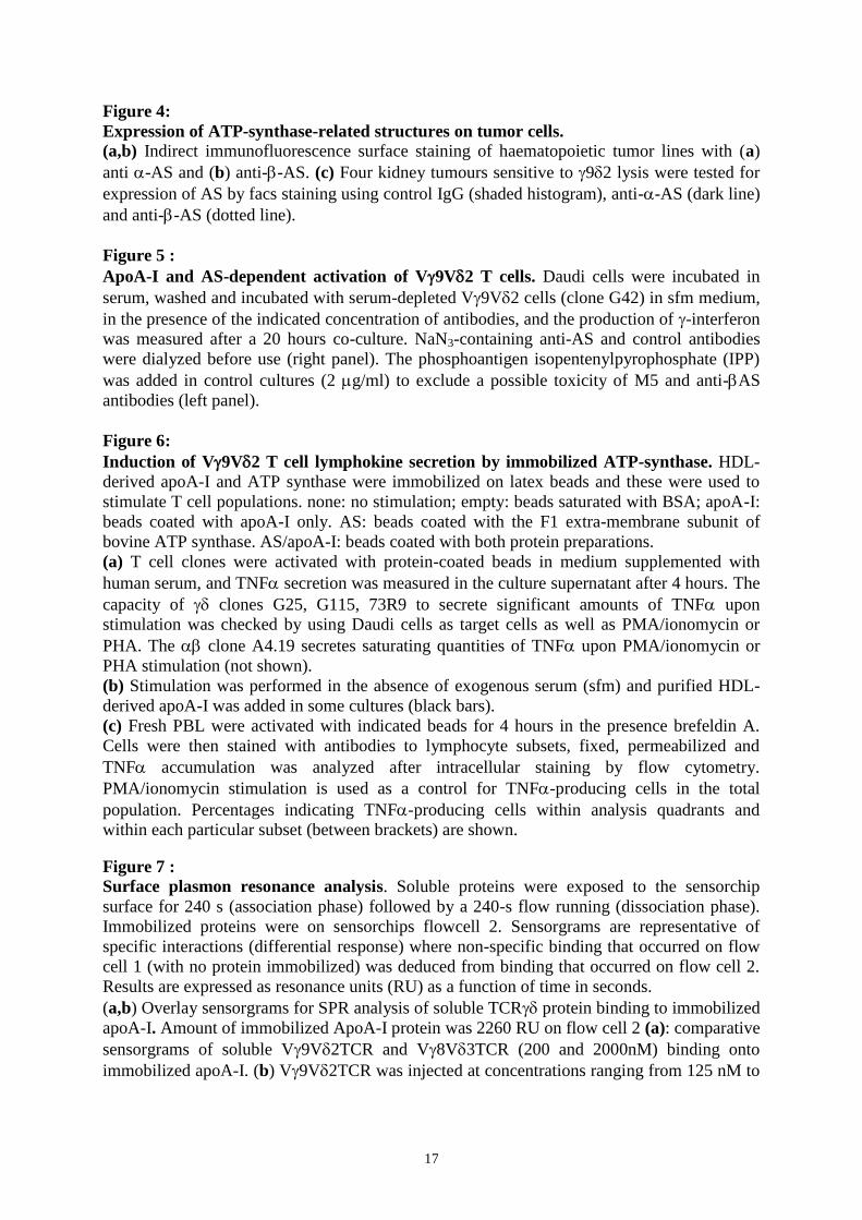
fulfilled both criteria was selected for further studies. M5 mAb stained Daudi cells as well as
several other hematopoietic tumors reported to be recognized by V9V2 T cells, including
cell lines of lymphoid (MOLT-4, RPMI 8226) and myeloid (K562, U937) origins. By contrast
several tumor cells resistant to V9V2 T cell killing such as Raji and B lymphoblastoid cells
were not stained by M5 mAb (Fig.1a). As M5 mAb binding to tumor cells was dependent on
the presence of serum in cell culture medium (Fig.1b), M5 was subsequently used to
immunopurify a putative ligand from bovine serum. A protein (M5L) running in
polyacrylamide gels as a ~28kDa polypeptide could be isolated and mass spectrometry
analysis of tryptic peptide digests identified apolipolipoprotein A-I as the likely ligand. M5
immunopurified a similar protein from human serum (hM5L) and its identity to apoA-I was
confirmed by its reactivity in immunoblotting experiments using the anti-human apoA-I
monoclonal antibody 4H1 (Fig.1d). Accordingly, like M5 Ab, 4H1 stained Daudi and RPMI
8226, another tumor line frequently killed by V9V2 T cells, but not Raji cells following
their incubation with a human lipid-free form of human apoA-I prepared from high density
lipoprotein particles (HDL-apoA-I) by ion exchange chromatography (Fig.1c).
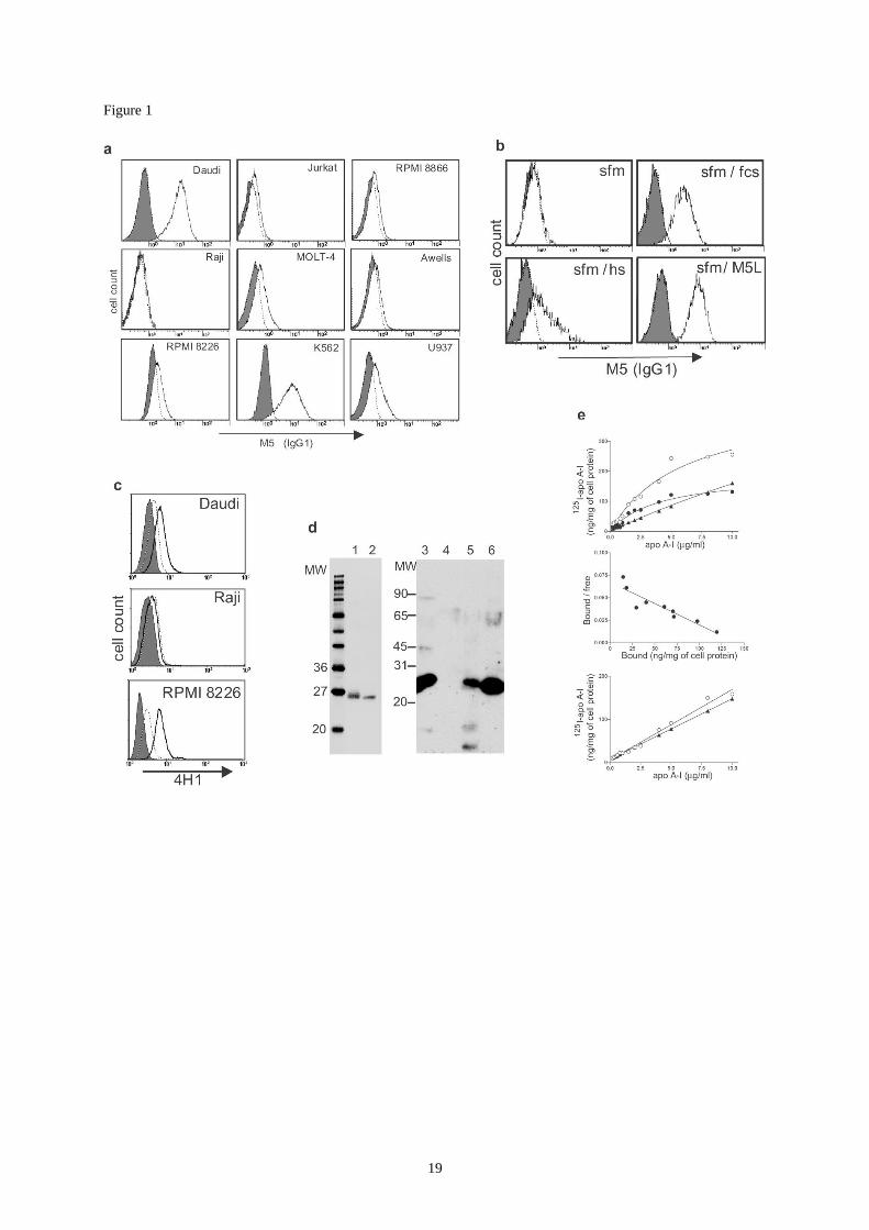
In order to assess the putative involvement of apo A-I in V9V2 cytolytic activity, we
performed cytotoxicity assays in serum-free medium (sfm), using target cells depleted of
serum after an overnight culture in sfm (Fig.2). Cytolytic activity of V9V2 T cell clones
against Daudi cells was decreased in serum-free conditions, and restored by addition of serum
during the cytotoxicity assay. Similarly, addition of purified M5L increased V9V2 T cell-
mediated lysis in a dose-dependent manner. By contrast Daudi cell lysis by a control V8V3
T cell clone (#73R9) was neither affected by serum deprivation nor by addition of M5L
(Fig.2). A similar effect on V9V2 T cell cytotoxicity was observed using HDL-apoA-I or a
commercial apoA-I preparation (Sigma, data not shown). Thus, tumor lysis by V9V2 T
cells is specifically affected by soluble extracellular apoA-I.
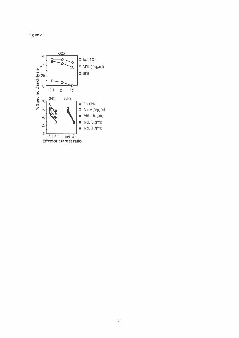
Modulation of V9V2 T cell activity by apo A-I could be due to apoA-I recognition by
either the V9V2 TCR itself or by accessory receptors such as toll-like receptors which are
known to be expressed on various conventional and non-conventional T cell subsets
(Caramalho et al., 2003 ; Mokuno et al., 2000 ; Sakaguchi, 2003). Possible involvement of
apoA-I in TCR engagement was studied by using recombinant soluble forms of the V9V2
TCR derived from the G115 clone (Allison et al., 2001), which were biotinylated and
tetramerized by fluorescent streptavidin (hereafter referred to as TCR tetramers). Like M5
mAb, V9V2 TCR tetramers bound to the V9V2 target cells Daudi, K562 and more
weakly to RPMI 8226, but neither to Raji cells nor to B lymphoblastoid cell lines (Fig.3a and
data not shown). As a control, V8V3 TCR tetramers derived from clone 73R9 bound to
Daudi but to a much lesser extent to K562 cells (Fig.3a) indicating that these two TCRs target
distinct structures on the tumor cell surface. K562 cells, which yielded the brightest staining
levels with V9V2 TCR tetramers, were used in further experiments. V9V2 TCR tetramer
binding to K562 was decreased when cells were cultured for 18h in the absence of serum (not
shown) and increased following addition of serum or human apoA-I (hM5L or HDL-apoA-I).
Importantly apoA-II, whose hydrophobicity was similar to that of apoA-I, did not enhance
5
V9V2-TCR tetramer binding (Fig.3b). This suggested that the effect of apoA-I was not
merely due to a non-specific adsorption of TCR tetramers, which might have been induced by
hydrophobic compounds. Accordingly neither serum, nor apoA-I, affected the binding of
V8V3 TCR tetramers (Fig.3b). Also consistent with a specific effect of apoA-I on V9V2
TCR tetramer binding, pre-incubation of cells with M5 and 4H1 antibodies decreased TCR
tetramer binding to cells coated with hM5L. 4H1 decreased tetramer binding on cells
preincubated with HDL-derived apoA-I whereas M5 did not (Fig.3c). This goes along with
the observation that M5 mAb does not bind to HDL-derived apoA-I (not shown) and suggests
structural differences between the immuno-purified and the HDL-derived forms of apoA-I,
although both are recognized by 4H1 and promote TCR binding. Although soluble V92
TCR no longer bound to serum-deprived Daudi cells (data not shown), they still stained
serum-deprived K562 cells (Fig.3b). This could be explained by the persistence of residual
apolipoproteins on the cell surface. However, it remained possible that the soluble TCR bound
to an as yet unidentified structure and that this binding was increased by apoA-I.
We have recently shown that components of the mitochondrial enzyme ATP-synthase
(AS) can be expressed on the surface of hepatocytes, and that this enzyme constitutes a high
affinity receptor for free, delipidated apoA-I on these cells (Martinez et al., 2003). We studied
expression of components of this enzyme on the surface of V9V2 tumor targets. Daudi,
K562 and RPMI 8226 were stained by mAb against the chain of AS (AS) whereas Raji,
leukaemic T cells and B-LCL were not (Fig.4a). Four kidney tumors which were lysed by
V9V2 CTL also expressed an AS-related surface component (Fig.4c). The subunit of
AS was also detected on Daudi, K562 and U932, a monocytic line not consistently killed by
V9V2 T cells. This chain was undetectable on RPMI 8226 and the kidney tumors
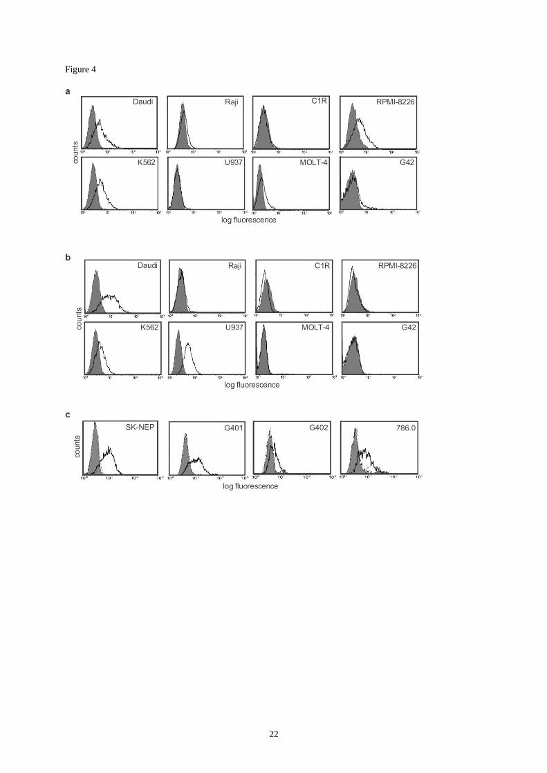
(Fig.4b,c). Therefore tumor susceptibility to V9V2 lysis more strongly correlated with
expression of theS subunit. Scatchard analysis of the binding of iodinated apoA-I to
Daudi cells revealed a single binding receptor on these cells (KD= 0.8x10⁻⁷
M; Fig.1e). Since
Daudi cells did not express other putative apoA-I receptors, such as Scavenger Receptor B1
(Murao et al., 1997) or ABCA-1 (Chambenoit et al., 2001) (data not shown), these
experiments strongly suggested that AS was the apoA-I receptor on tumor cells.
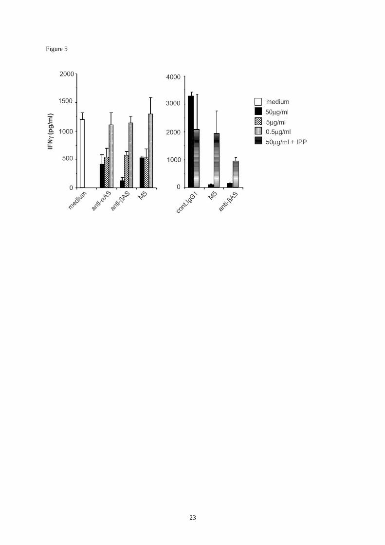
Two sets of experiments were performed to assess the involvement of AS in tumor cell
recognition by V9V2 T cells. Firstly, antibodies to apoA-I and to the extramembrane (F1)
subunit of AS were tested for their ability to modulate lymphokine secretion by V9V2 T
cells following incubation with Daudi cells. IFN secretion was strongly inhibited by anti-
S, anti-S and M5, as compared to a control Ig. This inhibition was not merely due to Ab
toxicity since V9V2 T activation was largely restored by addition of isopentenyl
pyrophosphate (IPP), a previously described V9V2 T cell antigen (Fig.5).
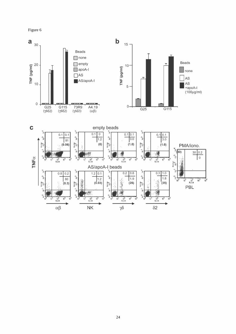
Secondly, the involvement of AS in V9V2 T cell activation was studied by testing the
ability of mitochondrial bovine AS immobilized on latex beads with and without apoA-I to
stimulate V9V2 T cells. Beads carrying apoA-I only or none of these proteins were also
tested (Fig.6). Co-culture with AS-coated beads induced a strong TNF secretion by V9V2
clones, whereas V8V3 and clones, otherwise able to produce TNF after stimulation by
phorbol myristate acetate and calcium ionophore, were not activated (Fig.6a). We could not
demonstrate any stimulatory activity of immobilized apoA-I alone. Similar experiments
performed in serum-free conditions indicated a stimulatory activity of beads carrying AS
alone, and a relatively minor increase of this stimulatory activity following addition of soluble
apoA-I (Fig.6b). This would suggest an accessory role of apoA-I in the stimulatory activity of
AS. However a possible carryover of apoA-I or related apolipoproteins by effector cells
cannot be ruled out. When added onto fresh PBL, beads carrying AS and apoA-I induced
6
selective TNF production by a large fraction of V2+ T cells but had no significant effect on
the and NK cell populations (Fig.6c).
Altogether the above results suggested a highly specific and TCR-mediated activation
of V9V2 cells by apoA-I and AS. However, the respective contribution of AS and apoA-I
in T cell / tumor cell interactions remained unclear. Although anti-apoA-I antibodies had a
substantial inhibitory effect on cytotoxicity and lymphokine secretion, these effects as well as
the modulatory properties of apoA-I could be explained by the proximity of apoA-I and AS
on the cell surface, possibly generating indirect perturbation of TCR-AS interactions. Surface
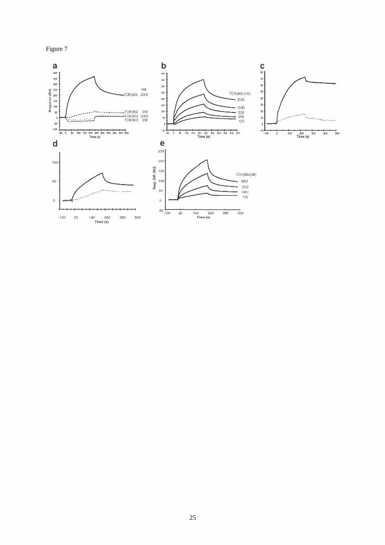
plasmon resonance analysis (SPR) has been previously used to demonstrate interactions
between apoA-I and ATP synthase (Martinez et al., 2003). We followed a similar approach to
assess TCR/apoA-I/AS interactions, after immobilization of either apoA-I, soluble G115 TCR
or purified AS onto SPR chips. Immobilized apoA-I specifically interacted with soluble
monomeric G115 TCR (KD≈0.8 M) whereas interaction with the 73R9 TCR was too low to
be measured (Fig.7a,b). Conversely, immobilized G115 TCR specifically interacted with
soluble apoA-I, but not with apoA-II (Fig.7c). When soluble TCR were similarly tested on
immobilized AS, a clear interaction was demonstrated for the V9V2 TCR (KD=1.5M)
whereas the binding of the V8V3 TCR was low and could not be measured (Fig.7d,e). Thus
SPR analysis confirmed the occurrence of specific interactions between V8V2 TCR, AS
and apo A-I.

7
DISCUSSION
Our results strongly suggest that during interaction between V9V2 T cells and their
tumoral targets, a ternary complex forms between the TCR on the T cell side and apoA-I plus
AS on the tumor cell side. It is not clear whether the structure expressed on the surface of
tumors is identical to mitochondrial AS : reactivity with available monoclonal antibodies
suggests that the AS and AS chains can be independently expressed on various tumors,
indicating some form of surface expression polymorphism of these proteins. Using a limited
panel of tumor lines we found that sensitivity to V9V2 T cells correlated better with AS
than with AS expression. Although AS might not be the primary tumor target antigen of
V9V2 T cells but instead a cross-reactive unrelated entity, the stimulatory potential of
immobilized mitochondrial AS would indicate that its homology to the putative primary
antigen(s) is high enough to induce a potent V9V2 T cell stimulation.
Direct binding of apoA-I to AS (Martinez et al., 2003) and the TCR (our present data)
have now been demonstrated. These interactions seem to be required for optimal T cell
activation as V9V2 T cell activation and soluble TCR binding were markedly decreased by
serum depletion and were restored by addition of exogenous soluble apoA-I. However, our
present results suggest a hierarchical importance of these interactions for V9V2 T cell
activation. Indeed, we could not demonstrate any stimulatory activity for immobilized apoA-I
whereas AS appeared to be stimulatory by itself when immobilized on latex beads. SPR
experiments however strongly suggest a direct interaction of apoA-I with both the TCR and
AS. Thus a possible role for apoA-I could be the stabilisation of the interaction between the
TCR and the AS, explaining the stimulatory effect of apoA-I in cytotoxicity assays.
ApoA-I is unlikely to be immunogenic by itself and, as already mentionned,
immobilized apoA-I did not activate V9V2 T cells. However, apoA-I may serve as a carrier
for antigenic ligands such as phosphoantigens, either derived from the tumor cells themselves
or from the extracellular medium, thus permitting their “presentation” by a surface AS-related
structure. This possibility would be consistent with the recently described correlation between
endogenous production of mevalonate pathway metabolites (including IPP) by tumor cells
and their susceptibility to V9V2 T cell-mediated lysis (Gober et al., 2003).
Ectopic expression of components of AS is not unprecedented as the presence of AS
was previously described on the surface of K562 cells (Das et al., 1994) and endothelial cells
(Moser et al., 1999) and linked to immunomodulatory effects in one case. The /AS
subunits are also found on hepatocytes where they promote binding of free apoA-I and
display enzymatic activity (Martinez et al., 2003). Whether these components are also
involved in an enzymatic complex on tumor cells is not known yet. Although expression of
this apoA-I receptor on normal tissue cells could potentially induce V9V2 T lymphocyte-
mediated autoimmunity, several recent studies have described mechanisms involving NK-like
receptors and permitting efficient control of this potential self reactivity (Fisch et al., 1997;
Halary et al., 1997 ).
Besides, together with the high expression of LDL receptors and apoE in intraepithelial
lymphocytes (Fahrer et al., 2001), and with studies documenting apoE binding to ATP-
synthase (Mahley et al., 1989), these data open a new field of investigations linking lipid
metabolism and anti-tumor immunosurveillance.
8
MATERIAL AND METHODS
1) Tumor cell lines, T cell clones and cultures
Daudi, Raji, RPMI 8226, K562, Jurkat, Molt-4, and U937, SK-NEP, G401, G402, and 786.0
were obtained from ATCC. Awells (EBV+ lymphoblastoid B cell line) is from the
International Histocompatibility Workshop (IHW#9090). C1R (HLA-A-B
--LCL) and RPMI
8866 (B-LCL) were obtained from Drs P.Lebouteiller and M.Colonna respectively. All tumor
cell lines were cultivated in RPMI 1640 medium supplemented with 10% fetal calf serum
(Invitrogen) except for serum deprivation experiments. In this case, cells were washed once in
RPMI 1640, incubated for 1 hour at 37°C in serum free culture medium (hybridoma sfm,
Invitrogen), pelletted and reincubated for at least 16 hours at 37°C in sfm before use. The G25
(V9V2) and 73R9 (V8V3) clones were obtained as described for G42 and G115 (Allison
et al., 2001; Davodeau et al., 1993) by anti-V monoclonal antibody selection and subsequent
amplification using PHA and IL2 and cloning.
3) Fluorescence analysis and antibodies
Immunofluorescence stainings were performed in PBS containing 1%BSA and devoid of
serum using FITC-conjugated goat F(ab)’2 anti-mouse Ig antibody (Caltag) as the second step
reagent. Irrelevant isotype-matched control antibodies were used as negative controls. In
apolipoprotein binding experiments, serum-deprived cells were incubated with serum or
purified protein preparations in PBS containing 1%BSA, at room temperature, 30 minutes
prior to antibody staining. 7H10 (anti--ATP synthase) and 3D5 (anti--ATP synthase) and
7F9 (anti--ATP synthase) are from Molecular Probes. 4H1 (anti-human apoA-I) (Collet et
al., 1997) was obtained from Dr Y. Marcel (Ottawa).
4) Generation of M5A12D10 hybridoma and antibody purification
Balb/c mice were injected intraperitoneally four times at two-weeks intervals with 15x106
Daudi cells washed and resuspended in PBS. Hybridoma were obtained by fusion of spleen
cells with P3X63Ag8 myeloma cells and were selected on the basis of tumor cell staining.
Subcloned hybridoma were subsequently amplified in sfm medium and antibodies were
purified on protein G affinity columns (Pharmacia), neutralized, dialyzed against PBS and
concentrated (Harlow and Lane, 1988). Finally, antibodies were tested for their ability to
modulate lysis of Daudi cells by effectors, leading to the selection of the M5A12D10
antibody (IgG1).
5) Purified proteins
The human and bovine ligands of M5A12D10 (hM5L and bM5L respectively) were isolated
by affinity chromatography: human and fetal calf serum diluted 1/20 in 3 M NaCl and 50 mM
Tris pH 7 were passed through the column carrying the covalently attached antibody. After
washing (last wash was in 3 M NaCl, 10 mM Tris pH 7), bound proteins were eluted with 100
mM glycine pH 2.7. Isolation of ApoA-I from High Density Lipoproteins (HDL apoA-I) by
ion-exchange chromatography has been already described (Mezdour et al., 1987). Purity of
apoA-I was checked by Western blot analyses using different antibodies directed against
human apoB, apoA-II, apo-C and apoA-I. The apolipoprotein A-I homogeneity was more than
99% (as measured by densitometry after SDS-PAGE and silver staining). Purified bovine
ATP synthase (F1 subunit) (Lutter et al., 1993) was obtained from John E. Walker
(Cambridge, UK).
6) Production of soluble fluorescent tetrameric TCR and usage for cell staining
9
The G115 (V9V2) (3) and 73R9 (V8V3) extracellular and chains (the latter carrying a
short 3’ Biotin tag) were expressed in Escherichia coli, then refolded together by rapid
dilution in 1 L of 1 M L-arginine, 0.1 M Tris-HCl, pH 8.0 and 0.2 mM reduced/0.2 mM
oxidized glutathione. After dialysis against 10 mM Tris-HCl pH 8.0, concentration by cation
exchange chromatography at pH 5.5, and purification by size exclusion chromatography at
pH 8.0, the refolded protein was biotinylated for 4 h at 30°C with 6 g/ml BirA and excess of
free biotin was removed by dialysis against 10 mM Tris-HCl pH 8.0, 150 mM NaCl.
Tetramers of TCR heterodimers were obtained by mixing the biotinylated TCR with
phycoerythrin-labeled streptavidin (Biosource) at a molar ratio of 10:1. For staining, 2.105
cells were incubated at room temperature for 45 min with phycoerythrin-labeled TCR
tetramers at a concentration of TCR of 30 g/ml in PBS plus 1% BSA. Background staining
was determined using phycoerythrin-labeled streptavidin at the same concentration.
7) Protein identification by proteomic analysis and mass spectrometry
Immunopurified material was analyzed using one dimensional electrophoresis and visualized
by coomassie blue staining. Protein bands were excised from the gel and subjected to several
washing steps, reduction alkylation reaction, in-gel trypsin digestion with modified trypsine
(Promega, Madison, WI) at 25 ng/µl in 50 mM NH4HCO3 and finally followed by peptide
extraction. The peptides purified with ZipTip C18 (Millipore) were mixed with equal volumes
(0.5 ml) of a saturated a-cyano-4-hydroxycinnamic acid in 50% acetonitrile, 0.1% TFA onto
the MALDI target and allowed to air-dry. Peptide mass fingerprinting were obtained by using
a PE Biosystems MALDI-TOF mass spectrometer (Voyager DE STR, Foster City, CA, USA)
on each protein band. Unknown proteins were identified using the data base fitting program
MS-Fit (Protein Prospector, (http://prospector.ucsf.edu)), searching against all eukaryotic
entries in Swiss Prot and NCBI non redundant protein data bases. We considered the
identifcation positive when a minimum of four measured peptide masses were matched and
provided at least around 20% sequence coverage. Mass accuracy of 10 ppm was obtained with
internal calibration using auto-digestion peaks of trypsin (M+H+, 842.51, 2211.10, and
2283.18).
8) Chromium and cytokine release assays
2h-51
Cr-release assays were performed in standard conditions except for the use of serum-free
conditions in some experiments: in sensitization experiments with apolipoprotein
preparations, target tumor cells were serum-deprived as described for facs analysis, loaded
with 51
Cr (100 Ci/106 cells, 1 hour, 37°C), extensively washed in RPMI and resuspended in
sfm medium. Effector T cells cultivated in serum-containing medium were washed
extensively in RPMI, incubated for 2 hours in sfm at 37°C and resuspended in sfm medium.
Target cells (3000/well, in triplicates) were first incubated with apolipoprotein preparations or
serum-containing medium for 30 min at room temperature in 96-well round-bottom
microculture plates at room temperature prior to the addition of effector cells. Cells were then
pelleted and incubated at 37°C for 2 hours. Supernatants were recovered for 51
Cr release
measurement. Spontaneous release (in the absence of effectors) was subtracted from
experimental data and was in the 10-30% range of maximum release (effector cells replaced
by same volume of 0.1 M HCl). Specific lysis was calculated as the percentage of maximum
release. For cytokine release measurements, similar experiments were performed.
supernatants were harvested after 4 hours (TNF) or 20 hours (IFN). IFN was titrated by a
specific Elisa technique, whereas TNF concentration was assessed by a biological assay
based on WEHI cells viability.
9) Binding assays
10
The binding experiments were performed at 4°C for 2 hours as previously described
(Barbaras et al., 1994). Briefly, cells (9 µg of cell proteins per point) were incubated in PBS
for 2 hours at 4°C with increasing concentrations of labeled apoA-I. Cells were filtered on
0.22 µm filters (GVWP Millipore-France) and washed four times with 1% BSA in PBS.
Filters were used for radioactivity measurements. Non-specific binding was determined in the
presence of a 100-fold excess (as compared to the KD value) of the corresponding unlabeled
ligand. Binding was analyzed using a weighted non-linear curve-fitting program, based on the
LIGAND analysis program (Prism-GraphPad).
10) Immobilization of proteins on latex beads.
107 sulfate latex beads (Interfacial Dynamics corp., Portland, OR) were washed in PBS and
incubated with apolipoproteins (100 g/ml), F1-ATP synthase (0.4 mg/ml) or a mixture of
both under constant agitation at room temperature for 16 hours. Beads were then washed,
saturated for 3 hours in PBS containing 1%BSA and washed extensively in PBS before use.
Control “empty” beads were similarly saturated with BSA. In stimulation experiments, beads
were mixed with cells at a 1:1 ratio.
11) Immobilization of proteins for SPR (Biacore) analysis.
ApoA-I and ATP synthase (F1 subunit) were immobilized by amine linkage on CM5 chips
(Biacore AB) following NHS-EDC activation; V9V2-biotinylated soluble TCRs were
immobilized on SA streptavidin-coated chips (Biacore AB) and binding was analyzed in a
Biacore 3000 apparatus (BiacoreAB). Soluble ligands were injected at a flow rate of 20
µl/min, exposed to the surface for 240 s (association phase) followed by a 240-s flow running
during which the dissociation occurred. Sensorgrams are representative of specific
interactions (differential response) where non-specific binding that occurred on flow cell 1
was deduced from binding that occurred on flow cell 2. Results are expressed as resonance
units (RU) as a function of time in seconds.
11
REFERENCES
Allison, T. J., Winter, C. C., Fournie, J. J., Bonneville, M., and Garboczi, D. N. (2001).
Structure of a human gammadelta T-cell antigen receptor. Nature 411, 820-4.
Barbaras, R., Collet, X., Chap, H., and Perret, B. (1994). Specific binding of free
apolipoprotein A-I to a high-affinity binding site on HepG2 cells: characterization of two
high-density lipoprotein sites. Biochemistry 33, 2335-40.
Beckman, E. M., Porcelli, S. A., Morita, C. T., Behar, S. M., Furlong, S. T., and Brenner, M.
B. (1994). Recognition of a lipid antigen by CD1-restricted alpha beta+ T cells [see
comments]. Nature 372, 691-694.
Bukowski, J. F., Morita, C. T., and Brenner, M. B. (1999). Human gamma delta T cells
recognize alkylamines derived from microbes, edible plants, and tea: implications for innate
immunity. Immunity 11, 57-65.
Bukowski, J. F., Morita, C. T., and Brenner, M. B. (1994). Recognition and destruction of
virus-infected cells by human gamma delta CTL. J. Immunol. 153, 5133-5140.
Bukowski, J. F., Morita, C. T., Tanaka, Y., Bloom, B. R., Brenner, M. B., and Band, H.
(1995). V gamma 2V delta 2 TCR-dependent recognition of non-peptide antigens and Daudi
cells analyzed by TCR gene transfer. J. Immunol. 154, 998-1006.
Caramalho, I., Lopes-Carvalho, T., Ostler, D., Zelenay, S., Haury, M., and Demengeot, J.
(2003). Regulatory T cells selectively express toll-like receptors and are activated by
lipopolysaccharide. J Exp Med 197, 403-11.
Chambenoit, O., Hamon, Y., Marguet, D., Rigneault, H., Rosseneu, M., and Chimini, G.
(2001). Specific docking of apolipoprotein A-I at the cell surface requires a functional
ABCA1 transporter. J Biol Chem 276, 9955-60.
Collet, X., Marcel, Y. L., Tremblay, N., Lazure, C., Milne, R. W., Perret, B., and Weech, P.
K. (1997). Evolution of mammalian apolipoprotein A-I and conservation of antigenicity:
correlation with primary and secondary structure. J Lipid Res 38, 634-44.
Constant, P., Davodeau, F., Peyrat, M. A., Poquet, Y., Puzo, G., Bonneville, M., and Fournie,
J. J. (1994). Stimulation of human gamma delta T cells by nonpeptidic mycobacterial ligands.
Science 264, 267-70.
Das, B., Mondragon, M. O., Sadeghian, M., Hatcher, V. B., and Norin, A. J. (1994). A novel
ligand in lymphocyte-mediated cytotoxicity: expression of the beta subunit of H+ transporting
ATP synthase on the surface of tumor cell lines. J Exp Med 180, 273-81.
Davodeau, F., Peyrat, M. A., Hallet, M. M., Gaschet, J., Houde, I., Vivien, R., Vie, H., and
Bonneville, M. (1993). Close correlation between Daudi and mycobacterial antigen
recognition by human gamma delta T cells and expression of V9JPC1 gamma/V2DJC delta-
encoded T cell receptors. J. Immunol. 151, 1214-1223.
12
Davodeau, F., Peyrat, M. A., Hallet, M. M., Houde, I., Vie, H., and Bonneville, M. (1993).
Peripheral selection of antigen receptor junctional features in a major human gamma delta
subset. Eur J Immunol 23, 804-8.
De Libero, G., Casorati, G., Giachino, C., Carbonara, C., Migone, N., Matzinger, P., and
Lanzavecchia, A. (1991). Selection by two powerful antigens may account for the presence of
the major population of human peripheral gamma/delta T cells. J. Exp. Med. 173, 1311-1322.
Fahrer, A. M., Konigshofer, Y., Kerr, E. M., Ghandour, G., Mack, D. H., Davis, M. M., and
Chien, Y. H. (2001). Attributes of gammadelta intraepithelial lymphocytes as suggested by
their transcriptional profile. Proc Natl Acad Sci U S A 98, 10261-6.
Fisch, P., Meuer, E., Pende, D., Rothenfusser, S., Viale, O., Kock, S., Ferrone, S., Fradelizi,
D., Klein, G., Moretta, L., Rammensee, H. G., Boon, T., Coulie, P., and Vanderbruggen, P.
(1997). Control Of B Cell Lymphoma Recognition Via Natural Killer Inhibitory Receptors
Implies a Role For Human V-Gamma/V-Delta-2 T Cells In Tumor Immunity. European
Journal of Immunology 27, 3368-3379.
Girardi, M., Oppenheim, D. E., Steele, C. R., Lewis, J. M., Glusac, E., Filler, R., Hobby, P.,
Sutton, B., Tigelaar, R. E., and Hayday, A. C. (2001). Regulation of cutaneous malignancy by
gammadelta T cells. Science 294, 605-9.
Gober, H. J., Kistowska, M., Angman, L., Jeno, P., Mori, L., and De Libero, G. (2003).
Human T cell receptor gammadelta cells recognize endogenous mevalonate metabolites in
tumor cells. J Exp Med 197, 163-8.
Groh, V., Steinle, A., Bauer, S., and Spies, T. (1998). Recognition of stress-induced MHC
molecules by intestinal epithelial gammadelta T cells. Science 279, 1737-40.
Halary, F., Peyrat, M. A., Champagne, E., Lopezbotet, M., Moretta, A., Moretta, L., Vie, H.,
Fournie, J. J., and Bonneville, M. (1997). Control Of Self-Reactive Cytotoxic T Lymphocytes
Expressing Gamma-Delta T Cell Receptors By Natural Killer Inhibitory Receptors. European
Journal of Immunology 27, 2812-2821.
Harlow, E., and Lane, D. (1988). Antibodies: a laboratory manual (New York: Cold Spring
Harbor Laboratory Ed.), pp. 312.
Lang, F., Peyrat, M. A., Constant, P., Davodeau, F., David-Ameline, J., Poquet, Y., Vie, H.,
Fournie, J. J., and Bonneville, M. (1995). Early activation of human V gamma 9V delta 2 T
cell broad cytotoxicity and TNF production by nonpeptidic mycobacterial ligands. J Immunol
154, 5986-94.
Lutter, R., Saraste, M., van Walraven, H. S., Runswick, M. J., Finel, M., Deatherage, J. F.,
and Walker, J. E. (1993). F1F0-ATP synthase from bovine heart mitochondria: development
of the purification of a monodisperse oligomycin-sensitive ATPase. Biochem J 295, 799-806.
Mahley, R. W., Hui, D. Y., Innerarity, T. L., and Beisiegel, U. (1989). Chylomicron remnant
metabolism. Role of hepatic lipoprotein receptors in mediating uptake. Arteriosclerosis 9, I14-
8.
13
Malkovska, V., Cigel, F., and Storer, B. E. (1994). Human T cells in hu-PBL-SCID mice
proliferate in response to Daudi lymphoma and confer anti-tumour immunity. Clin. Exp.
Immunol. 96, 158-165.
Malkovska, V., Cigel, F. K., Armstrong, N., Storer, B. E., and Hong, R. (1992).
Antilymphoma activity of human gamma delta T-cells in mice with severe combined immune
deficiency. Cancer. Res. 52, 5610-5616.
Martinez, L. O., Jacquet, S., Esteve, J. P., Rolland, C., Cabezon, E., Champagne, E., Pineau,
T., Georgeaud, V., Walker, J. E., Terce, F., Collet, X., Perret, B., and Barbaras, R. (2003).
Ectopic beta-chain of ATP synthase is an apolipoprotein A-I receptor in hepatic HDL
endocytosis. Nature 421, 75-9.
Mezdour, H., Clavey, V., Kora, I., Koffigan, M., Barkia, A., and Fruchart, J. C. (1987).
Anion-exchange fast protein liquid chromatographic characterization and purification of
apolipoproteins A-I, A-II, C-I, C-II, C-III0, C-III1, C-III2 and E from human plasma. J
Chromatogr 414, 35-45.
Mokuno, Y., Matsuguchi, T., Takano, M., Nishimura, H., Washizu, J., Ogawa, T., Takeuchi,
O., Akira, S., Nimura, Y., and Yoshikai, Y. (2000). Expression of toll-like receptor 2 on
gamma delta T cells bearing invariant V gamma 6/V delta 1 induced by Escherichia coli
infection in mice. J Immunol 165, 931-40.
Moody, D. B., Reinhold, B. B., Guy, M. R., Beckman, E. M., Frederique, D. E., Furlong, S.
T., Ye, S., Reinhold, V. N., Sieling, P. A., Modlin, R. L., Besra, G. S., and Porcelli, S. A.
(1997). Structural Requirements For Glycolipid Antigen Recognition By Cd1b-Restricted T
Cells. Science 278, 283-286.
Morita, C. T., Beckman, E. M., Bukowski, J. F., Tanaka, Y., Band, H., Bloom, B. R., Golan,
D. E., and Brenner, M. B. (1995). Direct presentation of nonpeptide prenyl pyrophosphate
antigens to human gamma delta T cells. Immunity. 3, 495-507.
Moser, T. L., Stack, M. S., Asplin, I., Enghild, J. J., Hojrup, P., Everitt, L., Hubchak, S.,
Schnaper, H. W., and Pizzo, S. V. (1999). Angiostatin binds ATP synthase on the surface of
human endothelial cells. Proc Natl Acad Sci U S A 96, 2811-6.
Murao, K., Terpstra, V., Green, S. R., Kondratenko, N., Steinberg, D., and Quehenberger, O.
(1997). Characterization of CLA-1, a human homologue of rodent scavenger receptor BI, as a
receptor for high density lipoprotein and apoptotic thymocytes. J Biol Chem 272, 17551-7.
Sakaguchi, S. (2003). Control of immune responses by naturally arising CD4+ regulatory T
cells that express toll-like receptors. J Exp Med 197, 397-401.
Spada, F. M., Grant, E. P., Peters, P. J., Sugita, M., Melian, A., Leslie, D. S., Lee, H. K., van
Donselaar, E., Hanson, D. A., Krensky, A. M., Majdic, O., Porcelli, S. A., Morita, C. T., and
Brenner, M. B. (2000). Self-recognition of CD1 by gamma/delta T cells: implications for
innate immunity. J Exp Med 191, 937-48.

14
Tanaka, Y., Sano, S., Nieves, E., De Libero, G., Rosa, D., Modlin, R. L., Brenner, M. B.,
Bloom, B. R., and Morita, C. T. (1994). Nonpeptide ligands for human gamma delta T cells.
Proceedings of the National Academy of Sciences of the USA 91, 8175-8179.
Wu, J., Groh, V., and Spies, T. (2002). T cell antigen receptor engagement and specificity in
the recognition of stress-inducible MHC class I-related chains by human epithelial gamma
delta T cells. J Immunol 169, 1236-40.

15
AKNOWLEDGMENTS
This work was supported by grants from Association pour la Recherche sur le Cancer (for
MB), from the Cancer Research Institute (EG), from the European Community (MB) and
from the ARECA network: "Proteomics and Cancer, new pharmacological targets" (RB and
BP). We thank Fatima L’Faqihi, and Carine Froment for assistance in cytometry, and mass
spectrometry analysis respectively, Yves Marcel (Ottawa) for the antibody 4H1 and John E.
Walker (Cambridge, UK) for providing purified F1 subunit of AS. We are very grateful to
Etienne Joly for stimulating discussions and comments.
16
FIGURE LEGENDS
Figure 1:
Binding of extracellular apoA-I to tumor cell membrane.
(a) Indirect immunofluorescence staining with M5 mAb or a control IgG1 antibody (shaded
histograms) of human cell lines cultured in presence of fetal calf serum (fcs). Daudi, Raji:
Burkitt’s lymphomas; Awells, RPMI8866: B-lymphoblastoid cell lines; RPMI 8226: B-cell
myeloma; K562: erythroid leukemia; U937: monocytic leukemia; MOLT-4, Jurkat: T cell
leukemias.
(b) Daudi cells were depleted of serum components by overnight culture in serum free
medium (sfm) and subsequently incubated in PBS supplemented with either 10% fcs
(sfm/fcs), 10% human serum (sfm/hs) or 1% BSA plus 100 g/ml of M5L (sfm/M5L) before
staining as in figure 1a.
(c) Serum-depleted cell lines were incubated with lipid-free HDL-apoA-I (solid lines and
shaded histograms) or BSA (dotted lines) before indirect staining with the anti-human apoA-I
antibody 4H1 (lines) or control IgG1 (shaded histograms).
(d) Left: coomassie-blue staining of M5-immunopurified material from fcs (lane 1) and
human serum (lane 2) after non-reducing SDS-polyacrylamide gel electrophoresis (PAGE).
Right: Immunoblot after reducing PAGE using anti-human apoA-I antibody 4H1 on : human
serum (lane 3); fetal calf serum (lane 4); HDL (lane 5); hM5L (human serum ligand of M5
mAb, lane 6).
(e): 125
I-labelled free apoA-I binding was measured after 2 hours incubation at 4°C on Daudi
and Raji cells. Top and bottom panels: binding isotherm of 125
I-labeled free apoA-I to Daudi
(top) and Raji (bottom) cells. ( ): total binding; (): non-specific binding; ( ): specific
binding. Middle panel: Scatchard representation of specific binding on Daudi cells. In absence
of specific binding on Raji cells, Scatchard representation could only be performed from
Daudi cells data. The results are representative of two different cell preparations.
Figure 2 :
ApoA-I-dependent activation of V9V2 T cells.
Cr release assays were performed in
serum-free medium (sfm) to assess dependance on serum and apoA-I of Daudi cytolysis by
different effector T cell populations. G25 and G42: V9V2 T cell clones, 73R9: V8V3 T
cell clone. M5L (bovine serum component immunopurified with M5 antibody) was tested at
the indicated concentration for its ability to substitute for serum (fcs). Recombinant annexin V
was used as a control purified protein.
Figure 3:
V9V2 TCR binding to tumor cells and apoA-I. PE-labeled tetrameric TCRs from clone
G115 and clone 73R9 were used to stain tumor cell lines and assess the influence of
apolipoproteins on their tumor recognition. Shaded histograms: concentration-matched
streptavidin-PE alone. Line histograms: binding of the indicated PE-labelled tetrameric TCR.
(a) Cell lines were cultured in 10% fcs before staining with tetramers.
(b) K562 cells were serum-depleted, incubated with 10 % human serum or the indicated
apolipoprotein preparations (100 g/ml) and stained with G115 or 73R9 TCR tetramers.
(c) Serum-depleted K562 cells were incubated with 100 g/ml of HDL hApoA-I (left
histogram) or hM5L (right histogram) and labeled with G115 TCR tetramers in the presence
of the indicated competing mAb (20 g/ml). Note that M5 does not recognize HDL hApoA-I
(see text) whereas 4H1 recognizes both forms of the human protein. Data are representative of
three experiments.
17
Figure 4:
Expression of ATP-synthase-related structures on tumor cells.
(a,b) Indirect immunofluorescence surface staining of haematopoietic tumor lines with (a)
anti -AS and (b) anti--AS. (c) Four kidney tumours sensitive to 92 lysis were tested for
expression of AS by facs staining using control IgG (shaded histogram), anti--AS (dark line)
and anti--AS (dotted line).
Figure 5 :
ApoA-I and AS-dependent activation of V9V2 T cells. Daudi cells were incubated in
serum, washed and incubated with serum-depleted V9V2 cells (clone G42) in sfm medium,
in the presence of the indicated concentration of antibodies, and the production of -interferon
was measured after a 20 hours co-culture. NaN3-containing anti-AS and control antibodies
were dialyzed before use (right panel). The phosphoantigen isopentenylpyrophosphate (IPP)
was added in control cultures (2 g/ml) to exclude a possible toxicity of M5 and anti-AS
antibodies (left panel).
Figure 6:
Induction of V9V2 T cell lymphokine secretion by immobilized ATP-synthase. HDL-
derived apoA-I and ATP synthase were immobilized on latex beads and these were used to
stimulate T cell populations. none: no stimulation; empty: beads saturated with BSA; apoA-I:
beads coated with apoA-I only. AS: beads coated with the F1 extra-membrane subunit of
bovine ATP synthase. AS/apoA-I: beads coated with both protein preparations.
(a) T cell clones were activated with protein-coated beads in medium supplemented with
human serum, and TNF secretion was measured in the culture supernatant after 4 hours. The
capacity of clones G25, G115, 73R9 to secrete significant amounts of TNF upon
stimulation was checked by using Daudi cells as target cells as well as PMA/ionomycin or
PHA. The clone A4.19 secretes saturating quantities of TNF upon PMA/ionomycin or
PHA stimulation (not shown).
(b) Stimulation was performed in the absence of exogenous serum (sfm) and purified HDL-
derived apoA-I was added in some cultures (black bars).
(c) Fresh PBL were activated with indicated beads for 4 hours in the presence brefeldin A.
Cells were then stained with antibodies to lymphocyte subsets, fixed, permeabilized and
TNF accumulation was analyzed after intracellular staining by flow cytometry.
PMA/ionomycin stimulation is used as a control for TNF-producing cells in the total
population. Percentages indicating TNF-producing cells within analysis quadrants and
within each particular subset (between brackets) are shown.
Figure 7 :
Surface plasmon resonance analysis. Soluble proteins were exposed to the sensorchip
surface for 240 s (association phase) followed by a 240-s flow running (dissociation phase).
Immobilized proteins were on sensorchips flowcell 2. Sensorgrams are representative of
specific interactions (differential response) where non-specific binding that occurred on flow
cell 1 (with no protein immobilized) was deduced from binding that occurred on flow cell 2.
Results are expressed as resonance units (RU) as a function of time in seconds.
(a,b) Overlay sensorgrams for SPR analysis of soluble TCR protein binding to immobilized
apoA-I. Amount of immobilized ApoA-I protein was 2260 RU on flow cell 2 (a): comparative
sensorgrams of soluble V9V2TCR and V8V3TCR (200 and 2000nM) binding onto
immobilized apoA-I. (b) V9V2TCR was injected at concentrations ranging from 125 nM to

18
2 µM. The apparent kinetic constants of the interaction were ka=8.8e3 6.08 e
2 M
-1s
-1, kd=
7.13 e-3
6.76 e-5
s-1
, KD=8.1 e-7
M.
(c) Comparative sensorgrams of purified apoA-I (full line) and apoA-II (dotted line) binding
to immobilized V9V2TCR (proteins were injected at 100 µg/ml). Amount of immobilized
TCR protein was 335 RU on flow cell 2.
(d,e) Comparative sensorgrams of soluble TCR binding to immobilized ATP synthase.
Amounts of immobilized ATP synthase (F1) was 22440 RU on flow cell 2. (d) Comparative
sensorgrams of purified monomeric G115 TCR (V9V2, full line) and 73R9 TCR (V8V3,
dotted line) binding to immobilized ATP synthase (F1). Proteins were injected at a
concentration of 2M. (e) Monomeric G115 soluble TCR (V9V2) was injected at
concentrations ranging from 0.5 to 4M. The apparent kinetic constants of the interaction
were ka=1.68 e3 2.16 M
-1s
-1, kd= 2.54 e
-3 2.9 e
-5 s
-1, KD=1.51 e
-6M.
19
Figure 1
20
Figure 2
21
Figure 3
22
Figure 4
23
Figure 5
24
Figure 6
25
Figure 7
Related Documents