TUMOR NECROSIS FACTOR ALPHA AND INTERFERON GAMMA COOPERATIVELY INDUCE OXIDATIVE STRESS AND MOTONEURON DEATH IN RAT SPINAL CORD EMBRYONIC EXPLANTS M. MIR, a V. J. ASENSIO, a L. TOLOSA, a M. GOU-FABREGAS, b R. M. SOLER, b J. LLADÓ a AND G. OLMOS a * a Grup de Neurobiologia Cel.lular, Institut Universitari d’Investigacions en Ciències de la Salut (IUNICS)/Departament de Biologia, Universitat de les Illes Balears, Cra. de Valldemossa, km 7.5, E-07122 Palma de Mallorca, Illes Balears, Spain b Unitat de Senyalització Neuronal, Departament de Ciències Mèdiques Bàsiques, Facultat de Medicina, Universitat de Lleida-IBRLLEIDA, Mont- serrat Roig, 2, E-25008-Lleida, Spain Abstract—The accumulation of reactive microglia in the de- generating areas of amyotrophic lateral sclerosis (ALS) tis- sue is a key cellular event creating a chronic inflammatory environment that results in motoneuron death. We have de- veloped a new culture system that consists in rat spinal cord embryonic explants in which motoneurons migrate outside the explant, growing as a monolayer in the presence of glial cells. The proinflammatory cytokines tumor necrosis factor alpha (TNF-) and interferon gamma (IFN-) have been pro- posed to be involved in ALS-linked microglial activation. In our explants, the combined exposure to these cytokines re- sulted in an increased expression of the pro-oxidative en- zymes inducible nitric oxide synthase (iNOS), the catalytic subunit of the nicotinamide adenine dinucleotide phosphate (NADPH) oxidase, gp91 phox and cyclooxygenase-2 (COX-2), as compared to each cytokine alone. This effect was related to their cooperation in the activation of the transcription factor nuclear factor kappa B (NF-B). TNF- and IFN- also cooperated to promote protein oxidation and nitration, thus increasing the percentage of motoneurons immunoreactive for nitrotyrosine. Apoptotic motoneuron death, measured through annexin V-Cy3 and active caspase-3 immunoreactiv- ities, was also found cooperatively induced by TNF- and IFN-. Interestingly, these cytokines did not affect the viabil- ity of purified spinal cord motoneurons in the absence of glial cells. It is proposed that the proinflammatory cytokines TNF- and IFN- have cooperative/complementary roles in inflammation-induced motoneuron death. © 2009 IBRO. Pub- lished by Elsevier Ltd. All rights reserved. Key words: amyotrophic lateral sclerosis, microglia, neuroin- flammation, nitric oxide, proinflammatory cytokines. Amyotrophic lateral sclerosis (ALS) is a progressive neu- rodegenerative disease characterized by degeneration of motor cortex, brain stem and spinal cord motoneurons (Shaw, 2005). ALS was traditionally regarded as a “neuron only disease,” however, increasing evidences have dem- onstrated that glial cells, in particular activated microglia, may contribute to the initiation and progression of this disease (Sargsyan et al., 2005; Weydt and Moller, 2005; Boillee et al., 2006). ALS tissue is characterized by neuroinflammatory changes observed in both sporadic and familial ALS and in the superoxide dismutase 1 (SOD1) transgenic mouse model of ALS (McGeer and McGeer, 2002; Mhatre et al., 2004; Weydt and Moller, 2005). The key cellular event signaling the presence of neuroinflammation in ALS is the accumulation of reactive microglia in the degenerating ar- eas (Henkel et al., 2004; Turner et al., 2004; Sargsyan et al., 2005). The microglial reactivity is initiated before mo- toneuron loss (Hall et al., 1998; Henkel et al., 2006), sug- gesting that microglia are activated early in the pathogen- esis of ALS, either sensing the earliest neuronal stress or triggering the process. In accordance with this microglial activation, numerous systemic immune alterations have been described both in ALS patients and in the transgenic mouse model (Zhang et al., 2005; Banerjee et al., 2008; Holmoy, 2008). Several studies have reported T cell (CD4, CD8 and CD40 positive) infiltration along the vessel walls of the spinal cord and brain of ALS patients (Troost et al., 1989; Kawamata et al., 1992; Graves et al., 2004); these activated T cells may directly activate macrophages/micro- glia through cell– cell contact or may release their own inflammatory mediators, in particular, the proinflammatory cytokine interferon gamma (IFN-), one of the most potent microglia-activating factors (Hanisch, 2002). IFN--acti- vated microglia are, in turn, a source of the proinflamma- tory cytokine tumor necrosis factor alpha (TNF-)(Ha- nisch, 2002). This cytokine has been demonstrated to be an autocrine/paracrine signal for microglia and proposed to be implicated in the chronic activation of microglia ob- served in neurodegenerative disorders (Ghezzi and Men- nini, 2001; Kuno et al., 2005). In this regard, increased levels of the cytokines IFN- and TNF- have been re- ported in the blood of ALS patients (Poloni et al., 2000; Babu et al., 2008) and in the spinal cord of the transgenic mouse model (Elliott, 2001; Hensley et al., 2003). *Corresponding author. Tel: 34-971-17-24-48; fax: 34-971-17-31-84. E-mail address: [email protected] (G. Olmos). Abbreviations: ALS, amyotrophic lateral sclerosis; AMT, 4H-1,3-thia- zin-2-amine, 5,6-dihydro-6-methyl-hydrochloride; ANOVA, analysis of variance; BSA, bovine serum albumin; COX-2, cyclooxygenase-2; DAB, diaminobenzidine; DNPH, 2,4-dinitrophenylhydrazine; GFAP, glial fibrillary acidic protein; IFN-, interferon gamma; iNOS, inducible nitric oxide synthase; LPS, lipopolysaccharide; MAP2, microtubule- associated protein 2; NADPH, nicotinamide adenine dinucleotide phosphate; NF-B, nuclear factor kappa B; NHS, normal horse serum; NO, nitric oxide; PBS, phosphate-buffered saline; SDS, sodium dode- cyl sulfate; SOD1, superoxide dismutase 1; TBS, Tris buffer saline; TNF-, tumor necrosis factor alpha; VAChT, vesicular acetylcholine transporter. Neuroscience 162 (2009) 959 –971 0306-4522/09 $ - see front matter © 2009 IBRO. Published by Elsevier Ltd. All rights reserved. doi:10.1016/j.neuroscience.2009.05.049 959

Welcome message from author
This document is posted to help you gain knowledge. Please leave a comment to let me know what you think about it! Share it to your friends and learn new things together.
Transcript
TCD
MMGa
edMb
Bs
Agsevetcaposzs(atfciftiIicTil
*EAzvDgnapNcTt
Neuroscience 162 (2009) 959–971
0d
UMOR NECROSIS FACTOR ALPHA AND INTERFERON GAMMAOOPERATIVELY INDUCE OXIDATIVE STRESS AND MOTONEURON
EATH IN RAT SPINAL CORD EMBRYONIC EXPLANTSKfl
Arm(oomdB
ctm2saeatgetabmHCo1agicmvtnabsnlpB
. MIR,a V. J. ASENSIO,a L. TOLOSA,a
. GOU-FABREGAS,b R. M. SOLER,b J. LLADÓa AND
. OLMOSa*
Grup de Neurobiologia Cel.lular, Institut Universitari d’Investigacionsn Ciències de la Salut (IUNICS)/Departament de Biologia, Universitate les Illes Balears, Cra. de Valldemossa, km 7.5, E-07122 Palma deallorca, Illes Balears, Spain
Unitat de Senyalització Neuronal, Departament de Ciències Mèdiquesàsiques, Facultat de Medicina, Universitat de Lleida-IBRLLEIDA, Mont-errat Roig, 2, E-25008-Lleida, Spain
bstract—The accumulation of reactive microglia in the de-enerating areas of amyotrophic lateral sclerosis (ALS) tis-ue is a key cellular event creating a chronic inflammatorynvironment that results in motoneuron death. We have de-eloped a new culture system that consists in rat spinal cordmbryonic explants in which motoneurons migrate outsidehe explant, growing as a monolayer in the presence of glialells. The proinflammatory cytokines tumor necrosis factorlpha (TNF-�) and interferon gamma (IFN-�) have been pro-osed to be involved in ALS-linked microglial activation. Inur explants, the combined exposure to these cytokines re-ulted in an increased expression of the pro-oxidative en-ymes inducible nitric oxide synthase (iNOS), the catalyticubunit of the nicotinamide adenine dinucleotide phosphateNADPH) oxidase, gp91phox and cyclooxygenase-2 (COX-2),s compared to each cytokine alone. This effect was relatedo their cooperation in the activation of the transcriptionactor nuclear factor kappa B (NF-�B). TNF-� and IFN-� alsoooperated to promote protein oxidation and nitration, thusncreasing the percentage of motoneurons immunoreactiveor nitrotyrosine. Apoptotic motoneuron death, measuredhrough annexin V-Cy3 and active caspase-3 immunoreactiv-ties, was also found cooperatively induced by TNF-� andFN-�. Interestingly, these cytokines did not affect the viabil-ty of purified spinal cord motoneurons in the absence of glialells. It is proposed that the proinflammatory cytokinesNF-� and IFN-� have cooperative/complementary roles in
nflammation-induced motoneuron death. © 2009 IBRO. Pub-ished by Elsevier Ltd. All rights reserved.
Corresponding author. Tel: �34-971-17-24-48; fax: �34-971-17-31-84.-mail address: [email protected] (G. Olmos).bbreviations: ALS, amyotrophic lateral sclerosis; AMT, 4H-1,3-thia-in-2-amine, 5,6-dihydro-6-methyl-hydrochloride; ANOVA, analysis ofariance; BSA, bovine serum albumin; COX-2, cyclooxygenase-2;AB, diaminobenzidine; DNPH, 2,4-dinitrophenylhydrazine; GFAP,lial fibrillary acidic protein; IFN-�, interferon gamma; iNOS, inducibleitric oxide synthase; LPS, lipopolysaccharide; MAP2, microtubule-ssociated protein 2; NADPH, nicotinamide adenine dinucleotidehosphate; NF-�B, nuclear factor kappa B; NHS, normal horse serum;O, nitric oxide; PBS, phosphate-buffered saline; SDS, sodium dode-yl sulfate; SOD1, superoxide dismutase 1; TBS, Tris buffer saline;
mNF-�, tumor necrosis factor alpha; VAChT, vesicular acetylcholine
ransporter.
306-4522/09 $ - see front matter © 2009 IBRO. Published by Elsevier Ltd. All rightoi:10.1016/j.neuroscience.2009.05.049
959
ey words: amyotrophic lateral sclerosis, microglia, neuroin-ammation, nitric oxide, proinflammatory cytokines.
myotrophic lateral sclerosis (ALS) is a progressive neu-odegenerative disease characterized by degeneration ofotor cortex, brain stem and spinal cord motoneurons
Shaw, 2005). ALS was traditionally regarded as a “neuronnly disease,” however, increasing evidences have dem-nstrated that glial cells, in particular activated microglia,ay contribute to the initiation and progression of thisisease (Sargsyan et al., 2005; Weydt and Moller, 2005;oillee et al., 2006).
ALS tissue is characterized by neuroinflammatoryhanges observed in both sporadic and familial ALS and inhe superoxide dismutase 1 (SOD1) transgenic mouseodel of ALS (McGeer and McGeer, 2002; Mhatre et al.,004; Weydt and Moller, 2005). The key cellular eventignaling the presence of neuroinflammation in ALS is theccumulation of reactive microglia in the degenerating ar-as (Henkel et al., 2004; Turner et al., 2004; Sargsyan etl., 2005). The microglial reactivity is initiated before mo-oneuron loss (Hall et al., 1998; Henkel et al., 2006), sug-esting that microglia are activated early in the pathogen-sis of ALS, either sensing the earliest neuronal stress orriggering the process. In accordance with this microglialctivation, numerous systemic immune alterations haveeen described both in ALS patients and in the transgenicouse model (Zhang et al., 2005; Banerjee et al., 2008;olmoy, 2008). Several studies have reported T cell (CD4,D8 and CD40 positive) infiltration along the vessel wallsf the spinal cord and brain of ALS patients (Troost et al.,989; Kawamata et al., 1992; Graves et al., 2004); thesectivated T cells may directly activate macrophages/micro-lia through cell–cell contact or may release their own
nflammatory mediators, in particular, the proinflammatoryytokine interferon gamma (IFN-�), one of the most potenticroglia-activating factors (Hanisch, 2002). IFN-�-acti-
ated microglia are, in turn, a source of the proinflamma-ory cytokine tumor necrosis factor alpha (TNF-�) (Ha-isch, 2002). This cytokine has been demonstrated to ben autocrine/paracrine signal for microglia and proposed toe implicated in the chronic activation of microglia ob-erved in neurodegenerative disorders (Ghezzi and Men-ini, 2001; Kuno et al., 2005). In this regard, increased
evels of the cytokines IFN-� and TNF-� have been re-orted in the blood of ALS patients (Poloni et al., 2000;abu et al., 2008) and in the spinal cord of the transgenic
ouse model (Elliott, 2001; Hensley et al., 2003).s reserved.
nnrflhamkuwfmswgcaItmao
M
RE2Cf
Rt
EbRemCMcw�(fg5wtdss
eciotwa
cM
I
TcfcovSsfrg(Li
kMpTXcNEtfoiFm2angsBu
pbfadamabimrc
mwam
Q
Tomdp
M. Mir et al. / Neuroscience 162 (2009) 959–971960
Among the secretion products of activated microglia,itric oxide (NO), produced after the induction of type IIitric oxide synthase (iNOS) in microglia, plays a criticalole in mediating neurotoxicity associated with the neuroin-ammation process (Dawson and Dawson, 1998). Weave recently addressed the interaction between IFN-�nd TNF-� in the regulation of NO generation in pureicroglial cultures and have demonstrated that both cyto-
ines are required, through a very specific interplay, for thep-regulation of the iNOS (Mir et al., 2008). In this study,e have developed a new culture system to assess the
unctionality of spinal cord motoneurons in the presence oficroglia: we have observed that motoneurons from rat
pinal cord embryonic explants, growing on plates coveredith polyornithine and laminin, have the singularity of mi-rating outside the spinal cord, and grow as a monolayer ofells surrounded by glial cells. This system offers somedvantages for the individual study of these motoneurons.
n this new model, we have assessed the interaction be-ween the proinflammatory cytokines TNF-� and IFN-� onicroglial activation, NO and oxidative stress generationnd, importantly, the functional implications on the viabilityf spinal cord motoneurons.
EXPERIMENTAL PROCEDURES
aterials
at and murine IFN-� or TNF-� was purchased from PeproTechC Ltd. (London, UK). The selective iNOS inhibitor 4H-1,3-thiazin--amine, 5,6-dihydro-6-methyl-hydrochloride [AMT] was fromayman Chemical (Ann Arbor, MI, USA). Other reagents were
rom Sigma-Aldrich (St. Louis, MO, USA), except when indicated.
at spinal cord embryonic explants and cytokinereatments
mbryos at gestational age of 15–16 days (E15-16) were removedy cesarean section from pregnant Sprague–Dawley rats (Charlesiver, Barcelona, Spain). Lumbar spinal cords were dissected fromach embryo with forceps, transferred to ice cold Leibowitz’s 15edium (L15) and the meninges and ganglia carefully removed.ords were transversely sectioned into 350-�m slices with aacIlwain tissue chopper (Gomshall, Surrey, UK). Sections were
arefully placed at a density of 20 sections per well on 35 mm-ells precoated with poly-DL-ornithine (30 �g/ml) and L-laminin (2g/ml) and containing 2 ml of Eagle’s minimal essential medium
MEM) supplemented with 5% heat-inactivated horse serum, 5%etal bovine serum, 2 mM glutamine, 0.6% glucose and 15 �g/mlentamicin. Explants were maintained at 37 °C under a humidified% CO2 atmosphere. To avoid the detachment of explants theells were not manipulated during the first 4 days after platting;
hen, medium was changed and thereby every 3 days. After 5ays in vitro, some cells of the spinal cord, including motoneurons,tarted the migration outside the explant (Fig. 1). All treatmentstarted 10 days after the explant procedure.
Where indicated, rat spinal cord embryonic explants werexposed for 48 h to IFN-� (10 ng/ml), TNF-� (10 ng/ml) or bothytokines together, in the presence or absence of the selectiveNOS and nicotinamide adenine dinucleotide phosphate (NADPH)xidase inhibitors, AMT (10 �M) and apocynin (1 mM), respec-ively. The time of exposure and the concentration of the cytokinesere chosen based on our previous work demonstrating microglial
ctivation, iNOS induction and NO generation with these particular bonditions (Mir et al., 2008), and other studies (He et al., 2002;ander and Brown, 2005; Gibbons and Dragunow, 2006).
mmunohistochemistry
he following primary antibodies were used for immunohisto-hemistry: anti–microtubule-associated protein 2 (MAP2) (1:1000)rom Chemicon (Temecula, CA, USA); SMI-32 (1:1000) from Ab-am plc (Cambridge, UK); anti-Islet1 (40.2D6, 1:200) from Devel-pmental Studies Hybridoma Bank (Iowa City, IA, USA); anti–esicular acetylcholine transporter (VAChT) (1:500) from Synapticystems (Göttingen, Germany); anti-iNOS (1:200) from BD Bio-ciences (Franklin Lakes, NJ, USA); anti-active caspase-3 (1:200)rom Cell Signaling Technology (Danvers, MA, USA); anti-nitroty-osine (1:750) from Upstate (Charlottesville, VA, USA) and anti–lial fibrillary acidic protein (anti-GFAP; 1:1000) from DAKOGlostrup, Denmark). For lectin cytochemistry, the lectin fromycopersicon esculentum (tomato lectin) labeled with fluorescein
sothiocyanate (FITC) (25 �g/ml) was used.Rat spinal cord embryonic explants were exposed to cyto-
ines as indicated and then fixed with 4% paraformaldehyde in 0.1phosphate buffer, pH 7.4 for 30 min at 21�1 °C. Explants were
ermeabilized for 5 min with methanol, washed three times withris buffer saline (TBS), pH 7.4 and then blocked in 0.1% Triton-100, 5% normal horse serum (NHS) in TBS for 1 h. For activeaspase-3 immunohistochemistry, 0.4% Triton X-100 and 20%HS were used for permeabilization and blocking, respectively.xplants were then incubated overnight at 4 °C with one or two of
he above primary antibodies. Immunohistochemical controls, per-ormed by omitting the primary antibody, resulted in the abolitionf the immunostaining. For immunofluorescence, sections were
ncubated for 1 h with the appropriate secondary antibody, Alexaluor 555 goat anti-rabbit IgG (1:200), Alexa Fluor 488 goat anti-ouse IgG (1:200) or Alexa Fluor 350 goat anti-mouse IgG (1:00) (Invitrogen, Carlsbad, CA, USA). Cultures were then washednd mounted using Gelmount solution. In some cases of immu-ohistochemistry the secondary antibody used was a biotinylatedoat–antimouse and explants were processed according to thetandard avidin–biotin complex procedure (Vector Laboratories,urlingame, CA, USA); the diaminobenzidine (DAB) reaction wassed for color development.
For co-localization of SMI-32 and Islet1-positive neurons, ex-lants were first incubated with Islet1 antibody, followed by incu-ation with biotinylated goat anti-mouse antibody and processedor the DAB reaction as described above. After incubating withvidin/biotin blocker (Vector Laboratories) to saturate all free avi-in or biotin residues, explants were then reprobed with SMI-32ntibody followed by incubation with Alexa Fluor 488 goat anti-ouse IgG. Because both SMI-32 and Islet1 are monoclonalntibodies, goat anti-mouse antibodies applied after SMI-32 incu-ation may also react with Islet1-positive cells. To test the spec-
ficity of the secondary antibody against SMI-32, the second pri-ary antibody (SMI-32) was omitted; in this case, no green fluo-
escence was detected (not shown), indicating that there was noross-reactivity.
Images were obtained using a Leica DMR epifluorescenceicroscope (Leica Microsystems, Wetzlar, Germany) equippedith a Leica DC300 camera and software. Some samples werelso imaged under a Leica TCS SP2 confocal laser scanningicroscope.
uantification of apoptotic motoneuron death
o assess whether a given cytokine treatment affected the viabilityf motoneurons, its ability to induce apoptotic cell death wasonitored by means of the APOAC annexin V-Cy3 apoptosisetection kit (Sigma-Aldrich); this kit detects the presence of phos-hatidylserine residues on the outer leaflet of the plasma mem-
rane by means of the protein annexin conjugated to the fluoro-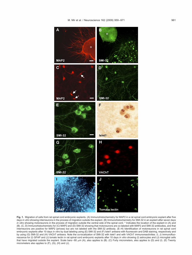
Fdi(iebrtm
M. Mir et al. / Neuroscience 162 (2009) 959–971 961
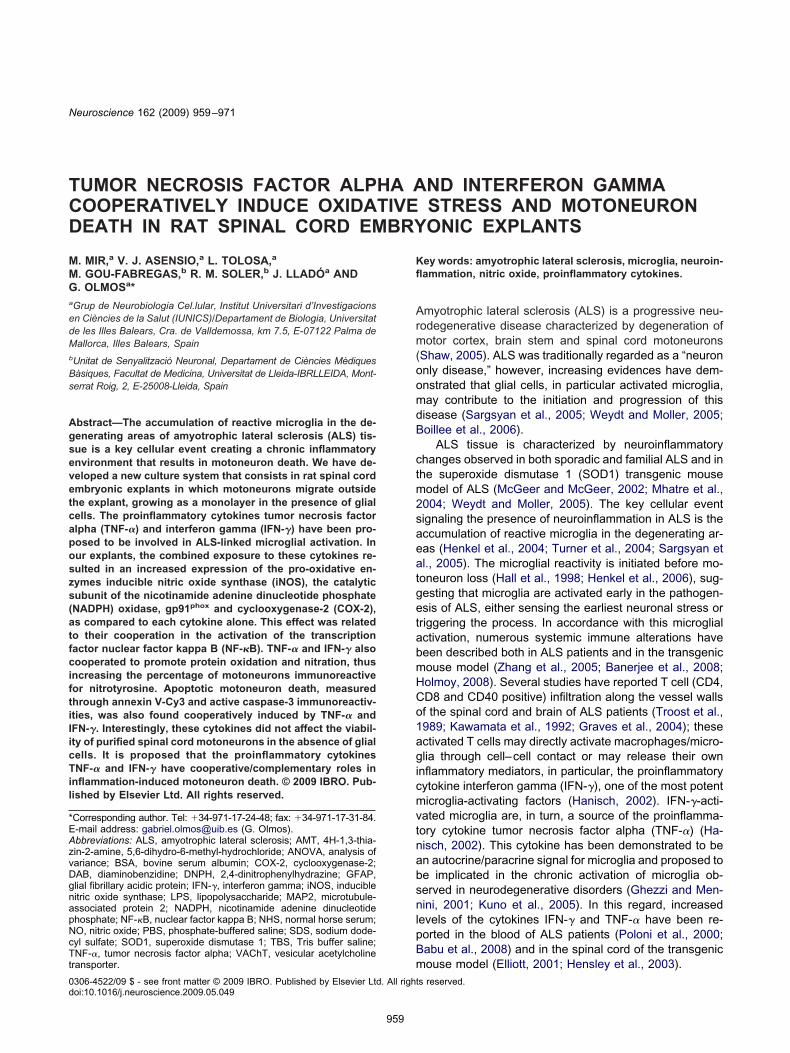
ig. 1. Migration of cells from rat spinal cord embryonic explants. (A) Immunohistochemistry for MAP2 in a rat spinal cord embryonic explant after fiveays in vitro showing interneurons in the process of migration outside the explant. (B) Immunohistochemistry for SMI-32 in an explant after seven days
n vitro showing motoneurons in the process of migration outside the ventral side of the spinal cord. * Indicates the location of the explant in (A) andB). (C, D) Immunohistochemistry for (C) MAP2 and (D) SMI-32 showing that motoneurons are co-labeled with MAP2 and SMI-32 antibodies, and thatnterneurons are positive for MAP2 (arrows) but are not labeled with the SMI-32 antibody. (E–H) Identification of motoneurons in rat spinal cordmbryonic explants after 10 days in vitro by dual labeling using (E) SMI-32 and (F) Islet1 antisera with fluorescein and DAB staining, respectively andy using (G) SMI-32 and (H) VAChT antisera. Note the co-localization of SMI-32 with Islet1 and with VAChT immunoreactivities. (I, J) Immunofluo-escence for (I) GFAP and (J) tomato lectin in rat spinal cord embryonic explants after 10 days in vitro showing (I) astrocytes and (J) microglial cellshat have migrated outside the explant. Scale bars�80 �m (A), also applies to (B). (C) Forty micrometers, also applies to (D) and (I). (E) Twenty
icrometers also applies to (F), (G), (H) and (J).
cr1brcdpmbbOcwtftfm
W
RicT(sPPp�ut1ao(Ctwbaoclpm
N
RItpaus
Nm
NpbA(w
aiiafs
P
Odnp(wrcmtoEP1ancstowiio1tp(Hwomi2
Mc
Md1Btpm70ci(AaifpCms
M. Mir et al. / Neuroscience 162 (2009) 959–971962
hrome Cy3.18 (Koopman et al., 1994). Briefly, explants wereinsed with phosphate-buffered saline (PBS), pH 7.4 at 37 °C for0 min, and then exposed to annexin V-Cy3 (1 �g/ml) in theinding buffer supplied by the manufacturer. Then, explants wereinsed again with PBS and processed for SMI-32 immunofluores-ence. The percentage of motoneurons undergoing apoptotic celleath was assessed by counting the number of annexin V-Cy3ositive cells that could be identified as motoneurons both byorphological and immunocytochemical criteria (i.e. double la-eled with annexin V-Cy3 and SMI-32), relative to the total num-er of motoneurons (SMI-32 positive) in the whole area of the well.nly motoneurons with their somata out of the explant wereounted. The mean number of counted motoneurons in controlells was 78�7 (n�20). Cell counts were done blindly as to the
reatment condition and at least three different wells were countedor each experimental condition. The procedure described here forhe quantification of annexin V positive motoneurons also appliesor the quantification of active caspase-3 and nitrotyrosine positiveotoneurons after the indicated experimental treatments.
estern blotting
at spinal cord embryonic explants were exposed to cytokines asndicated and, after treatments, explants were rinsed rapidly in iceold PBS and lysed in 2% sodium dodecyl sulfate (SDS), 125 mMris buffer, pH 6.8 containing a cocktail of protease inhibitorsComplete Mini; Roche Pharmaceuticals, Basel, Switzerland). Ly-ates were sonicated and proteins quantified by means of the DCrotein Assay from Bio-Rad Laboratories (Hercules, CA, USA).rotein equivalents from each sample were resolved in SDS–olyacrylamide gel electrophoresis and electrotransferred to 0.45m nitrocellulose membranes (Amersham, Buckinghamshire, UK)sing a Bio-Rad semidry trans-blot, according to the manufac-urer’s instructions. Membranes were blocked at 21�1 °C for
h with PBS containing 5% non-fat dry milk, 0.5% bovine serumlbumin (BSA) and 0.2% Tween 20. Membranes were probedvernight at 4 °C with the adequate primary antibodies: anti-iNOS1:4000) or anti–cyclooxygenase-2 (anti-COX-2; 1:2000) fromayman Chemical Company; anti-gp91phox (1:2000) or anti-�-
ubulin (1:10,000) from Sigma-Aldrich. Membranes were thenashed with PBS for 10 min at 21�1 °C (three times) and incu-ated for 2 h with the appropriate peroxidase-conjugated second-ry antibodies. These antibodies were used at the dilutions rec-mmended by the suppliers. Blots were finally developed with thehemiluminescent peroxidase substrate and visualized in chemi-uminescence film (Amersham). The apparent molecular weight ofroteins was determined by calibrating the blots with pre-stainedolecular weight markers (Bio-Rad).
O determination in culture medium
at spinal cord embryonic explants were exposed for 48 h toFN-� (10 ng/ml), TNF-� (10 ng/ml) or both cytokines together inhe presence or absence of the selective iNOS inhibitor AMT. NOroduced by the explants was determined in culture media byssaying the amount of nitrite, a stable oxidation product of NO,sing the Griess reagent (Green et al., 1982), as previously de-cribed (Mir et al., 2008).
uclear factor kappa B (NF-�B) activityeasurements
F-�B activation was assessed in rat spinal cord embryonic ex-lants exposed for 24 h to IFN-� (10 ng/ml), TNF-� (10 ng/ml) oroth cytokines together by means of the NF-�B transcription factorssay Kit (TransAMTM) from Active Motif, as previously described
Mir et al., 2008). Briefly, explants cultured in four 35 mm-wells
ere collected and mixed for every treatment to perform the rssay. Nuclear extracts were prepared and incubated with anmmobilized oligonucleotide containing an NF-�B consensus bind-ng site. The NF-�B complex bound was then detected with anntibody against an epitope of the p65 (RelA) subunit of NF-�B,
ollowed by incubation with the adequate peroxidase-conjugatedecondary antibody and with a developing solution.
rotein carbonyl measurement
xidative damage of rat spinal cord embryonic explants wasetermined after exposure for 48 h to IFN-� (10 ng/ml), TNF-� (10g/ml), or both cytokines together, by measuring the content ofrotein carbonyl as determined by the 2,4-dinitrophenylhydrazineDNPH) spectrophotometric assay. In this assay, DNPH reactsith protein carbonyls, forming a Schiff base to produce the cor-
esponding hydrazone, which can be analyzed spectrophotometri-ally (Levine et al., 1994). Embryonic explants cultured in four 35m-wells were collected and mixed for every treatment to perform
he assay. Explants were scraped and sonicated on ice in 500 �lf cold 50 mM sodium phosphate buffer pH 6.7 containing 1 mMDTA and a cocktail of protease inhibitors (Complete Mini; Rocheharmaceuticals), and homogenates centrifuged at 10,000�g for5 min at 4 °C. Supernatants were collected and the ratio ofbsorbances 280/260 nm was checked to be more than 1, asucleic acids may erroneously contribute to a higher estimation ofarbonyls. Total protein content in the supernatants was mea-ured by means of the DC Protein Assay from Bio-Rad Labora-ories. The supernatant fraction was divided in two 200 �l aliquots,ne sample was treated with 800 �l DNPH and the other sampleith an equal volume of 2.5 M HCl. Both samples were incubated
n the dark at 21�1 °C for 1 h and vortex-mixed at 15-minntervals. Both aliquots were precipitated on ice for 5 min with 1 mlf 20% (wt/vol) trichloroacetic acid and then centrifuged at0,000�g for 10 min at 4 °C. Supernatants were then reprecipi-ated with 10% trichloroacetic acid and centrifuged as above. Therecipitates were washed three times with ethanol/ethyl acetate1:1 vol/vol) and the final pellet was dissolved in 6 M guanidineCl in 20 mM sodium phosphate buffer, pH 6.7. Insoluble debrisas removed by centrifugation as above. The difference spectrumf the DNPH-treated sample versus the HCl control was deter-ined, and the results were expressed as nanomoles of DNPH
ncorporated per milligram of protein, based on the absorption of1.0 mM–1 cm–1 at 375 nm for aliphatic hydrazones.
ouse motoneurons isolation and quantification ofell survival
otoneurons were purified from mouse embryos by optimizingifferent steps of protocols described for chicken (Soler et al.,998) and mouse motoneurons (Arce et al., 1999) isolation.riefly, mouse embryo (E12.5) spinal cords were dissected and
he dorsal half was removed. Ventral cords were chopped intoieces and incubated for 10 min at 37 °C in GHEBS buffer (137M NaCl, 2.7 mM KCl, 22.2 mM glucose, 25 mM Hepes buffer pH.4 and 20 IU/ml penicillin plus 20 �g/ml streptomycin) containing.025% trypsin. Then cords were mechanically dissociated andollected under a 4% BSA cushion. The largest cells were thensolated by centrifugation (10 min at 520�g) on an OptiPrepiodixanol) (Axis-Shield plc, Dundee, Scotland) density gradient.t the end of this procedure, cells were again centrifuged throughBSA cushion. The collected cells were pooled in a tube contain-
ng culture medium, counted with a hemocytometer and plated onour-well culture dishes (Falcon, BD Biosciences) precoated witholy-DL-ornithine and L-laminin at a density of 1250 cells per well.ulture medium was Neurobasal (GIBCO, Invitrogen) supple-ented with the B27 supplement (GIBCO, Invitrogen), 2% horse
erum, 0.5 mM L-glutamine, 25 �M 2-mercaptoethanol and the
ecombinant neurotrophic factors: brain-derived neurotrophic fac-
t(c(
tbflttnpvrcbtcctt
S
Ae(eP
Cs
Wapog1mtr(csahwAg1mLpTwHllapr
wl
TCe
TstwnsmecTcNanId(etaic
gTt2taNoc(iwss(soccwoiTscCct
M. Mir et al. / Neuroscience 162 (2009) 959–971 963
or (BDNF) (1 ng/ml), glial cell line-derived neurotrophic factorGDNF) (10 ng/ml), ciliary neurotrophic factor (CNTF) (10 ng/ml),ardiotrophin-1 (CT-1) (10 ng/ml) and hepatocyte growth factorHGF) (10 ng/ml) (PeproTech EC Ltd.).
For survival experiments, motoneurons were exposed for 48 ho TNF-� (10, 50 or 100 ng/ml), IFN-� (10, 50 or 100 ng/ml), oroth cytokines together. The expression of receptors for proin-ammatory cytokines in spinal cord motoneurons at E12.5 gesta-ional age has already been reported (Sedel et al., 2004). Quan-ification of motoneuron survival was performed by counting theumber of large phase-bright neurons with long axonal/neuriterocesses in the whole area of the well. The percentage of sur-iving cells in the different conditions studied was normalized withespect to the number of cells present in control (non-treated)ultures. Motoneurons cultured for 48 h in supplemented Neuro-asal medium but in the absence of neurotrophic factors wereaken as a negative control of cell survival. The mean number ofounted motoneurons in control wells was 172�8 (n�9). Cellounts were done blindly as to the treatment condition and at leasthree different wells were counted for each experimental condi-ion.
tatistics
ll experiments were repeated at least three times. Data arexpressed as mean�SEM values. One-way analysis of varianceANOVA) followed by Bonferroni test was used for the statisticalvaluations. Differences were considered significant when the-value was �0.05.
RESULTS
haracterization of migrating motoneurons in ratpinal cord embryonic explants
e observed that in rat spinal cord embryonic explantsfter five days in vitro growing on plates covered witholyornithine and laminin, some cells started the migrationutside the explant. Neurons, identified by anti-MAP2, mi-rated from different points around the spinal cord (Fig.A). Motoneurons were first identified by anti–neurofila-ent heavy chain (NF-H, SMI-32) immunostaining and on
he basis of their morphology: large bodies (�20 �m) ofounded or triangular shape with a single well-defined axonsee also De Paola et al., 2008); these SMI-32-positiveells started the migration from the ventral side of thepinal cord (Fig. 1B). The SMI-32-positive population couldlso be labeled with the anti-MAP2 antibody (Fig. 1C, D),owever, the spinal cord interneurons, positive for MAP-2,ere not positive for the SMI-32 antibody (Fig. 1C, D).fter 10 days in vitro, motoneurons appeared formingroups growing in monolayers outside the explant (Fig.E). To further confirm that these cells corresponded tootoneurons, the co-localization of SMI-32 with Islet1, aIM homeodomain transcription factor assumed to be ex-ressed specifically by motoneurons (Yamada et al., 1993;suchida et al., 1994; Francisco-Morcillo et al., 2006) andith the cholinergic marker VAChT (Wetts et al., 2001;ärtig et al., 2007) was assessed. Migrating cells with
arge cell bodies and SMI-32 positive were found to beabeled both by the Islet1 (Fig. 1E, F) and by the VAChTntibody (Fig. 1G, H); thus indicating that the SMI-32-ositive population corresponded to spinal cord motoneu-
ons. Other migrating cells were identified as astrocytes as sell as microglia by means of GFAP (Fig. 1I) and tomatoectin (Fig. 1J) immunohistochemistry, respectively.
NF-� and IFN-� cooperate in iNOS, gp91phox andOX-2 expression via NF-�B in rat spinal cordmbryonic explants
he expression of the iNOS enzyme was assessed in ratpinal cord embryonic explants after different cytokinereatments. In control explants, low expression of iNOSas detected; however, exposure to IFN-� (10 ng/ml), butot TNF-� (10 ng/ml), significantly increased iNOS expres-ion (Fig. 2A). In agreement with our previous study in pureicroglial cultures (Mir et al., 2008), when explants werexposed to both cytokines together, iNOS expression waslearly increased in comparison with IFN-� alone (Fig. 2A).his result was confirmed by measuring NO production inulture media. The levels of nitrite, an oxidation product ofO, were consistent with the expression levels of iNOSfter the different cytokine treatments (i.e. the maximalitrite levels were obtained in the presence of TNF-� andFN-�). The selective iNOS inhibitor AMT blocked NO pro-uction stimulated by the combination of both cytokinesFig. 2B). To assess which cell of the spinal cord explantsxpressed iNOS in response to the cytokines, immunohis-ochemistry co-localization experiments were performednd showed that, after exposure to TNF-� and IFN-�, the
ncrease in iNOS expression occurred specifically in mi-roglia (Fig. 2D), but not in astroglia (Fig. 2E).
The transcription factor NF-�B plays a key role in iNOSene induction, and we have previously demonstrated thatNF-� and IFN-� cooperate in iNOS gene expression
hrough increased NF-�B activation in microglia (Mir et al.,008). To assess the mechanism underlying the coopera-ive effect between the two cytokines on iNOS expressionnd NO production; experiments were performed in whichF-�B binding to DNA was studied in nuclear extractsbtained from explants exposed to TNF-�, IFN-� or bothytokines together. In agreement with our previous studyMir et al., 2008), both TNF-� and IFN-� were able tonduce NF-�B activation above the basal levels; however,hen cells were challenged with both cytokines together, aignificant increase in NF-�B binding to DNA was ob-erved, as compared to the effect of each cytokine aloneFig. 2C). On the basis of the observation of iNOS expres-ion, we postulated that TNF-� and IFN-� might also co-peratively regulate the transcription of other genes alsoontaining �B motifs. Therefore, the expression of theatalytic subunit of the NADPH oxidase, gp91phox (other-ise known as Nox2) (Gauss et al., 2007) and the lipid-xidizing enzyme COX-2 (Nakao et al., 2002) were stud-
ed. As shown in Fig. 2A, these enzymes were induced byNF-� but not by IFN-�; however, and importantly to ourtudy, when both cytokines were added together an in-rease in the expression levels of both gp91phox andOX-2 was clearly detected. Together, these results indi-ated that TNF-� and IFN-� cooperate, via the transcrip-ion factor NF-�B, to increase the expression of oxidative
tress-inducing enzymes.
Ts
TkerfitbpWfo
ii
(pTiDTmpIi3dwtwh
FtitcufeaPmasc mistry foe graphs i
M. Mir et al. / Neuroscience 162 (2009) 959–971964
NF-� and IFN-� cooperate in inducing oxidativetress in rat spinal cord embryonic explants
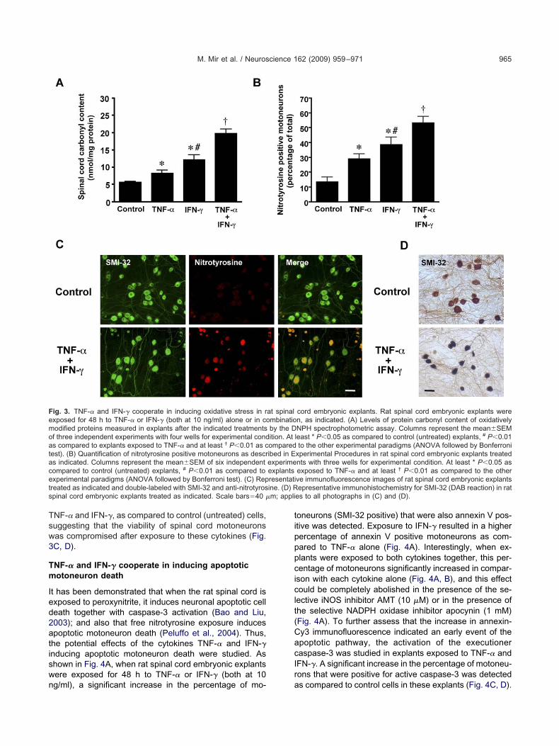
otal protein carbonyl content after exposure to the cyto-ines TNF-� and IFN-� was examined in rat spinal cordmbryonic explants to assess the extent of oxidative freeadical damage to proteins. When explants were exposedor 48 h to TNF-� or IFN-� (both at 10 ng/ml) a significantncrease in carbonyl levels as compared to control (un-reated) explants was observed. The levels of protein car-onyl content were significantly higher in the explants ex-osed to IFN-� than in those exposed to TNF-� (Fig. 3A).hen the above cytokines were added in combination a
urther and significant increase in oxidized proteins wasbserved (Fig. 3A).
iNOS and NADPH oxidase produce NO and superox-de (O �), respectively, which combine by a diffusion-lim-
ig. 2. TNF-� and IFN-� cooperate in iNOS, gp91phox and COX-2 expo TNF-� or IFN-� (both at 10 ng/ml) alone or in combination, as indican lysates (15 �g of protein) of rat spinal cord embryonic explants afteotal protein loaded per lane. A representative immunoblot of three indeord embryonic explants after the indicated treatments by measuring nised as a negative control to block NO production when indicated. Coluor experimental condition. * P�0.01 as compared to control (untreatexperimental paradigms (ANOVA followed by Bonferroni test). (C) Ratt 10 ng/ml) alone or in combination, as indicated, and nuclear extrrocedures. Columns represent values of optical density (OD) correean�SEM of three independent experiments with four wells for expet least † P�0.01 as compared to the other experimental paradigms (Apinal cord embryonic explants after the indicated treatments. The expo-localization with tomato lectin (merge). (E) Double immunohistochexposed to TNF-� and IFN-�. Scale bars�20 �m, applies to all photo
2
ted reaction to form the more toxic oxidant peroxynitrite c
ONOO–); this anion, in turn, can nitrate tyrosine groups ofroteins and forms the stable compound 3-nitrotyrosine.hus, nitrotyrosine immunoreactivity can be used as an
ndicator of NO and superoxide production (Dawson andawson, 1998). When explants were exposed for 48 h toNF-� or IFN-� a significant increase in the percentage ofotoneurons (SMI-32 positive) that were also nitrotyrosineositive was detected. However, exposure of explants toFN-� resulted in a higher percentage of nitrotyrosine pos-tive motoneurons than in those exposed to TNF-� (Fig.B). Interestingly, and in accordance with the previouslyescribed effects of TNF-� and IFN-� on NO production,hen explants were exposed to both cytokines together,
his percentage was significantly increased in comparisonith each cytokine alone (Fig. 3B, C). Moreover, immuno-istochemistry for SMI-32 showed shrunken somata and
ia NF-�B. Rat spinal cord embryonic explants were exposed for 48 hestern blots showing iNOS, gp91phox and COX-2 immunoreactivities
ated treatments. �-Tubulin immunoreactivity is shown as a control ofxperiments is shown. (B) Amounts of NO in culture media of rat spinalthe Griess reagent. The selective inhibitor of iNOS AMT (10 �M) was
esent the mean�SEM of six independent experiments with three wellsexplants exposed to TNF-� and † P�0.001 as compared to the otherrd embryonic explants were exposed for 24 h to TNF-� or IFN-� (bothared and assayed for NF-�B activity as described in Experimental
the micrograms of total nuclear proteins in the extract and are thecondition. * P�0.05 as compared to control (untreated) explants andllowed by Bonferroni test). (D) Immunohistochemistry for iNOS in rat
e also labeled with tomato lectin to show microglial cells. iNOS showsr iNOS (red) and GFAP (green) in rat spinal cord embryonic explantsn (D) and (E).
ression vted. (A) W
r the indicpendent etrite usingmns reprd) and tospinal coacts prepcted byrimentalNOVA fo
lants wer
ondensed cytoskeleton in motoneurons treated with
Tsw3
Tm
Ied2atiswn
tipppciclt(CacIr
Femoatacet ine. (D) Rs m; appli
M. Mir et al. / Neuroscience 162 (2009) 959–971 965
NF-� and IFN-�, as compared to control (untreated) cells,uggesting that the viability of spinal cord motoneuronsas compromised after exposure to these cytokines (Fig.C, D).
NF-� and IFN-� cooperate in inducing apoptoticotoneuron death
t has been demonstrated that when the rat spinal cord isxposed to peroxynitrite, it induces neuronal apoptotic celleath together with caspase-3 activation (Bao and Liu,003); and also that free nitrotyrosine exposure inducespoptotic motoneuron death (Peluffo et al., 2004). Thus,he potential effects of the cytokines TNF-� and IFN-�nducing apoptotic motoneuron death were studied. Ashown in Fig. 4A, when rat spinal cord embryonic explantsere exposed for 48 h to TNF-� or IFN-� (both at 10
ig. 3. TNF-� and IFN-� cooperate in inducing oxidative stress in rxposed for 48 h to TNF-� or IFN-� (both at 10 ng/ml) alone or in coodified proteins measured in explants after the indicated treatmentsf three independent experiments with four wells for experimental conds compared to explants exposed to TNF-� and at least † P�0.01 as cest). (B) Quantification of nitrotyrosine positive motoneurons as descrs indicated. Columns represent the mean�SEM of six independentompared to control (untreated) explants, # P�0.01 as compared toxperimental paradigms (ANOVA followed by Bonferroni test). (C) Repreated as indicated and double-labeled with SMI-32 and anti-nitrotyrospinal cord embryonic explants treated as indicated. Scale bars�40 �
g/ml), a significant increase in the percentage of mo- a
oneurons (SMI-32 positive) that were also annexin V pos-tive was detected. Exposure to IFN-� resulted in a higherercentage of annexin V positive motoneurons as com-ared to TNF-� alone (Fig. 4A). Interestingly, when ex-lants were exposed to both cytokines together, this per-entage of motoneurons significantly increased in compar-
son with each cytokine alone (Fig. 4A, B), and this effectould be completely abolished in the presence of the se-
ective iNOS inhibitor AMT (10 �M) or in the presence ofhe selective NADPH oxidase inhibitor apocynin (1 mM)Fig. 4A). To further assess that the increase in annexin-y3 immunofluorescence indicated an early event of thepoptotic pathway, the activation of the executioneraspase-3 was studied in explants exposed to TNF-� andFN-�. A significant increase in the percentage of motoneu-ons that were positive for active caspase-3 was detected
cord embryonic explants. Rat spinal cord embryonic explants weren, as indicated. (A) Levels of protein carbonyl content of oxidativelyNPH spectrophotometric assay. Columns represent the mean�SEMleast * P�0.05 as compared to control (untreated) explants, # P�0.01to the other experimental paradigms (ANOVA followed by Bonferroni
xperimental Procedures in rat spinal cord embryonic explants treatednts with three wells for experimental condition. At least * P�0.05 asexposed to TNF-� and at least † P�0.01 as compared to the otherve immunofluorescence images of rat spinal cord embryonic explantsepresentative immunohistochemistry for SMI-32 (DAB reaction) in rat
es to all photographs in (C) and (D).
at spinalmbinatioby the Dition. Atomparedibed in Eexperimeexplantsresentati
s compared to control cells in these explants (Fig. 4C, D).

Iapctot
Ts
Tapvstcss
arthdttkc
Soepti(
Fotacacmo( mbryonica in (B) an
M. Mir et al. / Neuroscience 162 (2009) 959–971966
nterestingly, the percentage of motoneurons positive forctive caspase-3 matched the percentage of annexin Vositive motoneurons found after exposure to the cytokineombination (Fig. 4A, C). Together, these results indicatedhat the proinflammatory cytokines TNF-� and IFN-� co-perate to induce apoptotic cell death in spinal cord mo-oneurons.
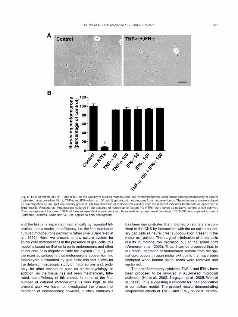
NF-� and IFN-� do not affect the viability of purifiedpinal cord motoneurons
o further confirm that the presence of activated microgliand the increased oxidative stress after exposure to theroinflammatory cytokines TNF-� and IFN-� may be in-olved in inducing motoneuron death, a culture of purifiedpinal cord motoneurons was used. In this culture, mo-oneurons exposed for 48 h to TNF-� or IFN-� alone or inombination, at the concentrations employed in the ratpinal cord embryonic explant, i.e. 10 ng/ml, appeared
ig. 4. Cooperative induction of apoptotic motoneuron death by TNF-�r IFN-� (both at 10 ng/ml) alone or in combination and, in this case,
he selective NADPH inhibitor apocynin (1 mM), as indicated. (A) Quas described in Experimental Procedures. Columns represent the mondition. At least * P�0.05 as compared to control (untreated) explans compared to the other experimental paradigms (ANOVA followed byord embryonic explants treated as indicated and double-labeled witotoneurons as described in Experimental Procedures in rat spinal corf three independent experiments with three wells for experimental conD) Representative immunofluorescence images of rat spinal cord enti-active caspase-3. Scale bars�40 �m, applies to all photographs
imilar to untreated cultures based on cell morphology; in a
ddition, no variation in the number of surviving motoneu-ons was observed (not shown). Thereafter, the concen-ration of the cytokines was increased to 50 and 100 ng/ml;owever, no effects could be observed in the parametersescribed above after treatments with TNF-� or IFN-� athese high cytokine concentrations (Fig. 5A, B). Thus,hese results indicated the lack of toxicity of these cyto-ines on spinal cord motoneurons in the absence of glialells.
DISCUSSION
everal models have been developed for the in vitro studyf the functionality of spinal cord motoneurons in the pres-nce of glial cells. The rat organotypic spinal cord culturereserves the in vivo horizontal architecture; however, inhis model, the detailed microscopic study of motoneuronss limited by the thickness of the spinal cord sectionsRothstein et al., 1993; Tolosa et al., 2008). The dissoci-
�. Rat spinal cord embryonic explants were exposed for 48 h to TNF-�sence or presence of the selective inhibitor of iNOS AMT (10 �M) or
of apoptotic motoneuron death by labeling them with annexinV-Cy3M of six independent experiments with three wells for experimental.01 as compared to explants exposed to TNF-� and at least † P�0.01oni test). (B) Representative immunofluorescence images of rat spinal
and annexin V-Cy3. (C) Quantification of active caspase-3 positivenic explants treated as indicated. Columns represent the mean�SEM�0.001 as compared to control (untreated) explants (Student’s t-test).explants treated as indicated and double-labeled with SMI-32 and
d (D).
and IFN-in the abntificationean�SEts, # P�0
Bonferrh SMI-32d embryodition. * P
ted spinal cord culture works with overall embryonic cords
aucasmstmtaarnpm
hfiamr(onds
baai
F(bEC with thre(
M. Mir et al. / Neuroscience 162 (2009) 959–971 967
nd the tissue is separated mechanically by repeated trit-ration. In this model, the efficiency, i.e. the final number ofultured motoneurons per well is rather small (Bar-Peled etl., 1999). Here, we present a new culture system forpinal cord motoneurons in the presence of glial cells; thisodel is based on that embryonic motoneurons and other
pinal cord cells migrate outside the explant (Fig. 1), andhe main advantage is that motoneurons appear formingonolayers surrounded by glial cells; this fact allows for
he detailed microscopic study of motoneurons and, prob-bly, for other techniques such as electrophysiology. Inddition, as the tissue has not been mechanically tritu-ated, the efficiency of this model, in terms of the finalumber of cultured motoneurons, is very high. In theresent work we have not investigated the process of
ig. 5. Lack of effects of TNF-� and IFN-� on the viability of purified muntreated) or exposed for 48 h to TNF-� and IFN-� (both at 100 ng/ml)y centrifugation on an OptiPrep density gradient. (B) Quantification oxperimental Procedures. Motoneurons cultured in the absence of nolumns represent the mean�SEM of three independent experiments
untreated) cultures. Scale bar�30 �m, applies to both photographs.
igration of motoneurons; however, in chick embryos it c
as been demonstrated that motoneuron somata are con-ned to the CNS by interactions with the so-called bound-ry cap cells (a neural crest subpopulation present in theotor exit points). The surgical elimination of these cells
esults in motoneuron migration out of the spinal cordVermeren et al., 2003). Thus, it can be proposed that, inur model, migration of motoneuron somata from the spi-al cord occurs through motor exit points that have beenisrupted when lumbar spinal cords were removed andectioned.
The proinflammatory cytokines TNF-� and IFN-� haveeen proposed to be involved in ALS-linked microglialctivation (He et al., 2002; Sargsyan et al., 2005; Wen etl., 2006), thus suggesting a rationale for their application
n our culture model. The present results demonstrating
ons. (A) Photomicrograph using phase-contrast microscopy of controld motoneurons from mouse embryos. The motoneurons were isolateduron viability after the different indicated treatments as described inic factors (no NTFs) were taken as negative control of cell survival.e wells for experimental condition. * P�0.001 as compared to control
otoneurspinal corf motone
eurotroph
ooperative effects of TNF-� and IFN-� on iNOS expres-

stctgeagNpm(gTpiw2smactuaci
N(ThstNtpaapTfC
ttccatp1apI(ieT
spoisIaoad2c(T(pCctppsclseis
ea4iwsbMetfitdemaafigtta2shcecm
M. Mir et al. / Neuroscience 162 (2009) 959–971968
ion and NO production (Fig. 2A, B) are in agreement withhose previously obtained in pure cultures of microglialells (Mir et al., 2008). In these cultures, we demonstratedhat the cooperative effects of TNF-� and IFN-� on micro-lial iNOS expression relied in part on their cooperativeffects on NF-�B and interferon regulatory factor-1 (IRF-1)ctivation, two key transcription factors regulating iNOSene expression (Mir et al., 2008). As similar results onF-�B activation were obtained when explants were ex-osed to the above cytokines (Fig. 2C) and iNOS wasainly induced in microglia (Fig. 2D), but not in astroglia
Fig. 2E), the same mechanisms described in pure micro-lial cultures may be claimed for the cooperative effects ofNF-� and IFN-� on iNOS expression found in our ex-lants. Interestingly, in spinal cords of ALS patients iNOS
mmunoreactivity was also found increased; however, itas observed mainly in reactive astrocytes (Sasaki et al.,000). The discrepancy with our results may be due to thehort-term period (48 h) of exposure to cytokines; thus,icroglia may respond earlier to TNF-� and IFN-� thanstroglia. iNOS has also been found up-regulated in glialells of the spinal cord of early symptomatic and end-stageransgenic mutant SOD1 mice and the time course of iNOSp-regulation paralleled that of motoneuron loss (Almer etl., 1999). Also in this sense, NO produced by lipopolysac-haride (LPS)-activated microglia has been shown to bemplicated in motoneuron injury (Zhao et al., 2004).
Superoxide is produced by the microglial enzymeADPH oxidase, also known as phagocytic oxidase
Phox) (Lambeth, 2004; Wilkinson and Landreth, 2006).he present results indicate that TNF-� and IFN-� alsoave cooperative effects on the induction of the catalyticubunit of the NADPH oxidase, the gp91phox (Fig. 2A). Ashe transcriptional regulation of this subunit is mediated viaF-�B (Gauss et al., 2007), the cooperative effects be-
ween the two cytokines tested on gp91phox expression arerobably related to their cooperative induction of NF-�Bctivation. However, the fact that IFN-� induced NF-�Bctivation similarly to TNF-� but failed to induce the ex-ression of gp91phox (Fig. 2A, C), strongly suggests thatNF-� activates additional transcription factors also needed
or the efficient expression of gp91phox (and similarly forOX-2).
As NO rapidly reacts with the superoxide anion to formhe potent oxidant peroxynitrite, our results suggested thathe coexposure to TNF-� and IFN-� could result in in-reased peroxynitrite generation, as compared both toontrol explants or explants exposed to each cytokinelone. Peroxynitrite has been proposed to mediate theoxic activities of NO by inducing lipid oxidation and bothrotein oxidation and nitration (Dawson and Dawson,998). To confirm this point, oxidative damage of proteins,s well as protein nitrotyrosination, was assessed. As ex-ected, the combined exposure of explants to TNF-� and
FN-� resulted both in increased protein carbonyl contentFig. 3A) and in increased percentage of nitrotyrosine pos-tive motoneurons (Fig. 3B, C), as compared to controlxplants or explants exposed to each cytokine alone.
hese results are in agreement with those obtained in the opinal cords of the transgenic mouse model and in ALSatients were it has been described increased proteinxidative damage (Ilieva et al., 2007; Liu et al., 2007), and
ncreased 3-nitrotyrosine immunoreactivity in degeneratingpinal cord neurons (Beal et al., 1997; Cha et al., 2000).FN-�, when added alone, induced more protein oxidationnd nitration than TNF-� alone (Fig. 3A, B). As reactivexygen species are produced as a consequence of normalerobic metabolism, it is likely that the increased NO pro-uction observed in explants exposed to IFN-� alone (Fig.B) also resulted in increased peroxynitrite production and,onsequently, in increased protein oxidation and nitrationsee also Bentz et al., 2000). In addition, the effects ofNF-� alone in terms of protein oxidation and nitrationFig. 3A, B) can be explained by the increased superoxideroduction as a result of the activity of the gp91phox andOX-2 enzymes induced by this cytokine, as compared toontrol explants (Fig. 2A). The superoxide produced byhese enzymes is expected to react with basal NO toroduce peroxynitrite. It is assumed that gp91phox is ex-ressed exclusively by activated microglia in the nervousystem (Wilkinson and Landreth, 2006) and in fact, afteroexposure to TNF-� and IFN-�, its immunoreactivity co-ocalized with that of tomato lectin in our explants (data nothown); by contrast, microglia appear to predominantlyxpress the COX-1 isoform, whereas COX-2 isoform is
nduced by TNF-� mainly in neurons and astrocytes (Con-ilvio et al., 2004 and other references therein).
The coexposure to TNF-� and IFN-� resulted in coop-rative effects on apoptotic motoneuron death, measureds the percentage of annexin V positive motoneurons (Fig.A). Interestingly, this effect could be abolished by inhib-
ting the enzymes iNOS with AMT or the NADPH oxidaseith apocynin (Fig. 4A). Recently, it has been demon-trated that inhibition of NADPH oxidase with apocyninlocks LPS-mediated motoneuron injury (Li et al., 2008).oreover, deletion of Nox gene significantly slowed dis-ase progression and improved survival in the SOD1ransgenic mice (Marden et al., 2007). Together, thesendings reinforce our hypothesis indicating that peroxyni-rite and other nitrogen and oxygen reactive species, pro-uced as a result of the activity of the above microglialnzymes, are key factors in neuroinflammation-inducedotoneuron death. The coexposure to TNF-� and IFN-�lso resulted in similar increases in the percentage ofctive caspase-3 positive motoneurons (Fig. 4C, D). Thesendings are in agreement with those obtained in the trans-enic mouse model of ALS, indicating that caspase-3 ac-ivation appears before (Wengenack et al., 2004), and athe onset of motor axon loss and that, in vitro, caspase-3ctivity can be induced by oxidative stress (Pasinelli et al.,000). In addition, a study in a motoneuron cell linehowed that cells transfected with mutant human SOD1ave more annexin V binding than do wild-type cells andells transfected with the normal human SOD1 (Cooksont al., 2002). In the spinal cord of ALS patients increasedaspase-3 activity has also been described, as well asotoneurons with morphological features resembling ap-
ptosis (Martin, 1999; Sathasivam and Shaw, 2005). The
pabBervtcTtcstioatmifinfTsonbTkf
IstkAoAbatcdeTai
AMPdMeat
Cp
A
A
B
B
B
B
B
B
B
C
C
C
D
D
E
E
F
F
G
M. Mir et al. / Neuroscience 162 (2009) 959–971 969
resent results also demonstrate that the cytokines TNF-�nd IFN-�, alone or in combination, do not affect the via-ility of motoneurons in the absence of glial cells (Fig. 5A,). These findings are in agreement with those in whichxposure of purified rat spinal cord embryonic motoneu-ons to TNF-� (100 ng/ml) did not affect motoneuron sur-ival; however, a redistribution of mitochondria was de-ected (Stommel et al., 2007). Also in this sense, theombined exposure of a mouse motoneuronal cell line toNF-� and IFN-� altered the ultrastructural features and
he functionality of mitochondria (Ferri et al., 2008). Mito-hondrial failure induced by these cytokines may not beufficient to induce a death phenotype on purified mo-oneurons, but may increase their susceptibility to glial-nduced oxidative stress, thus causing the release of ap-ptogenic mitochondrial mediators (Emerit et al., 2004). Inddition, TNF-� gene knockout did not affect life span orhe extent of motoneuron loss in the SOD1 transgenicice, thus suggesting that TNF-� alone is not a key factor
n motoneuron degeneration (Gowing et al., 2006). Thesendings can be explained first, because TNF-� has botheuroprotective and neurotoxic effects related to the dif-erent signaling pathways activated by their receptorsNFR1 and TNFR2 (Ghezzi and Mennini, 2001) and, con-equently, TNF-� acts synergistically/cooperatively withther cytokines (i.e. IFN-�, present results) to promoteeuronal death (see also He et al., 2002); and second,ecause some proinflammatory cytokines (i.e. IL-1� andNF-�) have redundant functions in vivo; thus, in thenockout of TNF-� an increase in the transcripts encodingor IL-1� was detected (Gowing et al., 2006).
CONCLUSION
n summary, we have developed a new culture system totudy the functionality of spinal cord motoneurons in whichhe activation of microglia with the proinflammatory cyto-ines TNF-� and IFN-� reproduces some of the features ofLS in terms of pro-oxidative enzymes expression, proteinxidation and nitration, and apoptotic motoneuron death.ctivation of microglial iNOS and NADPH oxidase haseen reported to act synergistically to kill neurons (Mandernd Brown, 2005); these findings prompt us to proposehat the proinflammatory cytokines TNF-� and IFN-� haveomplementary roles in inflammation-induced motoneuroneath; i.e IFN-� induces iNOS expression but not gp91phox
xpression and the opposite occurs in the presence ofNF-�; when the two cytokines act together both enzymesre simultaneously and increasingly induced, thus result-
ng in increased motoneuron death.
cknowledgments—This study was supported by the Spanishinistry of Health through the “Instituto de Salud, Carlos III” grantsI041507, PI051445, PI060680; “Govern Balear, Conselleria’Economia, Hisenda i Innovació” PROGECIB-3A; “Fundació Laarató de TV3” and “Grups de recerca consolidats” from “Gen-ralitat de Catalunya” (2005SGR00628). M.M. was supported bypre-doctoral fellowship from the “Fondo de Investigación Sani-
aria”; L.T. by a pre-doctoral fellowship from “Govern Balear,
onselleria d’Economia, Hisenda i Innovació” and M.G.-F. by are-doctoral fellowship from the “Universitat de Lleida.”
REFERENCES
lmer G, Vukosavic S, Romero N, Przedborski S (1999) Induciblenitric oxide synthase up-regulation in a transgenic mouse model offamilial amyotrophic lateral sclerosis. J Neurochem 72:2415–2425.
rce V, Garces A, de Bovis B, Filippi P, Henderson C, Pettmann BdeLapeyriere O (1999) Cardiotrophin-1 requires LIFRbeta to pro-mote survival of mouse motoneurons purified by a novel technique.J Neurosci Res 55:119–126.
abu GN, Kumar A, Chandra R, Puri SK, Kalita J, Misra UK (2008)Elevated inflammatory markers in a group of amyotrophic lateralsclerosis patients from northern India. Neurochem Res 33:1145–1149.
anerjee R, Mosley RL, Reynolds AD, Dhar A, Jackson-Lewis V,Gordon PH, Przedborski S, Gendelman HE (2008) Adaptive im-mune neuroprotection in G93A-SOD1 amyotrophic lateral sclero-sis mice. PLoS ONE 3:e2740.
ao F, Liu D (2003) Peroxynitrite generated in the rat spinal cordinduces apoptotic cell death and activates caspase-3. Neuro-science 116:59–70.
ar-Peled O, Knudson M, Korsmeyer SJ, Rothstein JD (1999) Motorneuron degeneration is attenuated in bax-deficient neurons in vitro.J Neurosci Res 55:542–556.
eal MF, Ferrante RJ, Browne SE, Matthews RT, Kowall NW, BrownRH Jr (1997) Increased 3-nitrotyrosine in both sporadic and familialamyotrophic lateral sclerosis. Ann Neurol 42:644–654.
entz BG, Simmons RL, Haines GK 3rd, Radosevich JA (2000) Theyin and yang of nitric oxide: reflections on the physiology andpathophysiology of NO. Head Neck 22:71–83.
oillee S, Vande Velde C, Cleveland DW (2006) ALS: a disease ofmotor neurons and their nonneuronal neighbors. Neuron 52:39 –59.
ha CI, Chung YH, Shin CM, Shin DH, Kim YS, Gurney ME, Lee KW(2000) Immunocytochemical study on the distribution of nitroty-rosine in the brain of the transgenic mice expressing a humanCu/Zn SOD mutation. Brain Res 853:156–161.
onsilvio C, Vincent AM, Feldman EL (2004) Neuroinflammation,COX-2, and ALS—a dual role? Exp Neurol 187:1–10.
ookson MR, Menzies FM, Manning P, Eggett CJ, Figlewicz DA,McNeil CJ, Shaw PJ (2002) Cu/Zn superoxide dismutase (SOD1)mutations associated with familial amyotrophic lateral sclerosis(ALS) affect cellular free radical release in the presence of oxida-tive stress. Amyotroph Lateral Scler Other Motor Neuron Disord3:75–85.
awson VL, Dawson TM (1998) Nitric oxide in neurodegeneration.Prog Brain Res 118:215–229.
e Paola M, Diana V, Bigini P, Mennini T (2008) Morphological fea-tures and responses to AMPA receptor-mediated excitotoxicity ofmouse motor neurons: comparison in purified, mixed anterior hornor motor neuron/glia cocultures. J Neurosci Methods 170:85–95.
lliott JL (2001) Cytokine upregulation in a murine model of familialamyotrophic lateral sclerosis. Brain Res Mol Brain Res 95:172–178.
merit J, Edeas M, Bricaire F (2004) Neurodegenerative diseases andoxidative stress. Biomed Pharmacother 58:39–46.
erri A, Nencini M, Cozzolino M, Carrara P, Moreno S, Carri MT (2008)Inflammatory cytokines increase mitochondrial damage in mo-toneuronal cells expressing mutant SOD1. Neurobiol Dis 32:454 – 460.
rancisco-Morcillo J, Hidalgo-Sanchez M, Martin-Partido G (2006)Spatial and temporal patterns of proliferation and differentiation inthe developing turtle eye. Brain Res 1103:32–48.
auss KA, Nelson-Overton LK, Siemsen DW, Gao Y, DeLeo FR,
Quinn MT (2007) Role of NF-kappaB in transcriptional regulation of
G
G
G
G
G
H
H
H
H
H
H
H
H
I
K
K
K
L
L
L
L
M
M
M
M
M
M
N
P
P
P
R
S
S
S
S
S
S
M. Mir et al. / Neuroscience 162 (2009) 959–971970
the phagocyte NADPH oxidase by tumor necrosis factor-alpha.J Leukoc Biol 82:729–741.
hezzi P, Mennini T (2001) Tumor necrosis factor and motoneuronaldegeneration: an open problem. Neuroimmunomodulation 9:178–182.
ibbons HM, Dragunow M (2006) Microglia induce neural cell deathvia a proximity-dependent mechanism involving nitric oxide. BrainRes 1084:1–15.
owing G, Dequen F, Soucy G, Julien JP (2006) Absence of tumornecrosis factor-alpha does not affect motor neuron disease causedby superoxide dismutase 1 mutations. J Neurosci 26:11397–11402.
raves MC, Fiala M, Dinglasan LA, Liu NQ, Sayre J, Chiappelli F, vanKooten C, Vinters HV (2004) Inflammation in amyotrophic lateralsclerosis spinal cord and brain is mediated by activated macro-phages, mast cells and T cells. Amyotroph Lateral Scler OtherMotor Neuron Disord 5:213–219.
reen LC, Wagner DA, Glogowski J, Skipper PL, Wishnok JS, Tan-nenbaum SR (1982) Analysis of nitrate, nitrite, and [15N]nitrate inbiological fluids. Anal Biochem 126:131–138.
all ED, Oostveen JA, Gurney ME (1998) Relationship of microglialand astrocytic activation to disease onset and progression in atransgenic model of familial ALS. Glia 23:249–256.
anisch UK (2002) Microglia as a source and target of cytokines. Glia40:140–155.
artig W, Stieler J, Boerema AS, Wolf J, Schmidt U, Weissfuss J,Bullmann T, Strijkstra AM, Arendt T (2007) Hibernation model oftau phosphorylation in hamsters: selective vulnerability of cholin-ergic basal forebrain neurons—implications for Alzheimer’s dis-ease. Eur J Neurosci 25:69–80.
e BP, Wen W, Strong MJ (2002) Activated microglia (BV-2) facilita-tion of TNF-alpha-mediated motor neuron death in vitro. J Neuro-immunol 128:31–38.
enkel JS, Beers DR, Siklos L, Appel SH (2006) The chemokineMCP-1 and the dendritic and myeloid cells it attracts are increasedin the mSOD1 mouse model of ALS. Mol Cell Neurosci 31:427–437.
enkel JS, Engelhardt JI, Siklos L, Simpson EP, Kim SH, Pan T,Goodman JC, Siddique T, Beers DR, Appel SH (2004) Presence ofdendritic cells, MCP-1, and activated microglia/macrophages inamyotrophic lateral sclerosis spinal cord tissue. Ann Neurol 55:221–235.
ensley K, Fedynyshyn J, Ferrell S, Floyd RA, Gordon B, Grammas P,Hamdheydari L, Mhatre M, Mou S, Pye QN, Stewart C, West M,West S, Williamson KS (2003) Message and protein-level elevationof tumor necrosis factor alpha (TNF alpha) and TNF alpha-modu-lating cytokines in spinal cords of the G93A-SOD1 mouse modelfor amyotrophic lateral sclerosis. Neurobiol Dis 14:74–80.
olmoy T (2008) T cells in amyotrophic lateral sclerosis. Eur J Neurol15:360–366.
lieva EV, Ayala V, Jove M, Dalfo E, Cacabelos D, Povedano M,Bellmunt MJ, Ferrer I, Pamplona R, Portero-Otin M (2007) Oxida-tive and endoplasmic reticulum stress interplay in sporadic amyo-trophic lateral sclerosis. Brain 130:3111–3123.
awamata T, Akiyama H, Yamada T, McGeer PL (1992) Immunologicreactions in amyotrophic lateral sclerosis brain and spinal cordtissue. Am J Pathol 140:691–707.
oopman G, Reutelingsperger CP, Kuijten GA, Keehnen RM, Pals ST,van Oers MH (1994) Annexin V for flow cytometric detection ofphosphatidylserine expression on B cells undergoing apoptosis.Blood 84:1415–1420.
uno R, Wang J, Kawanokuchi J, Takeuchi H, Mizuno T, Suzumura A(2005) Autocrine activation of microglia by tumor necrosis factor-alpha. J Neuroimmunol 162:89–96.
ambeth JD (2004) NOX enzymes and the biology of reactive oxygen.
Nat Rev Immunol 4:181–189.evine RL, Williams JA, Stadtman ER, Shacter E (1994) Carbonylassays for determination of oxidatively modified proteins. MethodsEnzymol 233:346–357.
i B, Guo YS, Sun MM, Dong H, Wu SY, Wu DX, Li CY (2008) TheNADPH oxidase is involved in lipopolysaccharide-mediated motorneuron injury. Brain Res 1226:199–208.
iu D, Bao F, Wen J, Liu J (2007) Mutation of superoxide dismutaseelevates reactive species: comparison of nitration and oxidation ofproteins in different brain regions of transgenic mice with amyotro-phic lateral sclerosis. Neuroscience 146:255–264.
ander P, Brown GC (2005) Activation of microglial NADPH oxidaseis synergistic with glial iNOS expression in inducing neuronaldeath: a dual-key mechanism of inflammatory neurodegeneration.J Neuroinflammation 2:20.
arden JJ, Harraz MM, Williams AJ, Nelson K, Luo M, Paulson H,Engelhardt JF (2007) Redox modifier genes in amyotrophic lateralsclerosis in mice. J Clin Invest 117:2913–2919.
artin LJ (1999) Neuronal death in amyotrophic lateral sclerosis isapoptosis: possible contribution of a programmed cell death mech-anism. J Neuropathol Exp Neurol 58:459–471.
cGeer PL, McGeer EG (2002) Inflammatory processes in amyotro-phic lateral sclerosis. Muscle Nerve 26:459–470.
hatre M, Floyd RA, Hensley K (2004) Oxidative stress and neuroin-flammation in Alzheimer’s disease and amyotrophic lateral sclero-sis: common links and potential therapeutic targets. J AlzheimersDis 6:147–157.
ir M, Tolosa L, Asensio VJ, Llado J, Olmos G (2008) Complementaryroles of tumor necrosis factor alpha and interferon gamma in induciblemicroglial nitric oxide generation. J Neuroimmunol 204:101–109.
akao S, Ogtata Y, Shimizu E, Yamazaki M, Furuyama S, Sugiya H(2002) Tumor necrosis factor alpha (TNF-alpha)-induced prosta-glandin E2 release is mediated by the activation of cyclooxygen-ase-2 (COX-2) transcription via NFkappaB in human gingival fibro-blasts. Mol Cell Biochem 238:11–18.
asinelli P, Houseweart MK, Brown RH Jr, Cleveland DW (2000)Caspase-1 and -3 are sequentially activated in motor neuron deathin Cu,Zn superoxide dismutase-mediated familial amyotrophic lat-eral sclerosis. Proc Natl Acad Sci U S A 97:13901–13906.
eluffo H, Shacka JJ, Ricart K, Bisig CG, Martinez-Palma L, Pritsch O,Kamaid A, Eiserich JP, Crow JP, Barbeito L, Estevez AG (2004)Induction of motor neuron apoptosis by free 3-nitro-L-tyrosine.J Neurochem 89:602–612.
oloni M, Facchetti D, Mai R, Micheli A, Agnoletti L, Francolini G, MoraG, Camana C, Mazzini L, Bachetti T (2000) Circulating levels oftumour necrosis factor-alpha and its soluble receptors are in-creased in the blood of patients with amyotrophic lateral sclerosis.Neurosci Lett 287:211–214.
othstein JD, Jin L, Dykes-Hoberg M, Kuncl RW (1993) Chronicinhibition of glutamate uptake produces a model of slow neurotox-icity. Proc Natl Acad Sci U S A 90:6591–6595.
argsyan SA, Monk PN, Shaw PJ (2005) Microglia as potential con-tributors to motor neuron injury in amyotrophic lateral sclerosis.Glia 51:241–253.
asaki S, Shibata N, Komori T, Iwata M (2000) iNOS and nitrotyrosineimmunoreactivity in amyotrophic lateral sclerosis. Neurosci Lett291:44–48.
athasivam S, Shaw PJ (2005) Apoptosis in amyotrophic lateral scle-rosis—what is the evidence? Lancet Neurol 4:500–509.
edel F, Bechade C, Vyas S, Triller A (2004) Macrophage-derivedtumor necrosis factor alpha, an early developmental signal formotoneuron death. J Neurosci 24:2236–2246.
haw PJ (2005) Molecular and cellular pathways of neurodegenera-tion in motor neurone disease. J Neurol Neurosurg Psychiatry76:1046–1057.
oler RM, Egea J, Mintenig GM, Sanz-Rodriguez C, Iglesias M, ComellaJX (1998) Calmodulin is involved in membrane depolarization-medi-ated survival of motoneurons by phosphatidylinositol-3 kinase- and
MAPK-independent pathways. J Neurosci 18:1230–1239.
S
T
T
T
T
V
W
W
W
W
W
Y
Z
Z
M. Mir et al. / Neuroscience 162 (2009) 959–971 971
tommel EW, van Hoff RM, Graber DJ, Bercury KK, Langford GM,Harris BT (2007) Tumor necrosis factor-alpha induces changes inmitochondrial cellular distribution in motor neurons. Neuroscience146:1013–1019.
olosa L, Mir M, Asensio VJ, Olmos G, Llado J (2008) Vascularendothelial growth factor protects spinal cord motoneurons againstglutamate-induced excitotoxicity via phosphatidylinositol 3-kinase.J Neurochem 105:1080–1090.
roost D, van den Oord JJ, de Jong JM, Swaab DF (1989) Lympho-cytic infiltration in the spinal cord of patients with amyotrophiclateral sclerosis. Clin Neuropathol 8:289–294.
suchida T, Ensini M, Morton SB, Baldassare M, Edlund T, JessellTM, Pfaff SL (1994) Topographic organization of embryonic motorneurons defined by expression of LIM homeobox genes. Cell79:957–970.
urner MR, Cagnin A, Turkheimer FE, Miller CC, Shaw CE, Brooks DJ,Leigh PN, Banati RB (2004) Evidence of widespread cerebralmicroglial activation in amyotrophic lateral sclerosis: an [11C] (R)-PK11195 positron emission tomography study. Neurobiol Dis 15:601–609.
ermeren M, Maro GS, Bron R, McGonnell IM, Charnay P, Topilko P,Cohen J (2003) Integrity of developing spinal motor columns isregulated by neural crest derivatives at motor exit points. Neuron37:403–415.
en W, Sanelli T, Ge W, Strong W, Strong MJ (2006) Activated
microglial supernatant induced motor neuron cytotoxicity is asso-ciated with upregulation of the TNFR1 receptor. Neurosci Res55:87–95.
engenack TM, Holasek SS, Montano CM, Gregor D, Curran GL,Poduslo JF (2004) Activation of programmed cell death markers inventral horn motor neurons during early presymptomatic stages ofamyotrophic lateral sclerosis in a transgenic mouse model. BrainRes 1027:73–86.
etts R, Vaughn JE (2001) Development of cholinergic terminalsaround rat spinal motor neurons and their potential relationship todevelopmental cell death. J Comp Neurol 435:171–183.
eydt P, Moller T (2005) Neuroinflammation in the pathogenesis ofamyotrophic lateral sclerosis. Neuroreport 16:527–531.
ilkinson BL, Landreth GE (2006) The microglial NADPH oxidasecomplex as a source of oxidative stress in Alzheimer’s disease.J Neuroinflammation 3:30.
amada T, Pfaff SL, Edlund T, Jessell TM (1993) Control of cellpattern in the neural tube: motor neuron induction by diffusiblefactors from notochord and floor plate. Cell 73:673–686.
hang R, Gascon R, Miller RG, Gelinas DF, Mass J, Hadlock K, Jin X,Reis J, Narvaez A, McGrath MS (2005) Evidence for systemicimmune system alterations in sporadic amyotrophic lateral sclero-sis (sALS). J Neuroimmunol 159:215–224.
hao W, Xie W, Le W, Beers DR, He Y, Henkel JS, Simpson EP, YenAA, Xiao Q, Appel SH (2004) Activated microglia initiate motorneuron injury by a nitric oxide and glutamate-mediated mecha-
nism. J Neuropathol Exp Neurol 63:964–977.(Accepted 21 May 2009)(Available online 27 May 2009)
Related Documents