Vaccines 2013, 1, 174-203; doi:10.3390/vaccines1020174 vaccines ISSN 2076-393X www.mdpi.com/journal/vaccines Review Tumor-Associated Glycans and Immune Surveillance Behjatolah Monzavi-Karbassi 1 , Anastas Pashov 2 and Thomas Kieber-Emmons 1, * 1 Winthrop P. Rockefeller Cancer Institute and Department of Pathology, University of Arkansas for Medical Sciences, Little Rock, AR 72205, USA 2 Stephan Angeloff Institute of Microbiology, BAS, Sofia 1113, Bulgaria * Author to whom correspondence should be addressed; E-Mail: [email protected]; Tel.: +1-501-526-5930; Fax: +1-501-526-5934. Received: 18 April 2013; in revised form: 18 April 2013 / Accepted: 6 June 2013 / Published: 17 June 2013 Abstract: Changes in cell surface glycosylation are a hallmark of the transition from normal to inflamed and neoplastic tissue. Tumor-associated carbohydrate antigens (TACAs) challenge our understanding of immune tolerance, while functioning as immune targets that bridge innate immune surveillance and adaptive antitumor immunity in clinical applications. T-cells, being a part of the adaptive immune response, are the most popular component of the immune system considered for targeting tumor cells. However, for TACAs, T-cells take a back seat to antibodies and natural killer cells as first-line innate defense mechanisms. Here, we briefly highlight the rationale associated with the relative importance of the immune surveillance machinery that might be applicable for developing therapeutics. Keywords: monoclonal antibodies; immunotherapy; cancer; mimics; vaccine; TACA; glycans; tumor; carbohydrate 1. Introduction A basic premise underlying immune modalities for cancer is that the immune system can mount a rejection strength response against neoplastically transformed cells [1]. Tumor targeting draws upon two immunological mediated paradigms. The first draws upon concepts of immune surveillance that bridges both innate and adaptive immunity. According to the immune surveillance hypothesis, tumor associated antigens are regarded as ―non-self‖ by the immune system, and a major function of the OPEN ACCESS

Welcome message from author
This document is posted to help you gain knowledge. Please leave a comment to let me know what you think about it! Share it to your friends and learn new things together.
Transcript
Vaccines 2013, 1, 174-203; doi:10.3390/vaccines1020174
vaccines ISSN 2076-393X
www.mdpi.com/journal/vaccines
Review
Tumor-Associated Glycans and Immune Surveillance
Behjatolah Monzavi-Karbassi 1, Anastas Pashov
2 and Thomas Kieber-Emmons
1,*
1 Winthrop P. Rockefeller Cancer Institute and Department of Pathology, University of Arkansas for
Medical Sciences, Little Rock, AR 72205, USA 2 Stephan Angeloff Institute of Microbiology, BAS, Sofia 1113, Bulgaria
* Author to whom correspondence should be addressed; E-Mail: [email protected];
Tel.: +1-501-526-5930; Fax: +1-501-526-5934.
Received: 18 April 2013; in revised form: 18 April 2013 / Accepted: 6 June 2013 /
Published: 17 June 2013
Abstract: Changes in cell surface glycosylation are a hallmark of the transition from
normal to inflamed and neoplastic tissue. Tumor-associated carbohydrate antigens
(TACAs) challenge our understanding of immune tolerance, while functioning as immune
targets that bridge innate immune surveillance and adaptive antitumor immunity in clinical
applications. T-cells, being a part of the adaptive immune response, are the most popular
component of the immune system considered for targeting tumor cells. However, for
TACAs, T-cells take a back seat to antibodies and natural killer cells as first-line innate
defense mechanisms. Here, we briefly highlight the rationale associated with the relative
importance of the immune surveillance machinery that might be applicable for
developing therapeutics.
Keywords: monoclonal antibodies; immunotherapy; cancer; mimics; vaccine; TACA;
glycans; tumor; carbohydrate
1. Introduction
A basic premise underlying immune modalities for cancer is that the immune system can mount a
rejection strength response against neoplastically transformed cells [1]. Tumor targeting draws upon
two immunological mediated paradigms. The first draws upon concepts of immune surveillance that
bridges both innate and adaptive immunity. According to the immune surveillance hypothesis, tumor
associated antigens are regarded as ―non-self‖ by the immune system, and a major function of the
OPEN ACCESS
Vaccines 2013, 1 175
immune system is to survey the body for the development of malignancy and to eliminate tumor cells
as they arise [2]. Innate immunity relies on biochemical and cellular defense mechanisms often
observed in the early phases of encounter with microbes. The cellular players include natural killer
(NK) cells, dendritic cells (DCs), macrophages, monocytes, γδ T-cells and natural killer T (NKT)-cells.
Adaptive immunity involves the expansion of T-cells and B-cells and their humoral and cellular
mediators, cytokines and antibodies. In particular, antibodies and NK cells are early participants in the
immune response and are particularly effective in eliminating blood-borne metastases [3]. In contrast,
T-cells are the effector cells responsible for specific, long-lasting immunity.
The second draws upon concepts associated with tissue-specific destruction in the context of acute
allograft (acute) rejection, flares of autoimmunity and response to acute infection. This second
paradigm requires an understanding of the distinct difference between an anti-tumor immune response
and outright tumor rejection. In this context, immune-mediated cancer rejection is a facet of autoimmunity,
where the target tissue is the cancer itself. The induction of immune-mediated tumor tissue rejection
represents an important conceptual approach to cancer immunotherapy and also remains an important
goal in tumor immunology [4,5]. Antigens that function as tumor rejection antigens are considered
self, nearly self or non-self [6]. The fact that a tumor antigen elicits a tumor-specific immune response
does not necessarily mean that the immune response will cause the rejection of the tumor in vivo. The
question remains as to which tumor antigen can or is better at inducing a clinically beneficial response [7].
Tumor-rejection antigen is therefore an operational term describing how well an immune response
elicited against a tumor antigen will impact on tumor growth. Tumor antigens can be poor,
intermediate or strong tumor rejection antigens, describing quantitatively the impact of the immune
response on tumor growth [6].
Among potential tumor rejection antigens are glycans expressed on glycoproteins and glycolipids.
Aberrant glycosylation is a universal feature of cancer cells with some tumor-associated carbohydrate
antigens (TACAs) considered tumor progression markers. A considerable body of evidence put
TACAs amongst the most challenging of clinical targets for cancer immunotherapy [8,9], yet immune
responses to glycans are noted that could lend to therapeutic strategies and approaches (Figure 1).
TACA expression on cancer cells is associated with organ tropism underlying extravasation and
metastases, because of glycan receptors on organ tissues [10] or their role in survival. A requisite for
metastases is cell survival. Anoikis resistance or survival in the absence of attachment to extracellular
matrix (ECM) is a prerequisite for the development of tumor metastases [11,12]. Anoikis resistance
has evoked special attention in cancer research because circulating tumor cells in the blood stream are
resistant to it. Signaling cascades are intimately interconnected with TACA expression and interaction
with the microenvironment. TACAs can regulate the interaction between integrin and Focal Adhesion
Kinase (FAK), for example, which, in turn, regulates cancer cell adhesion and invasion [13–20]. Many
of the targeted TACAs are found on structures upstream of FAK that can modulate the signaling
through FAK [14,17,19–21], whereby anti-TACA antibodies might reset anoikis of tumor cells.
Glycans are considered as priming agents for T-cells and for B-cells working in concert [22–24].
Natural antibodies and induced antibodies can mediate tumor cell killing and tissue destruction by
several mechanisms that include complement-dependent cytotoxicity (CDC) [25], antibody-dependent
cellular cytotoxicity (ADCC) [26] and through signal transduction pathways, leading to anti-proliferative
activity or apoptosis [27]. Antibodies to TACAs have other attributes, such as negating negative
Vaccines 2013, 1 176
signals to immune cells by forming immune complexes with shed TACAs or by blocking attachment
of tumor cells to microenvironment constituents. Remodeling the glycan surface of tumor cells either
by bio-engineering approaches to facilitate antigen uptake to improve tumor cell immunogenicity [28–30]
or through inhibitors that affect glycosylation in general may exacerbate the action of antibodies and
NK cells reactive with glycan signatures.
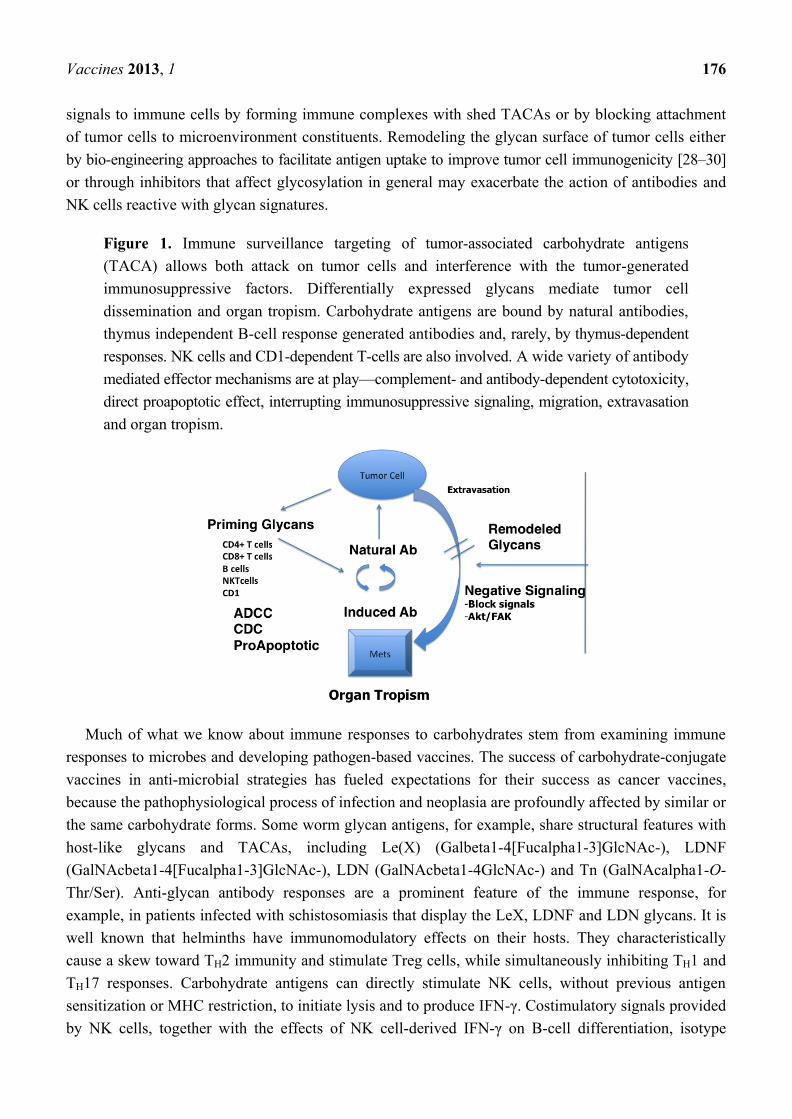
Figure 1. Immune surveillance targeting of tumor-associated carbohydrate antigens
(TACA) allows both attack on tumor cells and interference with the tumor-generated
immunosuppressive factors. Differentially expressed glycans mediate tumor cell
dissemination and organ tropism. Carbohydrate antigens are bound by natural antibodies,
thymus independent B-cell response generated antibodies and, rarely, by thymus-dependent
responses. NK cells and CD1-dependent T-cells are also involved. A wide variety of antibody
mediated effector mechanisms are at play—complement- and antibody-dependent cytotoxicity,
direct proapoptotic effect, interrupting immunosuppressive signaling, migration, extravasation
and organ tropism.
Much of what we know about immune responses to carbohydrates stem from examining immune
responses to microbes and developing pathogen-based vaccines. The success of carbohydrate-conjugate
vaccines in anti-microbial strategies has fueled expectations for their success as cancer vaccines,
because the pathophysiological process of infection and neoplasia are profoundly affected by similar or
the same carbohydrate forms. Some worm glycan antigens, for example, share structural features with
host-like glycans and TACAs, including Le(X) (Galbeta1-4[Fucalpha1-3]GlcNAc-), LDNF
(GalNAcbeta1-4[Fucalpha1-3]GlcNAc-), LDN (GalNAcbeta1-4GlcNAc-) and Tn (GalNAcalpha1-O-
Thr/Ser). Anti-glycan antibody responses are a prominent feature of the immune response, for
example, in patients infected with schistosomiasis that display the LeX, LDNF and LDN glycans. It is
well known that helminths have immunomodulatory effects on their hosts. They characteristically
cause a skew toward TH2 immunity and stimulate Treg cells, while simultaneously inhibiting TH1 and
TH17 responses. Carbohydrate antigens can directly stimulate NK cells, without previous antigen
sensitization or MHC restriction, to initiate lysis and to produce IFN-γ. Costimulatory signals provided
by NK cells, together with the effects of NK cell-derived IFN-γ on B-cell differentiation, isotype
Vaccines 2013, 1 177
switching and immunoglobulin secretion, ultimately result in augmentation of the IgG humoral
response against T-cell-independent antigens. In this mini-review, we place into context the selected
roles of TACAs reactive immune surveillance. In particular, we focus on glycan-mediated phenomena
associated with tissue rejection as a model to understand the rationale of controlling of tumor cell
growth by some immune modalities that target TACAs.
2. Glycans as Tumor Antigens
The rationale for targeting TACAs was elegantly discussed in terms of tissue distribution and
therapeutic importance [31]. The transition in glycosylation patterns of cancer cells reflect a myriad of
processes that correlate with poor prognosis of cancer, affecting cell signaling and communication, cell
motility and adhesion, angiogenesis and organ tropism. Both simple glycan structures and more
complex TACAs play a role in these processes. Glycan structures on the tumor cell surface result from
the combined action of glycotransferases and glycosidases. The carbohydrate antigens that have been
found to be tumor-associated (Table 1) include the mucin related Tn, sialyl Tn and Thomsen-Friedenreich
(TF/T) antigens, the blood group Lewis-related Lewis(Y), Sialyl Lewis(X) (SLeX) and Sialyl
Lewis(A) (SLeA), and Lewis(X) (also known as stage-specific embryonic antigen-1, SSEA-1), the
glycosphingolipids, Globo H, and stage-specific embryonic antigen-3 (SSEA-3), the sialic acid
containing glycosphingolipids, the gangliosides, GD2, GD3, GM2, fucosyl GM1 and Neu5GcGM3
and polysialic acid. SLeX and SLeA, in particular, are carbohydrate molecules that mediate the
adhesion between tumor cells and the endothelium. Overexpression of SLeX and SLeA is combined
with poor prognosis and malignant relapse [32]. The interaction of the antigen SLeX on tumor cells
and E-selectin on endothelial cells was shown to mediate adhesion of tumor cells to endothelial
cells [33], possibly facilitating tumor cell invasion in blood microvessels, extravasation and migration
into tissue. Additionally, colorectal tumor cells expressing SLeX might prefer the liver to form
clinically evident metastases, due to interaction with local E-selectin [34]. SLeX expression and
lymphatic microvessel density in primary tumors might predict disease recurrence, suggesting that for
some cancer, both lymphatic and hematogenous metastasis is mediated by SLeX interactions [35,36].
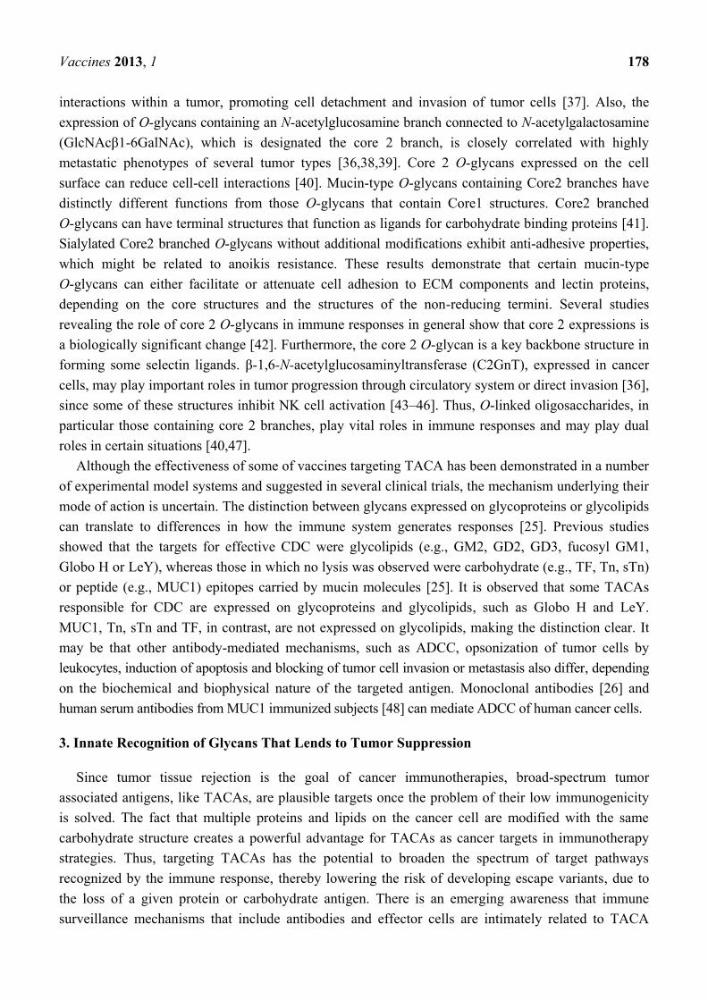
Table 1. Common carbohydrate antigens targets from tumor biopsy specimens.
Carbohydrate antigens Structure
Polysialic acid (PSA)
Tn
sialyl Tn
T antigen
Globo-H
LeY
SLeX
SLeA
α2,8-/α2,9 NeuAc
GalNAcSer/Thr
Neu5Acα2-6GalNAcaSer/Thr
Galβ1-3GalNAcαSer/Thr
Fucα1-2Galβ1-3GalNAcβ1-3Galα1-4Galβ1-4Glc
Fucα1-2Galβ1-4(Fucα1-3)GlcNAc-Galβ1-3(Fucα1-4)GlcNAc
Neu5Acα2-3Galβ1-4(Fucα1-3)GlcNAc
Neu5Acα2-3Galβ1-3(Fucα1-4)GlcNAc
Many cell-cell contacts are mediated by cell-surface glycans that are redundant on membrane
constituents that effect signaling pathways associated with anchorage independent growth and anoikis.
Upregulation of the N-glycan branching enzyme β-1,6-N-acetylglucosaminyltransferase V reduces cell-cell
Vaccines 2013, 1 178
interactions within a tumor, promoting cell detachment and invasion of tumor cells [37]. Also, the
expression of O-glycans containing an N-acetylglucosamine branch connected to N-acetylgalactosamine
(GlcNAcβ1-6GalNAc), which is designated the core 2 branch, is closely correlated with highly
metastatic phenotypes of several tumor types [36,38,39]. Core 2 O-glycans expressed on the cell
surface can reduce cell-cell interactions [40]. Mucin-type O-glycans containing Core2 branches have
distinctly different functions from those O-glycans that contain Core1 structures. Core2 branched
O-glycans can have terminal structures that function as ligands for carbohydrate binding proteins [41].
Sialylated Core2 branched O-glycans without additional modifications exhibit anti-adhesive properties,
which might be related to anoikis resistance. These results demonstrate that certain mucin-type
O-glycans can either facilitate or attenuate cell adhesion to ECM components and lectin proteins,
depending on the core structures and the structures of the non-reducing termini. Several studies
revealing the role of core 2 O-glycans in immune responses in general show that core 2 expressions is
a biologically significant change [42]. Furthermore, the core 2 O-glycan is a key backbone structure in
forming some selectin ligands. β-1,6-N-acetylglucosaminyltransferase (C2GnT), expressed in cancer
cells, may play important roles in tumor progression through circulatory system or direct invasion [36],
since some of these structures inhibit NK cell activation [43–46]. Thus, O-linked oligosaccharides, in
particular those containing core 2 branches, play vital roles in immune responses and may play dual
roles in certain situations [40,47].
Although the effectiveness of some of vaccines targeting TACA has been demonstrated in a number
of experimental model systems and suggested in several clinical trials, the mechanism underlying their
mode of action is uncertain. The distinction between glycans expressed on glycoproteins or glycolipids
can translate to differences in how the immune system generates responses [25]. Previous studies
showed that the targets for effective CDC were glycolipids (e.g., GM2, GD2, GD3, fucosyl GM1,
Globo H or LeY), whereas those in which no lysis was observed were carbohydrate (e.g., TF, Tn, sTn)
or peptide (e.g., MUC1) epitopes carried by mucin molecules [25]. It is observed that some TACAs
responsible for CDC are expressed on glycoproteins and glycolipids, such as Globo H and LeY.
MUC1, Tn, sTn and TF, in contrast, are not expressed on glycolipids, making the distinction clear. It
may be that other antibody-mediated mechanisms, such as ADCC, opsonization of tumor cells by
leukocytes, induction of apoptosis and blocking of tumor cell invasion or metastasis also differ, depending
on the biochemical and biophysical nature of the targeted antigen. Monoclonal antibodies [26] and
human serum antibodies from MUC1 immunized subjects [48] can mediate ADCC of human cancer cells.
3. Innate Recognition of Glycans That Lends to Tumor Suppression
Since tumor tissue rejection is the goal of cancer immunotherapies, broad-spectrum tumor
associated antigens, like TACAs, are plausible targets once the problem of their low immunogenicity
is solved. The fact that multiple proteins and lipids on the cancer cell are modified with the same
carbohydrate structure creates a powerful advantage for TACAs as cancer targets in immunotherapy
strategies. Thus, targeting TACAs has the potential to broaden the spectrum of target pathways
recognized by the immune response, thereby lowering the risk of developing escape variants, due to
the loss of a given protein or carbohydrate antigen. There is an emerging awareness that immune
surveillance mechanisms that include antibodies and effector cells are intimately related to TACA
Vaccines 2013, 1 179
reactivity that provides a template for developing strategies for cancer immunotherapy, because of the
display of glycans in the context of pattern recognition [49–52]. Glycans can be clustered representing
danger signals to the immune system. Pattern recognition receptors (PRRs) are sentinels of innate
immunity that instruct adaptive immunity mechanisms by which long-lived lymphocyte responses are
targeted to appropriate antigens [53,54]. Innate immune cells have evolved to sense microbial
pathogens through PRRs, coupling pathogen recognition to innate immunity through glycan-dependent
mechanisms [55]. The same mechanisms might be operative for glycans expressed on cancer cells.
Natural carbohydrate reactive antibodies have been described that mediate tumor cell apoptosis in
addition to modulating complement associated cell killing [27,56,57]. Preclinical studies support the
hypothesis that antibody-induced responses against TACAs might have their greatest impact in the
adjuvant setting, as antibody responses inhibit tumor outgrowth in metastatic models [58]. Such
observations suggest that sustained immunity against TACAs should be beneficial to prevent the
recurrence of disease, much like the natural ways of immune surveillance. Therefore, maximizing
sustained TACA-specific humoral immunity is considered an important goal in developing effective
antibody-based immunotherapies against cancer.
Like antibodies, NK cells are partners in immune surveillance. Three predominant superfamilies of
NK cell receptors (NKR) have been identified that can either inhibit or activate NK cell function:
(i) killer immunoglobulin (Ig)-like receptors (KIR) that bind to classical class I MHC molecules;
(ii) C-type lectin receptors that bind to non-classical class I MHC molecules or ―class I-like‖
molecules; and (iii) natural cytotoxicity receptors for which ligands are currently not well defined
(except for NKp30 binding to B7-H6 and BAG6) [59]. Interestingly, it is possible that some of the
natural cytotoxicity receptors may be binding to glycolipids [60]. NK cells can directly lyse virally
infected cells and tumor cells without prior sensitization and provide immunoregulatory cytokines that
shape the adaptive immune response. Cytolytic signals, triggered by inhibitory and activation receptors
on the cancer cell surface, regulate NK cell-mediated cytotoxicity and the production of chemokines
and inflammatory cytokines that mediate the immune response. The expression of some TACAs lend
to the evasion of NK cell immunity [43,44], while others activate NK cells [61]. Therefore, aberrant
glycosylation, while a target for immune surveillance, can regulate negative signaling of NK cells.
3.1. The Case for TACA-Directed Antibody-Mediated Tissue Rejection
The eradication of xenografts has been suggested as a model to provide important insights about the
role played by immunity in mediating tumor tissue rejection [5]. Tissue destruction occurs with
resolution of pathogenic processes (cancer, infection) or tissue damage and organ failure (autoimmunity,
allograft rejection) [5]. While xenograft rejection is highly mediated through innate immune
mechanisms, in tumor immunology, the primary focus for tumor tissue-rejection is focused on effector
cell-mediated tumor rejection and, particularly, the definition of cognate T-cell subsets that define
signatures for tumor cell rejection. Nevertheless, underlying the various mechanisms associated with
the biology of tissue damage or rejection emerges as a common pattern in tumor tissue-specific
destruction relevant to TACA targeting [5]. These patterns were elegantly reviewed by Marincola and
colleagues [5], which include the postulates that: (1) Tissue-specific destruction does not necessarily
only occur after non-self-recognition, but can also occur against self-or quasi-self-antigens. In the
Vaccines 2013, 1 180
context of tumor targets, TACA reactive antibodies are constantly produced, being inherent in the
innate and adaptive immunity. (2) The requirements for the induction of a cognate immune response
differ from those associated with the development of its effector phase. Natural circulating anti-TACA
antibodies are present and are known to be apoptotic to tumor cells. Therefore, antibodies can function
as both judge and jury. (3) Although the mechanisms prompting tissue-specific destruction differ
among immune pathologies, the effector phase converges into a common activation of adaptive and
innate cytotoxic mechanisms. In this context, glycan-reactive T-cells might work in unison with NK
cells and antibodies to target tumor cells. Furthermore, (4) adaptive immunity triggers a tissue-specific
reaction, but it is not always sufficient or necessary for tissue destruction. Carbohydrate-reactive
antibodies bind to both normal tissue and cancer cells. The binding to normal tissues does not
necessarily lend to normal tissue destruction, but may facilitate microenvironment interactions that
lend to tumor tissue rejection. Indeed, immune-based therapies have the potential to modulate the
tumor microenvironment by eliciting immune system cells that will initiate acute inflammation that
leads to tissue destruction [62].
Antibodies can mediate tissue rejection that validates targeting TACAs. A model for glycans as tissue
rejection antigens includes the response to the xeno-carbohydrate antigen Galα1-3Galβ1-4GlcNAc-R
(alpha-Gal) epitope. The majority of alpha-Gal antigens are built upon the Gal1, 4GlcNAc (type 2)
chain, but other inner-core saccharide chains also exist, especially on glycolipids [63–65]. Naturally
occurring anti-Gal antibody is produced as the most abundant antibody (1% of immunoglobulins)
throughout the life of all individuals [66]. Natural antibodies, such as anti-Gal or anti-blood groups
A/B antibodies, mediate hyperacute graft rejection and, thus, represent a major hurdle in
xenotransplantation [67] and blood transfusions, respectively. In the initial stage of the rejection,
anti-Gal IgG binds to R-Gal epitopes expressed on the surface of xenograft cells, triggering
antibody-dependent cell-mediated cytotoxicity by human blood monocytes and macrophages. The IgM
isotype of anti-Gal is believed to be responsible for the complement activation that leads to
complement-mediated lysis of the xenograft cells.
While early studies suggested an increased risk of cancer and poor prognosis associated with ABO
blood groups, such assertions have not been verified in breast cancer patients [68], but in ovarian
cancer, the presence of the B antigen was positively associated with ovarian cancer incidence, whereas
blood group A was not associated with risk [68]. One widely-occurring change observed in a large
variety of human cancers is deletion of the A or B epitope on tumor cells, associated with accumulation
of their precursor H (LeY, LeB), which causes enhanced malignancy [69]. The blood group reactive
lectin Griffonia simplicifolia (GS-I), which recognizes alpha-galactosyl moieties is recognized as a
surrogate marker to identify tumor expressed antigens reactive with anti-Gal antibodies [70], and GS-I
lectin is of utility to interrogate terminal α-GalNAc/Gal expression on human tissues [71].
The antibody-mediated tissue rejection model supports a rationale for targeting TACAs as
tumor-induced antibody responses resemble autoimmune responses [72]. Hyperacute rejection is a
complement-mediated response in recipients with pre-existing antibodies to the donor (for example,
ABO blood type antibodies). Tolerance to autologous ABO blood group antigens seems to depend in
part on peripheral control of antibody autoreactivity. However, normal human serum does contain
―hidden‖ natural antibodies reactive with autologous ABO blood group antigens [73]. These naturally
occurring antibodies, especially the anti-Gal response, might also have other clinical consequences for
Vaccines 2013, 1 181
immunotherapy [74] in the context of tolerance [75,76], cross-presentation of tumor antigens [77] and
increased immunogenicity of cell-based and protein-based vaccines [66]. Consequently, further
research is required to develop the translational and clinical applications.
3.2. The Case for Glycan-Directed T-Cell Mediated Tissue Rejection
As T-cell-dependent antigens, proteins have long been seen as the primary target of adaptive
immune responses. In contrast, carbohydrates are characterized as T-cell-independent (either Type 1 or
Type 2) antigens [78]; yet, early studies demonstrated that T-cells could recognize carbohydrate
antigens [79]. Post-translationally modified T-cell epitopes constitute a small fraction of both
MHC-I- and MHC-II-bound peptides, and a number of modifications are identified as natural MHC
ligands in vivo [80]. Computer-based sequence analysis suggests that only a minimal portion of
experimentally verified T-cell epitopes are potentially N- or O-glycosylated (2.26% and 1.22%,
respectively) [81] and T cells are demonstrated to react with processed glycopeptides and glycolipids
often representing TACA [82]. Some types of carbohydrates seem to be processed and presented to
T-cells by MHC-II [83,84] while others associate with the MHC-I groove [85–87]. The demonstration
that T-cells can recognize non-protein antigens has modified ideas on the breadth of antigens capable
of interacting with T-cells [88]. The size of the carbohydrate chains, as well as O- versus
N-glycosylation varies depending on tumor histotypes. However, recent studies suggest that
O-glycosylation (GalNAc) presentation on a peptide backbone, while inducing CD4+ T-cells can
impact negatively on CD8+ T-cell stimulation [85–87]. Structures of MHC Class II/peptide complexes
suggest analogies with helical carbohydrate structures that could fit the MHC Class II antigen-binding
groove [84]. In some cases, carbohydrate directly stimulates T-cells. Specific T-cell clones have been
generated from mice immunized with a meningococcal group C (alpha-2→9-sialic acid)
polysaccharide-tetanus toxoid conjugate [90]. These clones were MHC-independent, but still needed
contact with antigen presenting cells for optimal activation [90].
Crystal structure analysis of TCR-glycopeptide interactions validate that TCR can recognize
glycans presented on a peptide backbone [91,92]. Existing structures display the key interaction of the
core of the peptide ligand, with the TCR CDR3 region shaping a ―cavity‖ often accommodating
aromatic amino acid residues. The latter are successfully mimicked in size and conformation by short
glycans, like TF or the monomer, Tn. The ability of T-cells to recognize mono- and di-saccharides
attached to peptides with Ser or Thr might indicate that T-cells might be degenerate in recognizing
glycopeptides [51]. It should not be surprising that sometimes glycopeptides offer no significant
benefit as targets for cytotoxic immune response. In some cases, CTL, generated upon immunization
with glycopeptide, preferentially kills target cells treated with glycopeptide compared to those treated
with the core peptide. In other cases, it does not matter [93], and in some cases, it has been suggested
that other glycan receptors are involved in T-cell targeting [94]. This is particularly evident in the work
of Madsen et al. [89] that clearly suggest that natural processing of GalNAc on MUC1 might not be a
suitable for activating CTLs against MUC1. In general, this may or may not matter, because (a) some
activated CTLs are cross-reactive with both the glycosylated and non-glycosylated forms of the same
peptide and (b) glycopeptides are of low abundance on tumor target cells [93].
Vaccines 2013, 1 182
Polyclonal CTL have been observed to kill target cells expressing glycolipid [82]. It has been
suggested that glycopeptide-specific-restricted CTL and unrestricted glycan-specific CTL belong to
different T-cell populations with regard to TCR expression [95]. Such results demonstrate that
hapten-specific unrestricted CTL responses can be generated with MHC Class I-binding carrier
peptides. It is possible that CTLs activated with non-glycosylated peptides can cross-react with
glycopeptides and carbohydrate themselves. Such peptides have been referred to as carbohydrate
mimetic peptides (CMPs) or mimotopes. Sequences and structural properties of CMPs have been discussed
previously [96–99]. CMPs are known to generate T-cells cross-reactive with carbohydrates [100] and
to tumor cells [76,100–103]. The similarity of extended peptide structure and carbohydrates that can fit
within Class I or Class II groves has also been noted [97]. In addition, select amino acid residues can
spatially overlap glycans attached to peptides in the Class I grove [99]. T-lymphocytes from
CMP-immunized animals were shown to be activated in vitro by SLeX, triggering IFN-gamma
production in a MHC-dependent manner. Stimulation by peptide or carbohydrate resulted in loss of
L-selectin on CD4+ T-cells, confirming a Th1 phenotype. An enhancement in CTL activity in vitro
against SLeX-expressing Meth A cells using effector cells from Meth A-primed/peptide-boosted
animals was observed. CTL activity was inhibited by both anti-MHC class I and anti-L-selectin
antibodies. These results further support a role for L-selectin in tumor rejection, along with the
engagement by the TCR for most likely processed tumor-associated glycopeptides, focusing on peptide
mimetics as a means to induce carbohydrate reactive cellular responses. Immunization of mice with
this CMP reduced tumor cell growth in a transplanted mammary tumor model mediated, to a large
extent, by CD8+ T-cells [58], but without any damage to normal tissue after vaccination with the
CMP [104]. These observations are very important in understanding the complexity of the antitumor
response, especially in terms of abnormal glycan expression patterns and developing strategies in
vaccine design.
3.3. The Case for NK Cell-Mediated Rejection
Cell-mediated cytotoxicity is a primary effector function of NK cells. It has been known for a long
time that NK cells play a major role in tumor immune surveillance by serving as the first line of
antitumor immune defense [105,106]. The multifaceted steps early in NK immune surveillance include
an orchestrated activation and recruitment to the tissue sites where they, perform effector functions,
which may be associated with tumor reactive antibodies. Receptor diversity is crucial in allowing NK
cells to respond effectively, mediating their effects through direct cytolysis, release of cytokines and
regulation of subsequent adaptive immune responses [107–111]. NK cell lysis is regulated by a
balance of intracellular signals transmitted via stimulatory and inhibitory cell surface receptors after
specific binding to their respective target cell ligands [112–114]. Activation of endogenous NK cells
bears limited clinical benefit, as most cancer patients are treated with chemotherapy, and their immune
system is compromised. Consequently focus has been directed in recent years to first understand NK
suppression mechanisms and how better to exploit NK cell functionality.
Antibodies promote NK cell activation through antibody-dependent cell-mediated cytotoxicity. The
best example of combining an anti-GD2 antibody with NK cells is in neuroblastoma (NB) [115].
Treatment of patients with high-risk NB with monoclonal antibodies targeting the disialoganglioside
Vaccines 2013, 1 183
surface antigen GD2 has resulted in lower recurrence rates and improved overall survival [116–119].
In addition to complement-dependent cytotoxicity, the anti-GD2 monoclonal antibody 3F8 achieves
NB killing through antibody-dependent cell-mediated cytotoxicity mediated by myeloid and NK
cells [117]. To combine specific antibody-mediated recognition of NB cells with the potent cytotoxic
activity of NK cells, clonal derivatives of the clinically applicable human NK cell line NK-92 that
stably express a GD2-specific chimeric antigen receptor (CAR) comprising an anti-GD2 ch14.18
single chain Fv antibody fusion protein with CD3-δ chain as a signaling moiety has been
described [120]. CAR development in general is a hot topic area in immunotherapeutics, but mostly in
developing T-cells for adoptive therapy [121]. The therapeutic efficacy of endogenous NK cells
depends on the effectiveness of NK-activating agents to mobilize sufficient numbers of these cells to
tumor sites [122]. The clinical utilization of NK cells is considered at the forefront of cancer therapy. It
should be clear that adoptive transfer of NK cells should lead to high levels of circulating NK cells, but
that does not necessarily translate into mediating tumor regression [123]. This may result from
expression of glycans on the tumor cells in addition to glycans shed from the tumor cell surface.
A variety of studies have linked the nature of signaling with the glycan ligand NK receptor paring.
In this context, interest has focused on the N-glycan biosynthesis of glycoproteins and, in particular,
branching enzymes, such as N-acetylglucosaminyltransferase III (GnT-III), GnT-IV, GnT-V and a1-6
fucosyl- transferase (a1-6FucT) [124,125], that can regulate the further processing of the N-glycan
structures, which play a pivotal role in tumor development, metastasis and invasion. Heparan sulfate
proteoglycans play a role in NK cell initial recognition and activation [126,127]. The interaction of
SLeX antigen with lectin-like receptors on NK cells also triggers cytotoxicity [128,129]. Clustered
glycoconjugates sharing the common structure motif trisaccharide Le(x) [130] can enhance
cytotoxicity specifically by CD16+ NK cells. GlcNAc-terminated glycoclusters are found to be potent
inhibitors of receptors on natural killer cells [131]. N-acetyl-D-glucosamine (GlcNAc) transferases,
MGAT3 and MGAT5, have major involvement in linking terminating residues on glycans. MGAT5 is
responsible for adding β1-6 GlcNAc residues and forming branched structures, which are especially
abundant in cancer tissues with high metastatic potential. MGAT3 catalyzes the addition of β1-4
GlcNAc residues and forms a bisecting structure that disables further addition of GlcNAc by other
glycosyltransferases, like MGAT5. Expression of terminal GlcNAc is perceived to inhibit NK function
supported by experiments in which siRNA targeting these glycosyltransferases in tumor cells are
observed to increase NK cell activity towards tumors [132].
Some transformed cells evade immune surveillance and become resistant to NK cell cytotoxicity,
mainly because some shed TACA inhibits NK cell activation [45], leading to established primary
tumors [133–136]. In renal cell carcinoma, the presence of higher gangliosides correlates with
systematic metastasis. Disialosyl globopentaosylceramide (DSGb5) was identified previously as one of
the major gangliosides from renal cell carcinoma (RCC). Siglec-7 (sialic acid-binding Ig-like lectin-7),
expressed on NK cells as an inhibitory receptor, has a striking preference for internally branched
α2,6-linked disialic gangliosides, such as DSGb5 [135]. These results suggest that DSGb5 expressed
on RCC cells can downregulate NK cell cytotoxicity in a DSGb5-Siglec-7-dependent manner and that
RCC cells with DSGb5 create a favorable circumstance for their own survival and metastases [135].
Consequently, despite the enthusiasm of using NK cells in adoptive transfer protocols, in most cases,
NK functionality needs to be reset by remodeling the tumor glycan surface. The remodeling can lead to
Vaccines 2013, 1 184
activation of endogenous NK cells with anti-tumoral function. Studies exploring such possibilities are
warranted and under research.
3.4. Remodeling Glycan Signatures
Strategies for cell surface ―glycoform remodeling‖ promise to facilitate the investigation of
carbohydrate mediated cell-cell interactions [137] and as cancer vaccines [138]. Expression of the
human α1,2-fucosyltransferase, for example, in transgenic pigs modifies the cell surface carbohydrate
phenotype and confers resistance to human serum-mediated cytolysis [139]. The T-cell-independent
process of delayed xenograft rejection is suggested as a model for glycan remodeling, which augments
NK cell activity [140]. While natural antibodies against alpha-Gal epitope cause hyperacute rejection
of pig organs in primates, evidence for the role of alpha-Gal in the NK cell-mediated xeno-response
has been contradictory [141]. Therefore, while logic would dictate that glycan remodeling facilitates an
improved immune response [138], the nature of these responses might be limited to antibodies
and T-cells.
Nevertheless it was argued early on that inhibition of N-linked oligosaccharide processing in
malignant cells is associated with increased susceptibility to natural immunity [142]. Interference with
N-glycosylation has been shown both to reduce the membrane expression of MHC class I and to
increase the in vitro sensitivity of tumor cells to NK cell killing. It was long recognized that
compounds that inhibit glycosylation pathways could affect the growth of tumor cells in tumor bearing
animals. Castanospermine, swainsonine and tunicamycin block different steps in the pathways of
glycoprotein processing that affect tumor cell dissemination and tumor colonization. This suggested
blocking at one of at least two steps could have beneficial effects on tumor cell growth. The
antimetastatic effect of tunicamycin may be related to interference in tumor cell-extracellular matrix
interactions, whereas treatment with castanospermine or swainsonine appears to block at a stage distal
to initial tumor cell arrest [143]. Swainsonine, in particular, is interesting, as it inhibits the formation of
N-linked complex oligosaccharides with this inhibition correlative with enhancement with NK cell
function. Consequently, inhibitors of N-, as well as O-linked glycosylation need to be expanded,
because they should be useful for the treatment of cancer by effectively resetting NK functional
activity by disruption of negative signals; given that inhibitors can be specifically targeted to tumor
tissue [144]. More recently, it was shown that glycosylation regulates NK cell-mediated effector
function through the PI3K pathway [132].
Antibodies might also regulate glycan expression patterns in an undefined way that enhances NK
activity. The orchestration of glycan remodeling and galectin-1 upregulation by the tumor suppressor
p16INK4a
in pancreatic carcinoma cells to reconstitute susceptibility to anoikis underscores the potential
and tight control of this lectin [145]. Anti-glycan antibodies can function like lectins, mediating cell
death signals [58] and cell growth signals [146]. Other galectins can promote NK cell-mediated
anti-tumor activity by expanding unique phenotypes [147]. Co-culture of naive NK cells with
macrophages from Gal-9-treated mice resulted in enhanced NK activity, although Gal-9 itself did not
enhance the NK activity [147]. Antibodies can do the same. Clinical studies have indicated a role for
anti-ganglioside IgM antibodies (including anti-GD2) in passive and active immunity against some
cancers [148–150]. Their mechanism(s) of action is not clear, but a study in which mice transgenic for
Vaccines 2013, 1 185
anti-GD2 IgM antibody were protected from EL4 metastasis and death indicated a role for IgM,
complement and NK cells [151]. In these studies depletion of NK cells with anti-asialo GM1 rabbit
serum reduced or abrogated the observed anti-tumor effects, suggesting that NK cells play a major role
in tumor eradication or suppression [151]. It is possible that the GD2 model actually opens a window
on a more general innate circuitry, which has just been further elucidated. Macrophages activated by
Toll-like receptor (TLR) ligands appear to stimulate B cells, including through CD40-CD40L
interactions, to a state of activation (CD69+ CD25hi, CD317+) in which they produce IFN alpha and
stimulate NK cells nonspecifically [152].
4. Augmenting Responses to TACA
The clinical importance of targeting TACAs is highlighted by the success of carbohydrate-based
vaccines against infectious diseases, by the role of TACAs in autoimmune phenomena and by the
observed anti-TACA antibodies as clinical correlates of positive outcome seen in patients with cancer.
Carbohydrate-based vaccines against Haemophilus influenzae Type b, Neisseria meningitidis,
Streptococcus pneumoniae and Salmonella enterica serotype Typhi (S. Typhi) are already licensed, and
many similar products are in various stages of development. Therefore, factors contributing to the
successes and failures of these bacterial vaccines serve as guides to developing carbohydrate-targeting
cancer vaccines. The practical benefits of inducing TACA-reactive antibodies in patients with cancer
are further demonstrated by observations that patient survival significantly correlates with
ganglioside-reactive IgM levels [149,153]. Low affinity natural IgM antibodies have been found
indispensable for anti-viral responses [154,155]. An analogous role for natural antibodies as an innate
anti-cancer surveillance mechanism has been suggested, but has been underappreciated, so far [156,157].
The fact that survival rates of cancer patients are correlated with low (intrinsic) affinity and low-titer
TACA-reactive antibodies argues that more robust antibody responses may not be necessary.
The successful development of anti-microbial vaccines has proven that antibodies—particularly
those targeting carbohydrate antigens—are ideally suited for eradicating pathogens from the blood
stream and from early tissue invasion. Similarly, vaccines targeting TACAs may also prove beneficial
in treating micrometastases. This may be the case since anti-TACA antibodies correlate with beneficial
effects on the course of malignant disease and long-term patient survival [149]. However,
N-acetylglucosamine branch in O-glycans (core 2 O-glycans) expressing cancer cells acquire highly
metastatic phenotypes by surviving longer in host blood circulation [43–45]. The induction of TACA
reactive antibodies and NK cells to leukemic cells might prove specially beneficial, since simple
glycan profiles and commonly contained sialyl-T (NeuAcalpha2-3Galbeta1-3GalNAc) and disialyl-T
(NeuAcalpha2-3Galbeta1-3(NeuAcalpha2-6)GalNAc) antigens as major O-glycans are observed on
these cells [158] and receptors on NK cells bind to alpha2,3-NeuAc-containing glycoproteins [159].
Therefore, maximizing sustained antibody immunity against TACAs that express simple Core 1 and
Core 2 (C2GNT-1) structures is an important goal in developing effective cancer vaccines to combat
recurrent disease.
Vaccines 2013, 1 186
4.1. Taking Advantage of Natural Antibodies
The ability of the immune system to identify and destroy nascent cancer cells and, thereby, function
as a primary defense against cancer, is a long-standing debate. This raises expectations of therapeutic
development of antibodies derived from the promise of multifaceted biological potency compared to
stoichiometrically binding antibodies that just interfere with receptor binding. It is postulated that
anti-carbohydrate antibodies are part of immune surveillance, just as they are a first line of defense
against infectious agents. Natural antibodies may not only have a direct cytotoxic effect on intact
tumor cells, but also bystander effects. In addition, they are known to contain low affinity self-reactive
fraction representing a ―grey area‖ of the tolerance to self. It has been proposed that this ―grey‖
self-reactivity actually detects quantitative rather than qualitative changes of the antigenic
landscape—a function especially suited to detecting unnaturally increased expression of TACA [160].
Antibodies that bind to a broad spectrum of TACA can reduce tumor cell dissemination by multiple
mechanisms, including blocking the adhesion of metastatic cells to adhesion molecules and generally
functioning as regulatory molecules to thwart signaling processes that underlie migration and autocrine
and paracrine activities that grant immune privilege to cancer. FAK is a non-receptor tyrosine kinase
that plays an important role in signal transduction pathways that are initiated at sites of
integrin-mediated cell adhesions and by growth factor receptors. FAK is a key regulator of survival,
proliferation, migration and invasion: processes that are all involved in the development and
progression of cancer. FAK is also linked to oncogenes at both a biochemical and functional level.
Moreover, overexpression and/or increased activity of FAK is common in a wide variety of human
cancers, implicating a role for FAK in carcinogenesis. Given the important role of FAK in a large
number of processes involved in tumorigenesis, metastasis and survival signaling, FAK should be
regarded as a potential pathway target in the development of antibodies targeting TACAs that are
associated with anoikis [21] and blocking adhesion.
Determining populations of glycan reactive antibodies in the repertoire of natural autoantibodies
could lead to developing immunotherapies targeting cancer without affecting normal tissues or
resulting in adverse side-effects. Thus, the application of natural antibodies, like IVIg, has the potential
to be a supportive therapy for the treatment of cancer metastases and provide an opportunity to probe
yet undefined roles of natural antibodies relating broad-spectrum reactivity with anti-cancer functional
properties. Most anti-glycan antibodies recognize epitopes of two or three sugars. Consequently,
antibodies can cross-react with similar terminal structures. This property of recognizing epitopes
―shared‖ by different molecules is characteristic of anti-glycan antibodies and can be considered an
example of ―antigen mimicry‖. In this context, it would seem that anti-Gal antibodies should be
reactive with the histo-blood group antigens, LeB and LeY. Blood group B individuals show reactivity
to Tn antigen [161], and some anti-Gal antibodies are cross-reactive with the blood group B antigen [162].
Anti-Gal alpha(1,3)Gal antibodies are observed to react with mucin 1 (MUC1) found on the surface of
human breast cancer cells [163]. Thus, natural occurring anti-Gal alpha (1,3)Gal antibodies found in all
human serum can react with self (MUC1) peptides expressed in large amounts on the surface of tumor
cells, but not on normal cells. These findings are of interest and serve to explain reported findings that
human cells can, at times, express Gal alpha(1,3)Gal; such expression is suggested as an artifact in that
anti-Gal alpha(1,3)Gal antibodies react with mucin peptides [163].
Vaccines 2013, 1 187
The cross-reactivity of anti-Gal antibodies has been exploited in cell therapy, where autologous
cells processed to express alpha-Gal epitopes result in anti-Gal-mediated, in vivo targeting of
autologous tumor vaccine to antigen presenting cells (APC) [77,164]. Transfection of cells with the
enzyme 1,3galactosyltransferase (1,3GT) with concomitant expression of the Gal epitope followed by
immune complex formation by anti-Gal antibodies should increase transport to lymph nodes and
processing of anti-Gal complexed vaccines internalized by APC. Anticipated results include an
effective activation of vaccine-specific CD4(+) and CD8(+) T-cells and high cellular and humoral
immune response [77]. While manipulating the pre-existing anti-Gal response may facilitate an
efficacious vaccine response through antigen spreading to antitumor T-cell response, truly
tumor-specific antigens are needed to contribute decisively to tumor regression [165].
However, some antibodies display exquisite specificity, like those directed toward the TF
antigen [166]. Postpartum, carbohydrate structures on the cell walls of the gastrointestinal flora evoke
natural antibodies of presumed TF specificity. These antibodies may provide an early barrier against
TF-carrying tumor cells. The widely used regimen of neoadjuvant chemotherapy is demonstrated to
stimulate the immune response to TACA in some patients, as reviewed by Andre et al. [167]. Small
retrospective studies have suggested that post-chemotherapy lymphocyte infiltrates could be associated
with better outcome in patients who did not reach pathologic complete response [167]. The high levels
of anti-TF antibody before surgery is another example in which antibody targeting is associated with a
better survival of stage II breast cancer patients [168]. This may indicate that the selection of
immunopotentiating regimens of neoadjuvant chemotherapy might be beneficial for the host in
conjunction with the functional activity of natural anti-cancer antibodies.
On the other hand, the detectable spontaneous immune responses to T and Tn antigens are not
necessarily efficient, since the expression of these antigens correlates with worse prognosis, mostly
because of increased metastasis. The reason may be an escape of some cancer cells from the control by
immune responses to TACA, like T, Tn and sialyl-Tn. It is also possible that the correlation with
higher grade and metastasis is due to the observation that some tumors are resistant to immunoediting.
It would be interesting to differentiate between primarily TACA-negative tumors and secondarily
negative tumors that arise due to immunoediting. It is likely that specific suppressive influence of the
tumor on the production of TF antibodies is associated with the stage and grade of the tumor.
Postoperatively, these antibodies rebound, as do lymphocyte counts [169]. The observation of positive
correlation between the level of TF antibodies and the count of lymphocytes in TF-responders appears
to reflect the adaptive immune response and provides a further explanation for the involvement of
anti-TF IgG in cancer-associated immunosuppression. However, the possible protective mechanism of
TF antibodies in cancer has yet remained unclear, as is the role antibodies play in the natural
anti-cancer defense system. The signs of tumor-immune system interaction, together with the
ambivalence of the results, draw attention to the hypothesis that immune surveillance may be just an
epiphenomenon of the ―knowledge of self‖ or, at least, still very early in the process of evolutionary
optimization. The tools are there, but maybe they are yet to be tuned.
Vaccines 2013, 1 188
4.2. Bridging Humoral and Cellular Responses
Because of their characteristic immunogenicity and/or immunotolerance, most TACAs fail to
induce T-cell-mediated immunity that is critical for cancer therapy. Approaches to overcome this
limitation or improve their immunogenicity include coupling covalently TACA to proper carrier
molecules to form clustered or multi-epitopic conjugate vaccines, coupling TACAs to a T-cell peptide
epitope and/or an immunostimulant epitope to form fully synthetic multi-component glycoconjugate
vaccines [138]. Polyvalent vaccines containing a variety of tumor-associated antigens are being tested
under the hypothesis that a greater number of antigens in a vaccine will increase the probability of
containing the correct antigen(s) to stimulate an effective anti-tumor response. The case for a
polyvalent cancer vaccine to induce antibodies to TACAs has been made [170], although, in general,
there may be more heterogeneity in antibody responses to polyvalent vaccines than that anticipated
with monovalent vaccines [171]. More recent studies on carbohydrate-based vaccines are essentially
modifications to the basic premise of conjugate formulations [172–175].
The recognition that T-cell receptors can interact with glycopeptides has facilitated concepts for
new antigens being developed to activate anti-tumor responses. The feasibility of T-cell antigens
design based on carbohydrate structures is strongly supported by crystallography of several
HLA/peptide complexes. These include designer glycopeptides to facilitate CTL activation [176],
glycan modification of antigens to target to APC to enhance both CD4+ and CD8+ T-cell
responses [177–179]. One of the more important glycan decorated tumor antigens is human mucin 1
protein (MUC1). Attempts to develop MUC1-targeting cancer vaccines based on carrier-conjugated
unglycosylated MUC1 tandem repeat peptides or carrier-conjugated glycosylated epitopes have been
largely unsuccessful. Problems here partly relate to the conformational differences between
non-glycosylated vaccine sequences and tumor-expressed, aberrantly glycosylated MUC1. Moreover,
densely glycosylated MUC1 glycopeptide might be inefficiently processed by antigen-presenting cells,
which ultimately means T-helper cells and CTLS aren’t highly activated.
More promising results in tumor models have been reported using a two-component vaccine
approach based on an MHC I glycopeptide and a T-helper epitope [180]. A multicomponent vaccine
comprising a glycosylated MUC1-derived glycopeptide covalently linked to a T-helper epitope and
TLR immunoadjuvant elicited potent humoral and cellular immune responses, effectively reversing
tolerance and demonstrated potent anticancer effects. The vaccine candidate comprises the thiobenzyl
ester of Pam3CysSK4 as a TLR2 ligand adjuvant, together with the composite T-helper epitope and
aberrantly glycosylated MUC1 peptide, CKLFAVWKITYKDTGTSAPDT(αGalNAc)RPAP, formulated
into phospholipid-based small unilamellar vesicles. To test its effects in vivo, the tripart vaccine was
administered to experimental mice and the animals challenged with MUC1-expressing mammary
tumor cells after 35 days. A week after the cancer challenge, the mice were given another vaccine
boost. Control mice were administered with vaccine constructs comprising either the unglycosylated
vaccine or subunits of the overall vaccine structure, i.e., just the glycopeptide or the adjuvant.
Immunization with the multicomponent vaccine led to significant reductions in tumor burden and
weight when compared with treatment using either empty liposomes or immunization with a control
vaccine that didn’t contain the MUC1 glycopeptide epitope or an unglycosylated multicomponent
Vaccines 2013, 1 189
candidate. Immunization with the primary tripartite candidate also elicited robust IgG antibody
responses against the MUC1 glycopeptide, including a mixed Th1/Th2 response.
However, there are aspects of MUC1 that are largely ignored in the literature that might impact on
its utility as an immunogen. Recently it was found that several of the tumor-related glycoforms of
carcinoembryonic antigen, and MUC1 might affect CLR signaling and DC differentiation. These are
specific ligands for the pattern recognition receptors DC-SIGN [181] and macrophage galactose-type
C-type lectin (MGL) [182], expressed on DCs. MGL1/2-positive cells are interesting, as they represent
a distinct sub-population of macrophages, having unique functions in the generation and maintenance
of granulation tissue induced by antigenic stimuli [183]. MGL1 is postulated to be actively involved in
inflammatory processes [184]. Consequently, Tn glycans on MUC1 that bind MGL might instruct DC
to drive Th2-mediated responses, which, unlike those of Th1 effector cells, are thought not to
contribute to tumor cell eradication. This has several ramifications. Cancer patients with MUC1
expression profiles may exhibit a Th2-skewed cytokine profile within blood and tumor-infiltrating
lymphocytes. This Th1/Th2 imbalance would coincide with disease progression and immunotherapy
response. Various lines of evidence suggest that in vivo skewing of T-cell responses toward a Th2 type
is an important mechanism of immune evasion in cancer patients [185–187]. Terminal glycan
structures shared by both host and parasitic helminths include LeX, LDN and LDNF and the truncated
O-glycans known as the T (Galβ1-3GalNAcα1-O-Thr/Ser) and Tn antigens, all
glycan antigens that
may interact with host lectins that skew the immune response to Th2 profiles [188]. This skewing may
limit the efficacy of immunotherapeutic approaches [189]. Immunization with formulations that reflect a
Th2 bias of the native antigen might only exacerbate the Th2 response. Ensuring induction of a strong
type 1 response may be critical to the development of effective cancer vaccines.
MUC1-derived non-glycosylated peptides are also demonstrated to mimic carbohydrate antigens
that include the Gal epitope [76]. Non-glycosylated peptides that mimic TACAs are noted to induce
both humoral and cellular responses to tumors. CMPs can induce cellular responses, including
CMP- and TACA-reactive Th1 CD4+ and tumor-specific CD8
+ cells [100,101], and CMPs can prime
for memory responses to TACAs [190]. We have demonstrated that a single CMP can bind to
antibodies with differing TACA specificities that, upon immunization, can induce divergent antibody
responses that recognize a range of TACAs [191]. Thus, this important and novel feature of CMPs
effectively broadens the repertoire of reactive antibodies without inducing autoimmunity in animal
models. The capacity to induce a carbohydrate-cross-reactive humoral, a Th and a CTL response with
one single CMP is clearly a unique property of this approach. The observations that CMPs can induce
both antibody and cellular responses in the absence of autoimmunity emphasize the feasibility of
CMP-based vaccination strategies and the potential benefits of maximizing their effectiveness.
Furthermore, CMPs can be encoded into DNA and viral vectors to enhance long-term immunity,
which precludes the need for repeated TACA-based vaccination to maintain immune surveillance. This
approach has led to a phase I study of a carbohydrate mimetic peptide (manuscript in preparation) in
stage IV breast cancer subjects. This CMP shares homology with a region of MUC1, but involved
reverse engineering using antibody and lectin templates as the basis for CMP development [104].
The mimicking of MUC1 non-glycosylated peptides with the Gal epitope might also have
unintended consequences. For example, mimicry might lend to confusion in deciphering the difference
in natural antibody levels to MUC1 and clinical outcomes to MUC1-based vaccines if anti-Gal
Vaccines 2013, 1 190
antibodies cross-reactive with MUC1 are not considered [192]. In addition, this mimicry might also
skew Th2 type responses to MUC1 vaccines, which is contradictory to the present paradigm that
stresses Th1 responses to MUC1 and other tumor associated antigens. In fact, it is easy to see that as
MUC1 expressing cancer cells emerge, the Th2 response becomes set. Vaccines that are MUC1-based
might only stimulate B-cells and T-cells that are already primed as the Th2 type, exacerbating what
might be akin to ―original antigen sin‖ or an amnestic response to MUC1 of Th2 type [193]. In
addition, while transgenic mice expressing human MUC1 are perceived to be of importance to
understand the immune response to MUC1 in humans, it is often overlooked that these transgenics also
express murine MUC1 in which T cells generated to human MUC1 peptides cross-react with naturally
expressed murine MUC1 peptides. This cross-reactivity is seldom discussed and has the potential to
confound results.
5. Conclusions
Glycans or TACAs are important targets for cancer immunotherapy, as suggested by immune
surveillance mechanisms. TACAs display important biological effects in tumor biology and tumor
immunology. Most importantly, the recognition properties of glycans by immune effector cells have
suggested translational strategies in immune therapy. The diversity of regulatory mechanisms
involving glycans expands the range of possible effects of TACA targeting immunotherapeutic
approaches. Anti-TACA antibodies, thus, may be involved in more than direct tumor cytotoxicity.
Although the exact mechanism may represent a cascade of steps that are still to be established,
immunization targeting TACAs has already been shown to yield antitumor effects mediated by NK
cells or through neutralization of tumor immunosuppressive factors in the form of soluble
gangliosides. Future work should clarify the points of involvement of antibody/carbohydrate
interactions in modulating tumor growth and facilitating innate surveillance mechanisms.
The abrogation of negative regulatory signals imposed by glycans and the maintenance of the
activated phenotype of NK cells can significantly enhance NK cell activity against solid tumors.
Manipulating the balance between inhibitory and activating NK receptor signals, the sensitivity of
target cells to NK cell-mediated apoptosis and NK cell cross-talk with other immune effector cells
might hold therapeutic promise [194,195]. Efforts to modulate NK cell trafficking into inflamed tissues
and/or lymph nodes and to counteract NK cell suppressors, might prove fruitful in the clinic. However,
a greater understanding of how to downregulate negative signaling, the benefits of combination
therapy, characterization of the functional distinctions between NK cell subsets and the design of new
tools to monitor NK cell activity are needed to strengthen our ability to harness the power of NK cells
for therapeutic aims.
Conflict of Interest
The authors declare no conflict of interest.
References
1. Swann, J.B.; Smyth, M.J. Immune surveillance of tumors. J. Clin. Invest. 2007, 117, 1137–1146.
Vaccines 2013, 1 191
2. Burnet, F.M. The concept of immunological surveillance. Prog. Exp. Tumor Res. 1970, 13, 1–27.
3. Soloski, M.J. Recognition of tumor cells by the innate immune system. Curr. Opin. Immunol.
2001, 13, 154–162.
4. Wang, E.; Monaco, A.; Monsurro, V.; Sabatino, M.; Pos, Z.; Uccellini, L.; Wang, J.;
Worschech, A.; Stroncek, D.F.; Marincola, F.M. Antitumor vaccines, immunotherapy and the
immunological constant of rejection. IDrugs 2009, 12, 297–301.
5. Wang, E.; Worschech, A.; Marincola, F.M. The immunologic constant of rejection. Trends
Immunol. 2008, 29, 256–262.
6. Houghton, A.N.; Guevara-Patiño, J.A. Immune recognition of self in immunity against cancer.
J. Clin. Invest. 2004, 114, 468–471.
7. Schreiber, T.H.; Raez, L.; Rosenblatt, J.D.; Podack, E.R. Tumor immunogenicity and
responsiveness to cancer vaccine therapy: The state of the art. Semin. Immunol. 2010, 22,
105–112.
8. Hakomori, S. Tumor-associated carbohydrate antigens defining tumor malignancy: Basis for
development of anti-cancer vaccines. Adv. Exp. Med. Biol. 2001, 491, 369–402.
9. Xu, Y.; Sette, A.; Sidney, J.; Gendler, S.J.; Franco, A. Tumor-associated carbohydrate antigens:
A possible avenue for cancer prevention. Immunol. Cell Biol. 2005, 83, 440–448.
10. Ono, M.; Hakomori, S. Glycosylation defining cancer cell motility and invasiveness.
Glycoconj. J. 2004, 20, 71–78.
11. Sakamoto, S.; Kyprianou, N. Targeting anoikis resistance in prostate cancer metastasis.
Mol. Aspects Med. 2010, 31, 205–214.
12. Zhong, X.; Rescorla, F.J. Cell surface adhesion molecules and adhesion-initiated signaling:
Understanding of anoikis resistance mechanisms and therapeutic opportunities. Cell Signal.
2012, 24, 393–401.
13. Kornberg, L.J. Focal adhesion kinase and its potential involvement in tumor invasion and
metastasis. Head Neck 1998, 20, 745–752.
14. Hauck, C.R.; Hsia, D.A.; Schlaepfer, D.D. The focal adhesion kinase—A regulator of cell
migration and invasion. IUBMB Life 2002, 53, 115–119.
15. Sawai, H.; Okada, Y.; Funahashi, H.; Matsuo, Y.; Takahashi, H.; Takeyama, H.; Manabe, T.
Activation of focal adhesion kinase enhances the adhesion and invasion of pancreatic cancer cells
via extracellular signal-regulated kinase-1/2 signaling pathway activation. Mol. Cancer 2005, 4, 37.
16. Danker, K.; Reutter, W.; Semini, G. Glycosidated phospholipids: Uncoupling of signalling
pathways at the plasma membrane. Br. J. Pharmacol. 2010, 160, 36–47.
17. Zhang, D.; Wei, J.; Wang, J.; Liu, S.; Wang, X.; Yan, Q. Difucosylated oligosaccharide Lewis Y
is contained within integrin alphavbeta3 on RL95–2 cells and required for endometrial
receptivity. Fertil. Steril. 2011, 95, 1446–1451.
18. Valentino, L.A.; Ladisch, S. Tumor gangliosides enhance alpha2 beta1 integrin-dependent
platelet activation. Biochim. Biophys. Acta 1996, 1316, 19–28.
19. Chen, Y.X.; Chen, X.W.; Li, C.G.; Yue, L.J.; Mai, H.R.; Wen, F.Q. Effect of tumor gangliosides
on tyrosine phosphorylation of p125FAK in platelet adhesion to collagen. Oncol. Rep. 2013, 29,
343–348.
Vaccines 2013, 1 192
20. Ohkawa, Y.; Miyazaki, S.; Hamamura, K.; Kambe, M.; Miyata, M.; Tajima, O.; Ohmi, Y.;
Yamauchi, Y.; Furukawa, K. Ganglioside GD3 enhances adhesion signals and augments
malignant properties of melanoma cells by recruiting integrins to glycolipid-enriched
microdomains. J. Biol. Chem. 2010, 285, 27213–27223.
21. Aixinjueluo, W.; Furukawa, K.; Zhang, Q.; Hamamura, K.; Tokuda, N.; Yoshida, S.; Ueda, R.
Mechanisms for the apoptosis of small cell lung cancer cells induced by anti-GD2 monoclonal
antibodies: Roles of anoikis. J. Biol. Chem. 2005, 280, 29828–29836.
22. Christiansen, D.; Vaughan, H.A.; Milland, J.; Dodge, N.; Mouhtouris, E.; Smyth, M.J.;
Godfrey, D.I.; Sandrin, M.S. Antibody responses to glycolipid-borne carbohydrates require
CD4+ T cells but not CD1 or NKT cells. Immunol. Cell Biol. 2011, 89, 502–510.
23. Zajonc, D.M.; Kronenberg, M. Carbohydrate specificity of the recognition of diverse glycolipids
by natural killer T cells. Immunol. Rev. 2009, 230, 188–200.
24. Freire, T.; Zhang, X.; Deriaud, E.; Ganneau, C.; Vichier-Guerre, S.; Azria, E.; Launay, O.;
Lo-Man, R.; Bay, S.; Leclerc, C. Glycosidic Tn-based vaccines targeting dermal dendritic cells
favor germinal center B-cell development and potent antibody response in the absence of
adjuvant. Blood 2010, 116, 3526–3536.
25. Ragupathi, G.; Liu, N.X.; Musselli, C.; Powell, S.; Lloyd, K.; Livingston, P.O. Antibodies
against tumor cell glycolipids and proteins, but not mucins, mediate complement-dependent
cytotoxicity. J. Immunol. 2005, 174, 5706–5712.
26. Lavrsen, K.; Madsen, C.B.; Rasch, M.G.; Woetmann, A.; Odum, N.; Mandel, U.; Clausen, H.;
Pedersen, A.E.; Wandall, H.H. Aberrantly glycosylated MUC1 is expressed on the surface of
breast cancer cells and a target for antibody-dependent cell-mediated cytotoxicity. Glycoconj. J.
2013, 30, 227–236.
27. Vollmers, H.P.; Brandlein, S. Natural antibodies and cancer. N. Biotechnol. 2009, 25, 294–298.
28. Champion, E.; Andre, I.; Moulis, C.; Boutet, J.; Descroix, K.; Morel, S.; Monsan, P.;
Mulard, L.A.; Remaud-Simeon, M. Design of alpha-transglucosidases of controlled specificity
for programmed chemoenzymatic synthesis of antigenic oligosaccharides. J. Am. Chem. Soc.
2009, 131, 7379–7389.
29. Pon, R.A.; Biggs, N.J.; Jennings, H.J. Polysialic acid bioengineering of neuronal cells by N-acyl
sialic acid precursor treatment. Glycobiology 2007, 17, 249–260.
30. Bertozzi, C.R.; Kiessling, L.L. Chemical glycobiology. Science 2001, 291, 2357–2364.
31. Livingston, P.O.; Zhang, S.; Lloyd, K.O. Carbohydrate vaccines that induce antibodies against
cancer. 1. Rationale. Cancer Immunol. Immunother. 1997, 45, 1–9.
32. Nakagoe, T.; Fukushima, K.; Tanaka, K.; Sawai, T.; Tsuji, T.; Jibiki, M.; Nanashima, A.;
Yamaguchi, H.; Yasutake, T.; Ayabe, H.; Arisawa, K. Evaluation of sialyl Lewis(a), sialyl
Lewis(x), and sialyl Tn antigens expression levels as predictors of recurrence after curative
surgery in node-negative colorectal cancer patients. J. Exp. Clin. Cancer Res. 2002, 21, 107–113.
33. Walz, G.; Aruffo, A.; Kolanus, W.; Bevilacqua, M.; Seed, B. Recognition by ELAM-1 of the
sialyl-Lex determinant on myeloid and tumor cells. Science 1990, 250, 1132–1135.
34. Gout, S.; Tremblay, P.L.; Huot, J. Selectins and selectin ligands in extravasation of cancer cells
and organ selectivity of metastasis. Clin. Exp. Metastasis 2008, 25, 335–344.
Vaccines 2013, 1 193
35. Doekhie, F.S.; Morreau, H.; de Bock, G.H.; Speetjens, F.M.; Dekker-Ensink, N.G.; Putter, H.;
van de Velde, C.J.; Tollenaar, R.A.; Kuppen, P.J. Sialyl Lewis X expression and lymphatic
microvessel density in primary tumors of node-negative colorectal cancer patients predict disease
recurrence. Cancer Microenviron. 2008, 1, 141–151.
36. Shimodaira, K.; Nakayama, J.; Nakamura, N.; Hasebe, O.; Katsuyama, T.; Fukuda, M.
Carcinoma-associated expression of core 2 beta-1,6-N-acetylglucosaminyltransferase gene in
human colorectal cancer: Role of O-glycans in tumor progression. Cancer Res. 1997, 57,
5201–5206.
37. Dennis, J.W.; Pawling, J.; Cheung, P.; Partridge, E.; Demetriou, M. UDP-N-acetylglucosamine:
alpha-6-D-mannoside beta1,6 N-acetylglucosaminyltransferase V (Mgat5) deficient mice.
Biochim. Biophys. Acta 2002, 1573, 414–422.
38. Yousefi, S.; Higgins, E.; Daoling, Z.; Pollex-Kruger, A.; Hindsgaul, O.; Dennis, J.W.
Increased UDP-GlcNAc:Gal beta 1-3GaLNAc-R (GlcNAc to GaLNAc) beta-1,
6-N-acetylglucosaminyltransferase activity in metastatic murine tumor cell lines. Control of
polylactosamine synthesis. J. Biol. Chem. 1991, 266, 1772–1782.
39. Hagisawa, S.; Ohyama, C.; Takahashi, T.; Endoh, M.; Moriya, T.; Nakayama, J.; Arai, Y.;
Fukuda, M. Expression of core 2 beta1,6-N-acetylglucosaminyltransferase facilitates prostate
cancer progression. Glycobiology 2005, 15, 1016–1024.
40. Tsuboi, S.; Fukuda, M. Roles of O-linked oligosaccharides in immune responses. Bioessays
2001, 23, 46–53.
41. Fukuda, M. Roles of mucin-type O-glycans in cell adhesion. Biochim. Biophys. Acta 2002, 1573,
394–405.
42. Pang, P.C.; Tissot, B.; Drobnis, E.Z.; Morris, H.R.; Dell, A.; Clark, G.F. Analysis of the human
seminal plasma glycome reveals the presence of immunomodulatory carbohydrate functional
groups. J. Proteome Res. 2009, 8, 4906–4915.
43. Okamoto, T.; Yoneyama, M.S.; Hatakeyama, S.; Mori, K.; Yamamoto, H.; Koie, T.; Saitoh, H.;
Yamaya, K.; Funyu, T.; Fukuda, M.; et al. Core2 O-glycan-expressing prostate cancer cells are
resistant to NK cell immunity. Mol. Med. Rep. 2013, 7, 359–364.
44. Suzuki, Y.; Sutoh, M.; Hatakeyama, S.; Mori, K.; Yamamoto, H.; Koie, T.; Saitoh, H.;
Yamaya, K.; Funyu, T.; Habuchi, T.; et al. MUC1 carrying core 2 O-glycans functions as a
molecular shield against NK cell attack, promoting bladder tumor metastasis. Int. J. Oncol. 2012,
40, 1831–1838.
45. Tsuboi, S.; Sutoh, M.; Hatakeyama, S.; Hiraoka, N.; Habuchi, T.; Horikawa, Y.; Hashimoto, Y.;
Yoneyama, T.; Mori, K.; Koie, T.; et al. A novel strategy for evasion of NK cell immunity by
tumours expressing core2 O-glycans. EMBO J. 2011, 30, 3173–3185.
46. Tsuboi, S. Tumor defense systems using O-glycans. Biol. Pharm. Bull. 2012, 35, 1633–1636.
47. Galli-Stampino, L.; Meinjohanns, E.; Frische, K.; Meldal, M.; Jensen, T.; Werdelin, O.; Mouritsen, S.
T-cell recognition of tumor-associated carbohydrates: The nature of the glycan moiety plays a
decisive role in determining glycopeptide immunogenicity. Cancer Res. 1997, 57, 3214–3222.
48. Snijdewint, F.G.; von Mensdorff-Pouilly, S.; Karuntu-Wanamarta, A.H.; Verstraeten, A.A.;
Livingston, P.O.; Hilgers, J.; Kenemans, P. Antibody-dependent cell-mediated cytotoxicity can be
induced by MUC1 peptide vaccination of breast cancer patients. Int. J. Cancer 2001, 93, 97–106.
Vaccines 2013, 1 194
49. Pashov, A.; Monzavi-Karbassi, B.; Chow, M.; Cannon, M.; Kieber-Emmons, T. Immune
surveillance as a rationale for immunotherapy? Hum. Vaccin. 2007, 3, 224–228.
50. Van Kooyk, Y.; Rabinovich, G.A. Protein-glycan interactions in the control of innate and
adaptive immune responses. Nat. Immunol. 2008, 9, 593–601.
51. Pashov, A.; Monzavi-Karbassi, B.; Raghava, G.P.; Kieber-Emmons, T. Bridging innate and adaptive
antitumor immunity targeting glycans. J. Biomed. Biotechnol. 2010, doi:10.1155/2010/354068.
52. Freire, T.; Osinaga, E. The sweet side of tumor immunotherapy. Immunotherapy 2012, 4, 719–734.
53. Olive, C. Pattern recognition receptors: Sentinels in innate immunity and targets of new vaccine
adjuvants. Expert Rev. Vaccines 2012, 11, 237–256.
54. Krishnaswamy, J.K.; Chu, T.; Eisenbarth, S.C. Beyond pattern recognition: NOD-like receptors
in dendritic cells. Trends Immunol. 2013, 34, 224–233.
55. Davicino, R.C.; Elicabe, R.J.; di Genaro, M.S.; Rabinovich, G.A. Coupling pathogen recognition
to innate immunity through glycan-dependent mechanisms. Int. Immunopharmacol. 2011, 11,
1457–1463.
56. Schwartz-Albiez, R. Naturally occurring antibodies directed against carbohydrate tumor antigens.
Adv. Exp. Med. Biol. 2012, 750, 27–43.
57. Rodriguez-Zhurbenko, N.; Martinez, D.; Blanco, R.; Rondon, T.; Grinan, T.; Hernandez, A.M.
Human antibodies reactive to NeuGcGM3 ganglioside have cytotoxic anti-tumor properties.
Eur. J. Immunol. 2013, 14, 201242693.
58. Monzavi-Karbassi, B.; Artaud, C.; Jousheghany, F.; Hennings, L.; Carcel-Trullols, J.; Shaaf, S.;
Korourian, S.; Kieber-Emmons, T. Reduction of spontaneous metastases through induction of
carbohydrate cross-reactive apoptotic antibodies. J. Immunol. 2005, 174, 7057–7065.
59. Farag, S.S.; Caligiuri, M.A. Human natural killer cell development and biology. Blood Rev.
2006, 20, 123–137.
60. Koch, J.; Steinle, A.; Watzl, C.; Mandelboim, O. Activating natural cytotoxicity receptors of
natural killer cells in cancer and infection. Trends Immunol. 2013, 34, 182–191.
61. Porgador, A. Natural cytotoxicity receptors: Pattern recognition and involvement of carbohydrates.
ScientificWorldJournal 2005, 5, 151–154.
62. Disis, M.L. Immune regulation of cancer. J. Clin. Oncol. 2010, 28, 4531–4538.
63. Backer, A.E.; Holgersson, J.; Samuelsson, B.E.; Karlsson, H. Rapid and sensitive GC/MS
characterization of glycolipid released Galalpha1,3Gal-terminated oligosaccharides from small
organ specimens of a single pig. Glycobiology 1998, 8, 533–545.
64. Hallberg, E.C.; Holgersson, J.; Samuelsson, B.E. Glycosphingolipid expression in pig aorta:
Identification of possible target antigens for human natural antibodies. Glycobiology 1998, 8,
637–649.
65. Bouhours, D.; Liaigre, J.; Lemoine, J.; Mayer-Posner, F.; Bouhours, J.F. Two novel
isoneolacto-undecaglycosylceramides carrying Galalpha1-->3Lewis(x) on the 6-linked antenna
and N-acetylneuraminic acidalpha2-->3 or Galactose alpha1-->3 on the 3-linked antenna,
expressed in porcine kidney. Glycoconj. J. 1998, 15, 1001–1016.
66. Macher, B.A.; Galili, U. The Galalpha1,3Galbeta1,4GlcNAc-R (alpha-Gal) epitope: A
carbohydrate of unique evolution and clinical relevance. Biochim. Biophys. Acta 2008, 1780,
75–88.
Vaccines 2013, 1 195
67. Stussi, G.; Mueller, R.J.; Passweg, J.; Schanz, U.; Rieben, R.; Seebach, J.D. ABO blood group
incompatible haematopoietic stem cell transplantation and xenograft rejection. Swiss Med. Wkly.
2007, 137, 101S–108S.
68. Gates, M.A.; Wolpin, B.M.; Cramer, D.W.; Hankinson, S.E.; Tworoger, S.S. ABO blood group
and incidence of epithelial ovarian cancer. Int. J. Cancer 2011, 128, 482–486.
69. Hakomori, S. Antigen structure and genetic basis of histo-blood groups A, B and O: Their
changes associated with human cancer. Biochim. Biophys. Acta 1999, 1473, 247–266.
70. Lin, S.S.; Parker, W.; Everett, M.L.; Platt, J.L. Differential recognition by proteins of
alpha-galactosyl residues on endothelial cell surfaces. Glycobiology 1998, 8, 433–443.
71. Korourian, S.; Siegel, E.; Kieber-Emmons, T.; Monzavi-Karbassi, B. Expression analysis of
carbohydrate antigens in ductal carcinoma in situ of the breast by lectin histochemistry. BMC
Cancer 2008, 8, 136.
72. Preiss, S.; Kammertoens, T.; Lampert, C.; Willimsky, G.; Blankenstein, T. Tumor-induced
antibodies resemble the response to tissue damage. Int. J. Cancer 2005, 115, 456–462.
73. Spalter, S.H.; Kaveri, S.V.; Bonnin, E.; Mani, J.C.; Cartron, J.P.; Kazatchkine, M.D. Normal
human serum contains natural antibodies reactive with autologous ABO blood group antigens.
Blood 1999, 93, 4418–4424.
74. Galili, U. The alpha-gal epitope and the anti-Gal antibody in xenotransplantation and in cancer
immunotherapy. Immunol. Cell Biol. 2005, 83, 674–686.
75. Galili, U. Immune response, accommodation, and tolerance to transplantation carbohydrate
antigens. Transplantation 2004, 78, 1093–1098.
76. Apostolopoulos, V.; Osinski, C.; McKenzie, I.F. MUC1 cross-reactive Gal alpha(1,3)Gal
antibodies in humans switch immune responses from cellular to humoral. Nat. Med. 1998, 4,
315–320.
77. Abdel-Motal, U.M.; Wigglesworth, K.; Galili, U. Mechanism for increased immunogenicity of
vaccines that form in vivo immune complexes with the natural anti-Gal antibody. Vaccine 2009,
27, 3072–3082.
78. Cobb, B.A.; Kasper, D.L. Coming of age: Carbohydrates and immunity. Eur. J. Immunol. 2005,
35, 352–356.
79. Jackson, S.; Folks, T.M.; Wetterskog, D.L.; Kindt, T.J. A rabbit helper T cell clone reactive
against group-specific streptococcal carbohydrate. J. Immunol. 1984, 133, 1553–1557.
80. Petersen, J.; Purcell, A.W.; Rossjohn, J. Post-translationally modified T cell epitopes: Immune
recognition and immunotherapy. J. Mol. Med. 2009, 87, 1045–1051.
81. Szabo, T.G.; Palotai, R.; Antal, P.; Tokatly, I.; Tothfalusi, L.; Lund, O.; Nagy, G.; Falus, A.;
Buzas, E.I. Critical role of glycosylation in determining the length and structure of T cell
epitopes. Immun. Res. 2009, 5, 4.
82. Zhao, X.J.; Cheung, N.K. GD2 oligosaccharide: Target for cytotoxic T lymphocytes. J. Exp.
Med. 1995, 182, 67–74.
83. Tzianabos, A.O.; Finberg, R.W.; Wang, Y.; Chan, M.; Onderdonk, A.B.; Jennings, H.J.;
Kasper, D.L. T cells activated by zwitterionic molecules prevent abscesses induced by
pathogenic bacteria. J. Biol. Chem. 2000, 275, 6733–6740.
Vaccines 2013, 1 196
84. Cobb, B.A.; Kasper, D.L. Characteristics of carbohydrate antigen binding to the presentation
protein HLA-DR. Glycobiology 2008, 18, 707–718.
85. Haurum, J.S.; Arsequell, G.; Lellouch, A.C.; Wong, S.Y.; Dwek, R.A.; McMichael, A.J.;
Elliott, T. Recognition of carbohydrate by major histocompatibility complex class I-restricted,
glycopeptide-specific cytotoxic T lymphocytes. J. Exp. Med. 1994, 180, 739–744.
86. Haurum, J.S.; Hoier, I.B.; Arsequell, G.; Neisig, A.; Valencia, G.; Zeuthen, J.; Neefjes, J.;
Elliott, T. Presentation of cytosolic glycosylated peptides by human class I major histocompatibility
complex molecules in vivo. J. Exp. Med. 1999, 190, 145–150.
87. Haurum, J.S.; Tan, L.; Arsequell, G.; Frodsham, P.; Lellouch, A.C.; Moss, P.A.; Dwek, R.A.;
McMichael, A.J.; Elliott, T. Peptide anchor residue glycosylation: Effect on class I major
histocompatibility complex binding and cytotoxic T lymphocyte recognition. Eur. J. Immunol.
1995, 25, 3270–3276.
88. Carbone, F.R.; Gleeson, P.A. Carbohydrates and antigen recognition by T cells. Glycobiology
1997, 7, 725–730.
89. Madsen, C.B.; Petersen, C.; Lavrsen, K.; Harndahl, M.; Buus, S.; Clausen, H.; Pedersen, A.E.;
Wandall, H.H. Cancer associated aberrant protein O-glycosylation can modify antigen processing
and immune response. PLoS One 2012, 7, e50139.
90. Muthukkumar, S.; Stein, K.E. Immunization with meningococcal polysaccharide-tetanus toxoid
conjugate induces polysaccharide-reactive T cells in mice. Vaccine 2004, 22, 1290–1299.
91. Glithero, A.; Tormo, J.; Haurum, J.S.; Arsequell, G.; Valencia, G.; Edwards, J.; Springer, S.;
Townsend, A.; Pao, Y.L.; Wormald, M.; et al. Crystal structures of two H-2Db/glycopeptide
complexes suggest a molecular basis for CTL cross-reactivity. Immunity 1999, 10, 63–74.
92. Speir, J.A.; Abdel-Motal, U.M.; Jondal, M.; Wilson, I.A. Crystal structure of an MHC class I
presented glycopeptide that generates carbohydrate-specific CTL. Immunity 1999, 10, 51–61.
93. Stepensky, D.; Tzehoval, E.; Vadai, E.; Eisenbach, L. O-glycosylated versus non-glycosylated
MUC1-derived peptides as potential targets for cytotoxic immunotherapy of carcinoma.
Clin. Exp. Immunol. 2006, 143, 139–149.
94. Bohm, C.M.; Mulder, M.C.; Zennadi, R.; Notter, M.; Schmitt-Graff, A.; Finn, O.J.;
Taylor-Papadimitriou, J.; Stein, H.; Clausen, H.; Riecken, E.O.; et al. Carbohydrate recognition
on MUC1-expressing targets enhances cytotoxicity of a T cell subpopulation. Scand. J. Immunol.
1997, 46, 27–34.
95. Abdel-Motal, U.M.; Berg, L.; Rosen, A.; Bengtsson, M.; Thorpe, C.J.; Kihlberg, J.; Dahmen, J.;
Magnusson, G.; Karlsson, K.A.; Jondal, M. Immunization with glycosylated Kb-binding peptides
generates carbohydrate-specific, unrestricted cytotoxic T cells. Eur. J. Immunol. 1996, 26,
544–551.
96. Qiu, J.; Luo, P.; Wasmund, K.; Steplewski, Z.; Kieber-Emmons, T. Towards the development of
peptide mimotopes of carbohydrate antigens as cancer vaccines. Hybridoma 1999, 18, 103–112.
97. Luo, P.; Canziani, G.; Cunto-Amesty, G.; Kieber-Emmons, T. A molecular basis for functional
peptide mimicry of a carbohydrate antigen. J. Biol. Chem. 2000, 275, 16146–16154.
98. Pashov, A.; Perry, M.; Dyar, M.; Chow, M.; Kieber-Emmons, T. Carbohydrate mimotopes in the
rational design of cancer vaccines. Curr. Top. Med. Chem. 2005, 5, 1171–1185.
Vaccines 2013, 1 197
99. Monzavi-Karbassi, B.; Cunto-Amesty, G.; Luo, P.; Kieber-Emmons, T. Peptide mimotopes as
surrogate antigens of carbohydrates in vaccine discovery. Trends Biotechnol. 2002, 20, 207–214.
100. Monzavi-Karbassi, B.; Cunto-Amesty, G.; Luo, P.; Shamloo, S.; Blaszcyk-Thurin, M.;
Kieber-Emmons, T. Immunization with a carbohydrate mimicking peptide augments
tumor-specific cellular responses. Int. Immunol. 2001, 13, 1361–1371.
101. Monzavi-Karbassi, B.; Luo, P.; Jousheghany, F.; Torres-Quinones, M.; Cunto-Amesty, G.;
Artaud, C.; Kieber-Emmons, T. A mimic of tumor rejection antigen-associated carbohydrates
mediates an antitumor cellular response. Cancer Res. 2004, 64, 2162–2166.
102. Wondimu, A.; Zhang, T.; Kieber-Emmons, T.; Gimotty, P.; Sproesser, K.; Somasundaram, R.;
Ferrone, S.; Tsao, C.Y.; Herlyn, D. Peptides mimicking GD2 ganglioside elicit cellular, humoral
and tumor-protective immune responses in mice. Cancer Immunol. Immunother. 2008, 57,
1079–1089.
103. Wierzbicki, A.; Gil, M.; Ciesielski, M.; Fenstermaker, R.A.; Kaneko, Y.; Rokita, H.; Lau, J.T.;
Kozbor, D. Immunization with a mimotope of GD2 ganglioside induces CD8+ T cells that
recognize cell adhesion molecules on tumor cells. J. Immunol. 2008, 181, 6644–6653.
104. Monzavi-Karbassi, B.; Hennings, L.J.; Artaud, C.; Liu, T.; Jousheghany, F.; Pashov, A.;
Murali, R.; Hutchins, L.F.; Kieber-Emmons, T. Preclinical studies of carbohydrate mimetic
peptide vaccines for breast cancer and melanoma. Vaccine 2007, 25, 3022–3031.
105. Saijo, N.; Ozaki, A.; Beppu, Y.; Takahashi, K.; Fujita, J.; Sasaki, Y.; Nomori, H.; Kimata, M.;
Shimizu, E.; Hoshi, A. Analysis of metastatic spread and growth of tumor cells in mice with
depressed natural killer activity by anti-asialo GM1 antibody or anticancer agents. J. Cancer Res.
Clin. Oncol. 1984, 107, 157–163.
106. Wiltrout, R.H.; Herberman, R.B.; Zhang, S.R.; Chirigos, M.A.; Ortaldo, J.R.; Green, K.M., Jr.;
Talmadge, J.E. Role of organ-associated NK cells in decreased formation of experimental
metastases in lung and liver. J. Immunol. 1985, 134, 4267–4275.
107. Bezouska, K.; Yuen, C.T.; O’Brien, J.; Childs, R.A.; Chai, W.; Lawson, A.M.; Drbal, K.;
Fiserova, A.; Pospisil, M.; Feizi, T. Oligosaccharide ligands for NKR-P1 protein activate NK
cells and cytotoxicity. Nature 1994, 372, 150–157.
108. Brown, M.G.; Scalzo, A.A. NK gene complex dynamics and selection for NK cell receptors.
Semin. Immunol. 2008, 20, 361–368.
109. Cheent, K.; Khakoo, S.I. Natural killer cells: Integrating diversity with function. Immunology
2009, 126, 449–457.
110. Biassoni, R. Natural killer cell receptors. Adv. Exp. Med. Biol. 2008, 640, 35–52.
111. Lanier, L.L. NK cell recognition. Annu. Rev. Immunol. 2005, 23, 225–274.
112. Fauriat, C.; Ivarsson, M.A.; Ljunggren, H.G.; Malmberg, K.J.; Michaelsson, J. Education of
human natural killer cells by activating killer cell immunoglobulin-like receptors. Blood 2010,
115, 1166–1174.
113. Foley, B.; de Santis, D.; Lathbury, L.; Christiansen, F.; Witt, C. KIR2DS1-mediated activation
overrides NKG2A-mediated inhibition in HLA-C C2-negative individuals. Int. Immunol. 2008,
20, 555–563.
Vaccines 2013, 1 198
114. Omidvar, N.; Wang, E.C.; Brennan, P.; Longhi, M.P.; Smith, R.A.; Morgan, B.P. Expression of
glycosylphosphatidylinositol-anchored CD59 on target cells enhances human NK cell-mediated
cytotoxicity. J. Immunol. 2006, 176, 2915–2923.
115. Tarek, N.; Le Luduec, J.B.; Gallagher, M.M.; Zheng, J.; Venstrom, J.M.; Chamberlain, E.;
Modak, S.; Heller, G.; Dupont, B.; Cheung, N.K.; et al. Unlicensed NK cells target neuroblastoma
following anti-GD2 antibody treatment. J. Clin. Invest. 2012, 122, 3260–3270.
116. Cheung, N.K.; Kushner, B.H.; Cheung, I.Y.; Kramer, K.; Canete, A.; Gerald, W.; Bonilla, M.A.;
Finn, R.; Yeh, S.J.; Larson, S.M. Anti-G(D2) antibody treatment of minimal residual stage 4
neuroblastoma diagnosed at more than 1 year of age. J. Clin. Oncol. 1998, 16, 3053–3060.
117. Modak, S.; Cheung, N.K. Neuroblastoma: Therapeutic strategies for a clinical enigma. Cancer
Treat. Rev. 2010, 36, 307–317.
118. Yu, A.L.; Gilman, A.L.; Ozkaynak, M.F.; London, W.B.; Kreissman, S.G.; Chen, H.X.; Smith, M.;
Anderson, B.; Villablanca, J.G.; Matthay, K.K.; et al. Anti-GD2 antibody with GM-CSF,
interleukin-2, and isotretinoin for neuroblastoma. N. Engl. J. Med. 2010, 363, 1324–1334.
119. Alderson, K.L.; Luangrath, M.; Elsenheimer, M.M.; Gillies, S.D.; Navid, F.; Rakhmilevich, A.L.;
Sondel, P.M. Enhancement of the anti-melanoma response of Hu14.18K322A by alphaCD40+
CpG. Cancer Immunol. Immunother. 2012, 15, 15.
120. Esser, R.; Muller, T.; Stefes, D.; Kloess, S.; Seidel, D.; Gillies, S.D.; Aperlo-Iffland, C.;
Huston, J.S.; Uherek, C.; Schonfeld, K.; et al. NK cells engineered to express a GD2-specific
antigen receptor display built-in ADCC-like activity against tumour cells of neuroectodermal
origin. J. Cell. Mol. Med. 2012, 16, 569–581.
121. Hombach, A.A.; Holzinger, A.; Abken, H. The weal and woe of costimulation in the adoptive
therapy of cancer with chimeric antigen receptor (CAR)-redirected T cells. Curr. Mol. Med.
2012, 31, 31.
122. Albertsson, P.A.; Basse, P.H.; Hokland, M.; Goldfarb, R.H.; Nagelkerke, J.F.; Nannmark, U.;
Kuppen, P.J. NK cells and the tumour microenvironment: Implications for NK-cell function and
anti-tumour activity. Trends Immunol. 2003, 24, 603–609.
123. Parkhurst, M.R.; Riley, J.P.; Dudley, M.E.; Rosenberg, S.A. Adoptive transfer of autologous
natural killer cells leads to high levels of circulating natural killer cells but does not mediate
tumor regression. Clin. Cancer Res. 2011, 17, 6287–6297.
124. Kariya, Y.; Kawamura, C.; Tabei, T.; Gu, J. Bisecting GlcNAc residues on laminin-332
down-regulate galectin-3-dependent keratinocyte motility. J. Biol. Chem. 2009, 285, 3330–3340.
125. Chung, T.W.; Kim, K.S.; Kang, S.K.; Lee, J.W.; Song, E.Y.; Chung, T.H.; Yeom, Y.I.;
Kim, C.H. Remodeling of the major mouse xenoantigen, Galalpha1-3Galbeta1-4GlcNAc-R, by
N-acetylglucosaminyltransferase-III. Mol. Cells 2003, 16, 343–353.
126. Hershkovitz, O.; Jivov, S.; Bloushtain, N.; Zilka, A.; Landau, G.; Bar-Ilan, A.;
Lichtenstein, R.G.; Campbell, K.S.; van Kuppevelt, T.H.; Porgador, A. Characterization of the
recognition of tumor cells by the natural cytotoxicity receptor, NKp44. Biochemistry 2007, 46,
7426–7436.
127. Byrd, A.; Hoffmann, S.C.; Jarahian, M.; Momburg, F.; Watzl, C. Expression analysis of the
ligands for the Natural Killer cell receptors NKp30 and NKp44. PLoS One 2007, 2, e1339.
Vaccines 2013, 1 199
128. Higai, K.; Ichikawa, A.; Matsumoto, K. Binding of sialyl Lewis X antigen to lectin-like receptors
on NK cells induces cytotoxicity and tyrosine phosphorylation of a 17-kDa protein. Biochim.
Biophys. Acta 2006, 1760, 1355–1363.
129. Ito, K.; Higai, K.; Shinoda, C.; Sakurai, M.; Yanai, K.; Azuma, Y.; Matsumoto, K.
Unlike natural killer (NK) p30, natural cytotoxicity receptor NKp44 binds to multimeric
alpha2,3-NeuNAc-containing N-glycans. Biol. Pharm. Bull. 2012, 35, 594–600.
130. Kovalenko, E.I.; Abakushina, E.; Telford, W.; Kapoor, V.; Korchagina, E.; Khaidukov, S.;
Molotkovskaya, I.; Sapozhnikov, A.; Vlaskin, P.; Bovin, N. Clustered carbohydrates as a target
for natural killer cells: A model system. Histochem. Cell. Biol. 2007, 127, 313–326.
131. Bezouska, K.; Kren, V.; Kieburg, C.; Lindhorst, T.K. GlcNAc-terminated glycodendrimers form
defined precipitates with the soluble dimeric receptor of rat natural killer cells, sNKR-P1A.
FEBS Lett. 1998, 426, 243–247.
132. Benson, V.; Grobarova, V.; Richter, J.; Fiserova, A. Glycosylation regulates NK cell-mediated
effector function through PI3K pathway. Int. Immunol. 2010, 22, 167–177.
133. Grayson, G.; Ladisch, S. Immunosuppression by human gangliosides. II. Carbohydrate structure
and inhibition of human NK activity. Cell. Immunol. 1992, 139, 18–29.
134. Nicoll, G.; Avril, T.; Lock, K.; Furukawa, K.; Bovin, N.; Crocker, P.R. Ganglioside GD3
expression on target cells can modulate NK cell cytotoxicity via siglec-7-dependent and
-independent mechanisms. Eur. J. Immunol. 2003, 33, 1642–1648.
135. Kawasaki, Y.; Ito, A.; Withers, D.A.; Taima, T.; Kakoi, N.; Saito, S.; Arai, Y. Ganglioside
DSGb5, preferred ligand for Siglec-7, inhibits NK cell cytotoxicity against renal cell carcinoma
cells. Glycobiology 2010, 20, 1373–1379.
136. Belisle, J.A.; Horibata, S.; Jennifer, G.A.; Petrie, S.; Kapur, A.; Andre, S.; Gabius, H.J.;
Rancourt, C.; Connor, J.; Paulson, J.C.; et al. Identification of Siglec-9 as the receptor for
MUC16 on human NK cells, B cells, and monocytes. Mol. Cancer 2010, 9, 118.
137. Yarema, K.J.; Bertozzi, C.R. Chemical approaches to glycobiology and emerging
carbohydrate-based therapeutic agents. Curr. Opin. Chem. Biol. 1998, 2, 49–61.
138. Guo, Z.; Wang, Q. Recent development in carbohydrate-based cancer vaccines. Curr. Opin.
Chem. Biol. 2009, 13, 608–617.
139. Costa, C.; Zhao, L.; Burton, W.V.; Bondioli, K.R.; Williams, B.L.; Hoagland, T.A.; Ditullio, P.A.;
Ebert, K.M.; Fodor, W.L. Expression of the human alpha1,2-fucosyltransferase in transgenic pigs
modifies the cell surface carbohydrate phenotype and confers resistance to human serum-mediated
cytolysis. FASEB J. 1999, 13, 1762–1773.
140. Artrip, J.H.; Kwiatkowski, P.; Michler, R.E.; Wang, S.F.; Tugulea, S.; Ankersmit, J.;
Chisholm, L.; McKenzie, I.F.; Sandrin, M.S.; Itescu, S. Target cell susceptibility to lysis by
human natural killer cells is augmented by alpha(1,3)-galactosyltransferase and reduced by
alpha(1,2)-fucosyltransferase. J. Biol. Chem. 1999, 274, 10717–10722.
141. Horvath-Arcidiacono, J.A.; Porter, C.M.; Bloom, E.T. Human NK cells can lyse porcine
endothelial cells independent of their expression of Galalpha(1,3)-Gal and killing is enhanced by
activation of either effector or target cells. Xenotransplantation 2006, 13, 318–327.
142. Dennis, J.W. N-linked oligosaccharide processing and tumor cell biology. Semin. Cancer Biol.
1991, 2, 411–420.
Vaccines 2013, 1 200
143. Humphries, M.J.; Matsumoto, K.; White, S.L.; Olden, K. Inhibition of experimental metastasis
by castanospermine in mice: Blockage of two distinct stages of tumor colonization by
oligosaccharide processing inhibitors. Cancer Res. 1986, 46, 5215–5222.
144. Chen, L.; Sundback, J.; Olofsson, S.; Jondal, M. Interference with O-glycosylation in RMA
lymphoma cells leads to a reduced in vivo growth of the tumor. Int. J. Cancer 2006, 119,
1495–1500.
145. Andre, S.; Sanchez-Ruderisch, H.; Nakagawa, H.; Buchholz, M.; Kopitz, J.; Forberich, P.;
Kemmner, W.; Bock, C.; Deguchi, K.; Detjen, K.M.; et al. Tumor suppressor
p16INK4a—Modulator of glycomic profile and galectin-1 expression to increase susceptibility to
carbohydrate-dependent induction of anoikis in pancreatic carcinoma cells. FEBS J. 2007, 274,
3233–3256.
146. Wen, Y.J.; Mancino, A.; Pashov, A.; Whitehead, T.; Stanley, J.; Kieber-Emmons, T.
Antigen binding of human IgG Fabs mediate ERK-associated proliferation of human breast
cancer cells. DNA Cell Biol. 2005, 24, 73–84.
147. Nobumoto, A.; Oomizu, S.; Arikawa, T.; Katoh, S.; Nagahara, K.; Miyake, M.; Nishi, N.;
Takeshita, K.; Niki, T.; Yamauchi, A.; et al. Galectin-9 expands unique macrophages exhibiting
plasmacytoid dendritic cell-like phenotypes that activate NK cells in tumor-bearing mice.
Clin. Immunol. 2009, 130, 322–330.
148. Irie, R.F.; Morton, D.L. Regression of cutaneous metastatic melanoma by intralesional injection
with human monoclonal antibody to ganglioside GD2. Proc. Natl. Acad. Sci. USA 1986, 83,
8694–8698.
149. Takahashi, T.; Johnson, T.D.; Nishinaka, Y.; Morton, D.L.; Irie, R.F. IgM anti-ganglioside
antibodies induced by melanoma cell vaccine correlate with survival of melanoma patients.
J. Invest. Dermatol. 1999, 112, 205–209.
150. Perez, C.A.; Ravindranath, M.H.; Soh, D.; Gonzales, A.; Ye, W.; Morton, D.L. Serum
anti-ganglioside IgM antibodies in soft tissue sarcoma: Clinical prognostic implications.
Cancer J. 2002, 8, 384–394.
151. Kawashima, I.; Yoshida, Y.; Taya, C.; Shitara, H.; Yonekawa, H.; Karasuyama, H.; Tada, N.;
Furukawa, K.; Tai, T. Expansion of natural killer cells in mice transgenic for IgM antibody to
ganglioside GD2: Demonstration of prolonged survival after challenge with syngeneic tumor
cells. Int. J. Oncol. 2003, 23, 381–388.
152. Bao, Y.; Han, Y.; Chen, Z.; Xu, S.; Cao, X. IFN-alpha-producing PDCA-1+ Siglec-H-B cells
mediate innate immune defense by activating NK cells. Eur. J. Immunol. 2011, 41, 657–668.
153. Hsueh, E.C.; Gupta, R.K.; Qi, K.; Morton, D.L. Correlation of specific immune responses with
survival in melanoma patients with distant metastases receiving polyvalent melanoma cell
vaccine. J. Clin. Oncol. 1998, 16, 2913–2920.
154. Baumgarth, N.; Herman, O.C.; Jager, G.C.; Brown, L.E.; Herzenberg, L.A.; Chen, J. B-1 and B-2
cell-derived immunoglobulin M antibodies are nonredundant components of the protective
response to influenza virus infection. J. Exp. Med. 2000, 192, 271–280.
155. Diamond, M.S.; Sitati, E.M.; Friend, L.D.; Higgs, S.; Shrestha, B.; Engle, M. A critical role for
induced IgM in the protection against West Nile virus infection. J. Exp. Med. 2003, 198,
1853–1862.

Vaccines 2013, 1 201
156. Brandlein, S.; Pohle, T.; Ruoff, N.; Wozniak, E.; Muller-Hermelink, H.K.; Vollmers, H.P.
Natural IgM antibodies and immunosurveillance mechanisms against epithelial cancer cells in
humans. Cancer Res. 2003, 63, 7995–8005.
157. Vollmers, H.P.; Brandlein, S. The ―early birds‖: Natural IgM antibodies and immune
surveillance. Histol. Histopathol. 2005, 20, 927–937.
158. Yamada, K.; Kinoshita, M.; Hayakawa, T.; Nakaya, S.; Kakehi, K. Comparative studies on the
structural features of O-glycans between leukemia and epithelial cell lines. J. Proteome Res.
2009, 8, 521–537.
159. Xin, X.; Higai, K.; Imaizumi, Y.; Suzuki, C.; Ito, K.; Itoh, A.; Matsumoto, S.; Azuma, Y.;
Matsumoto, K. Natural killer group 2A (NKG2A) and natural killer group 2C (NKG2C) bind to
sulfated glycans and alpha2,3-NeuAc-containing glycoproteins. Biol. Pharm. Bull. 2011, 34,
480–485.
160. Hennings, L.; Artaud, C.; Jousheghany, F.; Monzavi-Karbassi, B.; Pashov, A.; Kieber-Emmons, T.
Carbohydrate mimetic peptides augment carbohydrate-reactive immune responses in the absence
of immune pathology. Cancers 2011, 3, 4151–4169.
161. Hirohashi, S.; Clausen, H.; Yamada, T.; Shimosato, Y.; Hakomori, S. Blood group A
cross-reacting epitope defined by monoclonal antibodies NCC-LU-35 and -81 expressed in
cancer of blood group O or B individuals: Its identification as Tn antigen. Proc. Natl. Acad. Sci.
USA 1985, 82, 7039–7043.
162. Galili, U.; Buehler, J.; Shohet, S.B.; Macher, B.A. The human natural anti-Gal IgG. III. The
subtlety of immune tolerance in man as demonstrated by crossreactivity between natural anti-Gal
and anti-B antibodies. J. Exp. Med. 1987, 165, 693–704.
163. Sandrin, M.S.; Vaughan, H.A.; Xing, P.X.; McKenzie, I.F. Natural human anti-Gal alpha(1,3)Gal
antibodies react with human mucin peptides. Glycoconj. J. 1997, 14, 97–105.
164. Manches, O.; Plumas, J.; Lui, G.; Chaperot, L.; Molens, J.P.; Sotto, J.J.; Bensa, J.C.; Galili, U.
Anti-Gal-mediated targeting of human B lymphoma cells to antigen-presenting cells: A potential
method for immunotherapy using autologous tumor cells. Haematologica 2005, 90, 625–634.
165. Corbiere, V.; Chapiro, J.; Stroobant, V.; Ma, W.; Lurquin, C.; Lethe, B.; van Baren, N.;
van den Eynde, B.J.; Boon, T.; Coulie, P.G. Antigen spreading contributes to MAGE
vaccination-induced regression of melanoma metastases. Cancer Res. 2011, 71, 1253–1262.
166. Butschak, G.; Karsten, U. Isolation and characterization of thomsen-friedenreich-specific
antibodies from human serum. Tumour Biol. 2002, 23, 113–122.
167. Andre, F.; Dieci, M.V.; Dubsky, P.; Sotiriou, C.; Curigliano, G.; Denkert, C.; Loi, S. Molecular
pathways: Involvement of immune pathways in the therapeutic response and outcome in breast
cancer. Clin. Cancer Res. 2013, 19, 28–33.
168. Kurtenkov, O.; Klaamas, K.; Mensdorff-Pouilly, S.; Miljukhina, L.; Shljapnikova, L.;
Chuzmarov, V. Humoral immune response to MUC1 and to the Thomsen-Friedenreich (TF)
glycotope in patients with gastric cancer: Relation to survival. Acta Oncol. 2007, 46, 316–323.
169. Smorodin, E.P.; Kurtenkov, O.A.; Sergeyev, B.L.; Kodar, K.E.; Chuzmarov, V.I.;
Afanasyev, V.P. Postoperative change of anti-Thomsen-Friedenreich and Tn IgG level: The
follow-up study of gastrointestinal cancer patients. World J. Gastroenterol. 2008, 14, 4352–4358.
Vaccines 2013, 1 202
170. Ragupathi, G.; Gathuru, J.; Livingston, P. Antibody inducing polyvalent cancer vaccines. Cancer
Treat. Res. 2005, 123, 157–180.
171. Johnston, D.; Bystryn, J.C. Heterogeneous antibody response to polyvalent melanoma vaccines
in syngeneic mice. Cancer Immunol. Immunother. 2005, 54, 345–350.
172. Liu, C.C.; Ye, X.S. Carbohydrate-based cancer vaccines: Target cancer with sugar bullets.
Glycoconj. J. 2012, 29, 259–271.
173. Yin, Z.; Huang, X. Recent development in carbohydrate based anti-cancer vaccines.
J. Carbohydr. Chem. 2012, 31, 143–186.
174. Hevey, R.; Ling, C.C. Recent advances in developing synthetic carbohydrate-based vaccines for
cancer immunotherapies. Future Med. Chem. 2012, 4, 545–584.
175. Zhu, J.; Warren, J.D.; Danishefsky, S.J. Synthetic carbohydrate-based anticancer vaccines: The
Memorial Sloan-Kettering experience. Expert Rev. Vaccines 2009, 8, 1399–1413.
176. Xu, Y.; Gendler, S.J.; Franco, A. Designer glycopeptides for cytotoxic T cell-based elimination
of carcinomas. J. Exp. Med. 2004, 199, 707–716.
177. Singh, S.K.; Stephani, J.; Schaefer, M.; Kalay, H.; Garcia-Vallejo, J.J.; den Haan, J.; Saeland, E.;
Sparwasser, T.; van Kooyk, Y. Targeting glycan modified OVA to murine DC-SIGN transgenic
dendritic cells enhances MHC class I and II presentation. Mol. Immunol. 2009, 47, 164–174.
178. Singh, S.K.; Streng-Ouwehand, I.; Litjens, M.; Kalay, H.; Saeland, E.; van Kooyk, Y.
Tumour-associated glycan modifications of antigen enhance MGL2 dependent uptake and MHC
class I restricted CD8 T cell responses. Int. J. Cancer. 2011, 128, 1371–1383.
179. Denda-Nagai, K.; Aida, S.; Saba, K.; Suzuki, K.; Moriyama, S.; Oo-Puthinan, S.; Tsuiji, M.;
Morikawa, A.; Kumamoto, Y.; Sugiura, D.; et al. Distribution and function of macrophage
galactose-type C-type lectin 2 (MGL2/CD301b): Efficient uptake and presentation of
glycosylated antigens by dendritic cells. J. Biol. Chem. 2010, 285, 19193–19204.
180. Lakshminarayanan, V.; Thompson, P.; Wolfert, M.A.; Buskas, T.; Bradley, J.M.;
Pathangey, L.B.; Madsen, C.S.; Cohen, P.A.; Gendler, S.J.; Boons, G.J. Immune recognition of
tumor-associated mucin MUC1 is achieved by a fully synthetic aberrantly glycosylated MUC1
tripartite vaccine. Proc. Natl. Acad. Sci. USA 2012, 109, 261–266.
181. Samsen, A.; Bogoevska, V.; Klampe, B.; Bamberger, A.M.; Lucka, L.; Horst, A.K.; Nollau, P.;
Wagener, C. DC-SIGN and SRCL bind glycans of carcinoembryonic antigen (CEA) and
CEA-related cell adhesion molecule 1 (CEACAM1): Recombinant human glycan-binding
receptors as analytical tools. Eur. Cell Biol. 2010, 89, 87–94.
182. Saeland, E.; van Vliet, S.J.; Backstrom, M.; van den Berg, V.C.; Geijtenbeek, T.B.; Meijer, G.A.;
van Kooyk, Y. The C-type lectin MGL expressed by dendritic cells detects glycan changes on
MUC1 in colon carcinoma. Cancer Immunol. Immunother. 2007, 56, 1225–1236.
183. Sato, K.; Imai, Y.; Higashi, N.; Kumamoto, Y.; Mukaida, N.; Irimura, T. Redistributions of
macrophages expressing the macrophage galactose-type C-type lectin (MGL) during
antigen-induced chronic granulation tissue formation. Int. Immunol. 2005, 17, 559–568.
184. Chun, K.H.; Imai, Y.; Higashi, N.; Irimura, T. Migration of dermal cells expressing a
macrophage C-type lectin during the sensitization phase of delayed-type hypersensitivity.
J. Leukoc. Biol. 2000, 68, 471–478.
Vaccines 2013, 1 203
185. Becker, Y. Molecular immunological approaches to biotherapy of human cancers—A review,
hypothesis and implications. Anticancer Res. 2006, 26, 1113–1134.
186. Botella-Estrada, R.; Escudero, M.; O’Connor, J.E.; Nagore, E.; Fenollosa, B.; Sanmartin, O.;
Requena, C.; Guillen, C. Cytokine production by peripheral lymphocytes in melanoma.
Eur. Cytokine Netw. 2005, 16, 47–55.
187. Knutson, K.L.; Disis, M.L. Tumor antigen-specific T helper cells in cancer immunity and
immunotherapy. Cancer Immunol. Immunother. 2005, 54, 721–728.
188. Van Die, I.; Cummings, R.D. Glycan gimmickry by parasitic helminths: A strategy for
modulating the host immune response? Glycobiology 2009, 20, 2–12.
189. Minkis, K.; Kavanagh, D.G.; Alter, G.; Bogunovic, D.; O’Neill, D.; Adams, S.; Pavlick, A.;
Walker, B.D.; Brockman, M.A.; Gandhi, R.T.; et al. Type 2 Bias of T cells expanded from the
blood of melanoma patients switched to type 1 by IL-12p70 mRNA-transfected dendritic cells.
Cancer Res. 2008, 68, 9441–9450.
190. Monzavi-Karbassi, B.; Shamloo, S.; Kieber-Emmons, M.; Jousheghany, F.; Luo, P.; Lin, K.Y.;
Cunto-Amesty, G.; Weiner, D.B.; Kieber-Emmons, T. Priming characteristics of peptide
mimotopes of carbohydrate antigens. Vaccine 2003, 21, 753–760.
191. Pashov, A.D.; Plaxco, J.; Kaveri, S.V.; Monzavi-Karbassi, B.; Harn, D.; Kieber-Emmons, T.
Multiple antigenic mimotopes of HIV carbohydrate antigens: Relating structure and antigenicity.
J. Biol. Chem. 2006, 281, 29675–29683.
192. Hermsen, B.B.; Verheijen, R.H.; Menko, F.H.; Gille, J.J.; van Uffelen, K.; Blankenstein, M.A.;
Meijer, S.; van Diest, P.J.; Kenemans, P.; von Mensdorff-Pouilly, S. Humoral immune responses
to MUC1 in women with a BRCA1 or BRCA2 mutation. Eur. J. Cancer 2007, 43, 1556–1563.
193. Klinman, N.R. The mechanism of antigenic stimulation of primary and secondary clonal
precursor cells. J. Exp. Med. 1972, 136, 241–257.
194. Joncker, N.T.; Fernandez, N.C.; Treiner, E.; Vivier, E.; Raulet, D.H. NK cell responsiveness is
tuned commensurate with the number of inhibitory receptors for self-MHC class I: The rheostat
model. J. Immunol. 2009, 182, 4572–4580.
195. Joncker, N.T.; Shifrin, N.; Delebecque, F.; Raulet, D.H. Mature natural killer cells reset their
responsiveness when exposed to an altered MHC environment. J. Exp. Med. 2010, 207,
2065–2072.
© 2013 by the authors; licensee MDPI, Basel, Switzerland. This article is an open access article
distributed under the terms and conditions of the Creative Commons Attribution license
(http://creativecommons.org/licenses/by/3.0/).
Related Documents