Acta Tropica 120 (2011) 167–172 Contents lists available at ScienceDirect Acta Tropica journa l h o me pa g e: www.elsevier.com/locate/actatropica Trypanosoma cruzi I–III in southern Brazil causing individual and mixed infections in humans, sylvatic reservoirs and triatomines Nilce Gomes Abolis a , Silvana Marques de Araújo a , Max Jean de Ornelas Toledo a , Maria Aparecida Fernandez b , Mônica Lúcia Gomes a,∗ a Laboratório de Parasitologia Humana, Departamento de Ciências Básicas da Saúde, Universidade Estadual de Maringá, Paraná, Brazil b Laboratório Organizac ¸ ão Funcional do Núcleo, Departamento de Biologia Celular e Genética, Universidade Estadual de Maringá, Paraná, Brazil a r t i c l e i n f o Article history: Received 12 November 2010 Received in revised form 23 July 2011 Accepted 2 August 2011 Available online 9 August 2011 Keywords: Trypanosoma cruzi DTUs rRNA COII Southern Brazil a b s t r a c t The aim of this study was to characterise Discrete Typing Units (DTUs) of 28 isolates of Trypanosoma cruzi from humans (15), triatomines (9), and opossums (4) in the state of Paraná, southern Brazil. For this purpose, we analysed the size polymorphism at the 3 end of the 24S ribosomal RNA gene (rRNA) and the restriction fragment length polymorphism (RFLP) of the partial 5 sequence of the mitochondrial Cytochrome Oxidase subunit II gene (COII). Band patterns of the isolates were compared with reference samples of T. cruzi I (Silvio X10 and Col 17G2), T. cruzi II (Esmeraldo and JG), T. cruzi III (222 and 231), T. cruzi IV (CAN III), T. cruzi V (SO3 cl5), and T. cruzi VI (CL Brener). Our results confirmed that rRNA analysis is of limited use for assessing T. cruzi DTUs. COII RFLP analysis was suitable for screening, but for one isolate it was necessary to determine the COII partial sequence to identify the DTU. Only one of the isolates from humans belonged to T. cruzi I; 13 isolates belonged to T. cruzi II and one to T. cruzi III. The four isolates from opossums and five isolates from triatomines were identified as T. cruzi I. Four isolates from triatomines showed patterns of both T. cruzi I and II, indicating mixed infections. This study contributes to the characterisation of the dynamics of T. cruzi populations in southern Brazil. © 2011 Elsevier B.V. All rights reserved. 1. Introduction Trypanosoma cruzi, the etiological agent of Chagas’ disease, cir- culates in nature among humans, vectors, domestic, and sylvatic reservoirs. At present, eight to nine million people are infected by this parasite in Latin America, and 25–90 million are at risk of infection (WHO, 2007; Hotez et al., 2008; Rassi et al., 2010). Classi- cally, the interaction of this parasite with sylvatic triatomines and mammalian reservoirs is known as the sylvatic transmission cycle, and its circulation between humans and domestic animals as the domestic transmission cycle (WHO, 2002; Macedo et al., 2004). Recently, based on different molecular markers, the Second Satellite Meeting (Zingales et al., 2009) recommended that T. cruzi should be classified into six Discrete Typing Units (DTUs – T. cruzi I–VI). T. cruzi I and T. cruzi II are frequently associated with differ- ent hosts and transmission cycles, but they have also been reported from the same host, indicating a mixed infection (Bosseno et al., ∗ Corresponding author at: Departamento de Ciências Básicas da Saúde, Uni- versidade Estadual de Maringá, Av. Colombo, 5790, Zona Sete, CEP: 87020-900, Maringá-Paraná, Brazil. Tel.: +55 44 3011 8988/+55 44 3011 4918; fax: +55 44 3011 5941. E-mail address: [email protected] (M.L. Gomes). 1996; Spitzner et al., 2007; Steindel et al., 2008; Ramírez et al., 2010). T. cruzi III is associated with terrestrial transmission cycles, armadillo reservoir hosts, and human infections (Freitas et al., 2006; Llewellyn et al., 2009). T. cruzi IV includes the strains belong- ing to the previously described zymodeme 3 (Miles et al., 1981) and has been found in sylvatic primates, Rhodnius spp., and humans with Chagas’ disease associated with oral transmission (Marcili et al., 2009). T. cruzi V and T. cruzi VI are prevalent among isolates obtained from humans, and have wide geographical distributions (Barnabé et al., 2000; Burgos et al., 2010). Nowadays it is well known that T. cruzi I shows high genetic diversity (Ia-Ie), through sequence analyses of the intergenic region of the mini-exon gene. These different genotypes have wide dis- tributions in the Americas, and have been found in domestic, peridomestic, and sylvatic transmission cycles (Cura et al., 2010; Herrera et al., 2009; Ramírez et al., 2011; Guhl and Ramírez, 2011). TcI has consistently been isolated from marsupials of the genus Didelphis, which lives in both arboreal and terrestrial sylvatic and peridomestic ecotopes in the Paraguayan Chaco region (Yeo et al., 2005) and the Amazon Basin (Marcili et al., 2009). Gaunt and Miles (2000) suggested that T. cruzi I has an evolutionary history associated with Didelphis, and possibly with triatomines of the tribe Rhodniini and with palm trees. Nevertheless, the finding that the same DTUs of T. cruzi infect mammals of different orders in 0001-706X/$ – see front matter © 2011 Elsevier B.V. All rights reserved. doi:10.1016/j.actatropica.2011.08.001

Welcome message from author
This document is posted to help you gain knowledge. Please leave a comment to let me know what you think about it! Share it to your friends and learn new things together.
Transcript
Ti
NMa
b
a
ARRAA
KTDrCS
1
crbicmad
SsIef
vMf
0d
Acta Tropica 120 (2011) 167– 172
Contents lists available at ScienceDirect
Acta Tropica
journa l h o me pa g e: www.elsev ier .com/ locate /ac ta t ropica
rypanosoma cruzi I–III in southern Brazil causing individual and mixed infectionsn humans, sylvatic reservoirs and triatomines
ilce Gomes Abolisa, Silvana Marques de Araújoa, Max Jean de Ornelas Toledoa,aria Aparecida Fernandezb, Mônica Lúcia Gomesa,∗
Laboratório de Parasitologia Humana, Departamento de Ciências Básicas da Saúde, Universidade Estadual de Maringá, Paraná, BrazilLaboratório Organizac ão Funcional do Núcleo, Departamento de Biologia Celular e Genética, Universidade Estadual de Maringá, Paraná, Brazil
r t i c l e i n f o
rticle history:eceived 12 November 2010eceived in revised form 23 July 2011ccepted 2 August 2011vailable online 9 August 2011
eywords:rypanosoma cruzi
a b s t r a c t
The aim of this study was to characterise Discrete Typing Units (DTUs) of 28 isolates of Trypanosomacruzi from humans (15), triatomines (9), and opossums (4) in the state of Paraná, southern Brazil. Forthis purpose, we analysed the size polymorphism at the 3′ end of the 24S� ribosomal RNA gene (rRNA)and the restriction fragment length polymorphism (RFLP) of the partial 5′ sequence of the mitochondrialCytochrome Oxidase subunit II gene (COII). Band patterns of the isolates were compared with referencesamples of T. cruzi I (Silvio X10 and Col 17G2), T. cruzi II (Esmeraldo and JG), T. cruzi III (222 and 231),T. cruzi IV (CAN III), T. cruzi V (SO3 cl5), and T. cruzi VI (CL Brener). Our results confirmed that rRNA
TUsRNAOIIouthern Brazil
analysis is of limited use for assessing T. cruzi DTUs. COII RFLP analysis was suitable for screening, butfor one isolate it was necessary to determine the COII partial sequence to identify the DTU. Only oneof the isolates from humans belonged to T. cruzi I; 13 isolates belonged to T. cruzi II and one to T. cruziIII. The four isolates from opossums and five isolates from triatomines were identified as T. cruzi I. Fourisolates from triatomines showed patterns of both T. cruzi I and II, indicating mixed infections. This studycontributes to the characterisation of the dynamics of T. cruzi populations in southern Brazil.
. Introduction
Trypanosoma cruzi, the etiological agent of Chagas’ disease, cir-ulates in nature among humans, vectors, domestic, and sylvaticeservoirs. At present, eight to nine million people are infectedy this parasite in Latin America, and 25–90 million are at risk of
nfection (WHO, 2007; Hotez et al., 2008; Rassi et al., 2010). Classi-ally, the interaction of this parasite with sylvatic triatomines andammalian reservoirs is known as the sylvatic transmission cycle,
nd its circulation between humans and domestic animals as theomestic transmission cycle (WHO, 2002; Macedo et al., 2004).
Recently, based on different molecular markers, the Secondatellite Meeting (Zingales et al., 2009) recommended that T. cruzihould be classified into six Discrete Typing Units (DTUs – T. cruzi
–VI). T. cruzi I and T. cruzi II are frequently associated with differ-nt hosts and transmission cycles, but they have also been reportedrom the same host, indicating a mixed infection (Bosseno et al.,∗ Corresponding author at: Departamento de Ciências Básicas da Saúde, Uni-ersidade Estadual de Maringá, Av. Colombo, 5790, Zona Sete, CEP: 87020-900,aringá-Paraná, Brazil. Tel.: +55 44 3011 8988/+55 44 3011 4918;
ax: +55 44 3011 5941.E-mail address: [email protected] (M.L. Gomes).
001-706X/$ – see front matter © 2011 Elsevier B.V. All rights reserved.oi:10.1016/j.actatropica.2011.08.001
© 2011 Elsevier B.V. All rights reserved.
1996; Spitzner et al., 2007; Steindel et al., 2008; Ramírez et al.,2010). T. cruzi III is associated with terrestrial transmission cycles,armadillo reservoir hosts, and human infections (Freitas et al.,2006; Llewellyn et al., 2009). T. cruzi IV includes the strains belong-ing to the previously described zymodeme 3 (Miles et al., 1981) andhas been found in sylvatic primates, Rhodnius spp., and humanswith Chagas’ disease associated with oral transmission (Marciliet al., 2009). T. cruzi V and T. cruzi VI are prevalent among isolatesobtained from humans, and have wide geographical distributions(Barnabé et al., 2000; Burgos et al., 2010).
Nowadays it is well known that T. cruzi I shows high geneticdiversity (Ia-Ie), through sequence analyses of the intergenic regionof the mini-exon gene. These different genotypes have wide dis-tributions in the Americas, and have been found in domestic,peridomestic, and sylvatic transmission cycles (Cura et al., 2010;Herrera et al., 2009; Ramírez et al., 2011; Guhl and Ramírez, 2011).TcI has consistently been isolated from marsupials of the genusDidelphis, which lives in both arboreal and terrestrial sylvatic andperidomestic ecotopes in the Paraguayan Chaco region (Yeo et al.,2005) and the Amazon Basin (Marcili et al., 2009). Gaunt and
Miles (2000) suggested that T. cruzi I has an evolutionary historyassociated with Didelphis, and possibly with triatomines of thetribe Rhodniini and with palm trees. Nevertheless, the finding thatthe same DTUs of T. cruzi infect mammals of different orders in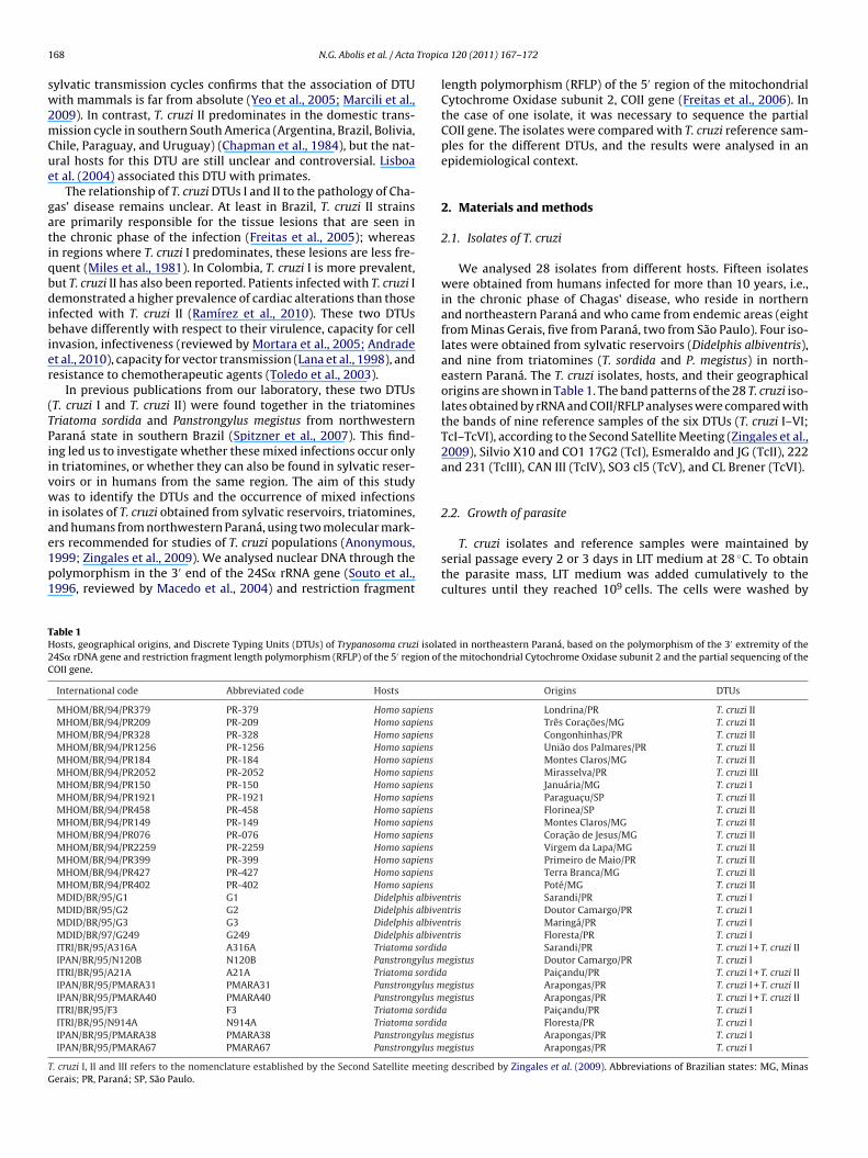
1 Tropic
sw2mCue
gatiqbdibier
(TPiivwiae1p1
TH2C
TG
68 N.G. Abolis et al. / Acta
ylvatic transmission cycles confirms that the association of DTUith mammals is far from absolute (Yeo et al., 2005; Marcili et al.,
009). In contrast, T. cruzi II predominates in the domestic trans-ission cycle in southern South America (Argentina, Brazil, Bolivia,
hile, Paraguay, and Uruguay) (Chapman et al., 1984), but the nat-ral hosts for this DTU are still unclear and controversial. Lisboat al. (2004) associated this DTU with primates.
The relationship of T. cruzi DTUs I and II to the pathology of Cha-as’ disease remains unclear. At least in Brazil, T. cruzi II strainsre primarily responsible for the tissue lesions that are seen inhe chronic phase of the infection (Freitas et al., 2005); whereasn regions where T. cruzi I predominates, these lesions are less fre-uent (Miles et al., 1981). In Colombia, T. cruzi I is more prevalent,ut T. cruzi II has also been reported. Patients infected with T. cruzi Iemonstrated a higher prevalence of cardiac alterations than those
nfected with T. cruzi II (Ramírez et al., 2010). These two DTUsehave differently with respect to their virulence, capacity for cell
nvasion, infectiveness (reviewed by Mortara et al., 2005; Andradet al., 2010), capacity for vector transmission (Lana et al., 1998), andesistance to chemotherapeutic agents (Toledo et al., 2003).
In previous publications from our laboratory, these two DTUsT. cruzi I and T. cruzi II) were found together in the triatominesriatoma sordida and Panstrongylus megistus from northwesternaraná state in southern Brazil (Spitzner et al., 2007). This find-ng led us to investigate whether these mixed infections occur onlyn triatomines, or whether they can also be found in sylvatic reser-oirs or in humans from the same region. The aim of this studyas to identify the DTUs and the occurrence of mixed infections
n isolates of T. cruzi obtained from sylvatic reservoirs, triatomines,nd humans from northwestern Paraná, using two molecular mark-
rs recommended for studies of T. cruzi populations (Anonymous,999; Zingales et al., 2009). We analysed nuclear DNA through theolymorphism in the 3′ end of the 24S� rRNA gene (Souto et al.,996, reviewed by Macedo et al., 2004) and restriction fragmentable 1osts, geographical origins, and Discrete Typing Units (DTUs) of Trypanosoma cruzi isola4S� rDNA gene and restriction fragment length polymorphism (RFLP) of the 5′ region of
OII gene.
International code Abbreviated code Hosts
MHOM/BR/94/PR379 PR-379 Homo sapiens
MHOM/BR/94/PR209 PR-209 Homo sapiens
MHOM/BR/94/PR328 PR-328 Homo sapiens
MHOM/BR/94/PR1256 PR-1256 Homo sapiens
MHOM/BR/94/PR184 PR-184 Homo sapiens
MHOM/BR/94/PR2052 PR-2052 Homo sapiens
MHOM/BR/94/PR150 PR-150 Homo sapiens
MHOM/BR/94/PR1921 PR-1921 Homo sapiens
MHOM/BR/94/PR458 PR-458 Homo sapiens
MHOM/BR/94/PR149 PR-149 Homo sapiens
MHOM/BR/94/PR076 PR-076 Homo sapiens
MHOM/BR/94/PR2259 PR-2259 Homo sapiens
MHOM/BR/94/PR399 PR-399 Homo sapiens
MHOM/BR/94/PR427 PR-427 Homo sapiens
MHOM/BR/94/PR402 PR-402 Homo sapiens
MDID/BR/95/G1 G1 Didelphis albiveMDID/BR/95/G2 G2 Didelphis albiveMDID/BR/95/G3 G3 Didelphis albiveMDID/BR/97/G249 G249 Didelphis albiveITRI/BR/95/A316A A316A Triatoma sordidIPAN/BR/95/N120B N120B Panstrongylus mITRI/BR/95/A21A A21A Triatoma sordidIPAN/BR/95/PMARA31 PMARA31 Panstrongylus mIPAN/BR/95/PMARA40 PMARA40 Panstrongylus mITRI/BR/95/F3 F3 Triatoma sordidITRI/BR/95/N914A N914A Triatoma sordidIPAN/BR/95/PMARA38 PMARA38 Panstrongylus mIPAN/BR/95/PMARA67 PMARA67 Panstrongylus m
. cruzi I, II and III refers to the nomenclature established by the Second Satellite meetinerais; PR, Paraná; SP, São Paulo.
a 120 (2011) 167– 172
length polymorphism (RFLP) of the 5′ region of the mitochondrialCytochrome Oxidase subunit 2, COII gene (Freitas et al., 2006). Inthe case of one isolate, it was necessary to sequence the partialCOII gene. The isolates were compared with T. cruzi reference sam-ples for the different DTUs, and the results were analysed in anepidemiological context.
2. Materials and methods
2.1. Isolates of T. cruzi
We analysed 28 isolates from different hosts. Fifteen isolateswere obtained from humans infected for more than 10 years, i.e.,in the chronic phase of Chagas’ disease, who reside in northernand northeastern Paraná and who came from endemic areas (eightfrom Minas Gerais, five from Paraná, two from São Paulo). Four iso-lates were obtained from sylvatic reservoirs (Didelphis albiventris),and nine from triatomines (T. sordida and P. megistus) in north-eastern Paraná. The T. cruzi isolates, hosts, and their geographicalorigins are shown in Table 1. The band patterns of the 28 T. cruzi iso-lates obtained by rRNA and COII/RFLP analyses were compared withthe bands of nine reference samples of the six DTUs (T. cruzi I–VI;TcI–TcVI), according to the Second Satellite Meeting (Zingales et al.,2009), Silvio X10 and CO1 17G2 (TcI), Esmeraldo and JG (TcII), 222and 231 (TcIII), CAN III (TcIV), SO3 cl5 (TcV), and CL Brener (TcVI).
2.2. Growth of parasite
T. cruzi isolates and reference samples were maintained byserial passage every 2 or 3 days in LIT medium at 28 ◦C. To obtainthe parasite mass, LIT medium was added cumulatively to thecultures until they reached 109 cells. The cells were washed by
ted in northeastern Paraná, based on the polymorphism of the 3′ extremity of thethe mitochondrial Cytochrome Oxidase subunit 2 and the partial sequencing of the
Origins DTUs
Londrina/PR T. cruzi IITrês Corac ões/MG T. cruzi IICongonhinhas/PR T. cruzi IIUnião dos Palmares/PR T. cruzi IIMontes Claros/MG T. cruzi IIMirasselva/PR T. cruzi IIIJanuária/MG T. cruzi IParaguac u/SP T. cruzi IIFlorinea/SP T. cruzi IIMontes Claros/MG T. cruzi IICorac ão de Jesus/MG T. cruzi IIVirgem da Lapa/MG T. cruzi IIPrimeiro de Maio/PR T. cruzi IITerra Branca/MG T. cruzi IIPoté/MG T. cruzi II
ntris Sarandi/PR T. cruzi Intris Doutor Camargo/PR T. cruzi Intris Maringá/PR T. cruzi Intris Floresta/PR T. cruzi Ia Sarandi/PR T. cruzi I + T. cruzi IIegistus Doutor Camargo/PR T. cruzi I
a Paic andu/PR T. cruzi I + T. cruzi IIegistus Arapongas/PR T. cruzi I + T. cruzi IIegistus Arapongas/PR T. cruzi I + T. cruzi II
a Paic andu/PR T. cruzi Ia Floresta/PR T. cruzi Iegistus Arapongas/PR T. cruzi Iegistus Arapongas/PR T. cruzi I
g described by Zingales et al. (2009). Abbreviations of Brazilian states: MG, Minas
Tropic
cw
2
t4Pti(twa8wa
2
SaR3(dcfiopbT12
2
2wwaocimfo
FO
N.G. Abolis et al. / Acta
entrifugation in KRT (Krebs–Ringer-Tris) buffer, and the cell massas stored at −20 ◦C before use.
.3. T. cruzi genomic DNA extraction and quantification
DNA was extracted as described by Macedo et al. (1992). Briefly,he cells of T. cruzi were resuspended in lysis buffer (80 mM NaCl,5 mM EDTA pH 8.0, and 1% SDS) at a concentration of 109 cells.roteinase K (Sigma Chemical Company Ltd.) was added and extrac-ion was done with phenol, phenol-chloroform, and precipitationn absolute ethanol. The DNA was resuspended in “Low TE” buffer10 mM Tris–HCl pH 8.0, and 1 mM EDTA pH 8.0) in the propor-ion of 100 �l/109 cells. The solutions of total DNA were quantifiedith the standard Low DNA Mass Ladder (Invitrogen, U.S.A.) in 2%
garose gel in TBE buffer (89 mM Tris–Borate, and 2 mM EDTA pH.0) stained with ethidium bromide. The solutions of total DNAere diluted to a concentration of 1 ng/�l for amplification and
nalysis.
.4. Ribosomal RNA (rRNA) analysis
The 24S� rRNA gene sequence was amplified as described byouto et al. (1996) and revised by Macedo et al. (2004). Eachmplification reaction was carried out in a thermocycler (MJesearch PTC-100) in a final volume of 12.5 �L. In each reaction,.1 picomoles of D71 (5′AAGGTGCGTCGACAGTGTGG3′) and D725′TTTTCAGAATGGCCGAACAGT3′) primers were used. After initialenaturing at 94 ◦C for 1 min, the samples were submitted to 30ycles (94 ◦C for 30 s more, 60 ◦C for 30 s, and 72 ◦C for 30 s), with anal extension at 72 ◦C for 10 min. The amplified products werebserved by silver staining in 6% polyacrylamide gel. A 110-bproduct could be produced by either TcI or TcIII DTUs; the 125-p product in TcII and TcVI; products between 120 and 130-bp incIV; and both rDNA products in TcV, with a low intensity of the25-bp band (Souto et al., 1996; Brisse et al., 2001; Lewis et al.,009).
.5. COII/RFLP analysis
Amplification of the mitochondrial Cytochrome Oxidase subunit gene (COII) was carried out as described by Freitas et al. (2006)ith modifications. Each amplification reaction was carried outith 3.1 picomoles of Tcmit-10 (5′-CCATATATTGTTGCATTATT-3′)
nd Tcmit-21 (5′-TTGTAATAGGAGTCATGTTT-3′) primers and 2 ngf total DNA, for a final reaction volume of 15.0 �L. The amplifi-ation was processed in a thermocycler (MJ96G, Biocycler) with
nitial denaturing at 94 ◦C for 1 min and 30 cycles of 94 ◦C for 30 sore, 48 ◦C for 2 min, 72 ◦C for 2 min, and final extension at 72 ◦Cor 10 min. Ten microlitres of the products of approximately 400 bpf the T. cruzi DNA maxicircle was digested with 10 units of the
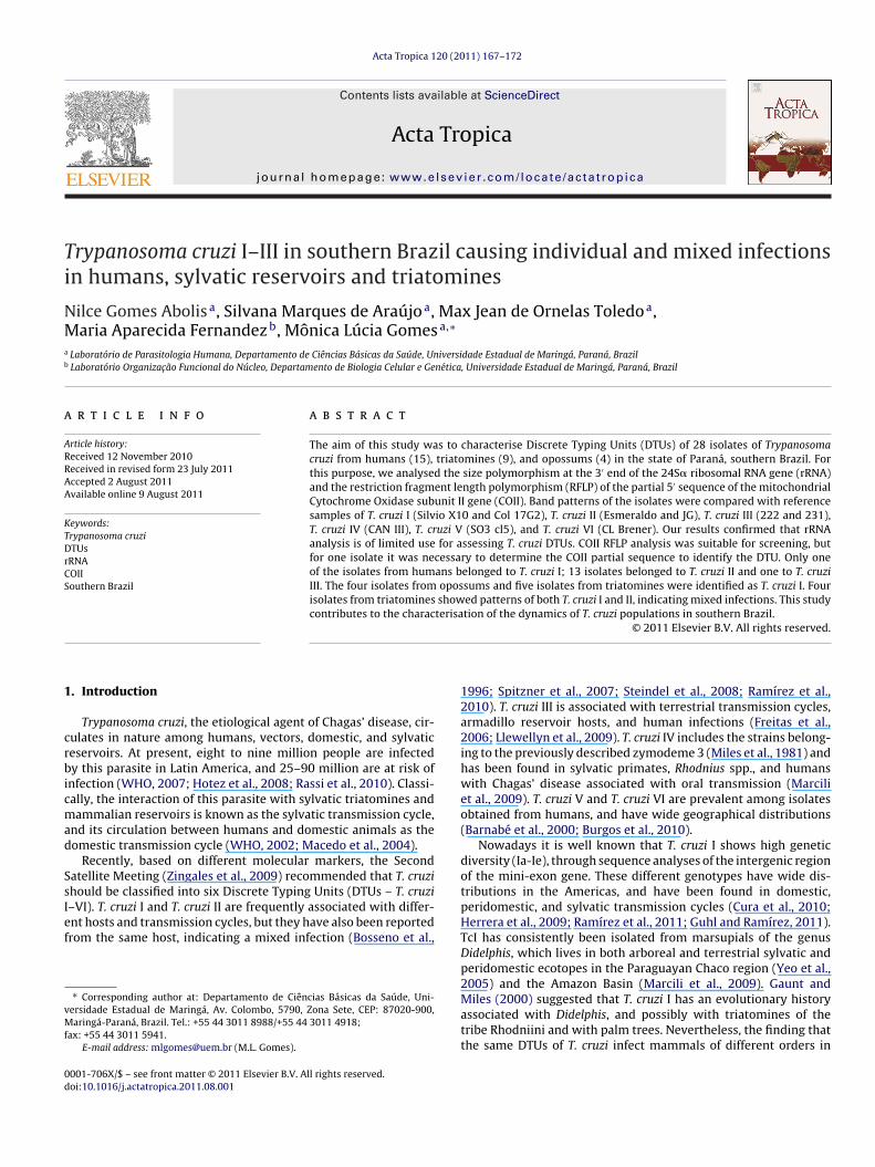
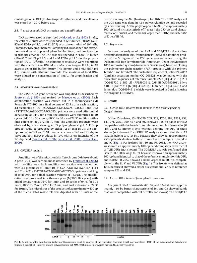
ig. 1. Genetic profiles from human isolates of Trypanosoma cruzi, by analysis of the rexidase II gene (COII) in silver-stained polyacrylamide gel. MM, 100 bp molecular weight
a 120 (2011) 167– 172 169
restriction enzyme AluI (Invitrogen) for 16 h. The RFLP analysis ofthe COII gene was done in 4.5% polyacrylamide gel and revealedby silver staining. At the polymorphic site AluI, the approximately300-bp band is characteristic of T. cruzi I, the 250-bp band charac-teristic of T. cruzi II, and the bands larger than 300 bp characteristicof T. cruzi III–VI.
2.6. Sequencing
Because the analyses of the rRNA and COII/RFLP did not allowdetermination of the DTU from isolate PR-2052, the amplified prod-uct of the 5′ region of the COII gene was sequenced using theDYEnamic ET Dye Terminator Kit (Amersham-Ge) in the MegaBace1000 automated system (Amersham Biosciences), based on cloningthe polymerase chain reaction (PCR) products with the primersTcmit-10 and Tcmit-21. The nucleotide sequence of isolate PR-2052(GenBank accession number GQ1206221) was compared with thenucleotide sequences of reference samples 222 (DQ3437191), 231(DQ3437201), SO3 cl5 (AF3590391), CAN III (AF3590301), SilvioX10 (DQ3437521), JG (DQ3437261), CL Brener (DQ3436451), andEsmeraldo (DQ3436461), which were deposited in GenBank, usingthe program ClustalW2.
3. Results
3.1. T. cruzi DTUs isolated from humans in the chronic phase ofChagas’ disease
Of the 15 isolates, 13 (PR-379, 209, 328, 1256, 184, 1921, 458,149, 076, 2259, 399, 427, and 402) showed 125-bp bands of rRNAcompatible with the bands from reference samples Esmeraldo, JG(TcII), and CL Brener (TcVI), without defining the DTU of thesestrains (not shown). The COII/RFLP analysis showed that these 13isolates belong to DTU TcII, because they showed approximately250-bp bands identical to those from reference samples Esmeraldoand JG (Fig. 1). For isolates PR-150 and PR-2052, the rRNA analy-sis showed an approximately 100-bp band compatible with the TcIor TcIII DTUs (not shown). The COII/RFLP analysis confirmed thatisolate PR-150 belongs to TcI, because it showed an approximately300-bp band identical to that of the reference samples for this DTU;and isolate PR-2052 showed a band larger than 300 bp, compati-ble with the III, V and VI DTUs (Fig. 1). This isolate was defined asTcIII, because it showed a closer nucleotide similarity to referencesamples 222 and 231.
3.2. T. cruzi DTUs isolated from sylvatic reservoirs
Analysis of rRNA from isolates G1, G2, and G249 showed approx-imately 110-bp bands characteristic of TcI, and G3 showed bandsthat were compatible with TcI or TcIII (not shown). The COII/RFLP
striction fragment length polymorphism (RFLP) of the mitochondrial Cytochrome marker. NC, negative control.
170 N.G. Abolis et al. / Acta Tropica 120 (2011) 167– 172
F rypant ide ge
ai
3
wmCtaiTmaptCnco
4
CpvTZTodR
TeaM1caaa2
abaqC
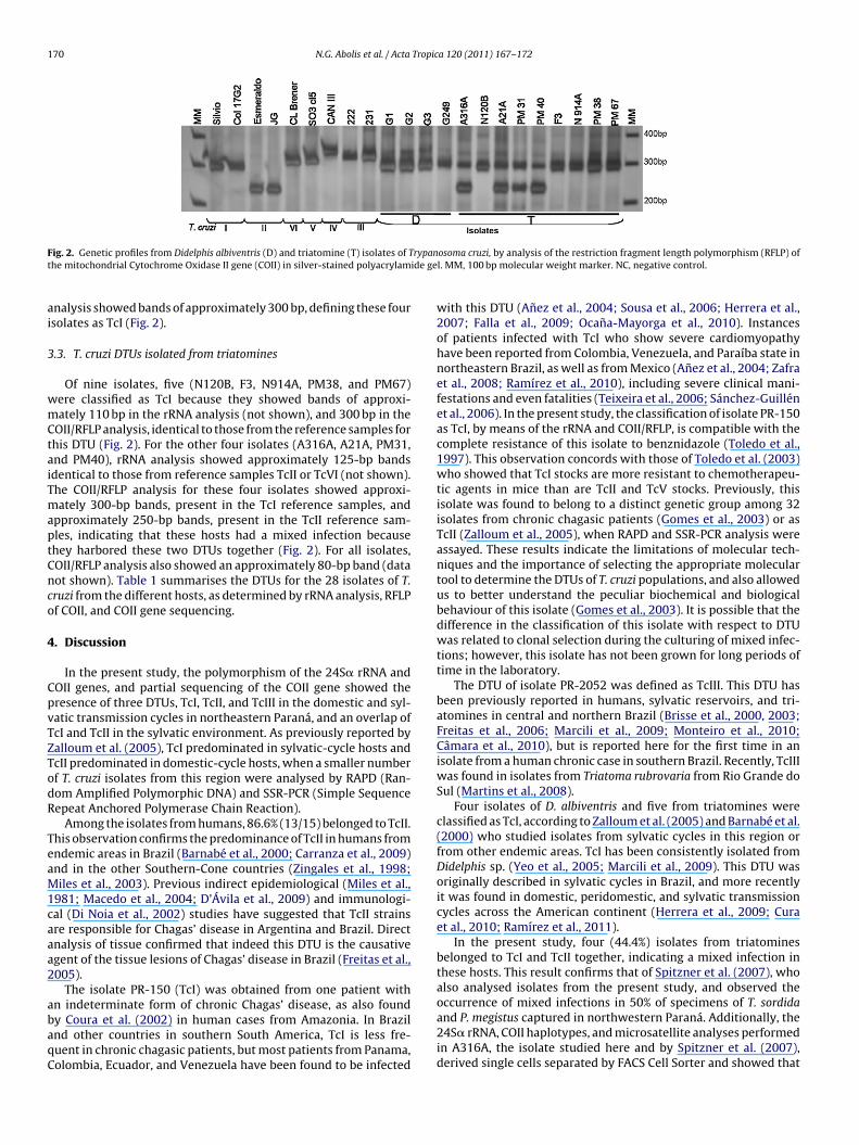
ig. 2. Genetic profiles from Didelphis albiventris (D) and triatomine (T) isolates of The mitochondrial Cytochrome Oxidase II gene (COII) in silver-stained polyacrylam
nalysis showed bands of approximately 300 bp, defining these foursolates as TcI (Fig. 2).
.3. T. cruzi DTUs isolated from triatomines
Of nine isolates, five (N120B, F3, N914A, PM38, and PM67)ere classified as TcI because they showed bands of approxi-ately 110 bp in the rRNA analysis (not shown), and 300 bp in the
OII/RFLP analysis, identical to those from the reference samples forhis DTU (Fig. 2). For the other four isolates (A316A, A21A, PM31,nd PM40), rRNA analysis showed approximately 125-bp bandsdentical to those from reference samples TcII or TcVI (not shown).he COII/RFLP analysis for these four isolates showed approxi-ately 300-bp bands, present in the TcI reference samples, and
pproximately 250-bp bands, present in the TcII reference sam-les, indicating that these hosts had a mixed infection becausehey harbored these two DTUs together (Fig. 2). For all isolates,OII/RFLP analysis also showed an approximately 80-bp band (dataot shown). Table 1 summarises the DTUs for the 28 isolates of T.ruzi from the different hosts, as determined by rRNA analysis, RFLPf COII, and COII gene sequencing.
. Discussion
In the present study, the polymorphism of the 24S� rRNA andOII genes, and partial sequencing of the COII gene showed theresence of three DTUs, TcI, TcII, and TcIII in the domestic and syl-atic transmission cycles in northeastern Paraná, and an overlap ofcI and TcII in the sylvatic environment. As previously reported byalloum et al. (2005), TcI predominated in sylvatic-cycle hosts andcII predominated in domestic-cycle hosts, when a smaller numberf T. cruzi isolates from this region were analysed by RAPD (Ran-om Amplified Polymorphic DNA) and SSR-PCR (Simple Sequenceepeat Anchored Polymerase Chain Reaction).
Among the isolates from humans, 86.6% (13/15) belonged to TcII.his observation confirms the predominance of TcII in humans fromndemic areas in Brazil (Barnabé et al., 2000; Carranza et al., 2009)nd in the other Southern-Cone countries (Zingales et al., 1998;iles et al., 2003). Previous indirect epidemiological (Miles et al.,
981; Macedo et al., 2004; D’Ávila et al., 2009) and immunologi-al (Di Noia et al., 2002) studies have suggested that TcII strainsre responsible for Chagas’ disease in Argentina and Brazil. Directnalysis of tissue confirmed that indeed this DTU is the causativegent of the tissue lesions of Chagas’ disease in Brazil (Freitas et al.,005).
The isolate PR-150 (TcI) was obtained from one patient withn indeterminate form of chronic Chagas’ disease, as also found
y Coura et al. (2002) in human cases from Amazonia. In Brazilnd other countries in southern South America, TcI is less fre-uent in chronic chagasic patients, but most patients from Panama,olombia, Ecuador, and Venezuela have been found to be infectedosoma cruzi, by analysis of the restriction fragment length polymorphism (RFLP) ofl. MM, 100 bp molecular weight marker. NC, negative control.
with this DTU (Anez et al., 2004; Sousa et al., 2006; Herrera et al.,2007; Falla et al., 2009; Ocana-Mayorga et al., 2010). Instancesof patients infected with TcI who show severe cardiomyopathyhave been reported from Colombia, Venezuela, and Paraíba state innortheastern Brazil, as well as from Mexico (Anez et al., 2004; Zafraet al., 2008; Ramírez et al., 2010), including severe clinical mani-festations and even fatalities (Teixeira et al., 2006; Sánchez-Guillénet al., 2006). In the present study, the classification of isolate PR-150as TcI, by means of the rRNA and COII/RFLP, is compatible with thecomplete resistance of this isolate to benznidazole (Toledo et al.,1997). This observation concords with those of Toledo et al. (2003)who showed that TcI stocks are more resistant to chemotherapeu-tic agents in mice than are TcII and TcV stocks. Previously, thisisolate was found to belong to a distinct genetic group among 32isolates from chronic chagasic patients (Gomes et al., 2003) or asTcII (Zalloum et al., 2005), when RAPD and SSR-PCR analysis wereassayed. These results indicate the limitations of molecular tech-niques and the importance of selecting the appropriate moleculartool to determine the DTUs of T. cruzi populations, and also allowedus to better understand the peculiar biochemical and biologicalbehaviour of this isolate (Gomes et al., 2003). It is possible that thedifference in the classification of this isolate with respect to DTUwas related to clonal selection during the culturing of mixed infec-tions; however, this isolate has not been grown for long periods oftime in the laboratory.
The DTU of isolate PR-2052 was defined as TcIII. This DTU hasbeen previously reported in humans, sylvatic reservoirs, and tri-atomines in central and northern Brazil (Brisse et al., 2000, 2003;Freitas et al., 2006; Marcili et al., 2009; Monteiro et al., 2010;Câmara et al., 2010), but is reported here for the first time in anisolate from a human chronic case in southern Brazil. Recently, TcIIIwas found in isolates from Triatoma rubrovaria from Rio Grande doSul (Martins et al., 2008).
Four isolates of D. albiventris and five from triatomines wereclassified as TcI, according to Zalloum et al. (2005) and Barnabé et al.(2000) who studied isolates from sylvatic cycles in this region orfrom other endemic areas. TcI has been consistently isolated fromDidelphis sp. (Yeo et al., 2005; Marcili et al., 2009). This DTU wasoriginally described in sylvatic cycles in Brazil, and more recentlyit was found in domestic, peridomestic, and sylvatic transmissioncycles across the American continent (Herrera et al., 2009; Curaet al., 2010; Ramírez et al., 2011).
In the present study, four (44.4%) isolates from triatominesbelonged to TcI and TcII together, indicating a mixed infection inthese hosts. This result confirms that of Spitzner et al. (2007), whoalso analysed isolates from the present study, and observed theoccurrence of mixed infections in 50% of specimens of T. sordida
and P. megistus captured in northwestern Paraná. Additionally, the24S� rRNA, COII haplotypes, and microsatellite analyses performedin A316A, the isolate studied here and by Spitzner et al. (2007),derived single cells separated by FACS Cell Sorter and showed that
Tropic
tTpr(fmAmpafiibut2emfrcuqeawaf
dtstihigso
t(c2
RtDltTitdmd
A
PSdD
N.G. Abolis et al. / Acta
his strain is composed by two subpopulations, one belonging tocII and another belonging to TcI. Furthermore, these two sub-opulations displayed distinct microsatellite profiles (unpublishedesults). To confirm the mixed infection, another molecular markerrRNA multiplex PCR for detection of T. rangeli and T. cruzi) was per-ormed with nine samples from triatomines naturally infected with
ulticlonal populations, including the four isolates studied here.lthough this marker does not show clearly the distinction betweenixtures of TcI and TcII, the natural mixed infection showed band
atterns similar to artificial mixtures obtained from culture formsnd infected triatomines, and different from the individual pro-les of TcI and TcII (personal communication). Mixed infections
n triatomines in southern Brazil appear to be common, becauseoth DTUs (TcI and TcII) were also detected in Triatoma tibiamac-lata in the state of Santa Catarina, when isolates from humans,riatomines, and sylvatic reservoirs were studied (Steindel et al.,008). The results presented here also agree with those of Bossenot al. (1996), Rozas et al. (2007), and Solari et al. (2001), who foundixed infections in isolates from Triatoma infestans in Bolivia, and
rom humans and sylvatic and peridomestic mammals in Chile,espectively. Although the mixed infection found in this study isonsistent with other studies, the rate of these infections may benderestimated, because mixtures of two parasites in equivalentuantities seem unlikely. More probable are infections of two orven three strains (Rozas et al., 2007) in very different ratios, whichre not detectable (Bosseno et al., 1996; Zafra et al., 2008). Here,e found no mixed infections for isolates of T. cruzi from sylvatic-
nimal reservoirs, perhaps because we analysed only a few isolatesrom these hosts.
The detection of mixed infections (TcI and TcII) appears toepend on the molecular marker used. The results of this study,ogether with those of Spitzner et al. (2007) and Zafra et al. (2008),howed that it was necessary to analyse also the COII gene in ordero identify mixed infections in triatomines in southern Brazil andn chronic chagasic patients in Colombia, respectively. On the otherand, limitations to rRNA analyses were observed in this study and
n others (Steindel et al., 2008; Zafra et al., 2008). Other investi-ators have shown that RAPD analyses (Spitzner et al., 2007) andpliced-leader gene analyses (Steindel et al., 2008) were incapablef identifying these two DTUs occurring together in the same host.
Another limitation of the 24S� rRNA gene analyses observed inhis study was that they did not clearly define the DTU in 71.4%20/28) of the isolates, confirming that this approach has insuffi-ient resolution for DTU assignment (Brisse et al., 2001; Lewis et al.,009; Souto et al., 1996; D’Ávila et al., 2009).
In summary, by analysing the rRNA gene and the COII geneFLP, and COII sequencing for one isolate, it was possible to assesshe genetic variability of T. cruzi isolated from humans, and threeTUs were found: TcI, TcII, and TcIII. TcI was also found in iso-
ates from sylvatic reservoirs and in the majority of isolates fromriatomines, which is common in the sylvatic transmission cycle.his study confirmed that mixed infections (TcI and TcII), at leastn southern Brazil, are still found only in triatomines. The informa-ion developed here opens prospects for studying the transmissionynamics of these parasite populations, to determine the sylvaticammal host or hosts of TcII in an endemic area for Chagas’
isease.
cknowledgments
To the Coordination for the Improvement of Higher Education
ersonnel (Coordenac ão de Aperfeic oamento Pessoal de Ensinouperior – CAPES) for granting a scholarship, to the Araucária Foun-ation (Fundac ão Araucária) Support for Scientific and Technologyevelopment of Paraná, and the National Council for Scientific anda 120 (2011) 167– 172 171
Technological Development (Conselho Nacional de Desenvolvi-mento Científico e Tecnológico – CNPq).
References
Andrade, L.O., Galvão, L.M.C., Meirelles, M.N.S.L., Chiari, E., Pena, S.D.J., Macedo, A.M.,2010. Differential tissue tropism of Trypanosoma cruzi strains: an in vitro study.Mem. Inst. Oswaldo Cruz 105, 834–837.
Anez, N., Crisante, G., da Silva, F.M., Rojas, A., Carrasco, H., Umezawa, E.S., Stolf,A.M., Ramírez, J.L., Teixeira, M.M., 2004. Predominance of lineage I among Try-panosoma cruzi isolates from Venezuelan patients with different clinical profilesof acute Chagas’ disease. Trop. Med. Int. Health 9, 1319–1326.
Anonymous, 1999. Recommendations from a Satellite Meeting. Mem. Inst. OswaldoCruz 94, 429–432.
Barnabé, C., Brisse, S., Tibayrenc, M., 2000. Population structure and genetic typ-ing of Trypanosoma cruzi, the agent of Chagas disease: a multilocus enzymeelectrophoresis approach. Parasitology 120, 513–526.
Bosseno, M.F., Telleria, J., Vargas, F., Yaksic, N., Noireau, F., Morin, A., Breniere, S.F.,1996. Trypanosoma cruzi: study of the distribution of two widespread clonalgenotypes in Bolivian Triatoma infestans vectors shows a high frequency ofmixed infections. Exp. Parasitol. 83, 275–282.
Brisse, S., Barnabé, C., Tibayrenc, M., 2000. Identification of six Trypanosoma cruziphylogenetic lineages by random amplified polymorphic DNA and multilocusenzyme electrophoresis. Int. J. Parasitol. 30, 35–44.
Brisse, S., Verhoef, J., Tibayrenc, M., 2001. Characterization of large and small subunitrRNA and mini-exon genes further supports the distinction of six Trypanosomacruzi lineages. Int. J. Parasitol. 31, 1218–1226.
Brisse, S., Henriksson, J., Barnabé, C., Douzery, E.J.P., Berkvens, D., Serrano, M., DeCarvalho, M.R.C., Buck, G.A., Dujardin, J., Tibayrenc, M., 2003. Evidence for geneticexchange and hybridization in Trypanosoma cruzi based on nucleotide sequencesand molecular karyotype. Infect. Genet. Evol. 2, 173–183.
Burgos, J.M., Diez, M., Vigliano, C., Bisio, M., Risso, M., Duffy, T., Cura, C., Brusses,B., Favaloro, L., Leguizamon, M.S., Lucero, R.H., Laguens, R., Levin, M.J., Favaloro,R., Schijman, A.G., 2010. Molecular identification of Trypanosoma cruzi discretetyping units in end-stage chronic chagas heart disease and reactivation afterheart transplantation. Clin. Infect. Dis. 51, 485–495.
Câmara, A.C.J., Varela-Freire, A.A., Valadares, H.M.S., Macedo, A.M., D’Ávila, D.A.,Machado, C.R., Lages-Silva, E., Chiari, E., Galvão, L.M.C., 2010. Genetic analysesof Trypanosoma cruzi isolates from naturally infected triatomines and humansin northeastern Brazil. Acta Trop. 115, 205–211.
Carranza, J.C., Valadares, H.M., D’Avila, D.A., Baptista, R.P., Moreno, M., Galvão,L.M., Chiari, E., Sturm, N.R., Gontijo, E.D., Macedo, A.M., Zingales, B., 2009. Try-panosoma cruzi maxicircle heterogeneity in Chagas disease patients from Brazil.Int. J. Parasitol. 39, 963–973.
Chapman, M.D., Baggaley, R.C., Godfrey-Fausset, P.F., Malpas, T.J., White, G., Canese,J., Miles, M.A., 1984. Trypanosoma cruzi from the Paraguayan Chaco: isoenzymeprofiles of strains isolated at Makthalawaiya. J. Protozool. 31, 482–486.
Coura, J.R., Junqueira, A.C., Fernandes, O., Valente, S.A., Miles, M.A., 2002. EmergingChagas disease in Amazonian Brazil. Trends Parasitol. 8, 171–176.
Cura, C.I., Mejia-Jaramillo, A.M., Duffy, T., Burgos, J.M., Rodriguero, M., Cardinal, M.V.,Kjos, S., Gurgel-Goncalves, R., Blanchet, D., De Pablos, L.M., Tomasini, N., da Silva,A., Russomando, G., Cuba, C.A., Aznar, C., Abate, T., Levin, M.J., Osuna, A., Gurtler,R., Diosque, P., Solari, A., Triana-Chavez, O., Schijman, A.G., 2010. Trypanosomacruzi I genotypes in different geographical regions and transmission cycles basedon a microsatellite motif of the intergenic spacer of spliced leader genes. Int. J.Parasitol. 40, 1599–1607.
D’Ávila, D.A., Macedo, A.M., Valadares, H.M.S., Gontijo, E.D., De Castro, A.M.,Machado, C.R., Chiari, E., Galvão, L.M.C., 2009. Probing population dynamics ofTrypanosoma cruzi during progression of the chronic phase in Chagasic patients.J. Clin. Microbiol. 47, 1718–1725.
Di Noia, J.M., Buscaglia, C.A., Marchi, C.R., Almeida, I.C., Frasch, A.C., 2002. ATrypanosoma cruzi small surface molecule provides the first immunological evi-dence that Chagas’ disease is due to a single parasite lineage. J. Exp. Med. 195,401–413.
Falla, A., Herrera, C., Fajardo, A., Montilla, M., Vallejo, G.A., Guhl, F., 2009. Haplotypeidentification within Trypanosoma cruzi I in Colombian isolates from severalreservoirs, vectors and humans. Acta Trop. 110, 15–21.
Freitas, J.M., Silva, E.L., Crema, E., Pena, S.D.J., Macedo, A.M., 2005. Real time PCRstrategy for the identification of major lineages of Trypanosoma cruzi directly inchronically infected human tissues. Int. J. Parasitol. 35, 411–417.
Freitas, J.M., Pinto, L.A., Pimenta, J.R., Rodrigues, L.B., Gonc alves, V.F., Teixeira, S.M.R.,Chiari, E., Junqueira, A.C.V., Fernandes, O., Macedo, A.M., Machado, C.R., Pena,S.J., 2006. Ancestral genomes, sex, and the population structure of Trypanosomacruzi. PLoS Pathog. 2, 226–235, doi:10.1371/journal.ppat.0020024, e24.
Gaunt, M.W., Miles, M.A., 2000. The ecotopes and evolution of triatomine bugs (Tri-atominae) and their associated trypanosomes. Mem. Inst. Oswaldo Cruz 95,557–565.
Gomes, M.L., Toledo, M.J.O., Nakamura, C.V., Bittencourt, N.R.L., Chiari, E., Marques-Araujo, S., 2003. Trypanosoma cruzi: genetic group with peculiar biochemicaland biological behavior. Mem. Inst. Oswaldo Cruz 98, 649–654.
Guhl, F., Ramírez, J.D., 2011. Trypanosoma cruzi I diversity: towards the need ofgenetic subdivision? Acta Trop. 119, 1–4.
Herrera, C., Bargues, M.D., Fajardo, A., Montilla, M., Triana, O., Vallejo, G.A., Guhl,F., 2007. Identifying four Trypanosoma cruzi I isolate haplotypes from differentgeographic regions in Colombia. Infect. Genet. Evol. 7, 535–539.
1 Tropic
H
H
L
L
L
L
M
M
M
M
M
M
M
M
O
R
R
R
72 N.G. Abolis et al. / Acta
errera, C., Guhl, F., Falla, A., Fajardo, A., Montilla, M., Vallejo, G.A., Bargues, M.D.,2009. Genetic variability and phylogenetic relationships within Trypanosomacruzi I isolated in Colombia based on miniexon gene sequences. J. Parasitol. Res.,doi:10.1155/2009/897364.
otez, P.J., Bottazzi, M.E., Franco-Paredes, C., Ault, S.K., Periago, M.R., 2008. Theneglected tropical diseases of Latin America and the Caribbean: a review of dis-ease burden and distribution and a roadmap for control and elimination. PLoSNegl. Trop. Dis. 2, 01–11.
ana, M., De Silveira, P.A., Da Barnabé, C., Quesney, V., Noel, S., Tibayrenc, M., 1998.Trypanosoma cruzi: compared vectorial transmissibility of three major clonalgenotypes by Triatoma infestans. Exp. Parasitol. 90, 20–25.
ewis, M.D., Ma, J., Yeo, M., Carrasco, H.J., Llewellyn, M.S., Miles, M.A., 2009. Geno-typing of Trypanosoma cruzi: Systematic Selection of Assays Allowing Rapidand Accurate Discrimination of All Known Lineages. Am. J. Trop. Med. Hyg. 81,1041–1049.
isboa, C.V., Mangia, R.H., Rubião, E., de Lima, N.R.C., Xavier, S.C.C., Picinatti, A., Fer-reira, L.F., Fernandes, O., Jansen, A.M., 2004. Trypanosoma cruzi transmission ina captive primate unit, Rio de Janeiro, Brasil. Acta Trop. 90, 97–106.
lewellyn, M.S., Lewis, M., Acosta, N., Yeo, M., Carrasco, H.J., Segovia, M., Vargas,J., Torrico, F., Miles, M.A., Gaunt, M.W., 2009. Trypanosoma cruzi IIc: phyloge-netic and phylogeographic insights from sequence and microsatellite analysisand potential impact on emergent Chagas disease. PLoS Negl. Trop. Dis. 3, 510,doi:10.1371/journal.pntd.0000510.
acedo, A.M., Machado, C.R., Oliveira, R.P., Pena, S.D.J., 2004. Trypanosoma cruzi:genetic structure of populations and relevance of populations and relevance ofgenetic variability to the pathogenesis of Chagas Disease. Mem. Inst. OswaldoCruz 99, 1–12.
acedo, A.M., Martins, M.S., Chiari, E., Pena, S.D.J., 1992. DNA fingerprinting ofTrypanosoma cruzi: a new tool for characterization of strains and clones. Mol.Biochem. Parasitol. 55, 147–154.
arcili, A., Valente, V.C., Valente, S.A., Junqueira, A.C.V., Da Silva, F.M., Pinto, A.Y.N.,Naiff, R.D., Campaner, M., Coura, J.R., Camargo, E.P., Miles, M.A., Teixeira, M.M.G.,2009. Trypanosoma cruzi in Brazilian Amazonia: lineages TCI and TCIIa in wildprimates, Rhodnius spp and in humans with Chagas disease associated with oraltransmission. Int. J. Parasitol. 39, 615–623.
artins, L.P.A., Marcili, A., Castanho, R.E.P., Therezo, A.L.S., De Oliveira, J.C.P.,Suzuki, R.B., Teixeira, M.M.G., Da Rosa, J.A., Speranc a, M.A., 2008. Rural Triatomarubrovaria from southern Brazil harbors Trypanosoma cruzi of Lineage IIc. Am. J.Trop. Med. Hyg. 3, 427–434.
iles, M.A., Cedillos, R.A., Povoa, M.M., De Souza, A.A., Prata, A., Macedo, V., 1981. Doradically dissimilar Trypanosoma cruzi strains (zymodemes) cause Venezuelanand Brazilian forms of Chagas disease? Lancet 1, 1338–1340.
iles, M.A., Feliciangeli, M.D., De Arias, A.R., 2003. American trypanosomiasis(Chagas’ disease) and the role of molecular epidemiology in guiding controlstrategies. Br. Med. J. 28, 1444–1448.
onteiro, W.M., Magalhães, L.K., Santana Filho, F., Borborema, M., Silveira, H., Bar-bosa, M.G.V., 2010. Trypanosoma cruzi TcIII/Z3 genotype as agent of an outbreakof Chagas disease in the Brazilian Western Amazonia. Trop. Med. Int. Health 15,1049–1051.
ortara, R.A., Andreoli, W.K., Taniwaki, N.N., Fernandes, A.B., Silva, C.V., Fernandes,M.C.D.C., L’Abbate, C., Silva, S., 2005. Mammalian cell invasion and intracellu-lar trafficking by Trypanosoma cruzi infective forms. An. Acad. Bras. Cienc. 77,77–94.
cana-Mayorga, S., Llewellyn, M.S., Costales, J.A., Miles, M.A., Grijalva, M.J., 2010. Sexsubdivision, and domestic dispersal of Trypanosoma cruzi lineage I in SouthernEcuador. PLoS Negl. Trop. Dis. 4, 915, doi:10.1371/journal.pntd.0000915.
amírez, J.D., Guhl, F., Rendón, L.M., Rosas, F., Marin-Neto, J.A., Morillo, C.A.,2010. Chagas cardiomyopathy manifestations and Trypanosoma cruzi geno-types circulating in chronic chagasic patients. PLoS Negl. Trop. Dis. 4, e899,doi:10.1371/journal.pntd.0000899.
amírez, J.D., Duque, M.C., Guhl, F., 2011. Phylogenetic reconstruction
based on Cytochrome b (Cytb) gene sequences reveals distinct geno-types within Colombian Trypanosoma cruzi I populations. Acta Trop.,doi:10.1016/j.actatropica.2011.04.009.assi Jr., A., Rassi, A., Marin-Neto, J.A., 2010. Chagas disease. Lancet 375,1388–1402.
a 120 (2011) 167– 172
Rozas, M., Botto-Mahan, C., Coronado, X., Ortiz, S., Cattan, P.E., Solari, A., 2007. Coex-istence of Trypanosoma cruzi genotypes in wild and periodomestic mammals inChile. Am. J. Trop. Med. Hyg. 77, 647–653.
Sánchez-Guillén, M.D.C., López-Colombo, A., Ordónez-Toquero, G., Gomez-Albino,I., Ramos-Jimenez, J., Torres-Rasgado, E., Salgado-Rosas, H., Romero-Diaz, M.,Pulido-Pérez, P., Pérez-Fuentes, R., 2006. Clinical forms of Trypanosoma cruziinfected individuals in the chronic phase of Chagas disease in Puebla, México.Mem. Inst. Oswaldo Cruz 101, 733–739.
Solari, A., Campillay, R., Ortiz, S., Wallace, A., 2001. Identification of Trypanosomacruzi genotypes circulating in Chilean chagasic patients. Exp. Parasitol. 97,226–233.
Sousa, O.E., Samudio, F., Juncá, C., Calzada, J.E., 2006. Molecular characterization ofhuman Trypanosoma cruzi isolates from endemic areas in Panama. Mem. Inst.Oswaldo Cruz 101, 455–457.
Souto, R.P., Fernandes, O., Macedo, A.M., Campbell Zingales, B., 1996. DNA Markersdefine two major phylogenetic lineages of Trypanosoma cruzi. Mol. Biochem.Parasitol. 83, 141–152.
Spitzner, F.L., Freitas, J.M., Macedo, A.M., Toledo, M.J.O., Araújo, S.M., Prioli, A.J.,Gomes, M.L., 2007. Trypanosoma cruzi – triatomine associations and the pres-ence of mixed infections in single triatomine bugs in Paraná state, Brazil. ActaParasitol. 52, 74–81.
Steindel, M., Pacheco, L.K., Scholl, D., Soares, M., Moraes, M.H., Eger, I., Kos-mann, C., Sincero, T.C.M., Stoco, P.H., Murta, S.M.F., Carvalho-Pinto, C.J., Grisard,E.C., 2008. Characterization of Trypanosoma cruzi isolated from humans,vectors, and animal reservoirs following an outbreak of acute human Cha-gas disease in Santa Catarina State, Brasil. Diagn. Microbiol. Infect. Dis. 60,25–32.
Teixeira, M.M.G., Da Silva, F.M., Marcili, A., Umezawa, E.S., Shikanai-Yasuda,M.A., Cunha-Neto, E., Kalil, J., Stolf, N., Stolf, A.M., 2006. Trypanosoma cruzilineage I in endomyocardial biopsy from a north-eastern Brazilian patientat end-stage chronic Chagasic cardiomyopathy. Trop. Med. Int. Health 3,294–298.
Toledo, M.J.O., Bahia, M.T., Carneiro, C.M., Martins-Filho, O.A., Tibayrenc, M., Barnabé,C., Tafuri, W.L., Lana, M., 2003. Chemotherapy with benznidazole and itra-conazole for mice infected with different Trypanosoma cruzi clonal genotypes.Antimicrob. Agents Chemother. 47, 223–230.
Toledo, M.J.O., Guilherme, A.L.F., Silva, J.C., Gasperi, M.V., Mendes, A.P., Gomes,M.L., Marques de Araújo, S., 1997. Trypanosoma cruzi: chemotherapy withbenznidazole in mice inoculated with strains from Paraná state and fromdifferent endemic areas of Brasil. Rev. Inst. Med. Trop. São Paulo 39,283–290.
WHO (World Health Organization), 2002. Control of Chagas Disease. Second Reportof the WHO Expert Committee, Series 905, 1–109.
WHO (World Health Organization), 2007. New global effort to eliminateChagas disease. Weekly epidemiological record 82 (28/29), 259–260.http://www.who.int/wer (accessed on 15.03.2011).
Yeo, M., Acosta, N., Llewellyn, M., Sánchez, H., Adamson, S., Miles, G.A.J., López, E.,González, N., Patterson, J.S., Gaunt, M.W., Arias, A.R., Miles, M.A., 2005. Originsof Chagas disease: Didelphis species are natural hosts of Trypanosoma cruzi I andarmadillos hosts of Trypanosoma cruzi II, including hybrids. Int. J. Parasitol. 35,225–233.
Zafra, G., Mantilla, J.C., Valadares, H.M., Macedo, A.M., González, C.I., 2008.Evidence of Trypanosoma cruzi II infection in Colombia. Parasitology 103,731–734.
Zalloum, L., Gomes, M.L., Kinoshita, A.T., Toledo, M.J.O., Prioli, A.J., Araújo, S.M., 2005.Trypanosoma cruzi: two genetic groups in Paraná state, southern Brazil. Exp.Parasitol. 111, 55–58.
Zingales, B., Souto, R.P., Mangia, R.H., Lisboa, C.V., Campbell, D.A., Coura, J.R., Jansen,A., Fernandes, O., 1998. Molecular epidemiology of American trypanosomiasisin Brazil based on dimorphisms of rRNA and mini-exon gene sequences. Int. J.Parasitol. 28, 105–112.
Zingales, B., Andrade, S.G., Briones, M.R.S., Campbell, D.A., Chiari, E., Fernandes, O.,Guhl, F., Lages-Silva, E., Macedo, A.M., Machado, C.R., Miles, M.A., Romanha,A.J., Sturm, N.R., Tibayrenc, M., Schijman, A.G., 2009. A new consensus forTrypanosoma cruzi intraspecific nomenclature: second revision meeting recom-mends TcI to TcVI. Mem. Inst. Oswaldo Cruz 104, 1051–1054.
Related Documents