33 The authors are with the School of Human Kinetics and Recreation, Memorial Uni- versity of Newfoundland, St. John’s, Newfoundland, A1C 5S7. Trunk Muscle Activity Increases With Unstable Squat Movements Kenneth Anderson and David G. Behm Catalogue Data Anderson, K.; and Behm, D.G. (2005). Trunk muscle activity increases with unstable squat movements. Can. J. Appl. Physiol. 30(1): 33-45. © 2005 Canadian Society for Exercise Physiology. Key words: electromyography, concentric, eccentric, resistance training, stabilizers Mots-clés: électromyographie, miométrique, pliométrique, entraînement à la force, stabilisateurs Abstract/Résumé The objective of this study was to determine differences in electromyographic (EMG) activ- ity of the soleus (SOL), vastus lateralis (VL), biceps femoris (BF), abdominal stabilizers (AS), upper lumbar erector spinae (ULES), and lumbo-sacral erector spinae (LSES) muscles while performing squats of varied stability and resistance. Stability was altered by doing the squat movement on a Smith machine, a free squat, and while standing on two balance discs. Fourteen male subjects performed the movements. Activities of the SOL, AS, ULES, and LSES were highest during the unstable squat and lowest with the Smith machine proto- col (p < 0.05). Increased EMG activity of these muscles may be attributed to their postural and stabilization role. Furthermore, EMG activity was higher during concentric contrac- tions compared to eccentric contractions. Performing squats on unstable surfaces may per- mit a training adaptation of the trunk muscles responsible for supporting the spinal column (i.e., erector spinae) as well as the muscles most responsible for maintaining posture (i.e., SOL). Cette étude se propose de présenter les différences d’activité myoélectrique (EMG) des muscles soléaire (SOL), vaste externe (VL), biceps fémoral (BF), stabilisateurs de l’abdomen (AS), érecteur du rachis lombaire supérieur (ULES), et érecteur du rachis lombo-sacré

Welcome message from author
This document is posted to help you gain knowledge. Please leave a comment to let me know what you think about it! Share it to your friends and learn new things together.
Transcript
Trunk Muscle Activity • 33
33
The authors are with the School of Human Kinetics and Recreation, Memorial Uni-versity of Newfoundland, St. John’s, Newfoundland, A1C 5S7.
Trunk Muscle Activity Increases With UnstableSquat Movements
Kenneth Anderson and David G. Behm
Catalogue DataAnderson, K.; and Behm, D.G. (2005). Trunk muscle activity increases with unstable squatmovements. Can. J. Appl. Physiol. 30(1): 33-45. © 2005 Canadian Society for ExercisePhysiology.
Key words: electromyography, concentric, eccentric, resistance training, stabilizersMots-clés: électromyographie, miométrique, pliométrique, entraînement à la force,stabilisateurs
Abstract/Résumé
The objective of this study was to determine differences in electromyographic (EMG) activ-ity of the soleus (SOL), vastus lateralis (VL), biceps femoris (BF), abdominal stabilizers(AS), upper lumbar erector spinae (ULES), and lumbo-sacral erector spinae (LSES) muscleswhile performing squats of varied stability and resistance. Stability was altered by doingthe squat movement on a Smith machine, a free squat, and while standing on two balancediscs. Fourteen male subjects performed the movements. Activities of the SOL, AS, ULES,and LSES were highest during the unstable squat and lowest with the Smith machine proto-col (p < 0.05). Increased EMG activity of these muscles may be attributed to their posturaland stabilization role. Furthermore, EMG activity was higher during concentric contrac-tions compared to eccentric contractions. Performing squats on unstable surfaces may per-mit a training adaptation of the trunk muscles responsible for supporting the spinal column(i.e., erector spinae) as well as the muscles most responsible for maintaining posture (i.e.,SOL).
Cette étude se propose de présenter les différences d’activité myoélectrique (EMG) desmuscles soléaire (SOL), vaste externe (VL), biceps fémoral (BF), stabilisateurs de l’abdomen(AS), érecteur du rachis lombaire supérieur (ULES), et érecteur du rachis lombo-sacré
34 • Anderson and Behm
(LSES) au cours de flexions accroupies, de stabilité et de résistance variables. La stabilitéest modifiée lors de flexions exécutées sur un appareil de marque Smith, exécutées librementou exécutées debout sur deux disques d’équilibre. Quatorze sujets masculins participent àl’expérience. Les activités des SOL, AS, ULES, et LSES sont plus élevées quand la condi-tion d’exécution est instable et plus faibles quand le mouvement est accompli sur la ma-chine de marque Smith (p < 0,05). L’augmentation de l’activité myoélectrique de ces musclesest probablement due à leur rôle postural et de stabilisation. De plus, l’activité myoélectriqueest plus grande au cours des actions miométriques qu’au cours des actions pliométriques.L’exécution des flexions accroupies sur des surfaces instables peut offrir un stimulusd’entraînement aux muscles du tronc qui soutiennent la colonne vertébrale (érecteur durachis) et aux muscles qui contribuent au maintien de la posture (soléaire).
Introduction
It is purported that greater instability of the human-surface interface will stress theneuromuscular system to a greater extent than traditional resistance training meth-ods using more stable benches and floors (Kornecki and Zschorlich, 1994). How-ever, it is important to identify to what extent instability will influence the acuteresponse of muscle. The advantage of an unstable training environment would bebased on the importance of neuromuscular adaptations with increases in strength.Strength gains can be attributed to increases in muscle cross-sectional area and toimprovements in neuromuscular coordination (Behm, 1995). A number of research-ers have reported that neural adaptations play the most important role in strengthgains in the early portion of a resistance training program (Behm, 1995; Sale,1988). Rutherford and Jones (1986) suggested that the specific neural adaptationoccurring with training was not increased recruitment or activation of motor unitsbut rather an improved coordination of agonist, antagonist, synergists, and stabi-lizers. Thus the inherently greater instability of the body-surface interface wouldchallenge the neuromuscular system to a greater extent, possibly enhancing strengthgains attributed to neural adaptations.
To our knowledge, few studies have examined the effect of unstable resis-tance training movements on muscle activation and force. Vera-Garcia et al. (2000)identified higher electromyographic (EMG) activity when performing a sit-up onan unstable surface vs. a flat surface. Siff (1991) found that the wider range ofmovement with Swiss balls is preferable to similar actions performed in most cir-cuit training gyms. Behm et al. (2002) reported that the decreases in quadricepsand plantar-flexor force and activation with unstable conditions were dependenton the degree of instability. Their research suggested that strength training adapta-tions of the limbs is possible if the degree of instability is moderate as opposed tosevere. Perhaps the importance of instability training is not only the unique stressplaced on the limb musculature but also its impact on trunk musculature responses.Thus the objective of this study was to examine the effect of differing levels ofinstability on trunk and limb muscle activation during a closed kinetic chain activ-ity (squat). It was hypothesized that as stability decreased, trunk muscle activitywould increase.
Trunk Muscle Activity • 35
Materials and Methods
SUBJECTS
Fourteen physically active young men (25.2 ± 6.2 yrs, 175.3 ± 6.5 cm, 82.6 ± 9.7kg) from Memorial University of Newfoundland participated in the study. All werecompetitive athletes (i.e., hockey, soccer, squash) with experience in resistancetraining (mean = 7.8 yrs ± 6.4); they were presently actively engaged with resis-tance training activities involving free weights, resistance machines, and instabil-ity devices. Six of the subjects only used instability devices such as Swiss balls fortrunk endurance activities such as sit-ups. All subjects read and signed a consentform prior to the experiment. Memorial University of Newfoundland’s human in-vestigation committee approved the study.
MEASUREMENT AND INSTRUMENTATION
EMG activity was measured during all protocols of varied stability and resistance.Surface EMG signals were measured from 6 muscle groups: mid-belly of vastuslateralis (VL), mid-belly of biceps femoris (BF), midline of soleus (SOL), upperlumbar erector spinae (ULES) at L1–L2 (6 cm lateral to the L1–L2 spinous pro-cesses), lumbo-sacral erector spinae (LSES) at L5–S1 (2 cm lateral to L5–S1 spinousprocesses), and abdominal stabilizers (AS), positioned superior to the inguinalligament and medial to the anterior superior iliac spine. As McGill et al. (1996)reported that surface electrodes could represent the activation profiles of deep ab-dominal muscles over a broad variety of tasks, and Ng et al. (1998) identifiedpossible contamination of EMG signals from more than one muscle (internal ob-liques and transverse abdominus), the term abdominal stabilizers will be used torefer to the EMG measured at the lower dorsal trunk position.
A number of studies have used a similar L5–S1 electrode placement (LSES)to measure the EMG activity of the multifidus (Danneels et al., 2001; Hermannand Barnes, 2001; Hodges and Richardson, 1996; Ng et al., 1998). In contrast,Stokes et al. (2003) reported that accurate measurement of the multifidus requiresintramuscular electrodes. Thus the EMG detected by these electrodes in the presentstudy is referred to as LSES muscle activity. Erector spinae muscles, according toanatomic nomenclature, include both superficial (spinalis, longissimus, iliocosta-lis) and deep (multifidus) vertebral muscles (Jonsson, 1969; Martini, 2001). Backmuscles have also been described as local and global stabilizing muscles based ontheir role in stabilizing the trunk (Berkmark, 1989). The multifidus is described asa component of the local stabilizing system while the longissimus contributes tothe global stabilizing system. The ULES EMG electrode positioning was morelateral than the LSES EMG positioning in order to diminish the detection of mul-tifidus activity and thus emphasize the measurement of global stabilizing muscles(longissimus).
EMG location sites were identified, shaved, sanded (to remove dead epithe-lial cells), and cleansed with rubbing alcohol to reduce resistance and achievemaximal adhesion of the electrode (Kendall® Medi-trace 100 series, Chikopee,MA). To maximize EMG sensitivity, the electrodes were aligned parallel to musclefiber orientation rather than in a perpendicular position (Ng et al., 1998). EMG
36 • Anderson and Behm
electrodes were placed collar to collar at a 2-cm distance. All muscles monitoredwere from the subject’s dominant side as determined by the leg preferentially usedto kick a soccer ball. EMG signals were then amplified (1000x), filtered (10–1000Hz) and smoothed (10 samples) (Biopac Systems MEC 100 amplifier, Santa Bar-bara, CA), and stored on computer after being directed through an analog-digitalconverter (Biopac MP100).
All data were recorded at a sampling rate of 2,000 Hz and analyzed with asoftware program (Acqknowledge 3.7.2., Biopac Systems). The maximum ampli-tude of the smoothed root mean square (RMS) of the EMG signal was evaluatedover the duration of the concentric and eccentric contractions of the squat. Theduration of eccentric and concentric activity was ascertained by using a marker ona separate channel controlled by the researcher to indicate the start and finish ofthe eccentric and concentric phases, respectively. The quadriceps EMG activityalso gave a clear indication of the different phases. There was no need to normal-ize the signal since the experiment was a repeated-measures design comparingwithin individuals with all conditions performed in one session.
PROTOCOL
Prior to experimental data collection, subjects were given a 2-week orientationsession in which they performed both stable and unstable squats (on balance discs)using only body mass for 3 sets of 10 repetitions on six occasions. Immediatelyprior to data collection, they underwent a 5-minute warm-up on a cycle ergometerat 70 rpm with a resistance of 1 kp. All testing was conducted in a single session.
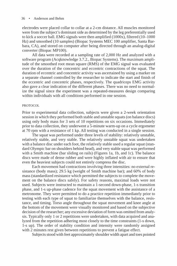
The squat was performed under three levels of stability: relatively unstable,relatively stable, and very stable. The relatively unstable squat was undertakenwith a balance disc under each foot, the relatively stable used a regular squat (stan-dard Olympic bar on shoulders behind head), and very stable squat was performedwith a Smith machine (bar sliding on rails) (Figures 1a, 1b, and 1c). The balancediscs were made of dense rubber and were highly inflated with air to ensure thateven the heaviest subjects could not entirely compress the disc.
Each movement had contractions involving three intensities: no external re-sistance (body mass); 29.5 kg (weight of Smith machine bar); and 60% of bodymass (standardized resistance which permitted the subjects to complete the move-ment on the balance discs safely). For safety reasons, maximal loads were notused. Subjects were instructed to maintain a 1-second down-phase, 1-s transitionphase, and 1-s up-phase cadence for the squat movement with the assistance of ametronome. They were permitted to do a practice repetition immediately prior totesting with each type of squat to familiarize themselves with the balance, resis-tance, and timing. Torso angle throughout the squat movement and knee angle atthe bottom of the movement were visually monitored and based on the subjectivedecision of the researcher; any excessive deviation of form was omitted from analy-sis. Typically only 1 or 2 repetitions were undertaken, with data acquired and ana-lyzed from the repetition adhering most closely to the time constraints (1-s down,1-s up). The order of stability condition and intensity were randomly assignedwith 2 minutes rest given between repetitions to prevent a fatigue effect.
Subjects stood with feet approximately shoulder width apart and toes pointed
Trunk Muscle Activity • 37
Figure 1. Squat methods.(top): Smith; (middle): Free;(bottom): Unstable.

38 • Anderson and Behm
straight ahead. The barbell was held behind the neck, across the shoulders andresting on the upper trapezius muscle. The grip was a little wider than shoulderwidth. Subjects held their breath during the down-phase of the lift and exhaledduring the up-phase. They were instructed to maintain heel contact with the floor.Escamilla (2001) reported peak quadriceps EMG activity occurring at approxi-mately 80–90° of knee flexion. Quadriceps activity remained fairly constant be-yond 80–90° of knee flexion, hence descending beyond 90o flexion (parallel squat)may not enhance quadriceps development (Escamilla, 2001). Therefore, subjectswere instructed to begin the up-phase once the upper leg was parallel to the floor(90° knee flexion).
STATISTICAL ANALYSIS
A three-way ANOVA (3 3 3 3 2: squat method {Smith machine, free squat, un-stable}, resistance {body mass, 29.5 kg, 60% body mass} and contraction type{eccentric-down, concentric-up}) repeated-measures was used (GB-STAT for MSWindows, Version 7.0. Silver Springs, MD). Upon review of collected data, theAS appeared to become highly active during the transition from eccentric to con-centric phases and the temporal data were analyzed with a repeated-measures one-way ANOVA. Differences were considered significant at p < 0.05. If significantdifferences were detected, a Bonferroni (Dunn’s) procedure was used to identifygroup differences. Reliability was assessed using an alpha (Cronbach) modelintraclass correlation coefficient (ICC) (Cohen, 1988) with all subjects. Repeated testswere conducted during the experimental session. Data were reported as means ± SD.
Results
EXTENT OF STABILITY
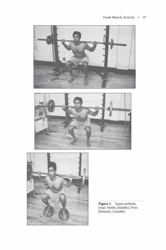
Trunk Muscles. The EMG activity of the AS during the Smith and freesquat were 29.6% (p < 0.01) and 18.6% (p < 0.05), respectively, less than duringthe unstable squat, while differences between the Smith and free squat were notsignificant. EMG activity of the LSES was 22.9% and 20% (both p < 0.05) lowerin the free and Smith squat, respectively, compared to the unstable squat; however,no significant differences between the Smith and free squat were identified. TheULES experienced a 33.8% (p < 0.01) decrease in the Smith compared to unstablesquat and a 22.9% (p < 0.05) decrease in the free compared to the unstable squat(Figure 2). There was also a 29% (p < 0.05) decrease in activity of the ULESduring the Smith compared to the free squat. All 14 subjects experienced increasesin ULES and LSES activation with the unstable squat vs. the Smith and free squat,whereas 12 of them experienced increased LAS activation with the unstable con-dition.
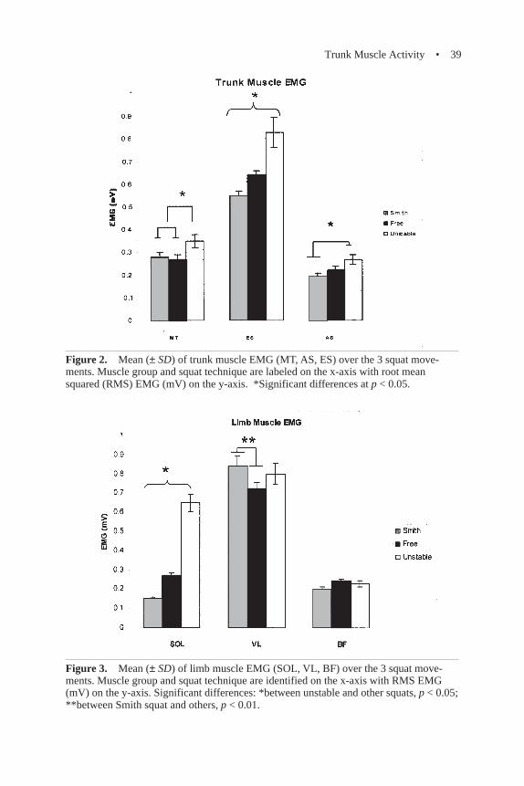
Limb Muscles. EMG of the SOL during the Smith and free squats were73.1% and 58.5% less, respectively, than during the unstable squat (p < 0.0001).Muscle activity of the VL was 4.8% (p < 0.05) lower in the unstable compared tothe Smith squat while the VL activity during the free squat was 14.3% (p < 0.01)lower than during the Smith squat. There were no significant BF differences be-tween the three squat protocols (Figure 3).
Trunk Muscle Activity • 39
Figure 2. Mean (± SD) of trunk muscle EMG (MT, AS, ES) over the 3 squat move-ments. Muscle group and squat technique are labeled on the x-axis with root meansquared (RMS) EMG (mV) on the y-axis. *Significant differences at p < 0.05.
Figure 3. Mean (± SD) of limb muscle EMG (SOL, VL, BF) over the 3 squat move-ments. Muscle group and squat technique are identified on the x-axis with RMS EMG(mV) on the y-axis. Significant differences: *between unstable and other squats, p < 0.05;**between Smith squat and others, p < 0.01.
40 • Anderson and Behm
RESISTANCE AND CONTRACTION TYPE
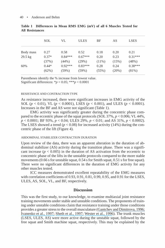
As resistance increased, there were significant increases in EMG activity of theSOL (p < 0.01), VL (p < 0.0001), LSES (p < 0.001), and ULES (p < 0.0001).Increases in the BF and AS were not significant (Table 1).
EMG activity was significantly greater during the concentric phase com-pared to the eccentric phase of the squat protocols (SOL 37%, p < 0.006; VL 44%,p < 0.0001; BF 93%, p < 0.04; ULES 29%, p < 0.01; and AS 31%, p < 0.0002).The LSES showed a trend (p < 0.08) for increased activity (14%) during the con-centric phase of the lift (Figure 4).
ABDOMINAL STABILIZER CONTRACTION DURATION
Upon review of the data, there was an apparent alteration in the duration of ab-dominal stabilizer (AS) activity during the transition phase. There was a signifi-cant increase (p < 0.005) in the duration of AS activation from the eccentric toconcentric phase of the lifts in the unstable protocols compared to the more stablemovements (0.66 s for unstable squat, 0.54 s for Smith squat, 0.51 s for free squat).There were no significant differences in the duration of EMG activity for anyother muscles tested.
ICC measures demonstrated excellent repeatability of the EMG measureswith correlation coefficients of 0.93, 0.91, 0.81, 0.99, 0.95, and 0.91 for the LSES,ULES, AS, SOL, VL, and BF, respectively.
Discussion
This was the first study, to our knowledge, to examine multiaxial joint resistancetraining movements under stable and unstable conditions. The proponents of train-ing under unstable conditions claim that resistance training under those conditionsprovides a greater stress to the overall musculature (Gantchev and Dimitrova, 1996;Ivanenko et al., 1997; Sheth et al., 1997; Wester et al., 1996). The trunk muscles(LSES, ULES, AS) were more active during the unstable squat, followed by thefree squat and Smith machine squat, respectively. This may be explained by the
Table 1 Differences in Mean RMS EMG (mV) of all 6 Muscles Tested forAll Resistances
SOL VL ULES BF AS LSES
Body mass 0.27 0.58 0.52 0.18 0.20 0.2129.5 kg 0.37* 0.84*** 0.67*** 0.20 0.23 0.31***
(37%) (44%) (29%) (11%) (15%) (48%)60% 0.44* 0.92*** 0.83*** 0.28 0.24 0.38***
(62%) (59%) (59%) (55%) (20%) (81%)
Parentheses identify the % increase from lowest value.Significant differences: *p < 0.05; ***p < 0.0001

Trunk Muscle Activity • 41
stabilizing roles of these muscles (Arokoski et al., 2001; De Troyer et al., 1990;Gardner-Morse et al., 1995). As subjects became more unstable with the balancedisc squats, the LSES, ULES, and AS and were recruited more to maintain stabil-ity of the spine and torso. Whereas Behm et al. (2002) reported that moderateinstability can still utilize resistance intensities that would enhance limb strength,the present study emphasized the more pronounced activity of the trunk stabilizerswith changes in stability. Therefore, performing unstable squat movements maynot only develop the prime movers but may also develop the trunk stabilizers.
It could be argued that changes in body position (i.e., torso angle) in re-sponse to the instability could have contributed to the changes in EMG activity. Asmentioned in the Methods, torso angle was visually monitored and based on thesubjective decision of the researcher; any excessive deviation of form was omittedfrom analysis. However, torso angle could not be entirely controlled since theanthropometrics and balance strategies of each individual varied. Greater degreesof instability inevitably result in greater movement fluctuations, which can onlybe controlled by adding stability. With the impact of researcher control (visualinspection) and the random movement strategies among individual subjects, it isunlikely that a main effect for torso position was present.
EMG activity in the SOL was also greater during the unstable squat move-ment compared to the more stable movements. The SOL is an important muscle inmaintaining erect posture, as it has a major role in controlling the ankle joint whichis often one of the first joints to help return the body to equilibrium after perturba-tion (Ivanenko et al., 1997). This is relevant in that strengthening of the SOL musclesmay help persons with balance difficulties to lessen the number of falls attributed
Figure 4. Comparison of mean (± SD) RMS EMG (mV) of all 6 muscles tested duringboth eccentric and concentric contractions (x-axis), as plotted on the y-axis. *Significantdifferences.
42 • Anderson and Behm
to uneven surfaces. Furthermore, sports performed on level (basketball, volleyball) orirregular (football, rugby) surfaces could also benefit from instability training.
Limb muscles including the BF and the VL did not show similar changes inthe amplitude of EMG activity under the unstable conditions as did the trunk andpostural muscles. There was no significant difference in the BF EMG amplitudebetween all three squat protocols, indicating that with this squat movement, variedstability had minimal effect on hamstring activity. There were similar findingswith the VL. As these muscles are primarily identified as prime movers in thesquat movement, with a minimal role in stability, the varied stability had littleeffect on the amplitude of the EMG activity. However, the elevated EMG activityof the VL during the Smith machine protocol may have been a result of foot place-ment and the stability (bar guided on rails) of the Smith machine. Subjects mayhave been able to use the VL to push posteriorly and vertically against the bar inorder to push backward as well as up.
The increased resistance placed on all movements resulted in a correspond-ing rise in EMG activity, as would be expected with the classic force:EMG rela-tionship (Bigland and Lippold, 1954; Genadry et al., 1988; Komi and Viitasalo,1976; Lippold, 1952). However, a similar increase in EMG was not identified inthe AS muscle. One possible explanation for the lack of increase in AS activity isthat it aids in spinal stabilization by increasing intra-abdominal pressure (IAP)(Rab et al., 1977). Cresswell and Thorstensson (1994) found that among the ab-dominal muscles, the highest level of activity and the best correlation to variationsin IAP was demonstrated by the transverse abdominus. Thus there may be a maxi-mum threshold for the AS in increasing IAP in untrained individuals.
Another hypothesis is that the AS “turn off” as part of a protective mecha-nism, whereas trunk flexion increases, increased abdominal activity can create ashearing moment at the lumbar spine. An alternate explanation may be that there isnot a linear relationship between increases in external resistance and IAP. How-ever, some authors (Cresswell and Thorstensson, 1994; Harmon et al., 1988) sug-gest that increased resistance does not result in elevated IAP, but there is no directcorrelation of this elevated IAP to AS activation. Therefore, increasing resistancemay not lead to a corresponding rise in AS activity.
Significant differences were also found in muscle activation between con-centric and eccentric phases of the movement. The SOL, VL, BF, ULES, and AShad significantly (p < 0.05) greater EMG activity during the concentric phase ofthe lifts compared to the eccentric phase, which are consistent with findings byGrabiner and Owings (2002) and Cresswell and Thorstensson (1994). However,only a trend (p < 0.08) for higher EMG during the concentric phase was evident inthe LSES. Since the LSES is a spinal stabilizer, it contracts isometrically duringboth the concentric and eccentric phases of the lifts, thus producing lower recog-nizable differences. Furthermore, one may argue that as the ULES and AS are alsostabilizers, why was there a change between contractions? McGill and Norman(1986) provide one explanation, that the ULES also contributes to spinal exten-sion as it works in unison with other spinal extensors to overcome the spinal bend-ing moment resulting from the load. Delitto et al. (1987) state that the increasedactivity of the AS during the concentric phase may be a requisite for increased IAPneeded to protect the spine due to the considerable degree of anterior shear force
Trunk Muscle Activity • 43
that can be generated by the upper body while extending the torso and combatinginertia.
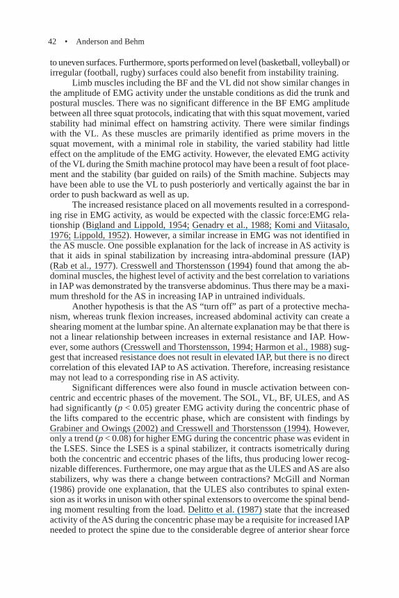
The AS were most active at the bottom of the movement with the transitionfrom eccentric to concentric phases (Figure 5). The possible mechanism respon-sible is known as the flexion-relaxation phenomenon (Newman and Gracovetsky,1995). McGill and Kippers (1994) found that during hip flexion, the lumbar exten-sors relaxed, as they were still able to generate substantial force elastically throughstretching. In the case of the squat, as subjects reached the bottom (lumbar flex-ion), LSES activity decreased (relying on elastic component) which resulted inincreased AS activity to maintain support to the spinal column anteriorly (Figure5). It would be interesting to discover whether individuals who are trained to acti-vate their LSES and AS (via trunk training) would show similar responses.
Conclusion
It was clear in this study that as subjects became more unstable, the activity oftheir trunk stabilizers and postural muscles increased whereas only negligible in-creases were observed in activity of the prime movers. Since previous studies (Behmet al., 2002) have shown significant decreases in force and activation of primemovers with unstable conditions, the use of unstable resistance training modalities
Figure 5. Raw data (EMG) from one subject that shows the flexion-relaxation phenom-enon (first line graph is AS, second line graph is LSES). Arrow indicates burst of AS ac-tivity.

44 • Anderson and Behm
may prove to be of more benefit to trunk stabilizers than prime movers. It shouldbe pointed out, however, that as only the acute response to an unstable movementwas measured, one should be cautious about making inferences as to possible train-ing effects.
References
Arokoski, J., Valta, T., Airaksinen, O., and Kankaanpaa, M. (2001). Back and abdominalmuscle function during stabilization exercises. Arch. Physical Med. Rehab. 82:1089-1098.
Behm, D. (1995). Neuromuscular implications and applications of resistance training. J.Strength Cond. Res. 9: 264-274.
Behm, D., Anderson, K., and Curnew, S. (2002). Muscle force and neuromuscular activa-tion under stable and unstable conditions. J. Strength Cond. Res. 16: 416-422.
Berkmark, A. (1989). Stability of the lumbar spine. A study in mechanical engineering.Acta Ortho. Scand. 230: 20-24.
Bigland, B., and Lippold, O. (1954). The relation between force, velocity and integratedelectrical activity in human muscles. J. Physiol. 123: 214-224.
Cohen, J. (1988). Statistical Power Analysis for the Behavioral Sciences. Hillsdale, NJ:Erlbaum.
Cresswell, A.G., and Thorstensson, A. (1994). Changes in intra-abdominal pressure, trunkmuscle activation and force during isokinetic lifting and lowering. Eur. J. Appl.Physiol. 68: 315-321.
Danneels, L.A., Cagnie, B.J., Cools, A.M., Vanderstraeten, G.G., Witvrouw, E.E., and DeCuyper, H.J. (2001). Intra-operator and inter-operator reliability of surface electromyo-graphy in the clinical evaluation of back muscles. Manual Ther. 6: 145-153.
DeLitto, D., Rose, S., and Apts, D. (1987). Electromyographic analysis of two techniquesfor squat lifting. Phys. Ther. 67: 1329-1334.
De Troyer, A., Estenne, M., Ninane, V., Van Gansbeke, D., and Gorini, M. (1990). Trans-verse abdominus muscle function in humans. J. Appl. Physiol. 68: 1010-1016.
Escamilla, R. (2001). Knee biomechanics of the dynamic squat exercise. Med. Sci SportsExerc. 33: 127-141.
Gantchev, G., and Dimitrova, D. (1996). Anticipatory postural adjustments associated witharm movements during balancing on unstable support surface. Int. J. Psychophysiol.22: 117-122.
Gardner-Morse, M., Stokes, I., and Laible, J. (1995). Role of muscles in lumbar spine sta-bility in maximum extension efforts. J. Orthop. Res. 13: 802-808.
Genadry, W., Kearney, R., and Hunter, I. (1988). Dynamic relationship between EMG andtorque at human ankle: Variation with contraction level and modulation. Med. Biol.Eng. Comput. 26: 489-496.
Grabiner, M., and Owings, T. (2002). EMG differences between concentric and eccentricmaximum voluntary contractions are evident prior to movement onset. Exp. BrainRes. 145: 505-511.
Harmon, E., Frykman, P., Clagett, E., and Kraemer, W. (1988). Intra-abdominal and intra-thoracic pressures during lifting and jumping. Med. Sci. Sports Exerc. 20: 195-201.
Hermann, K.M., and Barnes, W.S. (2001). Effects of eccentric exercise on trunk extensortorque and lumbar paraspinal EMG. Med. Sci Sports Exerc. 33: 971-977.
Trunk Muscle Activity • 45
Hodges, P.W., and Richardson, C.A. (1996). Inefficient muscular stabilization of the lum-bar spine associated with low back pain. Spine 21: 2640-2650.
Ivanenko, Y., Levik, Y., Taslis, V., and Gurfinkel, V. (1997). Human equilibrium on unstablesupport: The importance of feet-support interaction. Neurosci. Lett. 235: 109-112.
Jonsson, B. (1969) Morphology, innervation and electromyographic study of the erectorspinae. Spine 3: 638-641.
Komi, P., and Viitasalo, J. (1976). Signal characteristics of EMG at different levels of muscletension. Acta Physiol. Scand. 96: 267-276.
Kornecki, S., and Zschorlich, V. (1994). The nature of stabilizing functions of skeletalmuscles. J. Biomech. 27: 215-225.
Lippold, O. (1952). The relation between integrated action potentials in a human muscleand its isometric tension. J. Physiol. 117: 492-499.
Martini, F.H. (2001). Fundamentals of Anatomy and Physiology (5th ed.). EnglewoodCliffs, NJ: Prentice Hall.
McGill, S., Jukert, D., and Kropf, P. (1996). Appropriately placed surface EMG electrodesreflect deep muscle activity (psoas, quadratus lumborum, abdominal wall) in thelumbar spine. J. Biomech. 29: 1503-1507.
McGill, S., and Kippers, V. (1994). Transfer of loads between lumbar tissues during theflexion-relaxation phenomenon. Spine 19: 2190-2196.
McGill, S., and Norman, R. (1986). Partitioning of the L4-L5 dynamic moment into disc,ligamentous and muscular components during lifting. Spine 11: 666-678.
Newman, N., and Gracovetsky, S. (1995). Flexion-relaxation phenomenon (transfer of loadsbetween lumbar tissues during the flexion-relaxation phenomenon). Spine 20: 1739-1740.
Ng, J., Kippers, V., and Richardson, C. (1998). Muscle fibre orientation of abdominal musclesand suggested surface EMG electrode positions. Electromyogr. Clin. Neurophysiol.38: 51-58.
Rab, G., Chao, E., and Stauffer, R. (1977). Muscle force analysis of the lumbar spine. Orthop.Clin. North Am. 8: 193-199.
Rutherford, O.M., and Jones, D.A. (1986) The role of learning and co-ordination in strengthtraining. Eur. J. Appl. Physiol. 55: 100-105.
Sale, D. (1988). Neural adaptation to resistance training. Med. Sci Sports Exerc. 20: S135-S145.
Sheth, P., Yu, B., Laskowski, E., and An, K. (1997). Ankle disc training influences reactiontimes of selected muscles in a simulated ankle sprain. Am. J. Sports Med. 25: 538-543.
Siff, M. (1991). The functional mechanics of abdominal exercise. Am. J. Sports Med. 6:15-19.
Stokes, I.A.F., Henry, S.M., and Single, R.M. (2003). Surface EMG electrodes do not accu-rately record from lumbar multifidus muscles. Clin. Biomech. 18: 9-13.
Vera-Garcia, F., Grenier, S., and McGill, S. (2000). Abdominal muscle response duringcurl-ups on both stable and labile surfaces. Phys. Ther. 80: 564-569.
Wester, J., Jespersen, S., Nielson, K., and Neumann, L. (1996). Wobble board training afterpartial sprains of the lateral ligaments of the ankle: A prospective randomized study.J. Orthop. Sports Phys. Ther. 23: 332-336.
Received June 16, 2003; accepted in final form July 14, 2004.

Related Documents