TRPC3 Channels Are Necessary for Brain-Derived Neurotrophic Factor to Activate a Nonselective Cationic Current and to Induce Dendritic Spine Formation Michelle D. Amaral and Lucas Pozzo-Miller Department of Neurobiology, Civitan International Research Center and McKnight Brain Institute, University of Alabama at Birmingham, Birmingham, Alabama 35294 Abstract Brain-derived neurotrophic factor (BDNF) exerts prominent effects on hippocampal neurons, but the mechanisms that initiate its actions are poorly understood. We report here that BDNF evokes a slowly developing and sustained nonselective cationic current (I BDNF ) in CA1 pyramidal neurons. These responses require phospholipase C, IP 3 receptors, Ca 2+ stores, and Ca 2+ influx, suggesting the involvement of transient receptor potential canonical subfamily (TRPC) channels. Indeed, I BDNF is absent after small interfering RNA-mediated TRPC3 knockdown. The sustained kinetics of I BDNF appears to depend on phosphatidylinositol 3-kinase-mediated TRPC3 membrane insertion, as shown by surface biotinylation assays. Slowly emerging membrane currents after theta burst stimulation are sensitive to the scavenger TrkB–IgG and TRPC inhibitors, suggesting I BDNF activation by evoked released of endogenous, native BDNF. Last, TRPC3 channels are necessary for BDNF to increase dendritic spine density. Thus, TRPC channels emerge as novel mediators of BDNF-mediated dendritic remodeling through the activation of a slowly developing and sustained membrane depolarization. Keywords CA1 pyramidal neuron; hippocampus; TrkB receptor; biolistic transfection; surface biotinylation; confocal microscopy; organotypic slice culture; siRNA-mediated knockdown; theta-burst stimulation Introduction Brain-derived neurotrophic factor (BDNF), a member of the neurotrophin family, is a potent modulator of activity-dependent synaptic plasticity in the CNS (Poo, 2001). Activated neurotrophin Trk receptors trigger three signaling cascades, phospholipase Cγ (PLCγ)–IP 3 , Ras–Raf–extracellular signal-regulated kinase (ERK), and phosphatidylinositol 3 (PI3) kinase, all of which have been implicated in the varied actions of neurotrophins, ranging from modulation of gene expression, neuronal morphology, synaptic plasticity, and neurotransmitter release (Segal and Greenberg, 1996; Amaral et al., 2007). In addition to long-term modulation of ion channel expression, BDNF has been shown to evoke fast Na + currents through direct activation of Na v 1.9 channels (Blum et al., 2002) and slower nonselective cationic currents mediated by transient receptor potential canonical subfamily 3 (TRPC3) channels (Li et al., 1999). Ion channels of TRPC are activated by stimulation of G q /G 11 -type G-protein-coupled Correspondence should be addressed to Dr. Lucas Pozzo-Miller, Department of Neurobiology, SHEL-1002, University of Alabama at Birmingham, 1825 University Boulevard, Birmingham, AL 35294-2182. [email protected]. NIH Public Access Author Manuscript J Neurosci. Author manuscript; available in PMC 2010 January 14. Published in final edited form as: J Neurosci. 2007 May 9; 27(19): 5179–5189. doi:10.1523/JNEUROSCI.5499-06.2007. NIH-PA Author Manuscript NIH-PA Author Manuscript NIH-PA Author Manuscript

Welcome message from author
This document is posted to help you gain knowledge. Please leave a comment to let me know what you think about it! Share it to your friends and learn new things together.
Transcript

TRPC3 Channels Are Necessary for Brain-Derived NeurotrophicFactor to Activate a Nonselective Cationic Current and to InduceDendritic Spine Formation
Michelle D. Amaral and Lucas Pozzo-MillerDepartment of Neurobiology, Civitan International Research Center and McKnight Brain Institute,University of Alabama at Birmingham, Birmingham, Alabama 35294
AbstractBrain-derived neurotrophic factor (BDNF) exerts prominent effects on hippocampal neurons, but themechanisms that initiate its actions are poorly understood. We report here that BDNF evokes a slowlydeveloping and sustained nonselective cationic current (IBDNF) in CA1 pyramidal neurons. Theseresponses require phospholipase C, IP3 receptors, Ca2+ stores, and Ca2+ influx, suggesting theinvolvement of transient receptor potential canonical subfamily (TRPC) channels. Indeed, IBDNF isabsent after small interfering RNA-mediated TRPC3 knockdown. The sustained kinetics of IBDNFappears to depend on phosphatidylinositol 3-kinase-mediated TRPC3 membrane insertion, as shownby surface biotinylation assays. Slowly emerging membrane currents after theta burst stimulationare sensitive to the scavenger TrkB–IgG and TRPC inhibitors, suggesting IBDNF activation by evokedreleased of endogenous, native BDNF. Last, TRPC3 channels are necessary for BDNF to increasedendritic spine density. Thus, TRPC channels emerge as novel mediators of BDNF-mediateddendritic remodeling through the activation of a slowly developing and sustained membranedepolarization.
KeywordsCA1 pyramidal neuron; hippocampus; TrkB receptor; biolistic transfection; surface biotinylation;confocal microscopy; organotypic slice culture; siRNA-mediated knockdown; theta-burststimulation
IntroductionBrain-derived neurotrophic factor (BDNF), a member of the neurotrophin family, is a potentmodulator of activity-dependent synaptic plasticity in the CNS (Poo, 2001). Activatedneurotrophin Trk receptors trigger three signaling cascades, phospholipase Cγ (PLCγ)–IP3,Ras–Raf–extracellular signal-regulated kinase (ERK), and phosphatidylinositol 3 (PI3) kinase,all of which have been implicated in the varied actions of neurotrophins, ranging frommodulation of gene expression, neuronal morphology, synaptic plasticity, and neurotransmitterrelease (Segal and Greenberg, 1996; Amaral et al., 2007). In addition to long-term modulationof ion channel expression, BDNF has been shown to evoke fast Na+ currents through directactivation of Nav1.9 channels (Blum et al., 2002) and slower nonselective cationic currentsmediated by transient receptor potential canonical subfamily 3 (TRPC3) channels (Li et al.,1999). Ion channels of TRPC are activated by stimulation of Gq/G11-type G-protein-coupled
Correspondence should be addressed to Dr. Lucas Pozzo-Miller, Department of Neurobiology, SHEL-1002, University of Alabama atBirmingham, 1825 University Boulevard, Birmingham, AL 35294-2182. [email protected].
NIH Public AccessAuthor ManuscriptJ Neurosci. Author manuscript; available in PMC 2010 January 14.
Published in final edited form as:J Neurosci. 2007 May 9; 27(19): 5179–5189. doi:10.1523/JNEUROSCI.5499-06.2007.
NIH
-PA Author Manuscript
NIH
-PA Author Manuscript
NIH
-PA Author Manuscript

receptors [e.g., group I metabotropic glutamate receptor (mGluR)] and by receptor tyrosinekinases such as Trk receptors, leading to PLC-mediated formation of IP3 and diacylglycerol(DAG) (Clapham, 2003). TRPC channels are widely expressed in brain (Mizuno et al.,1999), including the hippocampus (Li et al., 1999; Strubing et al., 2001). Thus, PLC-dependent,non-voltage-gated cationic currents through TRPC channels are fundamentally novel forms ofCa2+ and Na+ entry in central neurons, mediating the slower glutamate and neurotrophinresponses through group I mGluRs (Kim et al., 2003) and TrkB receptors (Li et al., 1999),respectively.
Here, we present evidence that BDNF elicits a slowly developing and sustained nonselectivecationic current in hippocampal CA1 pyramidal neurons. This tetrodotoxin (TTX)- andsaxitoxin (STX)-insensitive current (IBDNF) required functional Trk receptors, PLC activity,IP3 receptors (IP3Rs), full intracellular Ca2+ stores, and extracellular Ca2+, suggesting theinvolvement of TRPC channels. Indeed, IBDNF was absent in neurons loaded with anti-TRPC3function-blocking antibodies or in those transfected with a small interfering RNA (siRNA)construct designed to knockdown the expression of TRPC3. BDNF also increased the levelsof surface accessible TRPC3 in cultured hippocampal neurons with a requirement for PI3kinase signaling and a time course that paralleled the activation of IBDNF, which was alsoblocked by a PI3 kinase inhibitor. The activation of a similar conductance by endogenouslyreleased BDNF was indicated by the blockade of slowly emerging membrane currents aftertheta burst stimulation (TBS) by the scavenger TrkB–IgG, as well as by Trk receptor and TRPCchannel inhibitors. Last, siRNA-mediated TRPC3 channel knockdown prevented thecharacteristic BDNF-induced increase of dendritic spine density in CA1 pyramidal neurons.In summary, TRPC channels emerge as novel mediators of BDNF-initiated dendriticremodeling through the activation of a slowly developing and sustained membranedepolarization.
Materials and MethodsOrganotypic slice culture
All animal procedures strictly adhered to national and international guidelines for the ethicaluse of research animals, such as the Public Health Service (PHS) Policy on Humane Care andUse of Laboratory Animals, as described athttp://grants.nih.gov/grants/olaw/references/phspol.htm, and the Policies on the Use ofAnimals and Humans in Neuroscience Research, as described at the Society for Neurosciencewebsitehttp://www.sfn.org/index.cfm?pagename=guidelinesPolicies_UseOfAnimalsandHumans.Last, the Institutional Animal Care and Use Committee of the University of Alabama atBirmingham reviews and approves all animal procedures described in the present report on anannual basis. Briefly, hippocampi were dissected from anesthetized postnatal day 7–11 SpragueDawley rats (Harlan, Indianapolis, IN or Charles River Laboratories, Wilmington, MA) andcut transversely into ~400-μm-thick slices using a custom-made wire slicer fitted with 20-μm-thick gold-coated platinum wire (Pozzo-Miller et al., 1995). Hippocampal slices wereindividually plated on Millicell-CM filter inserts (Millipore, Billerica, MA) and cultured in36°C, 5% CO2, 98% relative humidity incubators (Thermo-Forma, Waltham, MA). Slices weremaintained in culture media (Neurobasal-A plus B27; Invitrogen, Carlsbad, CA) containing20% equine serum for the first 4 d in vitro (div). To avoid the confounding effects of hormonesand growth factors in the serum, its concentration was gradually reduced over a period of 48h starting at 4 div (24 h each in 10 and 5% serum). After a period of 24 h in serum-free media(Neurobasal-A plus B27), 7–10 div slices were used for electrophysiology andimmunocytochemistry (Tyler and Pozzo-Miller, 2001). Some slice cultures remained in serum-
Amaral and Pozzo-Miller Page 2
J Neurosci. Author manuscript; available in PMC 2010 January 14.
NIH
-PA Author Manuscript
NIH
-PA Author Manuscript
NIH
-PA Author Manuscript
containing culture media (20% equine serum), as described in the original publications(Gahwiler, 1981; Yamamoto et al., 1989; Stoppini et al., 1991; Pozzo-Miller et al., 1993).
Primary culture of dissociated postnatal neuronsHippocampi were dissected from anesthetized postnatal day 2 Sprague Dawley rats (Harlan orCharles River Laboratories) and dissociated in papain (Worthington, Lakewood, NJ) for 45min at 37°C. The tissue was triturated to obtain a single-cell suspension, and the cells wereplated on dishes coated with poly-D-lysine/laminin and fed culture media (Neurobasal-A plusB27). Neurons were grown in 36°C, 5% CO2, 98% relative humidity incubators (Thermo-Forma). Cultures were maintained for 10–14 div before experimental procedures, with half ofthe medium changed every 4 d.
Whole-cell intracellular recordingsIndividual 7–10 div slices were transferred to a recording chamber mounted on a fixed-stageupright microscope (Axioskop FS; Zeiss, Oberkochen, Germany) and continuously perfused(2 ml/min) with artificial CSF (aCSF) at room temperature (24°C) containing the following(in mM): 124 NaCl, 2 KCl, 1.24 KH2PO4, 1.3 MgSO4, 17.6 NaHCO3, 2.5 CaCl2, 10 glucose,and 29.2 sucrose (310–320 mOsm). aCSF was bubbled with 95% O2/5% CO2, pH 7.4.Superficial CA1 pyramidal neurons were visualized with a water-immersion 63× objective [0.9(NA) numerical aperture] using infrared differential interference contrast (IR-DIC)microscopy. Whole-cell intracellular recordings were performed as described (Pozzo-Milleret al., 1995; Pozzo-Miller, 2006). Briefly, unpolished patch pipettes contained the following(in mM): 120 Cs-gluconate (or K-gluconate), 17.5 CsCl (or KCl), 10 Na-HEPES, 4 Mg-ATP,0.4 Na-GTP, 10 Na2 creatine phosphate, and 0.2 Na-EGTA (280–290 mOsm), pH 7.2(resistance of 3–4 MΩ). Nominally calcium-free extracellular aCSF was prepared by replacingCaCl2 with an equimolar concentration of MgCl2. Some drugs were dissolved in DMSO(≤0.01% final concentration) and others directly into the aCSF or intracellular solution; vehiclecontrols using 0.01% DMSO were routinely performed yielding no effects on membranecurrents or BDNF-induced responses. Membrane currents were recorded in the voltage-clampmode at a holding potential of −65 mV using an Axo-clamp 200B amplifier (MolecularDevices, Sunnyvale, CA), filtered at 2 kHz, and digitized at 10 kHz. Recordings were acceptedonly if access (series) resistance was ≤30 MΩ. CA1 neurons had whole-cell capacitances of~100 pF. Input resistance (Ri) was measured with hyperpolarizing voltage pulses (50 ms, −20mV), and cells were discarded if any of those cell parameters (Cm, Ri, Rs) changed by ≥20%during the course of an experiment. Current–voltage relationships were estimated from slowvoltage ramps between −85 and +45 mV (4 s; 32.5 mV/s) performed in the presence of TTX(0.5 μM) and Cd2+ (200 μM) to prevent the firing of Na+- and Ca2+-dependent action potentialsin distal dendrites that could escape voltage-clamp control. Electrophysiology data wereacquired on a single G4 Macintosh computer (Apple Computers, Cupertino, CA) runningcustom-written software (TIWorkBench, kindly provided Dr. T. Inoue, Tokyo University,Tokyo, Japan). All of the chemicals used for these experiments were obtained from Sigma (St.Louis, MO), Calbiochem (San Diego, CA), or Tocris (Ellisville, MO).
Human recombinant mature BDNF (supplied by Amgen, Thousand Oaks, CA) was pressureapplied from glass pipettes (~5 MΩ) using a Picospritzer-III (Parker Hannifin, Cleveland, OH).An application pipette was positioned ~100 μm above the slice and ~200 μm away from thesoma of the CA1 neuron under recording, aimed at its apical dendrites within stratum radiatum(~150 μm from the soma) and against the direction of aCSF perfusion flow. This arrangementproduced a stream of BDNF solution that overshoots the cell under recording and flows backover the slice, already diluted in the aCSF. Application of glutamate (100 μM, 8 psi, 9 s) fromsimilar pipettes was used to optimize this arrangement, yielding highly reproducible and stabletransient membrane currents. In addition, food coloring was used to assess the spatial spreading
Amaral and Pozzo-Miller Page 3
J Neurosci. Author manuscript; available in PMC 2010 January 14.
NIH
-PA Author Manuscript
NIH
-PA Author Manuscript
NIH
-PA Author Manuscript

of the applied solution over the slice. Pressure pulses of 30 psi lasting 25–30 s delivered a totalvolume of 2 μl of solution from ~5 MΩ glass pipettes. In most experiments, the pipettecontained 100 μg/ml BDNF in 0.0001–0.1% BSA. The extracellular BDNF scavenger TrkB–IgG (1 μg/ml; supplied by Regeneron, Tarrytown, NY; also from R & D Systems, Minneapolis,MN) was used to estimate the effective BDNF concentration required for IBDNF activation. Totest for dose–response relationships, some pipettes contained 50 or 75 μg/ml BDNF in 0.0001%BSA. BDNF denatured by boiling (10 min; 100 μg/ml), BSA alone (0.0001 or 0.1%), and aCSFwere used as pressure application controls. NGF, neurotrophin 4 (NT-4), and NT-3 (Promega,Madison, WI) (100 μg/ml in 0.0001% BSA) were applied in a similar manner.
Afferent fiber stimulation was performed with an extracellular patch pipette filled with bufferedand oxygenated aCSF (~4 MΩ) positioned within CA1 stratum radiatum to stimulate Schaeffercollaterals. Square constant-current pulses of 100 μs duration were produced by an isolatedstimulator (ISO-Flex; A.M.P.I., Jerusalem, Israel). High-frequency afferent stimulation wasdelivered as a theta burst pattern consisting of five bursts at 5 Hz, each burst having four pulsesat 100 Hz. A subthreshold concentration of the Na+ channel blocker TTX (10 nM) was includedin the aCSF to reduce membrane excitability and prevent polysynaptic responses, common inorganotypic slices. The aCSF contained the GABAA receptor antagonist picrotoxin (50 μM),whereas the K+ channels coupled to GABAB receptors were blocked by intracellular Cs+. TheaCSF also contained the noncompetitive antagonists of AMPA and NMDA receptors,GYKI-52466 [4-(8-methyl-9H-1,3-dioxolo [4,5-h][2,3]benzodiazepin-5-yl)-benzenaminehydrochloride] (20 μM) and MK-801 [(5S, 10R)-(+)-5-methyl-10,11-dihydro-5H-dibenzo[a,d] cyclohepten-5,10-imine maleate] (20 μM), respectively, as well as the antagonist of groupI mGluRs LY-367385 [(S)-(+)-α-amino-4-carboxy-2-methylbenzeneacetic acid] (100 μM);additional experiments were performed with the competitive antagonists CNQX (20 μM) andD,L-APV (50 μM). The constant-current intensity used for afferent stimulation (between 10and 50 μA) was never larger than 10 times that required to evoke AMPA receptor-mediatedfast EPSCs (~100–200 pA) at a holding potential of −65 mV in control aCSF.
ImmunocytochemistrySlices or cells were washed with PBS, and then fixed with 4% paraformaldehyde and 4%sucrose in phosphate buffer (PB) for 70 min. After rinsing with PBS containing 10 mMNH4Cl and then PBS alone, slices or cells were incubated with blocking and permeabilizationbuffer (0.4% fetal goat serum and 0.1% Triton X-100 in PBS) for 1 h and subsequentlyincubated with primary antibodies (anti-TRPC3; Alomone Labs, Jerusalem, Israel) diluted inblocking and permeabilization buffer. After incubation with FITC-conjugated secondaryantibodies, slices or cells were incubated with blocking and permeabilization buffer and thenincubated with NeuN (Millipore) primary antibodies. Slices were then incubated with TexasRed-conjugated secondary antibodies. Alternatively, biotinylated secondary antibodies wereused in combination with avidin-conjugated Quantum dots (488 nm excitation, 525 nmemission; Invitrogen). Finally, slices were mounted, sealed with Vectashield (VectorLaboratories, Burlingame, CA), and imaged with a laser scanning confocal microscopeequipped with argon and krypton lasers (FV300; Olympus Optical, Center Valley, PA) andoil-immersion lenses (60×, 1.2 NA or 100×, 1.4 NA). Controls were incubated with onlyprimary or secondary antibodies.
Surface biotinylationPrimary hippocampal cultures 10–14 div were washed once with aCSF and then stimulatedwith BDNF in aCSF. Sulfo-NHS-SS linked biotin (1.5 mg/ml in 1× PBS, pH 8.0) was thenadded to the neurons and incubated at 4°C for 45 min. Excess biotin was quenched with 100mM glycine, and then the cultures were washed with 1× PBS. The neurons were lysed withradioimmunoprecipitation assay buffer (50 mM Tris, pH 8.0, 150 mM NaCl, 1% Nonidet P-40,
Amaral and Pozzo-Miller Page 4
J Neurosci. Author manuscript; available in PMC 2010 January 14.
NIH
-PA Author Manuscript
NIH
-PA Author Manuscript
NIH
-PA Author Manuscript

0.5% sodium deoxycholate, and 0.1% SDS). Protein concentrations were determined via theLowry method, and 1 mg of each sample was added to streptavidin agarose (Pierce, Rockford,IL) and then incubated overnight at 4°C. The streptavidin agarose was washed three times withlysis buffer, and samples were eluted by adding 2× Laemli’s buffer (Bio-Rad, Hercules, CA)and then heating to 70°C for 30 min. Proteins were separated on SDS-PAGE gels, and Westernblots with anti-TRPC3 antibodies (Alomone Labs) were conducted. The two bands detectedin Western blots are not observed when the samples are preincubated with the control peptideantigen (HKLSE KLNPS VLRC, corresponding to residues 822–835 of mouse TRPC3;UniProtKB/Swiss-Prot accession number Q9QZC1). Anti-transferrin receptor (Zymed, SanFrancisco, CA) and anti-actin antibodies (Sigma) were used as loading controls for surface andintracellular proteins, respectively. Gels were scanned at 300 dpi in a flatbed scanner, and pixelintensity was measured using the gel analysis tool of NIH ImageJ software. The intensity ofTRPC3 bands was normalized to the intensity of the transferrin receptor bands.
TRPC3 siRNAsiRNA oligos (Invitrogen) were designed to target TRPC3 channel subunits. The sequence ofTRPC3 siRNA is as follows: Invitrogen-2476, sense sequence 5′ to 3′,GCACUCCAAGCAGUGA-CAU; antisense sequence 5′ to 3′,AUGUCACUGCUUGGAGUGC. Primary hippocampal neurons 10–14 div were transfectedwith 1 μg of fluorescein-labeled siRNA using the Gene Silencer siRNA Transfection Reagent(Gene Therapy Systems, San Diego, CA). Following the suggestions of the manufacturer,GeneSilencer and siRNA were diluted with serum-free medium (Neurobasal plus B27) andthen mixed together for 15 min at room temperature. To the cultured neurons, 1 ml of serum-free medium was added, followed by the siRNA mixture. After 24 h at 36°C, 2 ml of serum-free medium was added to the neurons. Four days later, the cultures were harvested andprocessed for Western blotting.
Particle-mediated gene transferHippocampal slices were cotransfected with a plasmid encoding enhanced yellow fluorescentprotein (eYFP) (Clontech, Mountain View, CA) and the TRPC3 siRNA oligos. A custom-modified Helios gene gun (Bio-Rad) was used to perform the biolistic transfection followingestablished protocols (Lo et al., 1994; Alonso et al., 2004). Briefly, plasmid cDNA and siRNAoligos were precipitated onto 1.6 μm colloidal gold at a ratio of 50 μg of eYFP plasmid to 100μg of siRNA oligo to 25 mg of gold. This mixture was coated onto Tefzel tubing using 0.06mg/ml polyvinylpyrrolidone. Slices were bombarded using helium pressure at 100 psi at adistance of 15 mm. For experiments using only eYFP, gene transfer was performed as above,except the plasmid encoding eYFP was precipitated onto 1.6 μm colloidal gold at a ratio of 50μg of DNA to 25 mg of gold.
BDNF and inhibitor treatments for confocal microscopyThree days after particle-mediated gene transfer, the respective inhibitors were added to theculture media 30 min before BDNF (250 ng/ml) application. To facilitate penetration of thereagents, an additional 50 μl of medium was gently placed on top of each slice. After 24 h ofBDNF exposure, slices were fixed in 4% paraformaldehyde and 100 mM PB for 60 min, rinsedin PB, and mounted on glass slides using Vectashield (Vector Laboratories).
Confocal microscopyA FluoView300 laser-scanning confocal microscope (Olympus Optical) fitted with a 100×,1.4 NA oil-immersion lens was used to acquire images of apical secondary and tertiarydendrites of eYFP-transfected CA1 neurons. eYFP and fluorescein-labeled siRNA were
Amaral and Pozzo-Miller Page 5
J Neurosci. Author manuscript; available in PMC 2010 January 14.
NIH
-PA Author Manuscript
NIH
-PA Author Manuscript
NIH
-PA Author Manuscript

excited with the 488 nm line of the argon laser and detected using standard FITC filters. Opticalsections in the z-axis were acquired at 0.1 μm intervals.
Spine density analysisDendritic spines were identified as small projections extending ≤3 μm from the adjacentdendrite, as described previously (Pozzo-Miller et al., 1999; Tyler and Pozzo-Miller, 2001,2003; Alonso et al., 2004). Spines were quantified on maximum-intensity projections of thez-stacks using NIH ImageJ software. To ensure that each spine was counted only once, theircourse was followed through the stack of z-sections. In addition, only those spines that appearedcontinuous with the parent dendrite were used for analysis. Spine density was calculated byquantifying the number of spines per length of imaged dendrite and then normalized to 10 μmof dendritic length. Supplemental Table 1 (available at www.jneurosci.org as supplementalmaterial) contains all of the measured parameters in the spine density analysis, including thetotal length of apical secondary and tertiary apical dendrites analyzed, the number of neuronsand slices, and the coefficient of variance (CV) of spine density values. Spine counts wereperformed “blindly” by an investigator unaware of the treatment groups.
Statistical analysisData were statistically analyzed using unpaired Student’s t test or ANOVA, followed byNewman–Keuls multiple-comparison post hoc test with Prism software package (GraphPadSoftware, San Diego, CA). p < 0.05 was considered significant. Data are presented as mean ±SEM; the SD of the mean was used to calculate the coefficient of variance (CV = mean/SD),which is given as a measure of consistency.
ResultsBDNF induces a slow depolarizing membrane current, IBDNF, in CA1 pyramidal neuronsthrough the activation of TrkB receptors
Long-term (minutes to hours) exposure to BDNF induces varied effects on hippocampalneurons, ranging from modulation of synaptic transmission and plasticity to structural changesof dendrites, spines, and presynaptic terminals (McAllister et al., 1999; Poo, 2001; Tyler et al.,2002b; Lu, 2003; Amaral et al., 2007). Conversely, brief (<300 ms) and highly targeted (<20μm) BDNF pulses have been shown to elicit transient EPSP-like membrane depolarizationsthrough the direct activation of Nav1.9 channels (Kafitz et al., 1999; Blum et al., 2002). Wechose to apply BDNF from picospritzer-controlled pipettes placed ~100 μm abovehippocampal slice cultures to avoid pressure and mechanical artifacts. BDNF-containingpipettes were positioned over CA1 neuron dendrites within stratum radiatum, ~200 μm awayfrom their cell bodies (Fig. 1A) to reproduce the spatiotemporal profile of a paracrineneuropeptide released from dense-core vesicles acting on perisynaptic receptors (Lessmann etal., 2003). Under these conditions, a single BDNF application (25–30 s) induced a delayed (54± 3.6 s) and slowly developing inward current with an average amplitude of 577.23 ± 41.07pA (n = 24 cells) in Cs+-filled CA1 pyramidal neurons held at −65 mV in the presence of TTX(500 nM) (Fig. 1B). These membrane currents, IBDNF, persisted for 13.03 ± 2.11 min and werehighly reproducible, because successive applications to the same cell (two to three; 20 minapart) evoked responses of similar amplitude (607.7 ± 19.98 pA; n = 4; p = 0.7703 vs singleapplication responses). The above responses were made in slices kept in serum-free media toavoid the confounding effects of hormones and growth factors in the serum; similar responseswere also observed in slice cultures maintained in serum-containing culture media (430.02 ±8.04 pA; n = 4). A significant decrease in membrane input resistance indicated an increase inmembrane conductance at the peak of the BDNF response (Ri = 202.05 ± 12 vs 106.23 ± 9.95MΩ; p < 0.0001). The current–voltage (I–V) relationship of IBDNF was estimated by voltageramp protocols (4 s from −85 to +45 mV) applied before and near the peak of IBDNF evoked
Amaral and Pozzo-Miller Page 6
J Neurosci. Author manuscript; available in PMC 2010 January 14.
NIH
-PA Author Manuscript
NIH
-PA Author Manuscript
NIH
-PA Author Manuscript

in the presence of TTX (500 nM) and Cd2+ (200 μM) to block voltage-gated Na+ and Ca2+
channels, respectively. The contribution of AMPA and NMDA receptor currents during thesevoltage ramps was eliminated by inclusion of CNQX (20 μM) and D,L-APV (100 μM),respectively. The positive slope conductance estimated from a linear regression of each I–Vplot was 3.54 ± 0.21 nS (n = 3). The average reversal potential of the subtracted BDNF-inducedcomponent of the current response to the voltage ramp was 27.96 ± 6.32 mV (n = 3), indicativeof a mixed cationic conductance (supplemental Fig. 1, available at www.jneurosci.org assupplemental material).
Consistent with the activation of an inward current at resting membrane potential, BDNFelicited a similarly delayed and slowly developing membrane depolarization in K+-filledneurons in the absence of TTX (22.18 ± 2.02 mV; n = 9), which led to an increase in spontaneousaction potentials and EPSPs (Fig. 1C). BDNF also caused a pronounced increase in thefrequency of spontaneous miniature EPSCs (in TTX), without affecting their amplitude (Fig.1B) (our unpublished observation), consistent with its known presynaptic actions onneurotransmitter release (Poo, 2001;Tyler et al., 2002a,2006). Together, these observationsdirectly demonstrate that BDNF has parallel actions on both sides of excitatory synapses,enhancing presynaptic transmitter release and directly activating a postsynaptic membranedepolarization.
The effects of BDNF on membrane conductance were confirmed to be specific, because vehiclealone (up to 0.1% BSA) or BDNF denatured by boiling (100 μg/ml, 10 min) (supplementalFig. 2, available at www.jneurosci.org as supplemental material) were entirely ineffective(28.04 ± 15 pA, n = 4, p < 0.0001; and 6.46 ± 5.25 pA, n = 3, p < 0.0001, respectively).Furthermore, IBDNF exhibited a dose–response relationship: pressure application from a pipettecontaining 50 μg/ml BDNF induced a current of 57.42 ± 4.23 pA (n = 4), whereas a largermembrane current was elicited by 75 μg/ml BDNF (178.75 ± 48.68 pA; n = 3) (supplementalFig. 2, available at www.jneurosci.org as supplemental material).
The extracellular BDNF scavenger TrkB–IgG was used to estimate the effective BDNFconcentration required for IBDNF activation. TrkB–IgG is a chimeric recombinant protein thatconsists of the ligand-binding domain of the TrkB receptor fused to the Fc domain of humanIgG (to make it soluble) (Shelton et al., 1995). Thus, TrkB–IgG neutralizes BDNF action byeffectively competing for BDNF binding to endogenous cell surface TrkB receptors, withoutphysically interacting with or affecting these receptors. Furthermore, it displays a dose-dependent inhibition of the effects of 200 ng/ml exogenously applied BDNF and completelyinhibits the effects of 200 ng/ml BDNF at an equimolar (~7 nM) equivalent of TrkB–IgG (2μg/ml) (McAllister et al., 1997). Bath application of 1 μg/ml TrkB–IgG completely preventedthe BDNF-induced current after pressure ejection from a pipette containing 100 μg/ml BDNF(in 0.0001% BSA), even after two to three successive pulses (Fig. 1D). Because ofaccumulation on the slice, subsequent BDNF applications did evoke a current, albeitsignificantly smaller than those recorded under control conditions, suggesting that 1 μg/mlTrkB–IgG (i.e., 3.8 nM) is saturated by two to three successive BDNF applications. Theseresults indicate that the effective BDNF concentration within the slice after a single applicationis 33–50 ng/ml or less; otherwise, it would have saturated the scavenger and evoked a currentin response to the first application. This range of concentrations, which are equivalent to 1.2–1.8 nM of the BDNF dimer, are considered to specifically activate TrkB receptors. For example,TrkB–IgG concentrations between 1 and 20 μg/ml have been shown to prevent the inductionof long-term potentiation in hippocampal slices (Figurov et al., 1996; Kang et al., 1997; Chenet al., 1999; Tyler et al., 2006), as well as BDNF-mediated increases in dendritic complexity(McAllister et al., 1997) and spine density (Tyler and Pozzo-Miller, 2001).
Amaral and Pozzo-Miller Page 7
J Neurosci. Author manuscript; available in PMC 2010 January 14.
NIH
-PA Author Manuscript
NIH
-PA Author Manuscript
NIH
-PA Author Manuscript

The requirement of Trk receptors was demonstrated by the observation that bath applicationof the tyrosine kinase inhibitor k-252a [(8R★,9S★,11S★)-(−)-9-hydroxy-9-methoxycarbonyl-8-methyl-2,3,9,10-tetrahydro-8,11-epoxy-1H,8H,11H-2,7b,11a-triazadibenzo(a,g)cycloocta(cde)trinden-1-one] completely prevented IBDNF (17.9 ± 4.8 pA;n = 9; p < 0.0001 vs control) (Fig. 1E), even after three successive BDNF applications to thesame cell. Moreover, inhibition of Trk receptors only in the postsynaptic cell by intracellularapplication of the membrane-impermeable analog k-252b (200 nM in 0.01% DMSO) alsoinhibited IBDNF (27.58 ± 16.51 pA; n = 4; p < 0.0001 vs control) (supplemental Fig. 2, availableat www.jneurosci.org as supplemental material); the concentrations used (200 nM in 0.01%DMSO for both) are specific for receptor tyrosine kinases of the trk gene family (Knusel andHefti, 1992). The specific role of TrkB receptors was further confirmed by applying otherneurotrophins in a similar manner. NT-3 (100 μg/ml), which binds primarily to TrkC but alsoto TrkB with a lower affinity (Barbacid, 1994), induced a membrane current with similarkinetics but smaller amplitude than IBDNF (238.75 ± 138 pA, n = 3, p = 0.0122 vs control) (Fig.1F). As expected from the low levels of TrkA expression in the postnatal hippocampus, NGF(100 μg/ml) had only minor effects on membrane currents (27.5 ± 13.2 pA; n = 3; p < 0.0001vs control) (Fig. 1F). Together, activation of TrkB receptors in the postsynaptic neuron underrecording is necessary for the activation of IBDNF.
It is important to note that BDNF-evoked Na+ currents mediated by Nav1.9 channels have veryshort latencies, as well as rapid activation and inactivation kinetics (Kafitz et al., 1999), inmarked contrast with the delayed, slowly activating, and long-lasting IBDNF described here inCA1 neurons and previously characterized in acutely dissociated pontine neurons (Li et al.,1999). In addition, Nav1.9-mediated currents were shown to be sensitive to the Na+ channelblocker STX (Blum et al., 2002), whereas IBDNF persisted in the presence of this toxin (10 nM;491.48 ± 30.18 pA; n = 3; p = 0.4756 vs control) (Fig. 1G). These differences demonstrate thattwo completely different mechanisms couple TrkB receptors to membrane cationconductances.
IBDNF requires both IP3R-dependent Ca2+ mobilization and Ca2+ influxNeurotrophin-activated Trk receptors stimulate PLCγ activity, leading to IP3 productionthrough hydrolysis of phosphatidylinositol-4,5-bisphosphate (PIP2) (Segal and Greenberg,1996). The requirement of this signaling pathway for the activation of IBDNF was demonstratedby the complete absence of responses when BDNF was applied in the presence of the PLCinhibitor U73122 (1-[6[[(17β)-3-methoxyestra-1,3,5(10)-triene-17-yl]-amino]hexyl]-1H-pyrrole-2,5-dione) (Bleasdale et al., 1990) (2 μM; 8.54 ± 7.14 pA; n = 3; p < 0.0001 vs control)(Fig. 2A), even after three successive BDNF applications to the same cell. Furthermore, BDNF-induced currents were significantly reduced in neurons loaded with xestospongin-C, an IP3Rinhibitor (Gafni et al., 1997) (1 μM in 0.01% DMSO; 54.06 ± 12 pA; n = 7; p < 0.0001 vscontrol) (Fig. 2B), even after three successive BDNF applications. In addition, intracellularapplication of heparin (100 μg/ml), another IP3R inhibitor (Ghosh et al., 1988), alsosignificantly reduced IBDNF amplitude (37.64 ± 15.34 pA; n = 6; p < 0.0001 vs control; evenafter four successive BDNF applications). Intriguingly, DAG produced by PIP2 hydrolysisappears to contribute to the activation of IBDNF, because the membrane-permeable DAG analogOAG (1-oleoyl-2-acetyl-sn-glycerol) (50 μM) also induced a membrane current, albeit smallerthan IBDNF, when locally applied to CA1 pyramidal neurons (359.82 ± 84.66; n = 7). Consistentwith a requirement of IP3R-dependent Ca2+ mobilization, pretreatment (30 min) with 1 μMthapsigargin (in 0.01% DMSO), which depletes intracellular Ca2+ stores by inhibitingsarcoendoplasmic reticulum Ca2+-ATPase (SERCA) pumps (Thastrup et al., 1990), alsoreduced IBDNF (51.59 ± 17.97 pA; n = 11; p < 0.0001 vs control) (Fig. 2C), even after fivesuccessive BDNF applications to the same cell. The requirement of intracellular Ca2+
elevations for IBDNF activation was confirmed by the observation that the Ca2+ chelator
Amaral and Pozzo-Miller Page 8
J Neurosci. Author manuscript; available in PMC 2010 January 14.
NIH
-PA Author Manuscript
NIH
-PA Author Manuscript
NIH
-PA Author Manuscript

BAPTA (20 mM in the pipette solution) significantly reduced IBDNF (65.13 ± 28.95 pA; n =7; p < 0.0001 vs control; even after five successive BDNF applications to the same cell) (Fig.2D). Intriguingly, IBDNF was also significantly reduced when BDNF was applied in a nominallyCa2+-free extracellular solution (52.9 ± 20.4 pA; n = 7; p < 0.0001 vs control; even after sixsuccessive BDNF applications to the same cell) (Fig. 2E), demonstrating that mobilizationfrom intracellular Ca2+ stores and Ca2+ influx are both necessary for the activation of IBDNF.Simultaneous whole-cell voltage-clamp recording and Ca2+ imaging experiments furthershowed that IBDNF is both preceded and associated with elevations of intracellular Ca2+
concentration (our unpublished observation). Together, these observations demonstrate thatTrk receptors, IP3Rs, full intracellular Ca2+ stores, and Ca2+ influx are required for BDNF-induced membrane currents, suggesting the involvement of TRPC channels (Montell et al.,2002; Clapham, 2003).
The role of TRPC channels in BDNF-induced membrane currentsTRPC3 channel subunits colocalize and coimmunoprecipitate with TrkB receptors (Li et al.,1999). Consistent with a potential role of TRPC channels in IBDNF, TRPC3 localized in thecell body and dendritic processes of pyramidal-like hippocampal neurons in primary culture,as well as in neuronal cell bodies and dendritic processes of the CA1 region of cultured slices(Fig. 3A). Furthermore, IBDNF in pontine neurons was shown to be mediated by channelscontaining TRPC3 subunits (Li et al., 1999). Consistently, IBDNF in CA1 neurons wasprevented by SKF-96365 [1-[β-3-(4-methoxyphenyl)-propoxy]-4-methoxyphenethyl]-1H-imidazole hydrochloride] (30 μM in 0.01% DMSO; 18.39 ± 10.16 pA; n = 7; p < 0.0001 vscontrol; even after five BDNF applications) (Fig. 3B), an inhibitor of store-operated Ca2+ entry(SOC) in several cell types (e.g., human neutrophils, platelets and endothelial cells, HL-60cells, rat thymic lymphocytes, and thyroid FRTL-5 cells) (Merritt et al., 1990), as well as incells heterologously expressing TRPC3 channels (Zhu et al., 1998). It was originally reportedthat SKF-96365 also inhibited voltage-gated Ca2+ channels in GH3 pituitary cells and rabbitear-artery smooth muscle cells (Merritt et al., 1990); however, a broad-spectrum Ca2+ channelblocker (i.e., 200 μM Cd2+) did not affect IBDNF in CA1 pyramidal neurons in our experiments(see above and supplemental Fig. 1, available at www.jneurosci.org as supplemental material).Introducing antibodies against intracellular domains of TRPC subunits through the whole-cellpipette has been shown to inhibit TRPC-like membrane conductances activated by BDNF (Liet al., 1999) and mGluR1/5 agonists (Kim et al., 2003; Faber et al., 2006). After 45 min ofwhole-cell access, anti-TRPC3 antibodies (1:100 dilution in the pipette solution) preventedIBDNF in CA1 neurons (26.51 ± 15 pA; n = 6; p < 0.0001 vs control) (Fig. 4C). Conversely,anti-TRPC5 antibodies (1:100) had no effect on IBDNF (479.04 ± 208.75 pA; n = 3; p = 0.4697vs control; 45 min of whole-cell access) (Fig. 4C), indicating that the effects of anti-TRPC3on IBDNF were specific.
To directly demonstrate the requirement of TRPC3 channel subunits for the activation ofIBDNF, we knocked down their expression using specific siRNA oligonucleotides. Theexpression levels of TRPC3 were significantly reduced in TRPC3 siRNA-transfected culturedhippocampal neurons, as demonstrated by Western blotting (Fig. 4A). Slice cultures were thenbiolistically transfected with gold particles coated with siRNA oligos and cDNA plasmidsencoding for eYFP for cell identification purposes. After 48 h of transfection, eYFP-transfectedCA1 pyramidal neurons were identified under fluorescence microscopy (Fig. 4B) and subjectedto whole-cell recording following our standard procedures (Alonso et al., 2004). Gene-guntransfection of this TRPC3 siRNA construct completely prevented the activation of IBDNF(42.89 ± 20.96 pA; n = 4; p < 0.05 vs control) (Fig. 4C), whereas transfecting eYFP or a randomsiRNA construct did not affect BDNF-induced currents (456.41 ± 220.44 pA; n = 3; p > 0.05vs control) (Fig. 4C). Together, these results demonstrate that IBDNF in CA1 neurons ismediated by nonselective cationic channels containing TRPC3 subunits.
Amaral and Pozzo-Miller Page 9
J Neurosci. Author manuscript; available in PMC 2010 January 14.
NIH
-PA Author Manuscript
NIH
-PA Author Manuscript
NIH
-PA Author Manuscript

BDNF increases the surface expression of TRPC3 in hippocampal neuronsIt has been shown recently that TRPC5 subunits are rapidly translocated to the plasmamembrane by growth factor stimulation of PI3 kinase, a mechanism required for the activationof TRPC5 currents in HEK293 cells (Bezzerides et al., 2004). Consistent with this model,IBDNF in CA1 neurons was reduced by the PI3 kinase inhibitor LY-294002 [2-(4-morpholinyl)-8-phenyl-1(4H)-benzopyran-4-one hydrochloride] (Vlahos et al., 1994) (10 μMin 0.01% DMSO; 58.13 ± 29.12 pA; n = 6; p < 0.0001 vs control) (Fig. 5A), even after twoBDNF applications. Wortmannin (100 nM in 0.01% DMSO), another PI3 kinase inhibitor, alsoprevented IBDNF (52.37 ± 20.94 pA; n = 5; p < 0.001 vs control) (Fig. 5B), even after threeBDNF applications.
To test the possibility that the sustained kinetics of IBDNF reflect membrane insertion of TRPCsubunits, we performed surface biotinylation reactions on cultured hippocampal neurons usingthe transferrin receptor as a surface protein loading control. Indeed, levels of surface-accessibleTRPC3 progressively increased after BDNF application with a time course that parallels thetime course of IBDNF activation (Fig. 5C). The pixel intensity of TRPC3 bands, normalized tothe transferrin receptor, increased from a baseline level of 0.02 ± 0.01 to 0.13 ± 0.07 after 2min and to 0.33 ± 0.02 after 4 min of BDNF exposure (n = 3; p < 0.05 vs baseline). In addition,the same PI3 kinase inhibitor that reduced IBDNF (LY-294002, 10 μM) also prevented thesurface translocation of TRPC3 (0.08 ± 0.03; n = 3; p > 0.05 vs baseline) (Fig. 5C), suggestingthat BDNF/TrkB signaling triggers rapid vesicular insertion of TRPC channels leading to asustained membrane current.
Activation of TRPC-like currents by endogenously released native BDNF during patternedafferent stimulation
The most effective stimulation to release endogenous native BDNF from cultured hippocampalcells is TBS (Balkowiec and Katz, 2002). Consistently, TBS of afferent fibers within CA1stratum radiatum in the presence of antagonists of AMPA, NMDA, mGluR1/5, and GABAAreceptors evoked inward currents in CA1 pyramidal neurons voltage clamped at a holdingpotential of −65 mV (20 μM GYKI-52466 or 20 μM CNQX; 20 μM MK-801 or 50 μM D,L-APV; 100 μM LY-367385; 50 μM picrotoxin; 10 nM TTX to reduce polysynaptic activity; andCs-gluconate patch solutions to block GABAB-coupled K+ channels). The postsynapticcurrents recorded under these conditions developed slowly and outlasted the five bursts of theTBS (Fig. 6A–C, arrows), had a mean amplitude of 40.62 ± 3.83 pA (n = 18), and returned tobaseline after ~2 s from the start of theta burst stimulation. These responses were highly stableduring baseline acquisition times ranging from 10 to 65 min. The coefficient of variance ofthese baseline responses was 0.20 (range of 0.04–0.38; n = 18), and they lack a significant“rundown.” Differences in amplitude and duration between exogenously applied BDNF andthese responses to afferent stimulation likely reflect a more limited amount of endogenousBDNF for release and/or a highly localized site of BDNF release activating a small number ofTrkB receptors. Similar differences are well known to exist between focal application ofmGluR agonists and synaptically mediated mGluR responses. The BDNF scavenger TrkB–IgG (1 μg/ml, equivalent to 3.8 nM of the receptor dimer) significantly reduced the amplitudeof these slow membrane currents (14.04 ± 2.9 vs 35.52 ± 3.7 pA; n = 5; p = 0.0013) (Fig.6A). In addition, slow TBS-induced currents were significantly inhibited by the Trk receptorinhibitor k-252a (200 nM; 10.1 ± 0.9 vs 23.78 ± 1.4 pA; n = 5; p = 0.0005) (Fig. 6B). Last, theTRPC channel inhibitor SKF-96365 (30 μM) also reduced these slow membrane responses(11.42 ± 1.9 vs 38.96 ± 9.2 pA; n = 5; p = 0.0127) (Fig. 6C). These results strongly suggestthat stimulation of afferent fibers within CA1 stratum radiatum is able to release endogenousBDNF, which in turn stimulates Trk receptors, activating a TRPC-dependent slow membranecurrent in CA1 pyramidal neurons reminiscent of IBDNF, the membrane current activated bybrief pulses of exogenously applied BDNF.
Amaral and Pozzo-Miller Page 10
J Neurosci. Author manuscript; available in PMC 2010 January 14.
NIH
-PA Author Manuscript
NIH
-PA Author Manuscript
NIH
-PA Author Manuscript
TRPC3 channels are necessary for BDNF to increase dendritic spine densityWe have shown previously that BDNF increases dendritic spine density in CA1 pyramidalneurons of hippocampal slice cultures through the activation of Trk receptors and the mitogen-activated protein kinase/ERK signaling pathway (Tyler and Pozzo-Miller, 2001; Alonso et al.,2004). Changes in the structure of dendritic spines, such as their formation, elimination, orvariations in morphology, are thought to depend on the dynamics of the actin cytoskeleton,whereas proteins affecting actin cytoskeleton dynamics are sensitive to changes in Ca2+
concentrations (Yuste and Bonhoeffer, 2001). Considering the sustained membranedepolarization (Fig. 1C), the increase in cytoplasmic Ca2+ concentration during activation ofIBDNF in CA1 neurons (our unpublished observation) and that ERK1/2 activation is Ca2+
sensitive (Agell et al., 2002), we hypothesized that the BDNF-induced increase in dendriticspine density requires functional TRPC channels. BDNF (250 ng/ml; 24 h) significantlyincreased dendritic spine density in eYFP-transfected CA1 pyramidal neurons (9.32 ± 0.85spines/10 μm dendrite, n = 16 cells from 10 slices vs 15.84 ± 2.02 spines/10 μm, n = 3 cellsfrom 3 slices; p < 0.01 ANOVA, followed by Newman-Keuls multiple comparison test) (Fig.7A,B). Consistent with a role of TRPC channels in BDNF-induced spine formation, theinhibitor SKF-96365 (30 μM) prevented the neurotrophin effect on spine density (SKF-96365plus BDNF, 10.56 ± 1.17 spines/10 μm, n = 6 cells from 4 slices vs SKF-96365, 9.95 ± 0.94spines/10 μm, n = 10 cells from 8 slices; p > 0.05) (Fig. 7A,B). Likewise, 2-aminoethoxydiphenyl borate (2-APB) (100 μM), another inhibitor of TRPC channels,prevented the increase in spine density caused by BDNF (2-APB plus BDNF, 4.15 ± 0.34spines/10 μm, n = 5 cells from 6 slices vs 2-APB, 4.69 ± 0.45 spines/10 μm, n = 7 cells from5 slices; p > 0.05) (Fig. 7 A, B). The significant reduction in spine density observed afterincubation with 2-APB alone compared with untreated control neurons (p < 0.001) may reflectits effect on IP3Rs and the potential role of intracellular Ca2+ stores on spine morphology and/or maintenance (Harris, 1999; Korkotian and Segal, 1999).
To directly implicate TRPC3 channels in these effects, we cotransfected hippocampal sliceswith the same siRNA oligonucleotides that reduced TRPC3 expression and abolished theactivation of IBDNF (Fig. 4), along with eYFP to visualize dendritic morphology. Indeed, BDNFfailed to increase spine density in CA1 pyramidal neurons transfected with TRPC3 siRNA(TRPC3 siRNA, 11.89 ± 0.32 spines/10 μm, n = 4 cells from 4 slices vs TRPC3 siRNA plusBDNF, 8.27 ± 0.6 spines/10 μm, n = 12 cells from 11 slices; p > 0.05, ANOVA followed byNewman–Keuls multiple comparison test) (Fig. 7 A, B). It is important to note that TRPC3siRNA oligonucleotides did not affect spine density compared with serum-free controls (p >0.05). Quantitative spine density data are summarized in supplemental Table 1 (available atwww.jneurosci.org as supplemental material). These observations directly demonstrate thatTRPC3 channels, which mediate IBDNF, are required for BDNF to increase spine density inCA1 pyramidal neurons. Together, these results support a significant physiological role for theBDNF-induced postsynaptic current mediated by TRPC3 channels, namely the formation ofdendritic spines.
DiscussionThe present study provides four novel insights into the immediate actions of BDNF onhippocampal neurons in addition to a direct consequence of those effects for neurotrophin-initiated dendritic remodeling of pyramidal neurons. First, CA1 pyramidal neurons expressIBDNF, a sustained nonselective cationic current that is evoked by BDNF stimulation ofpostsynaptic TrkB receptors leading to the activation of TRPC3-containing ion channels.Second, BDNF evokes rapid membrane insertion of TRPC3 subunits in hippocampal neurons,with a time course that parallels IBDNF activation. Third, afferent theta burst stimulation in theabsence of glutamatergic and GABAergic transmission revealed slow membrane currents that
Amaral and Pozzo-Miller Page 11
J Neurosci. Author manuscript; available in PMC 2010 January 14.
NIH
-PA Author Manuscript
NIH
-PA Author Manuscript
NIH
-PA Author Manuscript

are sensitive to the BDNF scavenger TrkB–IgG, as well as to Trk and TRPC inhibitors,resembling the pharmacological profile of the exogenous BDNF-activated membraneconductance. Last, siRNA-mediated TRPC3 knockdown and TRPC inhibitors prevented theincrease in spine density by BDNF, providing evidence of an intriguing consequence for theactivation of sustained membrane depolarizations associated with intracellular Ca2+ elevations.The steps in the BDNF-initiated signaling cascade leading to the activation of TRPC3-mediatedmembrane currents are summarized in the model shown in Figure 8.
IBDNF in CA1 neurons is prevented by bath application of tyrosine kinase inhibitors, such ask-252a and k-252b. NT-3, which binds primarily to TrkC but also to TrkB with a lower affinity(Barbacid, 1994), induces a membrane current with similar kinetics but smaller amplitude thanIBDNF, whereas NGF evokes only minor changes in membrane current. Consistent with thestimulation of PLCγ by TrkB, IBDNF is sensitive to the inhibition of PLC by U73122 and ofIP3Rs by xestospongin-C and heparin. Depletion of intracellular Ca2+ stores with thapsigarginalso inhibited IBDNF, as did the removal of extracellular Ca2+. Together with the sensitivity tointracellular Ca2+ buffering with BAPTA and the concurrent Ca2+ elevations, these resultsdemonstrate the critical requirement of TrkB-initiated Ca2+-dependent signaling in theactivation of IBDNF.
It has been known that BDNF elicits somatic Ca2+ elevations in cultured hippocampal neurons(Berninger et al., 1993), but the mechanisms remain unclear. These somatic Ca2+ elevationsin cultured neurons were reduced, but not completely blocked, in the absence of extracellularCa2+ (Finkbeiner et al., 1997; Li et al., 1998), suggesting that both Ca2+ influx and mobilizationcontribute to the responses. Thus, BDNF may evoke capacitative Ca2+ entry (Putney, 2003),a mechanism postulated to be mediated by some members of the TRPC channel subfamily(Birnbaumer et al., 1996; Mikoshiba, 1997; Montell et al., 2002) (but see Clapham, 2003).Indeed, TRPC3/6 channels mediate BDNF-evoked Ca2+ signals in growth cones of culturedcerebellar granule cells (Li et al., 2005), whereas xTRPC1, a Xenopus homolog of TRPC1,plays a similar role in BDNF-induced growth cone turning in vitro (Wang and Poo, 2005).Consistently, siRNA-mediated TRPC3 knockdown, or intracellular application of anti-TRPC3antibodies, but not anti-TRPC5, prevented the activation of IBDNF in CA1 neurons. Thus, ourresults suggest that IBDNF is mediated by ion channels containing at least TRPC3 subunits.
Additional support for the role of TRPC channels in these responses comes from the BDNF-induced increase of surface accessible TRPC3 subunits after a time course that parallelsIBDNF activation. The intriguing mechanism of rapid vesicular insertion of TRPC channels tothe plasma membrane has been shown to mediate homomeric TRPC5 currents andhippocampal cell neurite extension stimulated by epidermal growth factor (Bezzerides et al.,2004). Similarly, PLCβ-coupled muscarinic receptors cause membrane insertion of TRPC6(Cayouette et al., 2004) and of TRPC3 (Singh et al., 2004) (but see Smyth et al., 2006) inHEK293 cells. We show here that IBDNF and the increase in TRPC3 surface content inhippocampal neurons are both sensitive to a PI3 kinase inhibitor, as reported for TRPC5membrane translocation (Bezzerides et al., 2004). Thus, membrane delivery of TRPC3 subunitsmay contribute to sustained BDNF-induced membrane currents in hippocampal neurons.
Physiological relevance and potential function of IBDNFNonselective cationic currents resembling TRPC currents have been described in cortical andhippocampal neurons (Alzheimer, 1994; Haj-Dahmane and Andrade, 1996; Congar et al.,1997). Recent evidence indicates that TRPC1-containing channels mediate cation currentsactivated by the selective group I mGluR agonist (RS)-3,5-dihydroxyphenylglycine (DHPG)in Purkinje neurons (Kim et al., 2003). Similar DHPG-evoked currents sensitive to TRPCinhibitors were also recorded in hippocampal pyramidal neurons (Gee et al., 2003; Rae andIrving, 2004) and midbrain dopamine neurons (Tozzi et al., 2003). Consistently, we found that
Amaral and Pozzo-Miller Page 12
J Neurosci. Author manuscript; available in PMC 2010 January 14.
NIH
-PA Author Manuscript
NIH
-PA Author Manuscript
NIH
-PA Author Manuscript

the group I mGluR agonist DHPG evoked Ca2+ signals in CA1 neurons that preceded a slowinward current (data not shown), resembling BDNF-induced dendritic Ca2+ elevationsassociated with IBDNF (our unpublished observation). Thus, TRPC channels are mediators ofthe slow membrane responses initiated by mGluRs and neurotrophin receptors coupled to PLC-dependent signaling cascades.
It is worth noting that IBDNF recorded in pontine and CA1 neurons is markedly different fromthe much faster and transient TTX-insensitive Na+ current activated by TrkB ligands in severalregions of the brain (Kafitz et al., 1999). In addition, the BDNF-activated Na+ current is blockedby the Na+ channel blocker saxitoxin (Blum et al., 2002), whereas IBDNF in CA1 neurons isnot (Fig. 1G). BDNF, like classical neuropeptides, is synthesized and packaged in dense-corevesicles for their delivery to dendrites and axons, in which it is slowly released by largeCa2+ elevations evoked by high-frequency spike trains (Lessmann et al., 2003; Brigadski etal., 2005). Indeed, BDNF is most effectively released from hippocampal neurons by TBS(Balkowiec and Katz, 2002). Consistent with this mode of slow release, we present here thefirst direct evidence of a membrane response to endogenously released BDNF. After blockadeof glutamatergic and GABAergic ionotropic and metabotropic receptors, afferent fiberstimulation using a TBS pattern evoked a slowly developing inward current in CA1 neurons.Membrane inward currents outlasted the TBS train and were sensitive to the extracellularBDNF scavenger TrkB–IgG, as well as to Trk receptor and TRPC/SOC channel inhibitors,thus resembling the membrane currents induced by exogenously applied BDNF. In addition,these responses represent one of a handful of observations of activation of TRPC channels byafferent fiber stimulation. Membrane currents mediated by TRPC channels activated by groupI mGluRs in response to endogenously released glutamate have been observed in Purkinje cells(Kim et al., 2003), dopamine neurons of the substantia nigra compacta (Bengtson et al.,2004), and neurons in the lateral amygdala (Faber et al., 2006). We propose that a slowmembrane current, such as IBDNF, intricately linked with parallel presynaptic actions will havea long-lasting impact on neuronal function.
What is the role of BDNF-induced membrane currents? We have shown previously that BDNFincreases spine density in CA1 neurons (Tyler and Pozzo-Miller, 2001; Alonso et al., 2004),and, combined with miniature synaptic transmission, it increases the proportion of a particularspine type, stubby type I spines (Tyler and Pozzo-Miller, 2003). However, a specificmechanism by which BDNF modulates dendritic structure in CA1 neurons has not yet beenidentified. Our observations demonstrate that siRNA-mediated TRPC3 knockdown as well asTRPC inhibitors prevented the increase in spine density by BDNF. In this context, a long-lasting membrane depolarization associated with sustained intracellular Ca2+ elevations suchas those induced by brief BDNF applications seems particularly suited to trigger thecytoskeletal rearrangements necessary for spine remodeling and/or formation, a characteristicconsequence of BDNF signaling. Similarly, Ca2+ mobilization from intracellular stores(Korkotian and Segal, 1999) and activation of group I mGluRs (Vanderklish and Edelman,2002) induce changes in spine form and promote spine formation. The similarities of mGluRand TrkB signaling suggest that the convergence of these pathways on TRPC channels maytrigger a program of spine formation and/or maturation via Ca2+-dependent, actin-basedstructural remodeling. In this view, TRPC channels emerge as novel effectors of BDNF-mediated dendritic remodeling through the activation of a sustained depolarization associatedwith intracellular Ca2+ elevations.
Supplementary MaterialRefer to Web version on PubMed Central for supplementary material.
Amaral and Pozzo-Miller Page 13
J Neurosci. Author manuscript; available in PMC 2010 January 14.
NIH
-PA Author Manuscript
NIH
-PA Author Manuscript
NIH
-PA Author Manuscript

AcknowledgmentsThis work was supported by National Institutes of Health Grants RO1-NS40593 (L.P.-M.) and P30-HD38985(University of Alabama at Birmingham Mental Retardation Research Center) and by the Civitan InternationalFoundation. L.P.-M. is a McNulty Civitan Scientist. We thank Chris Chapleau for assistance with spine density studiesand initial observations of slow membrane currents induced by afferent stimulation. We are indebted to Dr. TakafumiInoue (University of Tokyo, Tokyo, Japan) for continuous support of the acquisition and analysis software (TIWork-Bench). We also thank Amgen (Thousand Oaks, CA) and Regeneron (Tarrytown, NY) for the supply of BDNF andTrkB-IgG, respectively.
ReferencesAgell N, Bachs O, Rocamora N, Villalonga P. Modulation of the Ras/Raf/MEK/ERK pathway by Ca2
+, and calmodulin. Cell Signal 2002;14:649–654. [PubMed: 12020764]Alonso M, Medina JH, Pozzo-Miller L. ERK1/2 activation is necessary for BDNF to increase dendritic
spine density in hippocampal CA1 pyramidal neurons. Learn Mem 2004;11:172–178. [PubMed:15054132]
Alzheimer C. A novel voltage-dependent cation current in rat neocortical neurones. J Physiol (Lond)1994;479:199–205. [PubMed: 7528275]
Amaral MD, Chapleau CA, Pozzo-Miller L. Transient receptor potential channels as novel effectors ofbrain-derived neurotrophic factor signaling: potential implications for Rett syndrome. Pharmacol Ther2007;113:394–409. [PubMed: 17118456]
Balkowiec A, Katz DM. Cellular mechanisms regulating activity-dependent release of native brain-derived neurotrophic factor from hippocampal neurons. J Neurosci 2002;22:10399–10407. [PubMed:12451139]
Barbacid M. The Trk family of neurotrophin receptors. J Neurobiol 1994;25:1386–1403. [PubMed:7852993]
Bengtson CP, Tozzi A, Bernardi G, Mercuri NB. Transient receptor potential-like channels mediatemetabotropic glutamate receptor EPSCs in rat dopamine neurones. J Physiol (Lond) 2004;555:323–330. [PubMed: 14724196]
Berninger B, Garcia DE, Inagaki N, Hahnel C, Lindholm D. BDNF and NT-3 induce intracellular Ca2+ elevation in hippocampal neurones. NeuroReport 1993;4:1303–1306. [PubMed: 7505114]
Bezzerides VJ, Ramsey IS, Kotecha S, Greka A, Clapham DE. Rapid vesicular translocation and insertionof TRP channels. Nat Cell Biol 2004;6:709–720. [PubMed: 15258588]
Birnbaumer L, Zhu X, Jiang M, Boulay G, Peyton M, Vannier B, Brown D, Platano D, Sadeghi H, StefaniE, Birnbaumer M. On the molecular basis and regulation of cellular capacitative calcium entry: rolesfor Trp proteins. Proc Natl Acad Sci USA 1996;93:15195–15202. [PubMed: 8986787]
Bleasdale JE, Thakur NR, Gremban RS, Bundy GL, Fitzpatrick FA, Smith RJ, Bunting S. Selectiveinhibition of receptor-coupled phospholipase C-dependent processes in human platelets andpolymorphonuclear neutrophils. J Pharmacol Exp Ther 1990;255:756–768. [PubMed: 2147038]
Blum R, Kafitz KW, Konnerth A. Neurotrophin-evoked depolarization requires the sodium channelNaV1.9. Nature 2002;419:687–693. [PubMed: 12384689]
Brigadski T, Hartmann M, Lessmann V. Differential vesicular targeting and time course of synapticsecretion of the mammalian neurotrophins. J Neurosci 2005;25:7601–7614. [PubMed: 16107647]
Cayouette S, Lussier MP, Mathieu EL, Bousquet SM, Boulay G. Exocytotic insertion of TRPC6 channelinto the plasma membrane upon Gq protein-coupled receptor activation. J Biol Chem2004;279:7241–7246. [PubMed: 14662757]
Chen G, Kolbeck R, Barde YA, Bonhoeffer T, Kossel A. Relative contribution of endogenousneurotrophins in hippocampal long-term potentiation. J Neurosci 1999;19:7983–7990. [PubMed:10479698]
Clapham DE. TRP channels as cellular sensors. Nature 2003;426:517–524. [PubMed: 14654832]Congar P, Leinekugel X, Ben-Ari Y, Crepel V. A long-lasting calcium-activated nonselective cationic
current is generated by synaptic stimulation or exogenous activation of group I metabotropicglutamate receptors in CA1 pyramidal neurons. J Neurosci 1997;17:5366–5379. [PubMed: 9204921]
Amaral and Pozzo-Miller Page 14
J Neurosci. Author manuscript; available in PMC 2010 January 14.
NIH
-PA Author Manuscript
NIH
-PA Author Manuscript
NIH
-PA Author Manuscript

Faber ES, Sedlak P, Vidovic M, Sah P. Synaptic activation of transient receptor potential channels bymetabotropic glutamate receptors in the lateral amygdala. Neuroscience 2006;137:781–794.[PubMed: 16289832]
Figurov A, Pozzo-Miller LD, Olafsson P, Wang T, Lu B. Regulation of synaptic responses to high-frequency stimulation and LTP by neurotrophins in the hippocampus. Nature 1996;381:706–709.[PubMed: 8649517]
Finkbeiner S, Tavazoie SF, Maloratsky A, Jacobs KM, Harris KM, Greenberg ME. CREB: a majormediator of neuronal neurotrophin responses. Neuron 1997;19:1031–1047. [PubMed: 9390517]
Gafni J, Munsch JA, Lam TH, Catlin MC, Costa LG, Molinski TF, Pessah IN. Xestospongins: potentmembrane permeable blockers of the inositol 1,4,5-trisphosphate receptor. Neuron 1997;19:723–733. [PubMed: 9331361]
Gahwiler BH. Organotypic monolayer cultures of nervous tissue. J Neurosci Methods 1981;4:329–342.[PubMed: 7033675]
Gee CE, Benquet P, Gerber U. Group I metabotropic glutamate receptors activate a calcium-sensitivetransient receptor potential-like conductance in rat hippocampus. J Physiol (Lond) 2003;546:655–664. [PubMed: 12562994]
Ghosh TK, Eis PS, Mullaney JM, Ebert CL, Gill DL. Competitive, reversible, and potent antagonism ofinositol 1,4,5-trisphosphate-activated calcium release by heparin. J Biol Chem 1988;263:11075–11079. [PubMed: 3136153]
Haj-Dahmane S, Andrade R. Muscarinic activation of a voltage-dependent cation nonselective currentin rat association cortex. J Neurosci 1996;16:3848–3861. [PubMed: 8656279]
Harris KM. Calcium from internal stores modifies dendritic spine shape. Proc Natl Acad Sci USA1999;96:12213–12215. [PubMed: 10535897]
Kafitz KW, Rose CR, Thoenen H, Konnerth A. Neurotrophin-evoked rapid excitation through TrkBreceptors. Nature 1999;401:918–921. [PubMed: 10553907]
Kang H, Welcher AA, Shelton D, Schuman EM. Neurotrophins and time: different roles for TrkBsignaling in hippocampal long-term potentiation. Neuron 1997;19:653–664. [PubMed: 9331355]
Kim SJ, Kim YS, Yuan JP, Petralia RS, Worley PF, Linden DJ. Activation of the TRPC1 cation channelby metabotropic glutamate receptor mGluR1. Nature 2003;426:285–291. [PubMed: 14614461]
Knusel B, Hefti F. K-252 compounds: modulators of neurotrophin signal transduction. J Neurochem1992;59:1987–1996. [PubMed: 1431889]
Korkotian E, Segal M. Release of calcium from stores alters the morphology of dendritic spines in culturedhippocampal neurons. Proc Natl Acad Sci USA 1999;96:12068–12072. [PubMed: 10518577]
Lessmann V, Gottmann K, Malcangio M. Neurotrophin secretion: current facts and future prospects.Prog Neurobiol 2003;69:341–374. [PubMed: 12787574]
Li HS, Xu XZ, Montell C. Activation of a TRPC3-dependent cation current through the neurotrophinBDNF. Neuron 1999;24:261–273. [PubMed: 10677043]
Li Y, Jia YC, Cui K, Li N, Zheng ZY, Wang YZ, Yuan XB. Essential role of TRPC channels in theguidance of nerve growth cones by brain-derived neurotrophic factor. Nature 2005;434:894–898.[PubMed: 15758952]
Li YX, Zhang Y, Lester HA, Schuman EM, Davidson N. Enhancement of neurotransmitter releaseinduced by brain-derived neurotrophic factor in cultured hippocampal neurons. J Neurosci1998;18:10231–10240. [PubMed: 9852560]
Lo DC, McAllister AK, Katz LC. Neuronal transfection in brain slices using particle-mediated genetransfer. Neuron 1994;13:1263–1268. [PubMed: 7993619]
Lu B. BDNF and activity-dependent synaptic modulation. Learn Mem 2003;10:86–98. [PubMed:12663747]
McAllister AK, Katz LC, Lo DC. Opposing roles for endogenous BDNF and NT-3 in regulating corticaldendritic growth. Neuron 1997;18:767–778. [PubMed: 9182801]
McAllister AK, Katz LC, Lo DC. Neurotrophins and synaptic plasticity. Annu Rev Neurosci1999;22:295–318. [PubMed: 10202541]
Amaral and Pozzo-Miller Page 15
J Neurosci. Author manuscript; available in PMC 2010 January 14.
NIH
-PA Author Manuscript
NIH
-PA Author Manuscript
NIH
-PA Author Manuscript

Merritt JE, Armstrong WP, Benham CD, Hallam TJ, Jacob R, Jaxa-Chamiec A, Leigh BK, McCarthySA, Moores KE, Rink TJ. SK&F 96365, a novel inhibitor of receptor-mediated calcium entry.Biochem J 1990;271:515–522. [PubMed: 2173565]
Mikoshiba K. The InsP3 receptor and intracellular Ca2+ signaling. Curr Opin Neurobiol 1997;7:339–345. [PubMed: 9232803]
Mizuno N, Kitayama S, Saishin Y, Shimada S, Morita K, Mitsuhata C, Kurihara H, Dohi T. Molecularcloning and characterization of rat trp homologues from brain. Brain Res Mol Brain Res 1999;64:41–51. [PubMed: 9889314]
Montell C, Birnbaumer L, Flockerzi V. The TRP channels, a remarkably functional family. Cell2002;108:595–598. [PubMed: 11893331]
Poo MM. Neurotrophins as synaptic modulators. Nat Rev Neurosci 2001;2:24–32. [PubMed: 11253356]Pozzo-Miller L. BDNF enhances dendritic Ca2+ signals evoked by coincident EPSPs and back-
propagating action potentials in CA1 pyramidal neurons. Brain Res 2006;1104:45–54. [PubMed:16797499]
Pozzo-Miller LD, Petrozzino JJ, Mahanty NK, Connor JA. Optical imaging of cytosolic calcium,electrophysiology, and ultrastructure in pyramidal neurons of organotypic slice cultures from rathippocampus. NeuroImage 1993;1:109–120. [PubMed: 9343562]
Pozzo-Miller LD, Petrozzino JJ, Connor JA. G protein-coupled receptors mediate a fast excitatorypostsynaptic current in CA3 pyramidal neurons in hippocampal slices. J Neurosci 1995;15:8320–8330. [PubMed: 8613765]
Pozzo-Miller LD, Inoue T, Murphy DD. Estradiol increases spine density and NMDA-dependent Ca2+
transients in spines of CA1 pyramidal neurons from hippocampal slices. J Neurophysiol1999;81:1404–1411. [PubMed: 10085365]
Putney JW Jr. Capacitative calcium entry in the nervous system. Cell Calcium 2003;34:339–344.[PubMed: 12909080]
Rae MG, Irving AJ. Both mGluR1 and mGluR5 mediate Ca2+ release and inward currents in hippocampalCA1 pyramidal neurons. Neuropharmacology 2004;46:1057–1069. [PubMed: 15111012]
Segal RA, Greenberg ME. Intracellular signaling pathways activated by neurotrophic factors. Annu RevNeurosci 1996;19:463–489. [PubMed: 8833451]
Shelton DL, Sutherland J, Gripp J, Camerato T, Armanini MP, Phillips HS, Carroll K, Spencer SD,Levinson AD. Human trks: molecular cloning, tissue distribution, and expression of extracellulardomain immunoadhesins. J Neurosci 1995;15:477–491. [PubMed: 7823156]
Singh BB, Lockwich TP, Bandyopadhyay BC, Liu X, Bollimuntha S, Brazer SC, Combs C, Das S,Leenders AG, Sheng ZH, Knepper MA, Ambudkar SV, Ambudkar IS. VAMP2-dependentexocytosis regulates plasma membrane insertion of TRPC3 channels and contributes to agonist-stimulated Ca2+ influx. Mol Cell 2004;15:635–646. [PubMed: 15327778]
Smyth JT, Lemonnier L, Vazquez G, Bird GS, Putney JW Jr. Dissociation of regulated trafficking ofTRPC3 channels to the plasma membrane from their activation by phospholipase C. J Biol Chem2006;281:11712–11720. [PubMed: 16522635]
Stoppini L, Buchs PA, Muller D. A simple method for organotypic cultures of nervous tissue. J NeurosciMethods 1991;37:173–182. [PubMed: 1715499]
Strubing C, Krapivinsky G, Krapivinsky L, Clapham DE. TRPC1 and TRPC5 form a novel cation channelin mammalian brain. Neuron 2001;29:645–655. [PubMed: 11301024]
Thastrup O, Cullen PJ, Drobak BK, Hanley MR, Dawson AP. Thapsigargin, a tumor promoter, dischargesintracellular Ca2+ stores by specific inhibition of the endoplasmic reticulum Ca2+-ATPase. Proc NatlAcad Sci USA 1990;87:2466–2470. [PubMed: 2138778]
Tozzi A, Bengtson CP, Longone P, Carignani C, Fusco FR, Bernardi G, Mercuri NB. Involvement oftransient receptor potential-like channels in responses to mGluR-I activation in midbrain dopamineneurons. Eur J Neurosci 2003;18:2133–2145. [PubMed: 14622174]
Tyler WJ, Pozzo-Miller LD. BDNF enhances quantal neurotransmitter release and increases the numberof docked vesicles at the active zones of hippocampal excitatory synapses. J Neurosci 2001;21:4249–4258. [PubMed: 11404410]
Tyler WJ, Pozzo-Miller L. Miniature synaptic transmission and BDNF modulate dendritic spine growthand form in rat CA1 neurones. J Physiol (Lond) 2003;553:497–509. [PubMed: 14500767]
Amaral and Pozzo-Miller Page 16
J Neurosci. Author manuscript; available in PMC 2010 January 14.
NIH
-PA Author Manuscript
NIH
-PA Author Manuscript
NIH
-PA Author Manuscript

Tyler WJ, Perrett SP, Pozzo-Miller LD. The role of neurotrophins in neurotransmitter release. TheNeuroscientist 2002a;8:524–531. [PubMed: 12467374]
Tyler WJ, Alonso M, Bramham CR, Pozzo-Miller LD. From acquisition to consolidation: on the role ofbrain-derived neurotrophic factor signaling in hippocampal-dependent learning. Learn Mem 2002b;9:224–237. [PubMed: 12359832]
Tyler WJ, Zhang XL, Hartman K, Winterer J, Muller W, Stanton PK, Pozzo-Miller L. BDNF increasesrelease probability and the size of a rapidly recycling vesicle pool within rat hippocampal excitatorysynapses. J Physiol (Lond) 2006;574:787–803. [PubMed: 16709633]
Vanderklish PW, Edelman GM. Dendritic spines elongate after stimulation of group 1 metabotropicglutamate receptors in cultured hippocampal neurons. Proc Natl Acad Sci USA 2002;99:1639–1644.[PubMed: 11818568]
Vlahos CJ, Matter WF, Hui KY, Brown RF. A specific inhibitor of phosphatidylinositol 3-kinase, 2-(4-morpholinyl)-8-phenyl-4H-1-benzopyran-4-one (LY294002). J Biol Chem 1994;269:5241–5248.[PubMed: 8106507]
Yamamoto N, Kurotani T, Toyama K. Neural connections between the lateral geniculate nucleus andvisual cortex in vitro. Science 1989;245:192–194. [PubMed: 2749258]
Yuste R, Bonhoeffer T. Morphological changes in dendritic spines associated with long-term synapticplasticity. Annu Rev Neurosci 2001;24:1071–1089. [PubMed: 11520928]
Zhu X, Jiang M, Birnbaumer L. Receptor-activated Ca2+ influx via human Trp3 stably expressed inhuman embryonic kidney HEK-293 cells. Evidence for a non-capacitative Ca2+ entry. J Biol Chem1998;273:133–142. [PubMed: 9417057]
Amaral and Pozzo-Miller Page 17
J Neurosci. Author manuscript; available in PMC 2010 January 14.
NIH
-PA Author Manuscript
NIH
-PA Author Manuscript
NIH
-PA Author Manuscript

Figure 1.BDNF activation of TrkB receptors induces a slowly developing and sustained depolarizingmembrane response that is not mediated by STX-sensitive Na+ channels. A, Representativehippocampal slice culture (from a postnatal day rat, 10 div) imaged by IR-DIC showing awhole-cell electrode patched on a CA1 neuron and the position of the BDNF applicationpipette. For clarity, the BDNF pipette has been lowered to the slice surface to show its positionrelative to the whole-cell electrode. B, Representative example of IBDNF, the membrane currentevoked by BDNF (100 μg/ml in the puffer pipette) in a CA1 pyramidal neuron voltage clampednear its resting membrane potential (Vh of −65 mV; Cs-gluconate electrode). C, Under currentclamp, BDNF induces a membrane depolarization with similar kinetics to that of IBDNF andincreases spontaneous synaptic activity and spike frequency (K-gluconate electrode, no TTX).D, The selective BDNF scavenger TrkB–IgG (1 μg/ml) prevented the responses to the first twoto three exogenous BDNF applications, suggesting that the effective BDNF concentrationnecessary to activate IBDNF is between 33 and 50 ng/ml, equivalent to 1.2–1.8 nM of the BDNFdimer, well within the TrkB receptor affinity. E, The tyrosine kinase inhibitor k-252a (200 nM)prevented the activation of IBDNF. F, NT-3 induced a membrane current of similar kinetics butsmaller amplitude than IBDNF (top), whereas NGF did not affect membrane conductance(bottom; both neurotrophins at 100 μg/ml in the application pipette). G, Contrary to the fastNa+ current mediated by Nav1.9 channels, IBDNF is insensitive to STX (10 nM). H, Averageamplitude of IBDNF in the above experimental conditions (*p < 0.0001, #p < 0.005 vs BDNFapplication in TTX).
Amaral and Pozzo-Miller Page 18
J Neurosci. Author manuscript; available in PMC 2010 January 14.
NIH
-PA Author Manuscript
NIH
-PA Author Manuscript
NIH
-PA Author Manuscript

Figure 2.IBDNF activation requires PLC activity, functional IP3 receptors, full intracellular stores,Ca2+ influx, and intracellular Ca2+ elevations. A, Bath application of the PLC inhibitor U73122completely blocked IBDNF. B, Intracellular application of xestospongin-C (Xes-C; 1 μM), aspecific inhibitor of IP3Rs, prevents IBDNF. C, Depletion of intracellular stores with the SERCApump inhibitor thapsigargin (1 μM) also abolishes IBDNF. D, Consistent with a requirement ofCa2+ signaling, IBDNF was also inhibited by loading neurons with the fast Ca2+ chelatorBAPTA (20 mM). E, Activation of IBDNF also requires extracellular Ca2+. F, Averageamplitude of IBDNF in the above experimental conditions (*p < 0.0001 vs BDNF applicationin TTX).
Amaral and Pozzo-Miller Page 19
J Neurosci. Author manuscript; available in PMC 2010 January 14.
NIH
-PA Author Manuscript
NIH
-PA Author Manuscript
NIH
-PA Author Manuscript

Figure 3.IBDNF is sensitive to inhibitors of TRPC currents, which are expressed in hippocampal neurons.A, TRPC3 localized in the cell body and dendritic processes of pyramidal-like hippocampalneurons in primary culture (top). TRPC3 was also detected in neuronal cell bodies and dendriticprocesses of the CA1 region of cultured slices (bottom). B, The TRPC/SOC inhibitorSKF-96365 (30 μM) prevents IBDNF. C, Antibodies against intracellular domains of TRPC3included in the whole-cell recording solution completely abolish IBDNF (top), whereas anti-TRPC5 had no effect (bottom). D, Average amplitude of IBDNF in the above experimentalconditions (*p < 0.0001 vs BDNF application in TTX).
Amaral and Pozzo-Miller Page 20
J Neurosci. Author manuscript; available in PMC 2010 January 14.
NIH
-PA Author Manuscript
NIH
-PA Author Manuscript
NIH
-PA Author Manuscript
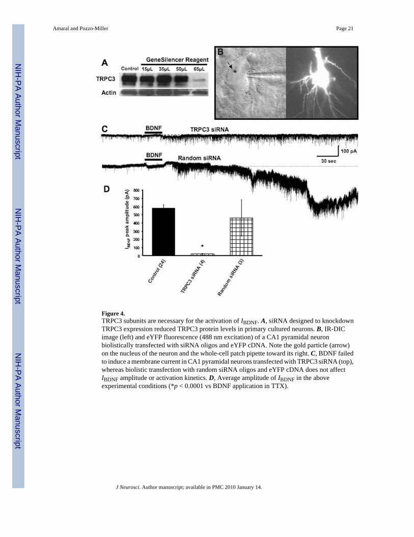
Figure 4.TRPC3 subunits are necessary for the activation of IBDNF. A, siRNA designed to knockdownTRPC3 expression reduced TRPC3 protein levels in primary cultured neurons. B, IR-DICimage (left) and eYFP fluorescence (488 nm excitation) of a CA1 pyramidal neuronbiolistically transfected with siRNA oligos and eYFP cDNA. Note the gold particle (arrow)on the nucleus of the neuron and the whole-cell patch pipette toward its right. C, BDNF failedto induce a membrane current in CA1 pyramidal neurons transfected with TRPC3 siRNA (top),whereas biolistic transfection with random siRNA oligos and eYFP cDNA does not affectIBDNF amplitude or activation kinetics. D, Average amplitude of IBDNF in the aboveexperimental conditions (*p < 0.0001 vs BDNF application in TTX).
Amaral and Pozzo-Miller Page 21
J Neurosci. Author manuscript; available in PMC 2010 January 14.
NIH
-PA Author Manuscript
NIH
-PA Author Manuscript
NIH
-PA Author Manuscript

Figure 5.PI3 kinase signaling is required for IBDNF and the rapid increase in TRPC3 surface content.A, IBDNF was reduced by the PI3 kinase inhibitor LY-294002 (10 μM). B, Average amplitudeof IBDNF in the above experimental conditions (*p < 0.0001 vs BDNF application in TTX).C, Surface biotinylation reactions on cultured hippocampal neurons revealed that levels ofsurface accessible TRPC3 progressively increased after BDNF application with a time coursethat parallels the time course of IBDNF activation (1, 2, and 4 min). In addition, the same PI3kinase inhibitor that reduced IBDNF also prevented the surface translocation of TRPC3. Bargraphs shows the quantitative analysis of surface biotinylation reactions, using the transferrinreceptor for normalization of pixel intensity.
Amaral and Pozzo-Miller Page 22
J Neurosci. Author manuscript; available in PMC 2010 January 14.
NIH
-PA Author Manuscript
NIH
-PA Author Manuscript
NIH
-PA Author Manuscript

Figure 6.A, Representative examples of membrane currents evoked by TBS in the presence of ionotropicand metabotropic glutamate and GABAA receptors outlasted the afferent train (arrows). Theseslow currents were completely blocked by TrkB–IgG (1 μg/ml). B, Slow currents evoked byTBS were completely blocked by k-252a (200 nM). C, TBS-evoked slow currents werecompletely blocked by SKF-96365 (30 μM).
Amaral and Pozzo-Miller Page 23
J Neurosci. Author manuscript; available in PMC 2010 January 14.
NIH
-PA Author Manuscript
NIH
-PA Author Manuscript
NIH
-PA Author Manuscript

Figure 7.Functional TRPC3 channels are required for BDNF to increase dendritic spine density in CA1pyramidal neurons. Slice cultures were transfected with either eYFP cDNA alone or incombination with TRPC3 siRNA oligonucleotides. Slices were exposed to BDNF (200 ng/mlin serum-free media) for 24 h, and spine density was measured by confocal microscopy 24 hafter transfection. A, Representative examples of segments of apical secondary and tertiarydendritic branches of eYFP-transfected CA1 pyramidal neurons. The TRPC inhibitorsSKF-96365 and 2-APB prevented the increase in spine density by BDNF. Likewise, siRNA-mediated knockdown of TRPC3 abolished the effect of BDNF on spine formation, withouteffects of its own. B, Average spine density in the above mentioned experimental groups. *p< 0.05 compared with serum-free controls, as per ANOVA followed by Newman–Keulsmultiple comparison post hoc test.
Amaral and Pozzo-Miller Page 24
J Neurosci. Author manuscript; available in PMC 2010 January 14.
NIH
-PA Author Manuscript
NIH
-PA Author Manuscript
NIH
-PA Author Manuscript

Figure 8.Schematic representation of the proposed model for intracellular signaling cascades initiatedby BDNF stimulation of TrkB receptors leading to the activation of membrane currents andCa2+ signals mediated by TRPC3-containing channels. Gab-1, Growth-associated binder 1;Grb2, growth factor receptor-bound protein 2; Shc, Src homology 2 domain-containingtransforming protein C1; P, phosphate groups; Y, tyrosine residues.
Amaral and Pozzo-Miller Page 25
J Neurosci. Author manuscript; available in PMC 2010 January 14.
NIH
-PA Author Manuscript
NIH
-PA Author Manuscript
NIH
-PA Author Manuscript
Related Documents