Am. J. Trop. Med. Hyg., 88(6), 2013, pp. 1196–1202 doi:10.4269/ajtmh.12-0714 Copyright © 2013 by The American Society of Tropical Medicine and Hygiene Tropical Diseases Screening in Immigrant Patients with Human Immunodeficiency Virus Infection in Spain Fernando Salvador,* Israel Molina, Elena Sulleiro, Joaquı´n Burgos, Adria ´n Curran, Eva Van den Eynde, Sara Villar del Saz, Jordi Navarro, Manuel Crespo, Inma Ocan ˜ a, Esteve Ribera, Vicenc ¸ Falco ´ , and Albert Pahissa Infectious Diseases Department and Microbiology Department, Hospital Universitari Vall d’Hebron, Universitat Auto ` noma de Barcelona, Spain Abstract. Latent parasitic infections can reactivate because of immunosuppression. We conducted a prospective observational study of all human immunodeficiency virus (HIV)–infected immigrants who visited the Infectious Diseases Department of the Hospital Universitari Vall d’Hebron, Barcelona, Spain, during June 2010–May 2011. Screening of the most prevalent tropical diseases (intestinal parasitosis, Chagas disease, leishmaniasis, malaria, schistosomiasis, and strongyloidiasis) was performed according to geographic origin. A total of 190 patients were included: 141 (74.2%) from Latin America, 41 (21.6%) from sub-Saharan Africa, and 8 (4.2%) from northern Africa. Overall, 36.8% (70 of 190) of the patients had at least one positive result for any parasitic disease: 5 patients with positive Trypanosoma cruzi serology, 11 patients with positive Schistosoma mansoni serology, 35 patients with positive Strongyloides stercoralis serology, 7 patients with positive Leishmania infantum serology, intestinal parasitosis were detected in 37 patients, malaria was diagnosed in one symptomatic patient. We propose a screening and management strategy of latent parasitic infections in immigrant patients infected with HIV. INTRODUCTION In recent years, the immigrant population in Spain has increased significantly, and in 2011 represented 12.2% of the total population (approximately 5.7 million persons). 1 Con- versely, in 2010 there were 34 million persons with human immunodeficiency virus (HIV) infection worldwide, and nearly 70% were concentrated in Africa and South America. 2 These factors have changed the demographic profile of HIV-infected patients treated in hospitals in Spain, with higher proportion of immigrants, mainly from sub-Saharan Africa and Latin America. Screening of latent infections is recommended in the initial approach for all HIV-infected patients because of the capabil- ity to reactivate when immunosuppression is established. A tuberculin skin test (TST), chest radiograph, and serologic tests (for Toxoplasma gondii, Treponema pallidum, and hep- atitis A, B and C viruses) should be performed. 3–5 However, immigrant patients may have other endemic infections in their countries, such as Strongyloides stercoralis hyperinfection syn- drome, or myocarditis and meningoencephalitis caused by Trypanosoma cruzi, 6,7 that can be reactivated or manifested as severe forms because of the immunosuppression. Immune reconstitution inflammatory syndrome has also been related to some parasitic infections, such as leishmaniasis, schisto- somiasis, and strongyloidiasis. 8 The aim of this study was to perform a screening for prev- alent tropical diseases in immigrants with HIV infection in Spain according to their geographic origin. With these results, we will suggest a screening, treatment and follow-up strategy. PATIENTS AND METHODS Study population and data collection. A prospective obser- vational study was conducted at the Infectious Diseases Department of the Hospital Universitari Vall d’Hebron, a University hospital in Barcelona, Spain. All HIV-infected patients from Latin America, sub-Saharan Africa, and north- ern Africa who visited the Infectious Diseases Department during June 2010–May 2011 were included. The study proto- col was approved by the institutional review board of the hospital and informed consent was obtained from all patients. The following data were collected: epidemiologic data (age, sex, country and district of origin, time since arrival in Spain, time since last travel to their country of origin, living in a rural environment); HIV infection–related data (CD4+ cell count, HIV plasma virus RNA, HIV acquisition risk factor, current antiretroviral therapy, previous opportunistic infections), eosinophil count, chest radiograph, TST and serologic data (Toxoplasma gondii, hepatitis B virus [HBV] surface antigen, antibodies against HBV core antigen and antibodies against hep- atitis C virus [HCV]). Eosinophilia was defined as eosinophil count ³ 500/mm 3 or a percentage ³ 7%. The TST (Mantoux method) result was considered positive when the transversal diameter of induration was ³ 5 mm, according to international recommendations. 5 Latent tuberculosis was diagnosed in patients with a positive TST result and a normal chest radiograph. Screening for tropical diseases. For infections with intesti- nal parasites, stool samples were collected on two days from all patients and placed in 10% formol saline. Microscopic examination was performed by using direct techniques (saline and iodine wet mounts) and after concentration techniques by using the formol-ether method or Ritchie’s technique. Auramine staining for Cryptosporidium and Isospora detec- tion was also performed. Specific treatment was offered to all patients with a parasite infection considered pathogenic. For Chagas disease, all patients from Latin America were tested for this disease (unless they came from the Caribbean islands). Serologic diagnosis of Chagas disease was performed by using two enzyme-linked immunosorbent assays (ELISAs) in parallel, one with recombinant antigen (Bioelisa Chagas; Biokit, Barcelona, Spain) and the other with crude antigen (Ortho T. cruzi ELISA; Johnson and Johnson, Piscataway, NJ) according to diagnostic criteria of the World Health Organization. Both ELISAs were performed according to the manufacturers’ instructions. We considered results posi- tive or negative if results of both tests were concordant. All discordant serum samples were tested by using an in-house * Address correspondence to Fernando Salvador, Infectious Diseases Department, Hospital Universitari Vall d’Hebron, Universitat Auto ` noma de Barcelona, Spain. E-mail: fmsalvad@ vhebron.net 1196

Welcome message from author
This document is posted to help you gain knowledge. Please leave a comment to let me know what you think about it! Share it to your friends and learn new things together.
Transcript
Am. J. Trop. Med. Hyg., 88(6), 2013, pp. 1196–1202doi:10.4269/ajtmh.12-0714Copyright © 2013 by The American Society of Tropical Medicine and Hygiene
Tropical Diseases Screening in Immigrant Patients with Human Immunodeficiency
Virus Infection in Spain
Fernando Salvador,* Israel Molina, Elena Sulleiro, Joaquın Burgos, Adrian Curran, Eva Van den Eynde, Sara Villar del Saz,Jordi Navarro, Manuel Crespo, Inma Ocana, Esteve Ribera, Vicenc Falco, and Albert Pahissa
Infectious Diseases Department and Microbiology Department, Hospital Universitari Vall d’Hebron, Universitat Autonoma de Barcelona, Spain
Abstract. Latent parasitic infections can reactivate because of immunosuppression. We conducted a prospectiveobservational study of all human immunodeficiency virus (HIV)–infected immigrants who visited the Infectious DiseasesDepartment of the Hospital Universitari Vall d’Hebron, Barcelona, Spain, during June 2010–May 2011. Screening of themost prevalent tropical diseases (intestinal parasitosis, Chagas disease, leishmaniasis, malaria, schistosomiasis, andstrongyloidiasis) was performed according to geographic origin. A total of 190 patients were included: 141 (74.2%) fromLatin America, 41 (21.6%) from sub-Saharan Africa, and 8 (4.2%) from northern Africa. Overall, 36.8% (70 of 190) ofthe patients had at least one positive result for any parasitic disease: 5 patients with positive Trypanosoma cruzi serology,11 patients with positive Schistosoma mansoni serology, 35 patients with positive Strongyloides stercoralis serology,7 patients with positive Leishmania infantum serology, intestinal parasitosis were detected in 37 patients, malariawas diagnosed in one symptomatic patient. We propose a screening and management strategy of latent parasitic infectionsin immigrant patients infected with HIV.
INTRODUCTION
In recent years, the immigrant population in Spain hasincreased significantly, and in 2011 represented 12.2% of thetotal population (approximately 5.7 million persons).1 Con-versely, in 2010 there were 34 million persons with humanimmunodeficiency virus (HIV) infection worldwide, and nearly70% were concentrated in Africa and South America.2 Thesefactors have changed the demographic profile of HIV-infectedpatients treated in hospitals in Spain, with higher proportion ofimmigrants, mainly from sub-SaharanAfrica and LatinAmerica.Screening of latent infections is recommended in the initial
approach for all HIV-infected patients because of the capabil-ity to reactivate when immunosuppression is established. Atuberculin skin test (TST), chest radiograph, and serologictests (for Toxoplasma gondii, Treponema pallidum, and hep-atitis A, B and C viruses) should be performed.3–5 However,immigrant patients may have other endemic infections in theircountries, such as Strongyloides stercoralis hyperinfection syn-drome, or myocarditis and meningoencephalitis caused byTrypanosoma cruzi,6,7 that can be reactivated or manifested assevere forms because of the immunosuppression. Immunereconstitution inflammatory syndrome has also been relatedto some parasitic infections, such as leishmaniasis, schisto-somiasis, and strongyloidiasis.8
The aim of this study was to perform a screening for prev-alent tropical diseases in immigrants with HIV infection inSpain according to their geographic origin. With these results,we will suggest a screening, treatment and follow-up strategy.
PATIENTS AND METHODS
Study population and data collection. A prospective obser-vational study was conducted at the Infectious DiseasesDepartment of the Hospital Universitari Vall d’Hebron, aUniversity hospital in Barcelona, Spain. All HIV-infected
patients from Latin America, sub-Saharan Africa, and north-ern Africa who visited the Infectious Diseases Departmentduring June 2010–May 2011 were included. The study proto-col was approved by the institutional review board of thehospital and informed consent was obtained from all patients.The following data were collected: epidemiologic data (age,
sex, country and district of origin, time since arrival in Spain,time since last travel to their country of origin, living in a ruralenvironment); HIV infection–related data (CD4+ cell count,HIV plasma virus RNA, HIV acquisition risk factor, currentantiretroviral therapy, previous opportunistic infections),eosinophil count, chest radiograph, TST and serologic data(Toxoplasma gondii, hepatitis B virus [HBV] surface antigen,antibodies againstHBVcore antigen and antibodies against hep-atitis C virus [HCV]). Eosinophilia was defined as eosinophilcount ³ 500/mm3 or a percentage ³ 7%. The TST (Mantouxmethod) result was considered positive when the transversaldiameter of induration was ³ 5 mm, according to internationalrecommendations.5Latent tuberculosiswasdiagnosed inpatientswith a positive TST result and a normal chest radiograph.Screening for tropical diseases. For infections with intesti-
nal parasites, stool samples were collected on two days fromall patients and placed in 10% formol saline. Microscopicexamination was performed by using direct techniques (salineand iodine wet mounts) and after concentration techniquesby using the formol-ether method or Ritchie’s technique.Auramine staining for Cryptosporidium and Isospora detec-tion was also performed. Specific treatment was offered to allpatients with a parasite infection considered pathogenic.For Chagas disease, all patients from Latin America were
tested for this disease (unless they came from the Caribbeanislands). Serologic diagnosis of Chagas disease was performedby using two enzyme-linked immunosorbent assays (ELISAs)in parallel, one with recombinant antigen (Bioelisa Chagas;Biokit, Barcelona, Spain) and the other with crude antigen(Ortho T. cruzi ELISA; Johnson and Johnson, Piscataway,NJ) according to diagnostic criteria of the World HealthOrganization. Both ELISAs were performed according tothe manufacturers’ instructions. We considered results posi-tive or negative if results of both tests were concordant. Alldiscordant serum samples were tested by using an in-house
*Address correspondence to Fernando Salvador, Infectious DiseasesDepartment, Hospital Universitari Vall d’Hebron, UniversitatAutonoma de Barcelona, Spain. E-mail: fmsalvad@ vhebron.net
1196
Western blotting method with a lysate from Trypanosomacruzi epimastigotes.9 When a positive result was obtained, chestradiograph, electrocardiogram, echocardiogram, esophagogram,and barium enema were performed and specific treatmentwas offered.For leishmaniasis, all patients were tested for this disease
by detection of IgG against Leishmania infantum by usingan ELISA (Novagnost Leishmania IgG; Siemens Diagnostics,Marburg, Germany) according to the manufacturer’s instruc-tions. Bone marrow aspiration was performed in patients withpositive serologic results and pancytopenia or hepatosple-nomegaly to diagnose visceral leishmaniasis (VL). Specifictreatment was offered.For malaria, patients from malaria-endemic areas (sub-
Saharan Africa and some Latin American regions) weretested for this disease by using a real-time polymerase chainreaction (PCR) with peripheral blood samples. This PCRcould identify four species of Plasmodium. DNA extractionwas conducted by using automatic silica-membrane tech-nology (NucliSENS EasyMag; bioMerieux, Marcy l’Etoile,France). Primers and probe for a satellite sequence wereselected and used in a TaqMan-based assay as described byRougemont and others.10 Positive results for the PCR wereconsidered diagnostic for malaria, and specific treatmentwas administered.For schistosomiasis, patients coming from schistosomiasis-
endemic areas (sub-Saharan Africa and some Latin Americanregions) were tested for this disease by detection of IgGagainst Schistosoma mansoni by using an ELISA (NovagnostSchistosoma mansoni IgG; Siemens Diagnostics) accordingto the manufacturer’s instructions. When results were pos-itive, stool and urine samples were test for parasite ovaand abdominal ultrasonography was performed. Specifictreatment was offered when schistosomiasis was confirmed(ova detection) or suspected (positive serologic results and
characteristic manifestations, such as eosinophilia, hematuria,or periportal fibrosis).For strongyloidiasis, all patients were tested for this disease
by detection of IgG against Strongyloides stercoralis by usingan ELISA (Strongyloides IgG; DRG Diagnostics, Marburg,Germany). When results were positive, three additionalstool samples were collected and specific fecal culture forS. stercoralis larvae (charcoal culture) was performed. Spe-cific treatment was offered when strongyloidiasis was con-firmed (larvae detection) or suspected (positive serologicresults and characteristic manifestations, such as eosinophilia,skin lesions, or intestinal disorders).Statistical analysis. Categorical data are presented as abso-
lute numbers and proportions, and continuous variables areexpressed as medians and interquartile ranges (IQRs). Thechi-square test or Fisher exact test, when appropriate, wasused to compare the distribution of categorical variables, andthe Mann-Whitney U test was used for continuous variables.Univariate and multivariate analyses using a forward stepwisemultiple regression model were conducted to identify variablesindependently associated with a specific parasitosis. Adjustedodds ratios and 95% confidence intervals were calculated.Results were considered statistically significant if the two-tailed P value was < 0.05. SPSS software for Windows version15.0 (SPSS Inc., Chicago, IL) was used for statistical analyses.
RESULTS
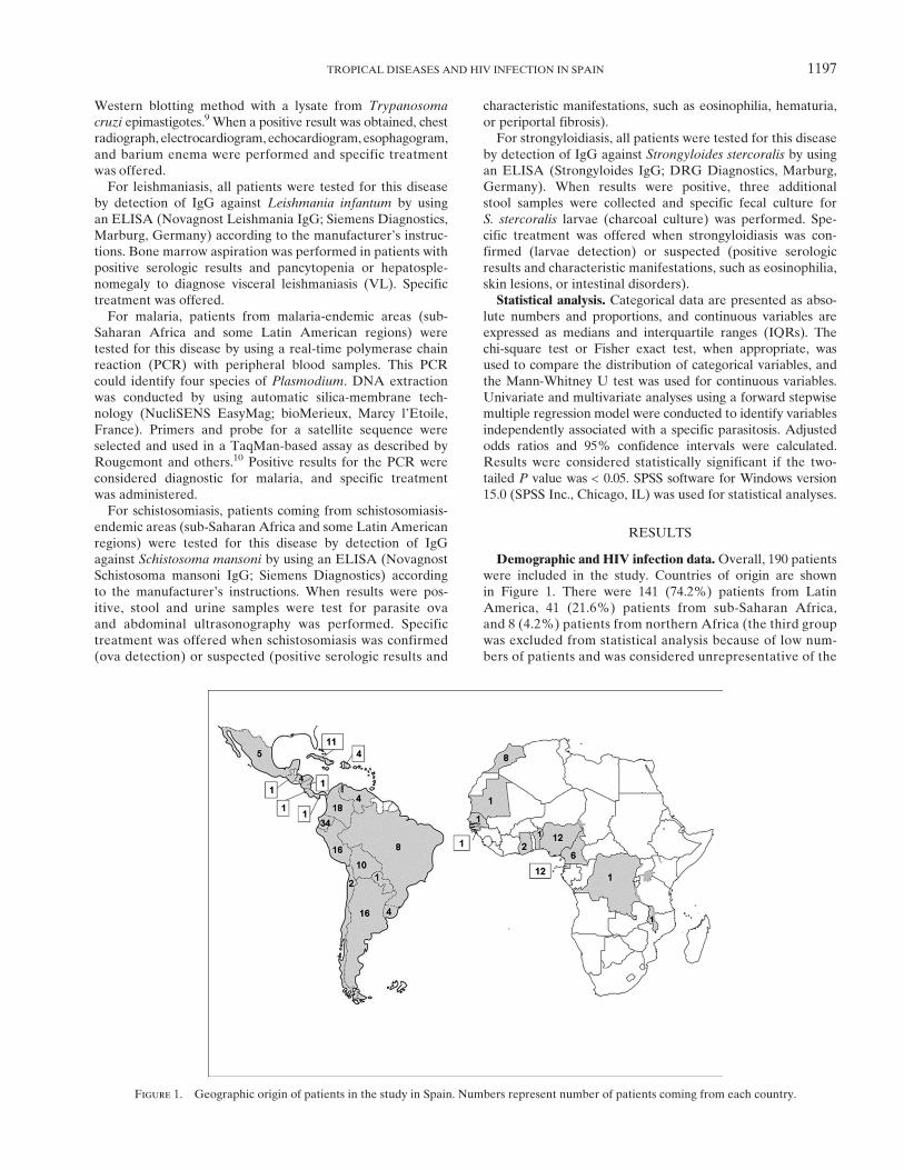
Demographic and HIV infection data.Overall, 190 patientswere included in the study. Countries of origin are shownin Figure 1. There were 141 (74.2%) patients from LatinAmerica, 41 (21.6%) patients from sub-Saharan Africa,and 8 (4.2%) patients from northern Africa (the third groupwas excluded from statistical analysis because of low num-bers of patients and was considered unrepresentative of the
Figure 1. Geographic origin of patients in the study in Spain. Numbers represent number of patients coming from each country.
TROPICAL DISEASES AND HIV INFECTION IN SPAIN 1197
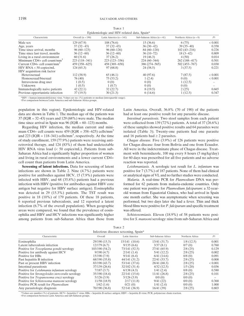
population in this region). Epidemiologic and HIV-relateddata are shown in Table 1. The median age of the patients was37 (IQR = 32–43) years and 129 (68%) were male. The mediantime since arrival in Spain was 96 (IQR = 60–123) months.Regarding HIV infection, the median current and mini-
mum CD4+ cell counts were 459 (IQR = 358–625) cells/mm3
and 223 (IQR = 118–341) cells/mm3, respectively. At the timeof study enrollment, 159 (77.9%) patients were receiving anti-retroviral therapy, and 124 (83%) of them had undetectableHIV RNA virus load (< 50 copies/mL). Patients from sub-Saharan Africa had a significantly higher proportion of womenand living in rural environments and a lower current CD4+cell count than patients from Latin America.Screening of latent infections. Data for screening of latent
infections are shown in Table 2. Nine (4.7%) patients werepositive for antibodies against HCV, 15 (7.9%) patients wereinfected with HBV, and 68 (35.8%) patients had a previousinfection with HBV (positive for antibodies against HBV coreantigen but negative for HBV surface antigen). Eosinophiliawas detected in 29 (15.3%) patients. The TST result waspositive in 18 (10%) of 179 patients. Of these 18 patients,6 reported previous tuberculosis, and 12 reported a latentinfection (6.7% of the overall population). When geographicareas were compared, we found that the prevalence of eosin-ophilia and HBV and HCV infections was significantly higheramong patients from sub-Saharan Africa than those from
Latin America. Overall, 36.8% (70 of 190) of the patientshad at least one positive result for any parasitic disease.Intestinal parasitosis. Two stool samples from each patient
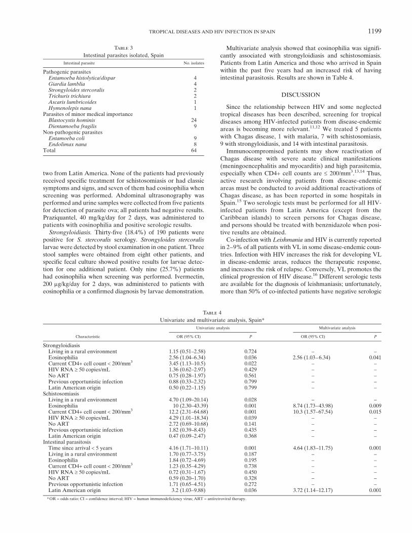
were collected from 139 (72%) patients. A total of 37 (26.6%)of these samples showed positive results and 64 parasites wereisolated (Table 3); Twenty-one patients had one parasiteand 16 patients had ³ 2 parasites.Chagas disease. Five (3.9%) of 126 patients were positive
for Chagas disease: four from Bolivia and one from Ecuador.All were in the indeterminate phase of Chagas disease. Treat-ment with benznidazole, 100 mg every 8 hours (5 mg/kg/day)for 60 days was prescribed for all five patients and no adversereaction was reported.Leishmaniasis. A serologic test result for L. infantum was
positive for 7 (3.7%) of 187 patients. None of them had clinicalor analytical signs of VL and no further studies were conducted.Malaria. A real-time PCR for Plasmodium DNA was per-
formed for 62 patients from malaria-endemic countries. Onlyone patient was positive for Plasmodium falciparum: a 32-year-old woman from Equatorial Guinea, who had arrived in Spainone month earlier. She was asymptomatic when screening wasperformed, but two days later she had a fever. Thin and thickblood filmswere positive forP. falciparum and specific treatmentwas offered.Schistosomiasis. Eleven (18.9%) of 58 patients were posi-
tive for S. mansoni serology: nine from sub-SaharanAfrica and
Table 1
Epidemiologic and HIV-related data, Spain*Characteristic Overall (n = 190) Latin America (n = 141) Sub-Saharan Africa (n = 41) Northern Africa (n = 8) P†
Male sex 129 (67.9) 108 (76.6) 15 (36.6) 6 (75) < 0.001Age, years 37 (32–43) 37 (32–43) 36 (30–42) 39 (35–40) 0.358Time since arrival, months 96 (60–123) 96 (60–126) 84 (60–120) 102 (43–216) 0.226Time since last travel, months 36 (12–60) 36 (12–60) 36 (10–72) 18 (3–42) 0.809Living in a rural environment 60 (31.6) 37 (26.2) 19 (46.3) 4 (50) 0.014Minimum CD4+ cell count/mm3 223 (118–341) 223 (123–336) 224 (60–344) 262 (188–417) 0.501Current CD4+ cell count/mm3 459 (358–625) 458 (369–658) 386 (274–565) 502 (453–767) 0.030HIV RNA < 50 copies/mL 124 (65.3) 97 (68.8) 24 (58.5) 3 (37.5) 0.221HIV acquisition risk factorHeterosexual 112 (58.9) 65 (46.1) 40 (97.6) 7 (87.5) < 0.001Homosexual/bisexual 76 (40) 75 (53.2) 1 (2.4) 0 (0) < 0.001Intravenous drug user 1 (0.5) 0 (0) 0 (0) 1 (12.5) –
Unknown 1 (0.5) 1 (0.7) 0 (0) 0 (0) –
Immunologically naive patients 42 (22.1) 32 (22.7) 8 (19.5) 2 (25) 0.665Previous opportunistic infection 37 (19.5) 30 (21.3) 6 (14.6) 1 (12.5) 0.347
*HIV = human immunodeficiency virus. Values are no. (%) patients or median (interquartile range).†For comparison between Latin America and sub-Saharan Africa groups.
Table 2
Infectious diseases screening, Spain*Characteristic Overall Latin America Sub-Saharan Africa Northern Africa P†
Eosinophilia 29/190 (15.3) 15/141 (10.6) 13/41 (31.7) 1/8 (12.5) 0.001Latent tuberculosis infection 12/179 (6.7) 9/135 (6.6) 3/37 (8.1) 0/7 (0) 0.760Positive for Toxoplasma gondii serology 103/190 (54.2) 73/141 (52.5) 27/41 (65.9) 2/8 (25) 0.129Positive for antibody against HCV 9/190 (4.7) 2/141 (1.4) 5/41 (12.2) 2/8 (25) 0.002Positive for HBs 15/190 (7.9) 9/141 (6.4) 6/41 (14.6) 0/8 (0) 0.091Past hepatitis B infection 68/190 (35.8) 44/141 (31.2) 22/41 (53.7) 2/8 (25) 0.008Past or present HBV infection 83/190 (43.7) 53/141 (37.6) 28/41 (68.3) 2/8 (25) < 0.001Intestinal parasitosis 37/139 (26.6) 32/102 (31.4) 4/32 (12.5) 1/5 (20) 0.036Positive for Leishmania infantum serology 7/187 (3.7) 6/138 (4.3) 1/41 (2.4) 0/8 (0) 0.580Positive for Strongyloides stercoralis serology 35/190 (18.4) 22/141 (15.6) 11/41 (26.8) 2/8 (25) 0.101Positive for Trypanosoma cruzi serology 5/126 (3.9) 5/126 (3.9) 0/0 (0) 0/0 (0) –
Positive for Schistosoma mansoni serology 11/58 (18.9) 2/17 (11.8) 9/41 (22) 0/0 (0) 0.368Positive PCR result for Plasmodium 1/62 (1.6) 0/21 (0) 1/41 (2.4) 0/0 (0) 1.000Any parasitologic diagnosis 70/190 (36.8) 52/141 (36.9) 16/41 (39) 2/8 (25) 0.803
*Values are number (%) of patients. HCV= hepatitis C virus; HBs = hepatitis B surface antigen; HBV = hepatitis B virus; PCR, polymerase chain reaction.†For comparison between Latin America and sub-Saharan groups.
1198 SALVADOR AND OTHERS
two from Latin America. None of the patients had previouslyreceived specific treatment for schistosomiasis or had classicsymptoms and signs, and seven of them had eosinophilia whenscreening was performed. Abdominal ultrasonography wasperformed and urine samples were collected from five patientsfor detection of parasite ova; all patients had negative results.Praziquantel, 40 mg/kg/day for 2 days, was administered topatients with eosinophilia and positive serologic results.Strongyloidiasis. Thirty-five (18.4%) of 190 patients were
positive for S. stercoralis serology. Strongyloides stercoralislarvae were detected by stool examination in one patient. Threestool samples were obtained from eight other patients, andspecific fecal culture showed positive results for larvae detec-tion for one additional patient. Only nine (25.7%) patientshad eosinophilia when screening was performed. Ivermectin,200 mg/kg/day for 2 days, was administered to patients witheosinophilia or a confirmed diagnosis by larvae demonstration.
Multivariate analysis showed that eosinophilia was signifi-cantly associated with strongyloidiasis and schistosomiasis.Patients from Latin America and those who arrived in Spainwithin the past five years had an increased risk of havingintestinal parasitosis. Results are shown in Table 4.
DISCUSSION
Since the relationship between HIV and some neglectedtropical diseases has been described, screening for tropicaldiseases among HIV-infected patients from disease-endemicareas is becoming more relevant.11,12 We treated 5 patientswith Chagas disease, 1 with malaria, 7 with schistosomiasis,9 with strongyloidiasis, and 14 with intestinal parasitosis.Immunocompromised patients may show reactivation of
Chagas disease with severe acute clinical manifestations(meningoencephalitis and myocarditis) and high parasitemia,especially when CD4+ cell counts are £ 200/mm3.13,14 Thus,active research involving patients from disease-endemicareas must be conducted to avoid additional reactivations ofChagas disease, as has been reported in some hospitals inSpain.15 Two serologic tests must be performed for all HIV-infected patients from Latin America (except from theCaribbean islands) to screen persons for Chagas disease,and persons should be treated with benznidazole when posi-tive results are obtained.Co-infection with Leishmania and HIV is currently reported
in 2–9% of all patients with VL in some disease-endemic coun-tries. Infection with HIV increases the risk for developing VLin disease-endemic areas, reduces the therapeutic response,and increases the risk of relapse. Conversely, VL promotes theclinical progression of HIV disease.16 Different serologic testsare available for the diagnosis of leishmaniasis; unfortunately,more than 50% of co-infected patients have negative serologic
Table 4
Univariate and multivariate analysis, Spain*
Characteristic
Univariate analysis Multivariate analysis
OR (95% CI) P OR (95% CI) P
StrongyloidiasisLiving in a rural environment 1.15 (0.51–2.58) 0.724 – –
Eosinophilia 2.56 (1.04–6.34) 0.036 2.56 (1.03–6.34) 0.041Current CD4+ cell count < 200/mm3 3.45 (1.13–10.5) 0.022 – –
HIV RNA ³ 50 copies/mL 1.36 (0.62–2.97) 0.429 – –
No ART 0.75 (0.28–1.97) 0.561 – –
Previous opportunistic infection 0.88 (0.33–2.32) 0.799 – –
Latin American origin 0.50 (0.22–1.15) 0.799 – –
SchistosomiasisLiving in a rural environment 4.70 (1.09–20.14) 0.028 – –
Eosinophilia 10 (2.30–43.39) 0.001 8.74 (1.73–43.98) 0.009Current CD4+ cell count < 200/mm3 12.2 (2.31–64.68) 0.001 10.3 (1.57–67.54) 0.015HIV RNA ³ 50 copies/mL 4.29 (1.01–18.34) 0.039 – –
No ART 2.72 (0.69–10.68) 0.141 – –
Previous opportunistic infection 1.82 (0.39–8.43) 0.435 – –
Latin American origin 0.47 (0.09–2.47) 0.368 – –
Intestinal parasitosisTime since arrival < 5 years 4.16 (1.71–10.11) 0.001 4.64 (1.83–11.75) 0.001Living in a rural environment 1.70 (0.77–3.75) 0.187 – –
Eosinophilia 1.84 (0.72–4.69) 0.195 – –
Current CD4+ cell count < 200/mm3 1.23 (0.35–4.29) 0.738 – –
HIV RNA ³ 50 copies/mL 0.72 (0.31–1.67) 0.450 – –
No ART 0.59 (0.20–1.70) 0.328 – –
Previous opportunistic infection 1.71 (0.65–4.51) 0.272 – –
Latin American origin 3.2 (1.03–9.88) 0.036 3.72 (1.14–12.17) 0.001
*OR = odds ratio; CI = confidence interval; HIV = human immunodeficiency virus; ART = antiretroviral therapy.
Table 3
Intestinal parasites isolated, Spain
Intestinal parasite No. isolates
Pathogenic parasitesEntamoeba histolytica/dispar 4Giardia lamblia 4Strongyloides stercoralis 2Trichuris trichiura 2Ascaris lumbricoides 1Hymenolepis nana 1
Parasites of minor medical importanceBlastocystis hominis 24Dientamoeba fragilis 9
Non-pathogenic parasitesEntamoeba coli 9Endolimax nana 8
Total 64
TROPICAL DISEASES AND HIV INFECTION IN SPAIN 1199
results, and there is serologic cross-reactivity between Leish-mania and other microorganisms, such as Trypanosoma ormycobacteria.17 In our study, none of the seven patients withpositive serologic results had clinical or analytical abnormali-ties, and Chagas disease was concurrently diagnosed in fourof these patients. Therefore, positive results were most proba-bly false-positive results caused by cross-reactivity. On thebasis of our results, serologic testing is not useful for VLscreening of VL HIV-infected persons.HIV infection and malaria are two of the most important
health problems in developing countries, mainly in sub-Saharan Africa. Studies about the impact of HIV infectionon the risk for severe malaria differ in their findings.18
Although real-time PCR is more sensitive than thick or thinblood films in post-arrival screening for malaria in asymptom-atic patients from disease-endemic areas, PCR did not pro-vide any advantage in our HIV population.19 In our experience,screening asymptomatic HIV-infected patients for malaria wasnot useful. This finding was probably caused by the fact thatmost analyzed patients were living in Spain for more thanfive years at the time of screening, which reduced the riskfor malaria.Chronic schistosomiasis results from the immune response
of the host to schistosome eggs and the granulomatous reac-tion produced by the antigens they secrete.20 Diminished eggexcretion efficiency has been found in HIV-infected patientswith schistosomiasis, which complicates diagnosis.21 The lackof sensitivity of classical methods and reduced egg excretionin HIV-infected patients make serologic analysis an interest-ing tool for schistosomiasis screening in this population.In our study, 11 patients had positive serologic results forS. mansoni, but results of screening for ova were negative.
Patients infected with HIV from area to which schistosomiasisis endemic should be screened by serologic testing, and stooland urine sample must be collected and tested when a positiveresult is obtained to confirm the diagnosis. On the basis of itsefficacy and safety, praziquantel should be offered to patientswith a confirmed diagnosis and those with positive serologicresults and indirect signs of infection (eosinophilia, hematu-ria, periportal fibrosis, urine bladder calcification). We do nothave any explanation for the association between schistoso-miasis and low CD4+ cell counts, but it was probably causedby the small number of patients.Strongyloides stercoralis infection is asymptomatic in most
infected patients, but a fulminant presentation that can befatal (S. stercoralis hyperinfection syndrome and disseminatedstrongyloidiasis) may occur in situations of compromised hostimmunity, as in HIV infection.22 Definitive diagnosis of stron-gyloidiasis is made on the basis of detection of larvae in stoolsamples. However, low parasite load and irregular larval out-put in chronic infections (especially in immunocompromisedpatients) make a diagnosis challenging.23 Currently, serologictests are sensitive and specific and are useful for screeningof asymptomatic patients and follow-up after specific treat-ment.24,25 Therefore, serologic testing for S. stercoralis infec-tion in an HIV-infected population seems to be the beststrategy. Stool samples must be collected for larvae detectionin persons with positive results to confirm the diagnosis. Spe-cific treatment with ivermectin should be offered to patientswith a confirmed diagnosis and patients with positive sero-logic results and other signs of infection (eosinophilia, pruri-tus, skin lesions, or gastrointestinal disorders).Higher prevalence of intestinal parasites in an HIV-
infected population has been reported.26 Cryptosporidium,
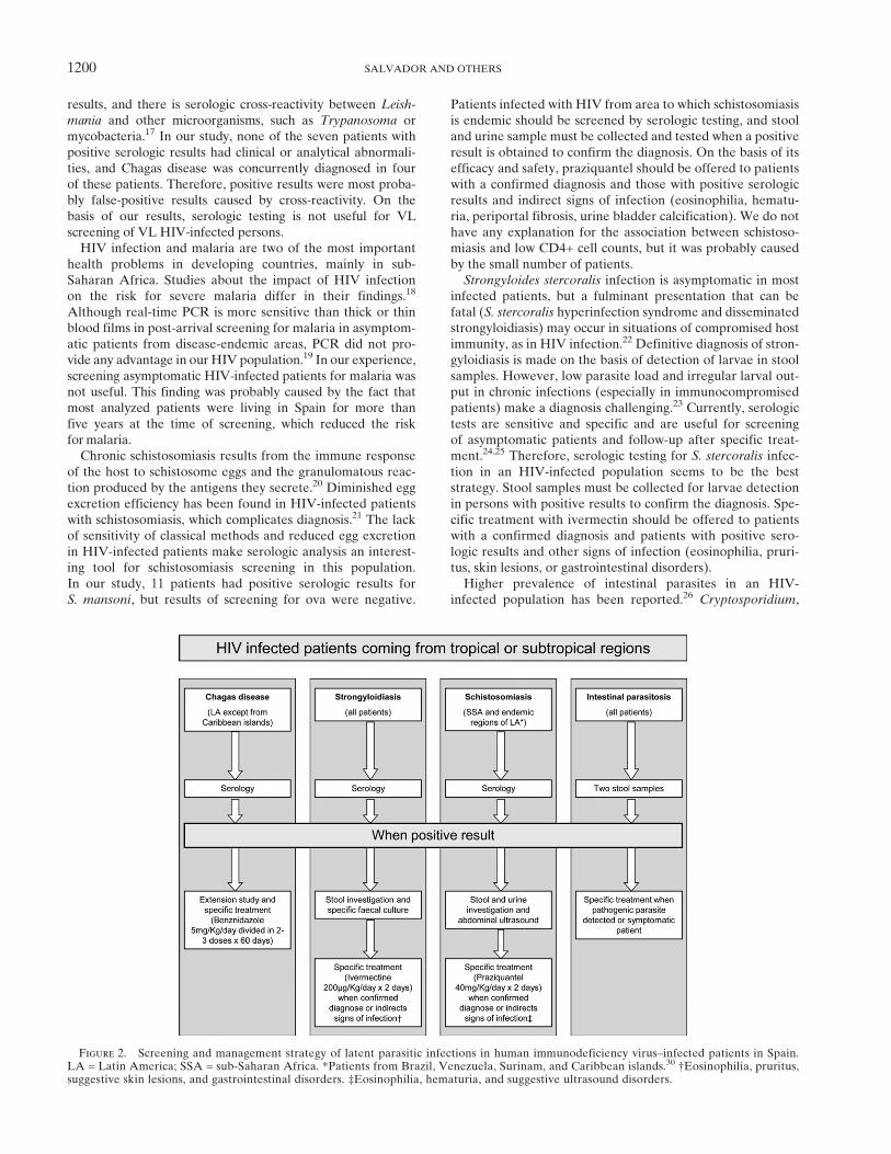
Figure 2. Screening and management strategy of latent parasitic infections in human immunodeficiency virus–infected patients in Spain.LA = Latin America; SSA = sub-Saharan Africa. *Patients from Brazil, Venezuela, Surinam, and Caribbean islands.30 †Eosinophilia, pruritus,suggestive skin lesions, and gastrointestinal disorders. ‡Eosinophilia, hematuria, and suggestive ultrasound disorders.
1200 SALVADOR AND OTHERS
Isospora, and other protozoan parasites have been relatedto advanced HIV infection, and other protozoa and helminthsare increasingly recognized as a significant problem inimmunocompromised persons.27,28 The higher prevalence ofintestinal parasitosis among immigrants living in Spain forless than five years can be explained by the fact that mostintestinal parasites interrupt their life cycle two or threeyears after leaving a favorable epidemiologic environment.We have found no explanation for the higher prevalence ofintestinal parasitosis among patients from Latin America inour study. Two stool samples should be collected from allHIV-infected patients from tropical and subtropical regionsto screen for intestinal parasitosis, even if these patients areasymptomatic (all patients with intestinal parasitosis wereasymptomatic in our study). Specific treatment must beoffered when pathogenic parasites are detected or in symp-tomatic patients.The low prevalence (4.7%) of HCV and HIV co-infection
compared with the entire HIV-infected population in Spain(approximately one third of the population) could be explainedby the main HIV acquisition risk factor in our study, thesexual transmission; only one patient was an intravenousdrug user. As it was expected, the prevalence of HBV infec-tion is higher in the group from sub-Saharan Africa than inthe group from Latin America.4
As suspected, parasitic infections among HIV-infectedpatients from tropical and subtropical regions are highlyprevalent, and screening programs for HIV populations fromdisease-endemic areas are highly recommended. In a similarstudy conducted in the United States,29 128 immigrantsinfected with HIV were screened by stool sample investiga-tion and serologic tests. Prevalence of antibodies againstStrongyloides (26%) and Schistosoma (29%) antigens werehigher than in our study (18.4% and 18.9%, respectively).Conversely, no patient in that study was positive for T. cruzi,whereas 3.9% of our patients had a positive test result. Thesedifferences were likely caused by differences in proportionsof patients from sub-Saharan Africa and Latin America inboth studies. As in our study, eosinophilia was strongly asso-ciated with strongyloidiasis and schistosomiasis, highlightingthe importance of testing for parasitic infections in patientswith high eosinophil counts.This study involved patients born in areas to which some
tropical diseases were endemic. Nevertheless, patients whotravel for long periods through the tropics are also at riskfor acquiring these endemic tropical diseases and should bescreened similar to patients born in these areas, especiallywhen they are symptomatic. Specific recommendations forscreening in returning travelers should to be made on the basisof travel destination, duration of travel, and risky activities.One of the limitations of our study is the use of a screening
method based on serologic tests. Immunosuppression causedby HIV infection may decrease the sensitivity of serologictests, and positive results may not differentiate between latentand past infections. Nevertheless, given the potential risk ofreactivation and safety of most of the specific treatments,treating parasitic infections based on serologic diagnosis inimmunocompromised patients seems to be beneficial. Anotherlimitation is that we used serologic tests for L. infantum andS. mansoni to screen for leishmaniasis and schistosomiasis,respectively, on the basis of cross-reactivity with other spe-cies, which could decrease test specificity. Finally, the study
population had specific social characteristics, which made itdifficult to follow-up these patients. A greater effort had to bemade by physicians; only 139 (72%) of 190 patients providedstool samples and further studies after positive serologicresults were obtained were difficult to perform.In summary, parasitic infections are prevalent in HIV-
infected patients from tropical and sub-tropical areas.Because of the risk of reactivation and severe forms of dis-eases, screening for parasitic infections is highly recom-mended in this population. On the basis of our results andother reported results, we propose screening for parasiticinfections by testing stool samples and using serologic testsfor Chagas disease, strongyloidiasis, and schistosomiasis in allHIV-infected patients according to their geographic origin.Malaria screening should be individualized according to risk(recent immigrants from malaria-endemic areas). Screeningand management strategy is summarized in Figure 2.
Received November 28, 2012. Accepted for publication February 18,2013.
Published online March 18, 2013.
Acknowledgments: The American Committee on Clinical TropicalMedicine and Travelers’ Health (ACCTMTH) assisted with publica-tion expenses.
Financial support: This study was partially supported by RedTematica de Investigacion en SIDA (RIS G03/173-RETIC RD06/0006/0039).
Disclosure: The funders had no role in study design, data collectionand analysis, decision to publish, or preparation of the manuscript.
Authors’ addresses: Fernando Salvador, Israel Molina, Elena Sulleiro,Joaquın Burgos, Adrian Curran, Eva Van den Eynde, Sara Villar delSaz, Jordi Navarro, Manuel Crespo, Inma Ocana, Esteve Ribera,Vicenc Falco, and Albert Pahissa, Infectious Diseases Department,Hospital Universitari Vall d’Hebron, Universitat Autonoma deBarcelona, Spain, E-mails: [email protected], [email protected], [email protected], [email protected], [email protected], [email protected], [email protected], [email protected],[email protected], [email protected], [email protected],[email protected], and [email protected].
REFERENCES
1. Instituto Nacional de Estadıstica. Available at: http://www.ine.es.Accessed March 2012.
2. World Health Organization, HIV/AIDS: Global Situation andTrends. Available at: http://www.who.int. AccessedMarch 2012.
3. Documento de Consenso de GESIDA/Plan Nacional Sobre el SIDASobre el Tratamiento Antirretroviral del Adulto (Actualizacion deEnero 2011). Available at: http://www.gesida.seimc.org.AccessedMarch 2012.
4. Recomendaciones de GESIDA/PNS/AEEH Sobre Tratamiento yManejo del Paciente Adulto Coinfectado por VIH y Virus deHepatitis A, B y C (Septiembre 2009). Available at: http://www.gesida.seimc.org. Accessed March 2012.
5. Gonzalez-Martın J, Garcıa-Garcıa JM, Anibarro L, Vidal R,Esteban J, Blanquer R, Moreno S, Ruiz-Manzano J, 2010.Consensus document on the diagnosis, treatment and preven-tion of tuberculosis. Enferm Infecc Microbiol Clin 28: e1–20.
6. Karp CL, Auwaerter PG, 2007. Coinfection with HIV and tropi-cal infectious diseases. I. Protozoal pathogens. Clin Infect Dis45: 1209–1213.
7. Karp CL, Auwaerter PG, 2007. Coinfection with HIV and tropi-cal infectious diseases. II. Helminthic, fungal, bacterial andviral pathogens. Clin Infect Dis 45: 1215–1220.
8. Lawn SD, Wilkinson RJ, 2006. Immune reconstitution diseaseassociated with parasitic infections following antiretroviraltreatment. Parasite Immunol 28: 625–633.
TROPICAL DISEASES AND HIV INFECTION IN SPAIN 1201
9. Riera C, Verges M, Iniesta L, Fisa R, Gallego M, Tebar S, PortusM, 2012. Identification of a Western blot pattern for thespecific diagnosis of Trypanosoma cruzi infection in humansera. Am J Trop Med Hyg 86: 412–416.
10. Rougemont M, Van Saanen M, Sahli R, Hinrikson HP, Bille J,Jaton K, 2004. Detection of four Plasmodium species in bloodfrom humans by 18 S rRNA gene subunit-based and species-specific real-time PCR assays. J Clin Microbiol 42: 5636–5643.
11. LilliePJ,BazazR,Greig JM, 2008. ScreeningAfricanHIVpositive1-patients for imported parasitic infections. J Infect 57: 481–484.
12. Sarner L, Fakoya AO, Tawana C, Allen E, Copas AJ, ChiodiniPL, Fenton KA, 2007. The utility of screening for parasiticinfections in HIV-1-infected Africans with eosinophilia inLondon. Int J STD AIDS 18: 626–629.
13. Lescure FX, le Loup G, Freilij H, Develoux M, Paris L, Brutus L,Pialoux G, 2010. Chagas disease: changes in knowledgeand management. Lancet Infect Dis 10: 556–570.
14. Sartori AM, Ibrahim KY, Nunes Westphalen EV, Braz LMA,Oliveira OC, Gakiya E, Lopes MH, Shikanai-Yasuda MA,2007. Manifestations of Chagas disease (American trypanoso-miasis) in patients with HIV/AIDS. Ann Trop Med Parasitol101: 31–50.
15. Rodrıguez-Guardado A, Asensi-Alvarez V, Rodrıguez-Perez M,Mejuto-Alvarez P, Flores-Chavez M, Alonso-Gonzalez P,Carton-Sanchez JA, 2011. Screening for Chagas disease inHIV-positive immigrants from endemic areas. EpidemiolInfect 139: 539–543.
16. Alvar J, Aparicio P, Aseffa A, Den Boer M, Canavate C, DedetJP, Gradoni L, Horst RT, Lopez-Velez R, Moreno J, 2008.The relationship between leishmaniasis and AIDS: the second10 years. Clin Microbiol Rev 21: 334–359.
17. Medrano FJ, Canavate C, Leal M, Rey C, Lissen E, Alvar J, 1998.The role of serology in the diagnosis and prognosis of visceralleishmaniasis in patients coinfected with human immunodefi-ciency virus type-1. Am J Trop Med Hyg 59: 155–162.
18. Flateau C, Le Loup G, Pialoux G, 2011. Consequences of HIVinfection on malaria and therapeutic implications: a systematicreview. Lancet Infect Dis 11: 541–556.
19. Matisz CE, Naidu P, Shokoples SE, Grice D, Krinke V, BrownSZ, Kowalewska-Grochowska K, Houston S, Yanow SK, 2011.Post-arrival screening for malaria in asymptomatic refugeesusing real-time PCR. Am J Trop Med Hyg 84: 161–165.
20. Ross AGP, Bartley PB, Sleigh AC, Olds GR, Li Y, WilliamsGM, McManus DP, 2002. Schistosomiasis. N Engl J Med346: 1212–1220.
21. Karanja DM, Colley DG, Nahlen BL, Ouma JH, Secor WE, 1997.Studies on schistosomiasis in western Kenia. I. Evidence forimmune-facilitated excretion of schistosome eggs from patientswith Schistosoma mansoni and human immunodeficiency viruscoinfections. Am J Trop Med Hyg 56: 515–521.
22. Keiser PB, Nutman TB, 2004. Strongyloides stercoralis in theimmunocompromised population. Clin Microbiol Rev 17:208–217.
23. Siddiqui AA, Berck SL, 2001. Diagnosis of Strongyloidesstercoralis infection. Clin Infect Dis 33: 1040–1047.
24. Bon B, Houze S, Talabani H, Magne D, Belkadi G, Develoux M,Senghor Y, Chandenier J, Ancelle T, Hennequin C, 2010.Evaluation of a rapid enzyme-linked immunosorbent assayfor diagnosis of Strongyloidiasis. J Clin Microbiol 48:1716–1719.
25. Biggs B-A, Caruana S, Mihrshahi S, Jolley D, Leydon J, Chea L,Nuon S, 2009. Short report: management of chronic strongyloi-diasis in immigrants and refugees: is serologic testing useful?Am J Trop Med Hyg 80: 788–791.
26. Sanyaolu AO, Oyibo WA, Fagbenro-Beyioku AF, GbadegeshinAH, IriemenamNC, 2011. Comparative study of entero-parasiticinfections among HIV sero-positive and sero-negative patientsin Lagos, Nigeria.Acta Trop 120: 268–272.
27. Modjarrad K, Zulu I, Redden DT, Njobvu L, Freedman DO,Vermund SH, 2005. Prevalence and predictors of intestinalhelminth infections among human immunodeficiency virustype 1-infected adults in an urban African setting. Am J TropMed Hyg 73: 777–782.
28. Lewthwaite P, Gill GV, Hart CA, Beeching NJ, 2005. Gastro-intestinal parasites in the immunocompromised. Curr OpinInfect Dis 18: 427–435.
29. Hochberg NS, Moro RN, Sheth AN, Montgomery SP, Steurer F,McAuliffe IT, Wang YF, Armstrong W, Rivera HN, LennoxJL, Franco-Paredes C, 2011. High prevalence of persistentparasitic infections in foreign-born, HIV-infected persons inthe United States. PLoS Negl Trop Dis 5: e1034.
30. World Health Organization, Schistosomiasis: Key Points andGeographical Distribution. Available at: http://www.who.int.Accessed March 2012.
1202 SALVADOR AND OTHERS
Related Documents