Go to: Go to: Indian J Orthop. 2008 OctDec; 42(4): 377–386. doi: 10.4103/00195413.43373 PMCID: PMC2740354 Treatment principles in the management of open fractures William W Cross, III and Marc F Swiontkowski Department of Orthopaedic Surgery, University of Minnesota Medical School, 2450 Riverside Ave., Ste R 200, Minneapolis, MN 55454, USA Address for correspondence: Dr. Marc F. Sw iontkow ski, Department of Orthopaedic Surgery, University of Minnesota Medical School, 2450 Riverside Ave., Suite R200, Minneapolis, MN 55454, USA. Email: sw [email protected] Copyright © Indian Journal of Orthopaedics This is an openaccess article distributed under the terms of the Creative Commons Attribution License, w hich permits unrestricted use, distribution, and reproduction in any medium, provided the original w ork is properly cited. This article has been cited by other articles in PMC. Abstract The management of open fractures continues to provide challenges for the orthopedic surgeon. Despite the improvements in technology and surgical techniques, rates of infection and nonunion are still troublesome. Principles important in the treatment of open fractures are reviewed in this article. Early antibiotic administration is of paramount importance in these cases, and when coupled with early and meticulous irrigation and debridement, the rates of infection can be dramatically decreased. Initial surgical intervention should be conducted as soon as possible, but the classic 6 h rule does not seem to be supported in the literature. All open fractures should be addressed for the risk of contamination from Clostridium tetani . When possible, early closure of open fracture wounds, either by primary means or by flaps, can also decrease the rate of infection, especially from nosocomial organisms. Early skeletal stabilization is necessary, which can be accomplished easily with temporary external fixation. Adhering to these principles can help surgeons provide optimal care to their patients and assist them in an early return to function. Keywords: Fracture principles, open fractures, trauma I NTRODUCTION It has been estimated that between 3.5 and 6 million fractures occur in the United States annually. Extrapolating from European data, we can estimate that more than 3%, i.e., 150,000, of these are open fractures. When adjusting for population differences, we predict that more than 4.5 million open fractures occur per year in India. This figure may be an underestimation, given the high population density in the large urban centers in India. These fractures can involve significant morbidity and are inherently worrisome, as the body's protective skin barrier has been broken and the potential for contamination is high. The correct and timely management of these injuries can benefit our patients and lead to more favorable outcomes. When deciding on the treatment strategy, the treating surgeon must consider the patient's condition, the mechanism of injury, and the fracture type. Although some of the most impressive injury patterns are from highenergy mechanisms, more commonly, patients present with an open fracture from a simple lowenergy mechanism such as a fall. Each fracture could conceivably be treated quite differently, ranging from external fixation and delayed closure or fixation to immediate irrigation, debridement, and primary closure. The status of the soft tissues surrounding the fracture site is of paramount importance in this decisionmaking process, which usually influences the initial management. 1 , 2 3 , 4

Welcome message from author
This document is posted to help you gain knowledge. Please leave a comment to let me know what you think about it! Share it to your friends and learn new things together.
Transcript

5/5/2015 Treatment principles in the management of open fractures
http://www.ncbi.nlm.nih.gov/pmc/articles/PMC2740354/ 1/13
Go to:
Go to:
Indian J Orthop. 2008 OctDec; 42(4): 377–386.doi: 10.4103/00195413.43373
PMCID: PMC2740354
Treatment principles in the management of open fracturesWilliam W Cross, III and Marc F Swiontkowski
Department of Orthopaedic Surgery, University of Minnesota Medical School, 2450 Riverside Ave., Ste R 200, Minneapolis, MN 55454, USAAddress for correspondence: Dr. Marc F. Sw iontkow ski, Department of Orthopaedic Surgery, University of Minnesota Medical School, 2450Riverside Ave., Suite R200, Minneapolis, MN 55454, USA. Email: sw [email protected]
Copyright © Indian Journal of Orthopaedics
This is an openaccess article distributed under the terms of the Creative Commons Attribution License, w hich permits unrestricted use, distribution,and reproduction in any medium, provided the original w ork is properly cited.
This article has been cited by other articles in PMC.
Abstract
The management of open fractures continues to provide challenges for the orthopedic surgeon. Despite the improvementsin technology and surgical techniques, rates of infection and nonunion are still troublesome. Principles important in thetreatment of open fractures are reviewed in this article. Early antibiotic administration is of paramount importance in thesecases, and when coupled with early and meticulous irrigation and debridement, the rates of infection can be dramaticallydecreased. Initial surgical intervention should be conducted as soon as possible, but the classic 6 h rule does not seem tobe supported in the literature. All open fractures should be addressed for the risk of contamination from Clostridiumtetani. When possible, early closure of open fracture wounds, either by primary means or by flaps, can also decrease therate of infection, especially from nosocomial organisms. Early skeletal stabilization is necessary, which can beaccomplished easily with temporary external fixation. Adhering to these principles can help surgeons provide optimal careto their patients and assist them in an early return to function.
Keywords: Fracture principles, open fractures, trauma
INTRODUCTION
It has been estimated that between 3.5 and 6 million fractures occur in the United States annually. Extrapolating fromEuropean data, we can estimate that more than 3%, i.e., 150,000, of these are open fractures. When adjusting forpopulation differences, we predict that more than 4.5 million open fractures occur per year in India. This figure may be anunderestimation, given the high population density in the large urban centers in India. These fractures can involvesignificant morbidity and are inherently worrisome, as the body's protective skin barrier has been broken and the potentialfor contamination is high. The correct and timely management of these injuries can benefit our patients and lead to morefavorable outcomes.
When deciding on the treatment strategy, the treating surgeon must consider the patient's condition, the mechanism ofinjury, and the fracture type. Although some of the most impressive injury patterns are from highenergy mechanisms,more commonly, patients present with an open fracture from a simple lowenergy mechanism such as a fall. Each fracturecould conceivably be treated quite differently, ranging from external fixation and delayed closure or fixation to immediateirrigation, debridement, and primary closure. The status of the soft tissues surrounding the fracture site is of paramountimportance in this decisionmaking process, which usually influences the initial management.
1,23,4

5/5/2015 Treatment principles in the management of open fractures
http://www.ncbi.nlm.nih.gov/pmc/articles/PMC2740354/ 2/13
Go to:
Goals of open fracture management are well known and include the prevention of infection, achievement of bony union,and the restoration of function. Current treatment strategies in the care of open fractures are continuously studied,improved, and adjusted as our literature base expands. Important principles include antibiotic utilization, timing of initialsurgical intervention, type of wound closure, antibiotic delivery methods, tetanus coverage, wound irrigation, andadjunctive therapies to assist with fracture union. This review aims to provide current information and references forfurther reading on these topics and provide a framework for decisionmaking when presented with an open fracture in theacute setting.
CLASSIFICATION SYSTEMS
The purpose of any fracture classification system in the clinical setting is to allow communication that infers fracturemorphology and treatment parameters. In the setting of open fractures, there are two classification systems that surgeonstreating these injuries should be familiar with. They are the Gustilo classification and the Mangled Extremity Severity Scale(MESS). The Gustilo classification has been the most widely used system and is generally accepted as the primaryclassification system for open fractures. This system takes into consideration the energy of the fracture, softtissuedamage, and the degree of contamination. It has been modified since the original classification to allow a more accurateprognosis for more severe injuries (i.e., Type III injuries). There has been some concern in the literature regarding theinterobserver reliability of this system. In this study, the surgeons interpreted color movies of examinations andradiographs of patients and then classified the injuries based on that screening. Overall, they demonstrated 60%agreement. We contend that classification of the injury should be made in the operating room at the conclusion of theinitial irrigation and debridements (see Table 1 for details).
Table 1Gustilo open fracture classification system
The second classification system, the MESS, was originally designed as an objective tool to assist the surgeon indecisionmaking for amputation vs limb salvage in complex lower extremity trauma. This rating scale takes intoconsideration the skeletal and soft tissue damage, limb ischemia time, presence of shock, and the patient's age. Multiplestudies have examined the efficacy of the MESS both retrospectively and prospectively and have found it to correlatewell with the treatment of major limb trauma. It has been suggested that a score of greater than or equal to 7 is predictiveof amputation with nearly 100% accuracy [Table 2].
Table 2The mangled extremity severity scale
More recent information has emerged in the management of severe lower extremity trauma and the effectiveness of theMESS and other lower extremity trauma scoring systems, including the Limb Salvage Index and the Predictive SalvageIndex. In one of the largest multicenter, prospective outcome studies pertaining to lower extremity trauma, the LowerExtremity Assessment Project (LEAP), found in a study of more than 500 patients with lower extremity trauma that injuryfactors with the highest significance in the decision for limb salvage were not solely the measures listed in the abovescoring systems. The LEAP group found that the most significant factors were tibial fracture pattern, presence of an openfoot fracture, bone loss, muscle injury, vein injury, arterial injury, and the absence of plantar sensation. Severe muscleinjury had the highest impact on the surgeon with loss of plantar sensation being second. More recent followup datahave challenged the importance of the insensate foot in the decision for amputation. Interestingly, this study found morethan onehalf of the patients presenting with an insensate foot treated with reconstruction ultimately regained sensation intwo years.
5–8
6,910
5,6
8
8,11–19
8
2021

5/5/2015 Treatment principles in the management of open fractures
http://www.ncbi.nlm.nih.gov/pmc/articles/PMC2740354/ 3/13
Go to:
The decision for amputation or limb salvage should be made with careful consideration of multiple factors to include notonly the MESS parameters and those from the LEAP study, but also the emotional impact, social impact, andpsychological recovery necessary for physical recovery. The assistance of the patient and their family is alsoencouraged. We ultimately advise initial surgical exploration prior to decisionmaking in the severely injured extremity.
CONTAMINATION OF OPEN FRACTURE AND USE OF ANTIBIOTICS
All open fractures are by definition contaminated and must be treated as such. The treatment methods may differdepending on the type of fracture. Infection risks also differ by fracture type and have been reported to be ranging from 0to 2% for Type I fractures, 2 to 10% for Type II fractures, and 10 to 50% for Type III fractures. More recentstudies have shown that the rates of clinical infection increased to 1.4% (7/497) for Type I fractures, 3.6% (25/695) forType II fractures, and to 22.7% (45/198) of Type III fractures. These data are similar to a more recent study on thetreatment of open tibia fractures.
Antibiotic treatment with open fracture management should be automatic with early administration being paramount [Table 3], ideally within 3 h of injury. The risk of infection has been shown to decrease sixfold with this practice.With the propensity for grampositive infections with Type I and II fractures, a firstgeneration cephalosporin is generallyrecommended. Some authors have advocated adding gramnegative coverage as well. Type III fractures oftenhave contamination from gramnegative organisms, and in the case of soilcontaminated wounds (i.e., farm injuries),additional coverage should be added for anaerobic bacteria. Typically, this would include penicillin for the risk of aClostridial infection. In the treatment of open fractures in the hospital setting, the surgeon must also be concerned fornosocomial infections, namely by Staphylococcus aureus and aerobic gramnegative bacilli such as Pseudomonas.Specific antibiotic coverage for these organisms may be indicated. The duration of antibiotic therapy in the treatment ofopen fractures has been suggested to be between 1 and 3 days without any solid agreement on a firm endpoint. We typically maintain antibiotic coverage until the wound is closed. Our recommended treatmentregimen is detailed in Table 3.
Table 3Recommendations for antibiotic therapy in open fracture management (allmedicine to be given intravenously)
Local antibiotic delivery must be considered when extensive contamination is present. This is commonly done with an“antibiotic beadpouch” construct formed with antibiotic powder and polymethylmethacrylate (PMMA) cement. Theseconstructs are available commercially or can also be easily made in the operating room with readily available equipment.A recommended technique we follow includes forming beads over 24gauge wire with 3.6 g of tobramycin mixed with40 g of PMMA cement. The beads are counted and then placed into the wound and covered with an impermeabledressing (i.e., Ioban, 3M, Minneapolis, MN). This simple technique when used in conjunction with systemic antibioticshas been shown to decrease infection rates from 12 to 3.7% in severe open fractures [Figure 1]. At our institution, thisbeadpouch technique is occasionally used after preliminary debridement when surgical plans dictate a return to theoperating room within 48 h for further debridement.
Figure 1(a) Clinical photograph of a open fracture leg shows antibiotic bead pouchbefore occlusive dressing application. (b) Antibiotic bead pouch with occlusivedressing applied
Wound contamination with dirt, saliva, or feces; puncture wounds, including unsterile injections; missile injuries; burns;
20,2217
5,9,23
2425
24,26
24,25,27
25
25,26,28–30
31
32
5/5/2015 Treatment principles in the management of open fractures
http://www.ncbi.nlm.nih.gov/pmc/articles/PMC2740354/ 4/13
Go to:
Go to:
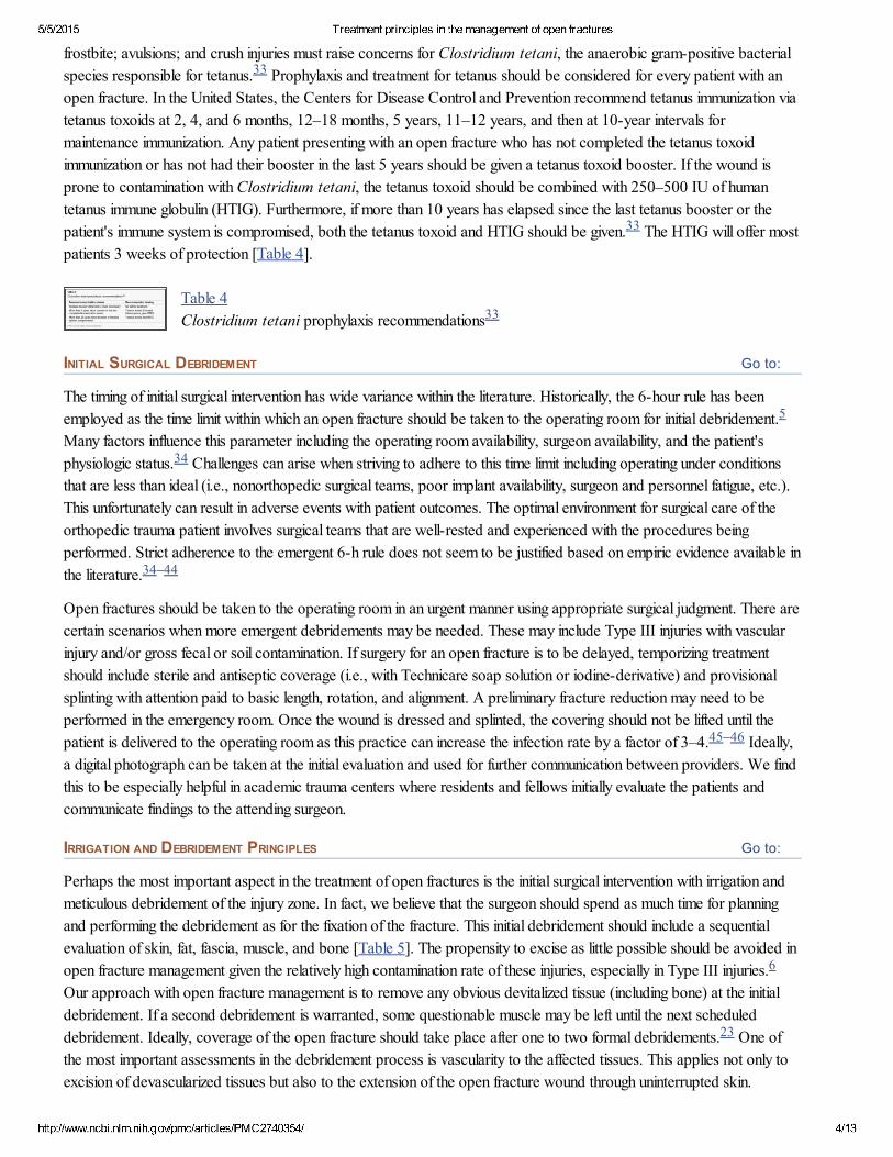
frostbite; avulsions; and crush injuries must raise concerns for Clostridium tetani, the anaerobic grampositive bacterialspecies responsible for tetanus. Prophylaxis and treatment for tetanus should be considered for every patient with anopen fracture. In the United States, the Centers for Disease Control and Prevention recommend tetanus immunization viatetanus toxoids at 2, 4, and 6 months, 12–18 months, 5 years, 11–12 years, and then at 10year intervals formaintenance immunization. Any patient presenting with an open fracture who has not completed the tetanus toxoidimmunization or has not had their booster in the last 5 years should be given a tetanus toxoid booster. If the wound isprone to contamination with Clostridium tetani, the tetanus toxoid should be combined with 250–500 IU of humantetanus immune globulin (HTIG). Furthermore, if more than 10 years has elapsed since the last tetanus booster or thepatient's immune system is compromised, both the tetanus toxoid and HTIG should be given. The HTIG will offer mostpatients 3 weeks of protection [Table 4].
Table 4Clostridium tetani prophylaxis recommendations
INITIAL SURGICAL DEBRIDEMENT
The timing of initial surgical intervention has wide variance within the literature. Historically, the 6hour rule has beenemployed as the time limit within which an open fracture should be taken to the operating room for initial debridement.Many factors influence this parameter including the operating room availability, surgeon availability, and the patient'sphysiologic status. Challenges can arise when striving to adhere to this time limit including operating under conditionsthat are less than ideal (i.e., nonorthopedic surgical teams, poor implant availability, surgeon and personnel fatigue, etc.).This unfortunately can result in adverse events with patient outcomes. The optimal environment for surgical care of theorthopedic trauma patient involves surgical teams that are wellrested and experienced with the procedures beingperformed. Strict adherence to the emergent 6h rule does not seem to be justified based on empiric evidence available inthe literature.
Open fractures should be taken to the operating room in an urgent manner using appropriate surgical judgment. There arecertain scenarios when more emergent debridements may be needed. These may include Type III injuries with vascularinjury and/or gross fecal or soil contamination. If surgery for an open fracture is to be delayed, temporizing treatmentshould include sterile and antiseptic coverage (i.e., with Technicare soap solution or iodinederivative) and provisionalsplinting with attention paid to basic length, rotation, and alignment. A preliminary fracture reduction may need to beperformed in the emergency room. Once the wound is dressed and splinted, the covering should not be lifted until thepatient is delivered to the operating room as this practice can increase the infection rate by a factor of 3–4. Ideally,a digital photograph can be taken at the initial evaluation and used for further communication between providers. We findthis to be especially helpful in academic trauma centers where residents and fellows initially evaluate the patients andcommunicate findings to the attending surgeon.
IRRIGATION AND DEBRIDEMENT PRINCIPLES
Perhaps the most important aspect in the treatment of open fractures is the initial surgical intervention with irrigation andmeticulous debridement of the injury zone. In fact, we believe that the surgeon should spend as much time for planningand performing the debridement as for the fixation of the fracture. This initial debridement should include a sequentialevaluation of skin, fat, fascia, muscle, and bone [Table 5]. The propensity to excise as little possible should be avoided inopen fracture management given the relatively high contamination rate of these injuries, especially in Type III injuries.Our approach with open fracture management is to remove any obvious devitalized tissue (including bone) at the initialdebridement. If a second debridement is warranted, some questionable muscle may be left until the next scheduleddebridement. Ideally, coverage of the open fracture should take place after one to two formal debridements. One ofthe most important assessments in the debridement process is vascularity to the affected tissues. This applies not only toexcision of devascularized tissues but also to the extension of the open fracture wound through uninterrupted skin.
33
33
33
5
34
34–44
45–46
6
23
5/5/2015 Treatment principles in the management of open fractures
http://www.ncbi.nlm.nih.gov/pmc/articles/PMC2740354/ 5/13
Go to:
Knowledge of angiosomes and attention to their patterns can help with avoiding woundhealing complications. Incisionsare optimally placed between angiosomes to prevent devascularizing portions of the wound.
Table 5Debridement principles in open fracture management
Irrigation, along with debridement, is absolutely crucial in the management of open fractures. The removal ofcontaminating debris and the decrease of potentially infective bacterial loads decrease the chances of acute and chronicinfections. Our institution uses a popular protocol that calls for 3 L for a Type I open fracture, 6 L for a Type II openfracture, and 9 L for a Type III open fracture [Table 6]. Surgeons should favor using a low to medium pressure lavagedevice as higherpressure devices have been associated with added tissue or bone damage. Alternatively, if powerirrigation is not available, bulb irrigation may be sufficient. Additives to irrigation have remained a source of controversy.Their use is based mostly on anecdotal reports of beneficial outcomes or avoidance of complications. Recently, a Level Ievidence study found that there is no significant difference between antibiotic and liquid castile soap solutions (TriadMedical, Franklin, Wisconsin) in wound infection or bonehealing rates in the management of open fractures.Interestingly, this same study also found a statistically significant link between woundhealing problems and antibiotic(bacitracin) irrigation. Overall, there is a lack of evidencebased recommendations in the literature to guide surgeons onthe appropriate additives for irrigations.
Table 6Irrigation principles in the open fracture management
TIMING OF WOUND CLOSURE
Options for wound closure in the treatment of open fractures include primary closure of the skin, splitthickness skingrafting, and the use of either free or local muscle flaps. The timing of open wound closure has proponents in theimmediate, early, and delayed categories. Although these terms are used frequently in the literature, their use has yet togain universal acceptance. Traditionally, immediate closure is defined as wound closure at the time of the initial surgicalintervention. Early closure is within the 24–72 h window, and delayed or late closure extends beyond 3 days.Historically, surgeons have opted to delay closure because of the perceived risks of clostridial infections and gasgangrene. This concern is certainly present in the grossly contaminated open fracture. Current treatment strategiescorrectly emphasize the importance of debridement and irrigation, and adhering to these principles has allowed surgeonsto consider earlier closure and immediate primary closure in some cases when certain criteria are met. These have beensuggested to include debridement performed within 12 h, no excess skin loss primarily or secondarily duringdebridement, skin approximation possible without tension, no gross soil or other similar contamination, and no vascularinsufficiency. Recent studies have shown that open fractures are often contaminated with nosocomial organisms (i.e.,Pseudomonas) and that early closure may help prevent these infections. Several studies have examinedimmediate closure of open tibia fractures and have documented that this practice resulted in decreased infection rates,decreased reoperations, and decreased time to bony union.
Our recommendation is toward primary closure of Type I, Type II, and a few selected Type IIIA fractures. The mostimportant factors in our decisionmaking process is the adequacy of the initial debridement and the degree of woundcontamination. If there is any doubt regarding the safety of primary closure, we opt to wait until the second surgicaldebridement and make further treatment decisions at that time. If a primary closure is conducted and there is questionabletissue viability noted postoperatively, we have a very low threshold for reopening the wound 48–72 h after initial closure.If possible, we aim to have coverage completed within 72 h preferably with primary closure. Particular attention must bepaid to tension across the wound closure site. Tension may interfere with wound healing by decreasing the vascularity
47–49
5051–52
53
54
5525,56–61
37,62–64

5/5/2015 Treatment principles in the management of open fractures
http://www.ncbi.nlm.nih.gov/pmc/articles/PMC2740354/ 6/13
Go to:
across the incision. Close relationships with plastic and tissue reconstructive teams can facilitate early closure if flapcoverage is necessary. A valuable adjunct to wound closure has been the wound vacuumassisted closure device (VAC;KCI, San Antonia, TX). It has been shown that this device aids in wound healing by reducing edema, enhancinggranulation tissue formation, and increasing local blood flow [Figure 2]. We utilize this vacuumassisted closureconcept often when immediate closure is not possible although it is important to realize that this method does notnecessarily reduce infection rates or allow a permissible delay in wound closure. The choice between the woundvacuumassisted closure device and the antibiotic beadpouch depends on the degree of wound contamination andsurgeon preference.
Figure 2(a) Clinical photograph of thigh shows Open wound prior to wound vacuumdressing. (b) Open wound appearance after wound vacuum dressing
SKELETAL STABILIZATION
Early stabilization of open fractures provides many benefits to the injured patient. It protects the soft tissues around thezone of injury by preventing further damage from mobile fracture fragments. It also restores length, alignment, androtation—all vital principles of fracture fixation. This restoration of length also helps decrease soft tissue dead spaces andhas been shown in studies to decrease the rates of infection in open fractures. Lastly, early fixation allowsimproved access to soft tissues surrounding the injury and facilitates the patient's early return to normal function. Thesurgeon has many choices when deciding on fixation constructs: skeletal traction, external fixation, and intramedullarynails and plates. The choice of fixation involves the bone fractured and the fracture location (intraarticular, metaphyseal,diaphyseal), the extent of the softtissue injury and the degree of contamination, and the physiologic status of thepatient. There are also certain situations in which more than one method may be used (i.e., fibula plating of a Pilonfracture in which fibula fixation helps restore length and rotation in conjunction with an external fixator placed across thetibialtalar joint).
Skeletal traction and external fixation are the quickest fixation constructs to employ. The use of skeletal traction should bereserved only for selected open fracture types (i.e., pelvis fractures and very proximal femur fractures) and if used, itshould only be for a short selected time. External fixation is a valuable tool in the surgeon's arsenal for acute open fracturemanagement. Indications for external fixation are grossly contaminated open fractures with extensive softtissuecompromise, the Type IIIAC injuries, and when immediate fixation is needed for physiologically unstable patients. Thislater indication involves the damage control concept of orthopedic trauma. When not being used for definitivefixation, external fixation is placed as a spanning construct leaving the zone of injury free of pins and easily accessible forimaging studies and future fixation. The surgeon should also be cognizant of future incision placement and avoid placingexternal fixation pins in these areas.
Plate fixation is generally indicated for open upper extremity fractures and periarticular fractures where reconstruction ofthe articular surface is paramount. The exception to early periarticular fixation is when a staged protocol is being used forextensive articular and softtissue involvement. Higher infections rates have been reported with plate fixation of openfractures, so diligence is needed when the decision is made to use plates. Current plating technology and lessinvasive techniques are lowering these rates and providing patients with good to excellent results.
Intramedullary nail fixation remains the mainstay of treatment for most open tibial shaft fractures and for selected femoralfractures. A recent study showed that more than 88% of surgeons use an intramedullary nail for open Type I and II tibialshaft fractures. Interestingly, this number decreases to 68% for Type IIIA and to 48% for Type IIIB fractures. The
65–6970,71
69,72
40,73,7475
23
76–79
80,8182,83
84–88
89
5/5/2015 Treatment principles in the management of open fractures
http://www.ncbi.nlm.nih.gov/pmc/articles/PMC2740354/ 7/13
Go to:
Go to:
Go to:
choice in the latter is external fixation. There has been considerable debate in the literature regarding reamed andnonreamed intramedullary nails with proponents for both methods. In an effort to answer this question, one of the largeststudies in orthopedic trauma surgery was recently completed. The Study to Prospectively evaluate ReamedIntramedullary Nails in Tibial fractures (SPRINT) enrolled more than 1300 patients and randomized them to reamed ornonreamed tibial nails. There were 400 open fractures enrolled in the study, and the major end point was reoperation.They found a 27% risk of revision in open fractures, regardless of the treatment used. Although not statistically significant,a trend was noted toward the need for revision surgery SPRINT (P = 0.16) when reamed nails were used in openfractures. It is important to note that study design did not allow any reoperations within 6 months of the indexprocedure. We continue to promote this recommendation at our institutions.
Conversion from external fixation to an intramedullary nail has received considerable attention in the literature. Originalreporting of this conversion had alarming results with infection and nonunion rates of 44 and 50%, respectively.Subsequent studies have demonstrated better results. Conclusions from these studies seem to indicate thatconversion from external fixation to an intramedullary nail is safe given two parameters: conversion in less than 2 weeksand absence of pin site infections. Conversion after pin site infections may require additional time and antibiotic treatmentafter removing the external fixator and placement of the intramedullary nail. We use this conversion frequently forcomplex trauma and Type III open fractures.
ADJUNCTIVE THERAPIES
An inherent risk in the treatment of open fractures is the occurrence of a nonunion. This is typically defined as a lack ofosseous union across three cortices as seen by radiographs 9 months postoperatively. This risk has been well quantifiedin the literature, especially with regard to open tibia fractures, with rates ranging from 5% for Type I open fractures and18–38% for Type IIIAC fractures. This statistic is not surprising in that the open fractures release theirvaluable fracture hematoma through the fracture site, which drastically reduces the concentration of valuable postinjuryhealing factors. There has been intense study into adjunctive therapies to assist the surgeon on the management andprevention of nonunions. With open fracture management, adjunctive therapies include prophylactic bone grafting and theapplication of bone morphogenic proteins (BMPs) at the initial operation.
In the largest study to date using BMPs, the BMP2 Evaluation in Surgery for Tibia Trauma (BESST) trial, demonstratedthat BMP2 can be used safely in open fractures. In the study of 145 open tibia fractures, there was a 44% reduction insecondary surgeries. Certainly not all fractures warrant BMP application, and the high costs associated with BMPsplay a major role in its utilization. At our institution, the use of BMPs in acute fracture management is limited to selectedGustilo Type III tibia fractures in patients with significant comorbidities that may impede fracture healing (diabetes,tobacco use, etc.).
Prophylactic bone grafting can also be used in the early treatment of open fractures. The literature has several examplesof studies pertaining to immediate or early prophylactic bone grafting, and this practice has reported to shorten the time tofracture union and reduce the rate of delayed union by more than 11 weeks. The utilization of prophylactic bonegrafting is not routine at our hospital, and we do not typically intervene before 6 months postoperatively.
CONCLUSIONS
The above review provides a framework that the surgeon can reference when treating patients with open fractures. Themanagement of open fractures involves the adherence to principles discussed earlier. Using a principlebased treatmentregimen can help improve patient outcomes while avoiding complications and adverse events. Ultimately, this is thesurgeon's goal, and patients will benefit from the early return to normal function.
Footnotes
Source of Support: Nil
Conflict of Interest: None.
89
90
90
9192–95
92
62,96–100
101
102
103–106

5/5/2015 Treatment principles in the management of open fractures
http://www.ncbi.nlm.nih.gov/pmc/articles/PMC2740354/ 8/13
Go to:REFERENCES
1. Praemer A, Furner S, Rice DP. Musculoskeletal conditions in the United States. Park Ridge, Ill: American Academyof Orthopaedic Surgeons; 1992.
2. Information about Orthopaedic Patients and Conditions AAOS. 2008. (4/8/2008)
3. CourtBrown CM, McQueen MM, Quaba AA. Management of open fractures. St. Louis; London: Mosby; MDunitz; 1996.
4. CourtBrown CM, Rimmer S, Prakash U, McQueen MM. The epidemiology of open long bone fractures. Injury.1998;29:529–34. [PubMed]
5. Gustilo RB, Anderson JT. Prevention of infection in the treatment of one thousand and twentyfive open fractures oflong bones: Retrospective and prospective analyses. J Bone Joint Surg Am. 1976;58:453–8. [PubMed]
6. Gustilo RB, Mendoza RM, Williams DN. Problems in the management of type III (severe) open fractures: A newclassification of type III open fractures. J Trauma. 1984;24:742–6. [PubMed]
7. Tscherne H, Oestern HJ. A new classification of softtissue damage in open and closed fractures. Unfallheilkunde.1982;85:111–5. [PubMed]
8. Johansen K, Daines M, Howey T, Helfet D, Hansen ST., Jr Objective criteria accurately predict amputation followinglower extremity trauma. J Trauma. 1990;30:568–72. [PubMed]
9. Gustilo RB, Gruninger RP, Davis T. Classification of type III (severe) open fractures relative to treatment and results.Orthopedics. 1987;10:1781–8. [PubMed]
10. Brumback RJ, Jones AL. Interobserver agreement in the classification of open fractures of the tibia: The results of asurvey of two hundred and fortyfive orthopaedic surgeons. J Bone Joint Surg Am. 1994;76:1162–6. [PubMed]
11. Helfet DL, Howey T, Sanders R, Johansen K. Limb salvage versus amputation: Preliminary results of the MangledExtremity Severity Score. Clin Orthop Relat Res. 1990;256:80–6. [PubMed]
12. Slauterbeck JR, Britton C, Moneim MS, Clevenger FW. Mangled extremity severity score: An accurate guide totreatment of the severely injured upper extremity. J Orthop Trauma. 1994;8:282–5. [PubMed]
13. Durham RM, Mistry BM, Mazuski JE, Shapiro M, Jacobs D. Outcome and utility of scoring systems in themanagement of the mangled extremity. Am J Surg. 1996;172:569–73. [PubMed]
14. O'Sullivan ST, O'Sullivan M, Pasha N, O'Shaughnessy M, O'Connor TP. Is it possible to predict limb viability incomplex Gustilo IIIB and IIIC tibial fractures? A comparison of two predictive indices. Injury. 1997;28:639–42.[PubMed]
15. Fagelman MF, Epps HR, Rang M. Mangled extremity severity score in children. J Pediatr Orthop. 2002;22:182–4.[PubMed]
16. Sharma S, Devgan A, Marya KM, Rathee N. Critical evaluation of mangled extremity severity scoring system inIndian patients. Injury. 2003;34:493–6. [PubMed]
17. Elsharawy MA. Arterial reconstruction after mangled extremity: Injury severity scoring systems are not predictive oflimb salvage. Vascular. 2005;13:114–9. [PubMed]
18. Togawa S, Yamami N, Nakayama H, Mano Y, Ikegami K, Ozeki S. The validity of the mangled extremity severityscore in the assessment of upper limb injuries. J Bone Joint Surg Br. 2005;87:1516–9. [PubMed]
19. Rush RM, Jr, Kjorstad R, Starnes BW, Arrington E, Devine JD, Andersen CA. Application of the Mangled
5/5/2015 Treatment principles in the management of open fractures
http://www.ncbi.nlm.nih.gov/pmc/articles/PMC2740354/ 9/13
Extremity Severity Score in a combat setting. Mil Med. 2007;172:777–81. [PubMed]
20. MacKenzie EJ, Bosse MJ, Kellam JF, Burgess AR, Webb LX, Swiontkowski MF, et al., editors. Factorsinfluencing the decision to amputate or reconstruct after highenergy lower extremity trauma. J Trauma. 2002;52:641–9.[PubMed]
21. Bosse MJ, McCarthy ML, Jones AL, Webb LX, Sims SH, Sanders RW, et al., editors. The insensate foot followingsevere lower extremity trauma: An indication for amputation? J Bone Joint Surg Am. 2005;87:2601–8. [PubMed]
22. Dougherty PJ. Open tibia fracture: Amputation versus limb salvage, Opinion: belowtheknee amputation. J OrthopTrauma. 2007;21:67–8. [PubMed]
23. Zalavras CG, Marcus RE, Levin LS, Patzakis MJ. Management of open fractures and subsequent complications. JBone Joint Surg Am. 2007;89:884–95. [PubMed]
24. Patzakis MJ, Wilkins J. Factors influencing infection rate in open fracture wounds. Clin Orthop Relat Res.1989;243:36–40. [PubMed]
25. Templeman DC, Gulli B, Tsukayama DT, Gustilo RB. Update on the management of open fractures of the tibialshaft. Clin Orthop Relat Res. 1998;350:18–25. [PubMed]
26. Patzakis MJ, Wilkins J, Moore TM. Considerations in reducing the infection rate in open tibial fractures. Clin OrthopRelat Res. 1983;178:36–41. [PubMed]
27. Patzakis MJ, Wilkins J, Moore TM. Use of antibiotics in open tibial fractures. Clin Orthop Relat Res. 1983;178:31–5. [PubMed]
28. Dellinger EP, Caplan ES, Weaver LD, Wertz MJ, Droppert BM, Hoyt N, et al., editors. Duration of preventiveantibiotic administration for open extremity fractures. Arch Surg. 1988;123:333–9. [PubMed]
29. Dellinger EP, Miller SD, Wertz MJ, Grypma M, Droppert B, Anderson PA. Risk of infection after open fracture ofthe arm or leg. Arch Surg. 1988;123:1320–7. [PubMed]
30. Zalavras CG, Patzakis MJ. Open fractures: Evaluation and management. J Am Acad Orthop Surg. 2003;11:212–9.[PubMed]
31. Zalavras CG, Patzakis MJ, Holtom P. Local antibiotic therapy in the treatment of open fractures and osteomyelitis.Clin Orthop Relat Res. 2004;427:86–93. [PubMed]
32. Ostermann PA, Seligson D, Henry SL. Local antibiotic therapy for severe open fractures. A review of 1085consecutive cases. J Bone Joint Surg Br. 1995;77:93–7. [PubMed]
33. Bleck TP. Clostridum tetani (Tetanus) In: Mandell G, Douglas R, Bennett J, editors. Principles and Practice ofInfectious Diseases. 6th ed. Philadelphia: Churchill Livingstone; 2005.
34. Pollak AN. Timing of debridement of open fractures. J Am Acad Orthop Surg. 2006;14:S48–51. [PubMed]
35. Ashford RU, Mehta JA, Cripps R. Delayed presentation is no barrier to satisfactory outcome in the management ofopen tibial fractures. Injury. 2004;35:411–6. [PubMed]
36. Bednar DA, Parikh J. Effect of time delay from injury to primary management on the incidence of deep infection afteropen fractures of the lower extremities caused by blunt trauma in adults. J Orthop Trauma. 1993;7:532–5. [PubMed]
37. Gopal S, Majumder S, Batchelor AG, Knight SL, De Boer P, Smith RM. Fix and flap: The radical orthopaedic andplastic treatment of severe open fractures of the tibia. J Bone Joint Surg Br. 2000;82:959–66. [PubMed]
38. Harley BJ, Beaupre LA, Jones CA, Dulai SK, Weber DW. The effect of time to definitive treatment on the rate of

5/5/2015 Treatment principles in the management of open fractures
http://www.ncbi.nlm.nih.gov/pmc/articles/PMC2740354/ 10/13
nonunion and infection in open fractures. J Orthop Trauma. 2002;16:484–90. [PubMed]
39. Khatod M, Botte MJ, Hoyt DB, Meyer RS, Smith JM, Akeson WH. Outcomes in open tibia fractures: Relationshipbetween delay in treatment and infection. J Trauma. 2003;55:949–54. [PubMed]
40. Merritt K. Factors increasing the risk of infection in patients with open fractures. J Trauma. 1988;28:823–7.[PubMed]
41. Sungaran J, Harris I, Mourad M. The effect of time to theatre on infection rate for open tibia fractures. ANZ J Surg.2007;77:886–8. [PubMed]
42. Reuss BL, Cole JD. Effect of delayed treatment on open tibial shaft fractures. Am J Orthop. 2007;36:215–20.[PubMed]
43. Crowley DJ, Kanakaris NK, Giannoudis PV. Debridement and wound closure of open fractures: The impact of thetime factor on infection rates. Injury. 2007;38:879–89. [PubMed]
44. Webb LX, Bosse MJ, Castillo RC, MacKenzie EJ. LEAP Study Group; Analysis of surgeoncontrolled variables inthe treatment of limbthreatening typeIII open tibial diaphyseal fractures. J Bone Joint Surg Am. 2007;89:923–8.[PubMed]
45. Browner BD. Skeletal trauma: Basic science, management, and reconstruction. 3rd ed. Philadelphia: Saunders;2003.
46. Tscherne H, Gotzen L. Fractures with soft tissue injuries. Berlin; New York: SpringerVerlag; 1984.
47. Taylor GI, Palmer JH. The vascular territories (angiosomes) of the body: Experimental study and clinical applications.Br J Plast Surg. 1987;40:113–41. [PubMed]
48. Attinger CE, Evans KK, Bulan E, Blume P, Cooper P. Angiosomes of the foot and ankle and clinical implications forlimb salvage: Reconstruction, incisions, and revascularization. Plast Reconstr Surg. 2006;117:261S–93S. [PubMed]
49. Taylor GI. The angiosomes of the body and their supply to perforator flaps. Clin Plast Surg. 2003;30:331–42.[PubMed]
50. Anglen JO. Wound irrigation in musculoskeletal injury. J Am Acad Orthop Surg. 2001;9:219–26. [PubMed]
51. Bhandari M, Schemitsch EH, Adili A, Lachowski RJ, Shaughnessy SG. High and low pressure pulsatile lavage ofcontaminated tibial fractures: An in vitro study of bacterial adherence and bone damage. J Orthop Trauma.1999;13:526–33. [PubMed]
52. Bhandari M, Thompson K, Adili A, Shaughnessy SG. High and low pressure irrigation in contaminated wounds withexposed bone. Int J Surg Investig. 2000;2:179–82. [PubMed]
53. Anglen JO. Comparison of soap and antibiotic solutions for irrigation of lowerlimb open fracture wounds: Aprospective, randomized study. J Bone Joint Surg Am. 2005;87:1415–22. [PubMed]
54. Levin LS. Early versus delayed closure of open fractures. Injury. 2007;38:896–9. [PubMed]
55. Rajasekaran S. Early versus delayed closure of open fractures. Injury. 2007;38:890–5. [PubMed]
56. CarsentiEtesse H, Doyon F, Desplaces N, Gagey O, Tancrede C, Pradier C, et al., editors. Epidemiology ofbacterial infection during management of open leg fractures. Eur J Clin Microbiol Infect Dis. 1999;18:315–23. [PubMed]
57. Byrd HS, Spicer TE, Cierney G., 3rd Management of open tibial fractures. Plast Reconstr Surg. 1985;76:719–30.[PubMed]
58. Cierny G, 3rd, Byrd HS, Jones RE. Primary versus delayed soft tissue coverage for severe open tibial fractures: A
5/5/2015 Treatment principles in the management of open fractures
http://www.ncbi.nlm.nih.gov/pmc/articles/PMC2740354/ 11/13
comparison of results. Clin Orthop Relat Res. 1983;178:54–63. [PubMed]
59. Caudle RJ, Stern PJ. Severe open fractures of the tibia. J Bone Joint Surg Am. 1987;69:801–7. [PubMed]
60. Fischer MD, Gustilo RB, Varecka TF. The timing of flap coverage, bonegrafting, and intramedullary nailing inpatients who have a fracture of the tibial shaft with extensive softtissue injury. J Bone Joint Surg Am. 1991;73:1316–22.[PubMed]
61. Sinclair JS, McNally MA, Small JO, Yeates HA. Primary freeflap cover of open tibial fractures. Injury.1997;28:581–7. [PubMed]
62. DeLong WG, Jr, Born CT, Wei SY, Petrik ME, Ponzio R, Schwab CW. Aggressive treatment of 119 open fracturewounds. J Trauma. 1999;46:1049–54. [PubMed]
63. Hertel R, Lambert SM, Muller S, Ballmer FT, Ganz R. On the timing of softtissue reconstruction for open fracturesof the lower leg. Arch Orthop Trauma Surg. 1999;119:7–12. [PubMed]
64. Godina M. Early microsurgical reconstruction of complex trauma of the extremities. Plast Reconstr Surg.1986;78:285–92. [PubMed]
65. Herscovici D, Jr, Sanders RW, Scaduto JM, Infante A, Di Pasquale T. Vacuumassisted wound closure (VACtherapy) for the management of patients with highenergy soft tissue injuries. J Orthop Trauma. 2003;17:683–8.[PubMed]
66. Tarkin IS, Clare MP, Marcantonio A, Pape HC. An update on the management of highenergy pilon fractures.Injury. 2008;39:142–54. [PubMed]
67. Hardwicke J, Paterson P. A role for vacuumassisted closure in lower limb trauma: A proposed algorithm. Int J LowExtrem Wounds. 2006;5:101–4. [PubMed]
68. Parrett BM, Matros E, Pribaz JJ, Orgill DP. Lower extremity trauma: Trends in the management of softtissuereconstruction of open tibiafibula fractures. Plast Reconstr Surg. 2006;117:1315. [PubMed]
69. Dedmond BT, Kortesis B, Punger K, Simpson J, Argenta J, Kulp B, et al., editors. The use of negativepressurewound therapy (NPWT) in the temporary treatment of softtissue injuries associated with highenergy open tibial shaftfractures. J Orthop Trauma. 2007;21:11–7. [PubMed]
70. Argenta LC, Morykwas MJ. Vacuumassisted closure: a new method for wound control and treatment: Clinicalexperience. Ann Plast Surg. 1997;38:563–76. [PubMed]
71. Morykwas MJ, Argenta LC, SheltonBrown EI, McGuirt W. Vacuumassisted closure: A new method for woundcontrol and treatment: animal studies and basic foundation. Ann Plast Surg. 1997;38:553–62. [PubMed]
72. Bhattacharyya T, Mehta P, Smith M, Pomahac B. Routine use of wound vacuumassisted closure does not allowcoverage delay for open tibia fractures. Plast Reconstr Surg. 2008;121:1263–6. [PubMed]
73. Worlock P, Slack R, Harvey L, Mawhinney R. The prevention of infection in open fractures: An experimental studyof the effect of fracture stability. Injury. 1994;25:31–8. [PubMed]
74. Merritt K, Dowd JD. Role of internal fixation in infection of open fractures: Studies with Staphylococcus aureus andProteus mirabilis. J Orthop Res. 1987;5:23–8. [PubMed]
75. Gustilo RB, Merkow RL, Templeman D. The management of open fractures. J Bone Joint Surg Am. 1990;72:299–304. [PubMed]
76. Hildebrand F, Giannoudis P, Kretteck C, Pape HC. Damage control: Extremities. Injury. 2004;35:678–89.[PubMed]

5/5/2015 Treatment principles in the management of open fractures
http://www.ncbi.nlm.nih.gov/pmc/articles/PMC2740354/ 12/13
77. Pape HC, Hildebrand F, Pertschy S, Zelle B, Garapati R, Grimme K, et al., editors. Changes in the management offemoral shaft fractures in polytrauma patients: From early total care to damage control orthopedic surgery. J Trauma.2002;53:452–62. [PubMed]
78. Pape HC, Krettek C. Damage control orthopaedic surgery. Unfallchirurg. 2003;106:85–6. [PubMed]
79. Roberts CS, Pape HC, Jones AL, Malkani AL, Rodriguez JL, Giannoudis PV. Damage control orthopaedics:Evolving concepts in the treatment of patients who have sustained orthopaedic trauma. Instr Course Lect. 2005;54:447–62. [PubMed]
80. Sirkin M, Sanders R, DiPasquale T, Herscovici D., Jr A staged protocol for soft tissue management in the treatmentof complex pilon fractures. J Orthop Trauma. 2004;18:S32–8. [PubMed]
81. Watson JT, Moed BR, Karges DE, Cramer KE. Pilon fractures: Treatment protocol based on severity of soft tissueinjury. Clin Orthop Relat Res. 2000;375:78–90. [PubMed]
82. Bach AW, Hansen ST., Jr Plates versus external fixation in severe open tibial shaft fractures: A randomized trial. ClinOrthop Relat Res. 1989;241:89–94. [PubMed]
83. Clifford RP, Beauchamp CG, Kellam JF, Webb JK, Tile M. Plate fixation of open fractures of the tibia. J Bone JointSurg Br. 1988;70:644–8. [PubMed]
84. Cole PA, Zlowodzki M, Kregor PJ. Treatment of proximal tibia fractures using the less invasive stabilization system:Surgical experience and early clinical results in 77 fractures. J Orthop Trauma. 2004;18:528–35. [PubMed]
85. Fankhauser F, Gruber G, Schippinger G, Boldin C, Hofer HP, Grechenig W, et al., editors. Minimalinvasivetreatment of distal femoral fractures with the LISS (Less Invasive Stabilization System): A prospective study of 30fractures with a follow up of 20 months. Acta Orthop Scand. 2004;75:56–60. [PubMed]
86. Kregor PJ, Stannard JA, Zlowodzki M, Cole PA. Treatment of distal femur fractures using the less invasivestabilization system: Surgical experience and early clinical results in 103 fractures. J Orthop Trauma. 2004;18:509–20.[PubMed]
87. Stannard JP, Wilson TC, Volgas DA, Alonso JE. Fracture stabilization of proximal tibial fractures with the proximaltibial LISS: Early experience in Birmingham, Alabama (USA) Injury. 2003;34:A36–42. [PubMed]
88. Syed AA, Agarwal M, Giannoudis PV, Matthews SJ, Smith RM. Distal femoral fractures: Longterm outcomefollowing stabilisation with the LISS. Injury. 2004;35:599–607. [PubMed]
89. Bhandari M, Guyatt GH, Tornetta P 3rd, Swiontkowski MF, Hanson B, Sprague S, et al., editors. Current practicein the intramedullary nailing of tibial shaft fractures: An international survey. J Trauma. 2002;53:725–32. [PubMed]
90. Bhandari M. 75th Annual Meeting of the American Academy of Orthopaedic Surgeons; 03/2008. San Francisco,California, USA San Francisco, CA: American Academy of Orthopaedic Surgeons; 2008. Reamed Versus NonReamed Tibial Intramedullary Nail Insertion on Reoperation Rates.
91. McGraw JM, Lim EV. Treatment of open tibialshaft fractures: External fixation and secondary intramedullary nailing.J Bone Joint Surg Am. 1988;70:900–11. [PubMed]
92. Nowotarski PJ, Turen CH, Brumback RJ, Scarboro JM. Conversion of external fixation to intramedullary nailing forfractures of the shaft of the femur in multiply injured patients. J Bone Joint Surg Am. 2000;82:781–8. [PubMed]
93. Blachut PA, Meek RN, O'Brien PJ. External fixation and delayed intramedullary nailing of open fractures of the tibialshaft: A sequential protocol. J Bone Joint Surg Am. 1990;72:729–35. [PubMed]
94. Dougherty PJ, Silverton C, Yeni Y, Tashman S, Weir R. Conversion from temporary external fixation to definitive

5/5/2015 Treatment principles in the management of open fractures
http://www.ncbi.nlm.nih.gov/pmc/articles/PMC2740354/ 13/13
fixation: Shaft fractures. J Am Acad Orthop Surg. 2006;14:S124–7. [PubMed]
95. Maurer DJ, Merkow RL, Gustilo RB. Infection after intramedullary nailing of severe open tibial fractures initiallytreated with external fixation. J Bone Joint Surg Am. 1989;71:835–8. [PubMed]
96. Hope PG, Cole WG. Open fractures of the tibia in children. J Bone Joint Surg Br. 1992;74:546–53. [PubMed]
97. Whorton AM, Henley MB. The role of fixation of the fibula in open fractures of the tibial shaft with fractures of theipsilateral fibula: Indications and outcomes. Orthopedics. 1998;21:1101–5. [PubMed]
98. Stegemann P, Lorio M, Soriano R, Bone L. Management protocol for unreamed interlocking tibial nails for opentibial fractures. J Orthop Trauma. 1995;9:117–20. [PubMed]
99. Bhandari M, Guyatt GH, Swiontkowski MF, Schemitsch EH. Treatment of open fractures of the shaft of the tibia. JBone Joint Surg Br. 2001;83:62–8. [PubMed]
100. CourtBrown CM, Keating JF, Christie J, McQueen MM. Exchange intramedullary nailing: Its use in aseptic tibialnonunion. J Bone Joint Surg Br. 1995;77:407–11. [PubMed]
101. Govender S, Csimma C, Genant HK, ValentinOpran A, Amit Y, Arbel R, et al., editors. Recombinant humanbone morphogenetic protein2 for treatment of open tibial fractures: A prospective, controlled, randomized study of fourhundred and fifty patients. J Bone Joint Surg Am. 2002;84:2123–34. [PubMed]
102. Swiontkowski MF, Aro HT, Donell S, Esterhai JL, Goulet J, Jones A, et al., editors. Recombinant human bonemorphogenetic protein2 in open tibial fractures: A subgroup analysis of data combined from two prospectiverandomized studies. J Bone Joint Surg Am. 2006;88:1258–65. [PubMed]
103. Blick SS, Brumback RJ, Lakatos R, Poka A, Burgess AR. Early prophylactic bone grafting of highenergy tibialfractures. Clin Orthop Relat Res. 1989;240:21–41. [PubMed]
104. Kesemenli CC, Kapukaya A, Subasi M, Arslan H, Necmioglu S, Kayikci C. Early prophylactic autogenous bonegrafting in type III open tibial fractures. Acta Orthop Belg. 2004;70:327–31. [PubMed]
105. Kobbe P, Frink M, Oberbeck R, Tarkin IS, Tzioupis C, NastKolb D, et al., editors. Treatment strategies forgunshot wounds of the extremities. Unfallchirurg. 2008;111:247–55. [PubMed]
106. Trabulsy PP, Kerley SM, Hoffman WY. A prospective study of early soft tissue coverage of grade IIIB tibialfractures. J Trauma. 1994;36:661–8. [PubMed]
Articles from Indian Journal of Orthopaedics are provided here courtesy of Medknow Publications
Related Documents