doi:10.1182/blood-2002-01-0172 Prepublished online June 7, 2002; Webb and Gritta Janka Alexandra H Filipovich, Helmut Gadner, Shinsaku Imashuku, Diane Komp, Stephan Ladisch, David Jan-Inge Henter, AnnaCarin Samuelsson-Horne, Maurizio Arico, R M Egeler, Goran Elinder, immuno-chemotherapy and bone marrow transplantation Treatment of hemophagocytic lymphohistiocytosis with HLH-94 (3722 articles) Clinical Trials and Observations Articles on similar topics can be found in the following Blood collections http://bloodjournal.hematologylibrary.org/site/misc/rights.xhtml#repub_requests Information about reproducing this article in parts or in its entirety may be found online at: http://bloodjournal.hematologylibrary.org/site/misc/rights.xhtml#reprints Information about ordering reprints may be found online at: http://bloodjournal.hematologylibrary.org/site/subscriptions/index.xhtml Information about subscriptions and ASH membership may be found online at: articles must include the digital object identifier (DOIs) and date of initial publication. priority; they are indexed by PubMed from initial publication. Citations to Advance online prior to final publication). Advance online articles are citable and establish publication yet appeared in the paper journal (edited, typeset versions may be posted when available Advance online articles have been peer reviewed and accepted for publication but have not Copyright 2011 by The American Society of Hematology; all rights reserved. Washington DC 20036. by the American Society of Hematology, 2021 L St, NW, Suite 900, Blood (print ISSN 0006-4971, online ISSN 1528-0020), is published weekly For personal use only. by guest on June 7, 2013. bloodjournal.hematologylibrary.org From

Welcome message from author
This document is posted to help you gain knowledge. Please leave a comment to let me know what you think about it! Share it to your friends and learn new things together.
Transcript

doi:10.1182/blood-2002-01-0172Prepublished online June 7, 2002;
Webb and Gritta JankaAlexandra H Filipovich, Helmut Gadner, Shinsaku Imashuku, Diane Komp, Stephan Ladisch, David Jan-Inge Henter, AnnaCarin Samuelsson-Horne, Maurizio Arico, R M Egeler, Goran Elinder, immuno-chemotherapy and bone marrow transplantationTreatment of hemophagocytic lymphohistiocytosis with HLH-94
(3722 articles)Clinical Trials and Observations �Articles on similar topics can be found in the following Blood collections
http://bloodjournal.hematologylibrary.org/site/misc/rights.xhtml#repub_requestsInformation about reproducing this article in parts or in its entirety may be found online at:
http://bloodjournal.hematologylibrary.org/site/misc/rights.xhtml#reprintsInformation about ordering reprints may be found online at:
http://bloodjournal.hematologylibrary.org/site/subscriptions/index.xhtmlInformation about subscriptions and ASH membership may be found online at:
articles must include the digital object identifier (DOIs) and date of initial publication. priority; they are indexed by PubMed from initial publication. Citations to Advance online prior to final publication). Advance online articles are citable and establish publicationyet appeared in the paper journal (edited, typeset versions may be posted when available Advance online articles have been peer reviewed and accepted for publication but have not
Copyright 2011 by The American Society of Hematology; all rights reserved.Washington DC 20036.by the American Society of Hematology, 2021 L St, NW, Suite 900, Blood (print ISSN 0006-4971, online ISSN 1528-0020), is published weekly
For personal use only. by guest on June 7, 2013. bloodjournal.hematologylibrary.orgFrom

Treatment of Hemophagocytic Lymphohistiocytosis
with HLH-94 Immuno-Chemotherapy
and Bone Marrow Transplantation
Jan-Inge Henter1,MD, PhD, AnnaCarin Samuelsson-Horne1, MD, Maurizio Aricò3, MD, R
Maarten Egeler4, MD, PhD, Göran Elinder2, MD, PhD, Alexandra H Filipovich5, MD, Helmut
Gadner6, MD, Shinsaku Imashuku7, MD, Diane Komp8, MD, Stephan Ladisch9, MD, David
Webb10, MD, Gritta Janka11, MD; for the Histiocyte Society.
1Childhood Cancer Research Unit, Karolinska Institutet, Department of Pediatric Hematology
and Oncology, Karolinska Hospital, Stockholm, Sweden; 2Department of Pediatrics,
Stockholm Söder Hospital, Karolinska Institutet, Stockholm, Sweden; 3Onco Ematologia
Pediatrica, Ospedale dei Bambini G di Cristina, Palermo, Italy; 4Department of Pediatrics,
Leiden University Medical Center, Leiden, the Netherlands; 5Children’s Hospital Medical
Center, Cincinnati OH, USA; 6St Anna Children´s Hospital, Vienna, Austria; 7Children’s
Research Hospital, Kyoto Prefectural University of Medicine, Japan; 8Department of
Pediatrics, Yale University School of Medicine, New Haven CT, USA; 9Children’s Research
Institute, Washington DC, USA; 10Great Ormond Street Hospital, London, UK; 11Department
of Hematology and Oncology, Children´s University Hospital, Hamburg, Germany
Running Head: Treatment of HLH.
Correspondence and reprints: Jan-Inge Henter, MD, PhD
Childhood Cancer Research Unit Q6:05, Karolinska Hospital, S-171 76 Stockholm, Sweden
Tel: +46 8 5177 2870; Fax: +46 8 5177 3184; e-mail: [email protected]
Copyright 2002 American Society of Hematology
Blood First Edition Paper, prepublished online June 7, 2002; DOI 10.1182/blood-2002-01-0172 For personal use only. by guest on June 7, 2013. bloodjournal.hematologylibrary.orgFrom

2
Supported by: The Children's Cancer Foundation of Sweden; the Medical Research Council
of Sweden (#12440); the Cancer Foundation of Sweden; the Ronald McDonald Foundation;
the Märta and Gunnar V Philipson Foundation; The Cancer and Allergy Foundation of
Sweden; Telethon Italy (#E755) (MA), IRCCS Policlinico San Matteo, Pavia (Ricerca
Corrente 390 RCR97/01 and #80291) (MA); and the Histiocytosis Association of America.
Word counts: Text (excluding tables, figure legends, and tables): 3564. Abstract: 223.
Suggested scientific heading: Clinical Observations, Interventions and Therapeutic Trials
For personal use only. by guest on June 7, 2013. bloodjournal.hematologylibrary.orgFrom

3
ABSTRACT
Background: Hemophagocytic lymphohistiocytosis (HLH) comprises familial (primary)
hemophagocytic lymphohistiocytosis (FHL) and secondary HLH (SHLH), both clinically
characterized by fever, hepatosplenomegaly, and cytopenia. FHL, an autosomal recessive
disease invariably fatal when untreated, is associated with defective triggering of apoptosis
and reduced cytotoxic activity, resulting in a widespread accumulation of T-lymphocytes and
activated macrophages. Methods: In 1994 the Histiocyte Society initiated a prospective
international collaborative therapeutic study (HLH-94), aiming at improved survival. It
combined chemotherapy and immunotherapy (etoposide, corticosteroids, cyclosporin A, and,
in selected patients, intrathecal methotrexate), followed by bone marrow transplantation
(BMT) in persistent, recurring and/or familial disease. Results: 113 eligible patients aged ≤15
years from 21 countries started HLH-94 between July 1, 1994 and June 30, 1998. They all
either had an affected sibling (n=25) and/or fulfilled the Histiocyte Society diagnostic criteria.
At a median follow-up of 3.1 years, the estimated 3-year probability of survival overall was
55% (95% confidence interval +/- 9%) and in the familial cases 51% (+/-20%). Twenty
enrolled children were alive and off-therapy for >12 months without BMT. For patients who
were transplanted (n=65), died prior to BMT (n=25) or were still on therapy (n=3), the 3-year
survival was 45% (+/-10%). The 3-year probability of survival after BMT was 62% (+/-12%).
Conclusions: HLH-94 is very effective, allowing BMT in most patients. Survival of children
with HLH has been greatly improved.
E-mail to the corresponding author: [email protected]
Key words: Familial hemophagocytic lymphohistiocytosis, treatment, etoposide,
cyclosporin A, dexamethasone, methotrexate, bone marrow transplantation, survival
For personal use only. by guest on June 7, 2013. bloodjournal.hematologylibrary.orgFrom

4
INTRODUCTION
Hemophagocytic lymphohistiocytosis (HLH) represents a spectrum of inherited and acquired
conditions with disturbed immune regulation of different severity, and it encompasses two
main conditions that have common clinical and pathobiological characteristics: familial
(primary) hemophagocytic lymphohistiocytosis (FHL) and secondary hemophagocytic
lymphohistiocytosis (SHLH). In contrast to SHLH, which may affect any age and which may
subside spontaneously, FHL is an invariably fatal inherited disease mostly seen in infancy and
early childhood (1-3). The annual childhood incidence of FHL has been estimated (in
Sweden) at 1.2 cases per 1,000,000, corresponding to 1:50,000 births (4). Common findings
include fever, hepatosplenomegaly, pancytopenia, and reduced cytotoxic T- and NK-cell
activity, as well as a widespread accumulation of T-lymphocytes and macrophages, some of
which may engage in hemophagocytosis (1-5). Central nervous system (CNS) involvement is
frequent, ranging from irritability, bulging fontanel, and neck stiffness, to convulsions, cranial
nerve palsies, ataxia, psychomotor retardation and coma (6-9).
A hypercytokinemia, mainly involving proinflammatory cytokines, mediates the clinical and
laboratory findings (10-14). A defective triggering of apoptosis in FHL was recently
suggested as the underlying pathophysiologic mechanism (15). In 1999, genetic studies
showed linkage to the chromosome regions 9q21.3-22 and 10q21-22 for some, but not all,
patients (16-18). Recent studies revealed perforin gene defects in 10q21-22-linked FHL-
patients (19-21), supporting the hypothesis that FHL, at least in some patients, is caused by a
deficiency in the triggering of apoptosis (15, 19-23). Of differential diagnostic importance is
that a few male patients with HLH recently have been shown to harbor germline mutations in
SH2D1A/SAP, the gene causing X-linked lymphoproliferative syndrome (XLP) (24).
For personal use only. by guest on June 7, 2013. bloodjournal.hematologylibrary.orgFrom

5
Chemotherapy with epipodophyllotoxin derivatives, etoposide (VP-16) and teniposide (VM-
26), combined with corticosteroids and intrathecal methotrexate (IT MTX) induce remission
in FHL (25-27). Remission can also be achieved with immunotherapy, i.e. antithymocyte
globulin (ATG) and steroids followed by cyclosporin A (CSA) (28). Ultimately, all FHL
patients relapsed and died until Fischer and coworkers showed that cure could be achieved
through allogeneic BMT (29), which was later confirmed by others (30-32).
Despite these improvements in treatment, multiple problems remained including death prior
to or during remission (2). Prompted by these therapeutic difficulties, in 1994 the Histiocyte
Society developed a treatment strategy (HLH-94) which combines (rather than randomizes
between) two previously reported regimens, chemotherapy (33) and immunotherapy (30).
HLH-94 is based on VP-16, corticosteroids, CSA, and, in selected patients, IT MTX, prior to
intended BMT (34). Herein we present results for 113 patients, recruited during a 4-year
period, with a major focus on survival and outcome.
MATERIAL AND METHODS
Treatment protocol
The HLH-94 treatment protocol includes 8 weeks of initial therapy, aiming at achieving a
clinical remission, followed by a continuation therapy aiming at keeping the children alive
and stable until an acceptable BMT donor was available (Fig 1). The initial therapy consists
of VP-16 (150 mg/m2 twice weekly for 2 weeks and then weekly) and dexamethasone
(initially 10 mg/m2 for 2 weeks followed by 5 mg/m2 for 2 weeks, 2.5 mg/m2 for 2 weeks,
1.25 mg/m2 for one week, and one week of tapering). After 8 weeks of initial treatment, it was
recommended that children with known familial disease or persistent non-familial disease
proceed to continuation therapy and BMT whereas children with resolved non-familial
For personal use only. by guest on June 7, 2013. bloodjournal.hematologylibrary.orgFrom
6
disease ceased therapy and restarted HLH-94 only in case of reactivation, in order to avoid
prolonged therapy and BMT for patients with presumably SHLH (Fig 2). The continuation
therapy from week 9 and onwards comprised dexamethasone pulses 10 mg/m2 for three days
every second week and VP-16 infusions 150 mg/m2 every alternating second week in
combination with daily oral CSA aiming at trough levels of 200 microgram/L. An important
role for the continuation therapy is to keep the children alive and in a stable condition during
the search of a marrow donor. IT MTX was administered at a maximum of four doses, but
was recommended only if there were progressive neurological symptoms or if an abnormal
CSF had not improved. Recommended supportive therapy included antimycotic treatment
during the initial dexamethasone therapy and continuous cotrimoxazole treatment, equivalent
to 5 mg/kg of trimethoprim three times weekly, as Pneumocystis Carinii prophylaxis.
It is often not possible to differentiate between inherited HLH and SHLH already at diagnosis,
unless there is already an affected child in the family. Most children with the inherited form of
the disease will appear as sporadic cases since the inheritance is autosomal recessive.
Moreover, an association with a viral infection cannot be used to justify the diagnosis of
SHLH, since such infections may be concomitant with the onset of FHL and even trigger the
disease (4,35-36). Because of these difficulties, children with severe or persistent disease are
recommended to start HLH- therapy also if there is no evidence of familial disease. In patients
without family history but with relapsing or non-responding disease, an underlying inherited
defect is most likely, as in the verified familial ones. On the contrary, in responding patients
that do not relapse when treatment is stopped, a secondary cause is likely. Genetic analyses
may provide a distinct diagnosis (16-21), but the results are rarely available at onset of the
disease. Moreover, the genetic explanation is presently known only for a minority of the
patients (20).
For personal use only. by guest on June 7, 2013. bloodjournal.hematologylibrary.orgFrom

7
BMT
The conditioning regimen and the graft-vs-host-disease (GVHD) prophylaxis were
determined by the treating transplantation unit, but a suggested protocol was provided. The
suggested conditioning consisted of busulfan 4 mg/kg on days -9, -8, -7, and -6,
cyclophosphamide 50 mg/kg on days -5, -4, -3, and -2, and VP-16 300 mg/m2 on days -5, -4,
and -3. In case of unrelated donor transplants, additional immunosuppression with horse ATG
was suggested, 15 mg/kg twice daily on days -2 and -1, and once daily on days +1 and +2.
GVHD prophylaxis included intravenous methotrexate 15 mg/m2 on day +1 and 10 mg/m2 on
days +3, +5 and +11, in combination with CSA beginning on day -3.
Patients
Altogether 119 children aged ≤ 15 years who had not received any previous cytotoxic or CSA
therapy and who were either familial cases or fulfilled the diagnostic criteria approved by the
Histiocyte Society (Table 1) started HLH-94 therapy between July 1, 1994 and June 30, 1998.
Of the 25 patients with an affected sibling, 17 fulfilled all diagnostic criteria, four had all
criteria but one and four had two missing criteria (the missing criteria being fever=3,
cytopenia=3, splenomegaly=0, hypertriglyceridemia/hypofibrinogenemia=1,
hemophagocytosis=5). The cut-off times were set in order to obtain a minimum of one-year
follow-up. Six children were subsequently found to have specific underlying disorders [non-
Hodgkin lymphoma (n=2), T-cell acute lymphoblastic leukemia, large granular lymphocytic
leukemia, juvenile rheumatoid arthritis, and a metabolic disease]. Thus 113 children (61 M/
52 F) were eligible for complete analyses, with a median follow-up after onset of therapy in
the surviving patients of 38 months (range 15-69). The 113 patients were recruited from 21
countries; Argentina, Austria, Canada, Denmark, Finland, Germany, Hong-Kong, Italy, Japan,
For personal use only. by guest on June 7, 2013. bloodjournal.hematologylibrary.orgFrom

8
Korea, the Netherlands, Norway, Saudi-Arabia, South-Africa, Spain, Sweden, Switzerland,
Turkey, United Kingdom, United States of America, and Yugoslavia.
Since it may be difficult and sometimes impossible to distinguish FHL from SHLH, and since
the mortality in SHLH is also high (35), the HLH-94 protocol, though primarily designed for
the treatment of FHL, was open for all HLH patients. Furthermore, bouts of FHL may be
triggered by infections (36), which is also true of SHLH, the latter commonly associated with
a strong macrophage activation and often referred to as infection- (virus-) associated
hemophagocytic syndrome (IAHS/VAHS) or malignancy-associated hemophagocytic
syndrome (MAHS) (35). Acknowledging these diagnostic difficulties, the protocol
recommended stopping therapy after 8 weeks for non-familial cases with resolved disease,
and treatment was restarted only in cases with reactivation.
Statistical analysis
The comparisons of different variables, such as age, disease status, etc., were performed by
univariate analyses. Log rank test comparing categories with respect to the cumulative
survival were performed with SPSS 10.0 (Chicago, IL) and illustrated by the Kaplan-Meier
method. The cut-off time for entering data was July 31, 2000, and the data reported refer to
the last information obtained. The study was approved by the Histiocyte Society and the
Ethics Committee of the Karolinska Institute.
RESULTS
Overall survival
Altogether 63 (56%) of the 113 children were alive at latest follow-up (median follow-up 37.5
months). Forty of these 63 patients (63%) had undergone BMT (Table 2). Fifty percent of the
For personal use only. by guest on June 7, 2013. bloodjournal.hematologylibrary.orgFrom
9
deceased children (25/50) had undergone BMT. The estimated 3-year probability of survival
of all 113 children is 55% (+/-9%, 95% confidence interval) (Fig 3A), and if the nine patients
who changed therapy are excluded the 3-year probability of survival of the remaining 104
children is 59% (+/-10%) (see Fig 2, bottom line). The 3-year survival in the 88 patients
without an affected sibling is 56% (+/-11%). If the 20 patients who are alive and off therapy
without BMT are not included in the analysis, 43/93 (46%) were alive with a 3-year
probability of survival of 45% +/-10% (n=93).
Family history, gender, and age at onset
Of the 113 children, 25 (13 M/12F) had a positive family history, i.e., an affected sibling,
either at diagnosis (n=22) or later during the study (n=3), the oldest being 6 years at onset.
The 3-year probability of survival in these 25 patients was 51% +/-20% (Fig 3B). Twenty of
these children had a BMT, 13 (65%) of whom are alive. None of the patients with verified
familial disease survived without BMT (Table 2), resulting in death from disease at days 2,
64, 89 (after a varicella infection), 111 (changed protocol day 29), and 294, respectively.
There was no difference in the 3-year overall survival with regard to gender (data not shown).
The mean age at onset was similar in the familial cases (13 mo) and the transplanted patients
(13 mo), whereas the corresponding age was higher (47 mo) in the 20 patients who are alive
and off therapy without BMT (Table 3). Overall the 3-year probability of survival was
significantly better in children aged ≥1 year at onset (72% (+/-13%) as compared to <1 year
42% (+/-12%) (p<0.005). However, if the 20 patients who are alive and off therapy without
BMT are not included in the analysis, there was no significant difference in survival
comparing these ages at onset (p=0.17).
For personal use only. by guest on June 7, 2013. bloodjournal.hematologylibrary.orgFrom

10
Initial and continuation immuno-chemotherapy
The initial and continuation therapy was successful in altogether 88/113 (78%) children, in
that they were either admitted for BMT (n=65) or were still alive at last follow-up (n=23)
(Table 4). Similarly, 80% (20/25) of the patients with a positive family history received BMT.
Out of the 25 deaths during the initial and continuation therapy, 20 were reported as death
from disease (at a median of 100 days after onset of therapy, range 2-899 days), four died of
toxicity (median 16 days, range 6-59), and one after a diagnostic biopsy. During the first 2
months of therapy (“the initial therapy”), 56 patients (53%) achieved a resolution (seven of
whom had a reactivation), 34 (32%) improved but had no resolution, whereas four (4%) did
not improve and twelve (11%) died (starting at day 2) (missing data in seven patients). Eight
children died during the subsequent 4 months and five died >6 months after onset of therapy
(days 221, 294, 345, 507, and 899) (Fig 1).
Results of BMT
Sixty-five children (41M/24F) underwent BMT, 40 (62%) (24M/16F) of whom are alive. The
3-year probability of survival after BMT is 62% (+/-12%) with a median follow-up after BMT
in the survivors of 30 months (range 10-63) (Fig 4) (estimated in 64 patients because of
missing date of BMT in one child). The median time from onset of therapy to BMT was 187
days (range 65-995), being 164 days (range 65-995) in the familial and 217 days (range 78-
933) in the non-familial cases. There was no difference in survival when comparing the
patients with BMT performed early (n=32, 62% +/-17% estimated 3-year-survival) versus late
(n=32, 61% +/-18%) after onset of therapy, using the median time to BMT as the cut-off time.
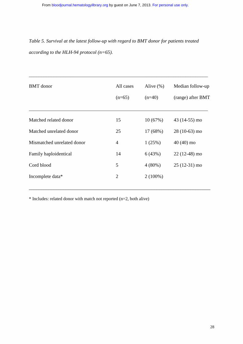
Survival with regard to donor is presented in Table 5. Donor-cell engraftment was achieved in
51/57 patients, two of the non-engrafted died within 17 days [no data (nd) in eight additional
For personal use only. by guest on June 7, 2013. bloodjournal.hematologylibrary.orgFrom

11
patients]. All surviving BMT-patients are free of disease. Among patients alive at
BMT+1year (n=41), only 1/26 patients (nd=15) had a mixed chimerism.
In the 25 deceased BMT patients, the death occurred prior to day +100 in 20 patients, caused
by BMT complications (n=17) or reactivation of HLH (n=3). One child died at day +121 (261
days after onset of therapy) of a surgical hemorrhage due to a disease unrelated to HLH. The
remaining four died of reactivation at day +112, Epstein-Barr virus (EBV)-associated
lymphoproliferative disease at +152, relapse and subsequent AML at +433, and unclear
respiratory disease at +550.
Surviving non-BMT patients
Altogether 23 children (8M/15F), all without a positive family history, were alive without
having received BMT, 20 of whom are off therapy and without evidence of disease for >12
months (follow-up range 1.1-4.2 years, mean 2.3) and three are on therapy for >1 year (1.9-
2.0 years) (two on CSA alone, one also receives corticosteroids). Ten of the 20 patients off
therapy received only initial therapy, and for the 10 patients that started continuation therapy
the mean duration of their total treatment was 359 days. Five of the latter 10 children had a
non-active disease at 2 months, whereas four had not received a resolution and one patient
had experienced a reactivation during the initial therapy. Almost all (19/20) of the patients off
therapy were >12 months old at onset (including four of the altogether 6 children aged ≥ 6
years), and the majority (n=12) (60%) were reported from East Asia.
Neurological symptoms and intrathecal therapy
Neurological alterations at onset were reported in 35/109 (32%) patients [missing data (md) in
four patients]. At 2 months, with 101 alive patients, neurological alterations were reported in
For personal use only. by guest on June 7, 2013. bloodjournal.hematologylibrary.orgFrom

12
13/95 (14%) patients (md=6). At the time of BMT 9/53 patients (17%) patients had
neurological alterations (md=12), and at BMT +100 days 8/36 patients (22%) (md=9). Out of
the 14 patients with neurological manifestations at onset that underwent BMT and were alive
at BMT +1-year, three had neurological alterations at that time and 10 had not (md=1).
Patients with neurological alterations at onset (n=35): At 2 months after onset, 21 of the
surviving 31 patients did not have any neurological abnormalities. Intrathecal therapy during
the first 2 months, administered to 15 of the 30 survivors (missing data on intrathecal therapy
in one patient), was associated with normalization of symptoms at 2 months in 10/15
individuals. In the patients who did not receive intrathecal therapy, the neurological
symptoms also normalized in 10/15 children.
Patients without neurological alterations at onset (n=74): Altogether 68 of these 74 patients
survived 2 months, during which period 11 had and 48 had not received intrathecal therapy
(md=9). At 2 months after onset, three of these 68 children had neurological alterations
(md=6), one of whom had received intrathecal therapy.
Prognostic factors
The prognostic influence of the factors age at onset, and the interval between onset of therapy
and BMT were analyzed, but neither of these factors were associated with significant
alterations in overall survival (data not shown).
DISCUSSION
Untreated FHL is invariably fatal with a median survival of 1-2 months (1). In 1993, a study
of 122 patients reported an estimated overall 5-year survival of 22% (2). Thus, the present
results with an estimated 3-year probability of survival of 51% (+/-20%) for the familial cases
For personal use only. by guest on June 7, 2013. bloodjournal.hematologylibrary.orgFrom

13
represents a great improvement. When comparing different HLH reports with regard to
therapeutic results, it is important to be aware that the percentage of patients with SHLH may
vary and, importantly, our data above refer to children with verified familial disease (Table 6).
This figure also represents a reasonable estimate of the final cure rate, since few deaths occur
later than 3 years after onset of therapy. In contrast to many reports, our study registered
patients prospectively from diagnosis, not only patients recruited for BMT. The HLH-94
protocol was effective in a wide range of institutions internationally, each treating very few
cases, and the present report includes patients from 21 countries.
In FHL, two steps are essential for survival, 1) effective initial and continuation therapy, and
2) a successful BMT. Altogether 20 (80%) of the 25 familial cases, i.e., children with an
affected sibling, survived the initial and continuation therapy and were admitted for BMT,
with only five deaths prior to BMT, indicating a high success rate of the immuno-
chemotherapy (Table 4). In the entire study population a total of 88/113 (78%) were either
admitted to BMT (n=65) or still alive without BMT, with at least 1-yr follow-up since onset
(n=23). The major toxicity of the pre-BMT therapy was neutropenia, in particular during the
first two months, but since neutropenia is common also in untreated HLH, it is sometimes
difficult to determine to what extent neutropenia was due to therapy or to active disease.
The 3-year probability of survival after BMT was 62% in our multi-institutional study (n=65).
It has to considered that only a minority of the BMTs were performed with matched related
donors (n=15). Recent BMT series have reported data ranging from a 3-year probability of
survival of 45 % (n=20) to an overall survival of 64% with HLA-nonidentical donors (n=14)
and 100% in a single-center material with matched sibling donors and unrelated donors
(n=12) (37-40).
For personal use only. by guest on June 7, 2013. bloodjournal.hematologylibrary.orgFrom

14
High mortality rates have previously been described also in infection-associated HLH (52% in
a review of all patients reported in the literature 1979-1996), in particular in EBV-associated
HLH (35). Treatment according to HLH-94, without BMT, appears beneficial also in
secondary HLH (41). Lack of signs of disease activity during a prolonged period (>12
months) after cessation of therapy, without previous BMT, will most likely suggest the
diagnosis of SHLH.
The inflammatory meningoencephalopathy in HLH, which may cause severe and permanent
CNS dysfunction (6-8), deserves prompt and adequate therapy considering the low
regenerative potential of nervous tissue. This was an important rationale for the intensive
initial systemic dexamethasone and etoposide therapy in HLH-94. Neurological alterations
were reported in 35/109 (32%) of the patients at registration. In these 35 affected individuals,
the symptoms normalized in 21 of the 31 survivors after 2 months of HLH-94 therapy. The
rate of normalization was similar whether intrathecal therapy was used (67%) or not (67%), as
an additional treatment to systemic corticosteroids, etoposide, and CSA, but the value of IT
MTX was not studied in a randomized fashion. Additional data will be required to better
evaluate the value of intrathecal therapy.
We speculate that the biology of the remarkably beneficial effects of etoposide in HLH,
previously not well understood, may be explained by the recent findings that FHL is
associated with a defective triggering of apoptosis (15,19,22), at least in a subset of patients,
since etoposide is known to be an excellent initiator of apoptosis (42). In contrast to
autoimmune lymphoproliferative syndrome (ALPS) with defective Fas-induced apoptosis,
FHL is characterized by lack of (lytic granule-dependent) apoptosis induction, i.e., lack of
For personal use only. by guest on June 7, 2013. bloodjournal.hematologylibrary.orgFrom

15
cytotoxic and NK cell-mediated apoptosis but, interestingly, in ALPS as well as in FHL the
result is a lymphoproliferation/ non-malignant accumulation of immune cells (22). Similarly,
the effect of dexamethasone might be explained by its anti-inflammatory and pro-apoptotic
properties, particularly valuable since the drug also penetrates well into the CNS, and CSA is
known to reduce T-cell activity, which is increased in HLH.
An increased incidence of acute myeloid leukemia (AML) and myelodysplastic syndrome
(MDS) has been reported following the use of epipodophyllotoxin derivatives (43). Etoposide
was included in the present protocol because it previously had shown to have a positive effect
in FHL, a disease that without treatment is uniformly fatal. We are aware of two reports on
AML/MDS in HLH following prolonged use of epipodophyllotoxins (44-45), and here we
report a third patient, a child without sustained engraftment after BMT, who relapsed and died
of AML on day 552 after onset of therapy (day +433 after BMT). An alternative pre-BMT-
approach entirely based on immunosuppression (ATG, corticosteroids, CSA, and IT MTX)
can also be effective, but it may result in a lower rate of complete remission at the time of
BMT as compared to treatment including the highly apoptosis-inducing drug etoposide
(38,42). We conclude that the risk of development of MDS/AML in HLH patients following
etoposide treatment is limited and acceptable considering its positive therapeutic effects but
that prolonged administration of epipodophyllotoxins if possible should be avoided.
Whereas HLH traditionally is separated in familial (primary) and secondary HLH, this
distinction may not be possible in the initial clinical setting until improved molecular
diagnosis is available, but the search for underlying gene mutations is encouraged (46).
Proving an acute infection at onset does not have any major therapeutic importance, since not
only SHLH but also FHL often features a triggering infectious agent (36). In less severe
For personal use only. by guest on June 7, 2013. bloodjournal.hematologylibrary.orgFrom

16
SHLH cases, either no treatment or a short duration of therapy might suffice, but future
studies are necessary to define these subsets, possibly with additional genetic markers. If the
disease is familial, relapsing, or severe and persistent even without family history, the BMT
from the best available donor is strongly recommended (38-40). Finally, this report also
demonstrates the value of international collaboration in conducting clinical studies of rare
disorders.
ACKNOWLEDGEMENTS
We would like to acknowledge all collaborating colleagues and families. We are also grateful
to our data managers Ulrika Kreicbergs, Anna-Maria Hasselgren-Häll and Martina Löfstedt.
For personal use only. by guest on June 7, 2013. bloodjournal.hematologylibrary.orgFrom

17
REFERENCES
1. Janka GE. Familial hemophagocytic lymphohistiocytosis. Eur J Pediatr 1983; 140: 221-
230
2. Arico M, Janka G, Fischer A, et al. Hemophagocytic lymphohistiocytosis: Diagnosis,
treatment and prognostic factors. Report of 122 children from the international registry.
Leukemia 1996; 10: 197-203
3. Henter J-I, Arico M, Elinder G, Imashuku S, Janka G. Familial hemophagocytic
lymphohistiocytosis (primary HLH). Hematol/Oncol Clin North Am 1998; 12: 417-433.
4. Henter J-I, Elinder G, Öst Å, and the FHL Study Group of the Histiocyte Society.
Diagnostic guidelines for hemophagocytic lymphohistiocytosis. Semin Oncol 1991; 18:
29-33.
5. Egeler RM, Shapiro RS, Loechelt B, Filipovich AH. Characteristic immune abnormalities
in hemophagocytic lymphohistiocytosis. J Pediat Hematol Onc 1996; 18: 340-345.
6. Henter J-I, Elinder G. Cerebromeningeal hemophagocytic lymphohistiocytosis. Lancet
1992; i: 104-107.
7. Haddad E, Sulis ML, Jabado N, Blanche S, Fischer A, Tardieu M. Frequency and severity
of central nervous system lesions in hemophagocytic lymphohistiocytosis. Blood 1997;
89: 794-800.
8. Henter J-I, Nennesmo I. Neuropathological findings and neurological symptoms in 23
children with hemophagocytic lymphohistiocytosis. J Pediatr 1997; 130: 358-365.
9. Henter J-I, Söder O, Öst Å, Elinder G. Incidence and clinical features of familial
hemophagocytic lymphohistiocytosis in Sweden. Acta Paediatr Scand, 1991; 80: 428-435.
10. Komp DM, McNamara J, Buckley P. Elevated soluble interleukin-2 receptor in childhood
hemophagocytic histiocytic syndromes. Blood 1989; 73; 2128-2132
For personal use only. by guest on June 7, 2013. bloodjournal.hematologylibrary.orgFrom
18
11. Howells DW, Strobel S, Smith I, Levinsky RJ, Hyland K. Central nervous system
involvement in the erythrophagocytic disorders of infancy: The role of cerebrospinal fluid
neopterins in their differential diagnosis and clinical management. Pediatr Res 1990; 28:
116-119
12. Henter J-I, Söder O, Hansson M, Elinder G, Andersson B, Andersson U.
Hypercytokinemia in familial hemophagocytic lymphohistiocytosis. Blood 1991; 78:
2918-2922.
13. Imashuku S, Ikushima S, Esumi N, et al. Serum levels of interferon-gamma, cytotoxic
factor and soluble interleukin-2 receptor in childhood hemophagocytic syndrome.
Leukemia Lymphoma 1991; 3: 287-292.
14. Henter J-I, Andersson B, Elinder G, Jakobson Å, Lübeck P-O, Söder O. Elevated
circulating IL-1 receptor antagonist but not IL-1 agonists in familial hemophagocytic
lymphohistiocytosis. Med Pediatr Oncol 1996; 27: 21-25.
15. Fadeel B, Orrenius S, Henter J-I. Induction of apoptosis and caspase activation in cells
obtained from familial haemophagocytic lymphohistiocytosis patients. Brit J Haematol
1999; 106: 406-415.
16. Ohadi M, Lalloz MR, Sham P, et al. Localization of a gene for familial hemophagocytic
lymphohistiocytosis at chromosome 9q21.3-22 by homozygosity mapping. Am J Hum
Genet 1999; 64: 165-171.
17. Dufurcq-Lagelouse R, Jabado N, Le Deist F, et al. Linkage of familial hemophagocytic
lymphohistiocytosis to 10q21-22 and evidence for heterogeneity. Am J Hum Genet 1999;
64: 172-179
18. Graham GE, Graham LM, Bridge PJ, et al. Further evidence for genetic heterogeneity in
familial hemophagocytic lymphohistiocytosis. Pediatr Res 2000; 48: 227-232.
For personal use only. by guest on June 7, 2013. bloodjournal.hematologylibrary.orgFrom
19
19. Stepp SE, Dufourcq-Lagelouse R, Le Deist F, et al. Perforin gene defects in familial
hemophagocytic lymphohistiocytosis. Science 1999; 286: 1957-1959.
20. Ericson KG, Fadeel B, Nilsson-Ardnor S, et al. Spectrum of perforin gene mutations in
familial hemophagocytic lymphohistiocytosis. Am J Hum Genet 2001, 68: 590-597.
21. Clementi R, zur Stadt U, Savoldi G, et al. Six novel mutations in the PRF1 gene in
children with haemophagocytic lymphohistiocytosis. J Med Genet. 2001; 38: 643-6.
22. Fadeel B, Orrenius S, Henter J-I. Familial hemophagocytic lymphohistiocytosis: too little
cell death may seriously damage your health. Leuk Lymphoma 2001; 42: 13-20.
23. Arico, Danesino C, Pende D, Moretta L. Pathogenesis of haemophagocytic
lymphohistiocytosis. Br J Haematol 2001; 114: 761-769.
24. Arico M, Imashuku S, Clementi R, et al. Hemophagocytic lymphohistiocytosis due to
germline mutations in SH2D1A, the X-linked lymphoproliferative disease gene. Blood
2001; 97: 1131-1133
25. Ambruso DR, Hays T, Zwartjes WJ, Tubergen DG, Favara BE. Successful treatment of
lymphohistiocytic reticulosis with phagocytosis with epipodophyllotoxin VP 16-213.
Cancer 1980; 45: 2516-2520
26. Fischer A, Virelizier JL, Arenzana-Seisdedos F, Perez A, Nezelof G, Griscelli C.
Treatment of four patients with erythrophagocytic lymphohistiocytosis by a combination
of epipodophyllotoxin, steroids, intrathecal methotrexate and cranial irradiation. Pediatrics
1985; 76: 263-268
27. Henter J-I, Elinder G, Finkel Y, Söder O. Successful induction with chemotherapy
including teniposide in familial erythrophagocytic lymphohistiocytosis. Lancet 1986; ii:
1402.
For personal use only. by guest on June 7, 2013. bloodjournal.hematologylibrary.orgFrom

20
28. Stephan JL, Donadieu J, Le Deist F, Blanche S, Griscelli C, Fischer A. Treatment of
familial hemophagocytic lymphohistiocytosis with antithymocyte globulins, steroids and
cyclosporin A. Blood 1993; 82: 2319-2323
29. Fischer A, Cerf-Bensussan N, Blanche S, et al. Allogeneic bone marrow transplantation
for erythrophagocytic lymphohistiocytosis. J Pediatr 1986; 108: 267-270
30. Blanche S, Caniglia M, Girault D, Landman J, Griscelli C, Fischer A. Treatment of
hemophagocytic lymphohistiocytosis with chemotherapy and bone marrow
transplantation: a single center study of 22 cases. Blood 1991; 78: 51-54
31. Todo S, Fujiwara F, Ikushima S, et al. Allogeneic bone marrow transplantation for
familial erythrophagocytic lymphohistiocytosis with high dose VP16-containing
conditioning regimen. Leukemia Lymphoma 1990; 1: 361-364
32. Nespoli L, Locatelli F, Bonetti F, et al. Familial hemophagocytic lymphohistiocytosis
treated with allogeneic bone marrow transplantation. Bone Marrow Transplant 1991; 7
(suppl 3): 139-142
33. Henter J-I, Elinder G. Familial hemophagocytic lymphohistiocytosis. Clinical review
based on the findings in seven children. Acta Paediatr Scand. 1991; 80: 269-277
34. Henter J-I, Arico M, Egeler M, et al. HLH-94: A treatment protocol for hemophagocytic
lymphohistiocytosis. Med Pediatr Oncol 1997; 28: 342-347.
35. Janka G, Elinder G, Imashuku S, Schneider M, Henter J-I. Infection- and malignancy-
associated hemophagocytic syndromes: Secondary hemophagocytic lymphohistiocytosis.
Hematol/Oncol Clin North Am 1998; 12: 435-444.
36. Henter J-I, Ehrnst A, Andersson J, Elinder G. Familial hemophagocytic
lymphohistiocytosis and viral infections. Acta Paediatr 1993; 82: 369-372.
For personal use only. by guest on June 7, 2013. bloodjournal.hematologylibrary.orgFrom

21
37. Baker KS, DeLaat CA, Steinbuch M, et al. Successful correction of hemophagocytic
lymphohistiocytosis with related or unrelated bone marrow transplantation. Blood 1997;
89: 3857-3863.
38. Jabado N, de Graeff-Meeder ER, Cavazzana-Calvo M, et al. Treatment of familial
hemophagocytic lymphohistiocytosis with bone marrow transplantation from HLA
genetically nonidentical donors. Blood 1997; 90: 4743-4748.
39. Imashuku S, Hibi S, Todo S, et al. Allogeneic hematopoietic stem cell transplantation for
patients with hemophagocytic syndrome (HPS) in Japan. Bone Marrow Transplant 1999;
23: 569-572.
40. Dürken M, Horstmann M, Bieling P, et al. Improved outcome in haemophagocytic
lymphohistiocytosis after bone marrow transplantation from related and unrelated donors:
a single-centre experience of 12 patients. Br J Haematol 1999; 106: 1052-1058
41. Imashuku S, Hibi S, Ohara T, et al. Effective control of Epstein-Barr virus-related
hemophagocytic lymphohistiocytosis with immunochemotherapy. Blood 1999; 93: 1869-
1874.
42. Martinsson P, Liminga G, Nygren P, Larsson R. Characteristics of etoposide-induced
apoptotic cell death in the U-937 human lymphoma cell line. Anticancer Drugs 2001; 12:
699-705
43. Pui C-H, Ribeiro RC, Hancock ML, et al. Acute myeloid leukemia in children treated with
epipodophyllotoxins for acute lymphoblastic leukemia. New Engl J Med 1991; 325:1632-
1687
44. Henter J-I, Elinder G, Lübeck P-O, Öst Å. Myelodysplastic syndrome following
epipodophyllotoxin therapy in familial hemophagocytic lymphohistiocytosis. Pediatr
Hematol Oncol 1993; 10: 163-68.
For personal use only. by guest on June 7, 2013. bloodjournal.hematologylibrary.orgFrom
22
45. Kitazawa J, Ito E, Arai K, Yokoyama M, Fukayama M, Imashuku S. Secondary acute
myelocytic leukemia after successful chemotherapy with etoposide for Epstein-Barr virus-
associated hemophagocytic lymphohistiocytosis. Med Pediatr Oncol 2001; 37:153-
46. Henter J-I. Biology and treatment of familial hemophagocytic lymphohistiocytosis:
importance of perforin in lymphocyte-mediated cytotoxicity and triggering of apoptosis.
Med Pediatr Oncol 2002, in press.
For personal use only. by guest on June 7, 2013. bloodjournal.hematologylibrary.orgFrom
23
LEGEND TO FIGURES
Fig 1. Overview of the treatment protocol HLH-94. Day of death for the 23 patients who died
the first year of therapy, except those who underwent BMT, is marked with an “!”. [BMT =
Go to BMT during continuation therapy as soon as an acceptable donor is available,
preferably when the disease is non-active. The patients without either familial or persistent
disease were recommended to cease therapy after the “initial therapy”, and restart in case of
reactivation. Dexa = Dexamethasone daily (pulses are 10 mg/m2 for 3 days). VP- 16 =
Etoposide 150 mg/m2 iv. CSA = Cyclosporin A. I.T. therapy = Intrathecal methotrexate (if
progressive neurological symptoms or if an abnormal CSF has not improved)].
Fig 2. Flow of 113 patients in the HLH-94 treatment protocol. (§: Of the 25 familial cases
reported, three were not known at onset and another three changed therapy prior to onset of
continuation therapy. #: Two were on cyclosporin A only and one also received
corticosteroids.*: Eight had received therapy for 2-12 months, and two for 12-24 months).
Fig 3. Kaplan-Meier survival curve for A/ all eligible study patients treated with HLH-94
(n=113), and B/ patients with an affected sibling (n=25).
Fig 4. Kaplan-Meier survival curve for patients who underwent BMT, starting at the time of
BMT (missing date of BMT in one of the 65 patients, leaving 64 patients for analysis).
For personal use only. by guest on June 7, 2013. bloodjournal.hematologylibrary.orgFrom

24
Table 1. Diagnostic Guidelines for HLH* (adapted from ref 4)
_____________________________________________________________________
Clinical criteria
* Fever
* Splenomegaly
Laboratory criteria
* Cytopenias (affecting ≥ 2 of 3 lineages in the peripheral blood):
Hemoglobin (< 90 g/L), platelets (<100 x 109/L), neutrophils (<1.0 x 10⁹/L)
* Hypertriglyceridemia and/or hypofibrinogenemia
(fasting triglycerides ≥2.0 mmol/L or ≥3 SD of the normal value for age,
fibrinogen ≤1.5 g/L or ≤3 SD)
Histopathologic criteria
* Hemophagocytosis in bone marrow or spleen or lymph nodes.
No evidence of malignancy
____________________________________________________________________
* All criteria required for the diagnosis of HLH. In addition, the diagnosis of FHL is justified by a positive family history, and parental consanguinity is suggestive.
Comments:1. If hemophagocytic activity is not proven at the time of presentation, further search for hemophagocytic activity is encouraged. If the bone marrow specimen is not conclusive, material may be obtained from other organs. Serial marrow aspirates over time may also be helpful.
2. The following findings may provide strong supportive evidence for the diagnosis: (a) Spinal fluid pleocytosis (mononuclear cells), (b) Histological picture in the liver resembling chronic persistent hepatitis (biopsy), (c) Low natural killer cell activity.
3. Other abnormal clinical and laboratory findings consistent with the diagnosis are:Cerebromeningeal symptoms, lymph node enlargement, jaundice, edema, skin rash. Hepatic enzyme abnormalities, hyperferritinemia, hypoproteinemia, hyponatremia, spinal fluid protein ↑, VLDL ↑, HDL ↓, circulating soluble IL-2 receptor ↑.
For personal use only. by guest on June 7, 2013. bloodjournal.hematologylibrary.orgFrom

25
Table 2. Overview of overall outcome in 113 patients with HLH treated according to the
HLH- 94 protocol
______________________________________________________________________
All patients Familial cases§
(n=113) (n=25)
______________________________________________________________________
Alive after BMT 40 13
Dead after BMT 25* 7*
Alive without BMT 23#
Dead prior to BMT 25 5
_______________________________________________________________________
§ All these patients had an affected sibling with HLH
* One child, with a positive family history, died of a surgical hemorrhage unrelated to HLH
# Three patients were still on therapy (two on CSA alone, one also received corticosteroids),
the others were all off therapy for >12 months
For personal use only. by guest on June 7, 2013. bloodjournal.hematologylibrary.orgFrom

26
Table 3. Survival at the latest follow-up with regard to age in 113 patients with HLH treated
according to the HLH-94 protocol.
___________________________________________________________________
Age at onset All patients Familial BMT Patients alive and off
of therapy cases patients therapy without BMT
(n=113) (n=25) (n=65) (n=20)
___________________________________________________________________
0 - <3 mo 12/30 (40%) 6/13 (46%) 11/21 (52%)
3 – <12 mo 15/34 (44%) 2/4 (50%) 14/22 (64%) 1/1
12- <24 mo 19/25 (76%) 3/4 (75%) 10/14 (71%) 8/8
≥ 24 mo 17/24 (71%) 2/4 (50%) 5/8 (62%) 11/11
____________________________________________________________________
TOTAL 63/113 (56%) 13/25 (52%) 40/65 (62%) 20/20
Mean age 19 mo 13 mo 13 mo 47 mo
Age range 0-145 mo 0-82 mo 0-84 mo 10-145 mo
____________________________________________________________________
For personal use only. by guest on June 7, 2013. bloodjournal.hematologylibrary.orgFrom

27
Table 4. Evaluation of the efficacy of immuno-chemotherapy during initial and continuation
period with regard to survival and possibility to obtain BMT
__________________________________________________________________________
All patients Familial cases
(n=113) (n=25)
__________________________________________________________________________
Patients admitted to BMT + patients alive without BMT 65+23 (78%) 20+0 (80%)
Patients dead during initial/continuation therapy* 25 (22%) 5 (20%)
__________________________________________________________________________
* Five of these patients died more than 6 months after onset of therapy (one with a positive
family history)
For personal use only. by guest on June 7, 2013. bloodjournal.hematologylibrary.orgFrom
28
Table 5. Survival at the latest follow-up with regard to BMT donor for patients treated
according to the HLH-94 protocol (n=65).
_________________________________________________________________________
BMT donor All cases Alive (%) Median follow-up
(n=65) (n=40) (range) after BMT
_________________________________________________________________________
Matched related donor 15 10 (67%) 43 (14-55) mo
Matched unrelated donor 25 17 (68%) 28 (10-63) mo
Mismatched unrelated donor 4 1 (25%) 40 (40) mo
Family haploidentical 14 6 (43%) 22 (12-48) mo
Cord blood 5 4 (80%) 25 (12-31) mo
Incomplete data* 2 2 (100%)
__________________________________________________________________________
* Includes: related donor with match not reported (n=2, both alive)
For personal use only. by guest on June 7, 2013. bloodjournal.hematologylibrary.orgFrom

29
Table 6. Survival as reported in the three largest reports on HLH.
______________________________________________________________________
Publication Year Number Survival
of report of patients
______________________________________________________________________
Janka (review) (1) 1983 121 5 % (1-yr)§
Arico et al (2) 1996 122 22 % (5-yr)*
Present study 2002 113 55 % (5-yr)*
Present study (familial cases) 2002 25 51 % (5-yr)*
_______________________________________________________________________
§ Out of 121 patients reviewed, 5/101 with follow-up data survived more than 12 months
* Probability of survival according to Kaplan-Meier estimate
For personal use only. by guest on June 7, 2013. bloodjournal.hematologylibrary.orgFrom

FIGURE 1 F
or personal use only. by guest on June 7, 2013.
bloodjournal.hematologylibrary.org
From

31
FIGURE 2
F
or personal use only. by guest on June 7, 2013.
bloodjournal.hematologylibrary.org
From

32
FIGURE 3A
F
or personal use only. by guest on June 7, 2013.
bloodjournal.hematologylibrary.org
From

33
FIGURE 3B
F
or personal use only. by guest on June 7, 2013.
bloodjournal.hematologylibrary.org
From

34
FIGURE 4
F
or personal use only. by guest on June 7, 2013.
bloodjournal.hematologylibrary.org
From
Related Documents