110 ABSTRACT Femoral neck fracture is a pathology that the trauma surgeon usually faces. When he or she decides to perform osteosynthesis, there are many factors to consider in order to achieve a good result and avoid complications. In this update article we seek to offer 10 keys to success when reduction and osteosynthesis are chosen as the therapeutic method. Key words: Fracture; neck; femur; osteosynthesis. Tratamiento de las fracturas de cuello femoral con osteosíntesis. Diez claves para el éxito RESUMEN Las fracturas de cuello femoral son un cuadro habitual para el cirujano traumatológico y cuando se elige realizar una osteosín- tesis, son muchos los factores por considerar para conseguir un buen resultado y evitar las complicaciones. En este artículo de actualización, intentamos dar 10 claves para el éxito cuando se opta por la reducción y la osteosíntesis como método terapéutico. Palabras clave: Fractura; cuello; fémur; osteosíntesis. INTRODUCTION Femoral neck fracture is a clinical picture traumatologists usually deal with. The treatment for such kind of frac- ture still represents a tremendous challenge. It presents a bimodal pattern, affecting to a large extent aged patients who have suffered a low-energy trauma, but also a smaller but growing peak of young patients who have suffered a high-energy trauma. When osteosynthesis is the treatment of choice, many factors should be considered in order to achieve good results and avoid complications, which, according to some reports, reach 45% of the cases 1,2 . Based on our experience and a careful literature review, we seek to examine the technical aspects of femoral neck fracture osteosynthesis in order to minimize mistakes, and detect modifiers of disease progression in order to identify risk groups in relation to osteosynthesis. Hence, a better therapeutic indication may be done, and, in case osteosynthe- sis is the treatment of choice, the outcome may be optimized. OBJECTIVES We seek to summarize in 10 items some points which can lead to achieve the best results in performing an os- teosynthesis to treat a fracture of the femoral neck. 1. Appropriate time for surgery It is a controversial issue to determine to what extent the time elapsed since the moment of trauma until the implementation of the osteosynthesis affects the progression of the femoral neck fracture healing. It is convention- ally suggested that surgery is performed within a period of 6 hours after the trauma. Treatment of Femoral Neck Fractures with Osteosynthesis. Ten Keys to Success. Fernando M. Bidolegui, Sebastián Pereira, Gabriel Vindver Orthopedics and Traumatology Service, Hospital Sirio Libanés, ECICARO (Buenos Aires, Argentina) UPDATE Rev Asoc Argent Ortop Traumatol 2021; 86 (1): 110-121 • ISSN 1852-7434 (online) Received on June 8 th , 2020. Accepted after evaluation on September 2 nd , 2020 • FERNANDO M. BIDOLEGUI, MD • [email protected] http://orcid.org/0000-0002-0502-2300 How to cite this paper: Bidolegui FM, Pereira S, Vindver G. Treatment of Femoral Neck Fractures with Osteosynthesis. Ten Keys to Success. Rev Asoc Argent Ortop Traumatol 2021;86(1):110- 121. https://doi.org/10.15417/issn.1852-7434.2021.86.1.1130 ID This Journal is licensed under Attribution-NonCommercial-ShareAlike 4.0 International Creative Commons (CC-BY-NC-SA 4.0).

Welcome message from author
This document is posted to help you gain knowledge. Please leave a comment to let me know what you think about it! Share it to your friends and learn new things together.
Transcript
110
AbstrActFemoral neck fracture is a pathology that the trauma surgeon usually faces. When he or she decides to perform osteosynthesis, there are many factors to consider in order to achieve a good result and avoid complications. In this update article we seek to offer 10 keys to success when reduction and osteosynthesis are chosen as the therapeutic method.Key words: Fracture; neck; femur; osteosynthesis.
tratamiento de las fracturas de cuello femoral con osteosíntesis. Diez claves para el éxito
resumen Las fracturas de cuello femoral son un cuadro habitual para el cirujano traumatológico y cuando se elige realizar una osteosín-tesis, son muchos los factores por considerar para conseguir un buen resultado y evitar las complicaciones. En este artículo de actualización, intentamos dar 10 claves para el éxito cuando se opta por la reducción y la osteosíntesis como método terapéutico.Palabras clave: Fractura; cuello; fémur; osteosíntesis.
IntroductIonFemoral neck fracture is a clinical picture traumatologists usually deal with. The treatment for such kind of frac-
ture still represents a tremendous challenge. It presents a bimodal pattern, affecting to a large extent aged patients who have suffered a low-energy trauma, but also a smaller but growing peak of young patients who have suffered a high-energy trauma. When osteosynthesis is the treatment of choice, many factors should be considered in order to achieve good results and avoid complications, which, according to some reports, reach 45% of the cases1,2. Based on our experience and a careful literature review, we seek to examine the technical aspects of femoral neck fracture osteosynthesis in order to minimize mistakes, and detect modifiers of disease progression in order to identify risk groups in relation to osteosynthesis. Hence, a better therapeutic indication may be done, and, in case osteosynthe-sis is the treatment of choice, the outcome may be optimized.
objectIvesWe seek to summarize in 10 items some points which can lead to achieve the best results in performing an os-
teosynthesis to treat a fracture of the femoral neck.
1. Appropriate time for surgeryIt is a controversial issue to determine to what extent the time elapsed since the moment of trauma until the
implementation of the osteosynthesis affects the progression of the femoral neck fracture healing. It is convention-ally suggested that surgery is performed within a period of 6 hours after the trauma.
Treatment of Femoral Neck Fractures with Osteosynthesis. Ten Keys to Success.Fernando m. bidolegui, sebastián Pereira, Gabriel VindverOrthopedics and Traumatology Service, Hospital Sirio Libanés, ECICARO (Buenos Aires, Argentina)
uPDAte
Rev Asoc Argent Ortop Traumatol 2021; 86 (1): 110-121 • ISSN 1852-7434 (online)
Received on June 8th, 2020. Accepted after evaluation on September 2nd, 2020 • FERnAndo M. BIdoLEguI, Md • [email protected] http://orcid.org/0000-0002-0502-2300
How to cite this paper: Bidolegui FM, Pereira S, Vindver G. Treatment of Femoral Neck Fractures with Osteosynthesis. Ten Keys to Success. Rev Asoc Argent Ortop Traumatol 2021;86(1):110-121. https://doi.org/10.15417/issn.1852-7434.2021.86.1.1130
Id
This Journal is licensed under Attribution-nonCommercial-ShareAlike 4.0 International Creative Commons (CC-BY-nC-SA 4.0).
Femoral neck fractures with osteosynthesis
Rev Asoc Argent Ortop Traumatol 2021; 86 (1): 110-121 • ISSN 1852-7434 (online) 111
Three decades ago, Manninger et al.1 compared surgeries performed within a period of 6 hours after the trauma to those performed between 6 and 24 hours after the trauma, and those performed more than 24 hours after the trauma. They reported that pseudoarthrosis and necrosis rates were significantly lower in the first group. However, in 2009, Loizou et al.2 examined 1023 patients with intracapsular hip fracture who were treated with osteosynthe-sis and the time of surgery ranged from <6 and 48 hours. There was no significant difference in the incidence of necrosis.
Upadhyay et al.3 compared two groups of patients with displaced femoral neck fracture who had undergone re-duction and osteosynthesis. The overall incidence of necrosis was 16.3%, and there was no significant difference between patients operated before or after 48 hours from the moment of trauma.
In 2015, Papakostidis et al.4 conducted a meta-analysis to evaluate the period of time which preceded the in-ternal fixation of fractures of the femoral neck. The authors concluded that well designed studies are needed and, although surgery should be performed as immediately as possible, there is no clear evidence about to what extent a reduction of the time elapsed since the moment of trauma affects the progression of the fracture healing. However, they suggest that the rate of pseudoarthrosis may increase if the period exceeds 24 hours.
In our series of 93 patients with femoral neck fracture treated with internal fixation, the average waiting time from trauma to surgery was 4.6 days. The rate of necrosis was 5.3%, which matches most of the previous studies, and, although we try to operate as soon as possible, the normal waiting time in our environment (>6 to 24 hours) did not affect our reports8.
2. Proper classificationIt is very important to classify femoral neck fractures correctly. The main distinction is between displaced and
undisplaced. The former include Garden types 1 and 2; the latter, Garden types 3 and 4. However, a proper clas-sification is often challenging, given the external rotation of the hip and its shortening, which hinders a correct interpretation of the radiographs.
In 2008, Egol et al.6 report that internal rotation traction radiographs are a helpful resource that increases the ability to accurately classify proximal femoral fractures in relation to fracture line and displacement, and have a direct impact in therapeutic decision. Lowenstein position is usually suggested for the initial evaluation of this type of fracture. However, by rotating the hip, displacement and thus vascularization may be worsened. Therefore, a surgical profile is recommended in order not to mobilize the injured hip7.
When osteosynthesis is the treatment of choice, the accuracy in determining the degree of displacement will directly affect the results. Frandsen et al.8 reported that patients with undisplaced femoral neck fractures presented a complication rate of 5% (necrosis or pseudoarthrosis), while patients with displaced fractures presented a rate of 40%, with no statistically significant difference between treatments. In their series of 93 patients, Pereira et al.5 report a complication rate of 4.2% in patients with undisplaced fractures, a number distinctly lower than the rate for displaced fractures, which reached 36.3%.
In conclusion, internal rotation traction radiographs constitute a simple, efficient and inexpensive tool to accu-rately determine both fracture line and displacement (Figure 1).
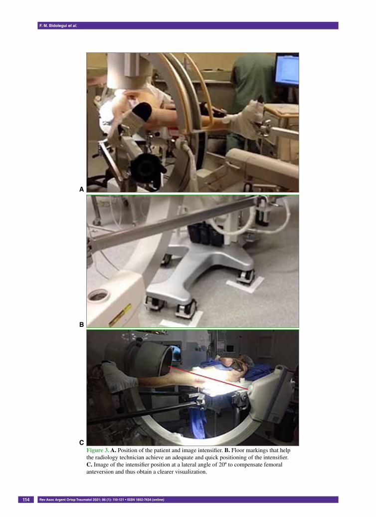
3. evaluation of posterior comminutionThe degree of comminution is a predictive factor whose complication rates and bad outcomes are higher in case
there is a femoral neck fracture with reduction and internal fixation. Huang et al.9 evaluated 146 patients, and sepa-rated the results depending on the presence of posterior comminution. The monitoring period was 4.6 years. The authors obtained statistically significant results according to which patients with posterior comminution presented more shortening, higher necrosis rates and a larger number of arthroplasty conversions.
Rawall et al.10 reported a significantly higher pseudoarthrosis rate in patients with cervical fractures whose pos-terior comminution was treated with osteoynthesis. Lastly, Pereira et al.5 reported consolidation without necrosis in 10 out of 14 comminuted fractures (71.4%), and 4 out of 8 non comminuted fractures (50%).
Based on what has been stated in the paragraphs above, we believe that, when treating femoral neck fracture patients with osteosynthesis, two studies are necessary: an adequate radiograph, and a CT scan to evaluate the posterior wall of the femoral neck. It should be taken into account that comminution increases the chance of failure for such therapeutic method.

112
F. m. bidolegui et al.
Rev Asoc Argent Ortop Traumatol 2021; 86 (1): 110-121 • ISSN 1852-7434 (online)
Posterior comminution of the femoral neck can be assessed through a surgical profile radiograph, CAT scans or MRI scans. In all images, it manifests as a posterior displacement of the femoral neck and head. (Figure 2).
Figure 1. A. Hip radiograph, anteroposterior, with no traction. Visualization of the fracture line is hindered. b. After traction and internal rotation, the femoral neck extends, allowing to identify a displaced medial fracture.
A b
Figure 2. Patient with femoral neck fracture. A. Surgical profile view. Comminution can be observed. b. CT scan. Posterior comminution is noticeable.
A b
Femoral neck fractures with osteosynthesis
Rev Asoc Argent Ortop Traumatol 2021; 86 (1): 110-121 • ISSN 1852-7434 (online) 113
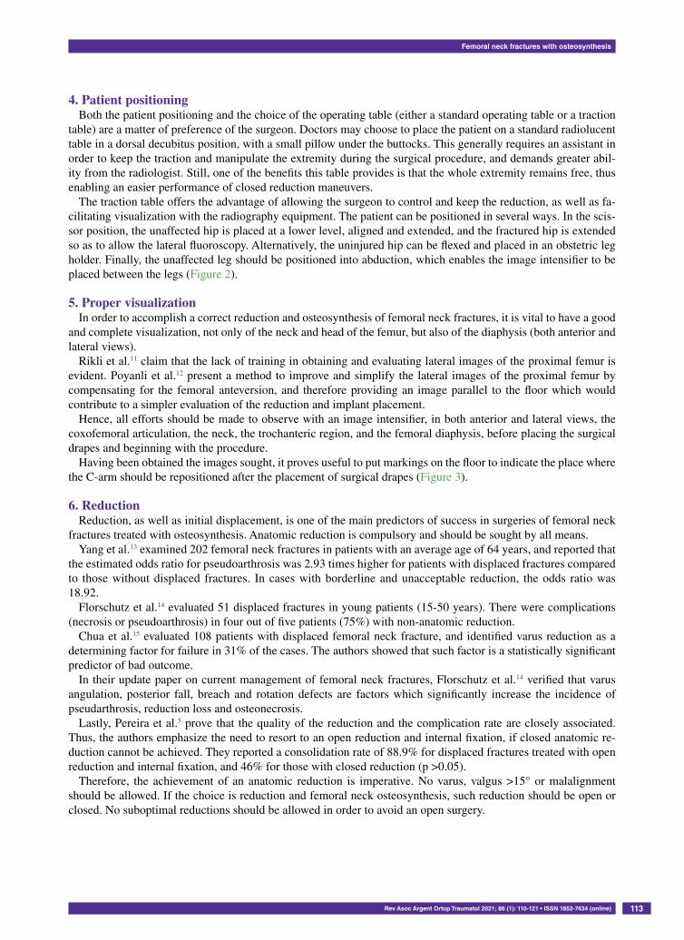
4. Patient positioningBoth the patient positioning and the choice of the operating table (either a standard operating table or a traction
table) are a matter of preference of the surgeon. Doctors may choose to place the patient on a standard radiolucent table in a dorsal decubitus position, with a small pillow under the buttocks. This generally requires an assistant in order to keep the traction and manipulate the extremity during the surgical procedure, and demands greater abil-ity from the radiologist. Still, one of the benefits this table provides is that the whole extremity remains free, thus enabling an easier performance of closed reduction maneuvers.
The traction table offers the advantage of allowing the surgeon to control and keep the reduction, as well as fa-cilitating visualization with the radiography equipment. The patient can be positioned in several ways. In the scis-sor position, the unaffected hip is placed at a lower level, aligned and extended, and the fractured hip is extended so as to allow the lateral fluoroscopy. Alternatively, the uninjured hip can be flexed and placed in an obstetric leg holder. Finally, the unaffected leg should be positioned into abduction, which enables the image intensifier to be placed between the legs (Figure 2).
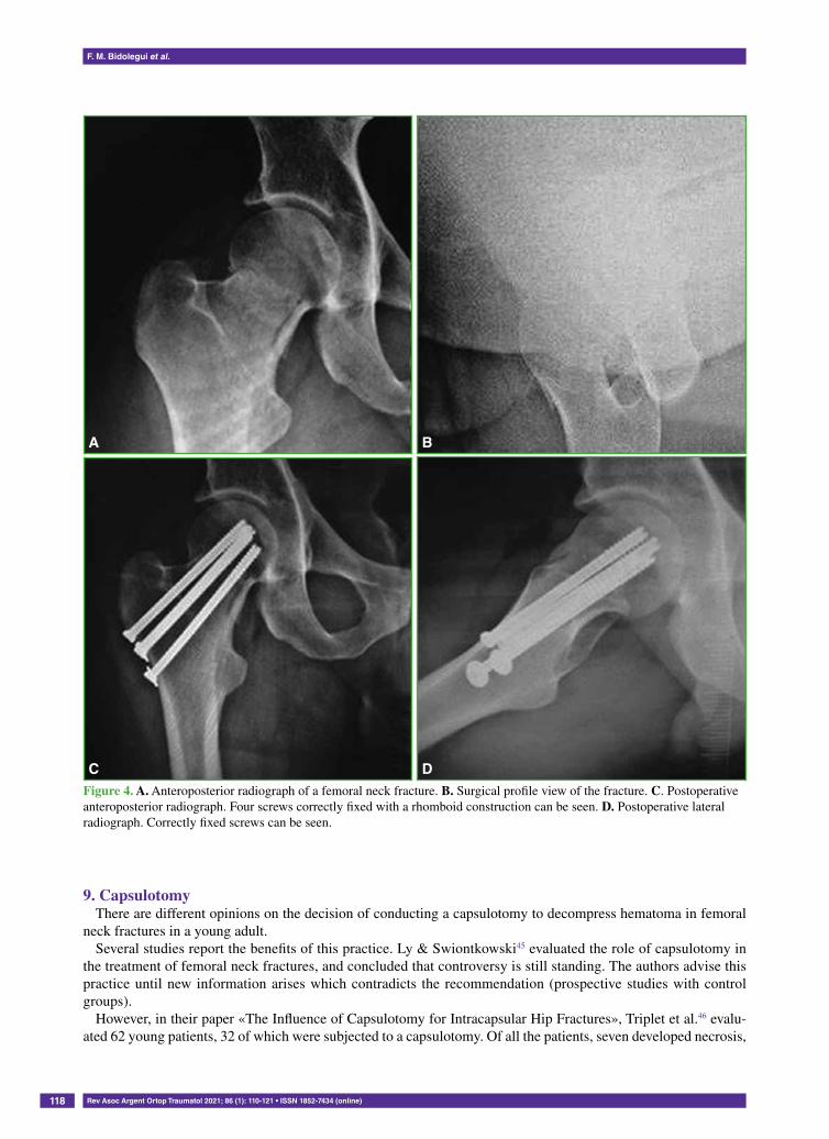
5. Proper visualizationIn order to accomplish a correct reduction and osteosynthesis of femoral neck fractures, it is vital to have a good
and complete visualization, not only of the neck and head of the femur, but also of the diaphysis (both anterior and lateral views).
Rikli et al.11 claim that the lack of training in obtaining and evaluating lateral images of the proximal femur is evident. Poyanli et al.12 present a method to improve and simplify the lateral images of the proximal femur by compensating for the femoral anteversion, and therefore providing an image parallel to the floor which would contribute to a simpler evaluation of the reduction and implant placement.
Hence, all efforts should be made to observe with an image intensifier, in both anterior and lateral views, the coxofemoral articulation, the neck, the trochanteric region, and the femoral diaphysis, before placing the surgical drapes and beginning with the procedure.
Having been obtained the images sought, it proves useful to put markings on the floor to indicate the place where the C-arm should be repositioned after the placement of surgical drapes (Figure 3).
6. reductionReduction, as well as initial displacement, is one of the main predictors of success in surgeries of femoral neck
fractures treated with osteosynthesis. Anatomic reduction is compulsory and should be sought by all means.Yang et al.13 examined 202 femoral neck fractures in patients with an average age of 64 years, and reported that
the estimated odds ratio for pseudoarthrosis was 2.93 times higher for patients with displaced fractures compared to those without displaced fractures. In cases with borderline and unacceptable reduction, the odds ratio was 18.92.
Florschutz et al.14 evaluated 51 displaced fractures in young patients (15-50 years). There were complications (necrosis or pseudoarthrosis) in four out of five patients (75%) with non-anatomic reduction.
Chua et al.15 evaluated 108 patients with displaced femoral neck fracture, and identified varus reduction as a determining factor for failure in 31% of the cases. The authors showed that such factor is a statistically significant predictor of bad outcome.
In their update paper on current management of femoral neck fractures, Florschutz et al.14 verified that varus angulation, posterior fall, breach and rotation defects are factors which significantly increase the incidence of pseudarthrosis, reduction loss and osteonecrosis.
Lastly, Pereira et al.5 prove that the quality of the reduction and the complication rate are closely associated. Thus, the authors emphasize the need to resort to an open reduction and internal fixation, if closed anatomic re-duction cannot be achieved. They reported a consolidation rate of 88.9% for displaced fractures treated with open reduction and internal fixation, and 46% for those with closed reduction (p >0.05).
Therefore, the achievement of an anatomic reduction is imperative. No varus, valgus >15° or malalignment should be allowed. If the choice is reduction and femoral neck osteosynthesis, such reduction should be open or closed. No suboptimal reductions should be allowed in order to avoid an open surgery.
114
F. m. bidolegui et al.
Rev Asoc Argent Ortop Traumatol 2021; 86 (1): 110-121 • ISSN 1852-7434 (online)
Figure 3. A. Position of the patient and image intensifier. b. Floor markings that help the radiology technician achieve an adequate and quick positioning of the intensifier. c. Image of the intensifier position at a lateral angle of 20º to compensate femoral anteversion and thus obtain a clearer visualization.
A
b
c

Femoral neck fractures with osteosynthesis
Rev Asoc Argent Ortop Traumatol 2021; 86 (1): 110-121 • ISSN 1852-7434 (online) 115
6.1. Closed reductionKrischak et al.16 claim that closed reduction should be anatomic, based on an examination of the relevance of
reduction quality on the clinical outcome. They treated 31 patients with closed reduction and reported that compli-cations were not related to non-anatomic reductions, especially in varus position, and that, when the fracture was valgized, the result was satisfactory.
The closed reduction technique requires a traction table. Traction, internal rotation and abduction or adduction should generally be applied to the limb, according to necessity. In case such technique should not result in the reduction sought, it is suggested to perform a percutaneous reduction or an open reduction.
6.2. Percutaneous reductionDespite not being very frequently applied, there are some percutaneous techniques which generally improve the
reduction and help avoid a direct surgical approach.Su et al.17 presented their experience with percutaneous reduction techniques in 25 cases in which fractures were
irreducible by means of external manipulative maneuvers. The suggested technique consists in the percutaneous insertion of two K-wires protecting the femoral artery by previous palpation, and the achievement of reduction and osteosynthesis by maneuvering the K-wires and inserting three cannulated screws. The consolidation rate was 100%, and there were two cases of necrosis.
In 2019, Yu et al.18 achieved very positive results with a similar technique applied in 32 patients. The authors particularly emphasized good planning and protection of noble structures.
Mahajan et al.19 suggest another percutaneous reduction technique, which consists in the insertion of a K-wire to fix the femoral head to the acetabulum so as to mobilize the distal fragment in order to achieve anatomic reduction. This technique was applied in 15 patients, and led to positive results without the need of focus opening.
6.3. Open reductionTwo basic approaches have been described and applied for an open reduction: anterolateral approach by Watson-
Jones, and anterior approach by Smith-Petersen. Each one has their advantages and disadvantages.Watson-Jones’s approach (1936) is developed using the space between the gluteus medius and the tensor fascia
lata. One of the advantages claimed for this approach is that the incision allows fracture reduction and placement of osteosynthesis. However, the impossibility to visualize subcapital fractures is a disadvantage.
On the other hand, Smith-Petersen’s approach (1917) is developed using the internervous plane of the sartorius (femoral nerve) and superior and the tensor fascia lata (gluteal nerve). A great advantage is that it provides the most direct access to the anterior aspect of the hip, which allows not only to adequately visualize the fracture, but also to easily insert reduction instruments. However, this method requires a secondary approach in order to insert the fixation implant21.
There are few studies which determine the most adequate approach for the treatment of displaced femoral neck fractures. Slobogean et al.22 conducted a survey involving many Canadian specialists and the Orthopaedic Trauma Association. The authors asked about the approaches chosen: 19.9% answered Watson-Jones, and 13.8% answered Smith-Petersen.
Molnar & Rout23 published the first paper with Smith-Petersen’s anterior approach for treatment of femoral neck fractures. The authors use, as in the traditional approach, the lower branch, and perform an anterior straight section, thus leading to a superb visualization of the neck of the femur with very good exposure, which allows different methods for reduction.
Lichstein et al.24 conducted a cadaveric study to compare both approaches, in order to determine which of them allows superior exposure and palpation. The authors reached the conclusion that Smith-Petersen’s approach, with-out rectus femoris tenotomy, provides superior exposure of the femoral neck (2.4 cm3), and of articular surface (1.6 cm3) over Watson-Jones’s approach. With tenotomy, the additional exposure over such approach is 3.3 cm3 and 1.6 cm3, respectively. However, anterior approach entails the risk of an injury of the femorocutaneous nerve in up to 60% of the cases. Therefore, the authors emphasize an adequate dissection in order to reduce the incidence of neurapraxia.
It is still a controverted matter to determine the best approach, which will frequently depend on the preference of the surgeon. It should be stressed that a traumatologist must be familiarized with both options to treat this clinical picture.

116
F. m. bidolegui et al.
Rev Asoc Argent Ortop Traumatol 2021; 86 (1): 110-121 • ISSN 1852-7434 (online)
7. selection of the implantAfter anatomic reduction, it is necessary to select the implant which best adjusts to the fracture. Currently, there
are multiple osteosynthesis options, but especially in our environment, the main controversy is probably between cannulated screws and compression hip screws (CHS).
In a biomechanical study, Giannoudis and Panteli25 compare the properties of the abovementioned kinds of im-plants and conclude that both cannulated screws and CHS seem to be an excellent choice when treating displaced fractures with a more stable line (Pawels I and II), whereas a CHS associated to a sliding compression screw pro-vides more stability to more vertical fractures (Pawels III).
Bhandari et al.26 arrive to a similar conclusion. They prefer CHS for more unstable lines and less collaborative patients.
Wu-ren Hou & Min-ou Xu27 performed a comparative study between CHS and three cannulated screws in young patients with comminuted femoral neck fractures. There was no significant difference in operating time, bleeding and hospital stay. However, they reported better consolidation time for CHS associated to anti-rotation screws (151 vs. 162; p <0,01) and better Harris Hip Score (91 vs. 85; p = 0,01).
In 2019, Cha et al.28 published a systematic review of literature on this topic. They evaluated 18 studies and concluded that most of them compare mechanic results instead of clinical, which hampers the possibility of deter-mining which implant is the best choice for each situation.
The FAITH study (Fracture Fixation in the Operative Management of Hip Fractures), which was multicentric, international and randomized, evaluated the risk of reoperation after the implementation of a femoral neck osteo-synthesis, using CHS and cannulated screws. The research involved 1108 patients, and there were no differences in reoperation rates. Even so, the study suggests sliding screw for smokers, displaced fractures and basicervical fractures29.
Ma et al.30 published a systematic review on the use of CHS over cannulated screws to treat femoral neck frac-tures. No significant differences were found in functional results (Harris Hip Score) regarding CHS. Such group presented fewer complications and a more rapid consolidation. Therefore, the authors conclude that the use of CHS seems to be more efficient in treating femoral neck fractures.
8. determining factors in osteosynthesis placementWhen the choice is cannulated screws of 6.5 or 7 mm, there are multiple factors which may condition the ef-
fectiveness of the method: What number? Which position? Which configuration? Which sequence? Although there is no absolute consensus, there is extensive agreement on certain technical aspects which are believed to favor a better outcome.
8.1. Entry pointScrews must be inserted proximal to the trochanter minor in the rubber area of the proximal lateral femur
and not the cortical area, in order to reduce stress concentration areas and a subsequent risk of subtrochanteric fracture.
Kloen et al.31 report four cases in which, after the insertion of cannulated screws, patients suffered a trochanteric fracture. The authors conclude that the only variable present in all cases is an entry point below the trochanter minor.
Sensoz et al.32 conducted a finite element analysis to evaluate entry point-related complications, and showed that insertion of screws below the trochanter minor significantly increases the chance of a subtrochanteric fracture.
8.2. Number of screwsThe number of screws that need to be used has a direct relation with the type of fracture treated. In 1987, Swiont-
kowski et al.33 conducted a study on cadaveric bone and compared the stability with three, four or five cannulated screws, using not only axial compression, but also torsion and flexion. The authors showed that there is no me-chanical benefit in using more than three screws, and bring attention to the importance of evaluating bone quality.
However, several studies reported a better assembly stability with three screws. In 2003, Maurer et al.34 compared the stability between two and three screws, and showed that the third screw improves axial compression resistance.
In 2006, Krastman et al.35 evaluated 112 cases of femoral neck fracture treated with two cannulated screws, and proved that this number is adequate in undisplaced fractures. Consolidation rate was 100% and there were no complications.

Femoral neck fractures with osteosynthesis
Rev Asoc Argent Ortop Traumatol 2021; 86 (1): 110-121 • ISSN 1852-7434 (online) 117
In our series, we sought to use at least three screws in every displaced fracture, whereas two screws were satis-factory in undisplaced or valgus-impacted fractures5.
8.3. WashersAnother controversial issue is the use of washers. In 2005, Zlowodzki et al.36 evaluated multiple failure predic-
tors for femoral neck fractures treated with osteosynthesis, and the non-use of washers was a statistically signifi-cant failure factor (p= o.03). Therefore, the authors advise use of washers.
Almost a decade later, the same author37 performed a biomechanical study comparing the maximum possible torque of screws inserted in osteoporotic bone, with or without washer. There was a statistically significant differ-ence (p <0.01) in favor of the use of washers, a method allowing more compression and torque when inserting the screw.
However, in daily practice, placing all the washers is frequently not possible, because the near screws do not al-low to do so. There are few papers on this topic, hence it is advisable to only use those washers which do not hinder the insertion of the screws, in order to achieve a more stable construction.
8.4. ConfigurationIn 2004, Selvan et al.38 conducted a biomechanical study comparing several kinds of screw fixation assembly.
Three of them had a triangle configuration, and three had a linear configuration. By conducting displacement, maximum load, and absorbed energy tests, the authors clearly proved that triangle configuration can resist a higher maximum load, lesser displacement and more energy absorption before failure.
Guo et al.39 have recently sought to determine the ideal assembly of screw fixation by evaluating a series of 96 patients with femoral neck fractures treated with osteosynthesis. They conclude that inverted triangle configuration leads to better outcomes, and emphasize the importance of placing the upper and lower screws in a level nearer the middle, in order to prevent a cortex violation of the posterior femoral neck.
In 2014, Gümüta et al.40 presented a biomechanical study comparing different osteosynthesis methods to treat unstable or vertical fractures. They report that, when adding a transverse screw into the calcar, the stability im-proves significantly.
Also seeking to improve stability in the worst scenarios, Collinge & Mir41 propose that screw fixation can be improved by adding a medial buttress plate to the medial face of the femoral neck as support, in order to reduce the chance of varus collapse.
In 2017, Ye et al.42 reported their experience with this method in 28 patients <60 years with Pawels III femoral neck fracture. Consolidation rate was 89% and there were no cases of necrosis. The authors conclude that it is an adequate tool and that it does not affect femoral neck vascularization.
In 2019, Hoffman et al.43 published a cadaveric study where they bring attention to the placement of the posterior screw in an inverted triangle configuration. They evaluated 10 specimens and demonstrated, through dissection and CT, that seven of them presented violation or perforation of the posterior cortex. Hence, the authors defend a middle placement of such screw.
There is limited literature discussing the distance between screws. Gurusamy et al.44 evaluated 395 femoral neck fractures, and reported that the distance between the screws is related to the chance of pseudoarthrosis, and that a longer distance between the screws represents a higher possibility to achieve consolidation. There seems to be enough evidence to choose an inverted triangle configuration, placing the screws separated as far as possible from each other, in order not to risk a violation of the cortex, especially with the proximal and posterior screws. For vertical fracture lines, the placement of a medial screw perpendicular to the calcar or a medial buttress plate seem a valid resource when there is an increased risk of varus collapse. The decision rests with the judgment of the surgeon.
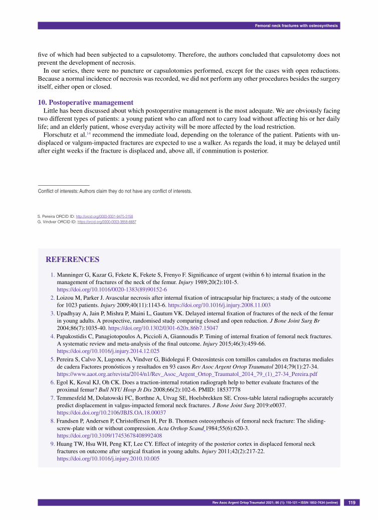
Lastly, it is important to highlight that a sequence for the placement of screws exists, which ensures a minimum possibility of reduction loss during the process. First, the lower middle screw should be placed, in order to resist varus collapse, but without allowing definitive compression. Then, the posterior screw should be placed, which reduces the chance of posterior fall. Finally, the anterior and upper screws should be placed. Once the screws are positioned, the upper screws should be first compressed, in order to avoid a varus axis deviation, which may occur if the lower screw is first compressed (Figure 4).
118
F. m. bidolegui et al.
Rev Asoc Argent Ortop Traumatol 2021; 86 (1): 110-121 • ISSN 1852-7434 (online)
9. capsulotomyThere are different opinions on the decision of conducting a capsulotomy to decompress hematoma in femoral
neck fractures in a young adult.Several studies report the benefits of this practice. Ly & Swiontkowski45 evaluated the role of capsulotomy in
the treatment of femoral neck fractures, and concluded that controversy is still standing. The authors advise this practice until new information arises which contradicts the recommendation (prospective studies with control groups).
However, in their paper «The Influence of Capsulotomy for Intracapsular Hip Fractures», Triplet et al.46 evalu-ated 62 young patients, 32 of which were subjected to a capsulotomy. Of all the patients, seven developed necrosis,
Figure 4. A. Anteroposterior radiograph of a femoral neck fracture. b. Surgical profile view of the fracture. c. Postoperative anteroposterior radiograph. Four screws correctly fixed with a rhomboid construction can be seen. d. Postoperative lateral radiograph. Correctly fixed screws can be seen.
A
c
b
D
Femoral neck fractures with osteosynthesis
Rev Asoc Argent Ortop Traumatol 2021; 86 (1): 110-121 • ISSN 1852-7434 (online) 119
five of which had been subjected to a capsulotomy. Therefore, the authors concluded that capsulotomy does not prevent the development of necrosis.
In our series, there were no puncture or capsulotomies performed, except for the cases with open reductions. Because a normal incidence of necrosis was recorded, we did not perform any other procedures besides the surgery itself, either open or closed.
10. Postoperative managementLittle has been discussed about which postoperative management is the most adequate. We are obviously facing
two different types of patients: a young patient who can afford not to carry load without affecting his or her daily life; and an elderly patient, whose everyday activity will be more affected by the load restriction.
Florschutz et al.14 recommend the immediate load, depending on the tolerance of the patient. Patients with un-displaced or valgum-impacted fractures are expected to use a walker. As regards the load, it may be delayed until after eight weeks if the fracture is displaced and, above all, if conminution is posterior.
S. Pereira ORCID ID: http://orcid.org/0000-0001-9475-3158G. Vindver ORCID ID: https://orcid.org/0000-0003-3858-6687
reFerences
1. Manninger G, Kazar G, Fekete K, Fekete S, Frenyo F. Significance of urgent (within 6 h) internal fixation in the management of fractures of the neck of the femur. Injury 1989;20(2):101-5. https://doi.org/10.1016/0020-1383(89)90152-6
2. Loizou M, Parker J. Avascular necrosis after internal fixation of intracapsular hip fractures; a study of the outcome for 1023 patients. Injury 2009;40(11):1143-6. https://doi.org/10.1016/j.injury.2008.11.003
3. Upadhyay A, Jain P, Mishra P, Maini L, Gautum VK. Delayed internal fixation of fractures of the neck of the femur in young adults. A prospective, randomised study comparing closed and open reduction. J Bone Joint Surg Br 2004;86(7):1035-40. https://doi.org/10.1302/0301-620x.86b7.15047
4. Papakostidis C, Panagiotopoulos A, Piccioli A, Giannoudis P. Timing of internal fixation of femoral neck fractures. A systematic review and meta-analysis of the final outcome. Injury 2015;46(3):459-66. https://doi.org/10.1016/j.injury.2014.12.025
5. Pereira S, Calvo X, Lugones A, Vindver G, Bidolegui F. Osteosíntesis con tornillos canulados en fracturas mediales de cadera Factores pronósticos y resultados en 93 casos Rev Asoc Argent Ortop Traumatol 2014;79(1):27-34. https://www.aaot.org.ar/revista/2014/n1/Rev_Asoc_Argent_Ortop_Traumatol_2014_79_(1)_27-34_Pereira.pdf
6. Egol K, Koval KJ, Oh CK. Does a traction-internal rotation radiograph help to better evaluate fractures of the proximal femur? Bull NYU Hosp Jt Dis 2008;66(2):102-6. PMID: 18537778
7. Temmesfeld M, Dolatowski FC, Borthne A, Utvag SE, Hoelsbrekken SE. Cross-table lateral radiographs accurately predict displacement in valgus-impacted femoral neck fractures. J Bone Joint Surg 2019:e0037. https://doi.doi.org/10.2106/JBJS.OA.18.00037
8. Frandsen P, Andersen P, Christoffersen H, Per B. Thomsen osteosynthesis of femoral neck fracture: The sliding-screw-plate with or without compression. Acta Orthop Scand 1984;55(6):620-3. https://doi.org/10.3109/17453678408992408
9. Huang TW, Hsu WH, Peng KT, Lee CY. Effect of integrity of the posterior cortex in displaced femoral neck fractures on outcome after surgical fixation in young adults. Injury 2011;42(2):217-22. https://doi.org/10.1016/j.injury.2010.10.005
––––––––––––––––––Conflict of interests: Authors claim they do not have any conflict of interests.
120
F. m. bidolegui et al.
Rev Asoc Argent Ortop Traumatol 2021; 86 (1): 110-121 • ISSN 1852-7434 (online)
10. Rawall S, Bali K, Upendra B, Garg B, Yadav CS, Jayaswal A. Displaced femoral neck fractures in the young: significance of posterior comminution and raised intracapsular pressure. Arch Orthop Trauma Surg 2012;132:73-9. https://doi.org/10.1007/s00402-011-1395-1
11. Rikli D, Goldhahn S, Blauth M, Mehta S, Cunningham M. PIP Study group. Optimizing intraoperative imaging during proximal femoral fracture fixation - A performance improvement program for surgeons. Injury 2018;49(2):339-44. https://doi.org/10.1016/j.injury.2017.11.024
12. Poyanli OS, Soylemez S, Ozkut AT, Uygur E, Kemah B. Precise placement of lag screws in operative treatment of trochanteric femoral fractures with a new guide system Injury 2015;46(11):2190-95. https://doi.org/10.1016/j.injury.2015.06.003
13. Yang J, Lin L, Chao K, Chuang S, Wu C. Risk factors for nonunion in patients with intracapsular femoral neck fractures treated with three cannulated screws placed in either a triangle or an inverted triangle configuration. J Bone Joint Surg Am 2013;95(1):61-9. https://doi.org/10.2106/JBJS.K.01081
14. Florschutz A, Langford J, Haidukewych G, Koval K. Femoral neck fractures: current management. J Orthop Trauma 2015;29(3):121-9. https://doi.org/10.1097/BOT.0000000000000291
15. Chua D, Jaglal SB, Schatzker J. Predictors of early failure of fixation in the treatment of displaced subcapital hip fractures. J Orthop Trauma 1998;12(4):230-4. https://doi.org/10.1097/00005131-199805000-00002
16. Krischak G , Beck A, Wachter N, Jakob R, Kinzl L, Suger G. Relevance of primary reduction for the clinical outcome of femoral neck fractures treated with cancellous screws. Arch Orthop Trauma Surg 2003;123(8):404-9. https://doi.org/10.1007/s00402-003-0571-3
17. Su Y, Chen W, Zhang Q, Li B, Guo M. An irreducible variant of femoral neck fracture: A minimally traumatic reduction technique. Injury 2011;42(2):140-5. https://doi.org/10.1016/j.injury.2010.05.008
18. Yu S, Xu X, Pandey N, Zhao Y, Jing J. A safe percutaneous technique for the reduction of irreducible femoral neck fractures using ultrasound localization of the femoral vascular and nervous structures at the hip. Medicine (Baltimore) 2019;98(15): e15163. https://doi.org/10.1097/MD.0000000000015163
19. Mahajan RH, Kumar S, Mishra B. Technique for gentle accurate reproducible closed reduction of intracapsular fracture of neck of femur. Injury 2017;48(3):789-790. https://doi.org/10.1016/j.injury.2016.10.030.
20. Watson-Jones R. Fractures of the neck of the femur. Br J Surg 1936;23(92):787-808. https://doi.org/10.1002/bjs.1800239213
21. Smith-Petersen MN. A new supra-articular subperiosteal approach to the hip joint. J Bone Joint Surg Am 1917;s2-15(8):59222. Slobogean GP, Sprague SA, Scott T, McKee M, Bhandari M. Management of young femoral neck fractures: is there a consensus? Injury 2015;46(3):435-40. https://doi.org/10.1016/j.injury.2014.11.028
23. Molnar RB, Routt ML. Open reduction of intracapsular hip fractures using a modified Smith-Petersen surgical exposure. J Orthop Trauma 2007;21(7):490-4. https://doi.org/10.1097/BOT.0b013e31804a7f7f
24. Lichstein P, Kleimeyer J, Githens M, Vorhies J, Gardner M, Bellino M, et al. Does the Watson-Jones or modified Smith-Petersen approach provide superior exposure for femoral neck fracture fixation? Clin Orthop Relat Res 2018;476(7):1468-76. https://doi.org/ 10.1097/01.blo.0000533627.07650.bb
25. Giannoudis P, Panteli M. Biomechanical rationale for implant choices in femoral neck fracture fixation in the non-elderly. Injury 2015;46(3):445-52. https://doi.org/10.1016/j.injury.2014.12.031
26. Bhandari M, Tornetta P 3rd, Hanson B, Swiontkowski M. Optimal internal fixation for femoral neck fractures: multiple screws or sliding hip screws? J Orthop Trauma 2009;23(6):403-7. https://doi.org/10.1097/BOT.0b013e318176191f
27. Wu-ren Hou, Min-ou Xu. [Comparison among three cannulated screws and dynamic hip screw combined with antirotation screw for comminuted fractures of femoral neck]. China J Orthop Traumatol 2015;28(9):796-801. [En chino] PMID: 26647559
28. Cha Y, Yoo J, Wang S, Kim K, Choy W, Hwang S. Biomechanical evaluation of internal fixation of Pauwels type III femoral neck fractures: A systematic review of various fixation methods. Clin Orthop Surg 2019;11(1):1-14. https://doi.org/10.4055/cios.2019.11.1.1
29. Fixation using Alternative Implants for the Treatment of Hip fractures (FAITH) Investigators. Fracture fixation in the operative management of hip fractures (FAITH): an international, multicentre, randomised controlled trial. Lancet 2017;389(10078):1519-27. https://doi.org/10.1016/S0140-6736(17)30066-1
30. Ma J, Kuang M, Xing F, Zhao Y, Chen H, Zhang L. Sliding hip screw versus cannulated cancellous screws for fixation of femoral neck fracture in adults: A systematic review. Int J Surg 2018;52:89-97. https://doi.org/10.1016/j.ijsu.2018.01.050

Femoral neck fractures with osteosynthesis
Rev Asoc Argent Ortop Traumatol 2021; 86 (1): 110-121 • ISSN 1852-7434 (online) 121
31. Kloen P, Rubel I, Lyden J, Helfet D. Subtrochanteric fracture after cannulated screw fixation of femoral neck fractures: A report of four cases. J Orthop Trauma 2003;17(3):225-9. https://doi.org/10.1097/00005131-200303000-00013
32. Sensoz E, Mehmet F, Acar V. Finite element analysis of the impact of screw insertion distal to the trochanter minor on the risk of iatrogenic subtrochanteric fracture. Proc Inst Mech Eng H 2018;232(8):807-18. https://doi.org/10.1177/0954411918789963
33. Swiontkowski MF, Harrington RM, Keller TS. Torsion and bending analysis of internal fixation techniques for femoral neck fracture: the roll of implant design and bone density. J Orthop Res 1987;5:433-44. https://doi.org/10.1002/jor.1100050316
34. Maurer S, Wright K, Koval K, Zuckerman F. Two or three screws for fixation of femoral neck fractures? Am J Orthop (Belle Mead NJ) 2003;32(9):438-42. PMID: 14560825
35. Krastman P, van den Bent RP, Krijnen P, Schipper IB. Two cannulated hip screws for femoral neck fractures: treatment of choice or asking for trouble? Arch Orthop Trauma Surg 2006;126(5):297-303. https://doi.org/10.1007/s00402-006-0143-4
36. Zlowodzki M, Weening B, Petrisor B, Bhandari M. The value of washers in cannulated screw fixation of femoral neck fractures. Trauma 2005;59(4):969-75. https://doi.org/10.1097/01.ta.0000188130.99626.8c
37. Zlowodzki M, Wijdicks C, Armitage B, Cole A. Value of washers in internal fixation of femoral neck fractures with cancellous screws: a biomechanical evaluation. J Orthop Trauma 2015;29(2):e69-72. https://doi.org/10.1097/BOT.0000000000000172
38. Selvan VT, Oakley J, Rangan A, Al-Lami MK. Optimum configuration of cannulated hip screw for the fixation of intracapsular hip fracture: a biomechanical study. Injury 2004;35(2):136-41. https://doi.org/10.1016/S0020-1383(03)00059-7
39. Guo I, Dong W, Qin S, Zhang Y. Definition of ideal configuration for femoral neck screw fixation in older people. Sci Rep 2019;9:12895. https://doi.org/10.1038/s41598-019-48258-2
40. Gümüştaş SA, Tosun HB, Ağır İ, Orak MM, Onay T, Okçu G. Influence of number and orientation of screws on stability in the internal fixation of unstable femoral neck fractures. Acta Orthop Traumatol Turc 2014;48(6):673-8. https://doi.org/10.3944/AOTT.2014.14.0088
41. Collinge C, Mir H. Application of a medial buttress plate may prevent many treatment failures seen after fixation of vertical femoral neck fractures in young adults. Med Hyp 2015;84(5):429-33. https://doi.org/10.1016/j.mehy.2015.01.029
42. Ye Y, Chen K, Tian K, Li W, Mauffrey C, Hak DJ. Medial buttress plate augmentation of cannulated screw fixation in vertical unstable femoral neck fractures: surgical technique and preliminary results. Injury 2017;48(10):2189-93. https://doi.org/10.1016/j.injury.2017.08.017
43. Hoffman J, Kellam J, Routt C, Gary J. Is the cranial and posterior screw of the “inverted triangle” configuration for femoral neck fractures safe? J Orthop Trauma 2019;33(7):331-4. https://doi.org/10.1097/BOT.0000000000001461
44. Gurusamy K, Parker MJ, Rowlands TK. The complication of displaced intracapsular fractures of the hip. The effect of screw positioning and angulation on fracture healing. J Bone Joint Surg Br 2005;87:632-4. https://doi.org/10.1302/0301-620X.87B5.15237
45. Ly TV, Swiontkowski MF. Treatment of femoral neck fractures in young adults. Instr Course Lect 2009;58:69-81. PMID: 19385521
46. Triplet JJ, Sullivan B, Taylor BC. The influence of capsulotomy for intracapsular hip fractures. J Long Term Eff Med Implants 2018;28(3):187-92. https://doi.org/10.1615/JLongTermEffMedImplants.2018028556
Related Documents