REVIEW Open Access Treating primary immunodeficiencies with defects in NK cells: from stem cell therapy to gene editing C. Eguizabal 1,2* , L. Herrera 1,2 , M. Inglés-Ferrándiz 1,2 and J. C. Izpisua Belmonte 3 Abstract Primary immunodeficiency diseases (PIDs) are rare diseases that are characterized by genetic mutations that damage immunological function, defense, or both. Some of these rare diseases are caused by aberrations in the normal development of natural killer cells (NKs) or affect their lytic synapse. The pathogenesis of these types of diseases as well as the processes underlying target recognition by human NK cells is not well understood. Utilizing induced pluripotent stem cells (iPSCs) will aid in the study of human disorders, especially in the PIDs with defects in NK cells for PID disease modeling. This, together with genome editing technology, makes it possible for us to facilitate the discovery of future therapeutics and/or cell therapy treatments for these patients, because, to date, the only curative treatment available in the most severe cases is hematopoietic stem cell transplantation (HSCT). Recent progress in gene editing technology using CRISPR/Cas9 has significantly increased our capability to precisely modify target sites in the human genome. Among the many tools available for us to study human PIDs, disease- and patient-specific iPSCs together with gene editing offer unique and exceptional methodologies to gain deeper and more thorough understanding of these diseases as well as develop possible alternative treatment strategies. In this review, we will discuss some immunodeficiency disorders affecting NK cell function, such as classical NK deficiencies (CNKD), functional NK deficiencies (FNKD), and PIDs with involving NK cells as well as strategies to model and correct these diseases for further study and possible avenues for future therapies. Keywords: Primary immunodeficiency diseases (PIDs), Induced pluripotent stem cells (iPSCs), Gene editing, CRISPR- Cas9, Stem cell therapy, Hematopoietic stem cells, Natural killer cells Background Primary immunodeficiency disorders (PIDs) are rare diseases caused by genetic mutations that damage immunological function, defense, or both. PIDs refer to over 130 disorders that result from developmental and/or functional defects in one or more cell types of the immune system. PIDs are generally classified as disorders of adaptive immunity (B cell, T cell, or combined immunodeficiencies) or innate immunity (NK cells, phagocyte, and complement disorders). Some of these diseases affect natural killer (NK) cells [1, 2]. NK cells are lymphocytes of the innate immune system poised to deliver a response immediately after recogniz- ing specific signals of “danger,” stress, or foreign origin. NK cells are initially defined as rapid cell-mediated cyto- toxicity cells, though they also initiate a slower receptor- mediated apoptosis, and efficiently produce soluble mediators, such as cytokines, and provide contact- dependent co-stimulation. Hence, NK cells contribute to the regulation of immune responses, in the surveillance of stress and cancer cells, and in the defense against © The Author(s). 2020 Open Access This article is licensed under a Creative Commons Attribution 4.0 International License, which permits use, sharing, adaptation, distribution and reproduction in any medium or format, as long as you give appropriate credit to the original author(s) and the source, provide a link to the Creative Commons licence, and indicate if changes were made. The images or other third party material in this article are included in the article's Creative Commons licence, unless indicated otherwise in a credit line to the material. If material is not included in the article's Creative Commons licence and your intended use is not permitted by statutory regulation or exceeds the permitted use, you will need to obtain permission directly from the copyright holder. To view a copy of this licence, visit http://creativecommons.org/licenses/by/4.0/. The Creative Commons Public Domain Dedication waiver (http://creativecommons.org/publicdomain/zero/1.0/) applies to the data made available in this article, unless otherwise stated in a credit line to the data. * Correspondence: [email protected] 1 Cell Therapy, Stem Cells and Tissues Group, Biocruces Bizkaia Health Research Institute, Barakaldo, Spain 2 Research Unit, Basque Center for Blood Transfusion and Human Tissues, Osakidetza, Galdakao, Spain Full list of author information is available at the end of the article Eguizabal et al. Stem Cell Research & Therapy (2020) 11:453 https://doi.org/10.1186/s13287-020-01964-5

Welcome message from author
This document is posted to help you gain knowledge. Please leave a comment to let me know what you think about it! Share it to your friends and learn new things together.
Transcript

REVIEW Open Access
Treating primary immunodeficiencies withdefects in NK cells: from stem cell therapyto gene editingC. Eguizabal1,2*, L. Herrera1,2, M. Inglés-Ferrándiz1,2 and J. C. Izpisua Belmonte3
Abstract
Primary immunodeficiency diseases (PIDs) are rare diseases that are characterized by genetic mutations thatdamage immunological function, defense, or both. Some of these rare diseases are caused by aberrations in thenormal development of natural killer cells (NKs) or affect their lytic synapse. The pathogenesis of these types ofdiseases as well as the processes underlying target recognition by human NK cells is not well understood. Utilizinginduced pluripotent stem cells (iPSCs) will aid in the study of human disorders, especially in the PIDs with defects inNK cells for PID disease modeling. This, together with genome editing technology, makes it possible for us tofacilitate the discovery of future therapeutics and/or cell therapy treatments for these patients, because, to date, theonly curative treatment available in the most severe cases is hematopoietic stem cell transplantation (HSCT). Recentprogress in gene editing technology using CRISPR/Cas9 has significantly increased our capability to precisely modifytarget sites in the human genome. Among the many tools available for us to study human PIDs, disease- andpatient-specific iPSCs together with gene editing offer unique and exceptional methodologies to gain deeper andmore thorough understanding of these diseases as well as develop possible alternative treatment strategies. In thisreview, we will discuss some immunodeficiency disorders affecting NK cell function, such as classical NK deficiencies(CNKD), functional NK deficiencies (FNKD), and PIDs with involving NK cells as well as strategies to model andcorrect these diseases for further study and possible avenues for future therapies.
Keywords: Primary immunodeficiency diseases (PIDs), Induced pluripotent stem cells (iPSCs), Gene editing, CRISPR-Cas9, Stem cell therapy, Hematopoietic stem cells, Natural killer cells
BackgroundPrimary immunodeficiency disorders (PIDs) are rarediseases caused by genetic mutations that damageimmunological function, defense, or both. PIDs refer toover 130 disorders that result from developmental and/orfunctional defects in one or more cell types of the immunesystem. PIDs are generally classified as disorders ofadaptive immunity (B cell, T cell, or combined
immunodeficiencies) or innate immunity (NK cells,phagocyte, and complement disorders). Some of thesediseases affect natural killer (NK) cells [1, 2].NK cells are lymphocytes of the innate immune system
poised to deliver a response immediately after recogniz-ing specific signals of “danger,” stress, or foreign origin.NK cells are initially defined as rapid cell-mediated cyto-toxicity cells, though they also initiate a slower receptor-mediated apoptosis, and efficiently produce solublemediators, such as cytokines, and provide contact-dependent co-stimulation. Hence, NK cells contribute tothe regulation of immune responses, in the surveillanceof stress and cancer cells, and in the defense against
© The Author(s). 2020 Open Access This article is licensed under a Creative Commons Attribution 4.0 International License,which permits use, sharing, adaptation, distribution and reproduction in any medium or format, as long as you giveappropriate credit to the original author(s) and the source, provide a link to the Creative Commons licence, and indicate ifchanges were made. The images or other third party material in this article are included in the article's Creative Commonslicence, unless indicated otherwise in a credit line to the material. If material is not included in the article's Creative Commonslicence and your intended use is not permitted by statutory regulation or exceeds the permitted use, you will need to obtainpermission directly from the copyright holder. To view a copy of this licence, visit http://creativecommons.org/licenses/by/4.0/.The Creative Commons Public Domain Dedication waiver (http://creativecommons.org/publicdomain/zero/1.0/) applies to thedata made available in this article, unless otherwise stated in a credit line to the data.
* Correspondence: [email protected] Therapy, Stem Cells and Tissues Group, Biocruces Bizkaia HealthResearch Institute, Barakaldo, Spain2Research Unit, Basque Center for Blood Transfusion and Human Tissues,Osakidetza, Galdakao, SpainFull list of author information is available at the end of the article
Eguizabal et al. Stem Cell Research & Therapy (2020) 11:453 https://doi.org/10.1186/s13287-020-01964-5

infections [3]. While these functions are not exclusive toNK cells, the ability to quickly mediate effector functionswithout the need of further development and/or matur-ation is a crucial distinctive feature of mature NK cellsand cytotoxic T lymphocytes (CTLs), both of which areefficient at mediating cytotoxicity.NK cell effector functions happen after ligation of
germline-encoded receptors and involve the secretion ofcytolytic molecules. These molecules are contained inpre-formed lytic granules of resting human NK cells. Asa result, the cytolytic process needs to be well controlledand may involve additional or enhanced mechanisms forcontrolling the secretion of lytic granule contents [4].Several NK cell deficiencies have been identified as
being present in PIDs, including some characterized byan impediment in their development (classic NK defi-ciencies or CNKD) or by an aberrant formation of thelytic synapse (functional NK deficiencies or FNKD). Todate, only one FNKD has been shown to impair only NKcell function. Many studies on how the lytic synapse isformed have been performed with T cells from patientswith PIDs. Though cytotoxic lymphocytes have criticalroles in host defense and immune regulation, NK celldeficiency can add to the clinical phenotypes [5].In this review, we will focus on the most relevant PIDs
with established functional defects in NK cells, such asCNKD [5] with mutations in GATA2 [6–8], MCM4,RTEL1, GINS1, and IRF8 genes; FNKD, with defects inthe FCGR3A gene and PIDs involving NK cells, likeWiskott–Aldrich syndrome (WAS) [9–11], Chèdiak–Higashi syndrome (CHS) [12, 13], Griscelli syndrometype II (GS2) [14, 15], familial hemophagocytic lympho-histiocytosis 2 (FHL2) [16, 17] and 4 (FHL4) [18–20],APDS and APDS-2 [21], severe combined immunodefi-ciency due to ADA deficiency (ADA-SCID), X-linkedsevere combined immunodeficiency (X-SCID), and X-linked chronic granulomatous disease (X-GCD) asdescribed in detail in Fig. 1 and Table 1 [5].The pathogenesis and processes underlying target rec-
ognition by human NK cells of all the PIDs mentionedbefore are still not well understood. For this reason, theuse of induced pluripotent stem cells (iPSCs), as a dis-ease modeling tool, will help the study of human NK celldisorders with the final goal of elucidating the diseasemechanisms to find novel gene and cell therapies for thetreatment of those PIDs.
iPSC generation as a disease modeling toolProf. Yamanaka in 2006 made the pivotal finding thatsomatic cells can be reprogrammed to pluripotent cellsby introducing a set of transcription factors, named in-duced pluripotent stem cells (iPSCs) [22]. This novelwork used fibroblasts from adult mouse tail tips in orderto create cells that resembled mouse embryonic stem
cells morphologically and in gene expression. Essentially,these iPSCs could be proliferated and differentiatedin vitro and in vivo, giving rise to mature cell types fromthe embryonic germ layers, endoderm, mesoderm, andectoderm as well as to germ cells. One year later, a par-allel procedure was successfully used to generate iPSCsfrom human fibroblasts. This achievement permitted theuse of this technology in the in vitro study of human dis-eases [23]. The most commonly used cell type to gener-ate iPSCs is the fibroblast, though other cell types havealso been used to derive iPSCs (keratinocytes, epithelialcells, blood cells, etc.) [24]. Later, several groups re-ported the success of generating mature and fully differ-entiated blood cells, including NK cells [25].Substantial improvement has also been made in the
development of other strategies to accomplish repro-gramming. Generation of iPSCs was initially based onusing four retroviral vectors [26]. Recently, non-integrative methods (episomal vectors and Sendai virus,among others) have been developed to reprogram in asafer manner [27, 28]. In 2011, Eguizabal and colleagueswere able to reprogram mature cells, such as dermalfibroblasts and blood cells, into iPSCs and later differen-tiate them into any desired cell type [25, 29–31]. Todate, several studies have been reported using thistechnology to generate patient-derived iPSCs for diseasemodeling and for future applications in cell and genetherapies [32–35].One of the first reports of a PID patient-derived iPSC
line was from a patient with an adenosine deaminase(ADA) deficit, which causes severe combined immuno-deficiency (ADA-SCID) [36]. While no follow-up studiesusing the ADA-SCID iPSCs have been reported to date,this crucial publication presented a proof of principlethat iPSCs can be generated from patients with PIDs forinvestigating and correcting this disease. In 2011,Pessach and colleagues also published the successfulgeneration of iPSCs from PID patients caused by specificmutations [37]. Another example of patient-specificiPSC generation was performed with cells from Fanconianemia patients. Izpisua Belmonte’s group conductedthis study, and interestingly, iPSCs could only be repro-grammed after correction of their mutation and gave riseto phenotypically normal myeloid and erythroid progeni-tors [38]. In addition, two papers from Malech’s labora-tory show the generation of iPSCs using peripheralhematopoietic stem cells from five different genotypes ofchronic granulomatous disease (CGD) patients. Both thepatient iPSCs and patient somatic cells before being re-programed into iPSCs can be corrected. Then, from thecorrected iPSCs by in vitro myeloid differentiation,normal granulocytes were generated [39, 40]. In 2016,Laskowski and colleagues generated iPSCs from aWiskott–Aldrich syndrome (WAS) patient and the WAS
Eguizabal et al. Stem Cell Research & Therapy (2020) 11:453 Page 2 of 10
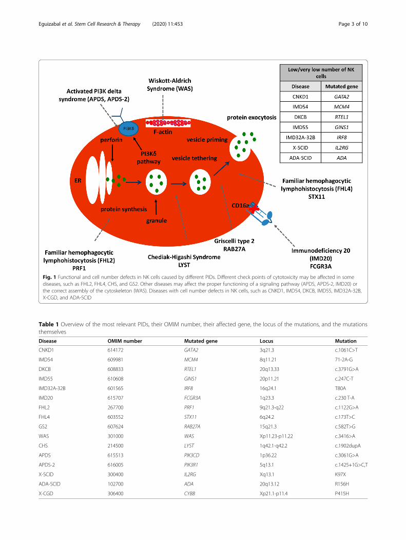
Table 1 Overview of the most relevant PIDs, their OMIM number, their affected gene, the locus of the mutations, and the mutationsthemselves
Disease OMIM number Mutated gene Locus Mutation
CNKD1 614172 GATA2 3q21.3 c.1061C>T
IMD54 609981 MCM4 8q11.21 71-2A-G
DKCB 608833 RTEL1 20q13.33 c.3791G>A
IMD55 610608 GINS1 20p11.21 c.247C-T
IMD32A-32B 601565 IRF8 16q24.1 T80A
IMD20 615707 FCGR3A 1q23.3 c.230 T-A
FHL2 267700 PRF1 9q21.3-q22 c.1122G>A
FHL4 603552 STX11 6q24.2 c.173T>C
GS2 607624 RAB27A 15q21.3 c.582T>G
WAS 301000 WAS Xp11.23-p11.22 c.3416>A
CHS 214500 LYST 1q42.1-q42.2 c.1902dupA
APDS 615513 PIK3CD 1p36.22 c.3061G>A
APDS-2 616005 PIK3R1 5q13.1 c.1425+1G>C,T
X-SCID 300400 IL2RG Xq13.1 K97X
ADA-SCID 102700 ADA 20q13.12 R156H
X-CGD 306400 CYBB Xp21.1-p11.4 P415H
Fig. 1 Functional and cell number defects in NK cells caused by different PIDs. Different check points of cytotoxicity may be affected in somediseases, such as FHL2, FHL4, CHS, and GS2. Other diseases may affect the proper functioning of a signaling pathway (APDS, APDS-2, IMD20) orthe correct assembly of the cytoskeleton (WAS). Diseases with cell number defects in NK cells, such as CNKD1, IMD54, DKCB, IMD55, IMD32A-32B,X-CGD, and ADA-SCID
Eguizabal et al. Stem Cell Research & Therapy (2020) 11:453 Page 3 of 10

locus was targeted in order to produce corrected WAS-iPSCs. This group proved that the defects showed byWAS-iPSC-derived lymphoid cells were completelycorrected for potential therapeutic use [41].Further studies with PID-mutation-corrected iPSCs
are needed to determine whether they are capable of dif-ferentiating into any target cell type as well as to gainmore thorough knowledge of the mechanisms behindspecific mutations [42]. Altogether, these publicationsreveal that disease–patient-specific iPSCs are an excep-tional tool for improved understanding of humandiseases and to develop novel and disease-specific celland gene treatment approaches (Fig. 2).
Gene editing revolutionDuring the DNA replication process, after experimentalmanipulation by using endonucleases or after exposureto chemotherapy or ionizing radiation, DNA damagecan occur. Luckily, DNA repair mechanisms are able torepair this damage, thus avoiding DNA mutations thatcan develop into disease. This repair process is the basisof gene editing therapies, which follow two main repairpathways for double-strand breaks (DSBs) in the DNA:
non-homologous end joining (NHEJ) and homologousrecombination (HR) [43].In the 1980s, pioneering scientists Prof. Capecchi,
Smithies, and Evens discovered the HR pathway thatrepaired genes in mammalian cells. Later in 2007,they were awarded the Nobel Prize in Medicine fortheir findings in introducing gene modifications inmice models by using embryonic stem cells and HR-mediated gene editing. A few years later, Prof. Jasinenhanced gene targeting in mammalian cells usingHR from yeast endonuclease I-SceI by using meganu-cleases. Since that time, next-generation gene editingtools have been used such as zinc-finger nucleases(ZFNs) and transcription activator-like effector nucle-ases (TALENs), which all edit DNA in combinationwith the FokI endonuclease [43].In the late 1980s, a bizarre topology at the 3′ end of
the alkaline phosphatase gene was revealed in E. coli.This was a clustered regularly interspaced short palin-dromic repeats (CRISPR) array, which is now one of themost commonly used gene editing technologies. Later,in 2005, the molecular mechanism was revealed, whichshowed that CRISPR arrays are transcribed into RNA to
Fig. 2 Obtaining hiPS cells from different cell sources in order to use them as a disease model, drug developmental model, or stem cell research.hiPS cells from a PID patient may be corrected with the goal of developing a cell-based therapy. Adapted from [37]
Eguizabal et al. Stem Cell Research & Therapy (2020) 11:453 Page 4 of 10

cleave and load into CRISPR-associated (Cas) proteins(Cas9) [43].For many years, scientists have been looking for a tool
to induce or repair mutations in a targeted manner.Several techniques have been used in the past, such asengineered meganucleases, ZFNs, and TALENs, all withlimited success, due to the fact that they are labor-intensive, expensive, or both. CRISPR-based methodolo-gies together with RNA-guided nuclease activity meantthat, theoretically, DSBs in eukaryotes can be induced,which was very hard to achieve before. In general, DSBsare repaired by DNA repair pathways, with NHEJ havingthe potential to induce indels—mutations caused by ran-dom insertion or deletion of nucleotides at the DSB site,whereas the HR repair pathway is more precise [44].The differences between engineered meganucleases,
ZFNs, TALENs, and CRISPR/Cas9 nucleases are de-scribed in detail in Fig. 3.Engineered meganucleases are derived from a huge
family of natural homing endonucleases [45], and somehave been designed with diverse strategies (structure-based design and yeast surface display) to identifynatural target sites in the genome [46, 47]. Historically,natural meganucleases have been the gold standard forspecificity, but have not been fully evaluated for transla-tional development.Zinc-finger nucleases (ZFNs) are artificial restriction
enzymes in which a DNA-cleavage domain from theenzyme FokI is fused to a zinc-finger DNA-bindingdomain [48, 49]. The nuclease domain must dimerize to
cut DNA efficiently. Consequently, a pair of ZFNsoriented correctly to allow dimerization must beengineered for each target site. A variety of strategiescan be engineered for novel target sites for zinc-fingerDNA-binding domains (modular assembly, phage dis-play, bacteria-based two-hybrid and one-hybrid systems,and combinatorial approaches) [50]. Though ZFN designstrategies are constantly being enhanced, engineering ofthese recombinant proteins with high activity and speci-ficity still remains a challenge. However, the highest-quality ZFNs generated are a mixture of phage andmodular display that are in an engineered T cell clinicaltrial [51].TAL effector nucleases (TALENs) are artificial proteins
with a similar structure to ZFNs with the fusion of theenzyme FokI nuclease domain to an engineered DNA-binding domain. This DNA-binding domain is engi-neered by gathering serial TAL repeats [52]. Each repeatmediates the interaction with a single nucleotidethrough a two amino acid repeat variable di-residue(RVD) that can be described by a simple code [53].Thus, generating a highly active TALEN is easier thangenerating a highly active ZFN. Moreover, TAL repeatsthat use engineered RVDs and not natural ones are nowbeing used to build TALENs and may have increasedspecificity over natural RVDs, though this still necessi-tates further study. A pair of TALENs must be engi-neered to recognize target sites of interest, as with ZFNs;thus, TALENs using TAL repeats with RVDs have su-perior specificity when compared to ZFNs.
Fig. 3 Diagram of the different types of existing gene editing tools: engineered meganucleases, zinc-finger nucleases (ZFNs), TAL effectornucleases (TALENs), and CRISPR/Cas9 nucleases
Eguizabal et al. Stem Cell Research & Therapy (2020) 11:453 Page 5 of 10
CRISPR/Cas9 nucleases (CRISPR stands for “clusteredregularly interspaced short palindromic repeats”) origin-ate from the immune system of bacteria and archea [54].The specificity of the CRISPR/Cas9 nuclease system isbased on RNA–DNA Watson–Crick base pairing insteadof protein–DNA interaction. In this system, a single-guide RNA (sgRNA) is constructed for the 20 nucleo-tides matching the target region. This target site must benext to a proto-spacer adjacent motif (PAM) sequence,which the Cas9 protein uses to recognize target sites[55]. The Cas9 protein, together with the sgRNA, is cap-able of unwinding double-stranded DNA, cross-examineif the single-guide adequately matches the target site,and generate a double-strand break in order to repair orintroduce mutations. CRISPR/Cas9 nucleases can beengineered very simply since they are active at thedesired target site.Gene editing technology is a powerful tool currently
being used in basic research, but the ultimate aim is totranslate these tools to be applied in therapeutic treat-ments. Being able to use gene editing technology in theclinic stems from the possibility of treating monogenicdiseases by developing a novel method to correct thedisease-associated mutation [56, 57]. There are severalcompanies (Cellectis, Sangamo Therapeutics, EditasCompany, CRISPR Therapeutics, Caribou Biosciences,Precision Biosciences, and Intellia Therapeutics) devel-oping gene editing-based approaches to treat monogenicdiseases like β-thalassemia, sickle cell anemia, cysticfibrosis (CF), hemophilia, Duchenne muscular dystrophy(DMD), alpha1-antitrypsin deficiency (A1ATD), Hun-tington’s disease, lysosomal storage disorders (LSDs),among others [44]. Unfortunately, no gene editing-basedstrategies to treat PIDs have been developed yet, butsurely, they are coming soon.Certainly, the use of gene editing tools in patient-
specific iPS cells will aid in the development of futuretreatments aimed at correcting the point mutations inPIDs with defects in NK cells.
Current gene and cell therapies for PIDs withdefects in NK cellsThe first time HSCT was used as a therapeutic optionfor treating PID in a severe combined immunodeficiency(SCID) patient was in 1965 [58], as shown in Fig. 4.Since then, HSCT has been the standard care for SCIDpatients with great survival rates [60–62]. However, de-pending on the HLA-match, HSCT could have somecomplications. On the one hand, when an HLA-identicalsibling donor is available, the success rates are very high.On the other hand, a less well-watched HLA allogenicdonor, such as haploidentical family members or unre-lated donors, could lead to some serious risks. Amongthese risks, the more worrisome ones are graft rejection
and graft versus host disease (GvHD). If either of theseconditions occur, patients require an urgent restorationof hematopoiesis to prevent complications from pro-longed pancytopenia [63]. In order to avoid these risks,during the last 20 years, genetic manipulation of a pa-tient’s own autologous immune cells has been studiedand is being translated into clinical trials. Key clinicaltrials using g-retroviral vectors for gene correction in ad-enosine deaminase SCID, Wiskott–Aldrich syndrome,and X-linked SCID had moderate hematopoietic celltransduction efficiency. Nevertheless, these trials servedas a proof of concept that gene correction could providea curative therapy since treated patients showed im-provement in immune function. Unfortunately, severalyears after treatment, patients developed leukemia as aconsequence of the activation of pro-oncogenes close tothe g-retroviral vector insertion points [64]. As a conse-quence, this led to the development of lentiviral vectors,which improved the efficacy and safety of the genetherapies. For example, Strimvelis (gene therapy forADA-SCID) is now open in Europe [65, 66] (Fig. 4).Currently, there are very few clinical trials that combinecell and gene therapy that are ongoing for several PIDsas shown in Table 2 [67, 68, 69], but none utilizes geneediting strategies.The triumph of gene therapy in treating PIDs is a
major advancement, though limitations in manufactur-ing disease-specific vectors remain a challenge [70]. Asthis field moves forward, more efficient proceduresoffering wider spread applications arise. Gene editingdefines a group of DNA editing approaches that can besimply designed for point mutations. Recently,programmable nucleases such as ZFNs, TALENs, andCRISPR-Cas9 have been developed as effective methodsfor editing the genome to correct the affected gene inPIDs [49, 71–76].Compared to lentiviral vectors, gene-specific editing
technologies has become a tremendously promising tool,as it has the potential to physiologically regulate geneexpression and prevent genome-wide vector integration.Some of the ongoing efforts are focused on developingsensitive techniques to detect genotoxicity derived fromunintended effects of endonucleases (off-target effects).In the case of CRISPR-Cas9 approaches for HSC
genome editing in PIDs, the design of the donortemplate is challenging and both the nature (single/mul-tiple mutations or deletions in one or more hotspotsdistributed along the gene) and the functional effect ofthe mutation (gain of function versus loss of function)have to be taken into consideration [77].Short donor templates (such as ssODN or linear or
plasmid dsDNA donors) have been used to correct lossof function (LOF) mutations of a single or few nucleo-tides. For example, De Ravin and colleagues [78] could
Eguizabal et al. Stem Cell Research & Therapy (2020) 11:453 Page 6 of 10

repair the mutation in the CYBB gene of CD34+ HSCsfrom patients with the immunodeficiency disorder X-linked chronic granulomatous disease (X-CGD) using achemically modified 100-bp ssODN that resulted in pro-duction of 15–20% functional mature human myeloidand lymphoid cells for up to 5 months.In contrast to small mutations, repair of large deletions
or insertions is not possible with short donor templatesand instead functional complementary DNA (cDNA)templates are inserted to target genes. Encouraging
preclinical studies have been published using this ap-proach for the treatment of X-SCID or X-CGD [79–81]and will be ready to translate to clinical trials soon.However, one limitation to consider for the application
of gene editing tools in a clinical setting might be theengraftment efficiency and HSC functionality of genetic-ally modified cells due to cellular effects of the gene edit-ing machinery. Indeed, global gene expression changeshave been observed upon delivery of CRISPR-Cas9 ma-chinery components into the cells. Immune response to
Table 2 Overview of current available clinical trials for different PIDs
Disease Mutated gene/protein Vector/target cell Conditioning Clinical trial reference
CNKD1 GATA2 Allogeneic HSCT Busulfan/fludarabine/cyclophosphamide/TBI NCT01861106
WAS WAS SIN-LV/BM/PBSCs RIC busulfan/fludarabine NCT01515462
NCT01347346
NCT01347242
NCT01410825
SIN-LV/PBSCs None NCT03837483
X-SCID IL2RG SIN-γRV/BM None NCT01410019
NCT01129544
NCT01175239
SIN-LV/PBSCs Busulfan 6 mg/kg NCT01306019
SIN-LV/BM Busulfan 6 mg/kg NCT01512888
ADA-SCID ADA SIN-LV/BM/PBSCs Busulfan 5 mg/kg NCT01380990
SIN-LV/BM/PBSCs Busulfan 4 mg/kg NCT01852071
NCT02022696
SIN-γRV self-inactivating γRV vector, SIN-LV self-inactivating lentiviral vector, BM bone marrow, PBSCs peripheral blood stem cells, MAC myeloablative conditioning,RIC reduced intensity conditioning, TBI total body irradiation
Fig. 4 Viral vector technology development and its application to human gene therapy. The line represents the timeline of this technology, fromthe 1960s to now. Adapted from [59]
Eguizabal et al. Stem Cell Research & Therapy (2020) 11:453 Page 7 of 10
viral infection, DNA damage response, apoptosis, andcell cycle processes have been reported as the most sig-nificantly enriched gene signatures [82]. The activationof these biological processes might negatively affect HSCstemness and hematopoietic lineage expansion and dif-ferentiation. Further studies are needed in order to bet-ter understand these mechanisms and therefore designmore efficient CRISPR-Cas9 strategies and improve HSCengraftment efficiency.Apart from CRISPR-Cas9, other genome editing tools
have been used to modify genes in different cell types in-cluding HSCs. ZFN and TALEN techniques have beenused to modify the IL2RG locus, which is responsible forSCID [83].Specifically in the case of PID with NK cell defects, a
good example of the evolution of treatment approachesis seen with WAS. The first clinical trial with gene ther-apy in WAS patients was performed using gamma-retroviral vectors. Even if 9 of 10 patients showed partialor complete resolution of immunodeficiency, auto-immunity, and other malignancies, 7 of them developedacute leukemia. This study demonstrated that gene ther-apy for WAS can be effective, although it was essentialto find an alternative to gamma-retroviruses given thehigh risk of leukemia after months or years [84]. Morerecently, self-inactivating lentiviral vectors have shownefficacy for several PIDs, including WAS, and they arenow in phase I/II clinical trials for a number of immunedisorders. To date, more than 20 patients have beentreated using lentiviral vectors and no evidence ofvector-related toxicity has been observed with any re-ports of leukemia [85]. However, platelet recovery hasbeen variable in those trials [86]. Although no pre-clinical studies have been published yet, nuclease-based gene editing approaches for repairing mutationsin PID with NK cell defects might represent the fu-ture of gene therapy, already demonstrated by studiestargeting other PIDs, as explained above. Hence,WAS gene targeting systems have been already testedin cell lines, providing the first hints for feasibility ofCRISPR-based and heterodimeric ZNF-based genetherapy strategies [87].In summary, thanks to a better understanding of
stem cell biology, bone marrow transplantation, vec-tor design, and genome editing, it is probable thatgene therapy will become the gold standard of carefor certain diseases in the future. In fact, its benefitshave already been demonstrated for WAS, ADA,SCID, and X-CGD. In addition, a number of preclin-ical studies using targeted gene editing strategiesshow promise [59, 79–81, 88] and a large number ofpatients treated so far in clinical trials indicate thatthe gene therapy field is fast becoming a therapeuticstandard.
Conclusions and future perspectivesDuring recent years, the genome editing field, togetherwith cell reprogramming field, has shown tremendousprogress. Currently, the first clinical trials with iPSC-derived NK cells (FT500) and gene-edited somatic cellshave started. Here, we described the advantages of mod-eling and correcting PIDs with gene editing technologieswhile avoiding the use of viral vectors. However, furtherrefinement of the genome editing tools is necessary ifthey are to be used in a clinical setting for PID treat-ment. Specifically, off-target mutagenesis has to beexamined and the yield of gene-corrected HSCs or otherblood cells (NK cells) needs to be improved so that anadequate number of cells for autologous transplantationand engraftment can be achieved. In spite of these is-sues, the influence of genome editing and stem cell ther-apies on modern medicine will be revolutionary for thePID field.
AbbreviationsPIDs: Primary immunodeficiency diseases; NK: Natural killer; CTLs: Cytotoxic Tlymphocytes; CNKD: Classical NK deficiencies; FNKD: Functional NKdeficiencies; WAS: Wiskott–Aldrich syndrome; CHS: Chèdiak–Higashisyndrome; GS2: Griscelli syndrome type II; FHL2/ FHL4: Familialhemophagocytic lymphohistiocytosis 2 and 4; APDS: Activated PI3K-deltasyndrome; PASLI: p110δ-activating mutation causing senescent T cells,lymphadenopathy, and immunodeficiency; DC: Dendritic cells; WASP: WASprotein; HLH: Hemophagocytic lymphohistiocytosis; MTOC: Microtubule-organizing center; EBV: Epstein–Barr virus; PI3K: Phosphoinositide 3-kinase;CMV: Cytomegalovirus; GOF: Gain of function; HSCT: Hematopoietic stem celltransplantation; XLP-1: X-linked lymphoproliferative syndrome type 1;iPSCs: Induced pluripotent stem cells; ADA: Adenosine deaminase;SCID: Severe combined immunodeficiency; CGD: Chronic granulomatousdisease; DSB: DNA double-strand break; NHEJ: Non-homologous end joining;HR: Homologous recombination; ZFN: Zinc-finger nuclease; TAL: Transcriptionactivator-like; TALEN: Transcription activator-like effector nucleases; CRISPR: Clustered regularly interspaced short palindromic repeats; RVD: Repeatvariable di-residue; sgRNA: Single-guide RNA; PAM: Proto-spacer adjacentmotif; CF: Cystic fibrosis; DMD: Duchenne muscular dystrophy;A1ATD: Alpha1-antitrypsin deficiency; LSD: Lysosomal storage disorder; HIV-1: Human immunodeficiency virus-1; HBV: Hepatitis B virus; CAR T: Chimericantigen receptor T cell; X-CGD: X-linked chronic granulomatous disease
AcknowledgementsWe apologize to our colleagues whose work was not cited due to spacelimitations.
DeclarationsNot applicable
Authors’ contributionsC.E., L.H., M.I.F., and J.C.I. conceived and designed the study and performedthe literature search and manuscript writing. All authors revised andapproved the final manuscript.
FundingThis work was supported by Health Department of Basque Government(Grant 2016111140), Instituto de Salud Carlos III (PI18/01299), and InocenteInocente Foundation (FII18-003-CPS). LH was supported by Jesus GangoitiBarrera Foundation and Asociación Española contra el Cáncer (AECC). Workin the laboratory of J.C.I.B. was supported by The Moxie Foundation.
Availability of data and materialsNot applicable
Eguizabal et al. Stem Cell Research & Therapy (2020) 11:453 Page 8 of 10
Ethics approval and consent to participateNot applicable
Consent for publicationNot applicable
Competing interestsThe authors declare no competing financial and non-financial interests.
Author details1Cell Therapy, Stem Cells and Tissues Group, Biocruces Bizkaia HealthResearch Institute, Barakaldo, Spain. 2Research Unit, Basque Center for BloodTransfusion and Human Tissues, Osakidetza, Galdakao, Spain. 3GeneExpression Laboratory, The Salk Institute for Biological Studies, 10010 NorthTorrey Pines Road, La Jolla, CA 93027, USA.
Received: 25 September 2020 Accepted: 5 October 2020
References1. Orange JS. Human natural killer cell deficiencies and susceptibility to
infection. Microbes Infect. 2002;4(15):1545–58.2. Orange JS. Human natural killer cell deficiencies. Curr Opin Allergy Clin
Immunol. 2006;6(6):399–409.3. Orange JS, Ballas ZK. Natural killer cells in human health and disease. Clin
Immunol. 2006;118(1):1–10.4. Wulfing C, Purtic B, Klem J, Schatzle JD. Stepwise cytoskeletal polarization as
a series of checkpoints in innate but not adaptive cytolytic killing. Proc NatlAcad Sci U S A. 2003;100(13):7767–72.
5. Mace EM, Orange JS. Emerging insights into human health and NK cellbiology from the study of NK cell deficiencies. Immunol Rev. 2019;287(1):202–25.
6. Vinh DC, Patel SY, Uzel G, Anderson VL, Freeman AF, Olivier KN, et al.Autosomal dominant and sporadic monocytopenia with susceptibility tomycobacteria, fungi, papillomaviruses, and myelodysplasia. Blood. 2010;115(8):1519–29.
7. Bigley V, Haniffa M, Doulatov S, Wang X-N, Dickinson R, McGovern N, et al.The human syndrome of dendritic cell, monocyte, B and NK lymphoiddeficiency. J Exp Med. 2011;208(2):227–34.
8. Rodrigues NP, Janzen V, Forkert R, Dombkowski DM, Boyd AS, Orkin SH,et al. Haploinsufficiency of GATA-2 perturbs adult hematopoietic stem-cellhomeostasis. Blood. 2005;106(2):477–84.
9. Orange JS, Stone KD, Turvey SE, Krzewski K. The Wiskott-Aldrich syndrome.Cell Mol Life Sci. 2004;61(18):2361–85.
10. Gismondi A, Cifaldi L, Mazza C, Giliani S, Parolini S, Morrone S, et al.Impaired natural and CD16-mediated NK cell cytotoxicity in patients withWAS and XLT: ability of IL-2 to correct NK cell functional defect. Blood.2004;104(2):436–43.
11. Orange JS, Harris KE, Andzelm MM, Valter MM, Geha RS, Strominger JL. Themature activating natural killer cell immunologic synapse is formed indistinct stages. Proc Natl Acad Sci U S A. 2003;100(24):14151–6.
12. Introne W, Boissy RE, Gahl WA. Clinical, molecular, and cell biologicalaspects of Chediak-Higashi syndrome. Mol Genet Metab. 1999;68(2):283–303.
13. Orange JS. Formation and function of the lytic NK-cell immunologicalsynapse. Nat Rev Immunol. 2008;8(9):713–25.
14. Ménasché G, Pastural E, Feldmann J, Certain S, Ersoy F, Dupuis S, et al.Mutations in RAB27A cause Griscelli syndrome associated withhaemophagocytic syndrome. Nat Genet. 2000;25(2):173–6.
15. Barral DC, Ramalho JS, Anders R, Hume AN, Knapton HJ, Tolmachova T,et al. Functional redundancy of Rab27 proteins and the pathogenesis ofGriscelli syndrome. J Clin Invest. 2002;110(2):247–57.
16. Sieni E, Cetica V, Hackmann Y, Coniglio ML, Da Ros M, Ciambotti B, et al.Familial hemophagocytic lymphohistiocytosis: when rare diseases shed lighton immune system functioning. Front Immunol. 2014;5:167.
17. Gholam C, Grigoriadou S, Gilmour KC, Gaspar HB. Familial haemophagocyticlymphohistiocytosis: advances in the genetic basis, diagnosis andmanagement. Clin Exp Immunol. 2011;163(3):271–83.
18. zur Stadt U, Schmidt S, Kasper B, Beutel K, Diler AS, Henter J-I, et al. Linkageof familial hemophagocytic lymphohistiocytosis (FHL) type-4 to
chromosome 6q24 and identification of mutations in syntaxin 11. Hum MolGenet. 2005;14(6):827–34.
19. Arneson LN, Brickshawana A, Segovis CM, Schoon RA, Dick CJ, Leibson PJ.Cutting edge: syntaxin 11 regulates lymphocyte-mediated secretion andcytotoxicity. J Immunol. 2007;179(6):3397–401.
20. Bryceson YT, Rudd E, Zheng C, Edner J, Ma D, Wood SM, et al. Defectivecytotoxic lymphocyte degranulation in syntaxin-11 deficient familialhemophagocytic lymphohistiocytosis 4 (FHL4) patients. Blood. 2007;110(6):1906–15.
21. Lucas CL, Chandra A, Nejentsev S, Condliffe AM, Okkenhaug K. PI3Kδ andprimary immunodeficiencies. Nat Rev Immunol. 2016;16(11):702–14.
22. Takahashi K, Yamanaka S. Induction of pluripotent stem cells from mouseembryonic and adult fibroblast cultures by defined factors. Cell. 2006;126(4):663–76.
23. Nishikawa S, Goldstein RA, Nierras CR. The promise of human inducedpluripotent stem cells for research and therapy. Nat Rev Mol Cell Biol. 2008;9(9):725–9.
24. Aasen T, Izpisúa Belmonte JC. Isolation and cultivation of humankeratinocytes from skin or plucked hair for the generation of inducedpluripotent stem cells. Nat Protoc. 2010;5(2):371–82.
25. Eguizabal C, Zenarruzabeitia O, Monge J, Santos S, Vesga MA, Maruri N,et al. Natural killer cells for cancer immunotherapy: pluripotent stem cells-derived NK cells as an immunotherapeutic perspective. Front Immunol.2014;5:439.
26. Lowry WE, Richter L, Yachechko R, Pyle AD, Tchieu J, Sridharan R, et al.Generation of human induced pluripotent stem cells from dermalfibroblasts. Proc Natl Acad Sci U S A. 2008;105(8):2883–8.
27. Kishino Y, Seki T, Fujita J, Yuasa S, Tohyama S, Kunitomi A, et al. Derivationof transgene-free human induced pluripotent stem cells from humanperipheral T cells in defined culture conditions. PLoS One. 2014;9(5):e97397.
28. González F, Boué S, Izpisúa Belmonte JC. Methods for making inducedpluripotent stem cells: reprogramming a la carte. Nat Rev Genet. 2011;12(4):231–42.
29. Eguizabal C, Montserrat N, Vassena R, Barragan M, Garreta E, Garcia-Quevedo L, et al. Complete meiosis from human induced pluripotent stemcells. Stem Cells. 2011;29(8):1186–95.
30. Tiscornia G, Vivas EL, Matalonga L, Berniakovich I, Barragán Monasterio M,Eguizábal C, et al. Neuronopathic Gaucher’s disease: induced pluripotentstem cells for disease modelling and testing chaperone activity of smallcompounds. Hum Mol Genet. 2013;22(4):633–45.
31. Montserrat N, Garreta E, González F, Gutiérrez J, Eguizábal C, Ramos V, et al.Simple generation of human induced pluripotent stem cells using poly-beta-amino esters as the non-viral gene delivery system. J Biol Chem. 2011;286(14):12417–28.
32. Tiscornia G, Vivas EL, Izpisúa Belmonte JC. Diseases in a dish: modelinghuman genetic disorders using induced pluripotent cells. Nat Med. 2011;17(12):1570–6.
33. Montserrat N, Nivet E, Sancho-Martinez I, Hishida T, Kumar S, Miquel L, et al.Reprogramming of human fibroblasts to pluripotency with lineagespecifiers. Cell Stem Cell. 2013;13(3):341–50.
34. Xu X, Qu J, Suzuki K, Li M, Zhang W, Liu G-H, et al. Reprogramming basedgene therapy for inherited red blood cell disorders. Cell Res. 2012;22(6):941–4.
35. Giorgetti A, Montserrat N, Aasen T, Gonzalez F, Rodríguez-Pizà I, Vassena R,et al. Generation of induced pluripotent stem cells from human cord bloodusing OCT4 and SOX2. Cell Stem Cell. 2009;5(4):353–7.
36. Park I-H, Arora N, Huo H, Maherali N, Ahfeldt T, Shimamura A, et al. Disease-specific induced pluripotent stem cells. Cell. 2008;134(5):877–86.
37. Pessach IM, Ordovas-Montanes J, Zhang S-Y, Casanova J-L, Giliani S,Gennery AR, et al. Induced pluripotent stem cells: a novel frontier in thestudy of human primary immunodeficiencies. J Allergy Clin Immunol. 2011;127(6):1400–1407.e4.
38. Raya A, Rodríguez-Pizà I, Guenechea G, Vassena R, Navarro S, Barrero MJ,et al. Disease-corrected haematopoietic progenitors from Fanconi anaemiainduced pluripotent stem cells. Nature. 2009;460(7251):53–9.
39. Merling RK, Sweeney CL, Chu J, Bodansky A, Choi U, Priel DL, et al. AnAAVS1-targeted minigene platform for correction of iPSCs from all fivetypes of chronic granulomatous disease. Mol Ther. 2015;23(1):147–57.
40. Zou J, Sweeney CL, Chou B-K, Choi U, Pan J, Wang H, et al. Oxidase-deficient neutrophils from X-linked chronic granulomatous disease iPS cells:functional correction by zinc finger nuclease-mediated safe harbortargeting. Blood. 2011;117(21):5561–72.
Eguizabal et al. Stem Cell Research & Therapy (2020) 11:453 Page 9 of 10
41. Laskowski TJ, Van Caeneghem Y, Pourebrahim R, Ma C, Ni Z, Garate Z, et al.Gene correction of iPSCs from a Wiskott-Aldrich syndrome patientnormalizes the lymphoid developmental and functional defects. Stem CellReports. 2016;7(2):139–48.
42. Karagiannis P, Yamanaka S, Saito MK. Application of induced pluripotentstem cells to primary immunodeficiency diseases. Exp Hematol. 2019;71:43–50.
43. Porteus MH. Towards a new era in medicine: therapeutic genome editing.Genome Biol. 2015;16:286.
44. Niederberger C, Pellicer A, Cohen J, Gardner DK, Palermo GD, O’Neill CL,et al. Forty years of IVF. Fertil Steril. 2018;110(2):185–324.e5.
45. Chevalier BS, Stoddard BL. Homing endonucleases: structural and functionalinsight into the catalysts of intron/intein mobility. Nucleic Acids Res. 2001;29(18):3757–74.
46. Silva G, Poirot L, Galetto R, Smith J, Montoya G, Duchateau P, et al.Meganucleases and other tools for targeted genome engineering:perspectives and challenges for gene therapy. Curr Gene Ther. 2011;11(1):11–27.
47. Jarjour J, West-Foyle H, Certo MT, Hubert CG, Doyle L, Getz MM, et al. High-resolution profiling of homing endonuclease binding and catalyticspecificity using yeast surface display. Nucleic Acids Res. 2009;37(20):6871–80.
48. Durai S, Mani M, Kandavelou K, Wu J, Porteus MH, Chandrasegaran S. Zincfinger nucleases: custom-designed molecular scissors for genomeengineering of plant and mammalian cells. Nucleic Acids Res. 2005;33(18):5978–90.
49. Urnov FD, Rebar EJ, Holmes MC, Zhang HS, Gregory PD. Genome editingwith engineered zinc finger nucleases. Nat Rev Genet. 2010;11(9):636–46.
50. Porteus MH, Carroll D. Gene targeting using zinc finger nucleases. NatBiotechnol. 2005;23(8):967–73.
51. Tebas P, Stein D, Tang WW, Frank I, Wang SQ, Lee G, et al. Gene editing ofCCR5 in autologous CD4 T cells of persons infected with HIV. N Engl J Med.2014;370(10):901–10.
52. Bogdanove AJ, Voytas DF. TAL effectors: customizable proteins for DNAtargeting. Science. 2011;333(6051):1843–6.
53. Boch J, Scholze H, Schornack S, Landgraf A, Hahn S, Kay S, et al. Breakingthe code of DNA binding specificity of TAL-type III effectors. Science. 2009;326(5959):1509–12.
54. Doudna JA, Charpentier E. Genome editing. The new frontier of genomeengineering with CRISPR-Cas9. Science. 2014;346(6213):1258096.
55. Hsu PD, Lander ES, Zhang F. Development and applications of CRISPR-Cas9for genome engineering. Cell. 2014;157(6):1262–78.
56. Suzuki K, Yamamoto M, Hernandez-Benitez R, Li Z, Wei C, Soligalla RD, et al.Precise in vivo genome editing via single homology arm donor mediatedintron-targeting gene integration for genetic disease correction. Cell Res.2019;29(10):804–19.
57. Suzuki K, Yu C, Qu J, Li M, Yao X, Yuan T, et al. Targeted gene correctionminimally impacts whole-genome mutational load in human-disease-specificinduced pluripotent stem cell clones. Cell Stem Cell. 2014;15(1):31–6.
58. Gatti RA, Meuwissen HJ, Allen HD, Hong R, Good RA. Immunologicalreconstitution of sex-linked lymphopenic immunological deficiency. Lancet.1968;2(7583):1366–9.
59. Lyu C, Shen J, Wang R, Gu H, Zhang J, Xue F, et al. Targeted genomeengineering in human induced pluripotent stem cells from patients withhemophilia B using the CRISPR-Cas9 system. Stem Cell Res Ther. 2018;9(1):92.
60. Buckley RH, Schiff SE, Schiff RI, Markert L, Williams LW, Roberts JL, et al.Hematopoietic stem-cell transplantation for the treatment of severecombined immunodeficiency. N Engl J Med. 1999;340(7):508–16.
61. Antoine C, Müller S, Cant A, Cavazzana-Calvo M, Veys P, Vossen J, et al.Long-term survival and transplantation of haemopoietic stem cells forimmunodeficiencies: report of the European experience 1968-99. Lancet.2003;361(9357):553–60.
62. Grunebaum E, Mazzolari E, Porta F, Dallera D, Atkinson A, Reid B, et al. Bonemarrow transplantation for severe combined immune deficiency. JAMA.2006;295(5):508–18.
63. Morgan RA, Gray D, Lomova A, Kohn DB. Hematopoietic stem cell genetherapy: progress and lessons learned. Cell Stem Cell. 2017;21(5):574–90.
64. Dunbar CE, High KA, Joung JK, Kohn DB, Ozawa K, Sadelain M. Genetherapy comes of age. Science. 2018;359(6372):eaan4672.
65. Ferrua F, Aiuti A. Twenty-five years of gene therapy for ADA-SCID: frombubble babies to an approved drug. Hum Gene Ther. 2017;28(11):972–81.
66. Aiuti A, Roncarolo MG, Naldini L. Gene therapy for ADA-SCID, the firstmarketing approval of an ex vivo gene therapy in Europe: paving the roadfor the next generation of advanced therapy medicinal products. EMBO MolMed. 2017;9(6):737–40.
67. Booth C, Gaspar HB, Thrasher AJ. Treating immunodeficiency through HSCgene therapy. Trends Mol Med. 2016;22(4):317–27.
68. Home - ClinicalTrials.gov [Internet]. [cited 2019 Oct 3]. Available from:https://clinicaltrials.gov/.
69. Evolving Gene Therapy in Primary Immunodeficiency.70. Poletti V, Charrier S, Corre G, Gjata B, Vignaud A, Zhang F, et al. Preclinical
development of a lentiviral vector for gene therapy of X-linked severecombined immunodeficiency. Mol Ther Methods Clin Dev. 2018;9:257–69.
71. Yeo NC, Chavez A, Lance-Byrne A, Chan Y, Menn D, Milanova D, et al. Anenhanced CRISPR repressor for targeted mammalian gene regulation. NatMethods. 2018;15(8):611–6.
72. Clarke R, Heler R, MacDougall MS, Yeo NC, Chavez A, Regan M, et al.Enhanced bacterial immunity and mammalian genome editing via RNA-polymerase-mediated dislodging of Cas9 from double-strand DNA breaks.Mol Cell. 2018;71(1):42–55.e8.
73. Genovese P, Schiroli G, Escobar G, Tomaso TD, Firrito C, Calabria A, et al.Targeted genome editing in human repopulating haematopoietic stemcells. Nature. 2014;510(7504):235–40.
74. Osborn MJ, Starker CG, McElroy AN, Webber BR, Riddle MJ, Xia L, et al. TALEN-based gene correction for epidermolysis bullosa. Mol Ther. 2013;21(6):1151–9.
75. Sander JD, Joung JK. CRISPR-Cas systems for editing, regulating andtargeting genomes. Nat Biotechnol. 2014;32(4):347–55.
76. Fu Y, Sander JD, Reyon D, Cascio VM, Joung JK. Improving CRISPR-Cas nucleasespecificity using truncated guide RNAs. Nat Biotechnol. 2014;32(3):279–84.
77. De Ravin SS, Brault J. CRISPR/Cas9 applications in gene therapy for primaryimmunodeficiency diseases. Emerg Top Life Sci. 2019;3(3):277–87.
78. De Ravin SS, Li L, Wu X, Choi U, Allen C, Koontz S, et al. CRISPR-Cas9 generepair of hematopoietic stem cells from patients with X-linked chronicgranulomatous disease. Sci Transl Med. 2017;11:9(372).
79. Kuo CY, Long JD, Campo-Fernandez B, de Oliveira S, Cooper AR, Romero Z,et al. Site-specific gene editing of human hematopoietic stem cells for X-linked hyper-IgM syndrome. Cell Rep. 2018;23(9):2606–16.
80. De Ravin SS, Reik A, Liu P-Q, Li L, Wu X, Su L, et al. Targeted gene additionin human CD34(+) hematopoietic cells for correction of X-linked chronicgranulomatous disease. Nat Biotechnol. 2016;34(4):424–9.
81. Schiroli G, Ferrari S, Conway A, Jacob A, Capo V, Albano L, et al. Preclinicalmodeling highlights the therapeutic potential of hematopoietic stem cellgene editing for correction of SCID-X1. Sci Transl Med. 2017;9(411):eaan0820.
82. Cromer MK, Vaidyanathan S, Ryan DE, Curry B, Lucas AB, Camarena J, et al.Global transcriptional response to CRISPR/Cas9-AAV6-based genome editingin CD34+ hematopoietic stem and progenitor cells. Mol Ther. 2018;26(10):2431–42.
83. Li H, Yang Y, Hong W, Huang M, Wu M, Zhao X. Applications of genomeediting technology in the targeted therapy of human diseases: mechanisms,advances and prospects. Signal Transduct Targeted Ther. 2020;5(1):1–23.
84. Braun CJ, Boztug K, Paruzynski A, Witzel M, Schwarzer A, Rothe M, et al.Gene therapy for Wiskott-Aldrich syndrome--long-term efficacy andgenotoxicity. Sci Transl Med. 2014;6(227):227ra33.
85. Abina SH-B, Gaspar HB, Blondeau J, Caccavelli L, Charrier S, Buckland K, et al.Outcomes following gene therapy in patients with severe Wiskott-Aldrichsyndrome. JAMA. 2015;313(15):1550–63.
86. Zhang Z-Y, Thrasher AJ, Zhang F. Gene therapy and genome editing forprimary immunodeficiency diseases. Genes Dis. 2020;7(1):38–51.
87. Gutierrez-Guerrero A, Sanchez-Hernandez S, Galvani G, Pinedo-Gomez J,Martin-Guerra R, Sanchez-Gilabert A, et al. Comparison of zinc fingernucleases versus CRISPR-specific nucleases for genome editing of theWiskott-Aldrich syndrome locus. Hum Gene Ther. 2018;29(3):366–80.
88. Thrasher AJ, Williams DA. Evolving Gene Therapy in PrimaryImmunodeficiency. Mol Ther. 2017 ;25(5):1132–41.
Publisher’s NoteSpringer Nature remains neutral with regard to jurisdictional claims inpublished maps and institutional affiliations.
Eguizabal et al. Stem Cell Research & Therapy (2020) 11:453 Page 10 of 10
Related Documents