HAL Id: tel-01375673 https://tel.archives-ouvertes.fr/tel-01375673 Submitted on 3 Oct 2016 HAL is a multi-disciplinary open access archive for the deposit and dissemination of sci- entific research documents, whether they are pub- lished or not. The documents may come from teaching and research institutions in France or abroad, or from public or private research centers. L’archive ouverte pluridisciplinaire HAL, est destinée au dépôt et à la diffusion de documents scientifiques de niveau recherche, publiés ou non, émanant des établissements d’enseignement et de recherche français ou étrangers, des laboratoires publics ou privés. Transposon regulation upon dynamic loss of DNA methylation Marius Walter To cite this version: Marius Walter. Transposon regulation upon dynamic loss of DNA methylation. Development Biology. Université Pierre et Marie Curie - Paris VI, 2015. English. NNT: 2015PA066672. tel-01375673

Welcome message from author
This document is posted to help you gain knowledge. Please leave a comment to let me know what you think about it! Share it to your friends and learn new things together.
Transcript
HAL Id: tel-01375673https://tel.archives-ouvertes.fr/tel-01375673
Submitted on 3 Oct 2016
HAL is a multi-disciplinary open accessarchive for the deposit and dissemination of sci-entific research documents, whether they are pub-lished or not. The documents may come fromteaching and research institutions in France orabroad, or from public or private research centers.
L’archive ouverte pluridisciplinaire HAL, estdestinée au dépôt et à la diffusion de documentsscientifiques de niveau recherche, publiés ou non,émanant des établissements d’enseignement et derecherche français ou étrangers, des laboratoirespublics ou privés.
Transposon regulation upon dynamic loss of DNAmethylationMarius Walter
To cite this version:Marius Walter. Transposon regulation upon dynamic loss of DNA methylation. Development Biology.Université Pierre et Marie Curie - Paris VI, 2015. English. �NNT : 2015PA066672�. �tel-01375673�
Université Pierre et Marie CurieÉcole doctorale Complexité du Vivant
Génétique et Biologie du developpement
Institut Curie` CNRS U934 - INSERM UMR 3215
Transposon regulation upon dynamic loss of DNA methylation
Thèse de Doctorat de Biologie présentée par
Marius WALTER
et dirigée par
Déborah BOURC'HIS
Présentée et soutenue le 10 Décembre 2015 devant un jury composé de:
Dr. Antonin Morillon
Pr. Wolf Reik
Pr. Donal O'Carroll
Dr. Vincent Colot
Dr. Michael Weber
Dr. Déborah Bourc'his
President du jury
Rapporteur
Rapporteur
Examinateur
Examinateur
Directrice de thèse

2
3
“There is large amount of evidence which suggests, but does not prove, that much DNA
in higher organisms is little better than junk. […] We therefore need to explain how such DNA
arose in the first place and why it is not speedily eliminated, since, by definition, it contributes
little or nothing to the fitness of the organism.”
Orgel and Crick, 1980
“In the future attention undoubtedly will be centered on the genome, and with greater
appreciation of its significance as a highly sensitive organ of the cell, monitoring genomic
activities and correcting common errors, sensing the unusual and unexpected events, and
responding to them, often by restructuring the genome.”
McClintock, 1983
“They didn’t know it was impossible so they did it.”
Mark Twain

4
5
Acknowledgements
I am grateful to the members of my jury who have agreed to evaluate my work: Pr. Wolf Reik and Pr. Donal O’Carroll, rapporteurs; Pr. Vincent Colot and Dr. Michael Weber, examinateur, and Dr. Antonin Morillon, president du jury. Being evaluated by such inspiring scientists is a rare privilege and I thank them in advance for their time and their help. This is a great honor.
I started to write this PhD manuscript being convinced that it was a massive waste of time. Indeed, there was so little time left before I had to leave the lab and so many cool ideas I wanted to test… I had to reconsider this statement after a few weeks, being forced to realize that it was exactly the opposite: an incredible gift of time. I could finally read and learn. Writing became an incredibly inefficient process and I spent days lost in the literature, often chasing exotic transposons in obscure species. The frustration never went away, and in fact grew continuously as new ideas accumulated and time to test them inexorably disappeared. Nonetheless, in the end I really enjoyed it. I therefore want to thank very warmly the members of my jury who have to read this manuscript. I really hope they will enjoy it.
To anybody else that would ever read this thesis, even a small part, or would just look at the figures and find them pretty, please send an email at [email protected]. I would add your name in the acknowledgment. Nothing could make me happier than being read, especially if you randomly found this thesis somewhere in the Internet. Please, really, send an email!
I joined the lab of Deborah Bourc’his almost five years ago. My interest for biology was
three months old and I had only a vague idea of what was DNA. Deborah nonetheless gave me a pipette, put me in the arms of two brave postdocs, and it was the start of an incredible human and scientific adventure. I have been incredibly lucky to work in this lab and with Deborah. I could not have been freer to do everything I wanted, nor receive better mentoring. I always had the help, the support and the trust of Deborah, which is invaluable. After five years spent in her lab, I feel that I think and act like a scientist. I am incredibly grateful for that.
But what made my PhD such a wonderful experience is also the general atmosphere of
the team and the department, a powerful mixing of scientific passion and true friendship. I especially want to thank Max and Joan, postdocs in the lab. Their curiosity, their apparently infinite knowledge and dedication for science constantly pushed me forward. They will never actually believe that I wrote that, but they represent the models of the young scientist I wanted to become. Beside, my debt to them (expressed in liters of beers they paid for me) is so staggering that it could never be repaid.
Natasha also occupies a very special place. She introduced me to the fantastic world of transposons and with her strange Australian accent, she is probably the best English teacher I ever had. Having my first (and for the moment only) paper published with her means a lot to me. She is one of the closest friends I made here, and was present at very important time. I never believed she would actually leave, but the lab and the whole BDD are now missing their rightful Queen. That being said, we will all probably become half deaf very young because of her, but well J.
I cannot write a paragraph for everybody, but I really enjoyed working with all the members of the team. Rachel that left too early, Raquel that is the nicest person I ever met, Julian that patiently taught me how to dissect tiny embryos, Tomek with its language impossible to master, Sophie that brought the world “crazy” to a new dimension. In a few
6
months I will transmit my crown of senior PhD to Juliane and I know she will make a good usage of it. I wish her all the failure she is impatiently looking for.
Finally, I am incredibly grateful to Aurelie. I had the chance to work with her on a daily-basis on the bioinformatic analysis for more than a year and I consider myself incredibly lucky. I could never have done half of what I needed without her help and I could not be more thankful. I am conscious that working with me was not always easy and that I often asked too much, but I think that at the end it was worth it. I am fully aware of what I owe her and I think that we can be proud.
I also want to acknowledge all the people in the department that contributed to my PhD, and especially all the team leaders. Their continual guidance during the course of this project, in unit seminars, thesis committees or elsewhere was very valuable and helpful. I especially want to thank Edith Heard, Raphael Margueron and their respective teams. I really learned a lot working in close contact with them. In particular, Michel and Elphège have been incredibly helpful, both in terms of technical help and scientific exchange.
I cannot think about a better place to work than BDD, our department. And I should
really write “live” and not “work” here, since I barely left the lab on this last year. At the end of my second year of PhD almost one year ago, I decided that I would finish it in three years instead of four. This manuscript shows it was possible, but at that time nobody believed I could do it. As consequence I seldom left the lab. The life of PhD students and postdocs sometimes involves insane amount of work. I survived because I could share that with close friends from all over the world. This department is full of amazing people, making almost unnecessary to maintain a social life out of it. Some might find that it is too much sometimes, but I had an amazing year. “Work hard, party hard” could definitely be the motto for PhDs and postdocs of our department. This is not the place to elaborate on that, but I want nonetheless to mention some of the people that contributed to the Friday night usual craziness. They made me happy during tough time, and I thank them for that.
Neuza because she is the best, for real; Joke, my climbing partner that once did the biggest baby step ever; Diana, because she is worth a hundred bubbles and is a very good ; Eskeww, the black devil that we should never listen to; Anahi because she is Anahi; Tim, because we are the proof that European Union works, Natasha, undisputed Queen of BDD; Ellis and Eve for making sure that Max would stay in France forever; Raquel, because she is the voice of reason; Juliane after midnight, because this is another person; Joan, for being always there with me on the late night shifts; Simao, of course; Sophie, for pushing the limit another step further every time; Ines, one of my numerous Portuguese teacher; The WOS, best bar of Paris, therefore the world, why would anybody want to go anywhere else ?; MacDonald, for 1! hamburgers; My bike, that always knew the way home; Jennifer Doudna and Emmanuelle Charpentier, for changing the word, the French state, for giving money to have fun in the lab; Adobe Illustrator, my one true love; The Point Ephemere, the only reason not to go to the WOS, and finally the girls of BDD, my sun and stars. !
J’ai aussi une pensée spéciale pour mes meilleurs amis, de l’X ou d’avant: Robi, Fouch,
Bellouch, Qt, Tom et Gabi en particulier. Vous êtes des mecs en or, ne changez rien. Je voudrais aussi remercier George, pour son soutien sans failles depuis tant d’années.
Delphine, aussi. Finalement je tiens à remercier ma famille. Leur amour inconditionnel est la fondation
sur laquelle je suis construit. Les avoir avec moi permet tout le reste.
climbing partner that once did the

7

8
9
Résumé
Les transposons sont des séquences d’ADN qui ont la capacité de se dupliquer de façon autonome, posant une menace pour l’intégrité et la stabilité du génome. De nombreux mécanismes existent pour contrôler l’expression des transposons, parmi lesquels la méthylation de l’ADN joue un rôle particulièrement important. Chez les mammifères, les profils de méthylation sont stables tout au long de la vie de l’individu, mis-à-part pendant deux moments clés du développement embryonnaire. Pendant ces deux périodes, la méthylation de l’ADN est globalement effacée, ce qui corrèle avec l’acquisition d’un état cellulaire pluripotent, puis rétablie. En utilisant un système cellulaire de reprogrammation de méthylation induite, ce travail s’est attaché à comprendre comment le génome parvient à maintenir le contrôle des transposons en l’absence de cette protection d’ordinaire essentielle, J’ai pu démontrer que divers mécanismes chromatiniens compensent progressivement la disparition de la méthylation de l’ADN pour le maintien de la répression des transposons. En particulier, la machinerie Polycomb prend en partie le relai et acquiert un rôle primordial, spécifiquement en l’absence de méthylation de l’ADN. Dans un second temps, la contribution du cofacteur d’ADN méthyltransférase DNMT3l lors de la méthylation de novo a été étudiée. Dans sa globalité, ces découvertes offrent des perspectives nouvelles sur la façon dont le génome se réorganise lors de moments clés du développement embryonnaire.
Mots clés : transposons – méthylation de l’ADN – chromatine – reprogrammation
Abstract
Transposons are DNA sequences that can duplicate autonomously in the genome, posing a threat for genome stability and integrity. To prevent their potentially harmful mobilization, eukaryotes have developed numerous mechanisms that control transposon expression, among which DNA methylation plays a particularly important role. In mammals, DNA methylation patterns are stable for life, at the exception of two key moments during embryonic development, gametogenesis and early embryogenesis. After a phase a global loss of genomic methylation accompanying the acquisition of pluripotent states, DNA methylation patterns are re- established de novo during differentiation. This work attempted to elucidate how the genome copes with the rapid loss of DNA methylation, in particular regarding the control of transposons in absence of this essential protective mark. Using an embryonic cellular model of induced methylation reprogramming, I showed that various chromatin-based mechanisms can compensate for the progressive loss of DNA methylation. In particular, my results suggest that the Polycomb machinery acquires a critical role in transposon silencing, providing a mechanistic relay specifically when DNA methylation patterns are erased. In a second phase, this work analyzed the contribution of the DNA methyltransferase cofactor DNMT3l during events of embryonic de novo methylation. Overall, these findings shed light onto the processes by which genome regulation adapts during DNA methylation reprogramming.
Key words: transposons – DNA methylation – chromatin – reprogramming

10
11
Résumé des travaux
Les éléments transposables, ou transposons, sont des séquences d’ADN qui ont la
capacité de se dupliquer de façon autonome dans le génome. Ils sont présents chez tous les
eucaryotes et représentent une force évolutive importante, contribuant au fonctionnement
normal du génome. Néanmoins, par leur potentiel mutagénique, ils constituent une menace
certaine et immédiate pour la stabilité du génome. En conséquence, les génomes eucaryotes
sont dotés de divers mécanismes de protection contre l’activité des transposons. En particulier,
la méthylation de l’ADN est l’un des mécanismes de défense les plus conservés, que l’on
trouve chez les plantes et les animaux. Chez les mammifères en particulier, les transposons se
sont multipliés dans des proportions impressionnantes, et représentent environ la moitié de la
masse génomique. La majorité de ces éléments ont accumulé au cours du temps des mutations
qui les rendent inactifs. Cependant, une petite minorité a conservé une activité de
mobilisation, avec des conséquences potentiellement délétères, comme en témoigne leur
implication dans de nombreuses pathologies, congénitales et acquises.
La méthylation de l’ADN exerce différentes fonctions chez les mammifères. Elle est l’un
des principaux mécanismes qui assure la répression des transposons, et est aussi associée au
contrôle de l’expression des gènes, en particulier ceux associés à l’empreinte génomique, ceux
soumis à l’inactivation du chromosome X chez les femelles enfin, les gènes impliqués dans la
pluripotence et les fonctions de la lignée germinale. Les profils de méthylation sont
remarquablement stables tout au long de la vie de l’individu, mis-à-part pendant deux
moments clés du développement, l’embryogénèse précoce et la gamétogénèse. Pendant ces
deux périodes, la méthylation de l’ADN est globalement effacée, ce qui corrèle avec
l’acquisition d’un état cellulaire pluripotent. Dans un second temps, les profils de méthylation
sont rétablis de novo lors de la différenciation de l’embryon ou de la spécification des gamètes.
La méthylation de novo dépend de l’activité catalytique des ADN méthyltransférases DNMT3a
et DNMT3b, assistées du cofacteur DNMT3l.
Mon travail de thèse s’est attaché à répondre à deux questions spécifiques :
- Comment le génome s’adapte-il à la perte rapide de méthylation de l’ADN, et
en particulier comment le génome parvient-il à maintenir le contrôle des
transposons en l’absence de cette protection d’ordinaire essentielle ?
- Quel est le rôle particulier de DNMT3l lors de la méthylation de novo du génome
de l’embryon ?
12
Pour ce faire, j’ai eu recours à des approches d’ingénierie génétique par les outils
CRISPR/Cas9 sur des cellules embryonnaires murines (ES), combinées à des méthodes de
cartographie à grande échelle des profils transcriptionnels (RNA-seq), de méthylation
génomique (par Whole Genome Bisulfite Sequencing, WGBS), et de modifications d’histones
(ChIP-seq) suivies d’analyses bioinformatiques pertinentes.
Afin de reproduire les vagues de déméthylation qui se produisent pendant le
développement embryonnaire et gamétique, j’ai utilisé un système de culture différentiel de
cellules ES murines. Le changement de ces cellules d’un milieu de culture contant du sérum à
un milieu contant des inhibiteurs chimiques (milieu « 2i) ») permet de convertir leur génome
d’un état globalement hyperméthylé à un état pratiquement dénué de méthylation, sans
modifier leur état de pluripotence. Pendant cette transition, j’ai observé que l’expression des
transposons suivaient deux phases distinctes. Dans un premier temps, pratiquement toutes les
familles de transposons montrent une réactivation. Puis dans un second temps, les transposons
sont remis sous silence. Cela indique que le contrôle des transposons peut être compensé par
des mécanismes alternatifs lorsque la méthylation de l’ADN disparait. A cet égard, j’ai pu
observer que la déméthylation du génome s’accompagnait d’une reconfiguration des profils
chromatiniens de type répressif : alors que la tri-méthylation de l’histone H3 sur la lysine 9
(H3K9me3) reste stable, H3K9me2 disparait totalement alors que les profils de H3K27me3,
une marque répressive associée à la machinerie Polycomb, se réorganisent et s’accumulent sur
les transposons. En utilisant des cellules mutantes pour des gènes du complexe Polycomb, j’ai
pu confirmé génétiquement que H3K27me3/Polycomb devenaient des importants
régulateurs des transposons en absence de méthylation de l’ADN.
De façon intéressante, j’ai de plus observé que H3K9me3 et H3K27me3 occupent des
familles de transposons distinctes, et ou des territoires séparés à l’intérieur de la séquence d’un
même transposon. Cela nous à permis de séparer les familles de transposons en trois classes
fonctionnelles, qui déterminent comment ces familles s’adaptent à la perte de méthylation de
l’ADN. Ces résultats n’étaient pas soupçonnés auparavant et mettent en lumière les
mécanismes possibles impliqués dans la répression des transposons pendant le développement
embryonnaire. En particulier, ce travail montre que des voies de répression différentes
agissent de concert spécifiquement en absence de méthylation de l’ADN pour sécuriser le
contrôle d’une large gamme de transposons, permettant ainsi le maintien de la stabilité du
génome lors de périodes développementales critiques.
13
La seconde partie de ma thèse a concerné l’étude du rôle de DNMT3l pendant la
méthylation de novo des cellules embryonnaires. DNMT3l est un cofacteur des ADN
méthyltransférases DNMT3a and DNMT3b. DNMT3l n’a pas d’activité enzymatique en soi,
mais stimule l’activité de DNMT3a et DNMT3b en stabilisant leur conformation
tridimensionnelle. Dans la lignée germinale, sa présence est absolument requise pour la
méthylation de novo. En revanche, sa fonction dans l’embryon précoce reste incertaine. Afin
d’analyser précisément la contribution de DNMT3l dans les évènements de méthylation
global du génome embryonnaire, j’ai utilisé des souris et des cellules ES mutantes pour
DNMT3l. J’ai ensuite cartographié à la base près la dynamique de la reméthylation de l’ADN
dans les embryons post-implantatoires et pendant la différenciation des cellules souches, en
comparaison avec des cellules de la lignée germinale.
Ces résultats montrent que pendant la différenciation des cellules souches mais pas dans
l’embryon post-implantatoire, DNMT3l accélère globalement la mise en place de la
méthylation de l’ADN. Ce retard de méthylation en l’absence de DNMT3l n’a néanmoins
que des conséquences minimes sur l’expression des gènes et ne semble pas affecter la
dynamique de différenciation cellulaire. De façon intéressante, nous avons remarqué que le
défaut de méthylation en l’absence de DNMT3l était pratiquement inexistant dans le corps
des gènes fortement exprimés, que ce soit en contexte embryonnaire ou germinal. Cela
indique que même si DNMT3l est nécessaire à l’acquisition rapide de la méthylation dans
l’ensemble du génome, sa présence est superflue au niveau des régions fortement transcrites.
Dans sa globalité, ce travail de thèse met en lumière la façon dont le génome s’adapte
aux changements globaux de méthylation de l’ADN. L’observation que la machinerie
Polycomb entre en jeu dans le contrôle des transposons spécifiquement en l’absence de
méthylation de l’ADN est particulièrement intéressant et novateur. Ce résultat illustre
comment des mécanismes différents se passent le relais pendant le développement
embryonnaire afin de préserver en continu l’intégrité du génome.

14
15
Table of content
INTRODUCTION 17
1 TRANSPOSONS 19 1.1 HISTORICAL PERSPECTIVE 19 1.2 BIOLOGY OF TRANSPOSABLE ELEMENTS 23 1.3 BIRTH, LIFE, DEATH AND AFTERLIFE OF TRANSPOSONS 34 1.4 DISTRIBUTION AND CONTRIBUTION OF TES 49 2 TRANSCRIPTIONAL CONTROL OF TRANSPOSONS 63 2.1 CHROMATIN 63 2.2 DNA METHYLATION 71 2.3 H3K9 METHYLATION 82 2.4 H3K27 METHYLATION AND POLYCOMB 93 2.5 METHYLATION(S) CROSSTALK 103 3 REPROGRAMMING 111 3.1 DNA METHYLATION REPROGRAMMING IN VIVO 113 3.2 REPROGRAMMING IN ES CELLS 118 3.3 TRANSPOSABLE ELEMENTS DURING REPROGRAMMING 120
RESULTS 123
1 AN EPIGENETIC SWITCH ENSURES TRANSPOSON REPRESSION UPON DYNAMIC LOSS
OF DNA METHYLATION IN ES CELLS 131 1.1 AUTHORS AND AFFILIATIONS 131 1.2 ABSTRACT 131 1.3 INTRODUCTION 131 1.4 RESULTS 135 1.5 DISCUSSION 161 1.6 EXPERIMENTAL PROCEDURES 165 1.7 ANNEXES 172 1.8 SUPPLEMENTAL TABLES 172 2 VARIOUS REQUIREMENTS FOR DNMT3L DURING EVENTS OF GENOME-WIDE DE NOVO
METHYLATION 183 2.1 AUTHORS AND AFFILIATIONS 183 2.2 INTRODUCTION 183 2.3 RESULTS 187

16
2.4 DISCUSSION 206 2.5 EXPERIMENTAL PROCEDURES 208 2.6 ANNEXES 212 2.7 SUPPLEMENTAL TABLES 213
DISCUSSION 219
3 DISCUSSION 220
REFERENCES 235

17
INTRODUCTION
19
1 TRANSPOSONS
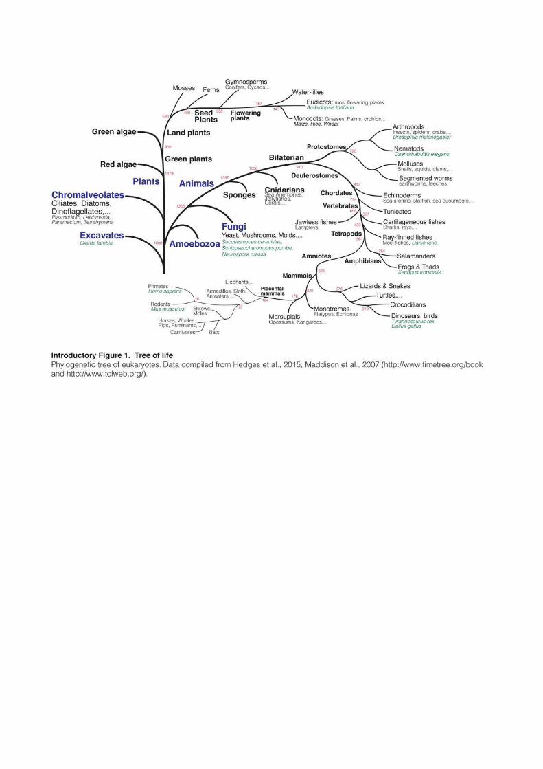
Long believed to be “junk” DNA without any function, Transposable Elements (TEs)
are now considered as major players for genome regulation and contributed to shape the
evolution of virtually every eukaryote (Introductory Figure 1).
1.1 Historical perspective
In the 1910s, Thomas Hunt Morgan discovered that chromosomes were the carriers of
genetic information and the material basis of Mendelian heredity (Morgan, 1915). After he
established the first genetic maps of Drosophila, genes started to be represented as fixed units
stably ordered in a linear pattern on chromosomes, like “beads-on-a-string”. Mutations were
thought to be permanent and irreversible, and considered to be the main driving force of
Darwinian evolution. This model would prevail during most of the 20th century. When
Barbara McClintock observed in 1950 that some genes could move along chromosomes, her
discovery received a very cold reception from the scientific community (McClintock, 1950).
McClintock was working on the mechanism of chromosome breakage and fusion in maize.
She had identified a locus on chromosome 9 where breakage was always occurring. She
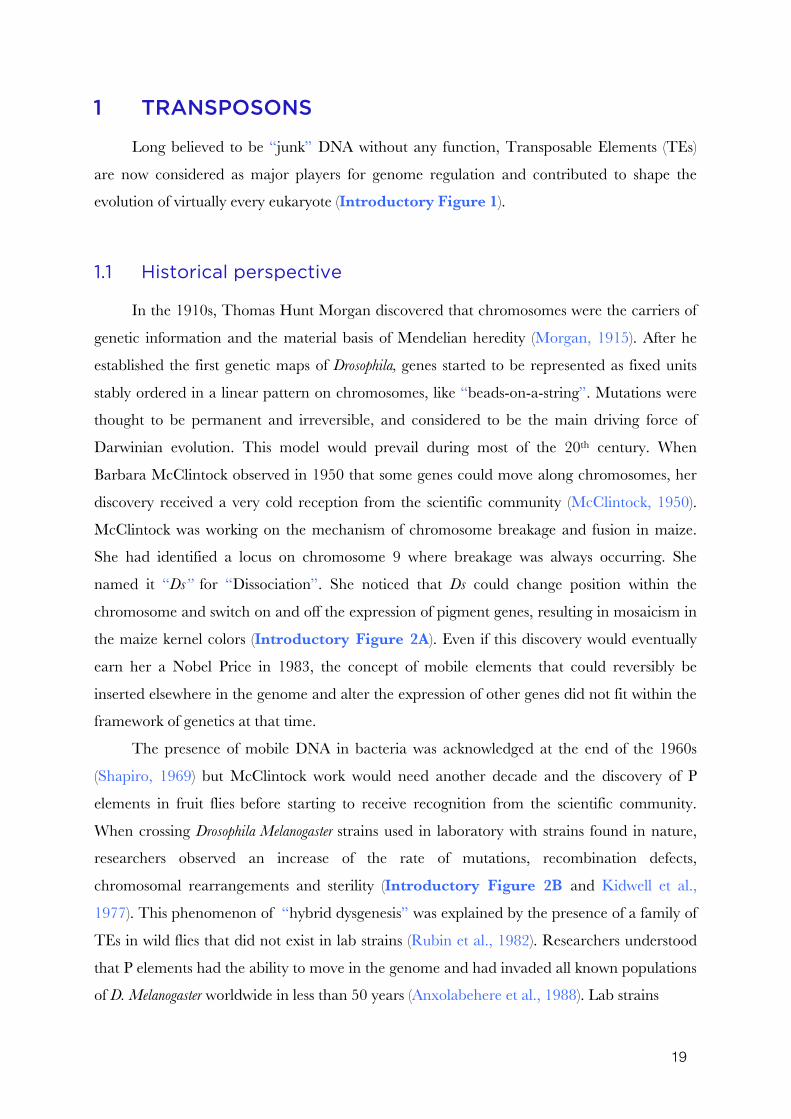
named it “Ds” for “Dissociation”. She noticed that Ds could change position within the
chromosome and switch on and off the expression of pigment genes, resulting in mosaicism in
the maize kernel colors (Introductory Figure 2A). Even if this discovery would eventually
earn her a Nobel Price in 1983, the concept of mobile elements that could reversibly be
inserted elsewhere in the genome and alter the expression of other genes did not fit within the
framework of genetics at that time.
The presence of mobile DNA in bacteria was acknowledged at the end of the 1960s
(Shapiro, 1969) but McClintock work would need another decade and the discovery of P
elements in fruit flies before starting to receive recognition from the scientific community.
When crossing Drosophila Melanogaster strains used in laboratory with strains found in nature,
researchers observed an increase of the rate of mutations, recombination defects,
chromosomal rearrangements and sterility (Introductory Figure 2B and Kidwell et al.,
1977). This phenomenon of “hybrid dysgenesis” was explained by the presence of a family of
TEs in wild flies that did not exist in lab strains (Rubin et al., 1982). Researchers understood
that P elements had the ability to move in the genome and had invaded all known populations
of D. Melanogaster worldwide in less than 50 years (Anxolabehere et al., 1988). Lab strains
21
isolated before the invasion burst had been protected. In the following years, it became
evident that TEs were present in almost every eukaryote species and represented a significant
proportion of genomes.
During the 1960s came also the realization that cells from different species could
contain very different amount of DNA. Whereas prokaryotes tend to have small genomes,
there is no correlation between genome size and evolutionary complexity for eukaryotes. This
observation is referred to as the “C-value Paradox” (Introductory Figure 2C, 2D and
Thomas, 1971). Some species of amphibians or fishes have for example 50 times as much
DNA per nucleus as humans. Even species of similar biology and complexity can harbor
striking differences in genome size. Some flowering plants, like the plant model system
Arabidopsis thaliana, harbor very small genomes (~100Mb, similar to Caenorhabditis elegans), while
other flowering plants contain 2,000 times as much DNA (Bennet and Leicht, 2012). These
observations were even more intriguing considering that the estimated number of genes
(defined as discrete, locatable and protein-coding units of DNA) does not vary in such
proportions among species. Indeed, gene number and genome size range from 2,000 genes in
2.3Mb for Encephalitozoon intestinalis, an intracellular fungal parasite (Corradi et al., 2010), to
around 20-30,000 genes in complex eukaryotes and an enormous size of 150Gb for the
flowering plant Paris japonica (Pellicer et al., 2010). To summarize, gene number varies in
eukaryotes in a 1 to 15 ratio, while genome sizes vary between 1 and 65,000.
In 1972, Susumu Ohno popularized the idea that the majority of DNA in the genome
had no function and could be considered as “junk” (Ohno, 1972). Furthermore, in order to
explain how such an amount of useless DNA could accumulate in genomes, two papers in
Nature in 1980 proposed that much of eukaryotic genomes was in fact composed of “selfish
DNA” (Doolittle and Sapienza, 1980; Orgel and Crick, 1980). These notions shifted from the
prevalent view that the whole nuclear DNA was functional and under heavy selective
pressure. On the contrary, and as long as it remained without consequences, DNA capable of
proliferating could theoretically accumulate in tremendous amount. Being neutral from an
evolutionary point of view, the concept of “junk” and “selfish” DNA solved the C-value
paradox and gave a reasonable explanation for the accumulation of TEs in most eukaryote
genomes. As Orgel and Crick stated it, “The spread of selfish DNA can be compared to the
spread of a not-too-harmful parasite within its host”.
This view of TEs as genomic parasites remained dominant for several decades. The
mutagenic potential of TEs turned them into powerful and popular genetic tools in many
model systems, but they otherwise received little attention. The interest changed with the era
23
of genome sequencing and the realization that TEs occupy much more genomic space than
anticipated. They constitute around 50% of the mouse or human genome and an impressive
90% of the corn genome (Lander et al., 2001; Schnable et al., 2009; Waterston et al., 2002).
In the recent years, TEs became an active topic for many fields of research, and are now seen
as major contributors of genome evolution and regulation in virtually every eukaryotes
(Fedoroff, 2012). Ironically, McClintock was initially presenting TEs as “controlling elements”
because of their ability to modify gene expression. Almost 50 years were necessary for the rest
of the scientific community to adopt this idea.
1.2 Biology of Transposable Elements
There are two major groups of TEs. Class II or DNA transposons move by a “cut-and-
paste” mechanism. Both Ds transposons discovered by McClintock in maize and P elements
in Drosophila are DNA transposons. Class I or retrotransposons move by a “copy-and-paste”
mechanism. They require an RNA intermediate and the insertion of its cDNA complement at
a new site in the genome (Finnegan, 1989).
DNA transposons and retrotransposons can further be subdivided into subclasses,
orders, superfamilies, families and subfamilies and an unified nomenclature has been
proposed (Wicker et al., 2007). In this classification, retrotransposons are subdivided into five
orders, the three main ones being LTR-retrotransposons, and Long and Short Interspersed
Nuclear Elements (LINEs and SINEs). In addition to these classical groups, two other orders
have been described and would only be named here for the sake of being exhaustive: DIRS-
like and Penelope-like retrotransposons. Their mechanism of retrotransposition differs from
the other orders and members have been detected in plants, fungi and animals (Evgen’ev and
Arkhipova, 2005; Poulter and Goodwin, 2005).
1.2.1 DNA-transposons
DNA transposons are found in almost all eukaryote genomes. They move by excising
themselves from the genome and integrating at a new location (Muñoz-López and García-
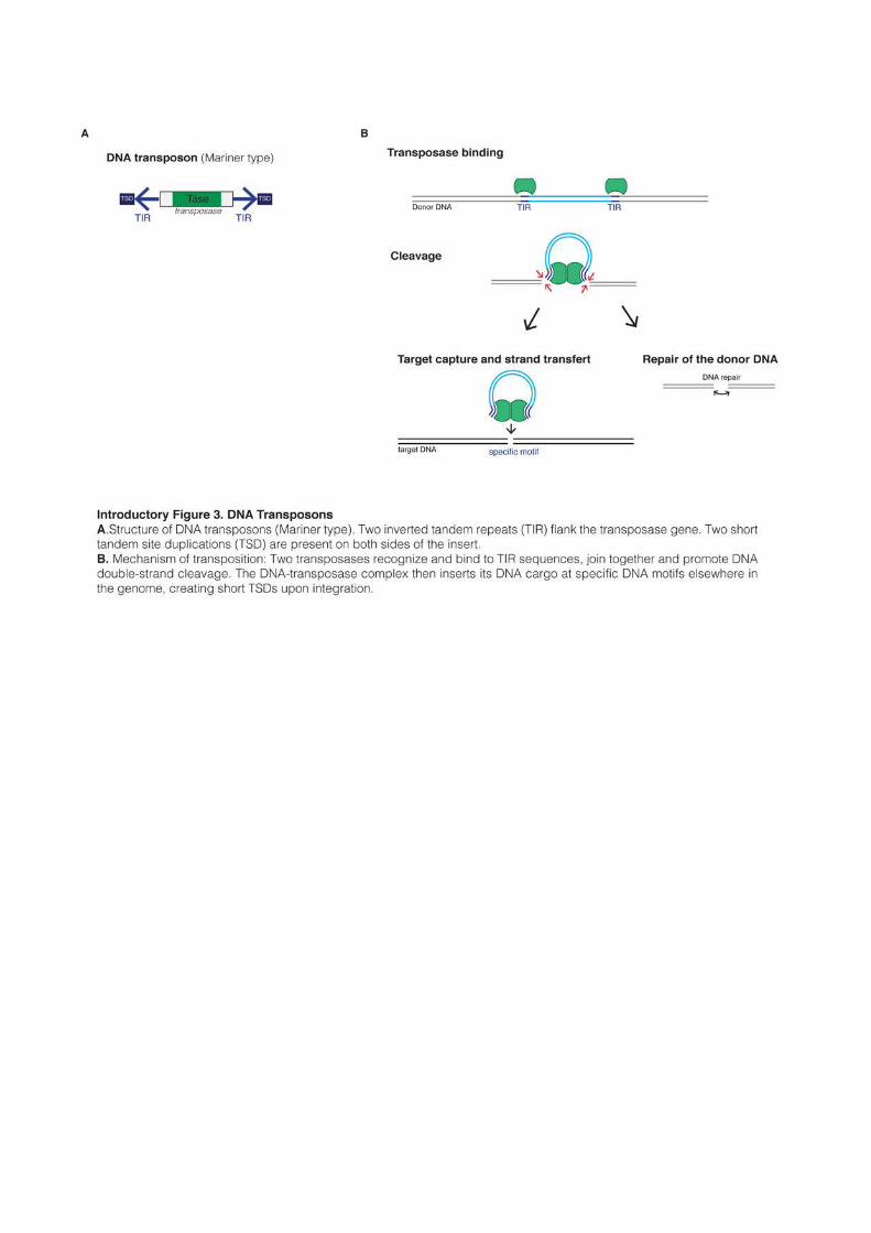
Pérez, 2010). Typical DNA transposons are 1.5-5kb in length and encode a transposase gene
flanked by two Tandem Inverted Repeats (TIRs) (Introductory Figure 3A). TIRs length
usually varies between 20pb to 1kb. Two transposase proteins bind to the TIRs and cleave the
5’-end of both repeats, forming a DNA-transposase dimer. Most DNA transposons then
recognize a specific target sequence elsewhere in the genome, specific for every family of
24
transposons. The cleaved DNA is then integrated into the new location. The transposition
process creates short Target Site Duplications (TSDs) of typically 4-8pb at both ends of the
insert (Introductory Figure 3B). At least nine superfamilies of DNA transposons were
reported to move according to this mechanism (Wicker et al., 2007).
However, some DNA transposons mobilize in a completely different way. For example,
Helitrons, which are present in protists, plants and animals, do not contain TIRs and move
through a complex “rolling-circle” mechanism (Kapitonov and Jurka, 2007). Mavericks, which
are found in diverse eukaryotes (except in plants), contain TIRs and encode between five and
eleven genes, including an integrase and a DNA polymerase (Pritham et al., 2007). Contrary
to most DNA transposons, Helitrons and Mavericks probably move trough a copy-and-paste
mechanism that involves the replication of a single-stranded DNA intermediate (Feschotte
and Pritham, 2007).
Except for Helitrons and Mavericks, mobilization of DNA transposons occurs in a non-
replicative manner. Multiplication of transposon copies can therefore only occur through
indirect mechanisms. For example, during DNA replication, transposition of a DNA
transposon from a newly replicated chromatid to an unreplicated one would lead to the gain
of one transposon copy in one of the daughter cell. Moreover, excision of a transposon from
its donor site creates DNA double-strand breaks that need to be repaired. One possible
pathway of repair is homologous recombination that uses the homologous chromosome (or
the sister chromatid) as a template. This scenario results in the regeneration of the transposon
at its site of origin and therefore in an increase in transposon copy number in the genome.
(Feschotte and Pritham, 2007). DNA repair can occur otherwise by Non-Homologous End-
Joining (NHEJ), leading to the formation of transposon “footprints” formed by the remaining
TSDs. Using these various mechanisms, DNA transposons have been able to accumulate in
large amounts in certain organisms.
Most DNA transposons belong to a handful of superfamilies that have been categorized
based on homology of their transposase gene (e.g. Tc1/mariner, hAT, piggyBac). Some of the
most widespread superfamilies, like Tc1/mariner, are found in almost every eukaryotic
kingdom, indicating a very ancient evolutionary origin (Capy et al., 1996; Plasterk et al.,
1999). Imperfect DNA repair can give rise to various internal deletions and create
degenerated transposon copies. However, because transposition only requires the terminal
repeats, degraded and non-autonomous copies can still be mobilized by intact transposases
encoded elsewhere by intact elements. For example, internally deleted Miniature Inverted-
25
repeat Transposable Elements (MITEs) have accumulated to large numbers in many genomes
and are often present in many more copies that intact elements (Feschotte et al., 2002).
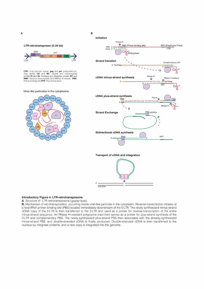
1.2.2 LTR-retrotransposons
LTR-retrotransposons are characterized by the presence of Long Terminal Repeats
(LTR) directly flanking an internal coding region. As retrotransposons, they mobilize through
reverse-transcription of their mRNA and integration of the newly created cDNA into another
location. Their mechanism of retrotransposition is shared with retroviruses, with the
difference that most LTR-retrotransposons do not form infectious particles that leave the cells
and therefore only replicate inside their genome of origin. Their size ranges from a few
hundred base pairs to 25kb, like the Ogre retrotransposon in the Pea genome (Neumann et
al., 2003). All functional LTR-retrotransposons encode a minimum of two genes, gag and pol,
that are sufficient for their replication (Introductory Figure 4A). Gag encodes a polyprotein
with a capsid and a nucleocapsid domain (Sandmeyer and Clemens, 2010). Gag proteins form
virus-like particles in the cytoplasm inside which reverse-transcription occurs. The Pol gene
produces three proteins: a protease (PR), a reverse-transcriptase endowed with an RT
(reverse-transcriptase) and an RNAse H domains, and an integrase (IN) (Wicker et al., 2007).
Typically, LTR-retrotransposon mRNAs are produced by the host RNA pol II acting
on a promoter located in their 5’ LTR. The Gag and Pol genes are encoded in the same
mRNA. Depending on the host species, two different strategies can be used to express the two
polyproteins: a fusion into a single open reading frame (ORF) that is then cleaved or the
introduction of a frameshift between the two ORFs (Gao et al., 2003). Occasional ribosomal
frameshifting allows the production of both proteins, while ensuring that much more Gag
protein is produced to form virus-like particles. Reverse-transcription usually initiates at a
short sequence located immediately downstream of the 5’-LTR and termed the primer
binding site (PBS). Specific host tRNAs bind to the PBS and act as primers for reverse-
transcription, which occurs in a complex and multi-step process, ultimately producing a
double-stranded cDNA molecule (Craig et al., 2002). The cDNA is finally integrated into a
new location, creating short TSDs and adding a new copy in the host genome (Introductory Figure 4B).
Based on phylogenic analyses of the conserved RT domain and on the order of the RT
and the IN domains in the Pol gene, LTR-retrotransposons can be classified into two main
superfamilies, copia-like and gypsy-like (named after copia/gyspy transposons in Drosophila
(Havecker et al., 2004; Xiong and Eickbush, 1990). Copia and gypsy LTR-retrotransposons can
27
be found in all eukaryote kingdoms, indicating a very ancient origin. Except in vertebrates,
these two superfamilies account for the vast majority of LTR-retrotransposons. Both copia and
gypsy elements sometime encode an additional envelope (env) gene with surface and
transmembrane units, giving to some retrotransposons the ability to infect other cells or other
organisms. In Drosophila, gypsy elements have for example been shown to be potentially
infectious (Kim et al., 1994).
A lack of a functional env gene is what distinguishes LTR-retrotransposons from bona fide
retroviruses. In vertebrates and especially in mammals, the vast majority of LTR-
retrotransposon is in fact thought to have resulted from the endogenization of retroviruses,
through inactivation or deletion of the domains that enable extracellular mobility (Boeke and
Stoye, 1997). In mammals, endogenous retroviruses (ERVs) are usually further categorized
into three classes, I, II and III, depending on the family of exogenous retroviruses (XRVs) they
are related to. The respective evolutionary origins of LTR-retrotransposons and ERVs are
complex and will be discussed later.
Many LTR-retrotransposons lack functional ORFs and cannot be replicated
autonomously. They can nonetheless be mobilized in trans by functional elements, the minimal
requirement being the presence of flanking LTRs and of the priming sequences in the PBS
necessary for the initiation of reverse-transcription. Many non-autonomous LTR-
retrotransposons can be identified in plants or animals, sometimes representing distinct
families like the VL30 or malR elements in mice, and have sometimes accumulated to
significant amounts (Stocking and Kozak, 2008). Additionally, in a majority of species, LTR
sequences can be found alone, without internal coding sequences. In mammalian genomes,
these “solo-LTRs” are ten-time more numerous than full-length copies. They are thought to
result from homologous recombination between the two LTRs of a retrotransposon, leaving a
solitary LTR after excision of most of the transposon sequence (Mager and Goodchild, 1989;
Sverdlov, 1998).
1.2.3 LINEs
LINEs are autonomous retrotransposons found in all eukaryotic kingdoms. Five
superfamilies have been described – R2, RTE, Jockey1, L1 and I – based on RT domain
phylogeny (Kapitonov et al., 2009; Wicker et al., 2007). All LINEs encode a least one protein,
ORF2, which contains an RT and an endonuclease (EN) domains. Except for the
evolutionary ancient R2 and RTE superfamilies, LINEs usually encode for another protein
1 L2 elements are for example member of the Jockey superfamily
29
named ORF1. LINE elements are relatively rare compared to LTR-retrotransposons in
plants, fungi or insects, but are dominant in vertebrates and especially in mammals, where
they represent around 20% of the genome. Most of the knowledge about LINE biology comes
from the study of L1 elements in mice and humans, even though the retrotransposition
mechanism was first elucidated from the study of an R2 element in the silkmoth Bombix mori
(Luan et al., 1993).
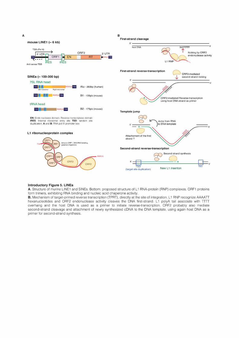
Full-length mammalian L1s are typically around 6kb long. They are composed of a 5’
untranslated region (UTR), which acts as an internal promoter, two ORFs and a 3’UTR
containing a polyadenylation signal and a polyA tail (Introductory Figure 5A and
Babushok et al., 2007). The 5’UTRs of mouse L1s contain a variable number of GC-rich
tandemly repeated monomers of around 200pb, followed by a short non-monomeric region.
Interestingly, the promoter activity of mouse 5’UTRs was shown to be proportional to the
number of monomers (DeBerardinis and Kazazian, 1999). Human 5’UTRs on the other hand
are ~900pb in length and do not contain such repeated motifs. All families of human L1s
harbor in their most 5’ extremity a binding motif for the transcription factor YY1 (Becker et
al., 1993). Younger families have also two binding sites for SOX-family transcription factors,
and both YY1 and SOX sites were shown to be required for human L1 transcription initiation
and activation (Athanikar et al., 2004; Tchenio, 2000). Both mouse and human 5’UTRs
contain as well a week antisense promoter of unknown function (Li et al., 2014; Speek, 2001).
L1s produce a single bicystronic mRNA encoding ORF1 and ORF2 proteins. In human
and mice, the two ORFs are non-overlapping (although not necessarily in the same reading
frame), but this is not the case in rats. In human, ORF2 is thought to be translated by an
unconventional termination/reinitiation mechanism (Alisch et al., 2006), while mouse L1s
contain an internal ribosome entry site (IRES) upstream of each ORF (Li et al., 2006b).
ORF1 is a 40kDa protein that lacks homology with any protein of known function. In
vertebrates, it contains a conserved C-terminus domain and a highly variable coiled-coil N-
terminus that mediates the formation of ORF1 trimetric complexes (Martin et al., 2003).
ORF1 trimmers have RNA-binding and nucleic acid chaperone activity that are necessary for
retrotransposition (Martin and Bushman, 2001; Martin et al., 2003, 2005).
ORF2 is a 150kDa protein with an endonuclease domain (EN), an RT domain and
sometimes, an RNAse H domain. The RNAse H domain is absent in mammalian L1s but is
present in Drosophila “I factors”. Both ORF1 and ORF2 proteins primarily associate in cis with
their encoding mRNA, forming a ribonucleoprotein (RNP) complex, likely composed of two
ORF2s and an unknown number of ORF1 trimers (Babushok et al., 2007; Kulpa and Moran,
30
2006). The complex is transported back into the nucleus, where the L1 endonuclease opens
the DNA at TTAAAA hexanucleotide motifs (Jurka, 1997). Reverse-transcription then occurs
directly at the site of integration trough a mechanism named target-primed reverse
transcription (TPRT) (Introductory Figure 5B), which was originally described for an
LINE R2 in silkworms (Cost et al., 2002; Luan et al., 1993). New insertions create short
TSDs, and the majority of new inserts are severely 5’-truncated (average insert size of 900pb
in humans) and often inverted (Szak et al., 2002). Because they lack their 5’UTR, most of new
inserts are non functional. LINE proteins sometimes fail to associate in cis with their encoding
mRNA and were shown to mobilize in trans various genic mRNAs, creating most of the time
“dead-on-arrival” pseudogenes lacking introns and promoters (Esnault et al., 2000).
1.2.4 SINEs
SINEs are the only TEs that are non-autonomous by nature, meaning that they did not
evolve from autonomous elements. They are small (80-500pb) and rely in trans on functional
LINEs for their replication, but their evolutionary origin is very distinct. SINEs can be found
in very diverse eukaryotes, but they have only accumulated to impressive amount in
mammals, where they represent between 5 and 15% of the genome with millions of copies.
SINEs typically possess a “head” with an RNA pol III promoter that enables autonomous
transcription, and a body of various composition (Introductory Figure 5A and Kramerov
and Vassetzky, 2005). SINEs replication mechanism was shown to rely on LINEs, either in
human or in fish (Dewannieux et al., 2003; Kajikawa and Okada, 2002). SINE RNAs form a
complex with LINE ORF2 proteins and are inserted into the genome by TPRT, creating
short TSDs upon insertion. Some SINE families are thought to rely on specific LINEs for
their replication, while others seem to be more generalist.
SINEs are postulated to originate from the accidental retrotransposition of various pol
III transcripts, and have appeared separately numerous times in evolution history (Kramerov
and Vassetzky, 2011). The type of pol III promoter defines the different superfamilies and
reveal their origin: tRNA, ribosomal 5S RNA or signal recognition particle 7SL RNA. Alu
and B1 elements, with their 1.1 million and 650,000 copies in the human and mouse
genomes, respectively, harbor a 7SL promoter. The 350,000 copies of B2 SINEs in the
mouse are on the other hand tRNA-related (Vassetzky and Kramerov, 2013). The origin of
the 3’ region is more obscure, and is thought to contain sequence elements allowing
recognition by the LINE proteins. Some SINE 3’-regions are indeed in some cases very
similar to the 3’-end of a LINE of the same genome (Kajikawa and Okada, 2002; Okada et
31
al., 1997). In other cases, the 3’ end of SINEs can be either A- or AT-rich, with short tandem
repeat or a poly-T tail (the pol III termination signal). The tail of Alu was, for example, shown
to be essential for their mobility (Dewannieux et al., 2003).
LINE proteins preferentially assemble in cis with their encoding mRNA, directly after
translation. The mechanism by which SINE RNAs are transported to the cytoplasm and
incorporated into LINE RNPs in place of LINE RNAs remains poorly understood. However,
the majority of SINEs are transcribed from promoters of RNAs usually involved in the
translation machinery. Some elements, like Alu and B1, can still form complexes with protein
associated with ribosomes (Weichenrieder et al., 2000). It has been proposed that most SINEs
maintain the ability to associate with the translation machinery, giving them the opportunity
to present their 3’-end to newly translated LINE proteins (Kramerov and Vassetzky, 2005).
Hominid genomes contain original elements termed SVA. They are composite
transposons formed by the fusion of a SINE-R and an Alu, separated by a variable number of
tandems repeats (Ostertag et al., 2003). Less than 3kb in length and apparently mobilized
using L1 machinery, they are around 2500-3000 copies in human or gorilla genomes, and less
than 1000 in orangutan (Wang et al., 2005). SVA are one of the youngest TE in great apes
genome and among the most active and polymorphic in the human population.
1.2.5 Evolutionary origin of Transposable Elements
DNA transposons and retrotransposons can be found in virtually every eukaryotes,
including very primitive unicellular protozoans, like the intestinal parasite Giarda Lambia
(Arkhipova and Meselson, 2000). The conservation of the structure and mode of replication of
TEs indicate that they appeared very early in evolution, and their presence seems to be a
constitutive feature of eukaryotic genomes. Even if numerous TEs have the capacity to
horizontally infect other cells or organisms, the transmission of TEs is thought to primarily
occur vertically from one cell to its daughter cells (Malik et al., 1999). Because TEs are mobile
by nature, it is easy to imagine that the integration of a transposon into a host gene or into
other transposons would allow the acquisition of increasingly complex new functions (Lerat et
al., 1999; Malik and Eickbush, 2001). The vertical mode of transmission enables the
reconstruction of an evolutionary history of TEs, from prokaryotic precursors to highly
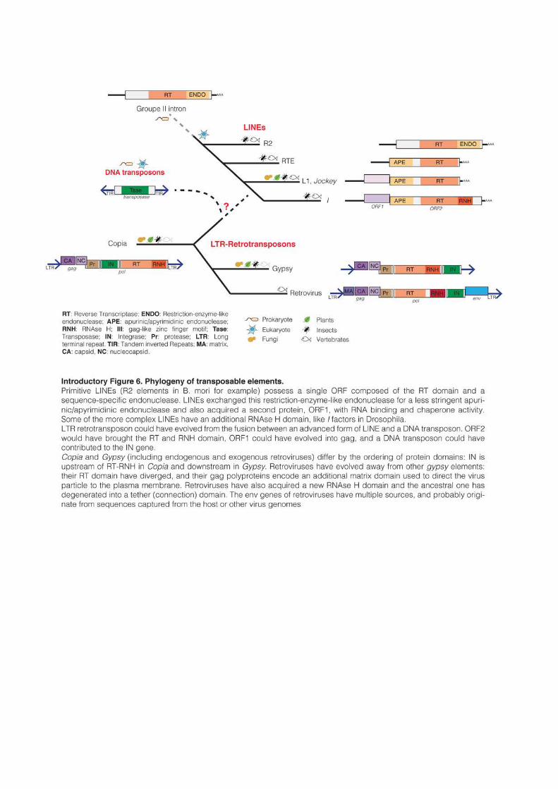
complex retroviruses (Introductory Figure 6). However, some crucial steps remain obscure
and speculative.
33
DNA transposons are thought to originate directly from the prokaryotic world.
“Insertion Sequences” in bacteria move by a mechanism that is nearly identical to DNA
transposons in eukaryotes (Cerveau et al., 2011). Some superfamilies of transposases even
appear to be shared between animals and bacteria, indicating that their emergence preceded
the evolution of eukaryotes (Feschotte, 2004).
The defining feature of autonomous retrotransposons is the presence of a reverse
transcriptase (RT) domain. Sequence similarity of the RT region has been used to establish
phylogenetic analyses of retroelements (Xiong and Eickbush, 1988, 1990). LINEs appear to be
the most ancient retrotransposons and are probably of prokaryotic origin. Their RT domain
and TPRT mode of replication is similar to group II introns, a class of mobile genetic
elements found in bacteria and mitochondria (Lambowitz and Zimmerly, 2011; Malik et al.,
1999). LINEs are also the only retrotransposons found in the very primitive eukaryote Giarda
lambia, (Arkhipova and Meselson, 2000). Primitive LINEs, like the well-studied R2 elements of
B. mori, possess a single ORF composed of the RT domain and of a sequence-specific
endonuclease similar to bacterial restriction enzymes (Luan et al., 1993). LINEs exchanged
this restriction-enzyme-like endonuclease for a less stringent apurinic/apyrimidinic
endonuclease, probably coming from the DNA repair machinery of the host cell (Malik et al.,
1999). They also acquired a second protein, ORF1, with RNA binding and chaperone
activity. Some of the more complex LINEs have an additional RNAse H domain, like the I
factors in Drosophila (Malik, 2005; Wicker et al., 2007). RNAse H is thought to be necessary to
remove the template RNA from the newly synthetized cDNA. This domain is absent from
mammalian L1s, which appears as a more primitive lineage that probably use host RNAse H
to carry out this function.
Based on RT domain phylogeny, LTR-retrotransposons (including ERVs and XRVs)
form a monophyletic group. Vertebrate retroviruses cluster together within the gypsy
superfamily, while copia is separated (Malik et al., 1999; Xiong and Eickbush, 1988, 1990).
This suggests that retroviruses evolved from gypsy elements by acquisition of env genes and
additional regulatory sequences, giving them the opportunity to invade other cells.
Invertebrate retroviruses are structurally similar to gypsy retrotransposons, except for the
presence of env genes that were probably acquired through recombination with other dsDNA
or ssRNA viruses (Malik, 2000; Pearson and Rohrmann, 2002). Vertebrate retroviruses on the
other hand have evolved separately from the rest of LTR-retrotransposons. Their RT domain
has diverged away from other gypsy retrotransposons and their gag polyproteins encode an
additional matrix domain used to direct the virus particle to the plasma membrane
34
(Sandmeyer and Clemens, 2010). Vertebrate retroviruses have also acquired a new RNAse H
domain and the ancestral one has degenerated into a tether (connection) domain (Malik and
Eickbush, 2001). The env gene of retroviruses has multiple sources, likely originating from
sequences captured from the host or from other viruses (Kim et al., 2004). These observations
converge towards the possibility that complex retroviruses have evolved from simpler LTR-
retrotransposons rather than the opposite. In fact, almost all mammalian LTR-
retrotransposons seem to result from the endogenization of retroviruses that have lost the
ability to infect other cells (Boeke and Stoye, 1997). Ironically, mammalian LTR-
retrotransposons are therefore the descendant of elements that initially managed to escape
their host genome, but were then trapped into another genome.
The emergence of the first LTR retrotransposon is obscure, and it has been proposed to
have occurred through the fusion between an advanced form of LINE and a DNA transposon
(Malik, 2005; Malik and Eickbush, 2001). Like DNA transposons, LTR-retrotransposons
integrate double-stranded DNA molecules into the genome and require tandem repeat
flanking the insert. A DNA transposon could have brought the integrase activity and the
necessity to harbor flanking repeats to early LTR-retrotransposons (Capy et al., 1997). The
LINEs I in Drosophila possess an RNase H domain and its ORF1 contains some zinc finger
motifs that are reminiscent of gag proteins (Martin, 2006; Wicker et al., 2007). Based on
RNAse H domain phylogeny, it has been proposed that LTR-retrotransposon pol gene could
have evolved from an I-like LINE element and the gag from ORF1 (Malik, 2005). In line with
this hypothesis, the entire lineage of LTR-retrotransposons appears to be no older than the
youngest lineage of LINEs (Malik and Eickbush, 2001). Even if this model is speculative, and
can not explain how LTR-retrotransposons developed their complex retrotransposition
mechanism, the only missing domain after a fusion between a LINE and a DNA transposon
would be the protease domain, which could have originated from an ancestral form of the
host pepsin gene (Lin et al., 1992).
1.3 Birth, life, death and afterlife of transposons
TEs are a constitutive feature of every eukaryote genomes. However, there is an
important diversity of TE distribution and genome composition between species. In order to
understand how TEs contribute to the genomic structure, it is necessary to analyze in details
the forces that facilitate or restrict their expansion.
35
1.3.1 Transposons: an heavy, diverse and ancient genomic load
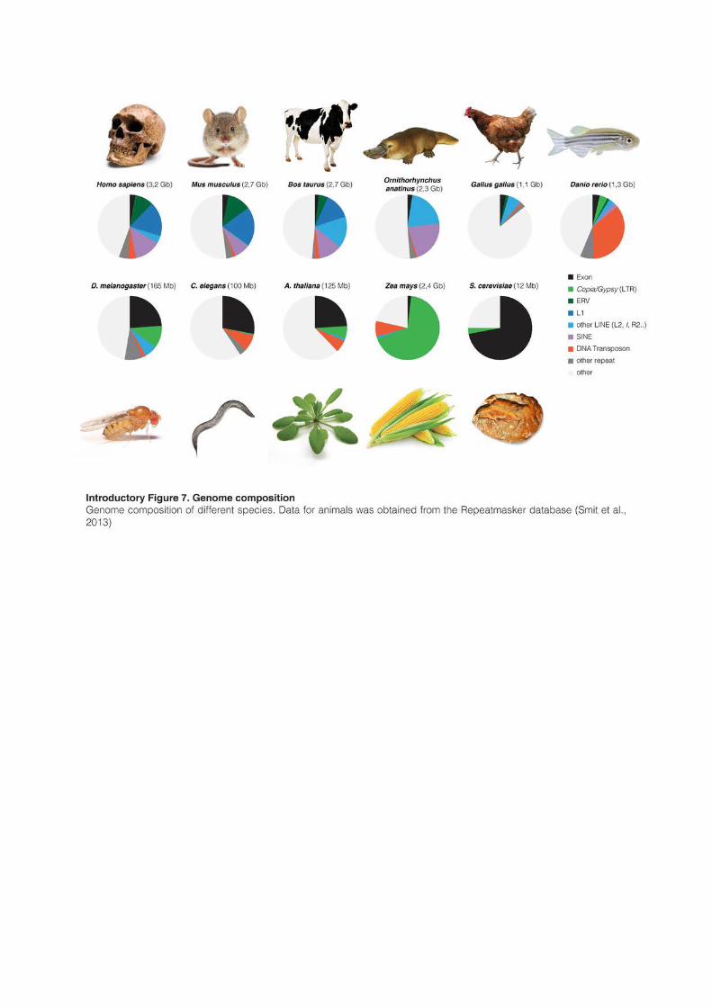
Because of their replicative nature, TEs have accumulated to significant proportion in the
majority of eukaryotes, and many different families of TE usually coexist inside the same
genome (Introductory Figure 7). However, the vast majority of them are usually not
functional anymore. Transposition itself often introduces errors and many new inserts carry
important mutations. For example, the majority of new LINE insertions in mammals are
severely 5’ truncated (Szak et al., 2002). Beside this, TEs are rarely under positive selection
pressure and rather accumulate mutations at a neutral rate over evolutionary time. Most TEs
are therefore merely molecular fossils, truncated, mutated and unable of further mobilization.
Out of the millions of copies that can populate a genome, only a subset is thought to be
potentially active. Among the 500,000 L1 copies that populate the human genome, it has
been estimated that between 5-7,000 are full-length (Khan et al., 2006; Lander et al., 2001),
that 80-100 are retrotransposition-competent, and that only six “hot L1s” contributed to the
majority of retrotransposition in the human population (Brouha et al., 2003).
TEs are not subject to the same selective constraints as protein coding genes. While the
coding part of the genome can be relatively similar even between distantly related species, the
transposon-derived fraction can evolve rapidly into a great diversity. One good example is the
comparison between the two distantly related frogs Nanorana parkeri and Xenopus tropicalis (Sun
et al., 2015). Despite having diverged 266 Myrs ago, the two frogs have conserved a
considerable genome synteny, but greatly differed in their TE content. The genome of N.
parekeri is significantly larger than the one of X. tropicalis (2.3 vs 1.5Gb): TEs account for most
of this difference, both in term of genomic mass (970 vs 318Mb of TEs) and composition
(mostly gypsy LTR retrotransposons in N. parkeri and DNA transposons X. tropicalis).
The comparison of TE composition between species reveals important differences in the
histories of expansion, contraction and activity of their genomes (Introductory Figure 7).
Saccharomyces cerevisiae harbors a very small genome (12Mb) and most of the genomic space is
occupied by exonic sequences. The budding yeast genome is probably under strong selective
pressure to maintain a small size and TEs only represent 3% of the DNA. The intestinal and
intracellular fungal parasite Encephalitozoon intestinalis represents an extreme case: it has the
smallest eukaryotic genome ever sequenced (2.3Mb) and is the only eukaryote that apparently
lacks TEs. Its genome is in fact so compact that non-coding sequences appear more conserved
that genes themselves (Corradi et al., 2010). D. melanogaster, C. elegans or A. thaliana have also
relatively dense genomes (100-150Mb). Exons occupy around one quarter of the genetic space
and TEs less than 15% (Arabidopsis Genome Initiative, 2000; Smit et al., 2013). Interestingly,
37
most of the TEs in these three genomes are relatively young, less than 20 millions year old,
and often active (Kapitonov and Jurka, 1999, 2003).
These features are in strong contrast with vertebrate genomes, which are usually much
bigger (>1 Gb). Exons represent only 2-3%, while old fossils of TEs are the main components
of the vertebrate DNA. It is commonly assumed that around 50% of the human genome is
composed of TEs, but this number is likely underestimated because very ancient TEs are
almost impossible to recognize and annotate. The TE-derived fraction of the human genome
might in fact be closer to two-third (de Koning et al., 2011). The diversity of TE composition
in vertebrate genomes is astonishing and reflects the diverse successful invasions of different
TE families in separated lineages. The zebrafish genome is composed of 35% of DNA
transposons; mammalian genomes are dominated by LINEs and SINEs; and gypsy-like LTR-
retrotransposons can make up to 30% of the Salamander genome (Smit et al., 2013; Sun et
al., 2012a). Birds on the other hand have the smallest genome among tetrapods (~1Gb),
which contains only 10% of TEs.
Except for a set of “hot L1s”, most human TEs are inactive. LINEs are in fact the most
successful transposons in all mammalian species, where they usually represent around 20% of
the genome. Mouse and human genomes are dominated by L1s, but other LINE
superfamilies have been successful in other mammals: Bov-B, an RTE-type LINe, represents
15% of the cow genome. L1s have been continuously amplifying for the last 160 Myrs in
mammals, and bursts of amplification have alternated with periods of low activity (Khan et
al., 2006; Pascale et al., 1990). However, it is estimated that the rate of human TE
amplification, including L1s, has been decreasing for the past 50 Myrs (Lander et al., 2001),
except for a peak of Alu (SINEs) expansion that occurred around 40 Myrs ago. The remnants
of past periods of amplification are, however, numerous. For example, the human genome
carries 3% of DNA transposons, which were active until 40-50Myrs ago in the primate
lineage (Pace and Feschotte, 2007). Similarly, L2 elements (2% of human DNA) have been
immobile for a long time, but they probably played an important role in the past. In contrast,
they represent 20% of the platypus genome, and domesticated L2-derived sequences are now
involved in T cell-specific gene regulation in humans (Donnelly et al., 1999). The most prolific
mammalian LTR retrotransposon elements (ERVL and MaLRs, 5.8% of the genome) greatly
multiplied 100Myrs ago but have been extinct for the last 40 Myrs (Lander et al., 2001). With
the potential exception of HERVK-HML2 (Subramanian et al., 2011), a recently acquired
ERV that is expressed in cancers and diseases, LTR retrotransposons appear to be on the
edge of extinction in humans.
38
The reasons for this decline in transposons expansion in the human lineage are obscure
and no good explanation has been offered so far. The mostly quiescent human genome stands
in opposition with the mouse genome, which harbors many active LINEs and ERVs. The
composition of the murine genome will be discussed in details later in this manuscript.
1.3.2 A subtle equilibrium between opposing forces
The evolution of the genome and, especially, of its repeated fraction is not a linear
process. When a new transposon copy inserts into the germline of an individual, this copy can
be transmitted to the next generation and then spreads into the population. Interestingly, the
multiplication of TEs seems to often occur by bursts of activity, followed by periods of decay.
Theoretical models have been proposed to reconstitute the initial invasion (Le Rouzic and
Capy, 2005) and long time evolution of TEs (Le Rouzic et al., 2007a). Mutations introduced
by TEs (inactivation of genes, chromosomal breaks, translocations, etc.) can be deleterious for
the host and its descendants. TEs were indeed shown to cause 50% of deleterious mutations in
Drosophila (Finnegan, 1992), and 10-15% in laboratory mice (Kazazian, 1998; Maksakova et
al., 2006). The spreading of TEs in a population is therefore essentially controlled by natural
selection (Charlesworth and Charlesworth, 1983). Moreover, eukaryotes have developed an
important and diverse range of defense mechanisms to protect themselves against the invasion
of TEs (Slotkin and Martienssen, 2007). It is often assumed that the genome is the theater of a
constant arms race between the TEs and their hosts (Lisch and Slotkin, 2011). Newly invading
elements are first not recognized by the host and can proliferate into the genome until
protective mechanisms evolve and slow down the multiplication process. Unable of further
mobilization, TEs then accumulate genetic mutations that definitely inactivate them. On the
long range, the accumulation of TEs is a subtle equilibrium between their own activity, host
defense mechanisms and natural selection.
1.3.3 Appearance of new transposons
New transposons can appear in a population either vertically, by modification of an
existing one, or horizontally by endogenization of sequences originating from other species or
from viruses.
LINEs and SINEs are transmitted essentially vertically (Burke et al., 1998; Pascale et al.,
1990). In mammals (at least), L1s constantly change their regulatory units by modifying their
5’UTR, while ORF1 and ORF2 sequences remain relatively conserved (Adey et al., 1994;
Khan et al., 2006). The 5’UTRs of L1s are often completely unrelated, especially between
39
different species. Only the 5’UTR of closely related L1 families seem to originate from the
modification of a common ancestor. On the contrary, L1s are thought to often acquire
completely novel regulatory units by inaccurately switching template during the
retrotransposition reaction (Hayward et al., 1997). The human L1 5’UTR was modified at
least eight time in the 70Myrs of primate evolution (Khan et al., 2006). Analysis of the mouse
genome reveals also that different L1 families (active or not) often recombine together,
exchanging regulatory or coding sequences to form new mosaic elements with renewed
activity (Saxton and Martin, 1998; Sookdeo et al., 2013). Interestingly, in most mammals
analyzed, L1 evolves as a single lineage: a new family emerges from the modification of an
existing one, amplifies to thousand of copies and becomes extinct after being replaced by
younger elements. This is currently the case in humans, where all L1s have derived from the
single dominant L1PA lineage over the last 40Myrs (Khan et al., 2006). Moreover, it appears
that concurrent L1 lineages only coexist when they harbor different promoter types. It is the
case in the mouse, where two lineages with different promoter types (A and F) are currently
active; it occurred as well in humans past before the extinction of the L1PB lineage 40Myrs
ago (Goodier et al., 2001; Sookdeo et al., 2013). As a good example of the arm race between
TEs and their hosts, the apparition of a new L1 family with modified regulatory or coding
units was often followed by a period of massive amplification of this new family. Moreover,
the coiled-coil domain of the human ORF1 protein appears to be rapidly evolving and under
high selective pressure (Boissinot and Furano, 2001; Khan et al., 2006).
Similar cycles of activity/quiescence can be observed for LTR-retrotransposons. For
example, ERV-L is a very ancient LTR retrotransposon that is present in all placental
mammals. The mouse genome contains the remains of several boosts of activity, and mouse
ERV-L (MERVL) is still one of the most active murine TEs (Bénit et al., 1999). The ability to
episodically modify their regulatory and coding sequences likely explains why some TEs,
especially LINEs, originate from the beginning of eukaryote existence and are still active
nowadays. Like perfect parasites, they are constantly adapting to their host.
Alternatively, the appearance of new TEs in a population can result from the horizontal
transfer of foreign DNA. The endogenization of retroviruses in vertebrates is probably the
most documented example. Exogenous retroviruses (XRVs) are similar to LTR
retrotransposons in terms of structure and mode of replication, with the difference that virus
particles leave (and often kill) the infected cells after retrotransposition. However, if a
retrovirus manages to invade the germline of an individual and is not too harmful for its host,
the retrovirus genome can be transmitted to the next generations. After the initial infection,
40
the env gene allowing cell-to-cell mobility is often lost or mutated, forcing the new transposons
to adopt an entirely cell-autonomous life cycle. Virtually every LTR retrotransposons in
mammals are the result of the endogenization of an ancient exogenous retrovirus (Boeke and
Stoye, 1997). For example, MLV (Mouse Leukemia Virus) elements have integrated the
mouse genome recently (<1.5Myr ago) and are still very similar to their exogenous
counterparts, with some members having maintained infectious properties (Stocking and
Kozak, 2008). Similarly, in Koalas, KoRVs have all the characteristics of a functional
retrovirus and endogenization into the Koala population is an ongoing process (Tarlinton et
al., 2006).
Examples of horizontal transfer are not restricted to ERVs but involve all types of TEs
in plants, animals and fungi (Wallau et al., 2012). Mariner and P elements (DNA transposons),
I factors (LINEs) and Gypsy (LTR retrotransposons) were shown to move between Drosophila or
other insect species (Abad et al., 1989; Daniels et al., 1990; Robertson, 1993). In vertebrates as
well, surprising cases of horizontal transfers were observed. Bats are the only mammals known
to harbor many active DNA transposons. Multiple waves of Mariner, hAT or Helitron
amplification have invaded the bat genome in the last 30Myrs and likely result from
horizontal transfer (Ray et al., 2007, 2008). Bov-B LINEs are present in all ruminants. They
represent 15% of the cow genome but are absent in related species (Jobse et al., 1995).
However, closely related LINEs were observed in the genome of different snakes and lizards
(Kordis and Gubensek, 1997). It was in fact established that Bov-B LINEs most probably
originated from the horizontal transfer 40-50 Myrs ago of elements from scaled reptiles to the
ancestor of ruminants (Kordis and Gubensek, 1998).
How exogenous TEs were horizontally transferred from one species to the other is often
mysterious. Retroviruses (and some gypsy LTR-retrotransposons) are naturally infectious but
the majority of TEs are normally unable to leave their host cell. It seems highly surrealistic
that a TE could jump directly from a snake to a cow: some sort of vector is necessary to
mediate the physical transfer of DNA between the donor cell and the recipient germline. This
vector needs to have access both to the intracellular and extracellular environment. Viruses,
parasites (insects, mites…) or intracellular parasites make suitable vectors for transfer between
natural populations, and examples of their involvement are numerous and increasing (Silva et
al., 2004).
Bats are important reservoirs of viruses (Calisher et al., 2006) and it has been postulated
that bat DNA transposons could have used dsDNA viruses as carriers to enter the bat genome
(Pace et al., 2008; Ray et al., 2008). By contrast with retroviruses that have ssRNA genomes
41
and need to integrate cDNA into their host genome, dsDNA viruses never use RNA
intermediates nor integrate into the host. Virus specialty is precisely to introduce new genetic
material into foreign cells. By jumping into the DNA of invading virus instead of their host
genome, TEs can find a way to be transported out of their genome of origin and potentially
toward completely unrelated species (depending on the infectious range of the virus).
Accordingly, the insertion of TEs into dsDNA virus has been observed in insects, with the case
of a gypsy-like retrotransposon transposing into the circular genome of a Baculovirus (Friesen
and Nissen, 1990), or in vertebrates by the presence of a snake-specific SINE in the genome of
a poxvirus infecting west African rodents (Piskurek and Okada, 2007).
Other lines of evidences suggest that TEs can use different parasites as carriers between
species. It was for example postulated that P elements were transferred between Drosophila
species by a semi-parasitic mite (Houck et al., 1991). Another DNA transposon was also
observed moving from a moth to its parasitoid wasp (Yoshiyama et al., 2001). In vertebrates, a
family of hAT DNA transposon, SPINs2, was shown to be highly conserved between a lizard, a
frog and five mammalian species (rodents, a primate, a bat, a tenrec and an opossum), but
could not be detected in closely related species (Pace et al., 2008). SPINs were further
identified in the genome of a triatomine bug, which feeds on the blood of various tetrapods,
and in a freshwater snail, which is a known intermediary for the parasite (Gilbert et al., 2010).
The combination snail/triatomine bug probably allowed the transfer of SPINs between these
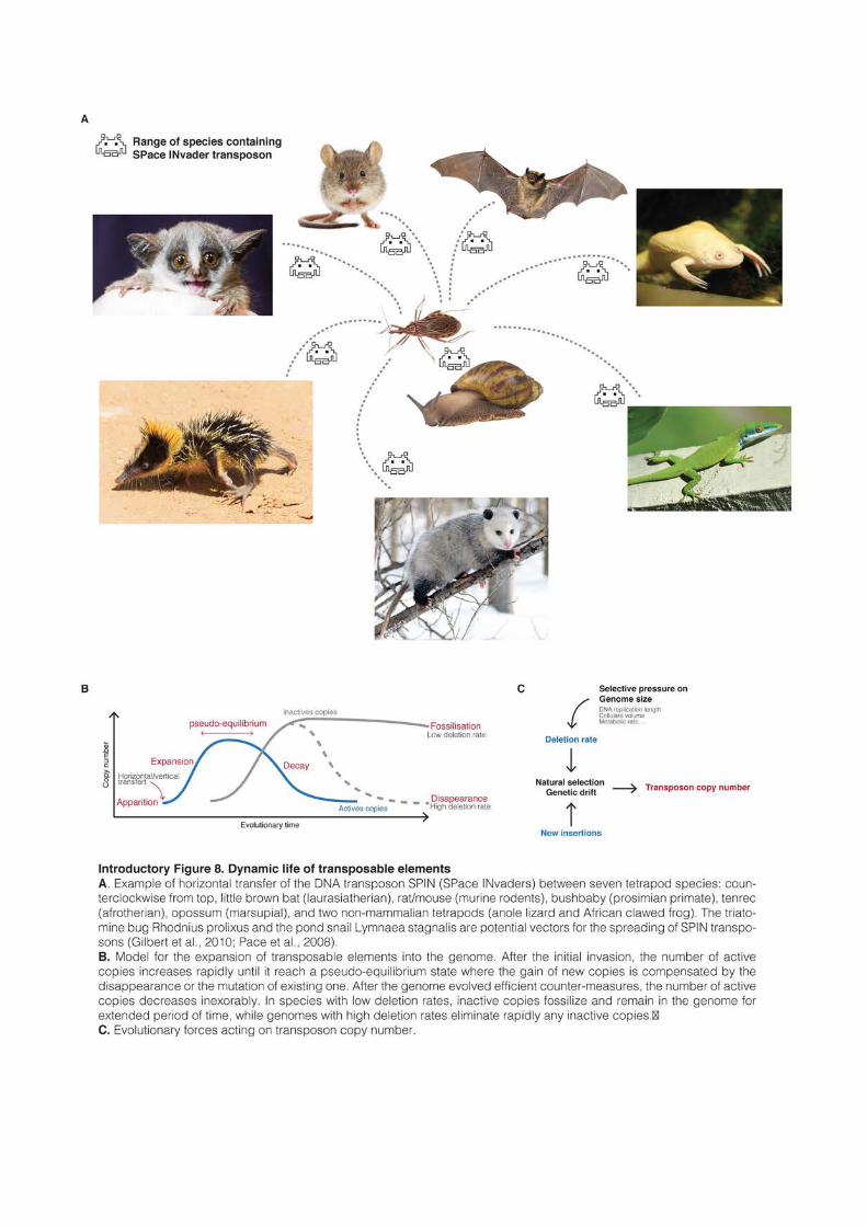
very distant tetrapod species (Introductory Figure 8A).
The favorite mode of appearance of new TEs, either by vertical modification or
horizontal transfer, probably varies across species and, as a consequence, affects the diversity
of TEs in various genomes. Species where horizontal transfer is prevalent (like probably
Drosophila) are expected to harbor many different and diverse TE families (Silva et al., 2004).
On the other hand, mammals seem to have comparatively few horizontal transfers, maybe
because their well-developed immune system efficiently guards against the transfer of DNA by
infectious vectors (Lander et al., 2001). Mammals tend in general to have few TE families, but
composed of many members. Indeed, half of TEs in the mouse and human genome come
from the different variants of a single family of LINEs (Khan et al., 2006).
1.3.4 Initial amplification and long-term maintenance
TEs are present as hundreds or thousands of copies in eukaryotic genomes. Every
successful TE families originally derive from a unique element, which integrated into the
2 For SPace INvaders…
43
germline of a single individual, started to be expressed and increased his copy number
throughout the population (Introductory Figure 8B). However, invasion by a new TE is not
always possible, especially if the founder element originates from the horizontal transfer from
a distant species; TEs are often adapted to their host and transposition into a different
environment might not be successful, especially if the transposition process requires specific
host factors. P elements use the Drosophila protein IRBP (inverted repeat binding protein) to be
excised from their locus (Beall and Rio, 1996; Beall et al., 1994). In relationship with the
polymorphic presence of this factor, P elements can only mobilize in species of the Drosophilidae
family, and not even in non-drosophilids fruit flies (Tephritidae) (O’brochta and Handler, 1988).
On the contrary, Mariner DNA transposons only require their own transposase for
mobilization (Vos et al., 1996), which probably explains why they are so widely distributed in
eukaryotes (Plasterk et al., 1999). Along with the necessity to escape build-in defense
mechanisms, adaptation of TEs to their former host probably restrains their ability to
successfully spread into new organisms.
Once the first element is integrated into the genome, the spread of a new TE family in
the population is a balance between the rate at which new copies arise and the rate at which
they are lost (Charlesworth and Charlesworth, 1983). Loss of a functional TE can happen
trough various mechanisms, including random genetic drift (the chromosome carrying a
transposon copy is not passed to the next generation by chance), inactivating mutations, and
natural selection against deleterious consequences. Mutations by small or large
insertions/deletions or nucleotide substitutions occur at a rate of around 0.1-100 per genome
per generation (Drake et al., 1998; Lynch, 2010); this is several orders of magnitude lower that
the transposition rate of an invading transposon, which can potentially adds several new
copies per generation. Immediately after the initial invasion, spontaneous mutations are
therefore only playing a negligible role and the spreading of a TE in a population is
principally governed by natural selection and genetic drift (Charlesworth et al., 1994). In the
first generations after the initial invasion, theoretical models show that low rates of
transposition likely lead to the rapid disappearance of the new transposon (Le Rouzic and
Capy, 2005; Le Rouzic et al., 2007a). The probability of fixation of any mutation depends on
the population size and the selective advantage or disadvantage brought by the mutation.
With too few copies in too few individuals, a new transposon would very unlikely be able to
get fixed in the population. On the other hand, a continuous high rate of transposition would
cause a massive amplification and natural selection against the deleterious consequences
would lead to the rapid elimination of the transposon from the population. These theoretical
44
models suggest in fact that a successful invasion need to be biphasic, with an initial burst of
amplification characterized by a (moderately) high transposition rate, followed by a longer
period where the transposition rate decreases (Le Rouzic and Capy, 2009). The initial burst of
TE amplification was observed several times in natural and laboratory populations, and the
most documented example is the rapid spread of P elements in all populations of D.
melanogaster worldwide, in less than 50 years (Anxolabehere et al., 1988).
The reason for the decrease of transposition rate is not well understood. However, it is
easy to speculate that once a TE managed to get fixed in a population, natural selection would
favor individuals that either developed defense mechanisms against the invader, or have
accumulated inactivating mutations of the invader. The appearance of non-autonomous
elements (SINEs, MITEs or degenerated copies) that hijack the activity of autonomous ones
was also proposed to slow down the accumulation of functional TEs (Feschotte and Pritham,
2007; Le Rouzic et al., 2007b). Indeed, autonomous and non-autonomous elements would
compete for the same proteins, decreasing the probability to insert new functional copies.
Some authors have speculated that transposon copy number can reach an equilibrium,
when the appearance of new elements is compensated by their loss (Biémont, 1994;
Charlesworth and Charlesworth, 1983). However, such an equilibrium would likely be
unstable and very transient (Le Rouzic and Capy, 2009). Indeed, the decay observed after the
initial invasion is mainly irreversible. Once the host has developed mechanisms to prevent
further transposition, active elements cannot be replaced and would either accumulate
mutations or disappear from the population because of natural selection. Depending on the
selective pressure, the mutation rate and the ability of a species to eliminate degenerated
sequences, the general dynamics of the invasion could take different forms. Strong selective
pressure and high mutation rate would rapidly eliminate remaining functional elements once
the peak of amplification is passed. On the other hand, a low mutation rate and a weak
selective pressure would allow for a very long decay phase, which may look like a pseudo-
equilibrium situation (Introductory Figure 8B).
1.3.5 Death and fossilization
Once their activity starts decreasing, TEs begin to accumulate mutations and, given
enough time, become molecular fossils. Human and mouse genomes share around 165Mb of
common ancestral repeats that date back to more than 100Myrs (Waterston et al., 2002). In
fact in the human genome, the majority of resident repeats precede the radiation of placental
mammals (Lander et al., 2001). This very ancient origin of mammalian TEs is in strong
45
opposition with the observed age of transposons in many other species. For example, D.
melanogaster contain only TEs younger than 20Myrs (Kapitonov and Jurka, 2003). All the LTR
retrotransposons present in the euchromatic part of the Drosophila genome appear to be
younger that their host species, having integrated well after the split with its closest relative (D.
simulans) 3Myrs ago (Bowen and McDonald, 2001). Similarly, S. cerevesiae LTR
retrotransposons are all younger than 100,000 years (Promislow et al., 1999) and the majority
of A. thaliana LTR retrotransposons have less than 4Myrs of age (Devos et al., 2002). The
maize has a genome size similar to mammals (2.4Gb) composed of 90% of TEs, but all these
elements appear nonetheless to be not older than 6Myrs (SanMiguel et al., 1998).
These differences in the age distribution of TEs can be explained by the differential
ability of species to eliminate nonessential DNA. Insertion of new TEs tend to increase
genome size, and the maize genome has apparently gained 1.2Gb of TE sequences in the last
3Myrs (SanMiguel et al., 1998). However, despite continuous TE activity, the majority of
individuals apparently managed to keep their genome size relatively constant, and some
species like A. thaliana are even actively shrinking (Hu et al., 2011). This indicates that
genomes have the ability to lose DNA over evolutionary time, counterbalancing the inflating
pressure of TE mobilization.
It was shown in Arabidopsis and Drosophila that an important proportion of global DNA
loss occurred by spontaneous deletion (Devos et al., 2002; Petrov et al., 1996). The
mechanisms that generate insertions and deletions are generally not well understood. They
could originate from mistakes during DNA replication (Levinson and Gutman, 1987), and it
was shown that DNA repair after a DNA double-strand break often generates insertions and
deletions ranging from a few base pairs to several kilobases (Kirik et al., 2000). In mammals,
plants and insects, deletions systematically outnumber insertions, both in terms of average size
and frequency, indicating that these genomes tend to naturally contract (Graur et al., 1989;
Kirik et al., 2000; Petrov, 2002). By analyzing pseudogenes (“dead” sequences that evolve at a
neutral rate), it was estimated that Drosophila is losing DNA 20 times faster than mammals
(Petrov and Hartl, 1998), while the nucleotide substitution rate is similar (Petrov and Hartl,
1999). In Drosophila, deletions are both more frequent and larger in average than in mammals
and the same appears true for C. elegans (Robertson, 2000). Nonessential sequences are
expected to lose half of their DNA in 14Myrs in Drosophila, compared to 884Myrs in mammals
(a period anyway so long that point substitutions would have made the sequence
unrecognizable long before that) (Petrov and Hartl, 1998).
46
DNA loss can also occur by unequal recombination between non-allelic but identical
regions. During meiosis, successful crossing-over involves the pairing of identical regions
located on matching chromosomes. Unequal homologous recombination occurs when
crossing-over happens between two non-allelic regions and can generate deletions,
duplications, inversions and other alterations (Sasaki et al., 2010). Unequal recombination can
occur as well during homology-dependent DNA double-strand break repair in somatic cells
(Hurles, 2005). Because TEs of the same family are nearly identical, they represent a very
good platform for unequal recombination. Solo-LTRs are precisely the consequence of
unequal recombination between the two flanking sequences of LTR-retrotransposons (Mager
and Goodchild, 1989). However, unequal recombination between two LTRs has probably a
limited contribution to the overall DNA loss, at least in Arabidopsis (Devos et al., 2002).
Similarly, recombination between two different TE copies located on the same chromosome
could lead to the deletion of one of the copy and of the sequence located between them. In
Arabidopsis, these events are even more rare that recombination between two-LTRs, probably
because large structural variations would be often counter-selected. A clear estimation of the
overall contribution of unequal recombination to total DNA loss is currently missing.
The differences in the rate of spontaneous deletions probably account for the important
variation observed in the age of transposons between species. In species with a high deletion
rate, non-expanding TEs are rapidly eliminated from the genome. Conversely, they can
remain almost forever in species with a slow deletion rate (Introductory Figure 8B). Even if
the rate is comparatively slow, DNA loss probably played an important role in mammals.
Estimation of ancestral genome sizes from fossils indicates that the common ancestor of mouse
and human may have had a genome of similar size (Organ et al., 2007). Given that the
human genome contain around 700Mb of lineage-specific repeats, it indicates that the human
genome lost an equal amount of DNA since its divergence with the mouse (Waterston et al.,
2002).
1.3.6 Solving the C-value paradox
The amplification of TEs is one of the main forces that increases genome size and TEs
often contribute to genomic gigantism, like in salamanders where genome size ranges from 14
to 120Gb (Sun et al., 2012a). On the other hand, DNA loss (most likely by spontaneous
deletion) has been proposed to be the main counterbalancing force (Petrov, 2001; Petrov et
al., 2000), even if other mechanisms are likely involved (unequal recombination, genes and
chromosome duplication/deletion, simple repeats expansion/shrinkage, etc.). To present it in
47
a simple manner, genomes with a high transposition rate increase in size, while genomes with
a high DNA loss rate shrink. For example, Drosophila, which loses DNA 20-time faster than
humans, has 20-fold smaller genome 3. Genome size is therefore a balance between these two
counteractive forces (Introductory Figure 8C). In order to further test that hypothesis, the
rate of DNA loss at pseudogenes was compared between Drosophila (genome size of 165Mb)
and two other insects: Laupala Hawaiian crickets (1.9Gb) and the grasshopper Podisma pedestris
(18.1Gb) (Bensasson et al., 2001; Petrov et al., 2000). In good correlation with its 11-time
bigger genome, Laupala tends to lose DNA 40 times slower than Drosophila. Podisma loses DNA
at an even slower rate, which probably explains its enormous genome. The deletion rate is in
fact not significantly different than the insertion rate in Podisma, meaning that genome size
cannot decrease and that new TE insertions would remain virtually forever, until nucleotide
substitution render them unrecognizable. Similarly, Salamanders have both the biggest
tetrapod genome and the slowest DNA loss rate (Sun et al., 2012b).
Why species have small or big genomes is probably not random. Genome size itself,
independently of the sequence content, is a cellular characteristic with many physiological
consequences upon which natural selection can act. Bigger genomes for example need more
time to replicate during mitosis and meiosis (Bennett, 1971). Genome size therefore puts a
limit on cell-cycle length and on the speed at which an organism can develop. Species that
need to develop fast are expected to require smaller genomes: in herbaceous plants,
ephemeral species have indeed significantly less DNA than species that live several years
(Bennett, 1972). Similarly, among salamanders, species with the biggest genome appear to
have also the longest embryonic development time (Jockusch, 1997).
Genome size also strongly correlates with cellular size (Cavalier-Smith, 1982; Szarski,
1970), which has a direct influence on a very wide range of mechanisms. In plants, genome
size was shown to positively correlate with seed and pollen size (Grotkopp et al., 2004; Knight
et al., 2010), especially for species that rely on the wind for their dispersion. In vertebrates, cell
size has a direct impact on the cell metabolic level (the rate at which a cell consume energy)
(Szarski, 1983). For example, an increase in cell size diminishes the energy necessary to
maintain the osmotic pressure between the interior and the exterior of the cells. An expanding
cell size can represent a good strategy for species leaving in energy-restricted environments.
Indeed, some salamanders or lungfishes leave in oxygen-poor fresh waters and have a very
low basal metabolic rate with very large cells (Licht and Lowcock, 1991; Szarski, 1970).
Conversely, small cells have bigger surface/volume ratios that facilitate gas exchanges and
3 Sometimes, numbers are magical.
48
they are probably a necessary condition to maintain a high metabolism (temperature
regulation, high mobility, complex nervous system), which is especially critical for red blood
cells that need to travel through small blood vessels (Gregory, 2001; Szarski, 1983). Probably
because of the high-energy cost required by flight, birds have the highest metabolic rate of
vertebrates and smaller cells than mammals (Szarski, 1983). Interestingly, they have also the
smallest vertebrate genomes (around 1Gb), with a very low TE content (around 10% in
chicken) (International Chicken Genome Sequencing Consortium, 2004). It was proposed that
small genome size was a necessary feature to evolve the high metabolism required by flight
(Hughes and Hughes, 1995), and indeed this hypothesis is reinforced by the observation that
bats, the only flying mammals, have also the smallest mammalian genomes (Van den Bussche
et al., 1995). Interestingly, estimation of ancestral genome size in dinosaur fossils indicate that
genome size reduction in the avian lineage long predated the acquisition of flight (Organ et
al., 2007).
These examples show that, in certain conditions, genome size can be a highly selectable
character that influences organism development and metabolism. This selective pressure on
genome size constrains the balance between amplification and deletion mechanisms
(Introductory Figure 8C). Species that favors big cell size like salamanders, or where
shrinking constraints are suddenly relaxed like in maize, would accumulate a lot of TEs. On
the other hand, organisms like Drosophila or Arabidopsis have small genomes and high deletion
rates, and maintain their genome small by actively eliminating DNA. Interestingly, it was
shown that natural selection is more likely to act on the mechanisms that create the deletions
(fidelity of DNA replication or of DNA repair for example), than on the individual deletions
themselves (Petrov and Hartl, 2000). Bird genomes are also small for vertebrates, but birds
probably use different mechanisms to control TE expansion. Indeed, birds TEs are very old
(International Chicken Genome Sequencing Consortium, 2004), which indicates that birds
probably maintain their genome small by somehow preventing efficiently the fixation of new
TE sequences.
Distribution of TEs can therefore be viewed as an indirect consequence of the selective
pressure acting on genome size. Mammals have moderately big genomes that do not appear
to be under strong decreasing or increasing pressure. As a consequence, TEs are let free to
accumulate and remain indefinitely as molecular fossils.
49
1.4 Distribution and contribution of TEs
We have reviewed in details the dynamic forces that control the expansion of TEs.
Parasitic by nature, TEs play no systematic role beyond their own selfish replication.
However, their self-centered proliferation has important consequences for the host genome.
Before analyzing in depth TEs in the mouse model, it is important to understand how TEs are
spatially distributed and how they sometimes directly contribute to genome function.
1.4.1 TEs create genetic variation, the substrate of natural selection
Evolution acts by selecting individuals that present advantageous variations compared to
the rest of the population (Darwin, 1859). By naturally creating genetic variation, TEs are
widely recognized as major drivers of evolution (Fedoroff, 2012). TEs cause 50% of
spontaneous mutation in Drosophila (Finnegan, 1992). In the mouse, ERV insertions make up
about 10-12% of mutations (Maksakova et al., 2006), and L1s account for another 2-3%
(Druker and Whitelaw, 2004). TEs are mostly silent in humans: they are responsible for only
~0.3% of human mutations, and all the reported insertions concern L1, Alu and SVA
elements (Callinan and Batzer, 2006). Nevertheless, based on the frequency of disease-causing
insertions and on comparisons between the human and chimpanzee genomes, it is estimated
that a new Alu element inserts into the genome every 20 births (Cordaux et al., 2006), and an
new L1 every 20 or 200 births, depending on the study (Kazazian, 1999; Xing et al., 2009).
The mutations created by TEs are of various types (Introductory Figure 9). The most
straightforward is the direct insertion into a new locus, potentially disrupting regulatory or
coding sequences. The LINE protein machinery is also able to insert in trans other types of
RNAs, leading to the insertion of SINEs or even cellular RNAs. Typical TE insertions have
short TSDs on both sides, but around 0.5-0.7% of human L1 and Alu insertions are non-
canonical and lack TSDs (Sen et al., 2007; Srikanta et al., 2009). These insertions were shown
to be independent of L1 endonuclease activity and linked to DNA double-strand break repair
(Morrish et al., 2002). It appears in fact that L1s and Alus can insert into an exiting DNA
break and repair it. Moreover, around 20% of L1 canonical insertions (and up to 90% of
endonuclease-independent ones) generate additional deletions at the site of integration,
ranging from 1pb to more than 20kb (Gilbert et al., 2002; Symer et al., 2002). By opposition,
L1s also sometimes insert additional DNA originating from the flanking genomic sequences of
the donor element (Moran et al., 1999). Named 5’- or 3’ transduction, these events probably
occur when L1 transcription uses alternative upstream promoters or downstream
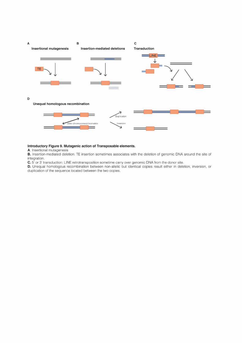
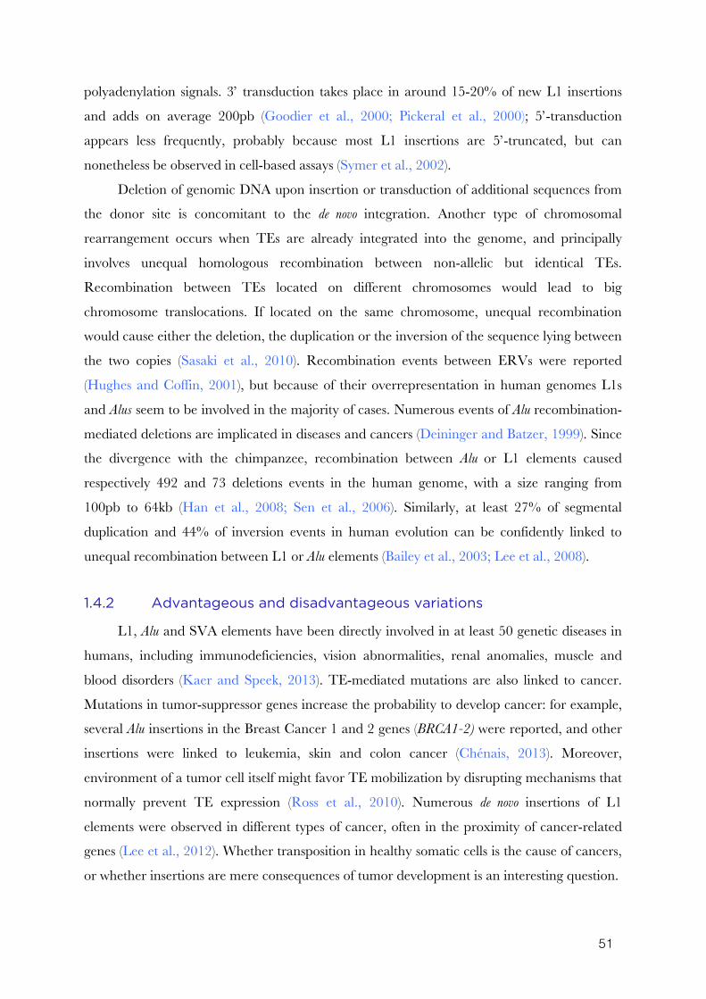
51
polyadenylation signals. 3’ transduction takes place in around 15-20% of new L1 insertions
and adds on average 200pb (Goodier et al., 2000; Pickeral et al., 2000); 5’-transduction
appears less frequently, probably because most L1 insertions are 5’-truncated, but can
nonetheless be observed in cell-based assays (Symer et al., 2002).
Deletion of genomic DNA upon insertion or transduction of additional sequences from
the donor site is concomitant to the de novo integration. Another type of chromosomal
rearrangement occurs when TEs are already integrated into the genome, and principally
involves unequal homologous recombination between non-allelic but identical TEs.
Recombination between TEs located on different chromosomes would lead to big
chromosome translocations. If located on the same chromosome, unequal recombination
would cause either the deletion, the duplication or the inversion of the sequence lying between
the two copies (Sasaki et al., 2010). Recombination events between ERVs were reported
(Hughes and Coffin, 2001), but because of their overrepresentation in human genomes L1s
and Alus seem to be involved in the majority of cases. Numerous events of Alu recombination-
mediated deletions are implicated in diseases and cancers (Deininger and Batzer, 1999). Since
the divergence with the chimpanzee, recombination between Alu or L1 elements caused
respectively 492 and 73 deletions events in the human genome, with a size ranging from
100pb to 64kb (Han et al., 2008; Sen et al., 2006). Similarly, at least 27% of segmental
duplication and 44% of inversion events in human evolution can be confidently linked to
unequal recombination between L1 or Alu elements (Bailey et al., 2003; Lee et al., 2008).
1.4.2 Advantageous and disadvantageous variations
L1, Alu and SVA elements have been directly involved in at least 50 genetic diseases in
humans, including immunodeficiencies, vision abnormalities, renal anomalies, muscle and
blood disorders (Kaer and Speek, 2013). TE-mediated mutations are also linked to cancer.
Mutations in tumor-suppressor genes increase the probability to develop cancer: for example,
several Alu insertions in the Breast Cancer 1 and 2 genes (BRCA1-2) were reported, and other
insertions were linked to leukemia, skin and colon cancer (Chénais, 2013). Moreover,
environment of a tumor cell itself might favor TE mobilization by disrupting mechanisms that
normally prevent TE expression (Ross et al., 2010). Numerous de novo insertions of L1
elements were observed in different types of cancer, often in the proximity of cancer-related
genes (Lee et al., 2012). Whether transposition in healthy somatic cells is the cause of cancers,
or whether insertions are mere consequences of tumor development is an interesting question.
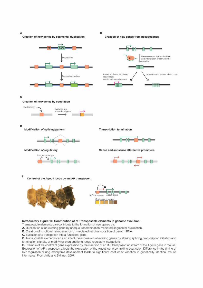

53
In the mouse, MLV LTR-retrotransposons have long been shown to be able to induce
leukemia (Jolicoeur et al., 1978)
Disease-causing mutations tend to disappear from the population, because of their
deleterious effects. But TE-derived mutations had many positive consequences during
evolution (Introductory Figure 10). The probability of evolving new protein-coding genes
de novo by random association of amino acids is very low. Since François Jacob introduced the
concept of “evolutionary tinkering”, it is commonly considered that in the majority of cases,
“The appearance of new molecular structures […] have rested on alteration of preexisting
ones” (Jacob, 1977). There are numerous examples where TEs helped creating new genes,
usually by duplicating existing ones. Around 40% of human or Drosophila genes are duplicated
somewhere else in the genome (Zhang, 2003). Considering that unequal recombination
between TEs is a major source of genomic duplication, unequal recombination probably
account for the creation of many duplicated genes (Introductory Figure 10A).
New genes are also created from intronless retrogenes created by LINE trans
mobilization (Introductory Figure 10B). Newly inserted retrogenes most often lack
regulatory sequences and are inert, becoming therefore pseudogenes. However, some appear
to have acquired new promoters or were transported with it. As a result, out of the 8,000
retrogenes identified in the mouse genome, around 1,000 are transcribed and a least 120 have
evolved into bona fide new genes (Vinckenbosch et al., 2006; Zhang et al., 2003). It is estimated
that at least one new functional retrogene per million year appeared during primate evolution
(Marques et al., 2005). Finally, at least one gene family in primates has also been created by
repeated 3’-transduction (Xing et al., 2006)
TEs did not only contribute to evolution by modifying the genome, but were also
sometimes directly domesticated by their host to evolve new functions Introductory Figure 10C and Volff, 2006). The majority of coopted TEs were DNA transposons. The most famous
case is the domestication of an ancient transposase 500Myrs ago to create the recombination-
activating Rag1 protein (Kapitonov and Jurka, 2005; Thompson, 1995). Rag1 (and Rag2) are
responsible for V(D)J recombination, the defining feature of the adaptive immune system of
jawed vertebrates. Another vital mechanism that originates from the domestication of an
ancient TE is the maintenance of chromosome telomeres. Telomerases are RNA-dependent
DNA polymerases and probably evolved from a primitive LINE-like retrotransposon
(Eickbush, 1997). In Drosophila, the ancestral telomerase was lost and telomere maintenance
evolved a second time by using, again, two LINE retrotransposons (Levis et al., 1993). Many
mammalian genes are also derived from the gag or env genes of LTR-retrotransposons (Volff,
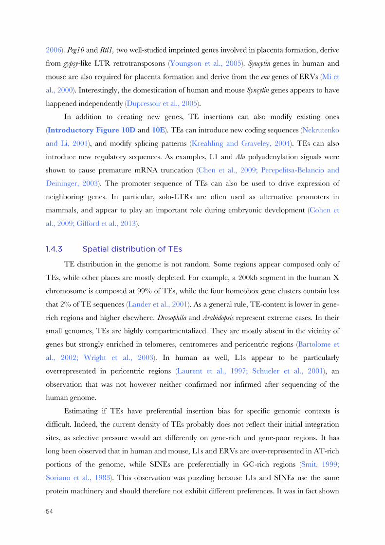
54
2006). Peg10 and Rtl1, two well-studied imprinted genes involved in placenta formation, derive
from gypsy-like LTR retrotransposons (Youngson et al., 2005). Syncytin genes in human and
mouse are also required for placenta formation and derive from the env genes of ERVs (Mi et
al., 2000). Interestingly, the domestication of human and mouse Syncytin genes appears to have
happened independently (Dupressoir et al., 2005).
In addition to creating new genes, TE insertions can also modify existing ones
(Introductory Figure 10D and 10E). TEs can introduce new coding sequences (Nekrutenko
and Li, 2001), and modify splicing patterns (Kreahling and Graveley, 2004). TEs can also
introduce new regulatory sequences. As examples, L1 and Alu polyadenylation signals were
shown to cause premature mRNA truncation (Chen et al., 2009; Perepelitsa-Belancio and
Deininger, 2003). The promoter sequence of TEs can also be used to drive expression of
neighboring genes. In particular, solo-LTRs are often used as alternative promoters in
mammals, and appear to play an important role during embryonic development (Cohen et
al., 2009; Gifford et al., 2013).
1.4.3 Spatial distribution of TEs
TE distribution in the genome is not random. Some regions appear composed only of
TEs, while other places are mostly depleted. For example, a 200kb segment in the human X
chromosome is composed at 99% of TEs, while the four homeobox gene clusters contain less
that 2% of TE sequences (Lander et al., 2001). As a general rule, TE-content is lower in gene-
rich regions and higher elsewhere. Drosophila and Arabidopsis represent extreme cases. In their
small genomes, TEs are highly compartmentalized. They are mostly absent in the vicinity of
genes but strongly enriched in telomeres, centromeres and pericentric regions (Bartolome et
al., 2002; Wright et al., 2003). In human as well, L1s appear to be particularly
overrepresented in pericentric regions (Laurent et al., 1997; Schueler et al., 2001), an
observation that was not however neither confirmed nor infirmed after sequencing of the
human genome.
Estimating if TEs have preferential insertion bias for specific genomic contexts is
difficult. Indeed, the current density of TEs probably does not reflect their initial integration
sites, as selective pressure would act differently on gene-rich and gene-poor regions. It has
long been observed that in human and mouse, L1s and ERVs are over-represented in AT-rich
portions of the genome, while SINEs are preferentially in GC-rich regions (Smit, 1999;
Soriano et al., 1983). This observation was puzzling because L1s and SINEs use the same
protein machinery and should therefore not exhibit different preferences. It was in fact shown
55
that young SINEs have the same AT-rich insertion bias than LINEs, but that there is a strong
(and unexplained) selective pressure to conserve older ones only in GC-rich portions (Lander
et al., 2001; Waterston et al., 2002). The preference of L1 insertions for AT-rich regions is
often explained by the requirement for L1 to insert at AATTTT hexanucleotides (Smit,
1999). However, gene-rich regions tend naturally to have a high GC-content, while gene-poor
regions are conversely AT-rich. The observed preference for AT-rich regions could simply be
the result of differential selective pressures.
The most unbiased method to estimate the real integration preferences of TEs is to rely
on artificially integrated constructs. Several studies with human and mouse artificial L1
transgenes showed that L1 insertions tend in reality to be distributed randomly throughout the
genome (Babushok et al., 2006; Gilbert et al., 2002; Symer et al., 2002). A modest preference
for intergenic regions could only be noted for mouse L1s. In line with these observations, the
most recent L1 insertions in the human genome are distributed randomly, and the observed
preference for AT-rich, gene-poor regions is probably a selective bias (Ovchinnikov et al.,
2001). Similarly, analysis of the integration sites of an artificially resurrected human ERV or
of bona fide retroviruses showed that retroviral sequences insert preferentially into gene-rich
regions, and especially inside genes (Brady et al., 2009; Mitchell et al., 2004). By contrast,
remaining ERVs are mostly excluded from genes, and in general ancient ERVs are
particularly underrepresented near genes compared to younger elements (Medstrand et al.,
2002).
Gene-rich regions are often associated with a permissive chromatin environment that
could make integration of new TEs easier. However, in the long term, the fixation and the
maintenance of TEs is probably mainly controlled by natural selection. As a consequence,
TEs are over-represented in gene-poor regions.
SINEs represent a very intriguing exception. They seem to integrate randomly but are
strongly selected to remain in the proximity of genes. This suggests that they are beneficial for
the organism (Lander et al., 2001), which fits with their incredible success in mammalian
genomes. SINEs could potentially play a role during meiosis and contribute to synapsis
formation between homologous chromosomes. During meiosis, sister chromatids adopt a
complex structure: they align on a protein-made axis with chromatin extending out in loops.
The protein axis of the two homologous chromosomes are brought together and joined via the
zipper-like synaptomenal complex. The formation of this complex is critical for correct
crossing-over, and recombination is thought to occur primarily within the loops and not in the
DNA located near the axis (Keeney et al., 2014). Interestingly, it was suggested that SYCP3,

56
(one of the core protein of the synaptomenal complex) was preferentially binding SINE DNA
in mouse (Johnson et al., 2013). SINEs could serve as a binding platform for the recruitment
of the synaptomenal complex in gene-rich regions. Because recombination occurs
preferentially in the chromatin loops that are not directly bound by the synaptomenal
complex, it would also ensure that potentially harmful DNA breaks do not occur directly in
genes, but only in their proximity. It was indeed shown in human that recombination
“hotspots” were preferentially located near genes, but principally outside the transcribed
domain (Myers et al., 2005). As synapsis formation is a genome-wide process, it would also
explain why the localization of SINEs in gene-rich regions is such a global pattern. However,
this hypothesis is highly speculative (Barau, 2015)4 and the reason for the accumulation of
SINEs near genes remains largely unknown.
1.4.4 TEs in the mouse genome5
The mouse genome is an interesting model to study TEs. It contains both ancestral
repeats that predate the divergence with humans, and also very recently integrated ones.
Rodent and primate lineages are estimated to have diverged between 65 and 100Myrs ago.
The broad composition of human and mouse genomes is similar, but closer analysis reveals
interesting differences (Waterston et al., 2002). The mouse genome (2.7Gb) is markedly
smaller than the human one (3.2Gb), and is composed of at least 44% of TEs, with around
1Gb (85%) that is mouse-specific. The rate of spontaneous deletions and nucleotide
substitutions is twice as high in mice than in humans. Fossilized repeats become then
unrecognizable faster in mouse: they disappear after 100-120Myrs, compared to 150-200Myrs
for humans. As a consequence, ancestral repeats comprise only 5% of the genome in mice,
and 22% in humans. Mouse TEs are also much younger in average: 25% of mouse repeats
have integrated in the last 25Myrs, while human TEs became almost extinct during that time.
Whereas the majority of human TEs are now inert, the mouse genome contains thousands of
active LINEs and ERVs.
L1s represent 20% of the mouse genome, with approximately 600,000 copies. Mouse
L1s all derive from the evolution of a single ancestral lineage, the last common ancestor
between humans and mice being L1MA6 (Smit et al., 1995; Waterston et al., 2002). Around
20,000 L1s are full-length and 3,000 copies are estimated to be retrotransposition-competent,
4 And also very personal 5 The names and the numbers given in this section are based on the last RepeatMasker annotation (Smit
et al., 2013), using criteria to categorize full-length elements that will be developed in the result section of this thesis. They are in good concordance with numbers given in Stocking and Kozak, 2008.
57
namely 30 times more than in humans (Goodier et al., 2001). Since the divergence with the
rat 13Myrs ago, the mouse L1 lineage has experienced 11 replacements of 5’UTR, and
numerous other recombinations in the coding part (Introductory Figure 11 and Sookdeo et
al., 2013). Recent active families are classified in two groups based on their promoter type (A
and F), while more older and extinct families have a V type promoter (Adey et al., 1994). The
F promoter replaced a V promoter by resuscitating a more ancestral form 6.4Myrs ago. The
A promoter further replaced this F-type 4.6Myrs ago. Respectively 2.2 and 1.2Myrs ago, A-
elements exchanged again their promoter and gave birth to Tf and Gf elements (both F-type
promoters) (Sookdeo et al., 2013). A-, Gf- and Tf-type account respectively for 900, 400 and
1,800 retrotransposition-competent elements (Goodier et al., 2001).
Whereas only one SINE lineage is active in the human lineage (Alu), mice have four
distinct SINE families (B1, B2, ID, B4). There is more SINEs in the mouse than in the human
genome (1.4 and 1.1 millions, respectively), but mouse SINEs occupy less genomic space (8
and 13%, respectively) because of their smaller size (Waterston et al., 2002). B1 and Alu
elements are both derived from a 7SL RNA and have probably a common origin (Quentin,
1994). On the other hand, B2 and ID are related to tRNAs. ID closely matches an Ala-tRNA
and is present with relatively few copies in the mouse genome, compared to its very successful
amplification in the rat genome (Gibbs et al., 2004). B4 elements represent an interesting case
of fusion between ID and B1 (Kramerov and Vassetzky, 2001). Both B4 and ID elements are
inactive in the mouse genome, whereas B1 and B2 show continuous signs of multiplication
(Gibbs et al., 2004).
Bona fide LTR-retrotransposons of gypsy or copia superfamilies cannot be found in the
mouse, but ERVs compose 12% of the genome. As a consequence, the two terms “LTR-
retrotransposons” and “ERVs” are often used as synonyms. ERVs are usually further
classified into three classes depending on the type of retrovirus they originate from
Introductory Figure 12A). Class-I relates to gamma- and epsilonretroviruses, class-II to
lentiviruses, alpha-, beta- and deltaretroviruses, while class-III is closer to spumaviruses
(Gifford et al., 2005; Jern et al., 2005). Class-III ERVs are often referred as class-L ERVs,
because in both mouse and human retrotransposition is initiated with a leucine tRNA (Bénit
et al., 1997; Cordonnier et al., 1995). Similarly, class-II is also often referred as class-K,
because the first identified elements (MMTV in mouse, HERV-K10 in human) use lysine
tRNAs as primers (Ono et al., 1986; Peters and Glover, 1980). However, this classification is
not correct since IAP, one of the most studied class-II family, appears to use a phenylalanine
tRNA (Rowe et al., 2010). In general, the nomenclature of ERVs is incredibly confusing. The
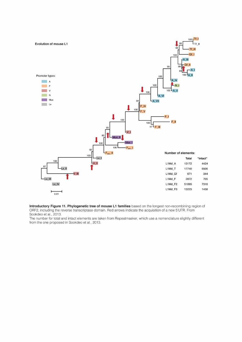
59
main families were discovered a long time ago and their current names (when they have one)
often reflect their initial description, but in the form of now meaningless initials (IAP, MLV,
MMTV, VL30, ETn, GLN, etc.). It is also difficult to have a clear estimation of the total
number of families because this varies depending on the criteria used to classify them. Some
studies proposed a limited number (around 20) (McCarthy et al., 2004), but the Repbase
depository separates LTRs from internal sequences and currently annotate 484 different
families (Jurka et al., 2005). Unfortunately, no coherent classification managed yet to replace
this unintelligible imbroglio. In fact, because they were changing the name of known
transposons, most of the attempts only added confusion (Baillie et al., 2004; McCarthy et al.,
2004).
Class-I ERVs represent only 1.2% of the mouse genome, with a lot of different families
composed of relatively few full-length copies (between 10 and 300 per genome). The best
characterized is the MLV family (Mouse Leukemia Virus). MLVs entered the mouse genome
recently (<1.5Myr ago) and intact elements remain potent retroviruses capable of infecting
other cells. MLV particles has long been shown to be able to promote cancer (Friend, 1957).
MLVs are in the process of endogenization and the number of element per genome varies
between 10 and <100 depending on the mouse species (Stocking and Kozak, 2008). RLTR6
(also named MmERV) and VL30 are two other interesting class-I elements. They share the
same LTRs, but RLTR6 is autonomous with predominantly intact gag, env and pol sequences,
whereas VL30 lacks coding regions (Bromham et al., 2001; French and Norton, 1997). VL30
are likely derived from RLTR6 elements that lost their coding region, and continue to use in
trans their protein machinery for their replication.
Class-II ERVs are very numerous and represent 4.9% of the mouse genome. The first
discovered ERV-II was linked to breast cancer (Mouse Mammary Tumor Virus: MMTV) and
was initially thought to be a bona fide retrovirus with a strong insertion preference for the same
genomic region (Nusse and Varmus, 1982). However, there is only 2-3 full-length MMTV
inserts in the mouse genome. Two related families are known as MusD6 and ETn (Early
Transposon), with around 300 intact copies each. ETns are very active and was first detected
in embryonic carcinoma (Brulet et al., 1983). ETn elements are however internally
recombined and lack coding sequences. They rely on retrotransposition-competent MusD
proteins for their activity (Baust et al., 2003; Mager and Freeman, 2000). The most studied
mouse ERVs are related to IAP (Intracisternal A-type Particles) that were initially observed in
the endoplasmic reticulum of various cancer cells (Introductory Figure 12B and Dalton et 6 In RepeatMasker nomenclature, MusD is named ETnERV or ERVB7_1 (sic)
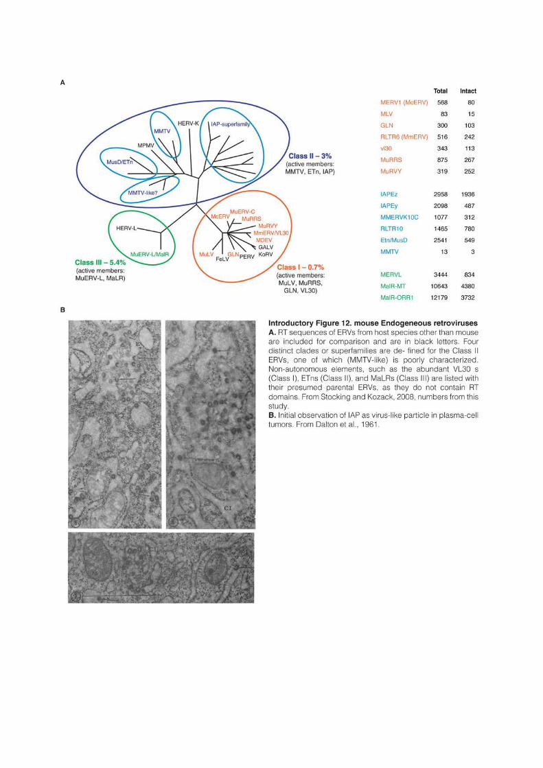
61
al., 1961; De Harven and Friend, 1958). Eight families can be distinguished (including for
example MMERVK10C or the severely internally deleted RLTR10) and they represent
together around 2,000 intact copies (McCarthy et al., 2004). ETn and IAP elements are the
most active transposons of the mouse genome and account for the vast majority of de novo
insertions (Maksakova et al., 2006).
As in most mammals, Class-III are the most abundant ERVs in the mouse genome
(5.9% of the total). They are also the most ancient, and they account for 80% of the ERVs
predating the human-mouse speciation (Waterston et al., 2002). Probably for this reason,
Class-III elements, and especially solo-LTRs, appear to have been coopted by mammalian
evolution and they play an important role during gametogenesis and embryonic development
(Gifford et al., 2013). Class-III is composed of only two types of elements: the
retrotransposition-competent MERVLs and the non-autonomous MalRs (Mammalian
apparent LTR-retrotransposons) (Bénit et al., 1997; Smit, 1993). MERVLs and MalRs share
similar LTR sequences and probably have a common ancestor (McCarthy et al., 2004).
MalRs are internally deleted and contain only non-coding repetitive DNA. The two main
types, MT and ORR1, have an average length of 2 and 2.4kb, respectively. MalRs, and
especially MT elements, have been incredibly successful, as they represent the vast majority of
class-III elements and outnumber MERVLs by a 10 to 1 ratio.
Further reading:
The following reviews were extensively used as starting material to write this chapter.
Biemont, 2010: history of TEs discovery; Feschotte and Pritham, 2007: DNA transposons; Kramerov and Vassetzky, 2005: SINEs. Babushok et al., 2007: LINE1; Cordaux and Batzer, 2009: genome evolution; Le Rouzic and Capy, 2009: transposon population genetics; Silva et al., 2004: horizontal transfer; Stocking and Kozak, 2008: murine LTR-retrotransposons. The following studies are cited in figure legends: Dalton et al., 1961; Fedoroff, 2012; Feschotte et al., 2002; Fleischmann et al., 2014; Gilbert et al., 2010; Gregory, 2015; Gregory et al., 2009; Griffiths et al., 2008; Hedges et al., 2015; Jirtle and Skinner, 2007; Kelley et al., 2014; Maddison et al., 2007; Pace et al., 2008; Pellicer et al., 2010; Smit et al., 2013; Sookdeo et al., 2013; Stocking and Kozak, 2008

62
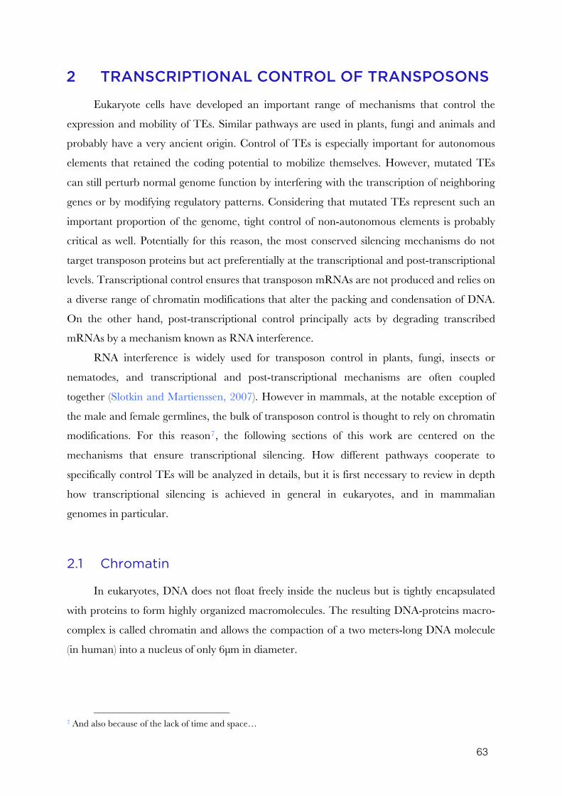
63
2 TRANSCRIPTIONAL CONTROL OF TRANSPOSONS
Eukaryote cells have developed an important range of mechanisms that control the
expression and mobility of TEs. Similar pathways are used in plants, fungi and animals and
probably have a very ancient origin. Control of TEs is especially important for autonomous
elements that retained the coding potential to mobilize themselves. However, mutated TEs
can still perturb normal genome function by interfering with the transcription of neighboring
genes or by modifying regulatory patterns. Considering that mutated TEs represent such an
important proportion of the genome, tight control of non-autonomous elements is probably
critical as well. Potentially for this reason, the most conserved silencing mechanisms do not
target transposon proteins but act preferentially at the transcriptional and post-transcriptional
levels. Transcriptional control ensures that transposon mRNAs are not produced and relies on
a diverse range of chromatin modifications that alter the packing and condensation of DNA.
On the other hand, post-transcriptional control principally acts by degrading transcribed
mRNAs by a mechanism known as RNA interference.
RNA interference is widely used for transposon control in plants, fungi, insects or
nematodes, and transcriptional and post-transcriptional mechanisms are often coupled
together (Slotkin and Martienssen, 2007). However in mammals, at the notable exception of
the male and female germlines, the bulk of transposon control is thought to rely on chromatin
modifications. For this reason7, the following sections of this work are centered on the
mechanisms that ensure transcriptional silencing. How different pathways cooperate to
specifically control TEs will be analyzed in details, but it is first necessary to review in depth
how transcriptional silencing is achieved in general in eukaryotes, and in mammalian
genomes in particular.
2.1 Chromatin
In eukaryotes, DNA does not float freely inside the nucleus but is tightly encapsulated
with proteins to form highly organized macromolecules. The resulting DNA-proteins macro-
complex is called chromatin and allows the compaction of a two meters-long DNA molecule
(in human) into a nucleus of only 6μm in diameter.
7 And also because of the lack of time and space…
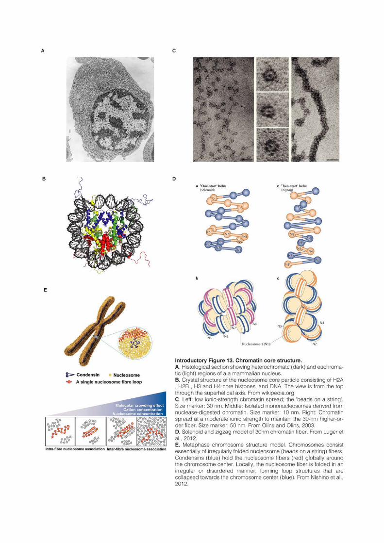
65
2.1.1 Historical perspective
Walther Flemming first introduced the term chromatin in 1882 to refer to a fibrous
scaffold in the cell nucleus that could be easily stained8 (Flemming, 1882). A decade earlier,
Friedrich Miescher had identified in the nucleus a strong phosphorus-rich acid (Miescher,
1871) and Albrecht Kossel proposed in 1884 that these “nucleic acids” might be bound to a
novel class of proteins that he had termed histones (Kossel, 1884). Histones and DNA
constitute indeed the core components of chromatin. For a long time, histones were even
thought to be the carrier of genetic information, while DNA was viewed as a mere linker
molecule (Schultz, 1941). This view completely shifted with the demonstration that DNA, and
not proteins, could by itself transform a harmless strain of Pneumococcus bacteria into a very
virulent one (Avery et al., 1944). The resolution of the double-helical structure of DNA then
provided the molecular basis of genetic inheritance (Watson and Crick, 1953) that led to the
enounciation of the central dogma of biology (Crick, 1956).
Chromatin was long established to exist in different forms. Chromosomes are at their
most condensed form during the metaphase stage of mitosis, and chromosomes decondense
during interphase. In 1928, Emil Heitz visualized chromosomal regions that do not undergo
post-mitotic decondensation and termed these domains “heterochromatin” (Heitz, 1928). By
opposition, “euchromatin” refers to chromosomal regions that decondense and spread
diffusely in the interphase nucleus (Introductory Figure 13A). Heitz immediately proposed
that the densely compacted heterochromatin could reflect a functionally inactive state of the
genome (Heitz, 1929, 1932). This hypothesis was first validated in Drosophila, where the
localization of the white gene next to an heterochromatic domain after an inversion was shown
to produce stochastic changes in eye color (Schultz, 1936). This view is still prevalent today:
heterochromatin is typically gene-poor and transcriptionally silent; whereas the less condensed
euchromatin is gene-rich and transcriptionally permissive. Heterochromatin was further
divided into constitutive and facultative heterochromatin to distinguish regions that are always
condensed on both homologous chromosomes and in every cell types (like centromeres and
telomeres), from regions that can alternate between different states in a development-specific
manner (like the X chromosome in female) (Brown, 1966).
The molecular structure of chromatin in its various flavors has received incredible
attention since then. While DNA is invariably composed of the same four nucleotides, the
proteins it is packaged with come in a seemingly infinite diversity. In particular, histones can
8 “chromo” stand for “color” in ancient Greek
66
harbor many different post-translational modifications and are further bound by numerous
other proteins. Furthermore, DNA itself can be modified and methyl group are often added at
cytosines. Both histone and DNA modifications directly influence chromatin condensation
and gene expression. Following the work of Jacob and Monod, gene expression was initially
believed to be regulated primarily by trans-acting factors that recognize specific DNA motifs
and either block or activate transcription (Jacob and Monod, 1961). The discovery in yeast
that histone themselves regulate gene expression (Kayne et al., 1988) was followed over the
years by the description of apparently limitless combinations between various chromatin
modifications. This development led to the “histone code” hypothesis9, which states that the
complex combination between various chromatin modifications “considerably extends the
information potential of the genetic code” and “represents a fundamental regulatory
mechanism that has an impact on most, if not all, chromatin-templated processes” (Jenuwein
and Allis, 2001; Strahl and Allis, 2000).
Since 1988, Chromatin ImmunoPrecipitation (ChIP) has been widely used to analyze
DNA-associated proteins and their modifications along chromosomes (Solomon et al., 1988).
Over the last decade, tremendous energy has been spent to map the local structure of
chromatin. These efforts, exemplified by the ENCODE project in mouse and human
(Bernstein et al., 2012), allowed the linear segmentation of chromosomes into hundred of
domains with different protein compositions. Moreover, the spatial organization of DNA in
the nucleus is not random. Local and long-range interaction between sequences and the
localization inside the nucleus appears highly controlled. For example in human or Drosophila,
hundreds of domains are preferentially associated with the nuclear lamina, and genes inside
these domains are transcriptional silent (Guelen et al., 2008; Pickersgill et al., 2006). The
developments of chromosome conformation capture technology (3C, 4C, 5C and Hi-C) gave
further insight into the global 3-dimensional structure of the genome and uncovered many
unsuspected long range interactions between DNA sequences (Dekker et al., 2013). In
Drosophila, mouse and human, chromosomes are composed of hundreds of discrete topological
domains of typically 100kb-1Mb, inside which DNA preferentially interact (Dixon et al.,
2012; Nora et al., 2012; Sexton et al., 2012).
Hundreds of chromatin modifications have been identified and the potential
combinations between them are infinite. However, most modifications preferentially associate
together in a reduced set of combination, allowing the definition of only a small number of
major chromatin types (Rando, 2012; van Steensel, 2011). One of the current challenges is to 9 that can be extended to incorporate DNA methylation and other non-histone proteins
67
integrate the enormous amount of linear and three-dimensional genomic data in order to
build a global and compressive picture of chromatin organization (Bickmore and van Steensel,
2013). These new descriptions are going beyond the original heterochromatin/euchromatin
dichotomy and allow for a better understanding of how local and large-scale structures affect
gene regulation and other nuclear functions. It was for example proposed that chromatin
complexity could be reduced to four “chromatin states” in Arabidopsis (Roudier et al., 2011),
five “chromatin colors” in Drosophila (Filion et al., 2010) or six “chromatin compartments” in
human (Rao et al., 2014).
2.1.2 Chromatin core structure and modifications
The first level of packaging is the nucleosome, which consists of 145-147pb of DNA
wrapped 1.7 times around an octamer of core histones (Kornberg, 1974; Luger et al., 1997).
Histone octamers are composed of two copies of the core histone proteins H2A, H2B, H3 and
H4. Histones possess flexible tails that extend away from the disk-like structure of the
nucleosome and that are involved in the interaction with other nucleosomes or various
nuclear factors (Introductory Figure 13B). Nucleosomes are separated by short linker
sequences of around 10-60bp that are often bound by the linker histone H1, with a global
stoichiometry in mouse nucleus of 0.5 to 0.8 H1 per nucleosome (Woodcock et al., 2006). H1
localize at the entry and exit points of DNA and stabilize the interaction of an additional 20pb
of DNA around the nucleosome, resulting in complete two-turn particle of ~166pb (Thoma et
al., 1979). The repeated array of nucleosomes can be easily observed by electronic microscopy
(Olins and Olins, 1974) and forms a “bead-on-a-string” primary structure known as the 10nm
fiber (Introductory Figure 13C).
The packing of DNA into nucleosomes results in a reduction of the fiber length of about
sevenfold. Chromatin can be further folded into a thicker secondary structure known as the
30nm fiber that was first observed by electron microscopy in salamander’s red blood cells
(Introductory Figure 13C and Gall, 1963). The 30nm fiber appears spontaneously in vitro
(Finch and Klug, 1976) and relies on linker histone H1 for its stabilization (Robinson and
Rhodes, 2006). Based on numerous in vitro studies, the 30nm fiber has long been thought to be
the natural state of chromatin folding. The high density of the 30nm fiber makes, however,
the analysis of its molecular conformation difficult. Two structures, the “zigzag” and
“solenoid” models, are still in competition to explain how the repeated array of nucleosomes
folds into the 30nm fiber (Introductory Figure 13D and Robinson et al., 2006; Schalch et
al., 2005). The conformation of the 30nm fiber appears moreover to depend on many
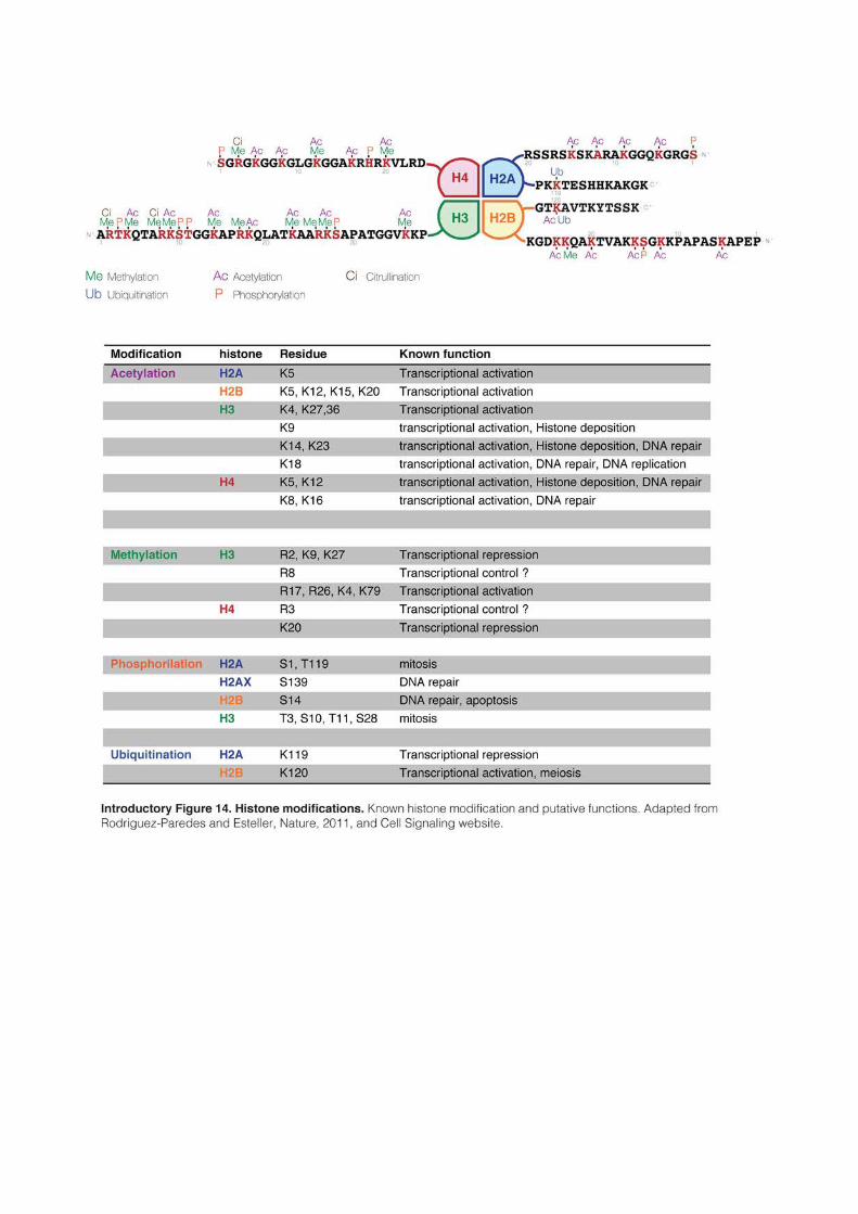
69
different parameters like salt conditions, length of linker DNA, histone tails modifications, etc.
(Hansen, 2002).
Chromatin can further fold into even denser tertiary structures, especially during
metaphase. It was long assumed that the 30nm fiber was further twisted and coiled to form
highly condensed domains. However, it was recently showed that 30nm fibers could not be
observed in metaphase chromosomes and that high degree of compaction was achieved by a
“fractal packaging” of irregularly arranged 10nm fibers (Introductory Figure 13E and
Nishino et al., 2012). Even in interphase chromosome, the direct observation of bona fide 30nm
fibers remains elusive (Tremethick, 2007) and some authors proposed that chromatin could in
reality only consist of dynamic and disordered 10nm fibers (Maeshima et al., 2014). In fact,
the 30nm fiber represents probably only one of many potential chromatin structures. In vivo
chromatin is presumably not uniform and likely harbor different conformations depending on
the context (transcriptional state, cell cycle stage, developmental stage, etc.).
Many “architectural” proteins have been described, including heterochromatin protein
1 (HP1), methyl-CpG-binding protein 2 (MeCP2), Polycomb group proteins and others.
These proteins usually form complexes that bind to multiple nucleosomes and induce further
chromatin compaction, sometime acting also as shields that block the access to the underlying
DNA. Architectural proteins are critical to form and maintain high-order chromatin
structures (McBryant et al., 2006).
Histone tails modifications are playing a very important role in chromatin compaction
and have a direct role in transcriptional control. Histone tails represent 25-30% of the total
mass of histones and provide an exposed surface for many protein-protein interactions. An
extensive literature documents dozen of different post-translational modifications including
acetylation, phosphorylation, methylation, ubiquitination and citrullination (Introductory Figure 14). Chromatin modifiers, ie proteins that add and remove (“writers”) and/or
interpret (“readers”) histone modifications have also been described at length (Kouzarides,
2007). Histone acetylation is one of the most studied modification and correlate with open
chromatin and active transcription (Grunstein, 1997). Acetylation neutralizes the positively
charged lysine residue and thereby decreases the interaction of histone tails with the
negatively charged DNA (Hong et al., 1993). This causes the nucleosomes to unfold (Norton
et al., 1989) and facilitates the recruitment of the transcription machinery (Lee et al., 1993).
Histone acetylation is primarily controlled by numerous histone acetyltransferase (HATs) and
deacetylases (HDACs) (Marmorstein and Roth, 2001; Thiagalingam et al., 2003).
70
By contrast, lysine mono-, di- and tri-methylation do not modify the histone tail charge
and can be linked either to active or inactive chromatin. In general, methylation of histone H3
lysine 4 (H3K4), H3K36 and H3K79 is associated with an active state, while H3K9, H3K27
and H4K20 correlate with repression and are prevalent in heterochromatin (Barski et al.,
2007). Many lysine methyltransferases (KMTs) and lysine demethylases (KDMs) are described
and a unified nomenclature based on the histone residue they target has been proposed (Allis
et al., 2007). For example H3K9-KMTs are members of the KMT1 family, H3K4-KMTS of
the KMT2 family, etc. At the notable exception of DOT1L10/KMT4 that deposit H3K79
methylation (Feng et al., 2002), histone lysine methyltransferases are all characterized by the
presence of a SET domain that catalyzes the methylation reaction using S-Adenosyl
methionine (SAM) as a methyl donor. SET stands for Su(var), Enhancer of zeste, and
Tritorax, the names of the tree proteins in which it was first identified in Drosophila (Jenuwein
et al., 1998).
The precise mechanisms that enable histone modifications to impact on chromatin
compaction and gene regulation are rarely well understood. Some modifications, like in
particular lysine acetylation, have a direct effect on chromatin compaction. The tail of histone
H4 is known to mediate interaction between adjacent nucleosomes (Tse and Hansen, 1997)
and the amino acids 16-25 of histone H4 are thought to interact with an acidic patch located
on the surface of H2A-H2B of an adjacent nucleosome (Luger et al., 1997). This interaction
has been shown to be required in vitro for the compaction into a 30nm fiber (Dorigo et al.,
2003). Furthermore, it was shown that acetylation of H4K16, a well-studied mark associated
with gene activation, was sufficient to prevent chromatin compaction (Shogren-Knaak et al.,
2006) but that by contrast, tri-methylation of H4K20 resulted in a even more condensed
chromatin (Lu et al., 2008). These examples show how histone modifications can directly
modify the local chromatin structure and probably impact transcriptional regulation as a
consequence.
Many chromatin modifications do not have a direct physical influence on chromatin
structure but act indirectly, by attracting or preventing the binding of “histone readers”. Over
the years, numerous histone readers and specific protein interaction domains have been
characterized. For example, methylation is recognized by PWWP, PHD or Chromo-like royal
domains (Chromo, Tudor MBT), while bromodomains recognize acetylation (Kouzarides,
2007). The functions associated with histone reader domains are very diverse. Some proteins
10 Because of the lack of significance it often represents, the origin and signification of many gene names will be
most of the time deliberately ignored.
71
are directly linked to chromatin structure like architectural proteins, chromatin remodelers or
chromatin modifiers, while others regulate various biological processes, including mRNA
transcription and elongation, DNA repair, V(D)J recombination or DNA replication. An
additional level of regulation is achieved by the use of histone variants. They are paralogs of
the canonical histones and their integration can affect nucleosome stability and help to create
functionally different environments. For example, H3 is replaced at centromeres by the
variant CENP-A, or by H3.3 in actively transcribed genes, regulatory regions and in some
cases in heterochromatin (Biterge and Schneider, 2014)
In plants, insects and mammals, two main type of repressive chromatin are identified
and will be reviewed in details in the following sections. The first one is best characterized by
H3K9 methylation, while the second is linked to H3K27 methylation. These two chromatin
states are structurally different. They are accompanied by distinct histone modifications and
rely on different protein complexes. H3K9 methylation mark essentially functional genomic
structure such as telomeres, centromeres, or transposable elements, while H3K27me3 is
primarily found in regulatory regions and is highly involved in gene regulation (Mikkelsen et
al., 2007).
Methylation of cytosine nucleotides, usually simply referred as DNA methylation, is a
really special chromatin modification, in the sense that it is the only one that actually has
DNA as substrate. DNA methylation cannot be unambiguously linked to any chromatin
compartment, as it is present throughout the genome in mammals. However, DNA
methylation has a direct effect on transcriptional regulation and is involved in many critical
biological processes.
2.2 DNA methylation
It has long been observed in a very diverse range of eukaryote species that a small subset
of cytosine nucleotides carries an additional methyl group at the fifth position of the six-atoms
aromatic ring (Introductory Figure 15A and Johnson and Coghill, 1925; Wyatt, 1950). In
mammals, around 4-6% of cytosines are typically methylated in somatic cells. This proportion
goes up to 25% in certain plant genomes, while other species like C. elegans, D. menanogaster (but
not other insects such as ants and bees) and some fungi (S. cerevisiae and S. pombe, but not N.
crassa) have no detectable levels (Zemach et al., 2010). In plants and other organisms, DNA
methylation is found in three different sequence contexts: CG (or CpG), CHG or CHH
(where H correspond to A,T or C). In mammals however, DNA methylation is almost
72
exclusively found in CpG dinucleotides, with the cytosines on both strand being usually
methylated. The sequence symmetry of this context led very early to the groundbreaking
hypothesis that DNA methylation could control transcriptional activity and serve as a form of
non-coded cellular memory during DNA replication (Holliday and Pugh, 1975; Riggs, 1975).
Indeed, DNA methylation has been shown to be stable trough multiple divisions or even from
parents to offspring, and is functionally involved in many important processes such as genomic
imprinting, X inactivation or silencing of transposable elements (Jones, 2012). In plants,
“epialleles” carrying different transcriptional outcomes depending on their methylation status
can be faithfully transmitted from one generation to the next. However, cases of
transgenerationnal inheritance of non-coded information in mammals appear (at best)
exceptional, especially because DNA methylation and other chromatin modifications are
extensively reprogrammed during embryonic development (Heard and Martienssen, 2014).
The question of these reprogramming events will be extensively addressed later, and the
following paragraphs focus principally on the function of DNA methylation in steady-state
cells.
The DNA methylation landscape of vertebrates is very particular compared to other
organisms (Introductory Figure 15B). In vertebrate, around 80% of CpG are methylated in
somatic cells and DNA methylation appears as a default state that has to be specifically
excluded from defined locations (Lister et al., 2009; Stadler et al., 2011). By contrast, the
genome of most plants, invertebrates, fungi or protists show “mosaic” methylation patterns,
where only specific genomic elements are targeted, and they are characterized by the
alternation of methylated and unmethylated domains (Suzuki and Bird, 2008; Zemach et al.,
2010). Some mammalian cell types such as embryonic stem (ES) cells or neurons contain also
a relatively high level of non-CpG methylation, but whether methylation in this context has a
function remains unclear (Lister et al., 2009, 2013)
High CpG methylation in mammalian genomes has an evolutionary cost because it
increases the frequency of spontaneous mutations. Loss of amino-groups occurs with a high
frequency for cytosines, with different consequences depending on their methylation.
Deamination of unmethylated cytosines results in uracils that are efficiently recognized and
removed by base-excision repair mechanisms. Deamination of methylated cytosines on the
other hand results in thymines, a proper genomic base that is not as efficiently repaired: this
results in frequent C to T transitions over evolutionary time (Bird, 1980; Walsh and Xu,
2006). As a consequence, CpGs are globally depleted in mammalian genomes, occurring at
only 20% of their excepted frequency (Lander et al., 2001; Swartz et al., 1962). The only
73
exception for this global CpG depletion resides in a specific category of GC- and CpG-rich
sequences termed CpG islands that are generally unmethylated and therefore retained the
expected CpG content (Bird, 1986; Bird et al., 1985).
CpG islands are usually defined as regions with 1) a length greater than 200pb, 2) a
G+C content greater than 50%, 3) a ratio of observed to expected CpG greater than 0.611,
although other definitions are sometimes used (Gardiner-Garden and Frommer, 1987).
Excluding repeated sequences, there are around 25,000 CpG islands in the human genome,
75% of which being less than 850pb long (Lander et al., 2001). They are major regulatory
units and around 50% of CpG islands are located in gene promoter regions, while another
25% lie in gene bodies, often serving as alternative promoters. Reciprocally, around 60-70%
of genes have a CpG island in their promoter region (Illingworth et al., 2010; Saxonov et al.,
2006). The majority of CpG islands are constitutively unmethylated and enriched for
permissive chromatin modification such as H3K4 methylation. In somatic tissues, only 10% of
CpG islands are methylated, the majority of them being located in intergenic and intragenic
regions, and this number drop to 1% in the male germline (Smallwood et al., 2011).
Of note, ES cells are derived from the inner cell mass of the blastocyst, one of the only
cell type during mouse development that is naturally depleted in DNA methylation. However,
when cultured classically in presence of fetal bovine serum, mouse ES cells have a DNA
methylation landscape similar to somatic cells: ~75% of CpGs are methylated and only CpG
islands are protected (Stadler et al., 2011). Human ES cells are similarly highly methylated
(Lister et al., 2009). Mouse ES cells can be also cultured without serum in presence of specific
inhibitors. In these conditions, the DNA methylation landscape is closer to the blastocyst and
cells are globally hypomethylated (Leitch et al., 2013). In almost all the studies referred to in
this section, mouse ES cells were classically grown in presence of serum, and except if
especially specified, it can therefore be considered that they are globally highly methylated.
2.2.1 DNA methyltransferases
DNA methylation is deposited by DNA (cytosine-5)-methyltransferase (DNMT)
enzymes, which catalyze the transfer of a methyl group to a cytosine nucleotide from a SAM
donor (Introductory Figure 15C). The first eukaryote DNMT to be identified and cloned
was the mammalian DNMT1, which is principally involved in the maintenance of DNA
methylation during cell divisions (Bestor and Ingram, 1983; Bestor et al., 1988). DNMT1
11 Observed/Expected ratio= (number of CpG * length of the sequence)/(number of C * number of G)
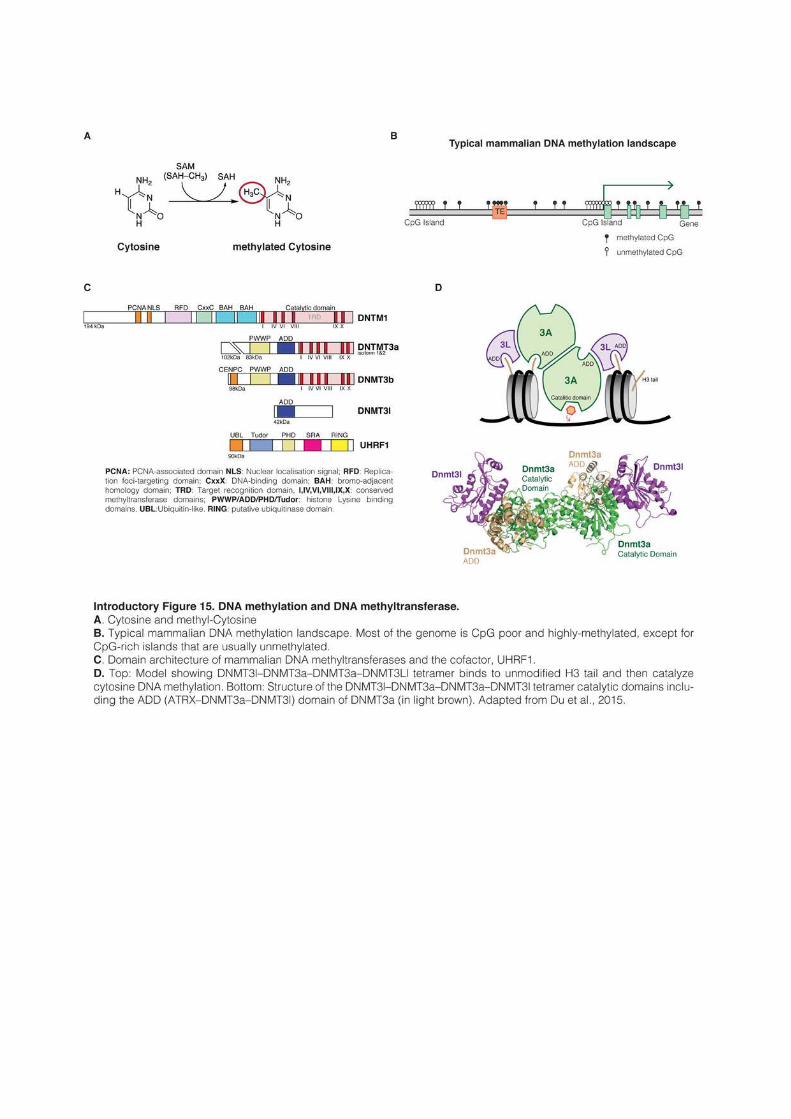
75
contains a replication foci-targeting domain (RFD), a DNA-binding CxxC domain, two
Bromo-adjacent homology domain (BAH) and a catalytic domain composed of core catalytic
motifs and of a target recognition domain that recognizes the sequence to be methylated
(Song et al., 2011; Takeshita et al., 2011). The overall structure is conserved with the plant
homolog MET1, and the catalytic domain of both enzymes is also shared with prokaryotic
DNMTs (Finnegan and Dennis, 1993). During mitosis, symmetrically methylated CpGs
become hemi-methylated and full methylation must be reestablished in both daughter cells.
DNMT1 is constitutively expressed in dividing cells and is most abundant during S phase. It is
recruited to the replication fork by interacting via its RFD domain with the proteins PCNA
and UHRF1. The latter binds specifically to hemimethylated CpG through its SRA domain
and orient DNMT1 to the newly synthetized, unmethylated DNA molecule (Bostick et al.,
2007; Chuang, 1997; Sharif et al., 2007). In addition, DNMT1 CxxC domain binds to
unmethylated CpG and prevent the catalytic domain to access DNA (Song et al., 2011). Both
UHRF1-mediated targeting to hemimethylated CpG and CxxC-mediated exclusion from
unmethylated CpG ensure the faithful copy of DNA methylation patterns during mitosis. This
maintenance activity is crucial in differentiated cells, and both UHRF1 and DNMT1-
knockout mice show embryonic lethality around mid-gestation (Bostick et al., 2007; Li et al.,
1992).
During embryonic development, DNA methylation is globally erased; it reaches a
minimum at the blastocyst stage and in primordial germ cells and is reestablished around the
time of implantation and during germ cell development (Kafri et al., 1992; Monk et al., 1987).
Moreover, ES cells lacking DNMT1 still harbor significant DNA methylation and can de novo
methylate foreign DNA sequences (Lei et al., 1996). These observations indicated that
additional DNMTs were present in the genome, and led to the identification of the two de novo
methyltransferases DNMT3a and DNMT3b (Okano et al., 1998). A third protein, DNM3l
(DNMT3-like) was the last to be identified. It shares strong homology with DNMT3a and
DNMT3b, but is severely truncated and lacks catalytic activity (Aapola et al., 2000). DNMT3
protein expression peaks during periods of de novo methylation in the peri-implantation
embryo and in germ cells, and their expression become undetectable in most adult tissues.
They are also highly expressed in ES cells, but downregulated after differentiation (Okano et
al., 1998, 1999). Dnmt3a-knockout mice survive until birth but die in the next four weeks,
while Dnmt3b knockout is lethal around mid-gestation. By contrast, mice depleted for Dnmt3a
in germ cells specifically are sterile, whereas Dnmt3b knockout has not effect (Kaneda et al.,
76
2004). Dnmt3l-knockout mice develop to term and have no obvious phenotype, but males and
females are infertile (Bourc’his et al., 2001).
DNMT3a and DNMT3b have non-redundant functions but share the same protein
structure (Introductory Figure 15C). They contain a PWWP motif, a PHD-related domain
termed ADD (Atrx-Dnmt3-Dnmt3l) and a catalytic domain similar to DNMT1. By contrast,
DNMT3l lacks the PWWP domain and its catalytic sites are mutated, but it conserves a
functional ADD domain (Chédin, 2011). DNMT3l does not bind DNA but its pseudo-
catalytic site physically interacts with the catalytic domain of DNMT3a and DNMT3b to
stimulate their activity (Introductory Figure 15D and Chen et al., 2005; Suetake et al.,
2004). Crystallographic studies show that DNMT3l and DNMT3a form tetramers composed
of DNMT3a and DNMT3l dimers, whereas in absence of DNMT3l, DNMT3a form long
oligomers with reduced activity and progressivity (Holz-Schietinger and Reich, 2010; Jia et
al., 2007). DNMT3a and DNMT3b preferentially methylate CpGs located in linker DNA
between nucleosomes (Morselli et al., 2015; Takeshima et al., 2008), and their activity leads to
the establishment of strand-specific patterns, with only one DNA strand being methylated (Lin
et al., 2002). Furthermore, both proteins show significant (but modest) intrinsic sequence
preference. For example, DNMT3a and DNMT3b appears to favor TNCGNC and TCGG
sites, and disfavor ANCGN and NCGC sites 12 , respectively (Wienholz et al., 2010).
Interestingly, CpG dinucleotides inside CpG islands tend to be depleted for flanking
sequences favored by DNMT3b, but enriched for disfavored sequences; this suggests that
DNMT3b flanking site preference could have shaped the evolution of CpG islands by
favoring sequences naturally resistant to de novo methylation (Wienholz et al., 2010). The
presence of DNMT3l does not alter the sequence preference of DNMT3a and DNMT3b, but
seems to increase the residence time of these enzymes on DNA, therefore increasing the
chance to methylate anyway disfavored sites, and resulting in more uniform DNA methylation
patterns.
PWWP and ADD domains regulate the interaction of Dmnt3 proteins with chromatin.
The PWWP domain of both DNMT3a and DNMT3b binds to H3K36me3, a mark strongly
associated with transcription elongation (Baubec et al., 2015; Dhayalan et al., 2010).
Accordingly, it was shown that upon artificial expression in yeast, DNMT3b is recruited to
H3K36me3-enriched chromatin (Morselli et al., 2015). By contrast, the ADD domain of
DNMT3a, DNMT3b and DNMT3l binds to histone H3 tails, and these interactions are
blocked by H3K4 methylation (Ooi et al., 2007; Otani et al., 2009). As a consequence, the de
12 N represents any bases
77
novo machinery is excluded from regions marked by H3K4me3, such as promoters and
enhancers. The anti-correlation between H3K4 and DNA methylation is observed genome-
wide, in particular in the context of CpG islands (Weber et al., 2007). Finally, DNMT3a and
DNMT3b appear to be specifically recruited to centromeric and pericentric regions.
DNMT3a and DNMT3b physically interact, probably via their N-terminal domains, with
CENP-C and Mis28α, two constitutive centromere proteins (Gopalakrishnan et al., 2009;
Kim et al., 2012). The PWWP domain was also shown to be necessary for recruiting
DNMT3a/b to pericentric heterochromatin, although the mechanism by which DNMT3a/b
are targeted there is unclear since the PWWP domain does not bind efficiently to H3K9 and
H4K20 methylation (Chen et al., 2004; Dhayalan et al., 2010).
While vertebrate genomes contain mainly CpG methylation in somatic cells, non-CpG
methylation is widespread in other organisms, especially in plants. MET1, the plant homolog
of Dnmt1, shares the same structure and maintains CpG methylation, while CRM2 is a plant-
specific chromomethylase that especially maintains CHG methylation. By contrast with its
mammalian homologs, the plant de novo methyltransferase DRM2 methylates cytosines in any
sequence context (Law and Jacobsen, 2010). Mammalian and plant genomes contain another
conserved DNMT-related enzyme, DNMT2. Despite its close analogy with authentic DNA
methyltransferases, DNMT2 was in fact shown to be a tRNA methyltransferase with little
affinity for DNA (Goll and Bestor, 2005). Sequence analysis of a wide range of eukaryotic
genomes further revealed the existence of a total of six families of DNMT enzymes, only three
of which exist in land plants and animals (Huff and Zilberman, 2014; Ponger and Li, 2005).
The DNMT,5 and 6 families are poorly characterized, but have a very ancient origin. Dnmt5
was, for example, shown to be present in very diverse unicellular organisms (including
diatoms, green algae or fungi) and to mediate CpG methylation maintenance of nucleosomal
linker DNA.
Whereas DNMTs methylate DNA, there are at least two mechanisms by which DNA
can be demethylated. Exclusion of DNMT1 from the replication fork leads to passive
demethylation by dilution of the methylation mark over cell divisions. Active demethylation is
mediated by the TET1/2/3 enzymes of the ten-eleven translocation (TET) family, which can
oxidize methylated cytosines to convert them into hydroxymethylated cytosines (Tahiliani et
al., 2009). Hydroxymethylated cytosines are then passively or actively reverted to
unmethylated cytosines: 1) UHRF1 correctly recognizes in vitro hemi-hydroxymethylated
cytosines, but Dnmt1 appears to have very low affinity for this substrate (Frauer et al., 2011;
Hashimoto et al., 2012). Hydroxymethylated cytosines would then be passively reverted to
78
unmethylated cytosines during DNA replication; 2) TET enzymes catalyze further oxidation
to formylcytosine and carboxylcytosine, which are specifically recognized, excised and
replaced by unmethylated cytosines through base-excision repair mechanisms (He et al.,
2011). In vivo observations indicate that both passive and active reversion of hydroxy-
methylation probably occur during embryonic development (Kohli and Zhang, 2013).
2.2.2 DNA methylation represses transcription of CpG-dense promoters
DNA methylation was probably present at some extent in very early eukaryote
ancestors. In virtually every organism analyzed, methylation in promoter regions correlates
negatively with gene expression (Feng et al., 2010; Zemach et al., 2010). CpG-dense
promoters of actively transcribed genes are never methylated, but reciprocally
transcriptionally silent genes do not necessarily carry a methylated promoter. In mouse and
human, around 60-70% of genes have a CpG island in their promoter region and most of
these CpG islands remain unmethylated independently of the transcriptional activity of the
gene, in both differentiated and undifferentiated cell types (Mohn et al., 2008; Weber et al.,
2007). Of note, CpG-poor promoters appear highly methylated in somatic cells, which does
not influence their activity, suggesting that DNA methylation can only repress transcription
when found in a CpG-dense context.
Whereas DNA methylation is not necessary per se for transcriptional silencing, it is
thought nonetheless to represent a “locked” state that definitely inactivates transcription. In
particular, DNA methylation appears critical for the maintenance of mono-allelic silencing in
the context of genomic imprinting and X chromosome inactivation (Beard et al., 1995; Li et
al., 1993). In these cases, expressed and silent alleles differ by their methylation status, and loss
of DNA methylation results in loss of imprinting and re-expression of Xist in somatic cells,
indicating that DNA methylation is instrumental to maintain efficient silencing. During
embryonic development, few genes change their methylation status, at the important
exception of many genes specifically expressed in the germline (Borgel et al., 2010).
Interestingly, germline genes are also among the few genes that have methylated promoters in
ES cells, and loss of DNA methylation leads to their reactivation, both in ES cells and in the
early embryo (Karimi et al., 2011b; Maatouk et al., 2006). These examples show that even if
DNA methylation-mediated repression is limited in amplitude, it is nonetheless absolutely
required for key biological processes. Of note, whereas DNA methylation of CpG islands is
unambiguously linked with transcriptional repression, the function of DNA methylation at
79
CG-poor promoterss remains unclear; albeit there is little evidence that it could be
functionally relevant (Schübeler, 2015).
DNA methylation appears absolutely required in differentiated cells, as knockout of any
of the three competent DNMT results in embryonic or post-partum lethality. By contrast, DNA
methylation is dispensable in undifferentiated cell types. The inner cell mass of the blastocyst
and primordial germ cells are naturally hypomethylated and ES cells lacking one or several
DNMTs can be isolated and do not present any obvious defect (Lei et al., 1996; Okano et al.,
1999; Tsumura et al., 2006). However, they die upon differentiation, which further indicates
that DNA methylation is a required feature of differentiated cell types. Since DNA
methylation appears to directly regulate only a limited number of genes, how precisely DNA
methylation absence causes the death of differentiated cells remain an open question.
2.2.3 Readers of DNA methylation
The early observation that methylated DNA sequences transfected into Xenopus oocytes
or mammalian cells are transcriptionally inactive indicates that methylation by itself has an
effect on transcription initiation (Stein et al., 1982; Vardimon et al., 1982). The link is thought
to be primarily indirect, and DNA methylation most likely acts by modulating the recruitment
of DNA binding factors, either preventing the recruitment of activators, or reversely,
attracting transcriptional repressors. Of note, in vitro assays indicate that DNA methylation
alone could modify the structure of chromatin by inducing a tighter wrapping of DNA around
nucleosomes, but the relevance of this observation in vivo remains to be investigated (Lee and
Lee, 2012)
Several methylation-sensitive transcriptional activators have been identified (Tate and
Bird, 1993), and for example YY1 recruitment was shown to be abolished by the methylation
of a single CpG inside its binding motif, playing a role in the regulation of the Peg3 imprinted
locus (Kim et al., 2003). By contrast, the KRAB-ZFP ZFP57 is known for binding only to
methylated motifs, and the subsequent recruitment of KAP113 is functionally linked to the
maintenance of genomic imprinting (Quenneville et al., 2012). However, many transcription
factors are completely insensitive to DNA methylation. Additionally, potentially because
transcriptional activators usually associate with H3K4 methylation, transcription factor-
binding sites are often hypomethylated and several transcription factors were even shown to
bind methylated sequences and subsequently promote their demethylation (Kress et al., 2006;
Stadler et al., 2011). How transcription factors influence DNA methylation and whether a
13 Cf. paragraph 2.3.2
80
given factor is sensitive or instructive to DNA methylation remains in most cases largely
unexplored.
Transcription factors have usually recognition sequences of several base pairs, which
can be sensitive or not to DNA methylation if the motif contains a CpG. Some proteins are by
contrast CpG-specific readers that recognize methylated or unmethylated DNA. In the mouse
genome, 12 proteins contain a CxxC domain that recognizes specifically unmethylated CpGs
(Long et al., 2013). At the exception of DNMT1 that uses its CxxC domain for the faithful
propagation of DNA methylation, CxxC-domains seem principally used to target various
proteins to unmethylated CpG islands and favor a transcriptionally permissive chromatin
environment. For example, the protein CFP1 is present in 80 % of CpG islands in the mouse
genome. Cfp1 is part of the SETD1 H3K4 methylation complex and mediate H3K4
methylation, even at transcriptionally silent regions (Thomson et al., 2010). Similarly, the
H3K4-KMT KMT2a and KMT2b also possess a CxxC-domain. The H3K36me2-KDMs
KDM2a and KDM2b associate genome-wide with 90% of CpG islands, which leads to
H3K36me2 depletion from CpG islands, whereas the rest of the genome is globally enriched
for this mark (Blackledge et al., 2010, 2014). Finally, TET1 and TET3 enzymes also possess a
CxxC domain. In mouse ES cells, TET1 is preferentially localized in CpG island-associated
promoters and appears to help maintaining their hypomethylation (Wu et al., 2011a). As a
whole, it appears that CxxC-proteins greatly contribute to defining the very particular
chromatin environment of CpG islands. Interestingly, this permissive environment appears as
the default state of many CpG islands, but is not necessarily coupled with active transcription
(Weber et al., 2007). Many DNA unmethylated and H3K4 methylated CpG island promoters
are silent, especially when Polycomb proteins are present as well.
While CxxX-domains recognizes unmethylated DNA, methylated CpGs are specifically
bound by the Methyl-CpG-binding domain (MBD) proteins MBD1/2/4 and MeCP2, which
are thought to contribute to transcriptional repression (Hendrich and Bird, 1998). MBD
proteins are principally attracted to CpG-dense and highly methylated regions such as
methylated CpG islands or pericentric major satellite sequences (Baubec et al., 2013). MeCP2
and Mbd2 promote transcriptional repression by recruiting histone deacetylase complexes
(Nan et al., 1998; Ng et al., 1999), and MeCP2 was also shown to compact chromatin by
assembling secondary structures (Georgel et al., 2003). MBD1 on the other hand associates
with H3K9-KMTs and histone chaperones to ensure the faithful propagation of H3K9me3
during DNA replication (Sarraf and Stancheva, 2004).
81
2.2.4 Conserved functions of DNA methylation
The aforementioned observations highlight the intrinsic nature of DNA methylation as
a powerful transcriptional repressor, at least in CpG dense contexts. Probably because most of
the DNA methylation landscape is established early during embryonic development and in
particular before organogenesis, and also because DNA methylation stays for life,
transcriptional repression of protein-coding genes appears essentially limited to very specific
classes of genes that need to be silent permanently and in all tissues. This situation is
exacerbated in plants, which, by contrast to mammals, do not reprogram their DNA
methylation during embryonic development. As a consequence, alterations of the DNA
methylation landscape were shown to be transmitted over several generations and to affect
several complex traits (Cortijo et al., 2014; Johannes et al., 2009). While DNA methylation
does not have the flexibility required for the fine-tuning of gene regulation, its stability is
perfect to ensure the permanent silencing of transposable elements. Transposon control is
indeed one the most ancient function of DNA methylation that is shared by animals, plants
and multiple protists (Huff and Zilberman, 2014). It is even suggested that DNA methylation
evolved precisely for this purpose (Yoder et al., 1997).
In plants, loss of CpG methylation by genetic means results in the reactivation of TEs
and their amplification in the genome for several generations, even if the DNA methylation
machinery is reintroduced and fully functional (Marí-Ordóñez et al., 2013; Tsukahara et al.,
2009). In mammals, it is canonically assumed that DNA methylation is required for
transposon silencing in differentiated tissues. But because differentiated cells cannot survive in
absence of DNA methylation, there are ironically few direct evidences for the role of DNA
methylation in TE silencing in vivo, and the same two observations originating from the same
laboratory are invariably cited: 1) Dnmt1-KO mouse embryos show a 1,000 fold reactivation
of IAPs elements, and 2) both LINEs and ERVs are activated in the male germline in absence
of Dnmt3l (Bourc’his and Bestor, 2004; Walsh et al., 1998; Zamudio et al., 2015). Similarly,
conditional knockout of Dnmt1 in differentiated cells in culture or in vivo result in the strong
reactivation of IAPs (Hutnick et al., 2010); it can further be added that in many cancer types,
TEs are severely hypomethylated and highly expressed (Schulz et al., 2006). By contrast,
DNA methylation depletion in undifferentiated ES cells does not result in major TE re-
activation. ES cells are in fact thought to be able to compensate for its absence by relying on
other mechanisms, in particular H3K9me-related pathways (Karimi et al., 2011b; Matsui et
al., 2010). This observation often led to the conclusion that DNA methylation is dispensable
82
for TE silencing in ES cells. The work presented in this manuscript however shows that this
statement needs to be revisited14.
Surprisingly, a function that appears even more conserved than transposon silencing is
positively correlated with gene expression. In almost all species where DNA methylation is
present, DNA methylation is especially enriched in the body of highly transcribed genes (Feng
et al., 2010). For example, some species such as the tunicate Ciona intestinalis, the anemone
Nematostella vectensis or the honeybee Apis mellifera do not methylate transposons, but have high
DNA methylation level in gene bodies (Suzuki et al., 2007; Zemach et al., 2010). The function
of gene body methylation is not well understood. A body of evidence suggests that it could
regulate splicing (Lev Maor et al., 2015) and suppress the activity of intragenic transcriptional
units (cryptic promoters or transposable elements) (Maunakea et al., 2010). Gene-body
methylation appears closely tied to H3K36 methylation. In yeast and mammals, H3K36
methylation is highly enriched in the body of highly transcribed genes. In yeast at least,
H3K36me3 recruits enzymes such as histone deacetylases to condense chromatin and prevent
the activation of cryptic start sites (Carrozza et al., 2005). In mammals, DNMT3a and
DNMT3b PWWP domain binds to H3K36me3 and the two enzymes are recruited to the
body of actively transcribed genes. In oocytes for example, DNA methylation is exclusively
found in the body of actively transcribed genes and this deposition is dependent on DNMT3a
and DNMT3l (Smallwood et al., 2011). By contrast in ES cells, DNMT3b, but not DNMT3a,
localizes preferentially in the body of active genes (Baubec et al., 2015).
2.3 H3K9 methylation
H3K9 methylation is the hallmark of what is often referred as constitutive
heterochromatin, especially around functional chromosome structures such as telomeres and
centromeres. Centromeres are made of repeated DNA sequences known as “minor satellites”
repeats. Pericentric heterochromatin refers to the large blocks of dense chromatin that
surround centromeres and is principally composed in mouse of large array of AT-rich “major
satellite” repeats interspersed with TEs (Guenatri et al., 2004). During interphase, pericentric
regions from several chromosomes cluster together to form chromocenters, which are easily
visualized as bright foci after DAPI staining or H3K9me3 immunofluorescence. H3K9
methylation is also present throughout the genome and is particularly enriched in TEs. H3K9
methylation exists at the mono-, di- or tri-methylated states, but only di- and tri- methylation
14 Cf. Result part
83
are linked to chromatin compaction and gene silencing. At least 11 different KMTs, and other
proteins such as HP1 and KRAB-associated protein 1 (KAP1), are implicated into H3K9-
mediated repression. Di- and tri-methylation, as well as the numerous proteins involved in
their deposition and reading, contribute to chromatin silencing in both overlapping and non-
overlapping manners. In ES cells, mass spectrometry analysis indicates that 50% of histone
H3 are unmodified at K9 residues, while 22% are mono-, 18% di- and 10% tri-methylated
(Peters et al., 2003).
Importantly, the localization of H3K9me3 is globally unaffected by the absence of DNA
methylation in Dnmt-tKO ES cells (Karimi et al., 2011b; Tsumura et al., 2006). Even if the
two modifications are often found together, this indicates that the recruitment and
maintenance of H3K9 methylation is essentially independent of DNA methylation, at least in
mouse ES cells.
2.3.1 H3K9-mediated chromatin compaction trough HP1 recruitment
H3K9 methylation is thought to promote chromatin compaction principally by
recruiting the architectural protein HP1, a highly conserved protein that was initially
discovered in Drosophila (James and Elgin, 1986) and that count three isoforms in mammals,
HP1α,β and γ (or CBX5, CBX1 and CBX3, respectively). HP1 is critical for the formation of
high-order chromatin structure, even if the precise mechanisms by which HP1 mediates
chromatin compaction remain unclear. HP1 proteins are small (~25kDA) and are
characterized by conserved N-terminal chromo- and C-terminal chromoshadow-domains,
separated by a central and more variable “hinge” region (Introductory figure 16A). HP1 is
recruited to chromatin by its chromodomain and specifically binds to di- or tri- methylated
H3K9 (Bannister et al., 2001; Lachner et al., 2001; Nielsen et al., 2002b). The
chromoshadow-domain promotes the interaction with other proteins and allows in particular
the formation of HP1 di- or multimeric complexes. In yeast, HP1 appears to bind
nucleosomes as tetramers that bridge toward homologous HP1-structures on adjacent
nucleosomes, which results in a highly compacted chromatin (Introductory figure 16B and
Canzio et al., 2011). Because it can interact simultaneously with multiple proteins, HP1
oligomers probably act as binding platforms for the assembly of large macromolecular
complexes in chromatin. HP1 also acts as a shield that blocks the interaction of the
transcriptional machinery with the underlying chromatin (Smallwood et al., 2008).
Interestingly, the presence of di- or tri-methylated H3K9 residues does not appear sufficient in
vivo to recruit HP1, but requires direct protein-protein interactions between HP1
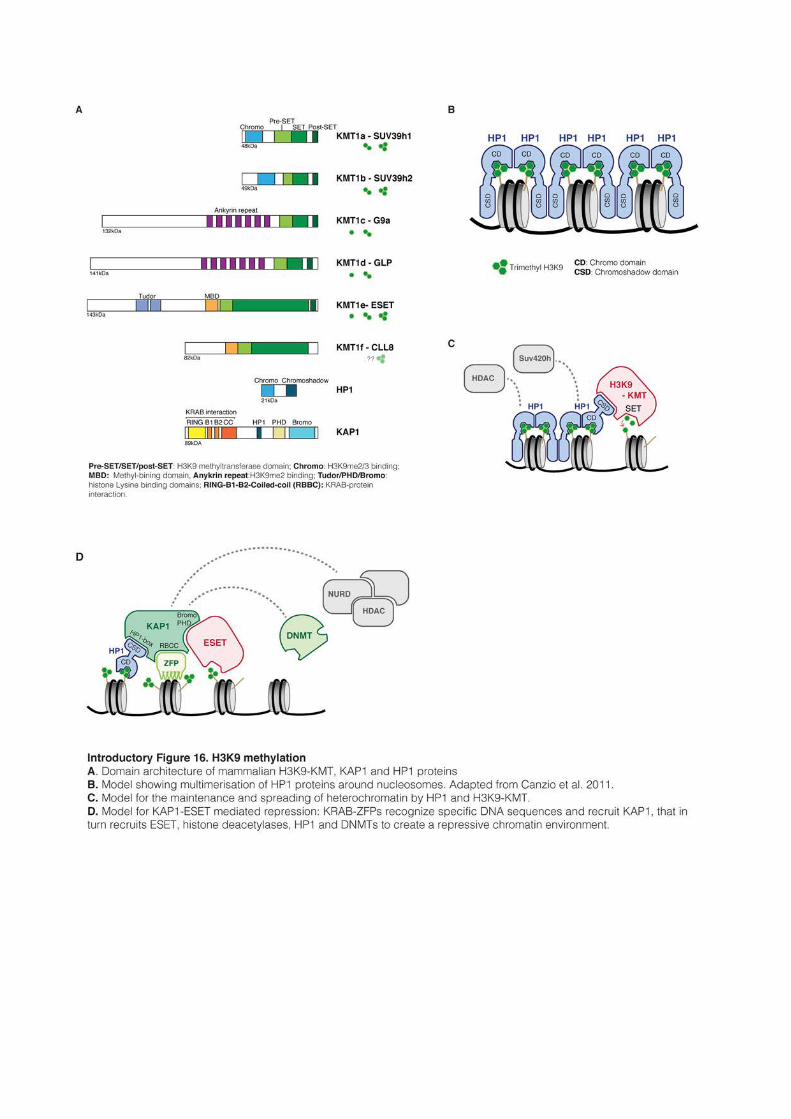
85
chromoshadow domains and the different KMTs that establish the mark (Chin et al., 2007;
Sripathy et al., 2006; Stewart et al., 2005). As a consequence of the interaction between HP1
and H3K9-KMTs, artificial tethering of HP1α to the genome was shown to result in the
deposition and spreading of H3K9me3 around the tethering site, forming large domains of
several kbs. Of note, these H3K9me3 domains are heritably transmitted through multiple cell
divisions, even after removal of the initiating stimulus, showing that once established,
H3K9me3 domain can be stably and autonomously maintained (Hathaway et al., 2012).
HP1 isoforms are similar in structure, but differ in their localization: HP1α is restricted
to pericentric regions, while HP1β and HP1γ can be found elsewhere in the genome (Minc et
al., 2000; Nielsen et al., 2001). In line with these distinct localization, the three isoform appear
to have also non-overlapping functions, as exemplified by the distinct phenotypes of each of
the individual mouse knockout: HP1α absence has no detectable effect, HP1β knockout mice
die around birth and HP1γ depletion leads to severe neonatal lethality and infertility (Aucott
et al., 2008; Brown et al., 2010). Furthermore, whereas HP1α, β and γ physically block
transcription initiation at promoters (Smallwood et al., 2008), in some cell types15 HP1γ (but
not H3K9me2/3) is also enriched in the body of highly transcribed genes and regulates
splicing by interacting with elongating forms of RNA polymerase II (Smallwood et al., 2012;
Vakoc et al., 2005).
Further compaction can probably be achieved through H4K20 methylation, which was
shown in vitro to physically condense chromatin. H4K20 mono-methylation is catalyzed by
SET8/KMT5a (Fang et al., 2002), while subsequent di- and tri-methylation rely successively
on SUV4-20h1/KMT5b and SUV4-20h2/KMT5c (Schotta et al., 2004, 2008). H4K20me1
and H4K20me2 are associated with DNA replication and DNA repair and are broadly
distributed in the genome, while H4K20me3 is found exclusively in heterochromatin and its
absence is associated with severe defects in overall chromatin structure and genome integrity
(Jørgensen et al., 2013). HP1, in particular α- and β-isoforms, was shown to bind directly to
SUV4-20h2, therefore mediating the deposition of H4K20me3 in H3K9 methylated regions
(Schotta et al., 2008). H4K20-dependent chromatin compaction is probably a two-step
process: H3K9 methylation first recruits HP1, which in turn interact with SUV4-20h2 that
deposits H4K20me3. In line with this model, H4K20me3 is abolished at pericentric regions in
absence of H3K9me3, whereas H3K9 methylation and HP1 association are maintained in
absence of SUV4-20h2. Potentially because SUV4-20h2 recruitment depends on specific HP1
isoforms, and because other factors are probably involved, H4K20me3 accompanies
15 Of note, it was not described in ES cells so far.
86
principally H3K9me3 in pericentric regions and TEs, but is more rarely associated with
H3K9me2.
Other factors than HP1 and H4K20 tri-methylation are probably involved in chromatin
compaction and transcriptional repression. For example, H3K9 methylation and HP1 are
thought to recruit HDACs (Schultz et al., 2001; Stewart et al., 2005), histone remodeling
complexes (Nielsen et al., 2002a) and favor the integration of the H3.3 histones variant by the
Daxx-Atrx chaperone complex (Eustermann et al., 2011; Lewis et al., 2010). Of note, H3K7
mono-methylation is also often found associated with H3K9me2/3 and H4K20me3,
especially in pericentric heterochromatin, but its function remain obscure (Peters et al., 2003).
2.3.2 H3K9 methyltransferases
Eight distinct H3K9-KMTs have been described so far in mouse and human
(Introductory figure 16A). The first discovered mammalian KMTs were
SUV39h1/KMT1a and the testis-restricted SUV39h2/KMT1b (Aagaard et al., 1999;
O’Carroll et al., 2000). SUV39h1/2 contain a conserved chromodomain and a SET domain.
They use mono-methylated lysine residues as substrate and specifically govern H3K9 tri-
methylation at pericentric heterochromatin. Absence of SUV39h enzymes results in severe
chromosomal instabilities (Peters et al., 2003, 2001; Rea et al., 2000). Importantly,
SUV39h1/2 bind to HP1 and HP1 is lost at pericentric regions in Suv39h-dKO; this suggests
a self-enforcing loop where H3K9me3 recruits HP1, that in turn attracts more SUV39h1/2,
allowing the spreading of H3K9me3 and HP1 along the chromatin fiber (Introductory figure 16C and Lachner et al., 2001). In yeast at least, the SUV39 homolog chromodomain
binds as well to H3K9 methylation and this interaction is necessary for the spreading of
heterochromatin. (Zhang et al., 2008a). In mammals, it was shown that the repressive
property of SUV39h1/2 was located in the N-terminal part containing the chromodomain,
and that the catalytic activity was not required for transcriptional silencing when the protein
was artificially tethered to the genome. (Firestein et al., 2000). It is postulated that the
chromodomain helps assembling multimeric repressive complexes on chromatin.
Interestingly, while HP1 binding on chromatin is highly dynamic and in a permanent on-off
flux, SUV39h remains immobile at pericentric heterochromatin and probably plays an
important stabilizing role (Cheutin et al., 2003; Krouwels et al., 2005)
Pericentric H3K9 mono-methylation is mediated specifically by PRDM3 and PRDM16
(Pinheiro et al., 2012). The methylation reaction occurs on the cytoplasmic pool of histones;
mono-methylated histones are then transported to pericentric heterochromatin to be
87
converted into H3K9me3 by SUV39h enzymes. Absence of PRDM3 and PRDM16 leads to
the disappearance of H3K9me3, H4K20me3 and HP1 from pericentric regions. Interestingly,
depletion of PRDM3 and PRDM16 further results in the disintegration of the DAPI-dense
heterochromatin foci and in severe destabilization of the nuclear lamina, which is not the case
in Suv39h-dKO cells (Pinheiro et al., 2012). This indicates a critical role for H3K9me1 in itself
in the formation and maintenance of pericentric heterochromatin. How H3K9me1-marked
histones and SUV39h-dependant H3K9 methylation are targeted to pericentric regions
remains largely unanswered questions.
While pericentric heterochromatin relies extensively on PRDM3, PRMD16 and
SUV39h1/2, H3K9 methylation is established by other KMTs in the rest of the genome.
G9a/KMT1c and GLP/KMT1d are the primary enzymes responsible for H3K9 mono- and
di-methylation in non-pericentric regions (Rice et al., 2003). G9a and GLP form heterodimers
via their SET domains, and the G9a/GLP complexes are functional in vivo, as both G9a and
GLP single KO contain only traces of H3K9me2 and severely reduced levels of H3K9me1
(Tachibana et al., 2002, 2005). G9a is able to methylate non-histone proteins, including itself,
and its auto-methylation is necessary for the further recruitment of HP1 (Chin et al., 2007).
G9a and GLP possess also a specific “ankyrin repeat domain” that binds to H3K9me1/2,
meaning that the two enzymes are capable of both read and write the same mark (Collins et
al., 2008). These self-enforcing loops are thought to favor the spreading of H3K9me2 into
very large blocks (up to 5Mb) that cover between 4% and 50% of the genome (in mouse
embryonic stem cells and in the liver, respectively) (Wen et al., 2009). In particular, large
repressive chromatin domains associated with the nuclear lamina are highly enriched for
H3K9me2, and the majority of genes upregulated in mouse G9a-KO ES cells are indeed
localized at the nuclear periphery (Guelen et al., 2008; Yokochi et al., 2009). H3K9me2-
mediated repression controls critical biological processes: in contrast with Suv39h1/2-knockout
mice that are viable (Peters et al., 2001), G9a and GLP-knockout mice are embryonic lethal
due to severe growth defects, and both male and female meiosis cannot be completed in
absence of G9a (Tachibana et al., 2002, 2005, 2007). The recruitment of G9a/GLP
complexes to chromatin likely involves multiple interactions with chromatin or sequence-
specific DNA-binding molecules. In human and mouse, G9a/GLP complexes were shown to
contain the zinc finger protein WIZ, and human complexes associate as well with ZNF644,
another DNA binding protein. WIZ and ZNF644 mediate the recruitment of G9a to specific
DNA sequences (Bian et al., 2015; Ueda et al., 2006). Other DNA-binding transcriptional
88
repressors such as E2F6 or CDP/cut were also shown to associate with G9a (Nishio and
Walsh, 2004; Ogawa et al., 2002).
The last characterized H3K9-KMTs are the related proteins ESET/SetDB1/KMT1e
and CLL8/SetDB2/KMT1f. Very little is known about CLL8. Depletion of the protein leads
to globally reduced levels of H3K9me3, but not of H3K9me2 (Falandry et al., 2010). CLL8
appears important for chromosomal segregation during mitosis and was shown to repress
specific genes during embryonic development in zebrafish and during anti-viral responses in
mouse (Schliehe et al., 2015; Xu et al., 2010).
On the other hand, ESET has received considerable attention, especially because its
biology is intimately linked with the KAP1 protein. ESET can successively mono-, di- and tri-
methylate H3K9 residues and is thought to be responsible for most of non-pericentric
H3K9me3 (Schultz et al., 2002; Wang et al., 2003). KAP1 is a multi-domain protein that acts
as a binding platform for the establishment of a repressive environment around specific DNA
sequences (Introductory figure 16D). It contains an N-terminal RBCC domain, a C-
terminal PHD-bromo domain and a central HP1 binding domain. The RBCC domain binds
specifically to the N-terminal KRAB (Krüppel-Associated box) domain of C2H2 zinc finger
proteins (ZFPs)(Friedman et al., 1996), a superfamily of DNA binding proteins that represents
around half of the annotated transcription factors. C2H2 zinc finger proteins recognize
specific DNA sequences and 40% possess a KRAB domain, representing namely 423 genes
encoding transcripts for 742 structurally distinct proteins in the human genome (Bellefroid et
al., 1991; Huntley et al., 2006). After being targeted to specific genomic loci by KRAB-ZFPs,
KAP1 further recruit ESET and the NuRD histone deacetylase complex through its PHD-
bromodomain and establishes a repressive environment that is stabilized by the interaction of
HP1 with both H3K9me3 and KAP1 (Ryan et al., 1999; Schultz et al., 2001, 2002).
Whereas KAP1 and ESET are ubiquitously expressed, cell-type specific expression of
KRAB-ZFPs and the diversity of their recognition sequences allow the fine-tuning of
numerous biological processes. In particular, both KAP1 and ESET are critical for embryonic
development, as knockout embryos die after 4-5 days of gestation (Cammas et al., 2000;
Dodge et al., 2004). KAP1 and ESET are also linked to pluripotency. KAP1 binds to
numerous promoters in mouse ES cells and its depletion induces spontaneous differentiation
(Hu et al., 2009). As another evidence for their critical role, ESET is the only H3K9-KMT for
which knockout is known to be lethal in mouse ES cells, a phenotype shared by Kap1 knockout
(Dodge et al., 2004; Rowe et al., 2010). The binding motifs of most KRAB-ZFPs are still
unknown and substantial efforts are currently undergoing to characterize the full repertoire of
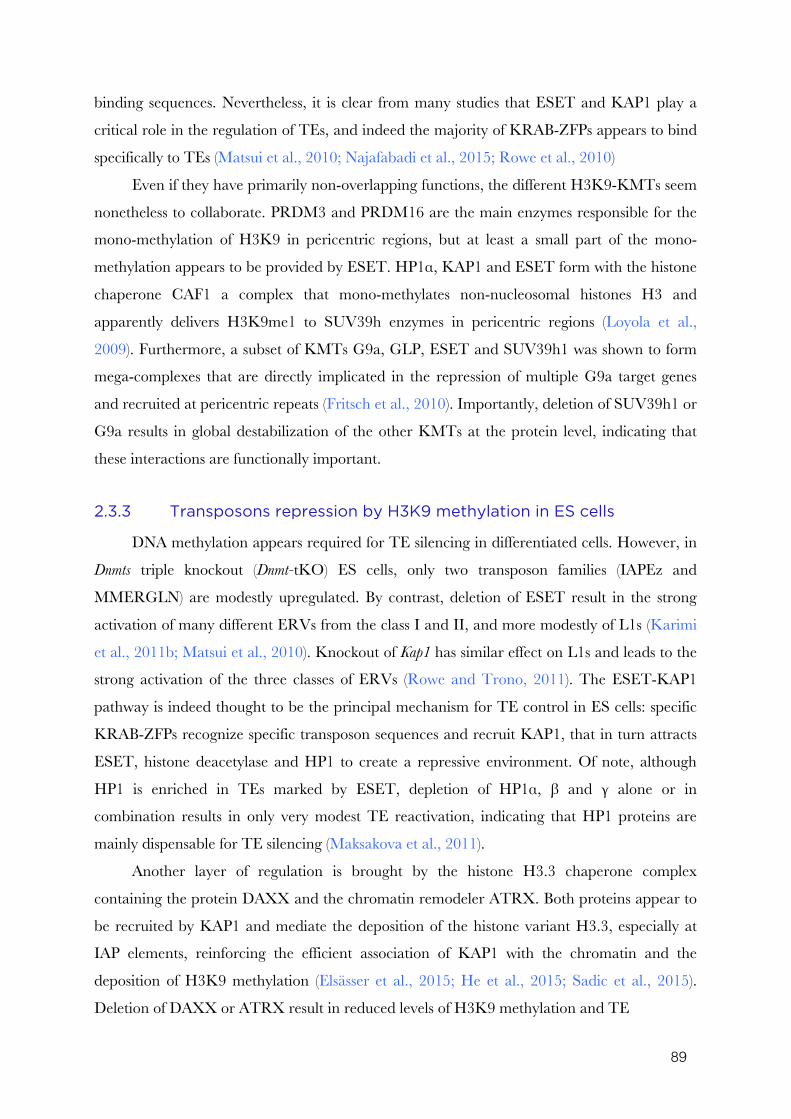
89
binding sequences. Nevertheless, it is clear from many studies that ESET and KAP1 play a
critical role in the regulation of TEs, and indeed the majority of KRAB-ZFPs appears to bind
specifically to TEs (Matsui et al., 2010; Najafabadi et al., 2015; Rowe et al., 2010)
Even if they have primarily non-overlapping functions, the different H3K9-KMTs seem
nonetheless to collaborate. PRDM3 and PRDM16 are the main enzymes responsible for the
mono-methylation of H3K9 in pericentric regions, but at least a small part of the mono-
methylation appears to be provided by ESET. HP1α, KAP1 and ESET form with the histone
chaperone CAF1 a complex that mono-methylates non-nucleosomal histones H3 and
apparently delivers H3K9me1 to SUV39h enzymes in pericentric regions (Loyola et al.,
2009). Furthermore, a subset of KMTs G9a, GLP, ESET and SUV39h1 was shown to form
mega-complexes that are directly implicated in the repression of multiple G9a target genes
and recruited at pericentric repeats (Fritsch et al., 2010). Importantly, deletion of SUV39h1 or
G9a results in global destabilization of the other KMTs at the protein level, indicating that
these interactions are functionally important.
2.3.3 Transposons repression by H3K9 methylation in ES cells
DNA methylation appears required for TE silencing in differentiated cells. However, in
Dnmts triple knockout (Dnmt-tKO) ES cells, only two transposon families (IAPEz and
MMERGLN) are modestly upregulated. By contrast, deletion of ESET result in the strong
activation of many different ERVs from the class I and II, and more modestly of L1s (Karimi
et al., 2011b; Matsui et al., 2010). Knockout of Kap1 has similar effect on L1s and leads to the
strong activation of the three classes of ERVs (Rowe and Trono, 2011). The ESET-KAP1
pathway is indeed thought to be the principal mechanism for TE control in ES cells: specific
KRAB-ZFPs recognize specific transposon sequences and recruit KAP1, that in turn attracts
ESET, histone deacetylase and HP1 to create a repressive environment. Of note, although
HP1 is enriched in TEs marked by ESET, depletion of HP1α, β and γ alone or in
combination results in only very modest TE reactivation, indicating that HP1 proteins are
mainly dispensable for TE silencing (Maksakova et al., 2011).
Another layer of regulation is brought by the histone H3.3 chaperone complex
containing the protein DAXX and the chromatin remodeler ATRX. Both proteins appear to
be recruited by KAP1 and mediate the deposition of the histone variant H3.3, especially at
IAP elements, reinforcing the efficient association of KAP1 with the chromatin and the
deposition of H3K9 methylation (Elsässer et al., 2015; He et al., 2015; Sadic et al., 2015).
Deletion of DAXX or ATRX result in reduced levels of H3K9 methylation and TE
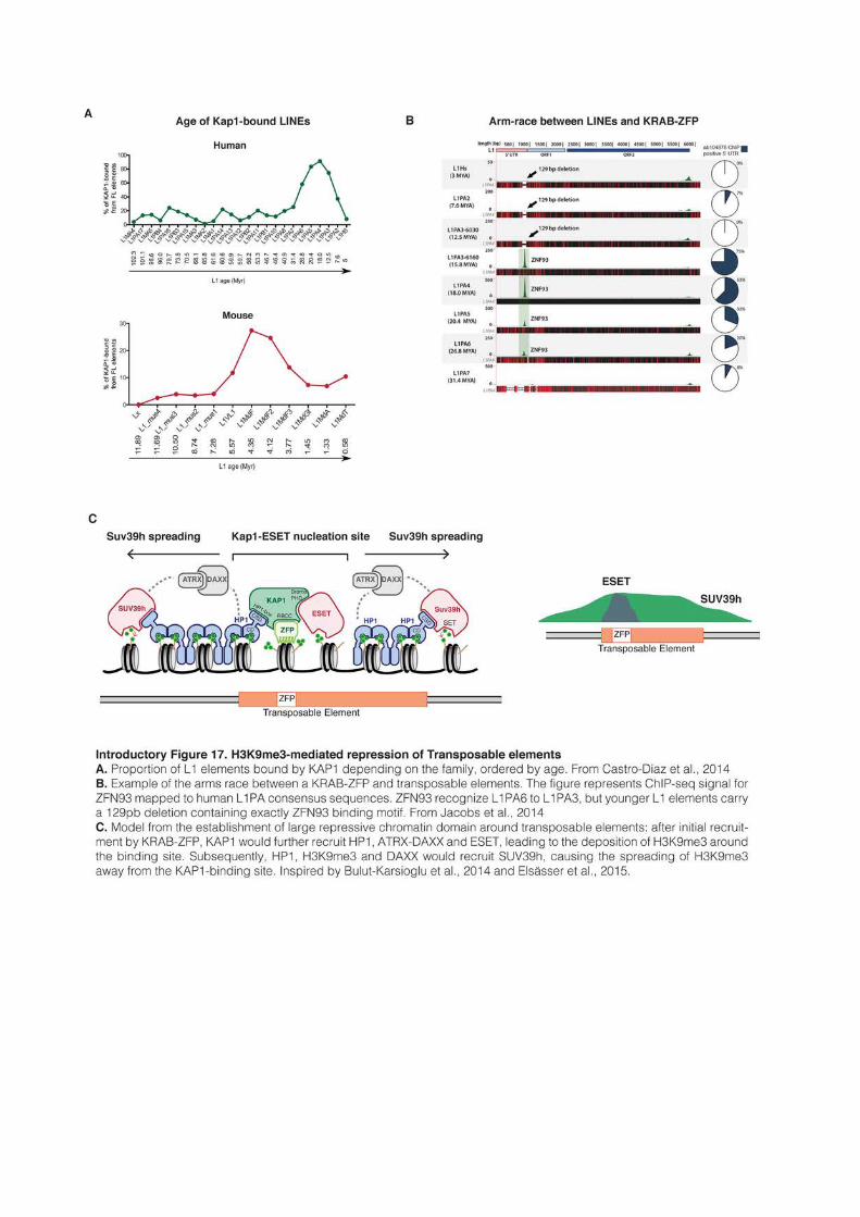
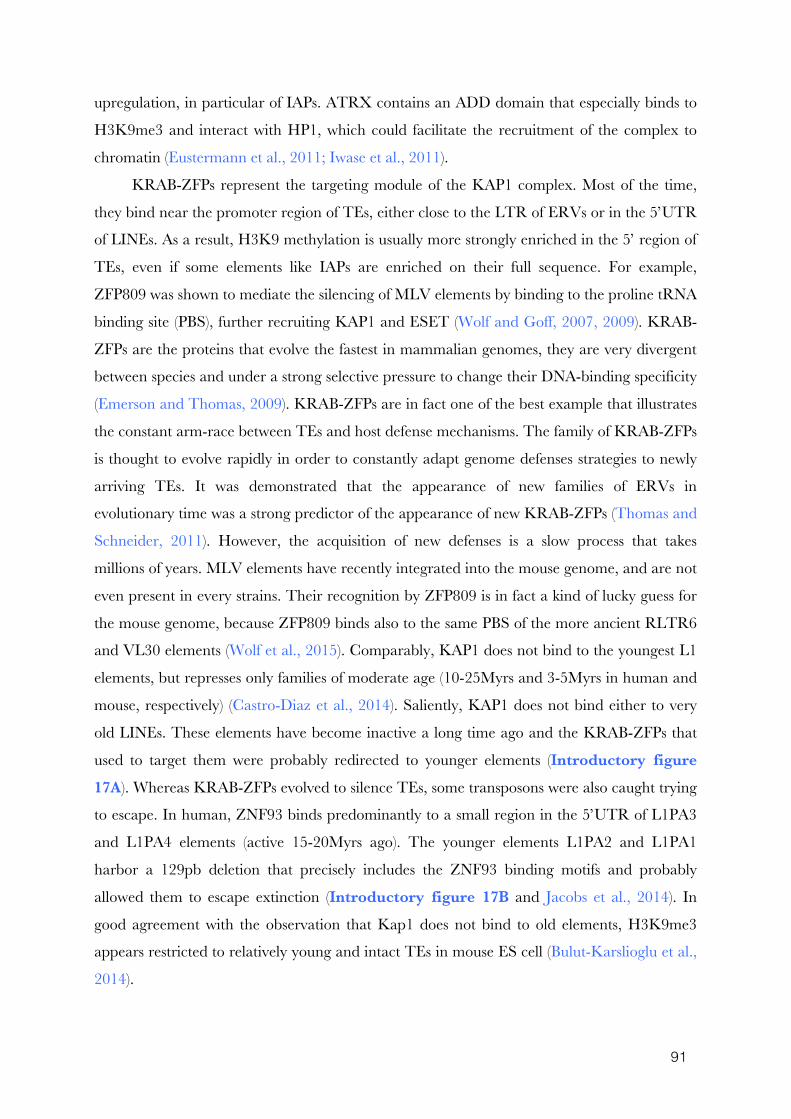
91
upregulation, in particular of IAPs. ATRX contains an ADD domain that especially binds to
H3K9me3 and interact with HP1, which could facilitate the recruitment of the complex to
chromatin (Eustermann et al., 2011; Iwase et al., 2011).
KRAB-ZFPs represent the targeting module of the KAP1 complex. Most of the time,
they bind near the promoter region of TEs, either close to the LTR of ERVs or in the 5’UTR
of LINEs. As a result, H3K9 methylation is usually more strongly enriched in the 5’ region of
TEs, even if some elements like IAPs are enriched on their full sequence. For example,
ZFP809 was shown to mediate the silencing of MLV elements by binding to the proline tRNA
binding site (PBS), further recruiting KAP1 and ESET (Wolf and Goff, 2007, 2009). KRAB-
ZFPs are the proteins that evolve the fastest in mammalian genomes, they are very divergent
between species and under a strong selective pressure to change their DNA-binding specificity
(Emerson and Thomas, 2009). KRAB-ZFPs are in fact one of the best example that illustrates
the constant arm-race between TEs and host defense mechanisms. The family of KRAB-ZFPs
is thought to evolve rapidly in order to constantly adapt genome defenses strategies to newly
arriving TEs. It was demonstrated that the appearance of new families of ERVs in
evolutionary time was a strong predictor of the appearance of new KRAB-ZFPs (Thomas and
Schneider, 2011). However, the acquisition of new defenses is a slow process that takes
millions of years. MLV elements have recently integrated into the mouse genome, and are not
even present in every strains. Their recognition by ZFP809 is in fact a kind of lucky guess for
the mouse genome, because ZFP809 binds also to the same PBS of the more ancient RLTR6
and VL30 elements (Wolf et al., 2015). Comparably, KAP1 does not bind to the youngest L1
elements, but represses only families of moderate age (10-25Myrs and 3-5Myrs in human and
mouse, respectively) (Castro-Diaz et al., 2014). Saliently, KAP1 does not bind either to very
old LINEs. These elements have become inactive a long time ago and the KRAB-ZFPs that
used to target them were probably redirected to younger elements (Introductory figure 17A). Whereas KRAB-ZFPs evolved to silence TEs, some transposons were also caught trying
to escape. In human, ZNF93 binds predominantly to a small region in the 5’UTR of L1PA3
and L1PA4 elements (active 15-20Myrs ago). The younger elements L1PA2 and L1PA1
harbor a 129pb deletion that precisely includes the ZNF93 binding motifs and probably
allowed them to escape extinction (Introductory figure 17B and Jacobs et al., 2014). In
good agreement with the observation that Kap1 does not bind to old elements, H3K9me3
appears restricted to relatively young and intact TEs in mouse ES cell (Bulut-Karslioglu et al.,
2014).
92
SUV39h enzymes bring another layer of control of TEs, and especially of LINEs. In Suv39h-
dKO, H3K9me3 diminishes at ERVs and almost totally disappears at L1s. As a result, some
L1s of the A subtype (but not T or F) are strongly upregulated, while ERVs show only a vey
modest activation (Bulut-Karslioglu et al., 2014). ESET and HP1 are preferentially enriched
inside the transposon sequence, and particularly nucleate in the 5’ region. SUV39h by
contrast appears to be especially involved in the spreading of H3K9me3 inside and outside
the transposon sequence and is found enriched in larger domains. Since little is known about
the mechanisms that target SUV39h in general, it is tempting to propose a model where
SUV39h would act downstream of KAP1, ESET and ATRX-DAXX, especially because
SUV39h1 was shown to physically interact with DAXX (He et al., 2015). After an initial
recruitment by KRAB-ZFPs, KAP1 would further recruit HP1, ATRX-DAXX and ESET,
leading to the deposition of H3K9me3 around the binding site. Subsequently, HP1,
H3K9me3 and DAXX would recruit SUV39h, causing the spreading of H3K9me3 distally
from the Kap1-binding site (Introductory figure 17C).
Importantly, H3K9me3-mediated silencing appears restricted to undifferentiated cells.
H3K9me3 marking at TEs is substantially lower in MEFs or upon differentiation of ES cells
and deletion of SUV39h, ESET or KAP1 in differentiated cells does not affect transposon
expression (Bulut-Karslioglu et al., 2014; Matsui et al., 2010; Rowe et al., 2013). This
indicates that other silencing mechanisms, probably involving DNA methylation, take over
during differentiation.
However, class III ERVs represent an exception: H3K9me3 is barely present in the
sequence of MERVL in any cell type. Class III ERVs seem preferentially regulated by
H3K9me2. In ES cells, MERVLs are highly enriched for H3K9me2 and are strongly
activated following G9a knockout (Maksakova et al., 2013). Of note, MERVL LTRs contain
the binding sequence of E2F6, a transcription factor that was shown to associate with G9a
and which could therefore be used to target H3K9me2 to MERVL elements (Ogawa et al.,
2002, personnal communication). But the picture is probably more complex. even though
MERVL lacks H3K9me3 enrichment, it is strongly upregulated in Kap1 and Suv39 mutant ES
cells, and modestly activated by Eset deletion. MERVL appears to be the only TE to be
repressed simultaneously by the three major H3K9 pathways, even though it could be trough
very indirect mechanism (Bulut-Karslioglu et al., 2014; Maksakova et al., 2013). Moreover,
MERVL elements are also upregulated in absence of KDM1a (LSD1), an H3K4me1/2
demethylase that appears necessary to remove activating histone modification from MERVL
promoters (Macfarlan et al., 2011).
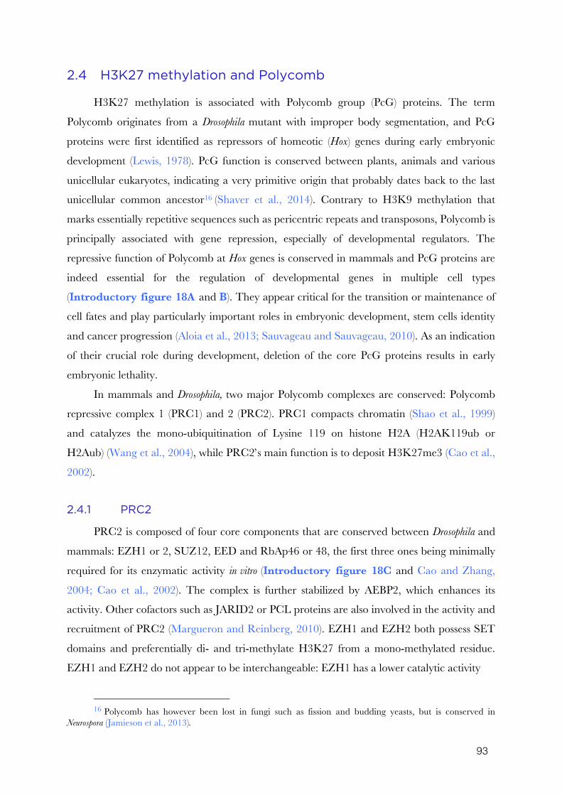
93
2.4 H3K27 methylation and Polycomb
H3K27 methylation is associated with Polycomb group (PcG) proteins. The term
Polycomb originates from a Drosophila mutant with improper body segmentation, and PcG
proteins were first identified as repressors of homeotic (Hox) genes during early embryonic
development (Lewis, 1978). PcG function is conserved between plants, animals and various
unicellular eukaryotes, indicating a very primitive origin that probably dates back to the last
unicellular common ancestor16 (Shaver et al., 2014). Contrary to H3K9 methylation that
marks essentially repetitive sequences such as pericentric repeats and transposons, Polycomb is
principally associated with gene repression, especially of developmental regulators. The
repressive function of Polycomb at Hox genes is conserved in mammals and PcG proteins are
indeed essential for the regulation of developmental genes in multiple cell types
(Introductory figure 18A and B). They appear critical for the transition or maintenance of
cell fates and play particularly important roles in embryonic development, stem cells identity
and cancer progression (Aloia et al., 2013; Sauvageau and Sauvageau, 2010). As an indication
of their crucial role during development, deletion of the core PcG proteins results in early
embryonic lethality.
In mammals and Drosophila, two major Polycomb complexes are conserved: Polycomb
repressive complex 1 (PRC1) and 2 (PRC2). PRC1 compacts chromatin (Shao et al., 1999)
and catalyzes the mono-ubiquitination of Lysine 119 on histone H2A (H2AK119ub or
H2Aub) (Wang et al., 2004), while PRC2’s main function is to deposit H3K27me3 (Cao et al.,
2002).
2.4.1 PRC2
PRC2 is composed of four core components that are conserved between Drosophila and
mammals: EZH1 or 2, SUZ12, EED and RbAp46 or 48, the first three ones being minimally
required for its enzymatic activity in vitro (Introductory figure 18C and Cao and Zhang,
2004; Cao et al., 2002). The complex is further stabilized by AEBP2, which enhances its
activity. Other cofactors such as JARID2 or PCL proteins are also involved in the activity and
recruitment of PRC2 (Margueron and Reinberg, 2010). EZH1 and EZH2 both possess SET
domains and preferentially di- and tri-methylate H3K27 from a mono-methylated residue.
EZH1 and EZH2 do not appear to be interchangeable: EZH1 has a lower catalytic activity
16 Polycomb has however been lost in fungi such as fission and budding yeasts, but is conserved in
Neurospora (Jamieson et al., 2013).
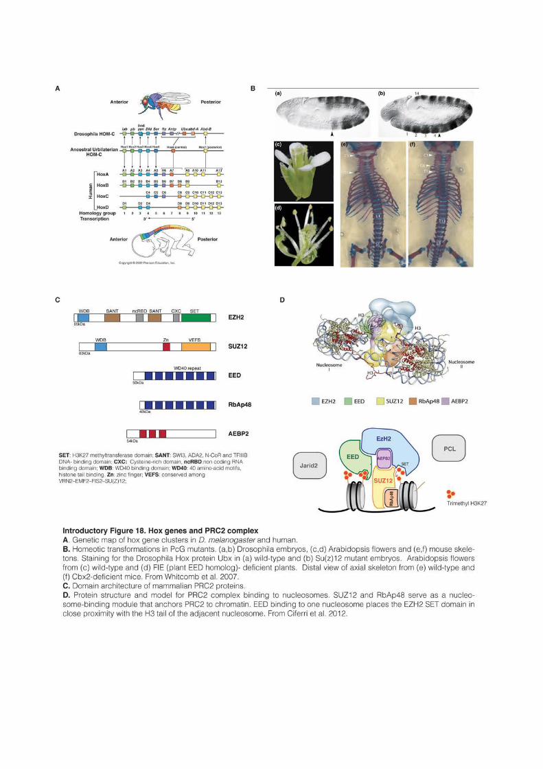
95
and its depletion has little consequence on global H3K27me2/3 levels (Margueron et al.,
2008). Importantly, global levels of H3K27me1 are unaffected by EZH1/2 depletion,
indicating that H3K27me1 is deposited by an unrelated and still unknown process. It is
sometimes pointed that G9a can methylate H3K27 in vitro (Tachibana et al., 2001) and
H3K27me1 levels appear indeed reduced in G9a-KO (Wu et al., 2011b). However,
H3K27me2/3 levels remain unaltered in G9a-KO and G9a cannot therefore be considered as
a good candidate for the global deposition H3K27me1.
EED binds specifically to H3K27me3 through its C-terminal WD40 domain and
H3K27me3 is abolished in absence of this interaction (Margueron et al., 2009). Additionally,
resolution of the molecular structure of the PRC2 complex suggests that EED binding to one
nucleosome places EZH2 SET domain in close proximity with the H3 tail of the next
nucleosome (Ciferri et al., 2012). These positive-feedback loops probably allow for the
maintenance and spreading of H3K27me3 along the chromatin fiber (Introductory figure 18D).
SUZ12 and RbAp46/48 are thought to serve as a nucleosome-binding modules that
anchor PRC2 to chromatin (Nekrasov et al., 2005). In particular, the SUZ12-RbAp46/48
subunit binds to unmodified H3 tails and this interaction is lost in presence of H3K4me3 and
H3K36me2-3, which subsequently prevents in cis PRC2 to methylate the same histone tail
(Schmitges et al., 2011). As H3K4 and H3K36 methylation are associated with active
transcription, this probably prevents the spreading of H3K27 methylation into active
chromatin territories. Interestingly, whereas H3K4me3/H3K36me2-3 and H3K27me3
cannot be present in the same H3 molecule, they can nonetheless be present in vivo in the
same nucleosome, each in a different H3 tail, forming “bivalent” structures marked at the
same time by activating and repressing marks (Bernstein et al., 2006a; Voigt et al., 2012).
H3K27 methylation is very abundant and is found in approximately 80% of histone H3
in mouse ES cells, with around 15% of mono-, 50% of di- and 15% of tri-methylation (Peters
et al., 2003). H3K27me2 and me3 deposition probably relies on slightly different PRC2
variants, as PCL cofactors are for example required for H3K27 tri- but not di-methylation
(Sarma et al., 2008). H3K27me2 does not appear to be significantly associated with
transcriptional repression, but likely represents an intermediate (or default) state that marks
genomic regions to be repressed. H3K27me3 usually harbors two distinct patterns in
mammalian cells, with either very large domains of more than 100kb, such as in Hox clusters
or in the inactive X in females, or smaller domains of a few kilobases. In ES cells, around 50-
60% of H3K27me3-enriched regions are localized in the proximity of promoters, marking
96
about 10% of the total number of genes, especially developmental regulators (Boyer et al.,
2006; Marks et al., 2012; Zhao et al., 2007). Interestingly, in mouse or human ES cells, the
majority (~80%) of H3K27me3-marked promoters are also enriched for H3K4me3, a mark
associated with transcription activation, forming bivalent domains (Bernstein et al., 2006a).
Bivalency is hypothesized to represent a specific state that poises key developmental genes for
either activation or repression, and indeed many lineage-specific bivalent promoters are
definitely activated or silenced following differentiation of ES cells (Mohn et al., 2008). A lot of
controversy has followed the description of bivalency, which was criticized as being an in vitro
artifact restricted to pluripotent ES cells in culture without an in vivo equivalent. However,
bivalent promoters are also observed in cultured differentiated cells such as neurons, mouse
embryonic fibroblasts (MEFs), hematopoietic progenitors and erythrocytes (Cui et al., 2009;
Mikkelsen et al., 2007; Mohn et al., 2008). Furthermore, its observation in vivo in the brain, in
primordial germ cells (PGCs) or in the sperm shows that bivalency likely represents an
important mechanism for the regulation of many critical developmental processes (Cui et al.,
2012; Hammoud et al., 2009; Sachs et al., 2013).
PRC2 and H3K27me3 have no obvious effect by themselves on chromatin compaction.
H3K27me3 could indirectly regulate transcription by preventing other proteins to access
chromatin. For example, levels of H3K27 acetylation increase upon loss of PRC2 and it was
proposed that H3K27me3 could act by preventing the binding of activating HATs (Pasini et
al., 2010). However, PRC2 and H3K27me3 are thought to control transcription mainly by
recruiting other factors, principally PRC1.
2.4.2 PRC1
In Drosophila, the canonical model for Polycomb-mediated silencing is relatively simple:
PRC2 1) is recruited to chromatin by recognizing specific sequences; 2) deposits H3K27me3,
which 3) further recruits PRC1 that 4) compacts chromatin and prevents transcription
(Levine et al., 2004). In mammals, the picture is much more complex. While the canonical
PRC1 is composed of four proteins in Drosophila, there are a total of 16 homologues in
mammals, and PRC1 is furthermore engaged into multiple non-canonical complexes (Simon
and Kingston, 2013). Since the initial description of a Drosophila-like PRC1 in mammals
(Levine et al., 2002), the variety of PCR1-related function has grown exponentially and a
comprehensive (and still incomplete) picture of PRC1 complexity has only started to emerge
recently (Introductory figure 19).
97
The defining structure of PCR1 complexes is the presence of the E3 ubiquitin ligases
RING1a and RING1b. E3 ligases carry out the final step of a three-enzymes cascade,
catalyzing the transfer of ubiquitin from an E2 donor enzyme (Berndsen and Wolberger,
2014). RING1a and RING1b catalyze mono-ubiquitination of Lysine 119 on histone H2A
(H2Aub) and seem both present in PRC1 complexes (Gao et al., 2012; Wang et al., 2004).
RING1b is the most active and broadly expressed of the two enzymes, its knockout is
embryonic lethal while RING1a-depleted mice survive (del Mar Lorente et al., 2000;
Voncken et al., 2003). However, even if Ring1b-KO ES cells have almost no H2Aub left, ES
cells stop to proliferate and spontaneously differentiate only when RING1a is additionally
depleted, indicating that RING1a can at least partially compensate for RING1b loss (Endoh
et al., 2008). The second defining feature of PRC1 is the exclusive presence of one of six
PCGF proteins (PCGF1 to 6). The association of RING1a/b with a specific PCGF protein
defines six distinct PRC1 complexes that can be referred as PRC1.1 to PRC1.6 (Gao et al.,
2012). The six PRC1 complexes have distinct protein partners and are functionally different.
In particular, they target different genomic regions and regulate different sets of genes.
RING1a/b-PCGF modules further form two types of complexes by associating with
either CBX proteins or with RYBP (or its homologue YAF2). CBX proteins and
RYBP/YAF2 compete for the same binding pocket on RING1a/b C-terminal domain and
are therefore mutually exclusive (Wang et al., 2010). CBX proteins are associated exclusively
with PGCF2 (MEL18) and PGCF4 (BMI1), forming in addition with the Polyhomeotic
proteins (PHC1/2/3) the canonical form of PRC1 (Introductory figure 19A and Gao et al.,
2012). The five CBX proteins17 (CBX2/4/6/7/8) are short proteins related to HP1 that bind
H3K27me3 through their chromodomain. Of note, whereas the Drosophila homologue
appears very specific, certain mammalian CBX proteins also recognize H3K9me3 in vitro
(Bernstein et al., 2006b; Fischle et al., 2003). H3K27me3 binding by CBX proteins allows the
recruitment of CBX-PRC1 to PRC2 targets, promoting chromatin compaction and gene
repression (Introductory figure 19A). Indeed, in mouse ES cells, CBX7 is the only CBX
protein associated with PRC1 and 90% of CBX7-binding sites in the genome are also
occupied by PRC2 (Morey et al., 2012). Depletion of CBX7 leads to reactivation of many
PRC2 targets, and loss of PRC2 greatly decreases the presence of PRC1 in gene promoters
(Leeb et al., 2010), indicating that CBX-PRC1 act as a transcriptional repressor downstream
of PRC2.
17 Formally, HP1α,β and γ are also members of the Cbx family, but the term Cbx would only be used here to
refer to the partners of PRC1.
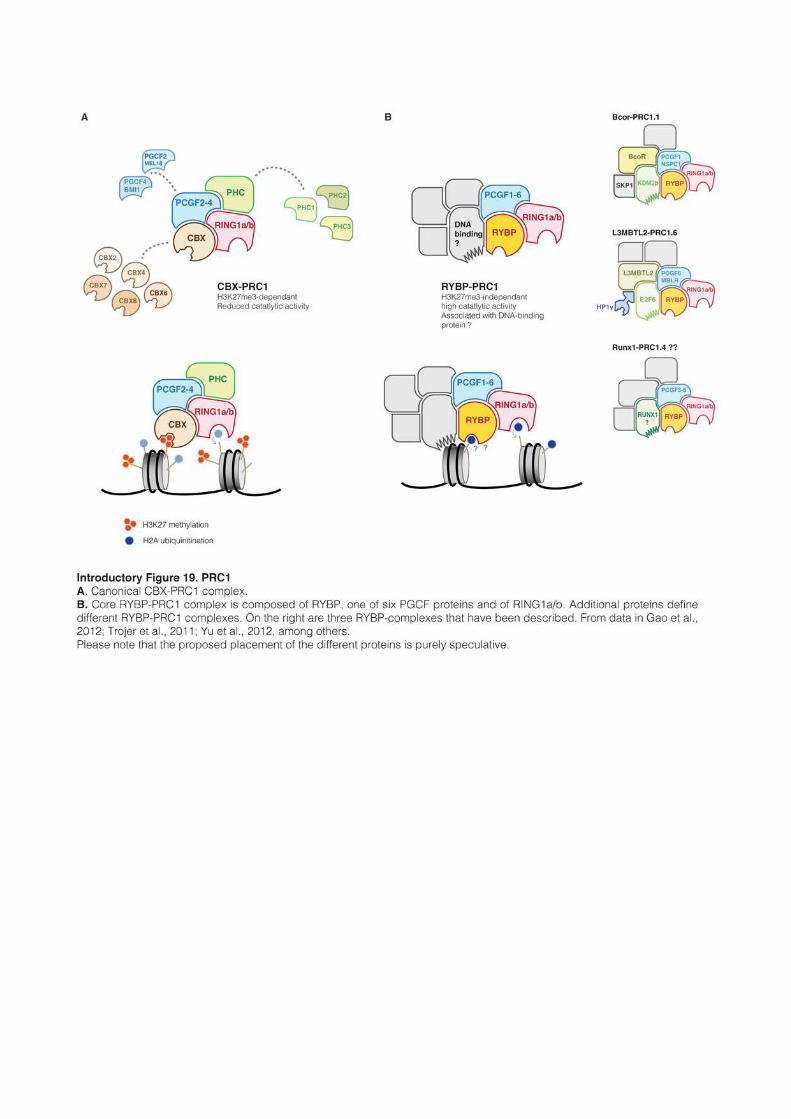
99
Interestingly, the multiplicity of potential combination between PGCF2 and 4 and the
five CBX proteins seems to provide an additional plasticity to PRC1 function. In pluripotent
ES cells, CBX7 associates preferentially with PGCF2 and represses many developmental
genes, including Pgcf4, Cbx2 and Cbx4. Upon differentiation, CBX7-PGCF2 complexes switch
for CBX2/CBX4-PGCF4 complexes that then regulate pluripotency genes, including Cbx7
(Morey et al., 2012). How the different CBX proteins distinguish their targets remains
mysterious. This fine-tuned regulation during cell differentiation highlights the complexity of
PRC1-mediated repression and indicates without doubt that H3K27me3 is not the only factor
implicated in PRC1 recruitment.
On the other hand, RYBP/YAF2 can be found with the six PGCF proteins and its
presence further exclude PCH proteins (Introductory figure 19B and Gao et al., 2012).
Importantly, RYBP-PRC1 does not contain a chromodomain subunit and is recruited to
chromatin independently of H3K27me3 (Tavares et al., 2012). Indeed, while in ES cells 90%
of RING1b binding sites are also occupied by H3K27me3, in other cells type like HEK-293T
most of RING1B binding sites are free of H3K27me3, and these H3K27me3-free binding
sites are also enriched for RYBP (Gao et al., 2012). Many examples show as well that PRC1
can be recruited to target sequences when PRC2 components are depleted, either at gene
promoters or in large domains such as the inactive X in females (Pasini et al., 2007;
Schoeftner et al., 2006). In several cases if not always, RYBP-PRC1 complexes contain DNA-
binding factors that trigger their recruitment to specific locations: PRC1.4 and PRC1.6
associates with the transcription factors RUNX1 and E2F6, respectively (Trojer et al., 2011;
Yu et al., 2012), whereas PRC1.1 contains the H3K36-KDM KDM2b that binds to
unmethylated CpG-rich sequences trough it CxxC domain (Wu et al., 2013). Of note, RYBP-
PRC1 appears to have a stronger catalytic activity than CBX-PRC1, as artificial tethering of
CBX-PRC1, but not of RYBP-PRC1, fails to deposit H2Aub (Introductory figure 20A and
Blackledge et al., 2014). Beside this, RYBP seems to have DNA and ubiquitin-binding activity
in vitro, which could help stabilizing the complex to target regions (Arrigoni et al., 2006; Neira
et al., 2009).
2.4.3 Polycomb recruitment and transcriptional repression
The interplay between PRC1 and PRC2 is complex and probably varies depending on
the cell type. CBX-PRC1 is recruited to regions marked by H3K27me3, while RYBP-PRC1
is recruited independently of this mark. In Drosophila, PRC2 binds to specific DNA sequences,
but such a mechanism does not seem to exist in mammals (Schuettengruber and Cavalli,
100
2009). At the exception of the large mega-domains found around Hox clusters or in the
inactive X chromosome, genome-wide analysis of H3K27me3 distribution reveals in fact that
PRC2 almost perfectly overlaps with CpG islands (Ku et al., 2008). Several studies showed
indeed that PRC2 was systematically attracted to GC-rich sequences, at the condition that
they are DNA unmethylated and depleted of activating transcription factors (Jermann et al.,
2014; Mendenhall et al., 2010). The precise mechanism by which PRC2 is recruited to GC-
rich sequences remains however unclear. JARID2, an important cofactor of PRC2, could be
involved, as it possesses a DNA binding domain that binds preferentially to GC-rich
sequences, especially short CCG tandem repeats (Li et al., 2010; Peng et al., 2009).
It appears clear as well that PRC2 can be recruited by long non-coding RNAs
(ncRNAs), especially to form large H3K27me3 mega-domains. PCR2 is recruited in cis by the
ncRNAs Xist and Kcnq1ot1 at the inactive X in females and at the Kcnq1 imprinted region,
respectively (Pandey et al., 2008; Plath et al., 2003), or in trans at the HoxD locus by the
ncRNA HOTAIR coming from the HoxC locus (Rinn et al., 2007). EZH2 strongly binds RNA,
but this interaction has very low specificity and 5-25% of the mouse transcriptome is
recovered after EZH2 pulldown assay (Zhao et al., 2010). JARID2 binds RNA as well and
might be more specific, as it as been shown to been required for the recruitment of PRC2 by
Xist to the inactive X chromosome (da Rocha et al., 2014).
In mouse ES cells, 90% of RING1b binding-sites are also marked by H3K27me3, most
of the time in the presence of CBX7, consistent with the canonical assumption that PRC1 is
recruited by H3K27me3 (Morey et al., 2012). However, reciprocally, some studies reported
that only half of H3K27me3-enriched regions are bound by Ring1b, indicating that CBX-
PRC1 recruitment by H3K27me3 is not automatic and that other mechanisms control CBX-
PRC1 recruitment (Ku et al., 2008).
In ES cells lacking PRC2, PRC1 enrichment decreases at sites normally occupied by
H3K27me3, but interestingly it does not disappear completely and remains at around one
quarter of its initial level. Moreover, H2A ubiquitination levels appear barely affected (Leeb et
al., 2010; Tavares et al., 2012). This indicates that PRC1 recruitment and H2Aub at PRC2
target sites are partially independent of PRC2. In fact, it was shown in ES cells that 1)
artificial tethering of RYBP-PRC1, but not of CBX-PRC1, resulted in PRC2 recruitment, 2)
PRC2-binding and H3K27me3 were globally lost after depletion of PRC1, and finally that 3)
H2A ubiquitination was specifically responsible for the recruitment of PRC2 (Introductory figure 20A and Blackledge et al., 2014; Cooper et al., 2014). These results shift the usual
paradigm of PRC1 and PRC2 recruitment and shows that PRC1 can efficiently attract PRC2
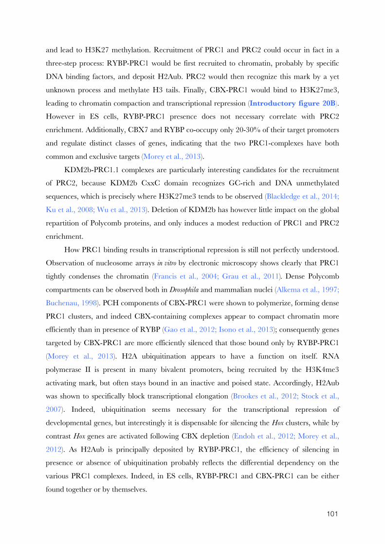
101
and lead to H3K27 methylation. Recruitment of PRC1 and PRC2 could occur in fact in a
three-step process: RYBP-PRC1 would be first recruited to chromatin, probably by specific
DNA binding factors, and deposit H2Aub. PRC2 would then recognize this mark by a yet
unknown process and methylate H3 tails. Finally, CBX-PRC1 would bind to H3K27me3,
leading to chromatin compaction and transcriptional repression (Introductory figure 20B).
However in ES cells, RYBP-PRC1 presence does not necessary correlate with PRC2
enrichment. Additionally, CBX7 and RYBP co-occupy only 20-30% of their target promoters
and regulate distinct classes of genes, indicating that the two PRC1-complexes have both
common and exclusive targets (Morey et al., 2013).
KDM2b-PRC1.1 complexes are particularly interesting candidates for the recruitment
of PRC2, because KDM2b CxxC domain recognizes GC-rich and DNA unmethylated
sequences, which is precisely where H3K27me3 tends to be observed (Blackledge et al., 2014;
Ku et al., 2008; Wu et al., 2013). Deletion of KDM2b has however little impact on the global
repartition of Polycomb proteins, and only induces a modest reduction of PRC1 and PRC2
enrichment.
How PRC1 binding results in transcriptional repression is still not perfectly understood.
Observation of nucleosome arrays in vitro by electronic microscopy shows clearly that PRC1
tightly condenses the chromatin (Francis et al., 2004; Grau et al., 2011). Dense Polycomb
compartments can be observed both in Drosophila and mammalian nuclei (Alkema et al., 1997;
Buchenau, 1998). PCH components of CBX-PRC1 were shown to polymerize, forming dense
PRC1 clusters, and indeed CBX-containing complexes appear to compact chromatin more
efficiently than in presence of RYBP (Gao et al., 2012; Isono et al., 2013); consequently genes
targeted by CBX-PRC1 are more efficiently silenced that those bound only by RYBP-PRC1
(Morey et al., 2013). H2A ubiquitination appears to have a function on itself. RNA
polymerase II is present in many bivalent promoters, being recruited by the H3K4me3
activating mark, but often stays bound in an inactive and poised state. Accordingly, H2Aub
was shown to specifically block transcriptional elongation (Brookes et al., 2012; Stock et al.,
2007). Indeed, ubiquitination seems necessary for the transcriptional repression of
developmental genes, but interestingly it is dispensable for silencing the Hox clusters, while by
contrast Hox genes are activated following CBX depletion (Endoh et al., 2012; Morey et al.,
2012). As H2Aub is principally deposited by RYBP-PRC1, the efficiency of silencing in
presence or absence of ubiquitination probably reflects the differential dependency on the
various PRC1 complexes. Indeed, in ES cells, RYBP-PRC1 and CBX-PRC1 can be either
found together or by themselves.
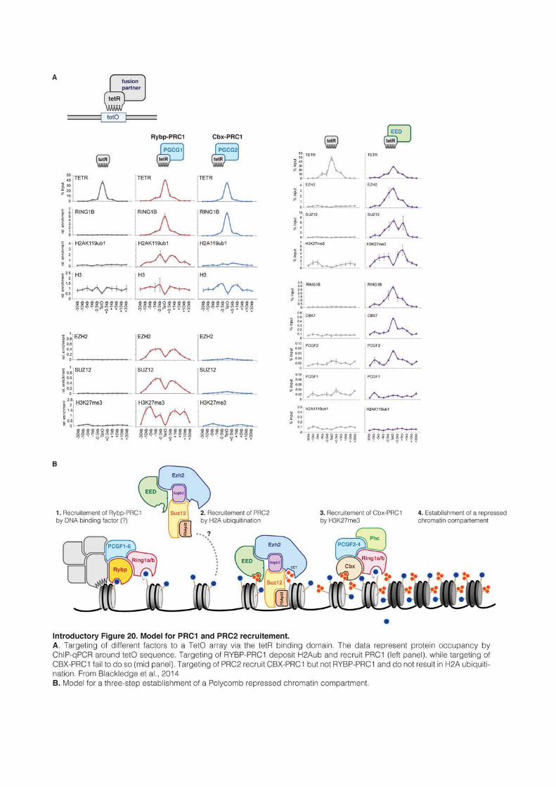
103
Finally, Polycomb was never reported so far as a major transposon regulator, at least in
cultured cells. At the exception of MLV elements, H3K27me3 is absent from transposon
sequences in ES cells. Moreover, TEs do not get reactivated following deletion of PRC1 or
PRC2, even though it has to be noted that the combined deletion of PRC1 and PRC2 leads
to a strong upregulation of MLV and IAP elements (Leeb et al., 2010).
2.5 Methylation(s) crosstalk
H3K9 methylation and H3K27 methylation mark functionally distinct genomic
compartments and are almost never observed together (Mikkelsen et al., 2007; Rao et al.,
2014). Whether the two marks are structurally incompatible or whether their physical
separation is merely the consequence of distinct recruitment mechanism is unclear. PRC2
catalytic activity is not affected by the presence of an H3K9 methylated histone tail (Schmitges
et al., 2011), and G9a methylates H3K27 in vitro, indicating that its catalytic activity is not
repelled by this mark (Tachibana et al., 2001). Whether the other H3K9-KMTs are affected
by H3K27 methylation remains an unexplored question. In fact, a potential mechanist link
between H3K9 and H3K27 methylation could be DNA methylation. Indeed, it is commonly
admitted that DNA and H3K9 methylation are positively correlated, while DNA and H3K27
methylation are mutually exclusive.
2.5.1 H3K9 methylation recruits DNA methylation
Since DNA methylation is present by default in mammalian genomes, stating that DNA
and H3K9 methylation are positively correlated is almost meaningless. In globally highly
methylated cells, it simply indicates that H3K9me2/3 is never found in unmethylated loci
such as CpG islands. Beside this, DNA methylation is clearly not a good predictor for the
presence of H3K9 methylation: DNA methylation is widespread, whereas H3K9 methylation
is localized to specific genomic places such as TEs, telomeres, centromeres and pericentric
regions. In contrast, correlation between DNA and H3K9 methylation is much stronger in
organisms that have mosaic DNA methylation pattern such as Neuraspora or Arabidopsis. In
Neuraspora, the link is unidirectional: H3K9me3 directly recruits the DNA methylation
machinery and all DNA methylation is lost in absence of H3K9me3 (Tamaru and Selker,
2001). In Arabidopsis, the pathways required to establish and maintain CHG and CHH
methylation are highly dependent on H3K9me3 and the two marks interact in complex self-
104
reinforcing loops, whereas maintenance of CG methylation by MET1 is by contrast
independent of H3K9me3 (Du et al., 2015).
In mammals, DNA methylation establishment and maintenance can occur
independently of H3K9 methylation. Nonetheless, H3K9 methylation functionally attracts the
DNA methylation machinery, which is especially relevant during developmental periods
where global DNA methylation is constitutively low. In early development, the genome is
globally hypomethylated at the blastocyst stage and IAP transposons and imprinting control
regions are among the rare sequences that maintain high DNA methylation (Lane et al., 2003;
Tremblay et al., 1997). Upon deletion of KAP1 in early embryo, DNA methylation is lost at
imprinted loci and IAPs are strongly reactivated, showing that KAP1 is necessary for the
DNA methylation of these genomic elements (Messerschmidt et al., 2012; Rowe et al., 2010).
On the same line, in ES cells cultured in a medium that promotes global hypomethylation,
residual DNA methylation is maintained specifically at IAPs, pericentric major satellite
repeats and imprinted loci, where it usually correlates with the presence of H3K9 methylation
(Ficz et al., 2013; Habibi et al., 2013). In ES cells and in the early embryo, maintenance of
imprinted methylation was shown to rely specifically on the KRAB-ZFP ZFP57, which
recognizes methylated binding-motif located in the imprinting control regions (Quenneville et
al., 2011, 2012). Finally, transfection in ES cells of reporter genes coupled with known KRAB-
ZFP binding motifs results in the rapid silencing of the reporter and de novo methylation of its
promoter, undoubtedly demonstrating that KAP1 and ESET action are not limited to the
protection of DNA methylation, but can also promote its initiation (Rowe et al., 2013).
By contrast with the ADD domain of ATRX, DNMT3a and 3b ADD domains have no
particular affinity for H3K9me3 (Iwase et al., 2011). DNMT3a and b are therefore not
directly recruited by H3K9me3, but by the different proteins associated with repressive
chromatin. For example, DNMT3a and 3b co-immunoprecipitate with ESET (Li et al.,
2006a) and KAP1 (Quenneville et al., 2011; Zuo et al., 2012). DNA methylation deposition at
pericentric heterochromatin is on the other hand mediated by direct interaction of DNMT3a
and 3b with SUV39h and HP1, and DNA methylation indeed diminishes in major satellite
repeats in Suv39h-dKO ES cells (Fuks, 2003; Lehnertz et al., 2003). Reciprocally, the
methylated DNA-binding protein MeCP2 associates with SUV39h, engaging DNA and
H3K9 methylation in a self-reinforcing loop (Lunyak et al., 2002). Of note, DNA methylation
of centromeric regions is not affected by SUV39h deletion, and DNMT3b is recruited directly
to minor satellite repeats by centromeric proteins independently of H3K9 methylation
(Gopalakrishnan et al., 2009).
105
In Dnmt3a/b double knockout (Dnmt3-dKO) ES cells, global DNA methylation reaches a
very low level at late passages, but TEs, such as IAP elements, and imprinted loci still remain
hypermethylated, indicating that DNMT1 as well can be recruited by H3K9 methylation
(Leung et al., 2014). DNMT1 has no specific affinity for H3K9 methylation by itself, but was
shown to bind to G9a (Estève et al., 2006). Moreover, UHRF1 recognizes H3K9me2/3 with
its Tudor and PHD domains, allowing DNMT1 recruitment to H3K9 methylated regions
(Arita et al., 2012; Rothbart et al., 2013). The recognition of H3K9me2/3 reinforces the
maintenance of DNA methylation, and can even direct some de novo deposition independently
of the presence of hemi-methylated CpGs (Arand et al., 2012; Liu et al., 2013).
G9a can also recruit DNMT3a and 3b, either directly or indirectly through the
chromodomain-containing protein MPP8 (Epsztejn-Litman et al., 2008; Kokura et al., 2010);
this correlates with a reduction of DNA methylation at transposons and germline genes in
G9a-KO ES cells (Dong et al., 2008). Interestingly, the recruitment by G9a is independent of
its catalytic activity. G9a is especially important for the inactivation and methylation of
pluripotent genes (such as Oct4) during ES cells differentiation. Furthermore, the transcription
factor E2F6 was shown to interact with G9a: as E2F6 mediates the proper methylation of
germline genes during early embryogenesis, this indicates that G9a is probably responsible for
methylating developmental gene in vivo as well (Ogawa et al., 2002; Velasco et al., 2010).
As a whole, the link between DNA methylation and H3K9 methylation appears mostly
unidirectional in mammals. H3K9me2/3-mediated DNA methylation appears especially
necessary during developmental periods where DNA methylation is either low or dynamic:
H3K9me2/3 allows the maintenance of DNA methylation at TEs and imprinted loci during
DNA methylation erasure, and promotes de novo methylation of developmental genes during
differentiation. Importantly, H3K9 methylation is no longer anymore once DNA methylation
has been established. Indeed, absence of KAP1, SUV39h or ESET in differentiated cells does
not result in DNA methylation defect nor in transcriptional activation of underlying sequences
(Bulut-Karslioglu et al., 2014; Matsui et al., 2010; Rowe et al., 2010).
2.5.2 DNA methylation excludes Polycomb from GC-rich regions
In ES cells, 80-90% of PRC1- or PRC2-enriched regions overlap with unmethylated
CpG islands and these regions tend to be hyper conserved during evolution (Ku et al., 2008;
Tanay et al., 2007). In fact, lack of DNA methylation and high CG content are the only
characteristics that have been shown so far to promote H3K27me3 recruitment in mammals.
Indeed, the introduction of GC-rich sequences in ES cells is sufficient to recruit PRC2, at the
106
condition that the region is protected against DNA methylation and devoid of activating
motifs (Jermann et al., 2014; Lynch et al., 2011; Mendenhall et al., 2010). However, the
restriction of PRC2 complexes to unmethylated CpG islands appears specific to ES cells. In
mouse or human differentiated cells, H3K27me3 expands away from CpG islands and forms
large domains spanning more than 10% of the genome and with a typical size of 15-40kb
(Hawkins et al., 2010; Pauler et al., 2009). These large H3K27me3 domains remain highly
methylated, indicating that H3K27me3 and DNA methylation are not always incompatible.
Nonetheless, even though H3K27me3 and DNA methylation can be found together in
differentiated cells throughout most of the genome, they remain mutually exclusive in CpG
islands (Brinkman et al., 2012; Statham et al., 2012), showing that the repulsive action of
DNA methylation requires high CpG density.
The exclusion of Polycomb from methylated and CpG-dense regions appears to be an
intrinsic feature of PRC2 complexes. In an attempt to mechanistically understand what
triggers the interaction of proteins with chromatin, reconstituted nucleosomes were incubated
with whole Hela cell extracts and used as a bait to pull-down interacting proteins. In this
assay, PRC1 and PRC2 complexes could be recovered when using unmethylated
nucleosomes, but the interaction was lost when nucleosomal DNA was methylated beforehand
(Bartke et al., 2010). DNA sequences used as baits in these experiments had very high CpG
contents; whether CpG-poor and methylated nucleosomes would also block the interaction
with PRC complexes was, unfortunately, not investigated. However, other studies reported
that PRC2 could methylate H3K27 in DNA methylated nucleosomes in vitro or upon artificial
tethering of PRC2 to methylated region in ES cells, even with high CpG density, suggesting
that DNA methylation alone is not sufficient to prevent PRC2 activity (Cooper et al., 2014).
These results are somehow contradictory, but might suggest that PRC2 binding, but not its
activity, is repelled by DNA methylation, and that PRC2 complexes act differentially in
differentiated and pluripotent cells. The in vivo existence of large GC-poor domains with high
H3K27 and DNA methylation in differentiated cells, but not ES cells, shows at least that
sparsely distributed methylation is not a major impediment for Polycomb activity.
The observation that H3K27me3 can spread in differentiated cells but is constrained to
unmethylated CpG islands in ES cells is intriguing and indicates that Polycomb has different
property in pluripotent and differentiated cells. In particular, in Dnmt-tKO ES cells,
H3K27me3 extends from CpG islands and covers large domains, in a pattern reminiscent of
the one observed in differentiated cells; this supports the concept that DNA methylation can
constrain Polycomb to unmethylated CpG islands in ES cells, but not in differentiated
107
contexts (Brinkman et al., 2012). Interestingly, promoters marked by H3K27me3 frequently
become DNA methylated during ES cell differentiation or carcinogenesis (Mohn et al., 2008;
Ohm et al., 2007), and a controversial study showed that PRC2 could physically interact with
DNMT1, DNMT3a and DNMT3b, leading to DNA methylation deposition at target
promoters in Hela cells (Viré et al., 2006). One possible explanation for this observation is that
silencing of these genes is initiated by Polycomb in pluripotent cells, and that Polycomb
further recruit DNA methylation to ensure long term silencing in differentiated cells.
Interestingly, artificial tethering of PRC2 to a transgene in ES cells leads to the recruitment of
Dnmt3a, but do not result in de novo methylation (Rush et al., 2009). This shows that
interaction of PRC2 and DNMTs is not restricted to differentiated cells, but that ES cells have
additional mechanisms to ensure that H3K27me3-enriched CpG islands remain
unmethylated.
Several mechanisms probably cooperate to prevent de novo methylation of PRC2 targets
in ES cells. For example, PRC2 was shown to recruit TET enzymes and promote active
demethylation of target sequences (Neri et al., 2013a). The majority of CpG islands marked
by H3K27me3 are also enriched for H3K4me3. Since the ADD domain of DNMT3 enzymes
is repelled by this mark, the bivalency of H3K27me3 domains could allow protection against
de novo methylation. Moreover, DNMT3l has also the ability to bind to PRC2, and its
presence outcompetes the interaction with DNMT3a and 3b. Since DNMT3l is only
expressed in undifferentiated cells, the exclusion of DNMT3a and 3b from PRC2 complexes
by DNTM3l ensures that PRC2 targets do not become de novo methylated, which is observed
in ES cell upon DNMT3l depletion (Neri et al., 2013b). Of note however, the interaction
between EZH2 and DNMT3l could not be reproduced in another study (Vlachogiannis et al.,
2015).
Enigmatically, the CxxX-containing protein KDM2b was also shown to be required for
the protection of H3K27me3-CpG islands. KDM2b is bound to virtually every unmethylated
CpG island promoters, and at a subset of them also associate with RYBP-PRC1.1 complexes
(Farcas et al., 2012; Wu et al., 2013). Deletion of KDM2b barely affects the enrichment of
PRC1 and PRC2 (Blackledge et al., 2014; He et al., 2013), but surprisingly two-third of PRC-
bound CpG islands become hypermethylated (Boulard et al., 2015). This result is puzzling,
because it suggests that in absence of KDM2b, PRC complexes and DNA methylation can be
found together in GC-dense regions, which stands against much of current knowledge.
The observation that Polycomb and DNA methylation are mutually exclusive appears
to be the consequence of a subtle equilibrium between several counterbalancing forces: in one
108
side, high density of unmethylated CpGs is sufficient for recruiting H3K27me3 and PRC
complexes are mechanistically excluded from methylated nucleosomes; on the other side,
PRC2 can directly recruit the DNA methylation machinery and H3K27me3-rich CpG
islands need to be actively protected against de novo deposition.
2.5.3 H3K27 and H3K9 methylations
In pluripotent and differentiated cells, H3K27 and H3K9 methylation are almost never
found together and their domains, large or small, do not overlap (Hawkins et al., 2010;
Mikkelsen et al., 2007). This is intriguing because the two marks do not seem mutually
exclusive per se. PRC2 was even shown to be preferentially attracted in vitro by nucleosomes
marked with H3K9me3 (Bartke et al., 2010). However, this interaction is lost if nucleosomes
are additionally DNA methylated. Since H3K9me2/3 is always found together with DNA
methylation, and H3K27me3 requires unmethylated sequences (at least in ES cells), it is
tempting to speculate that the mutual exclusion of H3K27me3 and H3K9me3 in most cell
types is caused by DNA methylation. In line with this hypothesis, the rare known sequences
where H3K27 and H3K9 methylation are observed together harbor low DNA methylation.
In Suv39h-KO ES cells, major satellite repeats lose both H3K9me3 and DNA
methylation and interestingly, some cells show concomitantly a strong H3K27me3
enrichment at chromocenters (Lehnertz et al., 2003; Peters et al., 2003). Similarly, in zygotes
lacking SUV39h enzymes, H3K9me3 is replaced in pericentric regions by PRC1
(Puschendorf et al., 2008). These observations suggest that PRC complexes can occupy
pericentric regions upon loss of H3K9me3. However, in DNA methylation-free ES cells,
H3K9me3 is not affected, but H3K27me3 relocalizes nonetheless to major satellite repeats
and forms bright foci in chromocenters that overlap with H3K9me3 (Cooper et al., 2014;
Marks et al., 2012; Saksouk et al., 2014). PRC2 and RYBP-PRC1, but not CBX-PRC1, are
present in pericentric heterochromatin in Dnmt-tKO ES cells. This shows that in the absence
of DNA methylation, H3K9 and Polycomb can coexist in pericentric regions. Major satellite
repeats are AT-rich and are not transcriptionally activated in Dnmt-tKO cells, making it
unlikely that Polycomb complexes are recruited by RNAs or by a high GC-content.
Recruitment of PRC2 was in fact shown to rely on the DNA-binding protein BEND3, which
specifically binds to major satellite repeats in absence of DNA methylation (Dai et al., 2013;
Saksouk et al., 2014). BEND3 could potentially recognize a motif in major satellite repeats in
a methylation-dependent manner.
109
A second example of co-occurrence of H3K27me3 and H3K9me3 in absence of DNA
methylation can be observed in primordial germ cells. At this stage, the genome is globally
unmethylated; in this context, H3K27me3 and H3K9me3 are present together in
transposons, including L1s and class I and II ERVs (Liu et al., 2014). Interestingly, both marks
decrease upon loss of ESET, indicating that their deposition might be functionally linked.
These limited examples suggest that in some specific contexts, H3K27me3 and
H3K9me3 could be present together, either at pericentric regions or in transposable elements.
At which extent is the absence of DNA methylation a necessary condition, or whether the two
marks can be present in the same nucleosome or the same H3 tail remain unaddressed
questions. In differentiated cells, H3K27me3 and H3K9me3 form large domains
independently of DNA methylation. However, these domains do not overlap, suggesting that
at least in differentiated cells, other mechanisms than DNA methylation may restrict the two
marks into their respective territory.
In ES cells lacking DNA methylation, H3K9me3 and H3K27me3 do not overlap, but
occupy distinct subdomains of pericentric heterochromatin, as determined by cytological tools
(Cooper et al., 2014). Therefore, H3K9me3 and H3K27me3 might be antagonistic, and in
line with this hypothesis, depletion of SUV39h in Dnmt-tKO cells leads to broader enrichment
of H3K27me3 in chromocenters. Since PRC2 activity is not blocked by H3K9 methylation
(Schmitges et al., 2011), the observed antagonism is probably indirect. In the zygote, it was
shown that HP1 proteins, but not H3K9me3, prevent the binding of CBX-PRC1 and PRC2
in pericentric heterochromatin, suggesting a mechanism for the mutual exclusion of the two
marks (Tardat et al., 2015). In contrast, PRC1 recruitment and H2Aub do not appear to be
antagonized by H3K9 methylation and both H3K9me3 and H2Aub overlap at
chromocenters in in Dnmt-tKO cells (Cooper et al., 2014).
Overall, H3K27me3 and H3K9me3 appear to be mutually exclusive, but what
mechanistically causes this exclusion is far for being understood.
Further reading:
The following reviews were used as starting material to write this chapter.
van Bemmel, 2012: historical perspective Luger et al., 2012: chromatin core structure Maison and Almouzni, 2004: HP1 and SUV39 Shinkai and Tachibana, 2011: G9a Iyengar and Farnham, 2011: KAP1 and ESET Margueron and Reinberg, 2011; Simon and Kingston, 2013: Polycomb
110
Chédin, 2011; Schübeler, 2015: DNA methylation Du et al., 2015; Rose and Klose, 2014: links between histone and DNA methylation The following studies are cited in figure legends: Blackledge et al., 2014; Bulut-Karslioglu et al., 2014; Canzio et al., 2011; Castro-Diaz et al., 2014; Ciferri et al., 2012; Du et al., 2015; Elsässer et al., 2015; Gao et al., 2012; Jacobs et al., 2014; Luger et al., 2012; Nishino et al., 2012; Olins and Olins, 2003; Rodriguez-Paredes and Esteller, 2011; Trojer et al., 2011; Whitcomb et al., 2007; Yu et al., 2012.
111
3 REPROGRAMMING
What ultimately defines a cell is not its chromatin landscape, but the ensemble of genes
that are expressed at a given moment. Consequently, cell type-specific transcription factors
are at the heart of cellular identity and control the gene expression profiles in an intricate
balance between antagonistic forces. Cells in culture are usually maintained in a stable state
by inhibiting or promoting defined regulatory pathways. Cell fate can also easily be switched
in vitro from pluripotent or multipotent to more differentiated states by targeting few key
signaling pathways, promoting the development towards a given cell fate while repressing
alternative possibilities (Graf and Enver, 2009). Conversely, differentiated cells can be
reversed artificially to a pluripotent state. John Gurdon showed that the introduction of the
nucleus of terminally differentiated intestinal cell into an enucleated Xenopus oocyte resulted in
the development of normal frogs, proving that the acquired somatic state of the nucleus can
return to a totipotent state, under the control of cytoplasmic proteins from the oocyte
(Gurdon, 1962; Gurdon et al., 1958). Shinya Yamanaka further showed that reprogramming
of somatic cells back to pluripotency only necessitates the expression of four core pluripotency
transcription factors (Takahashi and Yamanaka, 2006).
Transcription factors are sufficient by themselves to promote reprogramming, but the
process is inefficient and the chromatin landscape of differentiated cells represents a barrier
that has to be overcome. Somatic cells have large and static domains of repressive chromatin
and comparatively few active regions, whereas pluripotent stem cell are characterized by a
very plastic, dynamic and globally permissive chromatin environment (Meshorer et al., 2006;
Zhu et al., 2013). During reprogramming, the large repressive domains of differentiated cells
need to be released: accordingly, removal of DNA or H3K9 methylation greatly improves the
efficiency of somatic cell reprogramming (Chen et al., 2013; Mikkelsen et al., 2008).
During mammalian development, the majority of cell fate transitions are mono-
directional, from potent progenitors to more committed cell types. The process is driven by
the finely tuned temporal and spatial expression of transcription factors, promoting the
progressive compaction of the chromatin landscape. However, during two keys periods of
embryonic development, committed cells are reprogrammed and their developmental potency
is restored.
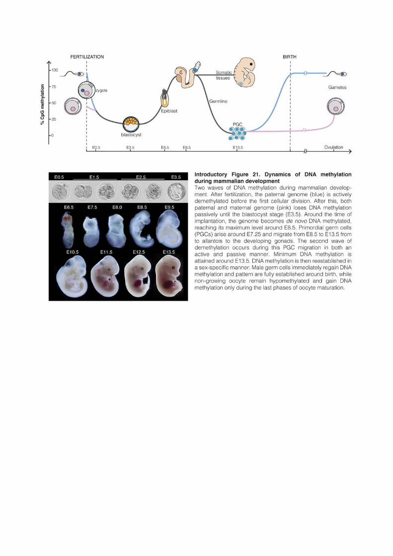
113
3.1 DNA methylation reprogramming in vivo
In mammalian development, cell fate reprogramming is intimately linked to the loss of
DNA methylation. Twice during development, DNA methylation patterns are erased on a
global scale before being reestablished (Introductory figure 21). The first DNA
demethylation event occurs in germ cells, the early fetal progenitors of spermatozoa and
oocytes; the second occurs in the zygote just after fertilization and culminates at the blastocyst
stage (Kafri et al., 1992; Monk et al., 1987). Resetting DNA methylation is absolutely required
for establishing male- and female-specific methylation patterns at imprinting control regions,
but it is very unlikely that global reprograming evolved for this function. Indeed, Xenopus
embryos appear to similarly go through a phase of global hypomethylation in the first hours
after fertilization and in their germline, while genomic imprinting is restricted to placental
mammals (Stancheva et al., 2002; Venkatarama et al., 2010). DNA methylation
reprogramming occurs concomitantly to the restoration of developmental potency, and it is
often assumed that hypomethylation is a constitutive feature of pluripotent states. However,
hypermethylated ES cells remain pluripotent and more importantly, early zebrafish embryos
stay hypermethylated throughout early development. The methylome inherited from the
zebrafish father remains even literally untouched, showing that hypomethylation is not a
requirement for pluripotency per se, at least in this species (Jiang et al., 2013; Potok et al.,
2013). Little is known in fact about the biological significance of DNA methylation erasure in
mammals, albeit it was proposed to be a way to erase any acquired epimutations, preventing
them from being transmitted to the next generation (Heard and Martienssen, 2014;
Seisenberger et al., 2013). In direct relevance with the focus of my thesis work, the following
paragraphs will review the known mechanisms that govern erasure and reestablishment of
DNA methylation in the early mammalian embryo.
3.1.1 DNA methylation erasure
In mouse, primordial germ cells (PGCs) arise from precursor cells in the epiblast at
around 7.25 day after fertilization (E7.25) (Ginsburg et al., 1990). First localized at the base of
the allantois around E8, PGCs migrate through the hindgut and reach the developing gonads
around E11.5 (Anderson et al., 2000; Molyneaux et al., 2001). Epiblast cells have already
acquired somatic developmental programs at this stage, and in particular germline genes are
methylated and silenced. The erasure of the somatic chromatin landscape appears necessary
to turn on the germline developmental program. Interestingly, even though the germline
114
lineage is a unipotent program that goes from early epiblast progenitors to very specialized
gametes, PGCs acquire the characteristics of pluripotent cells, such as the expression of
pluripotent transcription factors OCT4 and NANOG. Moreover, in vitro-derived embryonic
germ cells are in fact nearly indistinguishable from ES cells and can similarly form potent
chimeras, which can give rise to all somatic lineages and functional gametes when injected
into embryos (Leitch et al., 2013; Stewart et al., 1994).
Demethylation of PGCs occurs in two steps. From E8 to E10.5, DNA methylation
maintenance is impaired by downregulating and excluding DNMT1-UHRF1 from the
nucleus, and global levels of CpG methylation drop from around 70% in the E6.5 epiblast to
20-30% at E10.5 (Kobayashi et al., 2013; Seisenberger et al., 2012). While the bulk of the
genome is hypomethylated, some specific sequences such as imprinting control regions,
germline gene promoters and CpG islands on the X chromosome maintain methylation. This
remaining methylation requires a local protection against active demethylation, with both
hydroxy-methylation by TET1 and TET2 enzymes and the base excision repair pathway
(Hackett et al., 2013; Hajkova et al., 2010). The PGC genome reaches a minimum of 5-7% of
CpG methylation at E13.5, and the only sequences that remain significantly methylated at
that point are IAP transposable elements and other ERVs (Guibert et al., 2012). Interestingly,
H3K9me2 levels are concomitantly globally erased from E8 to E13.5, whereas H3K9me3
remains unaltered and H3K27me3 greatly increases (Hajkova et al., 2008; Seki et al., 2007).
Importantly, the global erasure of DNA methylation in PGCs was recently shown to be
conserved in humans, indicating that germline reprogramming is a general characteristic of
mammalian development, albeit the time scale is very different. The minimum of DNA
methylation is reached at around 9-10 weeks of gestation and levels remain low for several
weeks (Gkountela et al., 2015; Guo et al., 2015; Tang et al., 2015).
The second wave of DNA demethylation starts in the zygote just after fertilization. The
DNA methylation pattern inherited from the sperm and the oocyte are markedly different;
moreover, the two parental pronuclei follow distinct paths during the first cellular divisions.
The sperm nucleus is highly methylated (~90%) and tightly packed with protamines
(Kobayashi et al., 2012; Smith et al., 2012). The sperm genome is actively and rapidly
demethylated and loses half of its methylation before the first cellular division (Wang et al.,
2014b). The TET3 enzyme is highly enriched on the paternal pronucleus and converts a
substantial amount of methylated cytosines to hydroxymethylated cytosines (Gu et al., 2011).
How precisely demethylation occurs is still unclear, but active removal of methylated cytosines
was shown to occur before and during DNA replication and to involve base excision repair
115
(Hajkova et al., 2010; Santos et al., 2013). Analysis of Tet3-KO zygotes shows that hydroxy-
methylation is involved in active demethylation of 30-50% of the genome, but the global
increase of methylation in absence of Tet3 is fairly limited (~10%), indicating that passive
dilution during DNA replication is probably the main driver of DNA demethylation before
the first cellular division (Guo et al., 2014; Peat et al., 2014)
In comparison, the oocyte genome is markedly less methylated (~40%) than the sperm,
and the maternal pronucleus is protected against hydroxymethylation by the protein STELLA
(Nakamura et al., 2007, 2012). Active demethylation seems to occur nonetheless at some
extent in the maternal genome, but global maternal DNA methylation levels remain almost
constant during the first cellular division (Guo et al., 2014; Wang et al., 2014b). From the two-
cell embryo to the blastocyst stage, the remaining DNA methylation is thought to be lost
passively from the two parental genomes, to reach a minimal level of around 20% in the early
blastocyst. At that time, most of the genome is hypomethylated, and IAP transposons and
imprinting control regions are among the few regions that remain significantly methylated.
3.1.2 De novo DNA methylation
With the exception of the female germline, global hypomethylation is a very transient
state during mouse development. De novo DNA methylation begins in the late blastocyst
around E4, reaches ~60% at E6.5 and is complete around E7.5-8.5 (~80%) (Wang et al.,
2014b). This rapid gain of methylation correlates with pluripotency loss. For example,
injection of ES cells derived from the E2.5 to E4.5 inner cell mass into a blastocyst can form
chimaeras and contribute to the offspring, whereas epiblast stem cells (EpiSCs) originating
from E5.5 to E7.5 embryo fail to do so (Brons et al., 2007; Tesar et al., 2007).
Precise analysis of de novo methylation by RRBS (reduced-representation bisulfite
sequencing) between E3.5 and E8.5 shows that 50% of the gain occurs in a single day between
E4.5 and E5.5 and then progresses more slowly between E5.5 and E7.5 (Auclair et al., 2014).
Around 700 CpG islands are highly methylated at E8.5, around half of which were already
methylated in the E4.5 blastocyst. The majority of these CpG islands overlap with exons,
preferentially of important developmental genes, and most of the methylated CpG island
promoters regulate genes linked to the germline expression program (Auclair et al., 2014;
Borgel et al., 2010). The dynamics of de novo methylation is probably specific for every gene.
For example, methylation of the transcription factor Elf5 is thought to occur early, as this gene
governs the early separation between the epiblast and the trophectoderm lineages (Ng et al.,
2008). By contrast, Oct4 is still expressed in the E6.5 epiblast and its promoter gains
116
methylation only later during differentiation. De novo methylation in the peri-implentation
embryo relies mostly on DNMT3b, and absence of DNMT3a results in limited DNA
methylation defects (Auclair et al., 2014).
Because of the difficulty to isolate peri-implantation embryos, the methylome of E4.5
and E5.5 stages has not been analyzed with whole-genome base-pair precision level so far.
The technique used by Auclair et al., 2014, (RRBS) is biased toward CpG-rich sequences. As
a consequence, a precise overview de novo methylation dynamics at the level of the genome is
still missing, especially at CpG-poor regions such as gene bodies or intergenic sequences. The
reason for the different kinetics of DNA methylation is unclear. For example, DNMT3b was
shown in cellular systems to methylate preferentially CpG-rich sequences and to recognize
H3K36me3 in the body of highly transcribed genes, but whether this is the case in vivo as well
remains largely unexplored (Baubec et al., 2013; Morselli et al., 2015). Absence of DNMT3l
in this period is not critical since Dnmt3l-KO embryos develop to term and have no detectable
methylation defect; however, some studies have suggested it may have some subtle role both
in the embryo and in differentiating ES cells (Arand et al., 2012; Guenatri et al., 2013; Ooi et
al., 2010).
By contrast, DNMT3l is absolutely required in the germline and both males and females
cannot develop functional gametes in its absence (Bourc’his and Bestor, 2004; Bourc’his et al.,
2001). PGCs start to acquire sex-specific features around E12.5, when DNA methylation
reaches its minimum level. Male PGCs undergo mitotic arrest shortly after and remain non-
diving until they become spermatogonial stem cells a few days after birth, which is around 3-4
post-natal days (P3-4) in the laboratory mouse (Ewen and Koopman, 2010). A day-by-day
timeline of de novo methylation is missing, but global levels rise from ~7% at E13.5 to ~55% at
E16.5, are mostly completed before the beginning of meiosis (~75% at P2.5 or P10), and
reach a final level of 90% in the sperm (Kobayashi et al., 2012; Pastor et al., 2014).
Conditional knockout of DNMT3b has no consequences, whereas absence of DNMT3a or
DNMT3l leads to dramatic TE activation, meiosis arrest and germ cell apoptosis (Bourc’his
and Bestor, 2004; Kaneda et al., 2004). Consequently, DNMT3a and DNMT3l are assumed
to be the main responsible for de novo methylation in the male germline, but a comprehensive
analysis of the respective contribution of the three enzymes during that period is missing.
Interestingly, de novo DNA methylation of a subset of sequences relies on an original
pathway involving 25-30nt-long small RNAs and specialized Argonaute proteins (piRNAs and
PIWI proteins, respectively) in the fetal male germline. Most fetal piRNAs are complementary
to TE sequences and the piRNA pathway appears to be mostly dedicated to transposon
117
control, at least during this developmental window. Shortly, piRNAs originating from long
single-stranded RNA precursors are loaded into PIWI proteins in the cytoplasm and repress
TEs at the post-transcriptional level by degrading TE mRNAs, using especially MILI
endonuclease activity (De Fazio et al., 2011). A subset of PIWI-piRNA complexes relocalizes
to the nucleus and is thought to induce de novo methylation at complementary genomic targets
by a yet unknown mechanism (Aravin et al., 2008; Suh and Blelloch, 2011). Strikingly,
Dnmt3l, Mili or Miwi2 (two of the three PIWI proteins) knockout mouse have similar
phenotypes, with strong upregulation of young LINEs and ERVs and a meiotic catastrophe
(Aravin et al., 2007; Bourc’his and Bestor, 2004). In fact, even though piRNA-mediated DNA
methylation represents a very small proportion of the global methylation, piRNAs appear to
target and silence specifically ~17,000 active transposons (Molaro et al., 2014). In absence of
piRNAs or DNA methylation, TEs acquire some active chromatin marks during meiosis,
causing the relocalization of meiotic homologous recombination hotspots to TEs and major
meiotic defects (Zamudio et al., 2015).
At E12.5, female PGCs take a different developmental trajectory: they enter the
prophase of meiosis I at E13.5 and arrest at the end of metaphase I around the time of birth.
Oocytes remain blocked at this stage until meiosis resumes at puberty (Ewen and Koopman,
2010). Oocytes remain unmethylated during that whole period; de novo DNA methylation
occurs only during oocyte maturation, reaching a final level of around 40% (Kobayashi et al.,
2012; Smallwood et al., 2011). Interestingly in oocytes, high levels of genomic methylation are
highly correlated with transcription, and most of the DNA methylation is observed in the
body of active genes. As a consequence and contrary to the sperm where most of the genome
is methylated except CpG islands, intergenic regions are globally hypomethylated in oocytes.
On the other hand, CpG islands localized in transcriptional units are hypermethylated, which
is important for the setting of maternal genomic imprinting. DNA methylation is totally
abolished in absence of either DNMT3a or DNMT3l. These methylation-free mutant oocytes
develop to term and can be successfully fertilized, but the resulting embryos die of imprinting
defects at mid-gestation (Bourc’his et al., 2001). In contrast, mutations in DNMT3b or
DNMT1 have no effect (Shirane et al., 2013).
While DNMT3b-dependent deposition of DNA methylation is especially targeted to the
body of active genes in oocytes, the situation is different in ES cells where DNMT3b is
significantly enriched in gene bodies (Baubec et al., 2015; Smallwood et al., 2011). This would
suggest that DNMT3a and DNMT3b act differently depending on the context. Indeed, why
embryonic methylation relies on DNMT3b and germline methylation on DNMT3a and
118
DNMT3l remains a question largely unanswered. It is interesting to note that in both male ad
female germlines, de novo DNA methylation occurs in non-replicative cells, whereas in the
embryo cells are actively dividing. It would be interesting to analyze, in cell culture systems for
example, whether DNMT3a and DNMT3b have similar efficiency in dividing and non-
dividing cells.
3.2 Reprogramming in ES cells
In vivo, events of DNA methylation reprogramming occur in developmental stages that
are difficult to collect and that represent entities with a very limited number of cells. As a
consequence, analyzing chromatin modifications and transcriptional changes during these
periods is technically challenging. Technical innovations that allow performing ChIP, RNA
sequencing (RNA-seq) or whole genome DNA methylation mapping in a reduced number of
cells or even in single cells have only started to emerge recently (Brind’Amour et al., 2015;
Smallwood et al., 2014). These innovations will be instrumental for characterizing how
chromatin and transcriptional landscapes are dynamically regulated during critical
developmental periods.
Cultured ES cells have been very invaluable tools for developmental studies. Still, cells
in culture are artificially maintained in a steady state and consequently do not represent an
appropriate model to reproduce the dynamic events inherent to embryonic development. ES
cells can be easily differentiated, but since DNA methylation is already high when ES cells are
grown in classical serum-based conditions, even differentiation is not a good system to
recapitulate genome-wide de novo methylation. In the last years, the development of alternative
culture conditions in ES cells has offered the possibility to drastically modify the chromatin
and DNA methylation landscapes.
ES cells are derived from the inner cell mass of the blastocyst. They can proliferate
indefinitely in vitro without losing their identity and keep the potential to differentiate into any
embryonic cell lineages (but usually not into extra-embryonic lineages). Mouse ES cells were
initially established on feeder cells, usually immortalized embryonic fibroblasts, and cultured
in presence of fetal bovine serum (Evans and Kaufman, 1981; Martin, 1981). Feeders can be
removed when the cells are grown in presence of leukemia inhibitory factor (LIF), a small cell
signaling protein that is otherwise provided by the feeders (Smith et al., 1988; Williams et al.,
1988). “Serum” cells naturally express FGF4, a signaling protein that binds to FGF receptors
on the cellular membrane and can induce differentiation by activating the ERK/MAP kinase
119
pathway (Kunath et al., 2007). LIF and BMP4, a signaling protein present in the serum,
counteract FGF4-induced differentiation by activating alternative intracellular signaling
pathways. In particular, downstream transcription factors localize on the promoters of core
pluripotent factors such as NANOG, OCT4 and SOX2 and activate pluripotency
autoregulatory networks (Hirai et al., 2011). Of note, LIF blocks endodermal and mesodermal
differentiation, whereas BMP4 suppresses neural differentiation (Ying et al., 2003).
Importantly, FGF4 is expressed in serum-grown ES cells and the pluripotent pathways
induced by LIF and BMP4 do not prevent the expression of the FGF/ERK/MAP kinase
pathway, but act downstream to block differentiation (Ying et al., 2008). As a consequence,
serum-grown ES cells express both pluripotent and early differentiation genes. They are
thought to represent a metastable state primed for differentiation and express many genes
linked to developmental processes, such as mesoderm and ectoderm development (Marks et
al., 2012). In particular, DNMT3a, 3b and 3l are markers of early differentiation and are
highly expressed in serum conditions, which probably explains the hypermethylation of
serum-grown ES cells. These cells are also highly heterogeneous in terms of morphology and
gene expression. Key pluripotency genes such as Nanog, Rex1 or Stella show a high intercellular
heterogeneity in expression levels, which impacts on the developmental potential of individual
cells (Hayashi et al., 2008; Kalmar et al., 2009; Toyooka et al., 2008).
Disruption of the ERK/MAP kinase by small-molecule kinase inhibitors enables the
derivation of ES cells in minimal, serum-free media. The molecule PD0325901 inhibits the
MEP protein and blocks the MAP kinase pathway, preventing spontaneous differentiation
(Ying et al., 2008). Addition of CHIRON99021 inhibits the GSK3 protein and stimulates the
WNT pathway, enhancing ES cells self-renewal and allowing robust cell propagation.
Together, the two inhibitors (or 2i) are sufficient for the maintenance of pluripotency in the
absence of serum and LIF. There are around 3,500 genes that are differentially expressed
between serum and 2i and the two culture conditions are thought to represent two markedly
different states of pluripotency (Marks et al., 2012). ES cells in 2i appear closer to early
blastocyst cells, whereas serum cells are thought to represent later stages. In fact, 2i-grown ES
cells are postulated to represent the “ground state” of pluripotency, defined by Austin Smith
as “a basal proliferative state that is free of epigenetic restriction and has minimal
requirements for extrinsic stimuli” (Wray et al., 2010). Cells in the ground state are very
homogeneous in terms of cell morphology and gene expression and do not express
developmental genes. In particular, cells in 2i express at a high level the transcriptional
120
repressor PRDM14, which binds to the promoters of DNMT3a, 3b and 3l and prevent their
expression (Ficz et al., 2013; Leitch et al., 2013)
As a result and in contrast to serum-grown ES cells that are hypermethylated, genomic
methylation in 2i is severely reduced, with a global level of CpG methylation around 25-30%
(Ficz et al., 2013; Habibi et al., 2013; Leitch et al., 2013). The only sequences that retain
relatively high levels of DNA methylation are young ERVs, such as IAPs, and imprinted
control regions. Interestingly, the methylome of serum and 2i ES cells are inter-convertible,
and switching between 2i and serum media promotes gain or loss of DNA methylation. The
dynamics of demethylation from serum to 2i is rather slow, taking 2-3 weeks to go from a
hypermethylated to a hypomethylated genome. By comparison, de novo methylation upon
transition from 2i to serum occurs much faster, being completed in only a few days.
Interestingly, 2i-induced loss of DNA methylation is accompanied by a reconfiguration of the
chromatin landscape (Marks et al., 2012). In particular, H3K27me3 relocates from promoters
to satellite repeats. Consequently around two thirds of the H3K27me3/H3K4me3 bivalent
domains at promoters disappear during 2i conversion.
In addition, it was recently shown that the addition of vitamin C in the culture medium
can promote further demethylation (Blaschke et al., 2013). Vitamin C enhances the catalytic
activity of TET enzymes and leads to an increase of cytosine hydroxy-methylation, which is
followed by rapid and active loss of cytosine methylation. So far, the effect of vitamin C on
global DNA methylation levels has only been analyzed in a short time scale, either after 12 or
72 hours of treatment; the long-term effects of vitamin C remain to be explored.
3.3 Transposable elements during reprogramming
When McClintock discovered TEs, she referred to them as “controlling elements”
because of their capacity to affect the expression of neighboring protein-coding genes. Newly
integrated elements affect expression in a random manner, depending on their integration
site. Interestingly, in some species such as plants and insect, TE mobility is greatly increased
upon stressful conditions. It is thought that this increased mobility could serve to create
genetic variability, in order to adapt to the stressful event (Capy et al., 2000). Moreover, there
is increasing evidence that long-term TE residents play an important regulatory role on the
genome. In mammals in particular, the turnover of TEs in the genome is low: TEs fixed in a
population remain there for dozen of millions of years, providing enough evolutionary time
for old TEs to evolve important regulatory function.
121
In differentiated tissues, TEs are silenced by DNA methylation and do not appear to
significantly contribute to genome functions. However, in early phases of embryonic
development, TEs become expressed in a highly controlled manner and contribute to genome
regulation. From the oocyte to the blastocyst stages, TEs represent between 15 and 20% of
total capped RNAs, with both LINEs and ERVs being highly expressed (Fadloun et al., 2013).
Virus-like particles have long been detected in mouse embryos, originating either from IAP,
MusD or MERVL elements (Kuff and Lueders, 1988; Ribet et al., 2008). In human, virus-like
particles originating from young HERVK(HML-2) elements seem even to have acquire a
functional role by boosting the antiviral defense of early embryos (Grow et al., 2015).
Interestingly, TEs that contribute the most to embryonic development are class III
ERVs (MalR and MERVL), which incidentally are also the oldest ERVs of the mammalian
genomes. For example, MalR expression is particularly high in the unfertilized oocyte, where
these elements represent 13% of the transcriptome (Peaston et al., 2004). But the most striking
and best-studied case of influence of an ERV on embryonic genome regulation is provided by
MERVL. MERVL is one of the earliest transcripts to be expressed at the onset of zygotic
genomic activation: they can be already detected eight hours after fertilization, they are
upregulated 300-fold between the oocyte and the two-cell stage embryo, and decrease rapidly
during the following divisions (Kigami, 2002; Macfarlan et al., 2012). Moreover, MERVL
activation impacts on the expression of around 300 genes by the formation of chimeric
transcripts between MERVL LTR and exons. Most of these genes are specific to the two-cell
stage, suggesting that MERVL activation is an important early activator of embryonic
development. Importantly, MERVL expression is a hallmark of totipotency. In serum- based
culture, a small proportion of ES cells highly express MERVLs and genes specific to the two-
cell stage and almost all cells fluctuate in and out of this two-cell-like state (Macfarlan et al.,
2012). Akin to in vivo two-cell embryos, this subpopulation does not express the pluripotency
proteins OCT4, NANOG and SOX2, and are rather totipotent cells that can contribute to
both embryonic and extra-embryonic lineages. This can be explained by the fact that
MERVL elements drive the expression of the Gata4 and Tead4 genes, whose product is critical
for the specification of early extra-embryonic lineages.
By contrast, IAP expression peaks at the blastocyst stage and is rapidly silenced
afterwards (Peaston et al., 2004), but little is known as to whether IAPs contribute to
transcriptional regulation in a manner similar to MERVLs. In ES cells, numerous genes are
transcribed from alternative promoters located in TEs, either LINEs or ERVs, and many new
chimeric transcripts are formed upon deletion of ESET or other transposon repressors
122
(Karimi et al., 2011b). In fact, by rewiring gene-regulatory networks, the use of alternative
promoters located in TEs may be determinant for pluripotent states. In both mice and
humans, between 5 and 20% of NANOG and OCT4 binding sites are located in TEs,
especially class I in humans, and class II in mice (Kunarso et al., 2010). During events of
cellular reprogramming, these binding-sites probably serve as regulatory units that contribute
to define the pluripotent expression program. For example, naïve subpopulation of human ES
cells are characterized by elevated levels of a primate-specific ERV that provides functional
binding sites for pluripotency transcription factors and drive the expression of specific
alternative transcripts (Wang et al., 2014a).
Overall, TEs appear to play an important role during mammalian development. They
have been coopted to fulfill specific regulatory functions that probably contribute to define
pluripotency in the early embryo and in the germline.
Further reading:
The following reviews were used as starting material to write this chapter.
Seisenberger et al., 2013; Lee et al., 2014: DNA methylation reprogramming in vivo
Marks and Stunnenberg, 2014: Serum- and 2i-grown ES cells
Gifford et al., 2013: Transposons in development

123
RESULTS

124
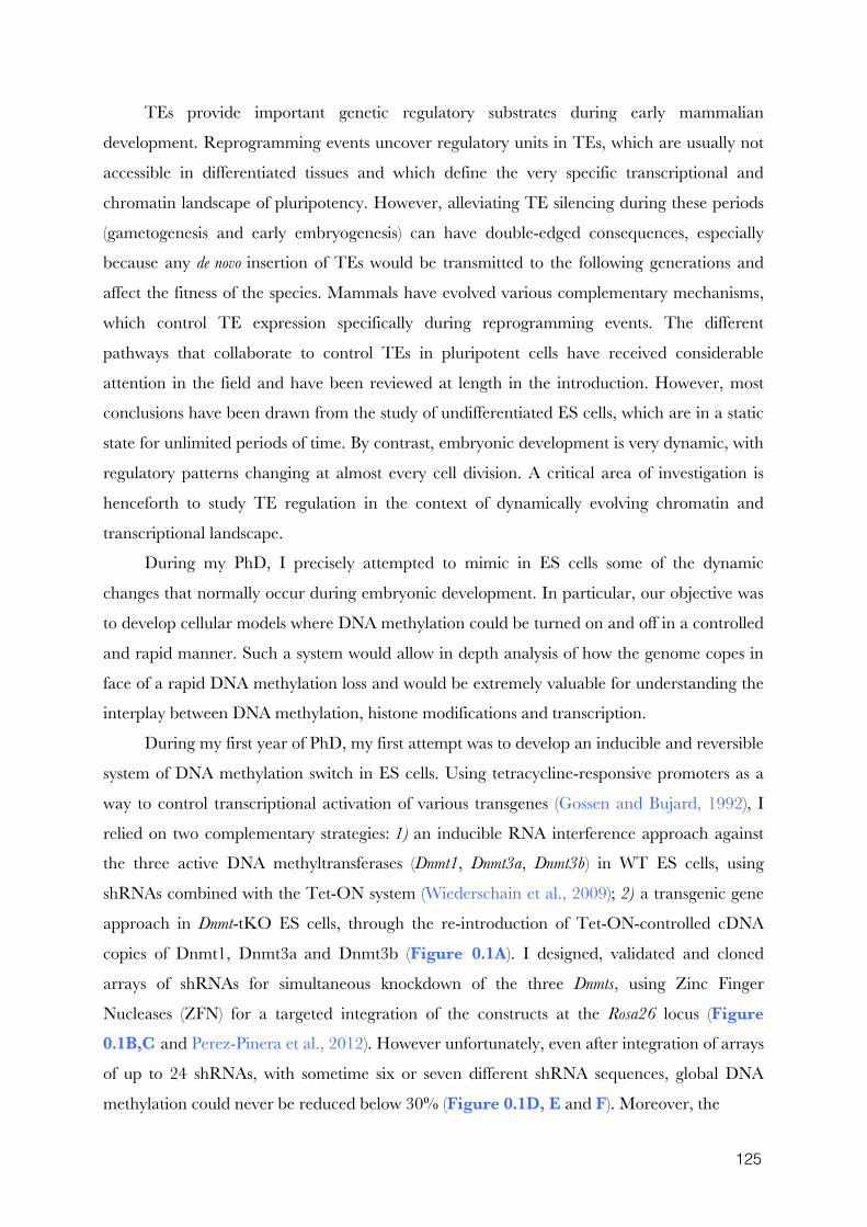
125
TEs provide important genetic regulatory substrates during early mammalian
development. Reprogramming events uncover regulatory units in TEs, which are usually not
accessible in differentiated tissues and which define the very specific transcriptional and
chromatin landscape of pluripotency. However, alleviating TE silencing during these periods
(gametogenesis and early embryogenesis) can have double-edged consequences, especially
because any de novo insertion of TEs would be transmitted to the following generations and
affect the fitness of the species. Mammals have evolved various complementary mechanisms,
which control TE expression specifically during reprogramming events. The different
pathways that collaborate to control TEs in pluripotent cells have received considerable
attention in the field and have been reviewed at length in the introduction. However, most
conclusions have been drawn from the study of undifferentiated ES cells, which are in a static
state for unlimited periods of time. By contrast, embryonic development is very dynamic, with
regulatory patterns changing at almost every cell division. A critical area of investigation is
henceforth to study TE regulation in the context of dynamically evolving chromatin and
transcriptional landscape.
During my PhD, I precisely attempted to mimic in ES cells some of the dynamic
changes that normally occur during embryonic development. In particular, our objective was
to develop cellular models where DNA methylation could be turned on and off in a controlled
and rapid manner. Such a system would allow in depth analysis of how the genome copes in
face of a rapid DNA methylation loss and would be extremely valuable for understanding the
interplay between DNA methylation, histone modifications and transcription.
During my first year of PhD, my first attempt was to develop an inducible and reversible
system of DNA methylation switch in ES cells. Using tetracycline-responsive promoters as a
way to control transcriptional activation of various transgenes (Gossen and Bujard, 1992), I
relied on two complementary strategies: 1) an inducible RNA interference approach against
the three active DNA methyltransferases (Dnmt1, Dnmt3a, Dnmt3b) in WT ES cells, using
shRNAs combined with the Tet-ON system (Wiederschain et al., 2009); 2) a transgenic gene
approach in Dnmt-tKO ES cells, through the re-introduction of Tet-ON-controlled cDNA
copies of Dnmt1, Dnmt3a and Dnmt3b (Figure 0.1A). I designed, validated and cloned
arrays of shRNAs for simultaneous knockdown of the three Dnmts, using Zinc Finger
Nucleases (ZFN) for a targeted integration of the constructs at the Rosa26 locus (Figure 0.1B,C and Perez-Pinera et al., 2012). However unfortunately, even after integration of arrays
of up to 24 shRNAs, with sometime six or seven different shRNA sequences, global DNA
methylation could never be reduced below 30% (Figure 0.1D, E and F). Moreover, the
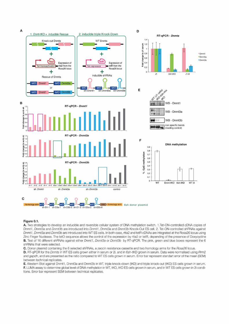
127
design of an inducible Dnmt rescue system in Dnmt-tKO ES cells proved to be a technical
nightmare, principally because Dnmt-tKO cells contain in their genome most of the usable
antibiotic resistant markers. After a year of fanatical cloning, these two complementary
approaches were finally dropped.
Two technological revolutions then allowed my PhD work to go around the wall it had
met. The first one is the worldwide revelation that CRISPR/Cas9 editing could be used to
manipulate virtually every biological systems, suddenly transforming ES cells into a very
powerful genetic model (Cong et al., 2013; Jinek et al., 2012). With CRISPR/Cas9, I was able
to knockout seven different genes in less than two years18: this allowed me to rapidly and
powerfully test hypotheses that would otherwise have remained unanswered. The second
revolution has not such a broad impact, and does not even deserve this term outside this
manuscript. However, the publication that vitamin C enhanced TET enzyme activity and
promoted active demethylation in ES cells was really the starting point of the work presented
here (Blaschke et al., 2013).
ES cultured in 2i conditions keep significant DNA methylation levels (~30% of CpG
methylation) and the transition between serum- and 2i-based media necessitates several weeks
to attain such methylation level (Ficz et al., 2013; Habibi et al., 2013). By contrast, we quickly
realized in the lab that conjunction of 2i culture system with vitamin C allowed reaching
unprecedented levels of hypomethylation in a short period of time. By switching culture
conditions from serum to 2i+vitamin C, I could promote fast and quasi-total demethylation of
the ES cell genome, providing clues as to how to answer the main question of my PhD work:
How do TEs react to the rapid loss of DNA methylation, and how the
genome adapts to the removal of this essential protective barrier?
By carefully following transposon behavior during global DNA demethylation, I was
able to unravel the mechanisms that ensure transposon regulation when DNA methylation is
lost. Our findings shed new lights about the way the genome can adapt to DNA methylation
reprogramming, with the particularly exciting evidence of the unsuspected role of Polycomb
proteins in transposon regulation. The results presented in the following section take the form
of a manuscript, which is currently under review at eLife, with a few cosmetic differences
necessary to fit the general style of this thesis.
18 So far
128
In a second time, we realized in the lab that the hypomethylated genome of ES cells
grown in 2i+vitamin represented an ideal starting point to study the dynamics of global de novo
methylation. Since they are already hypermethylated, serum-grown ES cells represent an
inadequate system to analyze DNA methylation dynamically. In contrast, by differentiating
2i+vitC-converted ES cells, we could reproduce the global de novo methylation events that
normally occur in the germline and early embryos. I used this system to characterize genome-
wide the contribution of DNMT3l in embryonic de novo methylation. Moreover, by comparing
the effect of Dnmt3l deficiency in differentiated ES cells, in vivo post-implantation embryos and
germ cells, I attempted to elucidate the developmental requirements of DNMT3l. The results
that I present in this thesis are still on-going work and represent a first version of a manuscript
that we plan to submit in the following months.
Of important note, massive amounts of genomic data were generated during my PhD,
and all of them were analyzed in close collaboration with Aurélie Teissandier, the
bioinformatician of our team.

129

130
131
1 An epigenetic switch ensures transposon repression upon dynamic loss of DNA methylation in ES cells
1.1 Authors and affiliations
Marius Walter1,2, Aurélie Teissandier1,2,3, Raquel Pérez Palacios1,
Déborah Bourc’his1*
1 Institut Curie, Dpt of Genetics and Developmental Biology, CNRS UMR3215, INSERM U934, Paris, France 2 UPMC, University Paris 06, Paris, France 3 Institut Curie, Inserm U900, Mines Paris Tech, Paris, France
1.2 Abstract
DNA methylation is extensively remodeled during mammalian gametogenesis and
embryogenesis. Most transposons become hypomethylated, raising the question of their
regulation in the absence of DNA methylation. To reproduce a rapid and extensive
demethylation, we subjected mouse ES cells to chemically defined hypomethylating culture
conditions. Surprisingly, we observed two phases of transposon regulation. After an initial
burst of de-repression, various transposon families were efficiently re-silenced, showing that
DNA methylation is involved in transposon repression in ES cells, but that alternative
mechanisms can compensate for its loss. This was accompanied by a reconfiguration of the
repressive chromatin landscape: while H3K9me3 was stable, H3K9me2 globally disappeared
and H3K27me3 accumulated at transposons. Interestingly, we observed that H3K9me3 and
H3K27me3 occupy different transposon families or different transposon territories within the
same family, defining three functional categories of adaptive chromatin responses to DNA
methylation loss. Our work highlights that H3K9me3 and H3K27me3 chromatin pathways
can secure the control of a large spectrum of transposons in periods of intense DNA
methylation change, ensuring longstanding genome stability.
1.3 Introduction
Millions of transposable elements reside in mammalian genomes, far surpassing in
number the approximately 25,000 protein-coding genes (Lander et al., 2001). Most of these
132
elements are retrotransposons, which utilize an RNA intermediate to duplicate and mobilize.
Through their activity or their mere presence, transposons can be both beneficial for the
evolution of the host genome and deleterious for its integrity. They can modify gene functions
through insertional mutagenesis, influence gene transcriptional outputs by acting as promoters
or enhancers or induce chromosomal rearrangements through non-allelic recombination
(Goodier and Kazazian, 2008). Accordingly, erratic transposon-related events have been
linked to congenital diseases, cancer and infertility (Kaer and Speek, 2013).
Successive waves of transposon expansion and decline have shaped mammalian
genomes over evolution, leading to a current occupancy rate of approximately half of the
genomic space. Reflecting their various evolutionary origin and multiplication success,
resident elements are greatly diverse in structures, numbers and functional properties, which
define discrete families of transposons. Long Terminal Repeat (LTR) sequences characterize
endogenous retroviruses (ERVs, 12% of the mouse genome), which can be further subdivided
into three families (ERV1, ERVK and ERVL), according to the infectious retroviruses they
derive from (Stocking and Kozak, 2008). Non-LTR elements comprise Long and Short
INterspersed Elements (LINEs and SINEs, 20% and 8% of the genome, respectively), and
also consist of specific sub-families (Babushok et al., 2007). The majority of transposons have
accumulated nullifying mutations and truncations, but around 1-2% of LINEs and ERVs
have intact sequences that embed the protein coding information necessary for their
mobilization. Notably, ERVK elements show the greatest level of activity, which causes at
least 10% of spontaneous mutations in laboratory mice (Maksakova et al., 2006).
To minimize their impact on genome fitness, multiple layers of control antagonize
transposons at different steps of their life cycle (Zamudio and Bourc’his, 2010). Notably,
restraining mechanisms can differ between cell types. In somatic cells and in the male
differentiating germline, DNA methylation is the main transcriptional suppressor of LTR and
non-LTR transposons. In these contexts, transposable elements are densely methylated
(Rollins et al., 2006; Smith et al., 2012) and DNA hypomethylation leads to their de-
repression (Bourc’his and Bestor, 2004; Walsh et al., 1998). In contrast, the early germline and
the early embryo manage to globally control their transposon burden without DNA
methylation. These cells naturally undergo genome-wide loss of DNA methylation, likely as
part of the acquisition of a pluripotent, flexible state (Seisenberger et al., 2013). Moreover,
genetic studies have demonstrated that mouse embryonic stem (ES) cells can use DNA
methylation-independent mechanisms to silence transposons: knocking-out the three active
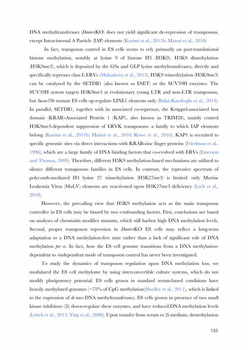
133
DNA methyltransferases (Dnmt-tKO) does not yield significant de-repression of transposons,
except Intracisternal A Particle (IAP) elements (Karimi et al., 2011b; Matsui et al., 2010)
In fact, transposon control in ES cells seems to rely primarily on post-translational
histone methylation, notably at lysine 9 of histone H3 (H3K9). H3K9 dimethylation
(H3K9me2), which is deposited by the G9a and GLP lysine methyltransferases, directly and
specifically represses class L ERVs (Maksakova et al., 2013). H3K9 trimethylation (H3K9me3)
can be catalyzed by the SETDB1 (also known as ESET) or the SUV39H enzymes. The
SUV39H system targets H3K9me3 at evolutionary young LTR and non-LTR transposons,
but Suvar39h mutant ES cells up-regulate LINE1 elements only (Bulut-Karslioglu et al., 2014).
In parallel, SETDB1, together with its associated co-repressor, the Krüppel-associated box
domain (KRAB)-Associated Protein 1 (KAP1, also known as TRIM28), mainly control
H3K9me3-dependent suppression of ERVK transposons- a family to which IAP elements
belong (Karimi et al., 2011b; Matsui et al., 2010; Rowe et al., 2010). KAP1 is recruited to
specific genomic sites via direct interactions with KRAB-zinc finger proteins (Friedman et al.,
1996), which are a large family of DNA binding factors that co-evolved with ERVs (Emerson
and Thomas, 2009). Therefore, different H3K9 methylation-based mechanisms are utilized to
silence different transposons families in ES cells. In contrast, the repressive spectrum of
polycomb-mediated H3 lysine 27 trimethylation (H3K27me3) is limited: only Murine
Leukemia Virus (MuLV) elements are reactivated upon H3K27me3 deficiency (Leeb et al.,
2010).
However, the prevailing view that H3K9 methylation acts as the main transposon
controller in ES cells may be biased by two confounding factors. First, conclusions are based
on analyses of chromatin modifier mutants, which still harbor high DNA methylation levels.
Second, proper transposon repression in Dnmt-tKO ES cells may reflect a long-term
adaptation to a DNA methylation-free state rather than a lack of significant role of DNA
methylation per se. In fact, how the ES cell genome transitions from a DNA methylation-
dependent to -independent mode of transposon control has never been investigated.
To study the dynamics of transposon regulation upon DNA methylation loss, we
modulated the ES cell methylome by using interconvertible culture systems, which do not
modify pluripotency potential. ES cells grown in standard serum-based conditions have
heavily methylated genomes (~75% of CpG methylation)(Stadler et al., 2011), which is linked
to the expression of de novo DNA methyltransferases. ES cells grown in presence of two small
kinase inhibitors (2i) down-regulate these enzymes, and have reduced DNA methylation levels
(Leitch et al., 2013; Ying et al., 2008). Upon transfer from serum to 2i medium, demethylation
135
occurs with a slow kinetics: several weeks are required to reach 20-30% of CpG methylation.
Notably, imprinted genes, major satellite repeats and IAP elements maintain persistent DNA
methylation after 2i adaptation (Ficz et al., 2013; Habibi et al., 2013). Addition of vitamin C
(vitC) can also lower the ES cell methylome. This compound promotes active demethylation
by stimulating the TET (Ten Eleven Translocation) enzymes, which oxidize 5-
methylcytosines to 5-hydroxymethylcytosines that are potential intermediates towards
unmethylated cytosines (Blaschke et al., 2013).
Here, by switching ES cells directly from a serum-based to a 2i+vitC medium, we were
able to induce rapid and extensive demethylation genome-wide, mimicking a situation
occurring in the early embryo. By combining DNA methylation, chromatin and
transcriptional profiling of transposons along with genetic analyses, we found that DNA
methylation represses multiple families of transposons in ES cells, but an epigenetic switch
towards histone-based control is progressively implemented as DNA methylation disappears.
Importantly, we reveal for the first time the specific and overlapping roles of H3K9 and
H3K27 trimethylation in controlling distinct transposon families upon DNA demethylation.
These findings have important implications for understanding the molecular underpinning of
transposon control in the pluripotent cells of the early mammalian embryo.
1.4 Results
DNA methylation is rapidly and extensively lost in ES cells during serum
to 2i+vitC media conversion
Dnmt-tKO ES cells are completely devoid of DNA methylation, yet expression levels of
most transposable elements remain globally similar to wild-type (WT) ES cells (Karimi et al.,
2011b; Tsumura et al., 2006). This may indicate the implementation of alternative
mechanisms that compensate for DNA methylation-based repression. To analyze dynamic
adaptation, we utilized a culture-based system that results in rapid DNA methylation loss:
converting ES cells from serum-based to 2i+vitamin C (2i+vitC) culture conditions. To
overcome confounding genetic effects, we used the J1 ES cell line, from which Dnmt-tKO
mutants were originally derived.
Quantification using the methyl-CpG sensitive restriction enzyme-based LUminometric
Methylation Assay (LUMA) (Karimi et al., 2011a) revealed that CpG methylation linearly
decreased from 77% to 13% in six days, and reached a minimal level of 6% after 14 days of
conversion (Figure 1.1A). In comparison, cells grown in serum+vitC or in 2i-only maintained
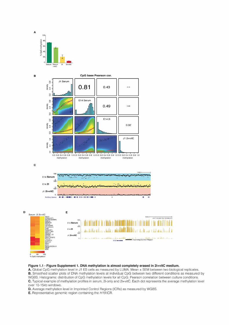
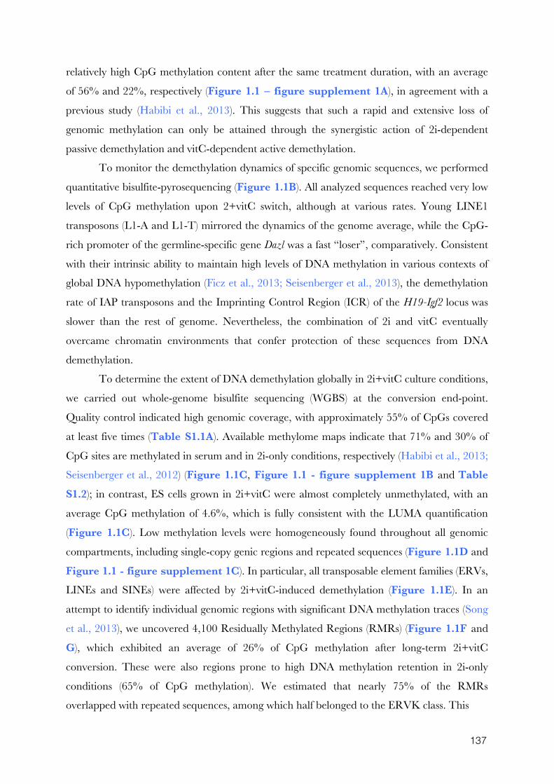
137
relatively high CpG methylation content after the same treatment duration, with an average
of 56% and 22%, respectively (Figure 1.1 – figure supplement 1A), in agreement with a
previous study (Habibi et al., 2013). This suggests that such a rapid and extensive loss of
genomic methylation can only be attained through the synergistic action of 2i-dependent
passive demethylation and vitC-dependent active demethylation.
To monitor the demethylation dynamics of specific genomic sequences, we performed
quantitative bisulfite-pyrosequencing (Figure 1.1B). All analyzed sequences reached very low
levels of CpG methylation upon 2+vitC switch, although at various rates. Young LINE1
transposons (L1-A and L1-T) mirrored the dynamics of the genome average, while the CpG-
rich promoter of the germline-specific gene Dazl was a fast “loser”, comparatively. Consistent
with their intrinsic ability to maintain high levels of DNA methylation in various contexts of
global DNA hypomethylation (Ficz et al., 2013; Seisenberger et al., 2013), the demethylation
rate of IAP transposons and the Imprinting Control Region (ICR) of the H19-Igf2 locus was
slower than the rest of genome. Nevertheless, the combination of 2i and vitC eventually
overcame chromatin environments that confer protection of these sequences from DNA
demethylation.
To determine the extent of DNA demethylation globally in 2i+vitC culture conditions,
we carried out whole-genome bisulfite sequencing (WGBS) at the conversion end-point.
Quality control indicated high genomic coverage, with approximately 55% of CpGs covered
at least five times (Table S1.1A). Available methylome maps indicate that 71% and 30% of
CpG sites are methylated in serum and in 2i-only conditions, respectively (Habibi et al., 2013;
Seisenberger et al., 2012) (Figure 1.1C, Figure 1.1 - figure supplement 1B and Table S1.2); in contrast, ES cells grown in 2i+vitC were almost completely unmethylated, with an
average CpG methylation of 4.6%, which is fully consistent with the LUMA quantification
(Figure 1.1C). Low methylation levels were homogeneously found throughout all genomic
compartments, including single-copy genic regions and repeated sequences (Figure 1.1D and
Figure 1.1 - figure supplement 1C). In particular, all transposable element families (ERVs,
LINEs and SINEs) were affected by 2i+vitC-induced demethylation (Figure 1.1E). In an
attempt to identify individual genomic regions with significant DNA methylation traces (Song
et al., 2013), we uncovered 4,100 Residually Methylated Regions (RMRs) (Figure 1.1F and G), which exhibited an average of 26% of CpG methylation after long-term 2i+vitC
conversion. These were also regions prone to high DNA methylation retention in 2i-only
conditions (65% of CpG methylation). We estimated that nearly 75% of the RMRs
overlapped with repeated sequences, among which half belonged to the ERVK class. This

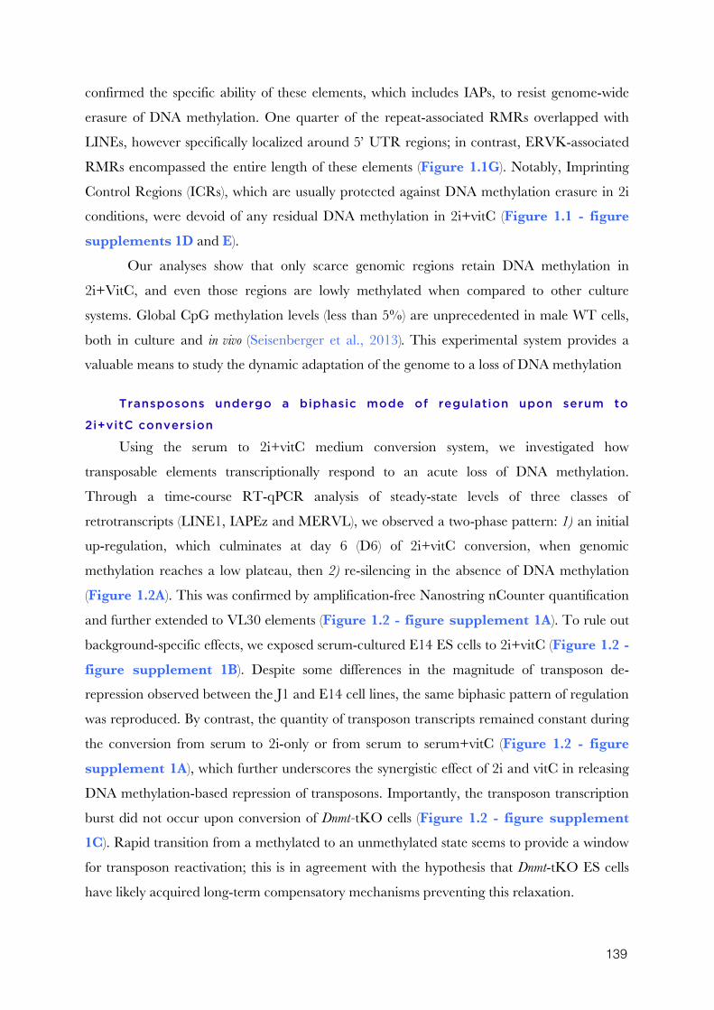
139
confirmed the specific ability of these elements, which includes IAPs, to resist genome-wide
erasure of DNA methylation. One quarter of the repeat-associated RMRs overlapped with
LINEs, however specifically localized around 5’ UTR regions; in contrast, ERVK-associated
RMRs encompassed the entire length of these elements (Figure 1.1G). Notably, Imprinting
Control Regions (ICRs), which are usually protected against DNA methylation erasure in 2i
conditions, were devoid of any residual DNA methylation in 2i+vitC (Figure 1.1 - figure supplements 1D and E).
Our analyses show that only scarce genomic regions retain DNA methylation in
2i+VitC, and even those regions are lowly methylated when compared to other culture
systems. Global CpG methylation levels (less than 5%) are unprecedented in male WT cells,
both in culture and in vivo (Seisenberger et al., 2013). This experimental system provides a
valuable means to study the dynamic adaptation of the genome to a loss of DNA methylation
Transposons undergo a biphasic mode of regulation upon serum to
2i+vitC conversion
Using the serum to 2i+vitC medium conversion system, we investigated how
transposable elements transcriptionally respond to an acute loss of DNA methylation.
Through a time-course RT-qPCR analysis of steady-state levels of three classes of
retrotranscripts (LINE1, IAPEz and MERVL), we observed a two-phase pattern: 1) an initial
up-regulation, which culminates at day 6 (D6) of 2i+vitC conversion, when genomic
methylation reaches a low plateau, then 2) re-silencing in the absence of DNA methylation
(Figure 1.2A). This was confirmed by amplification-free Nanostring nCounter quantification
and further extended to VL30 elements (Figure 1.2 - figure supplement 1A). To rule out
background-specific effects, we exposed serum-cultured E14 ES cells to 2i+vitC (Figure 1.2 - figure supplement 1B). Despite some differences in the magnitude of transposon de-
repression observed between the J1 and E14 cell lines, the same biphasic pattern of regulation
was reproduced. By contrast, the quantity of transposon transcripts remained constant during
the conversion from serum to 2i-only or from serum to serum+vitC (Figure 1.2 - figure supplement 1A), which further underscores the synergistic effect of 2i and vitC in releasing
DNA methylation-based repression of transposons. Importantly, the transposon transcription
burst did not occur upon conversion of Dnmt-tKO cells (Figure 1.2 - figure supplement 1C). Rapid transition from a methylated to an unmethylated state seems to provide a window
for transposon reactivation; this is in agreement with the hypothesis that Dnmt-tKO ES cells
have likely acquired long-term compensatory mechanisms preventing this relaxation.
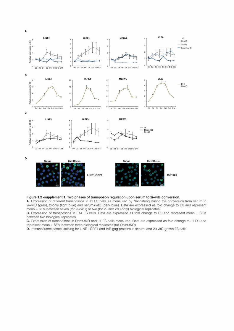
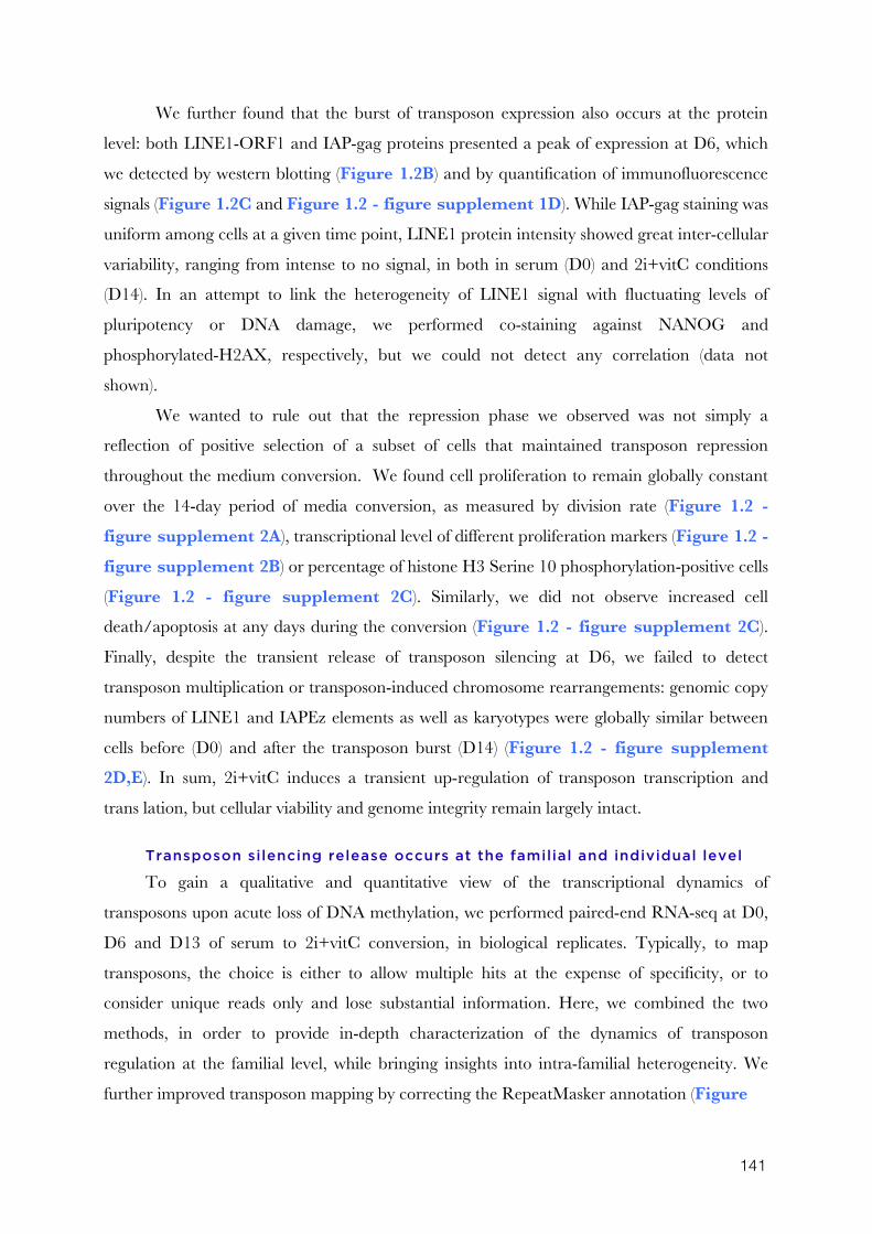
141
We further found that the burst of transposon expression also occurs at the protein
level: both LINE1-ORF1 and IAP-gag proteins presented a peak of expression at D6, which
we detected by western blotting (Figure 1.2B) and by quantification of immunofluorescence
signals (Figure 1.2C and Figure 1.2 - figure supplement 1D). While IAP-gag staining was
uniform among cells at a given time point, LINE1 protein intensity showed great inter-cellular
variability, ranging from intense to no signal, in both in serum (D0) and 2i+vitC conditions
(D14). In an attempt to link the heterogeneity of LINE1 signal with fluctuating levels of
pluripotency or DNA damage, we performed co-staining against NANOG and
phosphorylated-H2AX, respectively, but we could not detect any correlation (data not
shown).
We wanted to rule out that the repression phase we observed was not simply a
reflection of positive selection of a subset of cells that maintained transposon repression
throughout the medium conversion. We found cell proliferation to remain globally constant
over the 14-day period of media conversion, as measured by division rate (Figure 1.2 - figure supplement 2A), transcriptional level of different proliferation markers (Figure 1.2 - figure supplement 2B) or percentage of histone H3 Serine 10 phosphorylation-positive cells
(Figure 1.2 - figure supplement 2C). Similarly, we did not observe increased cell
death/apoptosis at any days during the conversion (Figure 1.2 - figure supplement 2C).
Finally, despite the transient release of transposon silencing at D6, we failed to detect
transposon multiplication or transposon-induced chromosome rearrangements: genomic copy
numbers of LINE1 and IAPEz elements as well as karyotypes were globally similar between
cells before (D0) and after the transposon burst (D14) (Figure 1.2 - figure supplement 2D,E). In sum, 2i+vitC induces a transient up-regulation of transposon transcription and
trans lation, but cellular viability and genome integrity remain largely intact.
Transposon silencing release occurs at the familial and individual level
To gain a qualitative and quantitative view of the transcriptional dynamics of
transposons upon acute loss of DNA methylation, we performed paired-end RNA-seq at D0,
D6 and D13 of serum to 2i+vitC conversion, in biological replicates. Typically, to map
transposons, the choice is either to allow multiple hits at the expense of specificity, or to
consider unique reads only and lose substantial information. Here, we combined the two
methods, in order to provide in-depth characterization of the dynamics of transposon
regulation at the familial level, while bringing insights into intra-familial heterogeneity. We
further improved transposon mapping by correcting the RepeatMasker annotation (Figure
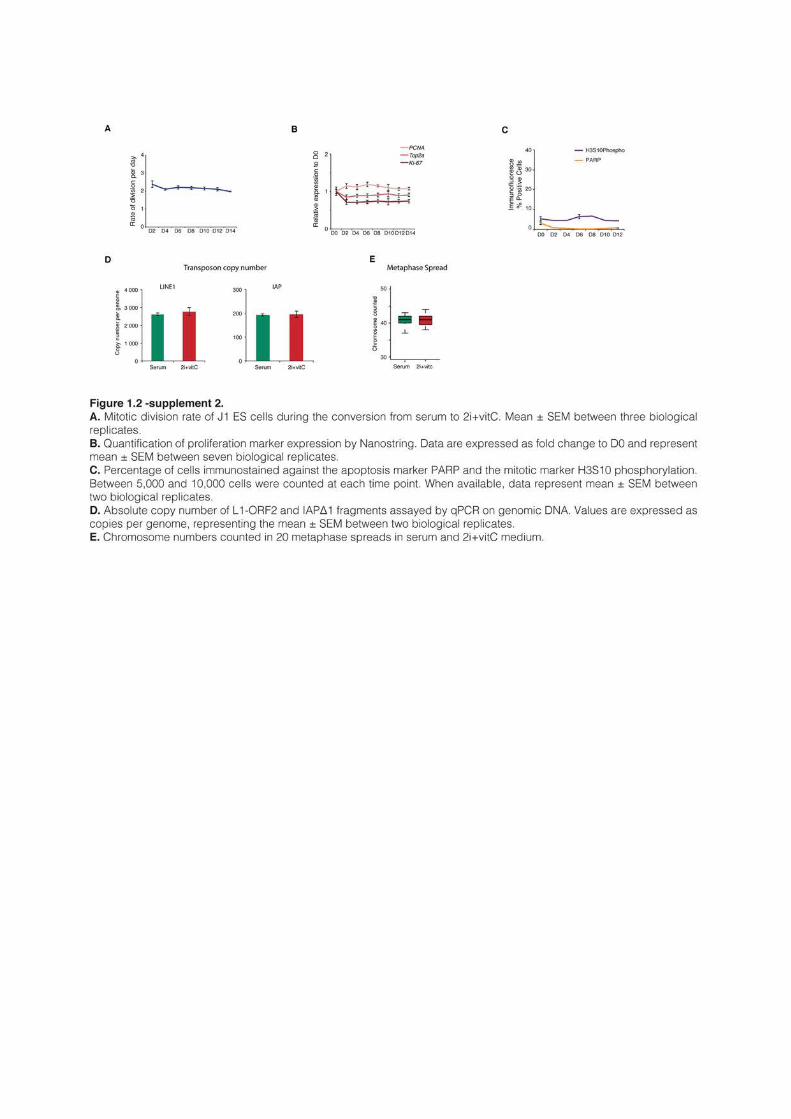
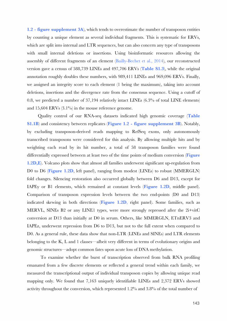
143
1.2 - figure supplement 3A), which tends to overestimate the number of transposon entities
by counting a unique element as several individual fragments. This is systematic for ERVs,
which are split into internal and LTR sequences, but can also concern any type of transposons
with small internal deletions or insertions. Using bioinformatic resources allowing the
assembly of different fragments of an element (Bailly-Bechet et al., 2014), our reconstructed
version gave a census of 588,739 LINEs and 497,706 ERVs (Table S1.3), while the original
annotation roughly doubles these numbers, with 989,411 LINEs and 969,096 ERVs. Finally,
we assigned an integrity score to each element (1 being the maximum), taking into account
deletions, insertions and the divergence rate from the consensus sequence. Using a cutoff of
0.8, we predicted a number of 37,194 relatively intact LINEs (6.3% of total LINE elements)
and 15,604 ERVs (3.1%) in the mouse reference genome.
Quality control of our RNA-seq datasets indicated high genomic coverage (Table S1.1B) and consistency between replicates (Figure 1.2 - figure supplement 3B). Notably,
by excluding transposon-derived reads mapping to RefSeq exons, only autonomously
transcribed transposons were considered for this analysis. By allowing multiple hits and by
weighting each read by its hit number, a total of 58 transposon families were found
differentially expressed between at least two of the time points of medium conversion (Figure 1.2D,E). Volcano plots show that almost all families underwent significant up-regulation from
D0 to D6 (Figure 1.2D, left panel), ranging from modest (LINEs) to robust (MMERGLN)
fold changes. Silencing restoration also occurred globally between D6 and D13, except for
IAPEy or B1 elements, which remained at constant levels (Figure 1.2D, middle panel).
Comparison of transposon expression levels between the two end-points (D0 and D13)
indicated skewing in both directions (Figure 1.2D, right panel). Some families, such as
MERVL, SINEs B2 or any LINE1 types, were more strongly repressed after the 2i+vitC
conversion at D13 than initially at D0 in serum. Others, like MMERGLN, ETnERV3 and
IAPEz, underwent repression from D6 to D13, but not to the full extent when compared to
D0. As a general rule, these data show that non-LTR (LINEs and SINEs) and LTR elements
belonging to the K, L and 1 classes—albeit very different in terms of evolutionary origins and
genomic structures—adopt common fates upon acute loss of DNA methylation.
To examine whether the burst of transcription observed from bulk RNA profiling
emanated from a few discrete elements or reflected a general trend within each family, we
measured the transcriptional output of individual transposon copies by allowing unique read
mapping only. We found that 7,163 uniquely identifiable LINEs and 2,372 ERVs showed
activity throughout the conversion, which represented 1.2% and 3.8% of the total number of
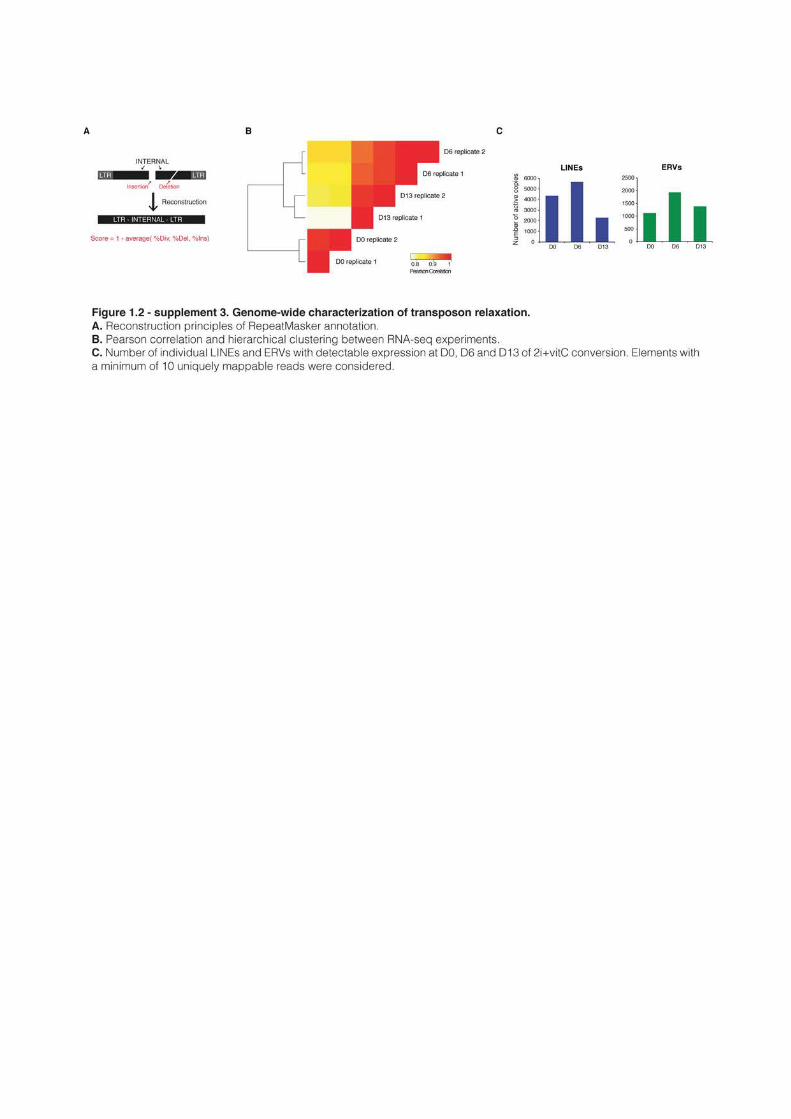
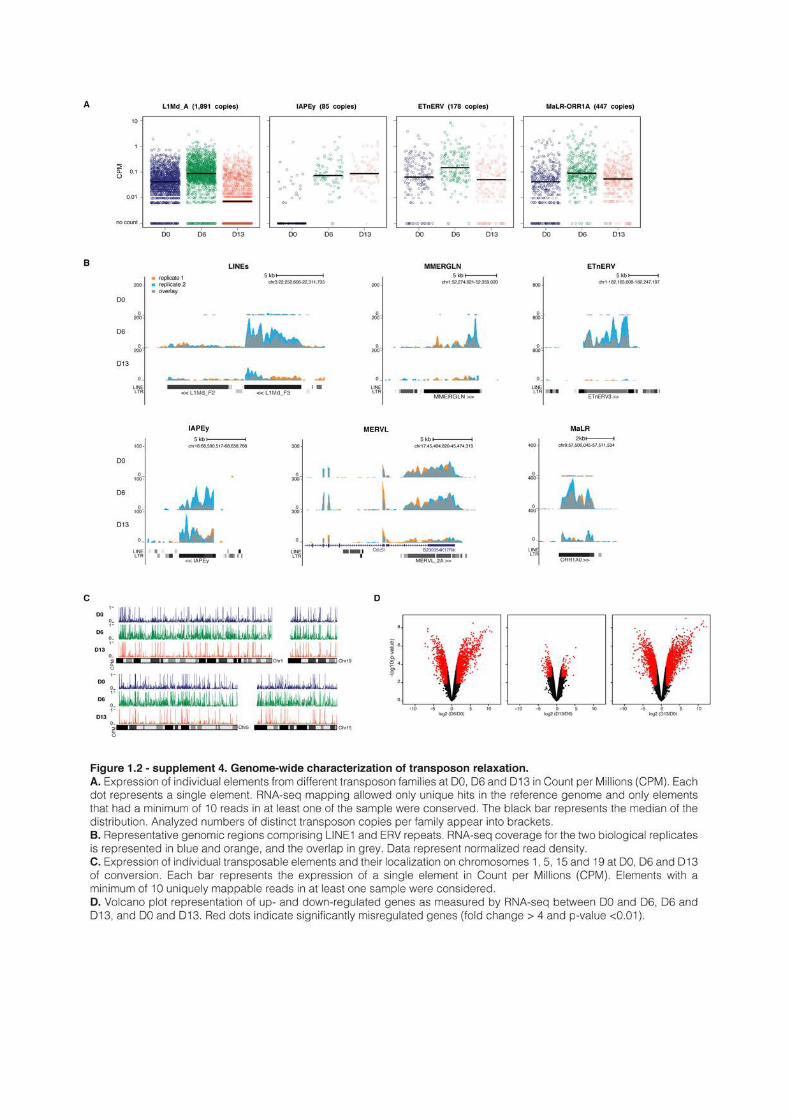
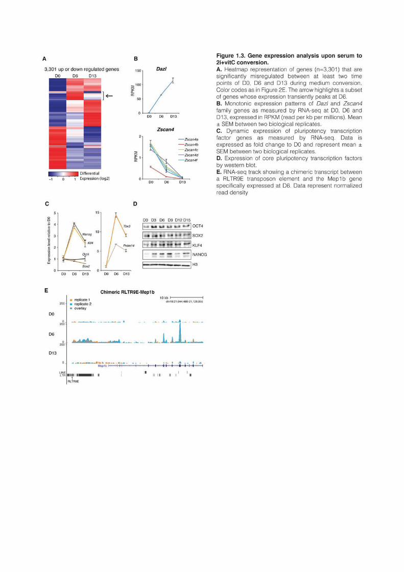
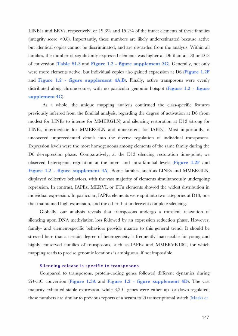
147
LINE1s and ERVs, respectively, or 19.3% and 15.2% of the intact elements of these families
(integrity score >0.8). Importantly, these numbers are likely underestimated because active
but identical copies cannot be discriminated, and are discarded from the analysis. Within all
families, the number of significantly expressed elements was higher at D6 than at D0 or D13
of conversion (Table S1.3 and Figure 1.2 - figure supplement 3C). Generally, not only
were more elements active, but individual copies also gained expression at D6 (Figure 1.2F
and Figure 1.2 - figure supplement 4A,B). Finally, active transposons were evenly
distributed along chromosomes, with no particular genomic hotspot (Figure 1.2 - figure supplement 4C).
As a whole, the unique mapping analysis confirmed the class-specific features
previously inferred from the familial analysis, regarding the degree of activation at D6 (from
modest for LINEs to intense for MMERGLN) and silencing restoration at D13 (strong for
LINEs, intermediate for MMERGLN and nonexistent for IAPEy). Most importantly, it
uncovered unprecedented details into the diverse regulation of individual transposons.
Expression levels were the most homogeneous among elements of the same family during the
D6 de-repression phase. Comparatively, at the D13 silencing restoration time-point, we
observed heterogenic regulation at the inter- and intra-familial levels (Figure 1.2F and
Figure 1.2 - figure supplement 4A). Some families, such as LINEs and MMERGLN,
displayed collective behaviors, with the vast majority of elements simultaneously undergoing
repression. In contrast, IAPEz, MERVL or ETn elements showed the widest distribution in
individual expression. In particular, IAPEz elements were split into two categories at D13, one
that maintained high expression, and the other that underwent complete silencing.
Globally, our analysis reveals that transposons undergo a transient relaxation of
silencing upon DNA methylation loss followed by an expression reduction phase. However,
family- and element-specific behaviors provide nuance to this general trend. It should be
stressed here that a certain degree of heterogeneity is frequently inaccessible for young and
highly conserved families of transposons, such as IAPEz and MMERVK10C, for which
mapping reads to precise genomic locations is ambiguous, if not impossible.
Silencing release is specific to transposons
Compared to transposons, protein-coding genes followed different dynamics during
2i+vitC conversion (Figure 1.3A and Figure 1.2 - figure supplement 4D). The vast
majority exhibited stable expression, while 3,301 genes were either up- or down-regulated;
these numbers are similar to previous reports of a serum to 2i transcriptional switch (Marks et
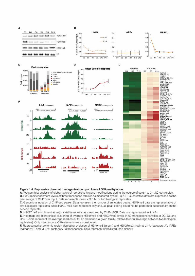
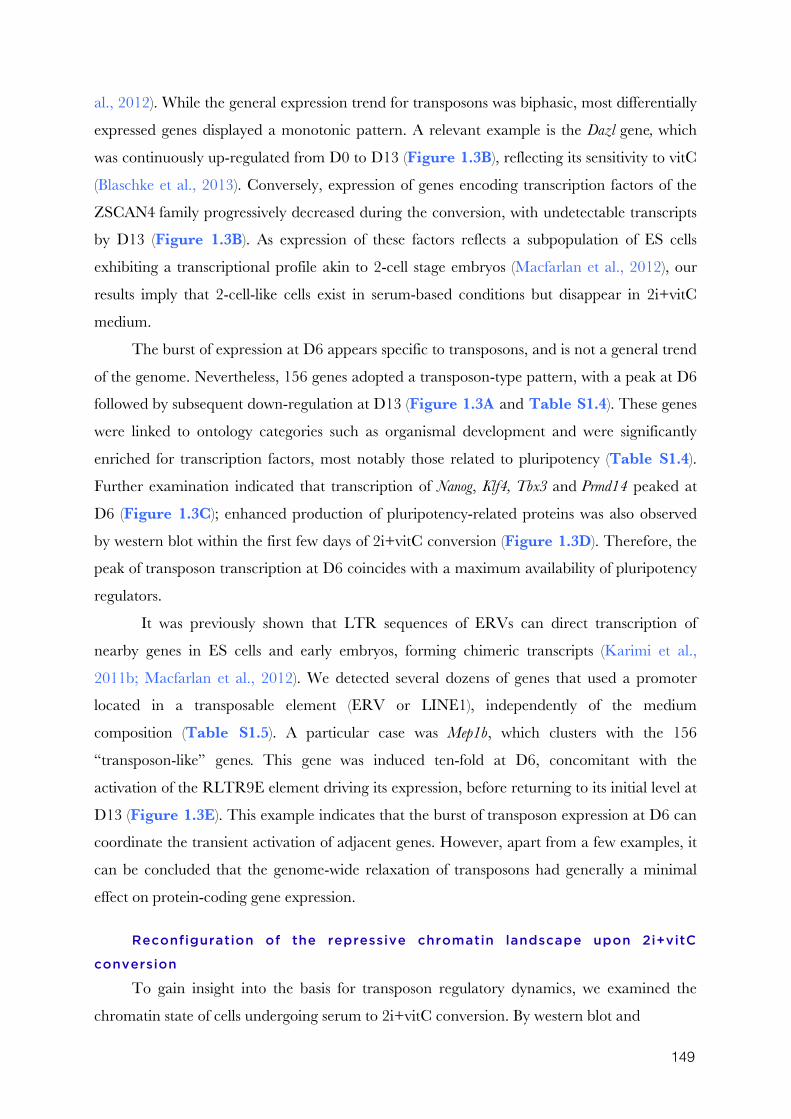
149
al., 2012). While the general expression trend for transposons was biphasic, most differentially
expressed genes displayed a monotonic pattern. A relevant example is the Dazl gene, which
was continuously up-regulated from D0 to D13 (Figure 1.3B), reflecting its sensitivity to vitC
(Blaschke et al., 2013). Conversely, expression of genes encoding transcription factors of the
ZSCAN4 family progressively decreased during the conversion, with undetectable transcripts
by D13 (Figure 1.3B). As expression of these factors reflects a subpopulation of ES cells
exhibiting a transcriptional profile akin to 2-cell stage embryos (Macfarlan et al., 2012), our
results imply that 2-cell-like cells exist in serum-based conditions but disappear in 2i+vitC
medium.
The burst of expression at D6 appears specific to transposons, and is not a general trend
of the genome. Nevertheless, 156 genes adopted a transposon-type pattern, with a peak at D6
followed by subsequent down-regulation at D13 (Figure 1.3A and Table S1.4). These genes
were linked to ontology categories such as organismal development and were significantly
enriched for transcription factors, most notably those related to pluripotency (Table S1.4).
Further examination indicated that transcription of Nanog, Klf4, Tbx3 and Prmd14 peaked at
D6 (Figure 1.3C); enhanced production of pluripotency-related proteins was also observed
by western blot within the first few days of 2i+vitC conversion (Figure 1.3D). Therefore, the
peak of transposon transcription at D6 coincides with a maximum availability of pluripotency
regulators.
It was previously shown that LTR sequences of ERVs can direct transcription of
nearby genes in ES cells and early embryos, forming chimeric transcripts (Karimi et al.,
2011b; Macfarlan et al., 2012). We detected several dozens of genes that used a promoter
located in a transposable element (ERV or LINE1), independently of the medium
composition (Table S1.5). A particular case was Mep1b, which clusters with the 156
“transposon-like” genes. This gene was induced ten-fold at D6, concomitant with the
activation of the RLTR9E element driving its expression, before returning to its initial level at
D13 (Figure 1.3E). This example indicates that the burst of transposon expression at D6 can
coordinate the transient activation of adjacent genes. However, apart from a few examples, it
can be concluded that the genome-wide relaxation of transposons had generally a minimal
effect on protein-coding gene expression.
Reconfiguration of the repressive chromatin landscape upon 2i+vitC
conversion
To gain insight into the basis for transposon regulatory dynamics, we examined the
chromatin state of cells undergoing serum to 2i+vitC conversion. By western blot and
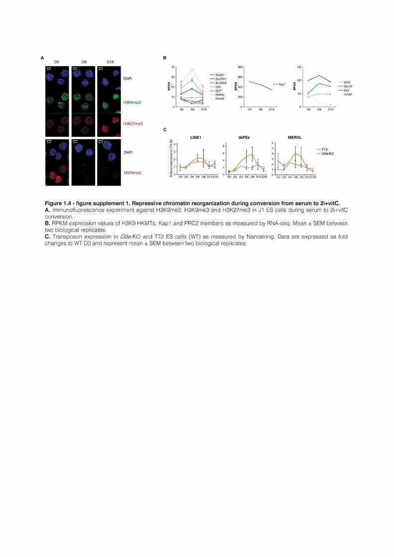
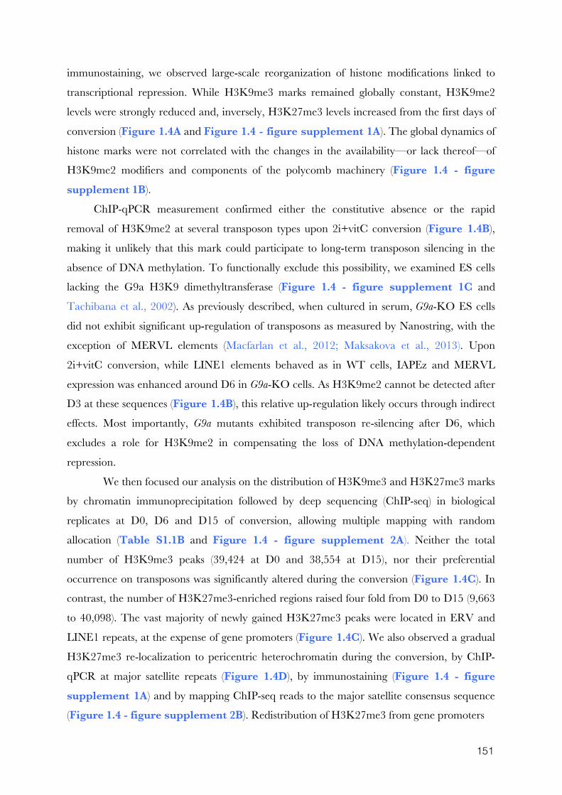
151
immunostaining, we observed large-scale reorganization of histone modifications linked to
transcriptional repression. While H3K9me3 marks remained globally constant, H3K9me2
levels were strongly reduced and, inversely, H3K27me3 levels increased from the first days of
conversion (Figure 1.4A and Figure 1.4 - figure supplement 1A). The global dynamics of
histone marks were not correlated with the changes in the availability—or lack thereof—of
H3K9me2 modifiers and components of the polycomb machinery (Figure 1.4 - figure supplement 1B).
ChIP-qPCR measurement confirmed either the constitutive absence or the rapid
removal of H3K9me2 at several transposon types upon 2i+vitC conversion (Figure 1.4B),
making it unlikely that this mark could participate to long-term transposon silencing in the
absence of DNA methylation. To functionally exclude this possibility, we examined ES cells
lacking the G9a H3K9 dimethyltransferase (Figure 1.4 - figure supplement 1C and
Tachibana et al., 2002). As previously described, when cultured in serum, G9a-KO ES cells
did not exhibit significant up-regulation of transposons as measured by Nanostring, with the
exception of MERVL elements (Macfarlan et al., 2012; Maksakova et al., 2013). Upon
2i+vitC conversion, while LINE1 elements behaved as in WT cells, IAPEz and MERVL
expression was enhanced around D6 in G9a-KO cells. As H3K9me2 cannot be detected after
D3 at these sequences (Figure 1.4B), this relative up-regulation likely occurs through indirect
effects. Most importantly, G9a mutants exhibited transposon re-silencing after D6, which
excludes a role for H3K9me2 in compensating the loss of DNA methylation-dependent
repression.
We then focused our analysis on the distribution of H3K9me3 and H3K27me3 marks
by chromatin immunoprecipitation followed by deep sequencing (ChIP-seq) in biological
replicates at D0, D6 and D15 of conversion, allowing multiple mapping with random
allocation (Table S1.1B and Figure 1.4 - figure supplement 2A). Neither the total
number of H3K9me3 peaks (39,424 at D0 and 38,554 at D15), nor their preferential
occurrence on transposons was significantly altered during the conversion (Figure 1.4C). In
contrast, the number of H3K27me3-enriched regions raised four fold from D0 to D15 (9,663
to 40,098). The vast majority of newly gained H3K27me3 peaks were located in ERV and
LINE1 repeats, at the expense of gene promoters (Figure 1.4C). We also observed a gradual
H3K27me3 re-localization to pericentric heterochromatin during the conversion, by ChIP-
qPCR at major satellite repeats (Figure 1.4D), by immunostaining (Figure 1.4 - figure supplement 1A) and by mapping ChIP-seq reads to the major satellite consensus sequence
(Figure 1.4 - figure supplement 2B). Redistribution of H3K27me3 from gene promoters
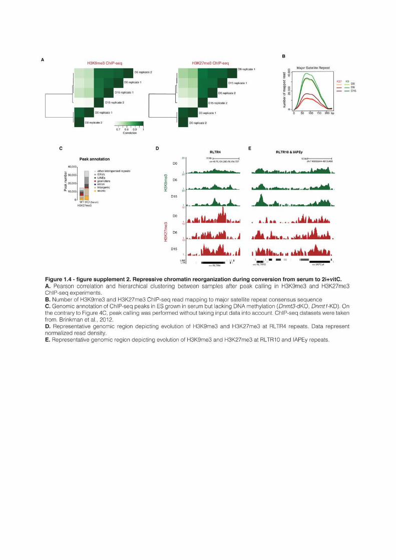
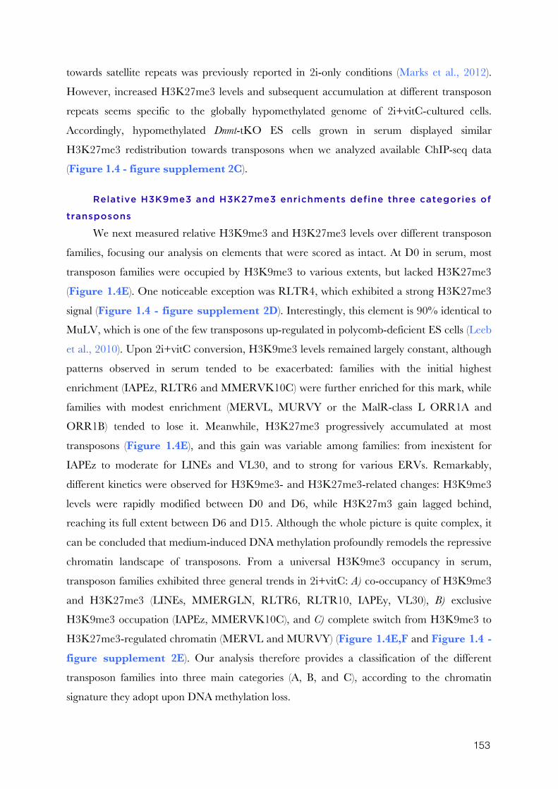
153
towards satellite repeats was previously reported in 2i-only conditions (Marks et al., 2012).
However, increased H3K27me3 levels and subsequent accumulation at different transposon
repeats seems specific to the globally hypomethylated genome of 2i+vitC-cultured cells.
Accordingly, hypomethylated Dnmt-tKO ES cells grown in serum displayed similar
H3K27me3 redistribution towards transposons when we analyzed available ChIP-seq data
(Figure 1.4 - figure supplement 2C).
Relative H3K9me3 and H3K27me3 enrichments define three categories of
transposons
We next measured relative H3K9me3 and H3K27me3 levels over different transposon
families, focusing our analysis on elements that were scored as intact. At D0 in serum, most
transposon families were occupied by H3K9me3 to various extents, but lacked H3K27me3
(Figure 1.4E). One noticeable exception was RLTR4, which exhibited a strong H3K27me3
signal (Figure 1.4 - figure supplement 2D). Interestingly, this element is 90% identical to
MuLV, which is one of the few transposons up-regulated in polycomb-deficient ES cells (Leeb
et al., 2010). Upon 2i+vitC conversion, H3K9me3 levels remained largely constant, although
patterns observed in serum tended to be exacerbated: families with the initial highest
enrichment (IAPEz, RLTR6 and MMERVK10C) were further enriched for this mark, while
families with modest enrichment (MERVL, MURVY or the MalR-class L ORR1A and
ORR1B) tended to lose it. Meanwhile, H3K27me3 progressively accumulated at most
transposons (Figure 1.4E), and this gain was variable among families: from inexistent for
IAPEz to moderate for LINEs and VL30, and to strong for various ERVs. Remarkably,
different kinetics were observed for H3K9me3- and H3K27me3-related changes: H3K9me3
levels were rapidly modified between D0 and D6, while H3K27m3 gain lagged behind,
reaching its full extent between D6 and D15. Although the whole picture is quite complex, it
can be concluded that medium-induced DNA methylation profoundly remodels the repressive
chromatin landscape of transposons. From a universal H3K9me3 occupancy in serum,
transposon families exhibited three general trends in 2i+vitC: A) co-occupancy of H3K9me3
and H3K27me3 (LINEs, MMERGLN, RLTR6, RLTR10, IAPEy, VL30), B) exclusive
H3K9me3 occupation (IAPEz, MMERVK10C), and C) complete switch from H3K9me3 to
H3K27me3-regulated chromatin (MERVL and MURVY) (Figure 1.4E,F and Figure 1.4 - figure supplement 2E). Our analysis therefore provides a classification of the different
transposon families into three main categories (A, B, and C), according to the chromatin
signature they adopt upon DNA methylation loss.
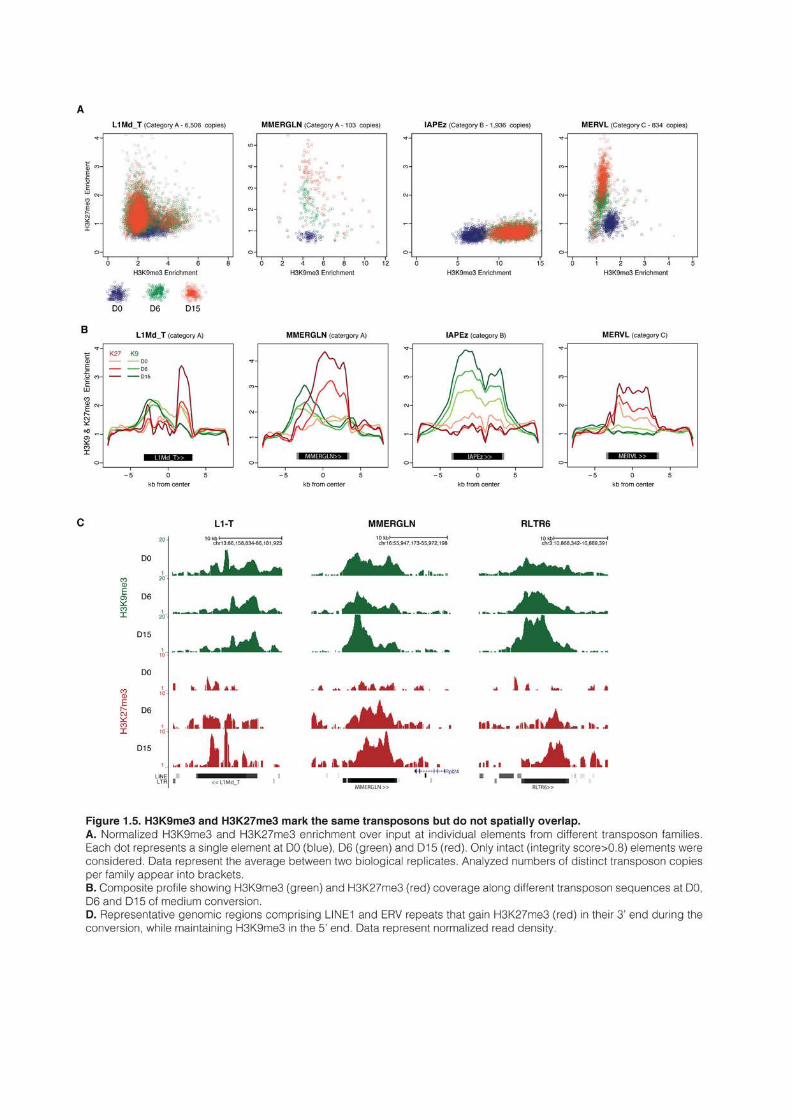
155
To assess the behavior of individual elements among these three generic patterns, we
attempted to analyze unique mappers, but the coverage on individual transposons was too low
to extract reliable information. Nevertheless, to gain insight into the question of intra-familial
heterogeneity, we plotted H3K9me3 and H3K27me3 enrichment for every intact transposons
(score>0.8) per family during the conversion. We found that elements of the B and C
categories tended to be very homogeneous. IAPEz elements (category B) collectively gained
H3K9me3 from D0 to D6; the MERVL and the Y-specific MURVY families (category C)
also showed compact patterns, with individual elements transitioning together from H3K9m3
enrichment at D0 to H3K27me3 at D15 (Figure 1.5A and Figure 1.5 - figure supplement 1B,C). The A category, which is enriched for both H3K9me3 and H3K27me3,
was more diverse, with some families displaying homogeneous patterns, while others showed
intra-familial dispersion in chromatin fates upon conversion. Within the MMERGLN,
RLTR6 or RLTR10 families, all elements gained H3K27me3 while maintaining or gaining
high levels of H3K9me3. Within the L1-T and IAPEy families, the majority of elements
gained H3K27me3, but a subset maintained H3K9me3 without acquiring H3K27me3
(Figure 1.5A and Figure 1.5 - figure supplement 1A). Another case of intra-familial
heterogeneity is provided by RLTR4, which specifically carries H3K27me3 marks at D0 in
serum: we demonstrate here that this enrichment was restricted to a small proportion of
elements, as was previously suspected (Figure 1.5 - figure supplement 1D and Reichmann
et al., 2012). By extracting single-element information from RNA-seq and ChIP-seq data, it is
clear that both transcriptional and chromatin heterogeneity exists among some transposon
families. Our analysis reveals that caution should be taken when interpreting average familial
behaviors, as they may be representative of only a few individual elements inside a given
family.
H3K27me3 occupies DNA methylation- and H3K9me3-free territories of
transposon sequences
H3K27me3 and H3K9me3 marks usually do not occur concomitantly (Mikkelsen et al.,
2007). We were therefore intrigued to observe that H3K27me3 and H3K9me3 were
simultaneously enriched at transposon families of the A category in 2i+vitC medium (Figure 1.4E and Figure 1.4 - figure supplement 1G,H). To map the relative position of
H3K9me3 and H3K27me3, we determined their average profile over full-length individual
elements of all transposon families, including their immediate genomic vicinity (+/- 5kb from
the center of each element). Notably, H3K9me3 domains often spread out on adjacent
genomic regions, whereas H3K27me3 was confined to transposon sequences (Figure 1.5B,C
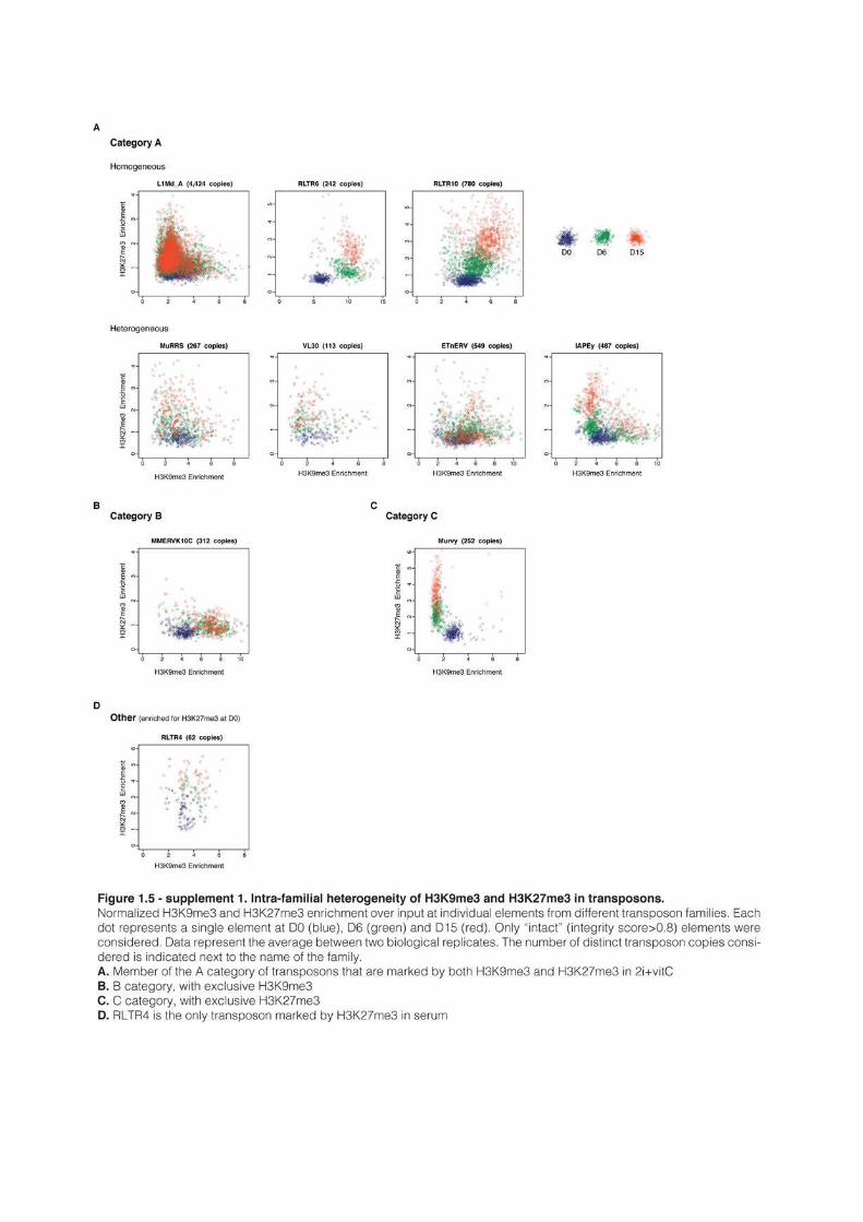
157
and Figure 1.5 - figure supplement 2A). It was previously described that H3K9me3
enrichment is restricted to the 5’ UTR of LINEs, while being evenly distributed along the
entire length of ERVs (Bulut-Karslioglu et al., 2014; Pezic et al., 2014). In fact, we found this
to be valid for specific ERVK elements only, namely IAPEz, IAPEy and MMERKV10C.
Our most striking finding was the observation of a spatial separation between the two marks
in category A transposons: H3K9me3 tended to occupy the 5’ end, while H3K27me3
preferentially targeted the 3’ end. This was observed for a significant proportion of LINEs and
for several ERVs of the 1 or K classes (MMERGLN, RLTR6, MuRRS, RLTR10) (Figure 1.5C for visual examples). However, some category A families showed H3K9me3 and
H3K27me3 co-localization in their 5’ region (VL30, IAPEy, ETnERV). Notably, these
transposon families harbor the greatest individual chromatin heterogeneity upon conversion
(Figure 1.5 - figure supplement 1A): we presume that the metaplot figures likely represent
an average among different individual elements and/or different cell populations.
Having demonstrated that culture-induced DNA demethylation leads to increased and
family-specific distribution of H3K27me3 on transposon sequences, we reasoned that similar
features might occur upon genetically induced DNA demethylation. Fulfilling this prediction,
analysis of available ChIP-seq datasets (Brinkman et al., 2012) showed concordant
H3K27me3 patterns between serum-grown Dnmt-tKO ES cells and 2i+vitC-grown ES cells:
entire coverage of MERVL sequences, 3’ localization in MMERGLN and RLTR6, and 5’
localization in VL30 and ETnERV (Figure 1.5 - figure supplement 2B). These results
support the notion that the pattern of H3K27me3 distribution on transposon sequences
corresponds to an adaptation to the lack of DNA methylation.
In summary, upon 2i+vitC-induced DNA demethylation, H3K27me3 and H3K9me3
can converge on category A transposon sequences, but they occupy different territories.
IAPEz (category B) and MERVL (C) represent extreme cases of exclusivity, with the former
being entirely covered by H3K9me3, and the latter by H3K27me3. Our study provides
unprecedented evidence that H3K27me3 deposition at transposons is a default response to
the absence of both DNA methylation and H3K9me3.
Chromatin silencing pathways play diverse roles at transposons
Our analysis reveals that H3K9me3 and H3K27me3 jointly or separately decorate
transposon sequences upon DNA methylation loss. Through genetic analyses, we aimed to
discern the functional relevance of these marks in controlling the three categories of
transposons that we defined. Regarding H3K9me3-dependent pathways, we used
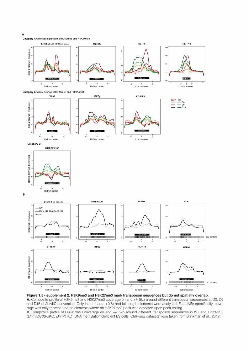
159
CRISPR/Cas9 editing to generate a double-knockout ES cell line for the H3K9
trimethyltransferases, SUV39H1 and SUV39H2 (Suvar39-dKO). Additionally we created an
haploinsufficient mutant for the KAP1 co-repressor (Kap1+/-), which interacts with the H3K9
trimethyltransferase SETDB1 (Figure 1.6 - figure supplement 1A,B); complete KAP1
removal is not compatible with ES cell survival (Rowe et al., 2010). The role of H3K27me3
was studied in mutant ES cells for the EED protein (Eed-KO, Schoeftner et al., 2006), which is
required for H3K27me3 catalysis by the Polycomb Repressive Complex 2 (PRC2)
(Margueron and Reinberg, 2011). Nanostring quantification of transposon transcripts was
performed upon serum to 2i+vitC transfer of these three cell lines, which, importantly, share
the same J1 cell background.
As representatives of transposon A category, young LINE1 elements maintain
H3K9me3 while gaining H3K27me3 during medium conversion. Previous studies concluded
that L1 repression in serum relies on SUV39H-dependent H3K9me3 (Bulut-Karslioglu et al.,
2014): through the analysis of our genetic mutants, we found this was the case for L1-A, and
very modestly for L1-T elements (Figure 1.6A). While L1-A elements appear under exclusive
SUV39H-dependent H3K9me3 control, in contrast, L1-T remained elevated in Eed-KO cells
specifically, indicating that the gain of H3K27me3 plays a role in silencing these elements as
DNA methylation is lost. The category B of transposons is exemplified by IAPEz elements,
which harbor exclusive H3K9me3 enrichment in all culture conditions. Although this profile
would predict a continuous and exclusive dependence towards H3K9me3 upon medium
adaptation, we observed complex behaviors in the different mutants (Figure 1.6B). During
conversion, Kap+/- cells showed enhanced IAPEz up-regulation and repression failure after
D6, in line with a major role of SETDB1-related H3K9me3 for controlling these elements in
ES cells (Matsui et al., 2010). However, SUV39H depletion led to an unexpected IAPEz
suppression upon conversion. One possible explanation is that Suvar39h-dKO cells have
acquired long-term compensatory mechanisms that prevent transient IAPEz activation upon
DNA methylation loss. Moreover, IAPEz elements were more strongly expressed in Eed-KO
compared to WT cells during conversion, which is at odds with their apparent lack of
H3K27me3 enrichment in ChIP-seq data (Figures 1.4E and F). This could be due to
indirect effects of the Eed deficiency and/or from a few H3K27me3-enriched elements, which
we were unable to detect by ChIP-seq.
Finally, the H3K9me3- to H3K27me3-chromatin transition undergone by category C
elements was very clearly illustrated in chromatin modifier mutants (Figure 1.6C). At D0,
H3K9me3-driven silencing of MERVL dominantly relied on SUV39H in serum. While
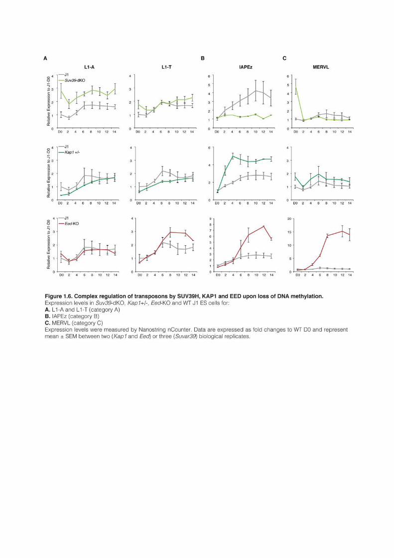
161
H3K9me3 became dispensable upon conversion, the switch towards H3K27me3 control was
perfectly correlated with a 15-fold expression increase we observed in Eed-KO cells. MERVL
represent a striking model of epigenetic switch, which occurs subsequently to DNA
methylation loss.
1.5 Discussion
Our study provides unprecedented insight into the dynamic adaptation of the
pluripotent genome to a loss of DNA methylation-based control of transposons. This was
achieved through detailed kinetic assessment of transcription and chromatin states during
conversion of WT ES cells from serum to 2i+vitC media, as a way to reproduce the DNA
methylation erasure that occurs during embryogenesis. Despite their heterogeneous origins
and structures, we found that various transposon families residing in the mouse genome
adopted a common regulatory fate: after an initial transcriptional burst, repression was re-
established in a DNA methylation-independent manner. Distinct combinations of H3K9me3
and H3K27me3 were observed among transposon families, defining three functional
categories of chromatin-based responses to DNA methylation loss: joint H3K9me3 and
H3K27m3 (A), H3K9me3-exclusive (B), and H3K27me3-exclusive (C) (Figure 1.7).
Importantly, Dnmt-tKO cells, which have endured long-term adaptation to a DNA
methylation-free state, displayed similar transposon-specific chromatin patterns when grown
in serum, which excludes a medium-related effect. In conclusion, our work revises the
previous assumption that DNA methylation is dispensable for transposon silencing in ES cells;
rather, we reveal here that various histone-based repression strategies are implemented upon
DNA methylation loss, thereby safeguarding pluripotent cells against a multitude of
heterogeneous transposon entities.
Upon 2i+vitC-mediated DNA methylation loss, the repertoire of repressive histone
marks of transposons is profoundly remodeled (Figure 1.7A): H3K9me2 enrichment
decreases, while H3K27me3 is enhanced. In contrast, H3K9me3 levels are globally constant:
this highlights that DNA methylation does not exert significant control over H3K9me3-
targeting of transposons in ES cells. Interestingly, we consistently observed that regions of
persistent DNA methylation (RMRs) coincide with high H3K9me3 enrichment on transposon
sequences in fully 2i+vitC-converted cells, e.g. on the 5’ end of LINE1 elements and
throughout the length of ERVK elements. This supports previous evidence that H3K9me3
can confer protection against DNA demethylation (Leung et al., 2014). Inversely, the rapid
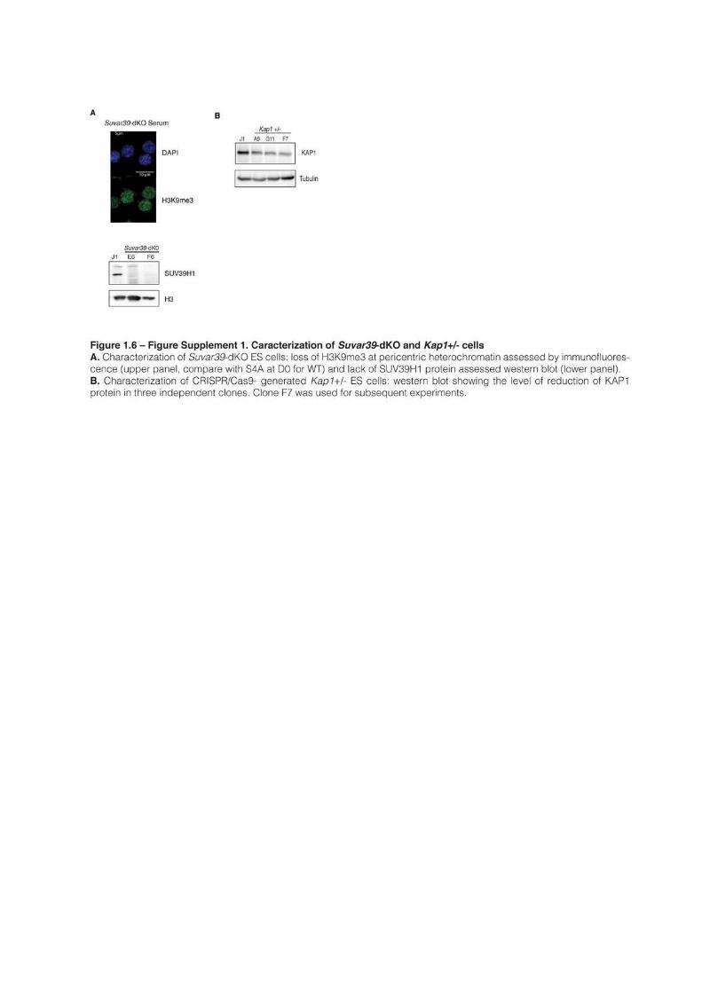
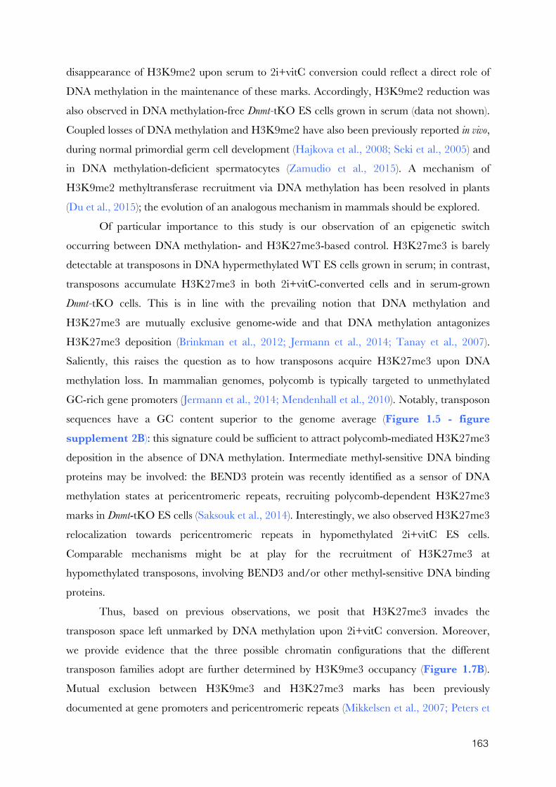
163
disappearance of H3K9me2 upon serum to 2i+vitC conversion could reflect a direct role of
DNA methylation in the maintenance of these marks. Accordingly, H3K9me2 reduction was
also observed in DNA methylation-free Dnmt-tKO ES cells grown in serum (data not shown).
Coupled losses of DNA methylation and H3K9me2 have also been previously reported in vivo,
during normal primordial germ cell development (Hajkova et al., 2008; Seki et al., 2005) and
in DNA methylation-deficient spermatocytes (Zamudio et al., 2015). A mechanism of
H3K9me2 methyltransferase recruitment via DNA methylation has been resolved in plants
(Du et al., 2015); the evolution of an analogous mechanism in mammals should be explored.
Of particular importance to this study is our observation of an epigenetic switch
occurring between DNA methylation- and H3K27me3-based control. H3K27me3 is barely
detectable at transposons in DNA hypermethylated WT ES cells grown in serum; in contrast,
transposons accumulate H3K27me3 in both 2i+vitC-converted cells and in serum-grown
Dnmt-tKO cells. This is in line with the prevailing notion that DNA methylation and
H3K27me3 are mutually exclusive genome-wide and that DNA methylation antagonizes
H3K27me3 deposition (Brinkman et al., 2012; Jermann et al., 2014; Tanay et al., 2007).
Saliently, this raises the question as to how transposons acquire H3K27me3 upon DNA
methylation loss. In mammalian genomes, polycomb is typically targeted to unmethylated
GC-rich gene promoters (Jermann et al., 2014; Mendenhall et al., 2010). Notably, transposon
sequences have a GC content superior to the genome average (Figure 1.5 - figure supplement 2B): this signature could be sufficient to attract polycomb-mediated H3K27me3
deposition in the absence of DNA methylation. Intermediate methyl-sensitive DNA binding
proteins may be involved: the BEND3 protein was recently identified as a sensor of DNA
methylation states at pericentromeric repeats, recruiting polycomb-dependent H3K27me3
marks in Dnmt-tKO ES cells (Saksouk et al., 2014). Interestingly, we also observed H3K27me3
relocalization towards pericentromeric repeats in hypomethylated 2i+vitC ES cells.
Comparable mechanisms might be at play for the recruitment of H3K27me3 at
hypomethylated transposons, involving BEND3 and/or other methyl-sensitive DNA binding
proteins.
Thus, based on previous observations, we posit that H3K27me3 invades the
transposon space left unmarked by DNA methylation upon 2i+vitC conversion. Moreover,
we provide evidence that the three possible chromatin configurations that the different
transposon families adopt are further determined by H3K9me3 occupancy (Figure 1.7B).
Mutual exclusion between H3K9me3 and H3K27me3 marks has been previously
documented at gene promoters and pericentromeric repeats (Mikkelsen et al., 2007; Peters et
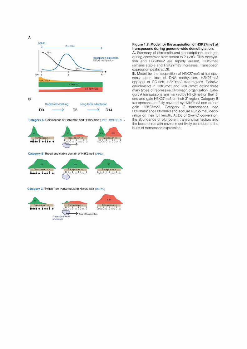
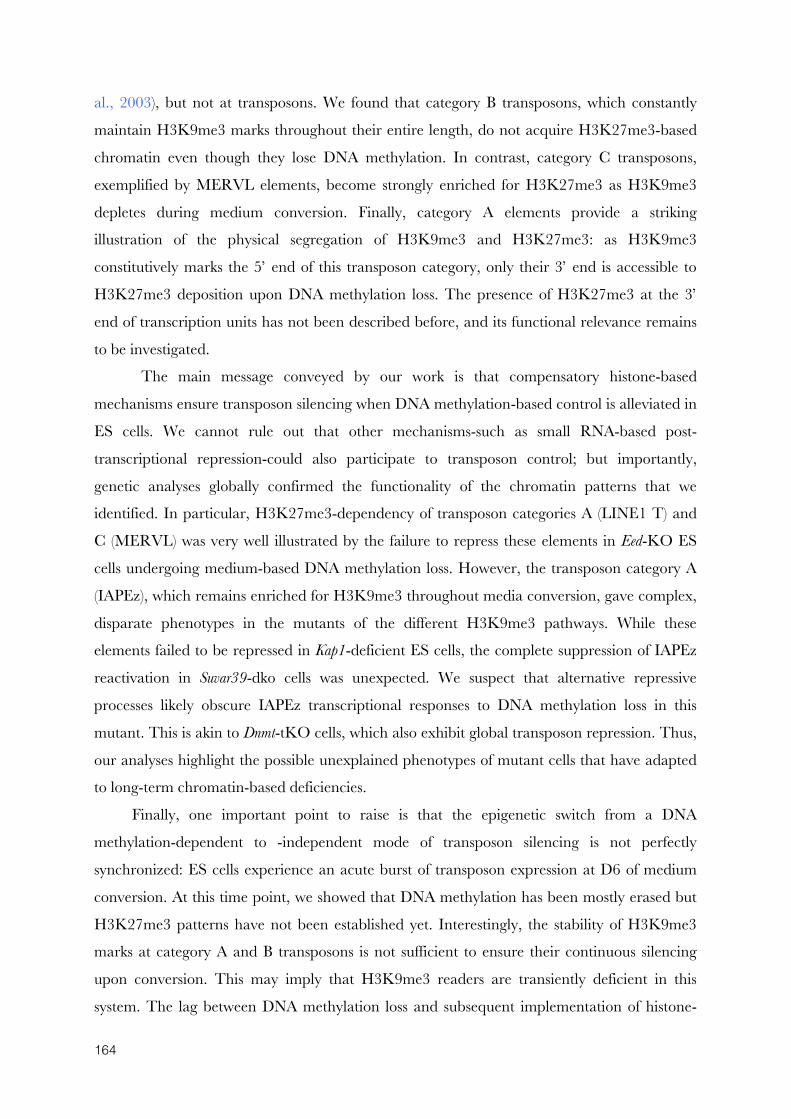
164
al., 2003), but not at transposons. We found that category B transposons, which constantly
maintain H3K9me3 marks throughout their entire length, do not acquire H3K27me3-based
chromatin even though they lose DNA methylation. In contrast, category C transposons,
exemplified by MERVL elements, become strongly enriched for H3K27me3 as H3K9me3
depletes during medium conversion. Finally, category A elements provide a striking
illustration of the physical segregation of H3K9me3 and H3K27me3: as H3K9me3
constitutively marks the 5’ end of this transposon category, only their 3’ end is accessible to
H3K27me3 deposition upon DNA methylation loss. The presence of H3K27me3 at the 3’
end of transcription units has not been described before, and its functional relevance remains
to be investigated.
The main message conveyed by our work is that compensatory histone-based
mechanisms ensure transposon silencing when DNA methylation-based control is alleviated in
ES cells. We cannot rule out that other mechanisms-such as small RNA-based post-
transcriptional repression-could also participate to transposon control; but importantly,
genetic analyses globally confirmed the functionality of the chromatin patterns that we
identified. In particular, H3K27me3-dependency of transposon categories A (LINE1 T) and
C (MERVL) was very well illustrated by the failure to repress these elements in Eed-KO ES
cells undergoing medium-based DNA methylation loss. However, the transposon category A
(IAPEz), which remains enriched for H3K9me3 throughout media conversion, gave complex,
disparate phenotypes in the mutants of the different H3K9me3 pathways. While these
elements failed to be repressed in Kap1-deficient ES cells, the complete suppression of IAPEz
reactivation in Suvar39-dko cells was unexpected. We suspect that alternative repressive
processes likely obscure IAPEz transcriptional responses to DNA methylation loss in this
mutant. This is akin to Dnmt-tKO cells, which also exhibit global transposon repression. Thus,
our analyses highlight the possible unexplained phenotypes of mutant cells that have adapted
to long-term chromatin-based deficiencies.
Finally, one important point to raise is that the epigenetic switch from a DNA
methylation-dependent to -independent mode of transposon silencing is not perfectly
synchronized: ES cells experience an acute burst of transposon expression at D6 of medium
conversion. At this time point, we showed that DNA methylation has been mostly erased but
H3K27me3 patterns have not been established yet. Interestingly, the stability of H3K9me3
marks at category A and B transposons is not sufficient to ensure their continuous silencing
upon conversion. This may imply that H3K9me3 readers are transiently deficient in this
system. The lag between DNA methylation loss and subsequent implementation of histone-

165
based repression could create an opportunistic window for transposon reactivation, provided
that adequate transcription factors are available. Several studies have previously pointed out
that transposons are enriched in pluripotency transcription factor binding motifs (Kunarso et
al., 2010; Wang et al., 2014a), in particular for NANOG and OCT4, and that upregulation of
these transcription factors was sufficient to promote transposon expression (Grow et al., 2015).
We propose that the simultaneous disappearance of DNA methylation marks and
increased availability of pluripotency activators create favorable conditions to transposon
expression at D6 of serum to 2i+vitC conversion (Figure 7B). After a brief silencing release,
functional repressive chromatin is recovered, in an H3K9me3 and/or H3K27me3-dependent
manner. Notably, we repeatedly observed a peak of massive cell death of H3K27me3-
deficient Eed-KO ES cells between D6 and D10 of medium conversion, when DNA
methylation has mostly disappeared (data not shown). This observation supports the critical
role for H3K27me3 in supplementing DNA methylation-based control in ES cells.
1.6 Experimental procedures
ES cell lines
J1 and Dnmt-tKO ES cells were a gift from M. Okano (Tsumura et al., 2006). E14 ES
cells were kindly provided by E. Heard. WT TT2 and G9a-KO ES cells (Tachibana et al.,
2002), and Eed-KO (Schoeftner et al., 2006) (on a J1 background) were gifts from Y. Shinkai
and A. Wutz, respectively. Kap1-/+ and Suvar39-dKO were generated from J1 ES cells using
CRISPR/Cas9 editing. Briefly, guide-RNAs specific to the target sequences were designed
using the online CRISPR Design Tool (Hsu et al., 2013)(Table S1.6) and incorporated into
the X330 backbone (Cong et al., 2013). Five millions J1 ES cells grown in serum were
transfected with 1-3μg of plasmid using Amaxa 4d nucleofector (Lonza) and plated at a low
density. Individual clones were picked and screened by PCR; mutated alleles were confirmed
by Sanger sequencing. Suvar39-dKO cells were obtained by creating a frame-shift in Suvar39h1
exon 4 and by deleting Suvar39h2 exon 4; Kap1+/- cells were generated by deleting exon 3.
ES cell culture
ES cells were grown in two different media, serum and 2i, defined as follow. Serum:
Glasgow medium (Sigma), 15% FBS (Gibco), 2mM L-Glutamine, 0.1mM MEM non essential
amino acids (Gibco), 1mM sodium pyruvate (Gibco), 0.1mM β-mercaptoethanol, 1000U/mL
leukemia inhibitory factor (LIF, Miltenyi); 2i: 50% neurobasal medium (Gibco), 50%
166
DMEM/F12 (Gibco), 2mM L-glutamine (Gibco), 0.1mM β-mercaptoethanol, Ndiff Neuro2
supplement (Milipore), B27 serum-free supplement (Gibco), 1000U/mL LIF, 3μM Gsk3
inhibitor CT-99021, 1μM MEK inhibitor PD0325901. Vitamin C (Sigma) was added at a
concentration of 100ug/mL (Blaschke et al., 2013).
When in serum, J1, Dnmt-tKO, E14, Kap-/+ and Suvar39-dKO ES cells were grown in
feeder-free conditions on gelatin-coated plates. TT2, G9a-KO and Eed-KO were cultured on
a monolayer of mitomycin C-treated mouse embryonic fibroblasts. ES cells were passaged
with TrypLE Express Enzyme (Life Technologies). All 2i ES cells were grown in gelatin-
coated plates and passaged every two or three days with Accutase (Life Technologies).
DNA methylation analyses
Genomic DNA was isolated using the GenElute Mammalian Genomic DNA Miniprep
Kit (Sigma) with RNase treatment. Global CpG methylation levels were assessed using
LUminometric Methylation Assay (LUMA) as described previously (Karimi et al., 2011a;
Richard Pilsner et al., 2010). Briefly, 500ng of genomic DNA was digested with MspI/EcoRI
and HpaII/EcoRI (NEB) in parallel reactions. HpaII is a methylation-sensitive restriction
enzyme and MspI is its methylation insensitive isoschizomer. EcoRI is included as an internal
reference. The overhangs created by the enzymatic digestion were quantified by
Pyrosequencing (PyroMark Q24, Qiagen) with the dispensation order:
GTGTGTCACACAGTGTGT. Global CpG methylation levels were calculated from the
peak heights at the position 7,8,13,14 as follows: 1-sqrt([p8*p14/p7*p13]HpaII
/[p8*p14/p7*p13]MspI)
CpG methylation at specific loci was assessed by bisulfite-pyrosequencing using the
Imprint DNA modification Kit (Sigma) for conversion. PCR and sequencing primers (Table S1.6) were designed with the PyroMark Assay Design Software and quantification of DNA
methylation was performed according to the recommended protocol.
Whole-Genome Bisulfite Sequencing libraries were prepared from 50ng of bisulfite-
converted genomic DNA using the EpiGnome/Truseq DNA Methylation Kit (Illumina)
following the manufacturer instructions. Sequencing was performed in 100pb paired-end
reads at a 30X coverage using the Illumina HiSeq2000 platform (Table S1.1C).
RNA expression analyses
Total RNA was extracted using Trizol (Life Technologies). cDNAs were prepared after
DNase treatment (Turbo DNase, Ambion) using random priming with Superscript III (Life
Technologies). Real-time quantitative PCR was performed using the SYBR Green Master
167
Mix on the Viia7 thermal cycling system (Applied Biosystem). Relative expression levels were
normalized to the arithmetic mean of the housekeeping genes Gapdh and Arp0 and to WT-D0
with the ΔΔCt method. Primers are given in Table S4.
Nanostring nCounter quantification was performed using 100ng of total RNA per
sample on a custom expression Codeset (sequences available upon request). Actin, Ppia, Gapdh
and Arp0 were used for normalization. Data are presented as the fold change compared to
WT-D0. Expression data for the different mutants are presented next to WT data that were
processed on the same Nanostring run. The same WT data can be used in several figures.
When necessary and in order to calculate mean and standard error of the mean between
replicates every two days, we extrapolated linearly the expression value of a given day using
data of immediately adjacent time points (for both RT-qPCR and Nanostring).
RNA-seq libraries were prepared from 500ng of DNase-treated RNA with the TruSeq
Stranded mRNA kit (Illumina). Sequencing was performed in 100pb paired-end reads using
the Illumina HiSeq2000 platform (Table S1.1B).
Chromatin Immunoprecipitation (ChIP)
Cells were cross-linked directly in culture plates with 1% formaldehyde (culture medium
supplemented with 1% formaldehyde, 0.015M NaCl, 0.15mM EDTA, 0.075mM EGTA,
15mM Hepes pH 8). After quenching with 0.125M glycine, cells were washed in PBS and
pelleted. Cells were then incubated at 4°C for 10 min in buffer 1 (Hepes-KOH pH 7.5
50mM, NaCl 140mM, EDTA pH 8.0 1mM, glycerol 10% NP-40 0.5%, Triton X-100 0.25%
and the protease inhibitors: PMSF 1mM, Aprotinin 10μg/ml, leupeptin 1μg/ml and
pepstatin1μg/ml), then at room temperature for 10 min in buffer 2 (NaCl 200mM, EDTA pH
8.0 1mM, EGTA pH 8.0 0.5mM and 10mM Tris pH 8 and the same protease inhibitors as
buffer 1) and finally resuspended in buffer 3 (EDTA pH 8.0 1mM, EGTA pH 8.0 0.5mM,
Tris pH8 10mM, N-lauroyl-sarcosine 0.5%; protease inhibitors as buffer 1). Chromatin was
sonicated with a Bioruptor (Diagenode) to reach a fragment size around 200bp. Chromatin
corresponding to 10μg of DNA was incubated overnight at 4°C with 3-5μg of antibody in
incubation buffer (buffer 3 supplemented with 0.5 volume of 3% Triton, 0.3% sodium
deoxycholate, 15mM EDTA; protease inhibitors). A fraction of chromatin extracts (5%) were
taken aside for inputs. Antibody-bound chromatin was recovered using Protein G Agarose
Columns (Active Motif). Briefly, the antibody-chromatin mix was incubated in the column for
4 hours, washed eight times with modified RIPA buffer (Hepes pH7.6 50mM, EDTA pH 8.0
10mM, sodium deoxycholate 0.7%, NP-40 1%, LiCl 500mM, PMSF 1 mM, 1μg/ml
168
leupeptin and 1μg/ml pepstatin), and washed one last time with TE-NaCl (50mM Tris pH
8.0, 10mM EDTA, 50mM NaCl). Chromatin was eluted with pre-warmed TE-SDS (50mM
Tris pH 8.0, 10mM EDTA, 1% SDS). ChIP-enriched sample and inputs were then reverse
cross-linked at 65°C overnight and treated with RNase A and proteinase K. DNA was
extracted with phenol/chloroform/isoamyl alcohol, precipitated with glycogen in sodium
acetate and ethanol and finally resuspended in TE. Enrichment compared to input was
analyzed by qPCR. A quantity of 20ng of ChIP- or input-DNA were used for ChIP-seq.
Remaining large DNA fragments were first eliminated using SPRIselect beads (Beckman
Coulter) and libraries were prepared using the TruSeq ChIP Sample Prep kit (Illumina).
Sequencing was performed in 50pb paired-end reads using the Illumina HiSeq2000 platform
(Table S1.1C). Antibodies are listed in Table S1.7
Western blotting
To prepare total protein extracts, cells were resuspended in BC250 lysis buffer (25mM
Tris pH 7.9, 0.2mM EDTA, 20% Glycerol, 0.25M KCl and protease inhibitor coktail from
Roche), sonicated and centrifuged to pellet debris. To prepare nuclear protein extracts, cells
were incubated for 10 min on ice in buffer A (Hepes pH 7.9 10mM, MgCl2 5mM, Sucrose
0.25M, NP40 0.1%, DTT 1mM and protease inhibitors) and centrifuged. The pellet was
resuspended in buffer B (Hepes pH 7.9 25mM, glycerol 20%, MgCl2 1.5mM, EDTA 0.1
mM, NaCl 700mM, DTT 1mM and protease inhibitors), sonicated and centrifuged to pellet
debris. Total and nuclear proteins were quantified by Bradford assay. Proteins (10-20μg per
gel lane) were separated by electrophoresis in 8-15% poly-acrylamide gels and transferred
onto nitrocellulose membranes using the Trans-Blot turbo transfer system (Biorad). After
incubation with primary antibodies and HRP-conjugated secondary antibodies, signal was
detected using ECL prime kit (Amersham) and ImageQuant Las-4000 mini biomolecular
Imager. Antibodies are listed in Table S1.7.
Immunofluorescence
Cells were harvested with Trypsin or Accutase, resuspended in PBS and plated for
10min on Poly-L-Lysine-coated glass cover slips. Cells were first fixed with 3%
paraformaldehyde for 10 min at room temperature, then rinsed three times with PBS and
permeabilized for 4 min with 0.5X Triton on ice. After blocking in 1% BSA for 15 min,
samples were incubated at room temperature for 40 min with primary antibodies, 45 min
with secondary antibodies and 3 min in 0.3μg/mL DAPI. Slides were mounted with Prolong
169
Gold mounting media (Invitrogen). Images were obtained with an Upright Widefield
microscope (Leica) or a Zeiss LSM700 inverted confocal microscope. Quantification of
immunofluorescence intensity in individual cells was performed using custom ImageJ and R
scripts. Between 1,000 and 5,000 cells were analyzed per sample.
Metaphase spreading
Cells were cultured for two hours with 0.04μg/mL colchicine and harvested by
trypsinization. Cell pellets were incubated in hypotonic buffer (15% FBS in water) for 7 min at
37°C and fixed with 66% acetic acid/33% ethanol. After centrifugation, cells were
resuspended in 1.5mL fixative and dropped from ~1m height onto glass slides. Slides were
dried and DNA was stained with DAPI. Chromosomes were counted with an Upright
Widefield microscope (Leica). Around 20 cells were analyzed per cell line.
Quantification of transposon genomic copy number
Absolute copy numbers of IAP and LINE1 were calculated by qPCR by establishing
standard curves plotting absolute Ct values of genomic DNA against serial dilutions of PCR
targets cloned into the pCR2.1-TOPO vector (Life Technologies), as described in Zamudio et
al., 2015.
Reconstruction of RepeatMasker
As described in Bailly-Bechet et al., 2014, a dictionary was constructed for LTR
retrotransposons that associated elements corresponding to the internal sequence and those
corresponding to LTR sequences. With the latter and the RepeatMasker database, fragments
of transposable elements corresponding to the same copy were merged. Divergence, deletion
and insertion percentages were recalculated from RepeatMasker and an integrity score for
each transposon were calculated as follow: score = 1-average(%divergence, %deletions,
%insertions)
WGBS data analysis
Whole-genome bisulfite sequencing reads generated in this study or recovered from
available datasets were treated as follow. The first eight base pairs of the reads were trimmed
using FASTX-Toolkit v0.0.13 (http://hannonlab.cshl.edu/fastx_toolkit/index.html). Adapter
sequences were removed with Cutadapt v1.3 (https://code.google.com/p/cutadapt/) and reads
shorter than 16bp were discarded. Cleaned sequences were aligned onto the Mouse reference
genome (mm10) using Bismark v0.12.5 (Krueger and Andrews, 2011) with Bowtie2-2.1.0
(Langmead and Salzberg, 2012) and default parameters. Only reads mapping uniquely on the
170
genome were conserved. Methylation calls were extracted after duplicate removal. Only CG
dinucleotides covered by a minimum of 10 reads were conserved for the rest of the analysis.
The R-package Methylkit v0.9.2 (Akalin et al., 2012) was used to provide Pearson’s
correlation scores between samples. To analyze the distribution of CpG methylation in
different genomic compartments, the mouse genome was divided into different partitions. The
RefSeq gene annotation and the RepeatMasker database were downloaded from UCSC table
browser and used for transcript and repeat annotations, respectively. Promoters were defined
as the -1kb to +100pb region around transcription start sites. CpG islands (CGIs) were
defined as in Illingworth et al., 2010. Intergenic partitions were defined as genomic regions
that did not overlap with promoters, CGI, exons, introns or repeats. Whole-genome mapping
of CpG methylation was then intersected with the different genomic compartments using
Bedtools (Quinlan and Hall, 2010).
Average CpG methylation on individual transposons was extracted from
RepeatMasker with Bedtools, average CpG methylation in the different transposon families
was calculated and plotted using R. Heatmap for average CpG methylation in Imprinted
control regions (ICRs) was generated similarly after retrieving ICR genomic coordinates from
the WAMIDEX database (Schulz et al., 2008). Residually methylated regions (RMRs) in
2i+vitC samples were identified using the MethPipe pipeline (Song et al., 2013) with default
parameters. RMRs located less than 1kb from each others were concatenated.
RNA-seq data analysis
In order to quantify gene expression, Paired-end 2x100bp reads were mapped onto
mm10 using Tophat v2.0.6 and RefSeq gene annotation (Kim et al., 2013) allowing five
mismatches. Gene-scaled quantification was performed with HTSeq v0.6.1 (Anders et al.,
2014).
In order to quantify transposon expression, reads mapping to ribosomal RNA (rRNA)
sequences (GenBank identifiers: 18S NR_003278.3, 28S NR_003279.1, 5S D14832.1, 5.8S
K01367.1) were first removed with Bowtie v1.0.0 allowing three mismatches. The rRNA-
depleted libraries were then mapped onto mm10 using Bowtie v1.0.0 allowing zero mismatch
and 10,000 best alignments per read. Exonic reads were removed. In order to count reads
mapping to transposable elements, reads were weighted by the number of mapping sites and
each library was intersected with the reconstructed RepeatMasker annotation, conserving
only reads overlapping at least at 80% with a given transposon.
171
For each library, read counts for genes and transposons were combined into a single
table. TMM normalization from the edgeR package v3.6.2 (Robinson and Oshlack, 2010)
was first applied. As described in the guideline of limma R-package v3.20.4, normalized
counts were processed by the voom method (Law et al., 2014) to convert them into log2
counts per million with associated precision weights. The differential expression was estimated
with the limma package. Genes and transposons were called differentially expressed when two
criteria were met: 1) the fold-change between two conditions was higher than four and two,
respectively, and 2) the adjusted p-value using the Benjamini Hochberg procedure was below
0.05.
For the analysis of RNA-seq libraries with uniquely mapped reads, the mapping was
performed as previously with Bowtie v1.0.0, except that only uniquely mapping reads were
conserved. Read counts on individual reconstructed element were quantified using HTSeq
v0.6.1. Only elements with at least 10 reads in at least one sample were conserved for further
analysis and read counts were subsequently normalized by the library size. Normalized read
counts for individual elements belonging to different families were then plotted using custom
R script. Tracks were created using HOMER software v4.7 (Heinz et al., 2010).
In order to identify and characterize chimeric transcripts, reads were mapped onto
mm10 using Tophat v2.0.6, without providing a gene annotation. Cufflinks v2.2.1 (Trapnell
et al., 2010) was used to reconstruct the transcriptome and quantify the different isoforms.
Transcripts were considered chimeric when the first exon overlapped with a transposon
annotated in Repeatmasker and one of the other exon was annotated in RefSeq.
ChIP-seq data analysis
Paired-end 2x50bp reads were mapped onto mm10 using Bowtie v1.0.0 allowing 3
mismatches. Reads mapping to multiple locations were randomly allocated. Duplicate reads
were removed using Picard v1.65 (http://broadinstitute.github.io/picard/). Tracks were created
using HOMER software v4.7 (Heinz et al., 2010) and Peak calling was performed with
MACS2 v2.0.10 (Zhang et al., 2008b) using the broad option and a 5% FDR threshold.
Detected peaks were annotated using RefSeq and RepeatMasker databases. In order to
construct the heatmap and the scatter plots, the total number of read counts for every
annotated transposable element was computed using Bedtools and the reconstructed
RepeatMasker annotation. Enrichment was normalized by the size of the element and Input
data. Metaplots for average enrichment and GC content on and around different transposons
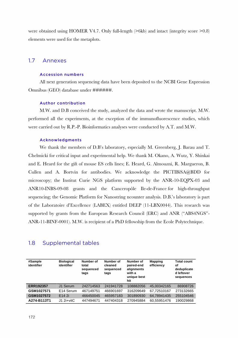
172
were obtained using HOMER V4.7. Only full-length (>6kb) and intact (integrity score >0.8)
elements were used for the metaplots.
1.7 Annexes
Accession numbers
All next generation sequencing data have been deposited to the NCBI Gene Expression
Omnibus (GEO) database under ######.
Author contribution
M.W. and D.B conceived the study, analyzed the data and wrote the manuscript. M.W.
performed all the experiments, at the exception of the immunofluorescence studies, which
were carried out by R.P.-P. Bioinformatics analyses were conducted by A.T. and M.W.
Acknowledgments
We thank the members of D.B’s laboratory, especially M. Greenberg, J. Barau and T.
Chelmicki for critical input and experimental help. We thank M. Okano, A. Wutz, Y. Shinkai
and E. Heard for the gift of mouse ES cells lines; E. Heard, G. Almouzni, R. Margueron, B.
Cullen and A. Bortvin for antibodies. We acknowledge the PICTIBiSA@BDD for
microscopy; the Institut Curie NGS platform supported by the ANR-10-EQPX-03 and
ANR10-INBS-09-08 grants and the Canceropôle Ile-de-France for high-throughput
sequencing; the Genomic Platform for Nanostring ncounter analysis. D.B.’s laboratory is part
of the Laboratoire d’Excellence (LABEX) entitled DEEP (11-LBX0044). This research was
supported by grants from the European Research Council (ERC) and ANR (“ABS4NGS”-
ANR-11-BINF-0001). M.W. is recipient of a PhD fellowship from the Ecole Polytechnique.
1.8 Supplemental tables
#Sample identifier
Biological identifier
Number of total sequenced tags
Number of cleaned sequenced tags
Number of paired-end alignments with a unique best hit
Mapping efficiency
Total count of deduplicated leftover sequences
ERR192357 J1 Serum 242714563 241941728 108882056 45,00342165 86908726 GSM1027571 E14 Serum 467149751 466901697 316209649 67,72510167 273132665 GSM1027572 E14 2i 466450045 465957183 301890930 64,78941435 255104546 A274-B113T1 J1 2i+vitC 447494671 447404318 270945884 60,55951476 190029868
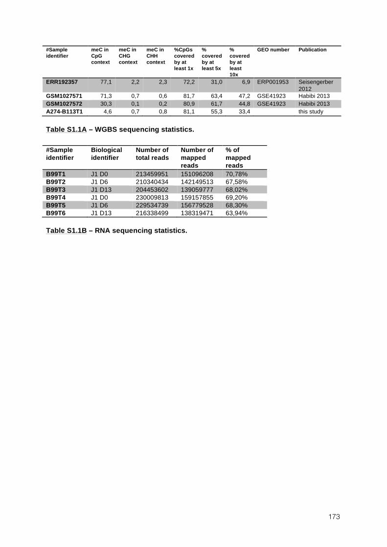
173
#Sample identifier
meC in CpG context
meC in CHG context
meC in CHH context
%CpGs covered by at least 1x
% covered by at least 5x
% covered by at least 10x
GEO number Publication
ERR192357 77,1 2,2 2,3 72,2 31,0 6,9 ERP001953 Seisengerber 2012
GSM1027571 71,3 0,7 0,6 81,7 63,4 47,2 GSE41923 Habibi 2013 GSM1027572 30,3 0,1 0,2 80,9 61,7 44,8 GSE41923 Habibi 2013 A274-B113T1 4,6 0,7 0,8 81,1 55,3 33,4 this study
Table S1.1A – WGBS sequencing statistics.
#Sample identifier
Biological identifier
Number of total reads
Number of mapped reads
% of mapped reads
B99T1 J1 D0 213459951 151096208 70,78% B99T2 J1 D6 210340434 142149513 67,58% B99T3 J1 D13 204453602 139059777 68,02% B99T4 J1 D0 230009813 159157855 69,20% B99T5 J1 D6 229534739 156779528 68,30% B99T6 J1 D13 216338499 138319471 63,94%
Table S1.1B – RNA sequencing statistics.
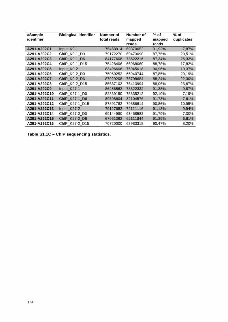
174
#Sample identifier
Biological identifier Number of total reads
Number of mapped reads
% of mapped reads
% of duplicates
A291-A292C1 Input_K9-1 75468914 69370052 91,92% 7,87% A291-A292C2 ChIP_K9-1_D0 79172270 69473090 87,75% 20,51% A291-A292C3 ChIP_K9-1_D6 84177608 73522216 87,34% 26,32% A291-A292C4 ChIP_K9-1_D15 75428406 66968060 88,78% 17,82% A291-A292C5 Input_K9-2 83496606 75945018 90,96% 10,37% A291-A292C6 ChIP_K9-2_D0 75060252 65940744 87,85% 20,19% A291-A292C7 ChIP_K9-2_D6 87029208 76798684 88,24% 22,30% A291-A292C8 ChIP_K9-2_D15 85637102 75413994 88,06% 23,67% A291-A292C9 Input_K27-1 86256562 78822332 91,38% 9,87% A291-A292C10 ChIP_K27-1_D0 82339150 75835212 92,10% 7,19% A291-A292C11 ChIP_K27-1_D6 89508604 82104576 91,73% 7,61% A291-A292C12 ChIP_K27-1_D15 87891782 79856614 90,86% 10,95% A291-A292C13 Input_K27-2 79127682 72111116 91,13% 9,94% A291-A292C14 ChIP_K27-2_D0 69144980 63468582 91,79% 7,30% A291-A292C15 ChIP_K27-2_D6 67961062 62111844 91,39% 6,61% A291-A292C16 ChIP_K27-2_D15 70720000 63983318 90,47% 8,20%
Table S1.1C – ChIP sequencing statistics.
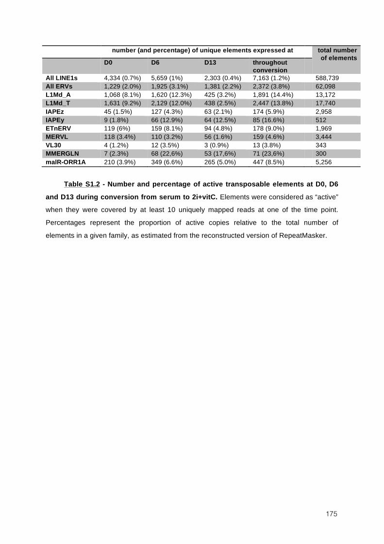
175
number (and percentage) of unique elements expressed at total number of elements D0 D6 D13 throughout
conversion
All LINE1s 4,334 (0.7%) 5,659 (1%) 2,303 (0.4%) 7,163 (1.2%) 588,739 All ERVs 1,229 (2.0%) 1,925 (3.1%) 1,381 (2.2%) 2,372 (3.8%) 62,098 L1Md_A 1,068 (8.1%) 1,620 (12.3%) 425 (3.2%) 1,891 (14.4%) 13,172 L1Md_T 1,631 (9.2%) 2,129 (12.0%) 438 (2.5%) 2,447 (13.8%) 17,740 IAPEz 45 (1.5%) 127 (4.3%) 63 (2.1%) 174 (5.9%) 2,958 IAPEy 9 (1.8%) 66 (12.9%) 64 (12.5%) 85 (16.6%) 512 ETnERV 119 (6%) 159 (8.1%) 94 (4.8%) 178 (9.0%) 1,969 MERVL 118 (3.4%) 110 (3.2%) 56 (1.6%) 159 (4.6%) 3,444 VL30 4 (1.2%) 12 (3.5%) 3 (0.9%) 13 (3.8%) 343 MMERGLN 7 (2.3%) 68 (22,6%) 53 (17,6%) 71 (23,6%) 300 malR-ORR1A 210 (3.9%) 349 (6.6%) 265 (5.0%) 447 (8.5%) 5,256
Table S1.2 - Number and percentage of active transposable elements at D0, D6
and D13 during conversion from serum to 2i+vitC. Elements were considered as “active” when they were covered by at least 10 uniquely mapped reads at one of the time point.
Percentages represent the proportion of active copies relative to the total number of
elements in a given family, as estimated from the reconstructed version of RepeatMasker.
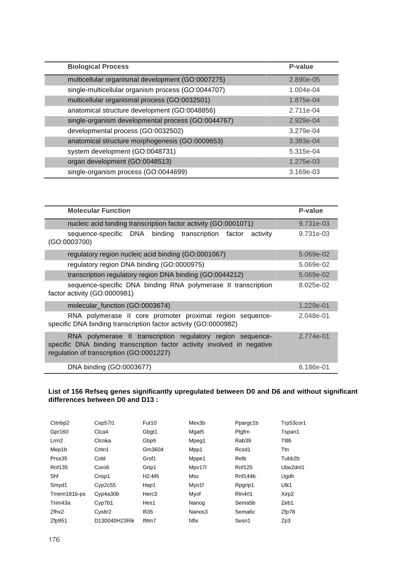
176
Biological Process P-value
multicellular organismal development (GO:0007275) 2.890e-05 single-multicellular organism process (GO:0044707) 1.004e-04 multicellular organismal process (GO:0032501) 1.875e-04 anatomical structure development (GO:0048856) 2.711e-04 single-organism developmental process (GO:0044767) 2.929e-04 developmental process (GO:0032502) 3.279e-04 anatomical structure morphogenesis (GO:0009653) 3.383e-04 system development (GO:0048731) 5.315e-04 organ development (GO:0048513) 1.275e-03 single-organism process (GO:0044699) 3.169e-03
Molecular Function P-value
nucleic acid binding transcription factor activity (GO:0001071) 9.731e-03 sequence-specific DNA binding transcription factor activity
(GO:0003700) 9.731e-03
regulatory region nucleic acid binding (GO:0001067) 5.069e-02 regulatory region DNA binding (GO:0000975) 5.069e-02 transcription regulatory region DNA binding (GO:0044212) 5.069e-02 sequence-specific DNA binding RNA polymerase II transcription
factor activity (GO:0000981) 8.025e-02
molecular_function (GO:0003674) 1.229e-01 RNA polymerase II core promoter proximal region sequence-
specific DNA binding transcription factor activity (GO:0000982) 2.048e-01
RNA polymerase II transcription regulatory region sequence-specific DNA binding transcription factor activity involved in negative regulation of transcription (GO:0001227)
2.774e-01
DNA binding (GO:0003677) 6.186e-01
List of 156 Refseq genes significantly upregulated between D0 and D6 and without significant differences between D0 and D13 :
Cttnbp2 Cep57l1 Fut10 Mex3b Ppargc1b Trp53cor1 Gpr160 Clca4 Gbgt1 Mgat5 Ptgfrn Tspan1 Lrrn2 Clcnka Gbp9 Mpeg1 Rab39 Ttll6 Mep1b Cntn1 Gm3604 Mpp1 Rcsd1 Ttn Prss35 Cobl Grsf1 Mppe1 Relb Tubb2b Rnf135 Coro6 Grtp1 Mpv17l Rnf125 Ube2dnl1 Shf Crisp1 H2-M5 Msc Rnf144b Ugdh Smyd1 Cyp2c55 Hap1 Myo1f Rpgrip1 Ulk1 Tmem181b-ps Cyp4a30b Herc3 Myof Rtn4rl1 Xirp2 Trim43a Cyp7b1 Hes1 Nanog Sema5b Zeb1 Zfhx2 Cysltr2 Ifi35 Nanos3 Sema6c Zfp78 Zfp951 D130040H23Rik Ifitm7 Nfix Sesn1 Zp3
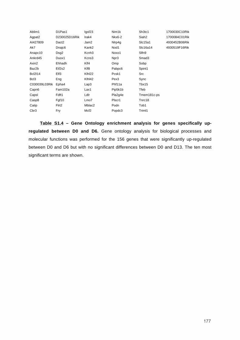
177
Ablim1 D1Pas1 Igsf23 Nim1k Sh3tc1 1700030C10Rik Agpat2 D230025D16Rik Irak4 Nkx6-2 Siah2 1700084C01Rik AI427809 Dact2 Jam2 Nlrp4g Slc15a1 4930452B06Rik Ak7 Dnajc6 Kank2 Nod1 Slc16a14 4930519F16Rik Anapc10 Dsg2 Kcnh3 Noxo1 Slfn9 Ankrd45 Duox1 Kcns3 Npr3 Smad3 Axin2 Ehhadh Klf4 Omp Sobp Baz2b Eif2s2 Klf8 Pabpc6 Spint1 Bcl2l14 Elf3 Klhl22 Pcsk1 Src Bcl3 Eng Klhl42 Pex3 Sync C030039L03Rik Epha4 Lap3 Phf11a Tbx15 Capn6 Fam102a Lax1 Pip5k1b Tfeb Capsl Fdft1 Ldlr Pla2g4e Tmem181c-ps Casp8 Fgf10 Lmo7 Plscr1 Tnrc18 Catip Flrt2 Mblac2 Podn Tob1 Cbr3 Fry Mcf2 Popdc3 Triml1
Table S1.4 – Gene Ontology enrichment analysis for genes specifically up-
regulated between D0 and D6. Gene ontology analysis for biological processes and
molecular functions was performed for the 156 genes that were significantly up-regulated between D0 and D6 but with no significant differences between D0 and D13. The ten most
significant terms are shown.
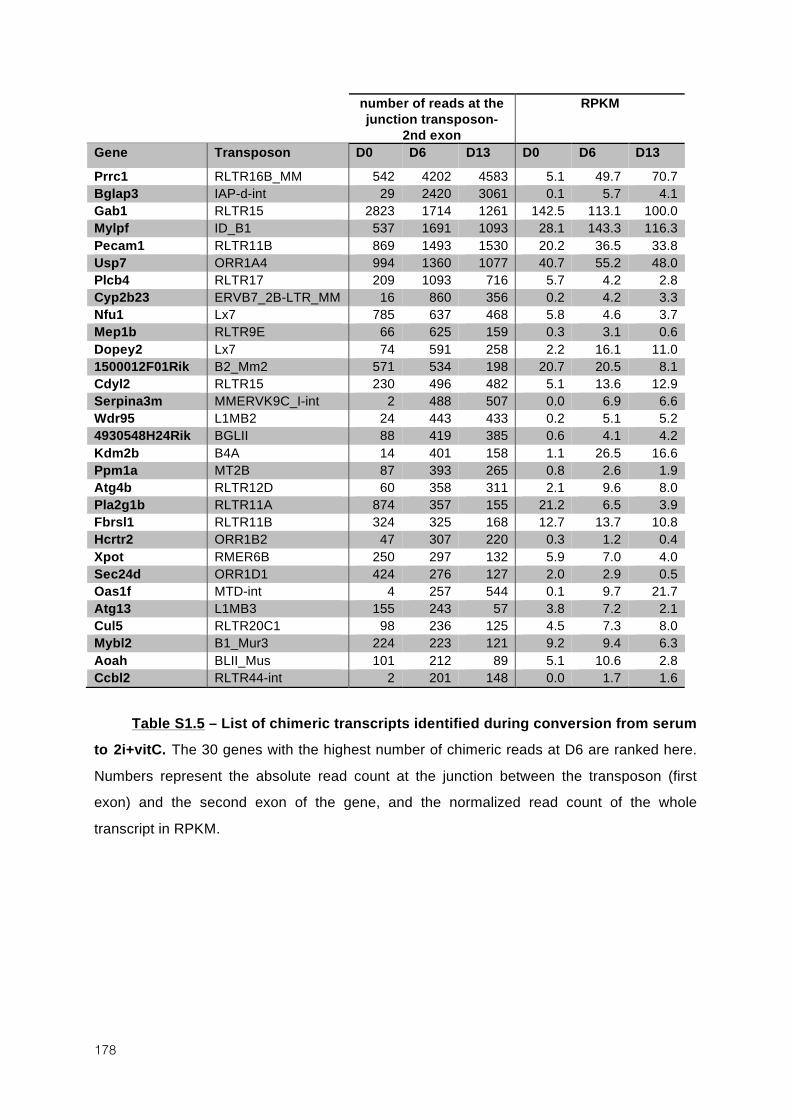
178
number of reads at the junction transposon-
2nd exon
RPKM
Gene Transposon D0 D6 D13 D0 D6 D13 Prrc1 RLTR16B_MM 542 4202 4583 5.1 49.7 70.7 Bglap3 IAP-d-int 29 2420 3061 0.1 5.7 4.1 Gab1 RLTR15 2823 1714 1261 142.5 113.1 100.0 Mylpf ID_B1 537 1691 1093 28.1 143.3 116.3 Pecam1 RLTR11B 869 1493 1530 20.2 36.5 33.8 Usp7 ORR1A4 994 1360 1077 40.7 55.2 48.0 Plcb4 RLTR17 209 1093 716 5.7 4.2 2.8 Cyp2b23 ERVB7_2B-LTR_MM 16 860 356 0.2 4.2 3.3 Nfu1 Lx7 785 637 468 5.8 4.6 3.7 Mep1b RLTR9E 66 625 159 0.3 3.1 0.6 Dopey2 Lx7 74 591 258 2.2 16.1 11.0 1500012F01Rik B2_Mm2 571 534 198 20.7 20.5 8.1 Cdyl2 RLTR15 230 496 482 5.1 13.6 12.9 Serpina3m MMERVK9C_I-int 2 488 507 0.0 6.9 6.6 Wdr95 L1MB2 24 443 433 0.2 5.1 5.2 4930548H24Rik BGLII 88 419 385 0.6 4.1 4.2 Kdm2b B4A 14 401 158 1.1 26.5 16.6 Ppm1a MT2B 87 393 265 0.8 2.6 1.9 Atg4b RLTR12D 60 358 311 2.1 9.6 8.0 Pla2g1b RLTR11A 874 357 155 21.2 6.5 3.9 Fbrsl1 RLTR11B 324 325 168 12.7 13.7 10.8 Hcrtr2 ORR1B2 47 307 220 0.3 1.2 0.4 Xpot RMER6B 250 297 132 5.9 7.0 4.0 Sec24d ORR1D1 424 276 127 2.0 2.9 0.5 Oas1f MTD-int 4 257 544 0.1 9.7 21.7 Atg13 L1MB3 155 243 57 3.8 7.2 2.1 Cul5 RLTR20C1 98 236 125 4.5 7.3 8.0 Mybl2 B1_Mur3 224 223 121 9.2 9.4 6.3 Aoah BLII_Mus 101 212 89 5.1 10.6 2.8 Ccbl2 RLTR44-int 2 201 148 0.0 1.7 1.6
Table S1.5 – List of chimeric transcripts identified during conversion from serum to 2i+vitC. The 30 genes with the highest number of chimeric reads at D6 are ranked here.
Numbers represent the absolute read count at the junction between the transposon (first exon) and the second exon of the gene, and the normalized read count of the whole
transcript in RPKM.
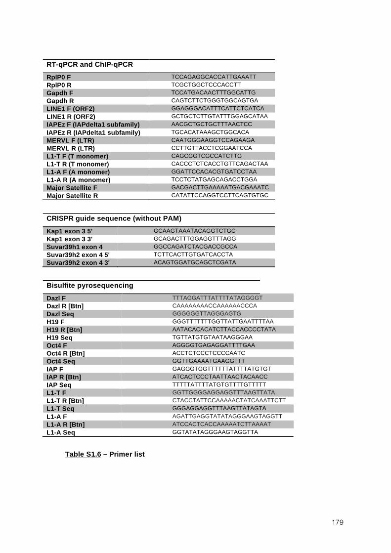
179
RT-qPCR and ChIP-qPCR RplP0 F TCCAGAGGCACCATTGAAATT RplP0 R TCGCTGGCTCCCACCTT Gapdh F TCCATGACAACTTTGGCATTG Gapdh R CAGTCTTCTGGGTGGCAGTGA LINE1 F (ORF2) GGAGGGACATTTCATTCTCATCA LINE1 R (ORF2) GCTGCTCTTGTATTTGGAGCATAA IAPEz F (IAPdelta1 subfamily) AACGCTGCTGCTTTAACTCC IAPEz R (IAPdelta1 subfamily) TGCACATAAAGCTGGCACA MERVL F (LTR) CAATGGGAAGGTCCAGAAGA MERVL R (LTR) CCTTGTTACCTCGGAATCCA L1-T F (T monomer) CAGCGGTCGCCATCTTG L1-T R (T monomer) CACCCTCTCACCTGTTCAGACTAA L1-A F (A monomer) GGATTCCACACGTGATCCTAA L1-A R (A monomer) TCCTCTATGAGCAGACCTGGA Major Satellite F GACGACTTGAAAAATGACGAAATC Major Satellite R CATATTCCAGGTCCTTCAGTGTGC
CRISPR guide sequence (without PAM) Kap1 exon 3 5' GCAAGTAAATACAGGTCTGC Kap1 exon 3 3' GCAGACTTTGGAGGTTTAGG Suvar39h1 exon 4 GGCCAGATCTACGACCGCCA Suvar39h2 exon 4 5' TCTTCACTTGTGATCACCTA Suvar39h2 exon 4 3' ACAGTGGATGCAGCTCGATA
Bisulfite pyrosequencing Dazl F TTTAGGATTTATTTTATAGGGGT Dazl R [Btn] CAAAAAAAACCAAAAAACCCA Dazl Seq GGGGGGTTAGGGAGTG H19 F GGGTTTTTTTGGTTATTGAATTTTAA H19 R [Btn] AATACACACATCTTACCACCCCTATA H19 Seq TGTTATGTGTAATAAGGGAA Oct4 F AGGGGTGAGAGGATTTTGAA Oct4 R [Btn] ACCTCTCCCTCCCCAATC Oct4 Seq GGTTGAAAATGAAGGTTT IAP F GAGGGTGGTTTTTTATTTTATGTGT IAP R [Btn] ATCACTCCCTAATTAACTACAACC IAP Seq TTTTTATTTTATGTGTTTTGTTTTT L1-T F GGTTGGGGAGGAGGTTTAAGTTATA L1-T R [Btn] CTACCTATTCCAAAAACTATCAAATTCTT L1-T Seq GGGAGGAGGTTTAAGTTATAGTA L1-A F AGATTGAGGTATATAGGGAAGTAGGTT L1-A R [Btn] ATCCACTCACCAAAAATCTTAAAAT L1-A Seq GGTATATAGGGAAGTAGGTTA
Table S1.6 – Primer list
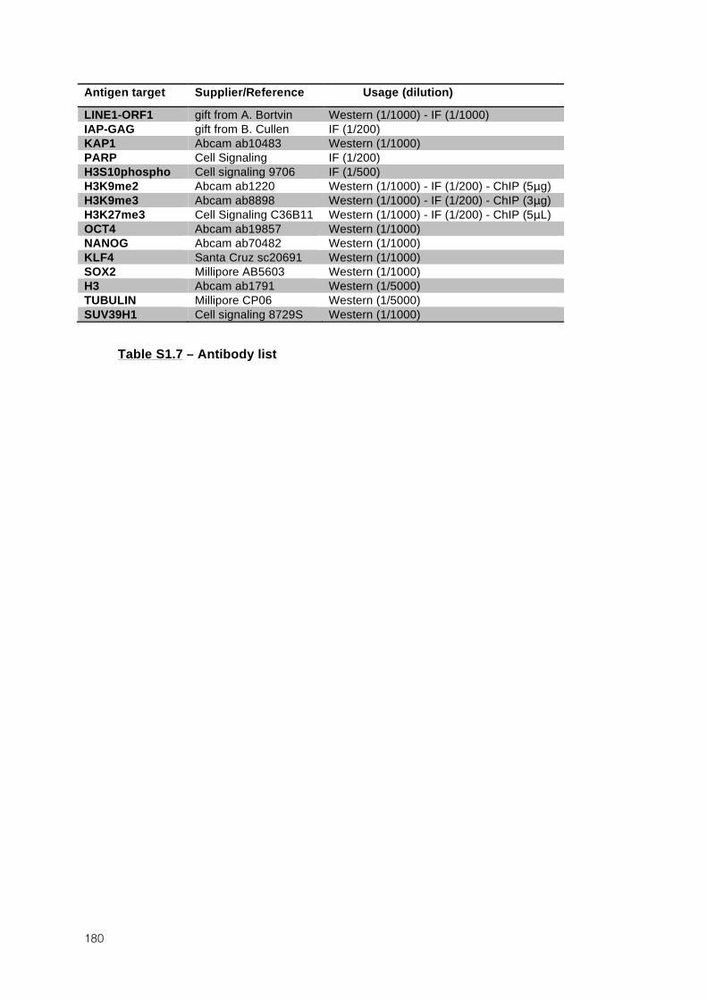
180
Antigen target Supplier/Reference Usage (dilution)
LINE1-ORF1 gift from A. Bortvin Western (1/1000) - IF (1/1000) IAP-GAG gift from B. Cullen IF (1/200) KAP1 Abcam ab10483 Western (1/1000) PARP Cell Signaling IF (1/200) H3S10phospho Cell signaling 9706 IF (1/500) H3K9me2 Abcam ab1220 Western (1/1000) - IF (1/200) - ChIP (5μg) H3K9me3 Abcam ab8898 Western (1/1000) - IF (1/200) - ChIP (3μg) H3K27me3 Cell Signaling C36B11 Western (1/1000) - IF (1/200) - ChIP (5μL) OCT4 Abcam ab19857 Western (1/1000) NANOG Abcam ab70482 Western (1/1000) KLF4 Santa Cruz sc20691 Western (1/1000) SOX2 Millipore AB5603 Western (1/1000) H3 Abcam ab1791 Western (1/5000) TUBULIN Millipore CP06 Western (1/5000) SUV39H1 Cell signaling 8729S Western (1/1000)
Table S1.7 – Antibody list

182
183
2 Various requirements for DNMT3L during events of genome-wide de novo methylation
2.1 Authors and affiliations
Marius Walter1,2, Aurélie Teissandier1,2,3, Julian Iranzo1, Sebastien A Smallwood4, William
A Pastor5, Steven E Jacobsen5, Gavin Kelsey4, Déborah Bourc’his1*
1 Institut Curie, Dpt of Genetics and Developmental Biology, CNRS UMR3215, INSERM U934, Paris,
France 2 UPMC, University Paris 06, Paris, France 3 Institut Curie, Inserm U900, Mines Paris Tech, Paris, France 4 Babraham Institute, Cambridge, UK 5 Department of Molecular, Cell and Developmental Biology, University of California, Los Angeles,
Los Angeles, United States
2.2 Introduction19
In eukaryotes, chromatin plays a fundamental role in the establishment and
maintenance of cell fate, allowing tight control of transcriptional programs in a tissue-specific
and developmentally regulated manner. Nucleosome organization, histone post-translational
modifications, histone variants and many other players contribute to the specification of
multiple functional chromatin states. Methylation of cytosines at the fifth position of the six-
atoms aromatic ring, or DNA methylation, is a widespread DNA modification found in the
majority of eukaryote kingdoms (Zemach et al., 2010). In mammals, DNA methylation is
essentially found in CpG dinucleotides, and the symmetry of this sequence context allows for
the faithful transmission of DNA methylation patterns during cell divisions. In mammals,
DNA methylation is critical for many important biological processes such as genomic
imprinting, X-chromosome inactivation in females, transposon repression and regulation of
gene expression (Smith and Meissner, 2013). In particular, aberrant DNA methylation
patterns is a hallmark of cancer and many human diseases are linked to imprinting defects
(Peters, 2014; You and Jones, 2012).
19 Some paragraphs of this introduction are almost directly taken from the main introduction of this thesis
manuscript.
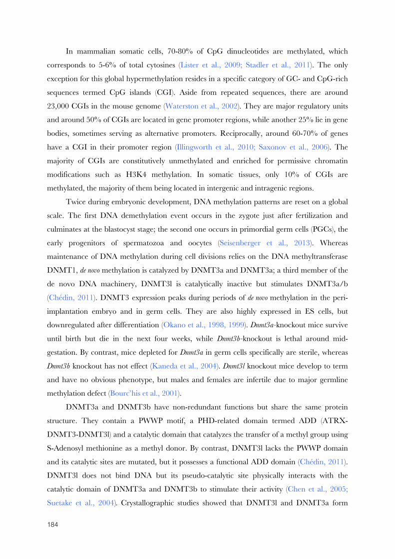
184
In mammalian somatic cells, 70-80% of CpG dinucleotides are methylated, which
corresponds to 5-6% of total cytosines (Lister et al., 2009; Stadler et al., 2011). The only
exception for this global hypermethylation resides in a specific category of GC- and CpG-rich
sequences termed CpG islands (CGI). Aside from repeated sequences, there are around
23,000 CGIs in the mouse genome (Waterston et al., 2002). They are major regulatory units
and around 50% of CGIs are located in gene promoter regions, while another 25% lie in gene
bodies, sometimes serving as alternative promoters. Reciprocally, around 60-70% of genes
have a CGI in their promoter region (Illingworth et al., 2010; Saxonov et al., 2006). The
majority of CGIs are constitutively unmethylated and enriched for permissive chromatin
modifications such as H3K4 methylation. In somatic tissues, only 10% of CGIs are
methylated, the majority of them being located in intergenic and intragenic regions.
Twice during embryonic development, DNA methylation patterns are reset on a global
scale. The first DNA demethylation event occurs in the zygote just after fertilization and
culminates at the blastocyst stage; the second one occurs in primordial germ cells (PGCs), the
early progenitors of spermatozoa and oocytes (Seisenberger et al., 2013). Whereas
maintenance of DNA methylation during cell divisions relies on the DNA methyltransferase
DNMT1, de novo methylation is catalyzed by DNMT3a and DNMT3a; a third member of the
de novo DNA machinery, DNMT3l is catalytically inactive but stimulates DNMT3a/b
(Chédin, 2011). DNMT3 expression peaks during periods of de novo methylation in the peri-
implantation embryo and in germ cells. They are also highly expressed in ES cells, but
downregulated after differentiation (Okano et al., 1998, 1999). Dnmt3a-knockout mice survive
until birth but die in the next four weeks, while Dnmt3b-knockout is lethal around mid-
gestation. By contrast, mice depleted for Dnmt3a in germ cells specifically are sterile, whereas
Dnmt3b knockout has not effect (Kaneda et al., 2004). Dnmt3l knockout mice develop to term
and have no obvious phenotype, but males and females are infertile due to major germline
methylation defect (Bourc’his et al., 2001).
DNMT3a and DNMT3b have non-redundant functions but share the same protein
structure. They contain a PWWP motif, a PHD-related domain termed ADD (ATRX-
DNMT3-DNMT3l) and a catalytic domain that catalyzes the transfer of a methyl group using
S-Adenosyl methionine as a methyl donor. By contrast, DNMT3l lacks the PWWP domain
and its catalytic sites are mutated, but it possesses a functional ADD domain (Chédin, 2011).
DNMT3l does not bind DNA but its pseudo-catalytic site physically interacts with the
catalytic domain of DNMT3a and DNMT3b to stimulate their activity (Chen et al., 2005;
Suetake et al., 2004). Crystallographic studies showed that DNMT3l and DNMT3a form
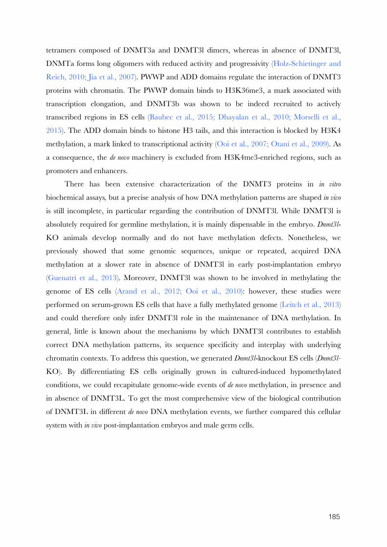
185
tetramers composed of DNMT3a and DNMT3l dimers, whereas in absence of DNMT3l,
DNMTa forms long oligomers with reduced activity and progressivity (Holz-Schietinger and
Reich, 2010; Jia et al., 2007). PWWP and ADD domains regulate the interaction of DNMT3
proteins with chromatin. The PWWP domain binds to H3K36me3, a mark associated with
transcription elongation, and DNMT3b was shown to be indeed recruited to actively
transcribed regions in ES cells (Baubec et al., 2015; Dhayalan et al., 2010; Morselli et al.,
2015). The ADD domain binds to histone H3 tails, and this interaction is blocked by H3K4
methylation, a mark linked to transcriptional activity (Ooi et al., 2007; Otani et al., 2009). As
a consequence, the de novo machinery is excluded from H3K4me3-enriched regions, such as
promoters and enhancers.
There has been extensive characterization of the DNMT3 proteins in in vitro
biochemical assays, but a precise analysis of how DNA methylation patterns are shaped in vivo
is still incomplete, in particular regarding the contribution of DNMT3l. While DNMT3l is
absolutely required for germline methylation, it is mainly dispensable in the embryo. Dnmt3l-
KO animals develop normally and do not have methylation defects. Nonetheless, we
previously showed that some genomic sequences, unique or repeated, acquired DNA
methylation at a slower rate in absence of DNMT3l in early post-implantation embryo
(Guenatri et al., 2013). Moreover, DNMT3l was shown to be involved in methylating the
genome of ES cells (Arand et al., 2012; Ooi et al., 2010): however, these studies were
performed on serum-grown ES cells that have a fully methylated genome (Leitch et al., 2013)
and could therefore only infer DNMT3l role in the maintenance of DNA methylation. In
general, little is known about the mechanisms by which DNMT3l contributes to establish
correct DNA methylation patterns, its sequence specificity and interplay with underlying
chromatin contexts. To address this question, we generated Dnmt3l-knockout ES cells (Dnmt3l-
KO). By differentiating ES cells originally grown in cultured-induced hypomethylated
conditions, we could recapitulate genome-wide events of de novo methylation, in presence and
in absence of DNMT3L. To get the most comprehensive view of the biological contribution
of DNMT3L in different de novo DNA methylation events, we further compared this cellular
system with in vivo post-implantation embryos and male germ cells.
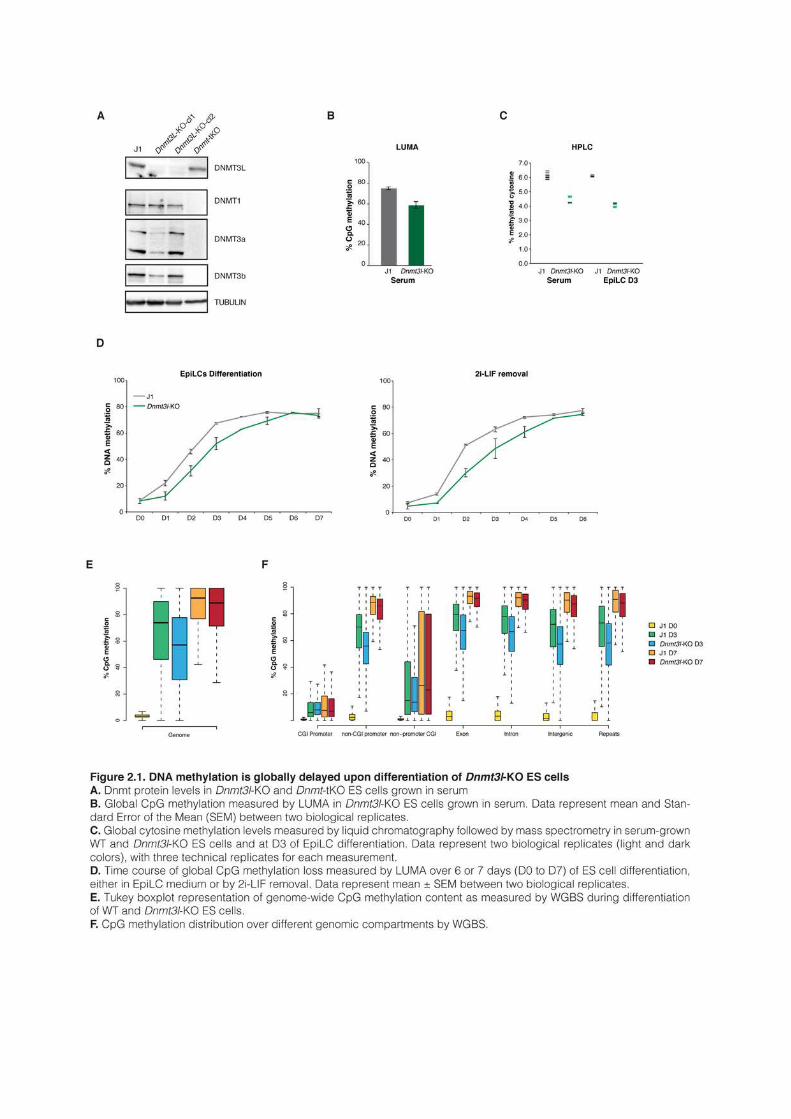
187
2.3 Results
Acquisition of DNA methylation is globally delayed in absence of DNMT3l
during ES cell differentiation
In order to generate Dnmt3l-KO ES cells, we used CRISPR/Cas9 editing to delete
Dnmt3l second exon in J1 WT ES cells, introducing a stop codon that efficiently knocked out
DNMT3l protein (Figure 2.1A). CRISPR/Cas9-mediated deletion produced two
independent knockout cell lines that we used as biological replicates during this study.
Importantly, protein levels of DNMT1, DNMT3a and DNMT3b were globally unaltered by
DNMT3l absence, even though DNMT3a and DNMT3b appeared slightly reduced in one of
the replicate (Figure 2.1A). We measured global CpG methylation in serum-grown ES cells
using LUminometric Methylation Assay (LUMA) and observed a reduction from ~75% to
~60% of CpG methylation in absence of DNMT3l (Figure 2.1B). Similarly, measurement of
the total proportion of methylated cytosines by liquid chromatography followed by mass
spectrometry showed a reduction from ~6% to ~4% in Dnmt3l-KO cells (Figure 2.1C).
Importantly, hydroxymethylation levels remained identical in absence of DNMT3l, suggesting
that active demethylation catalyzed the TET enzymes is unlikely to explain the
hypomethylation of Dnmt3l-KO ES cells (data not shown). The decreased level of DNA
methylation in Dnmt3l-KO cells is in line with the observation that serum-grown ES cells
lacking DNMT3a or DNMT3b have also reduced levels of DNA methylation (Arand et al.,
2012; Okano et al., 1999). This indicates that maintenance of hypermethylated state in serum-
grown ES cell necessitates a continuous involvement of the de novo DNA methylation
machinery.
In order to study DNMT3l contribution to de novo DNA methylation, we first reduced
global DNA methylation levels in Dnmt3L-KO and J1 WT ES cells by culturing them for three
weeks in presence of two small kinase inhibitors (2i) and vitamin C (Blaschke et al., 2013; Ying
et al., 2008), two conditions that promote almost complete erasure of DNA methylation in ES
cells, as we showed previously (Walter et al., submitted). Cells were then differentiated, either
into epiblast-like cells (EpiLC) by switching to EpiLC-specific medium (Guo et al., 2009), or
by 2i-LIF removal. In both differentiating conditions, global DNA methylation increased
rapidly in WT J1 ES cells and flattened out at roughly 75% after four days (Figure 2.1D). In
Dnmt3l-KO ES cells, DNA methylation acquisition was globally delayed, with 10-20% of
decreased methylation observed from day 1 (D1) to D5 of differentiation. However, Dnmt3l-
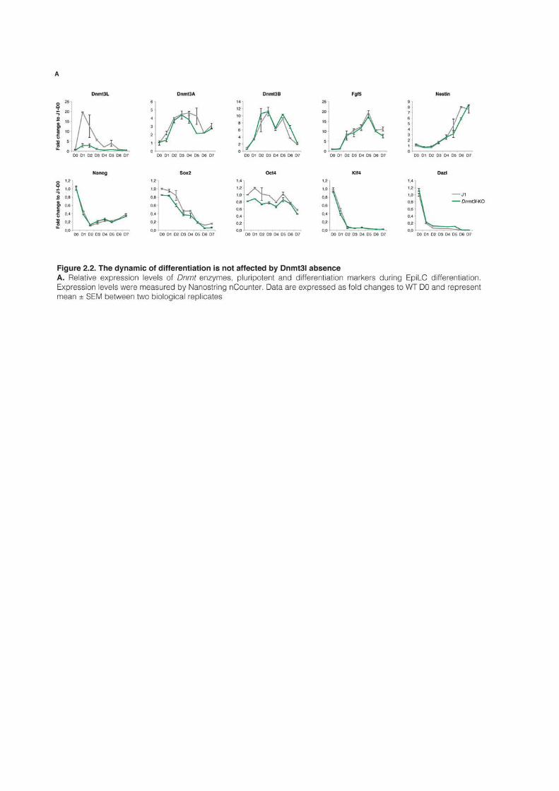
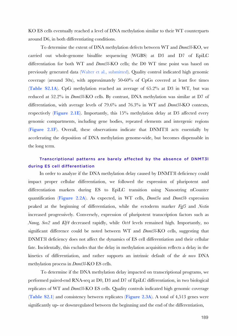
189
KO ES cells eventually reached a level of DNA methylation similar to their WT counterparts
around D6, in both differentiating conditions.
To determine the extent of DNA methylation defects between WT and Dnmt3l-KO, we
carried out whole-genome bisulfite sequencing (WGBS) at D3 and D7 of EpiLC
differentiation for both WT and Dnmt3l-KO cells; the D0 WT time point was based on
previously generated data (Walter et al., submitted). Quality control indicated high genomic
coverage (around 30x), with approximately 50-60% of CpGs covered at least five times
(Table S2.1A). CpG methylation reached an average of 65.2% at D3 in WT, but was
reduced at 52.2% in Dnmt3l-KO cells. By contrast, DNA methylation was similar at D7 of
differentiation, with average levels of 79.6% and 76.3% in WT and Dnmt3l-KO contexts,
respectively (Figure 2.1E). Importantly, this 15% methylation delay at D3 affected every
genomic compartments, including gene bodies, repeated elements and intergenic regions
(Figure 2.1F). Overall, these observations indicate that DNMT3l acts essentially by
accelerating the deposition of DNA methylation genome-wide, but becomes dispensable in
the long term.
Transcriptional patterns are barely affected by the absence of DNMT3l
during ES cell differentiation
In order to analyze if the DNA methylation delay caused by DNMT3l deficiency could
impact proper cellular differentiation, we followed the expression of pluripotent and
differentiation markers during ES to EpiLC transition using Nanostring nCounter
quantification (Figure 2.2A). As expected, in WT cells, Dnmt3a and Dnmt3b expression
peaked at the beginning of differentiation, while the ectoderm marker Fgf5 and Nestin
increased progressively. Conversely, expression of pluripotent transcription factors such as
Nanog, Sox2 and Klf4 decreased rapidly, while Oct4 levels remained high. Importantly, no
significant difference could be noted between WT and Dnmt3l-KO cells, suggesting that
DNMT3l deficiency does not affect the dynamics of ES cell differentiation and their cellular
fate. Incidentally, this excludes that the delay in methylation acquisition reflects a delay in the
kinetics of differentiation, and rather supports an intrinsic default of the de novo DNA
methylation process in Dnmt3l-KO ES cells.
To determine if the DNA methylation delay impacted on transcriptional programs, we
performed paired-end RNA-seq at D0, D3 and D7 of EpiLC differentiation, in two biological
replicates of WT and Dnmt3l-KO ES cells. Quality controls indicated high genomic coverage
(Table S2.1) and consistency between replicates (Figure 2.3A). A total of 4,513 genes were
significantly up- or downregulated between the beginning and the end of the differentiation,
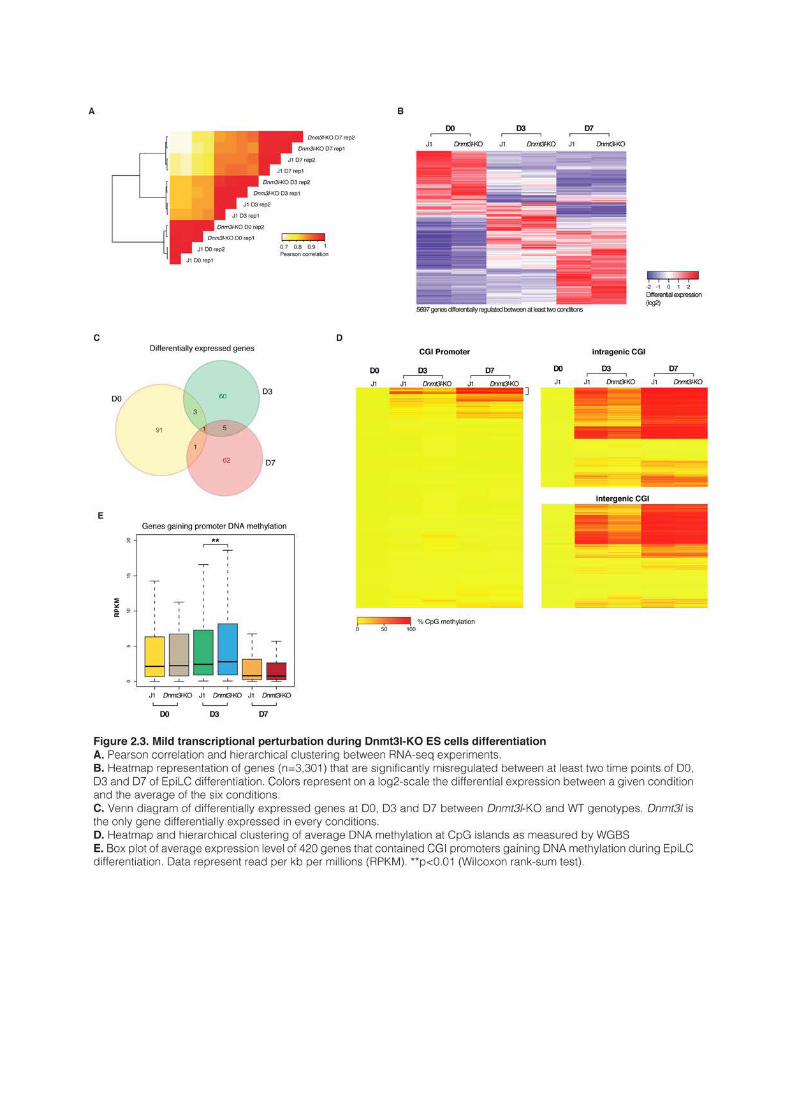
191
and WT and Dnmt3l-KO followed globally the same trend (Figure 2.3C). Closer analysis
revealed that 96, 69 and 69 genes were differentially expressed between WT and Dnmt3l-KO
at D0, D3 and D7 of differentiation, respectively (Table S2.2 and Figure 2.2D).
Around 75% of genes misrelegulated at D3 were upregulated in Dnmt3l-KO cells, and
this upregulation primarily affected genes that gained promoter DNA methylation during
differentiation. For example, the germline genes Dazl, Asz1 or Tuba3a were significantly
upregulated at D3, but not at D7, which correlates with a delayed acquisition of DNA
methylation in their CGI promoters (Figure 2.3 – supplemental figure 1A). Of note,
many of these genes were strongly expressed at D0 and experienced dramatic repression upon
differentiation, being almost completely silenced at D7. The delayed acquisition of DNA
methylation at their promoter correlates therefore with a delayed transcriptional repression.
By contrast, at D7, the majority (~80%) of the 69 differentially expressed genes were
downregulated in Dnmt3L-KO cells, suggesting indirect mechanisms. However, a few genes,
like for example the testis-specific Ldhc gene, were upregulated at D7 between Dnmt3l-KO WT
and WT cells, which correlated with reduced methylation of their promoters (Figure 2.3 – supplemental figure 1B).
We reasoned that delayed acquisition of DNA methylation in the Dnmt3l-KO context
would principally affect transcription of genes that gain promoter DNA methylation during
differentiation. During WT EpiLC differentiation, around 10% of CGIs located in promoter
regions and roughly 40% of non-promoter CGIs, acquire methylation (Figure 2.3D). More
specifically, we distinguished 420 genes that gained promoter DNA methylation during
differentiation, a number in good agreement with the 411 CpG-rich promoters that were
found highly methylated in 9.5 days (E9.5) embryos (Borgel et al., 2010). As expected, gene
ontology revealed that the majority of these genes were involved in germline development
(Table S2.3). Among these 420 genes, 180 were significantly transcribed at some point during
the differentiation. As anticipated, in average, their expression decreased importantly over the
course of the differentiation. In Dnmt3l-KO EpiLC differentiation, as for the rest of the
genome, CGI methylation was reduced after three days of differentiation, but attained WT
levels after seven days. The 180 germline genes that are expressed showed a slight but
significant upregulation could at D3 of Dnmt3l-KO differentiation (Figure 2.3E).
Another class of genomic elements that gained methylation upon differentiation was
transposable elements. We followed 69 representative families of transposons over the course
of the EpiLC differentiation and observed that virtually every families became heavily
methylated, with a delay in Dnmt3l-KO cells similar to the rest of the genome (Figure 2.3 –
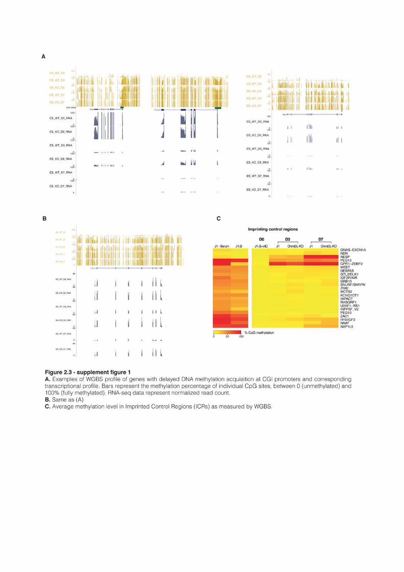
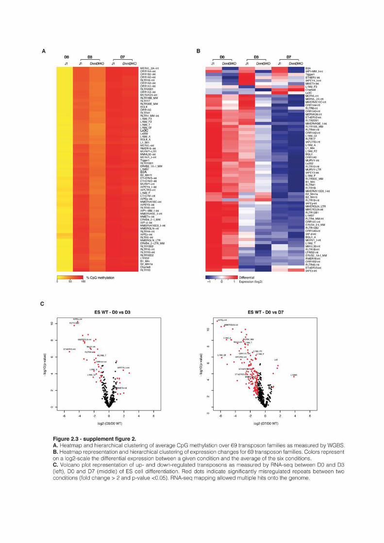
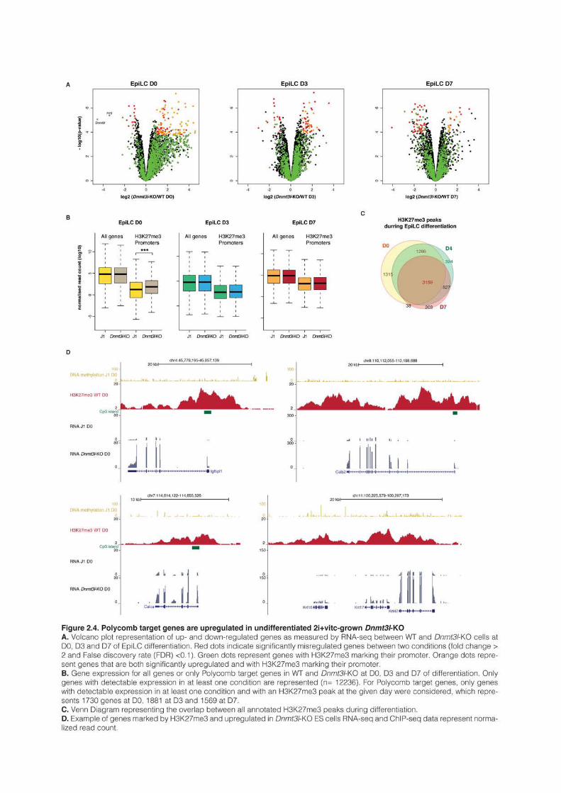
195
supplemental figure 2A). DNA methylation is described to repress transposable elements
in differentiated cells (Hutnick et al., 2010; Walsh et al., 1998), and indeed the gain of DNA
methylation correlated with the strong repression of the majority of transposon family during
WT differentiation (Figure 2.3 – supplemental figure 2B,C). Surprisingly, at both D0 and
D3, transposons appeared more expressed in WT than in Dnmt3l-KO cells. Considering the
naturally weak expression of Dnmt3l and the global absence of DNA methylation at D0, this
result was puzzling and difficult to interpret.
Notably, we followed the dynamics of de novo methylation in imprinting control regions
(Figure 2.3 – supplemental figure 1C). In globally hypermethylated ES cells grown in
serum, the majority of these regions showed, as expected, a methylation level of around 50%,
while some had lost their imprint, by gain or loss of methylation, according to the genetic drift
undergone by these cells (Greenberg and Bourc’his, 2015). As shown previously, DNA
methylation was completely lost in 2i+vitC (Walter et al., submitted). Interestingly, during
differentiation, the majority of the imprinting control regions did not undergo de novo
methylation, while a small subset became by contrast fully hypermethylated (Nesp, Peg13 and
Gpr1-Zdbf2). This showed that 2i+vitC treatment durably erases imprinting, and that the two
alleles of each imprinted regions are epigenetically similar in this context.
Overall this analysis showed that transcriptional patterns are barely affected in general
by the methylation delay in differentiating Dnmt3l-KO cells. Nonetheless, a subset of genes
that usually gain promoter methylation showed delayed repression during EpiLC
differentiation, with apparently no long-term consequences.
The majority of genes up-regulated in undifferentiated Dnmt3l-KO cells
have H3K27me3-enriched promoters
The observation that many genes were differentially expressed at D0 in Dnmt3l-KO
cells, the majority of them (~85%) being upregulated, is puzzling. Indeed, Dnmt3l is globally
repressed in 2i+vitC-grown ES cells (Leitch et al., 2013), and the genome is also completely
unmethylated in this context, making it difficult to propose an explanation for this
upregulation. Closer analysis revealed in fact that 722 genes were upregulated more than two-
fold between Dnmt3l-KO and WT cells. However, most of these genes were lowly expressed,
preventing them to meet our significance threshold.
To investigate what could explain this unexpected upregulation, we used H3K27me3
ChIP-seq data generated in the lab during EpiLC differentiation of E14 ES cells, another
male ES cells from a similar 129Sv background than J1 cells. ChIP-seq was carried at D0, D4
and D7 of differentiation. Peak calling and annotation indicated that 6,084 genes had a
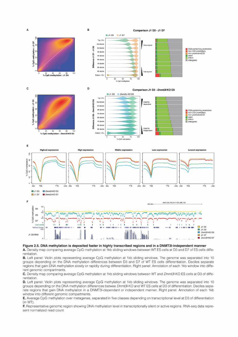
197
promoter marked by H3K27me3 in D0 undifferentiated 2i+vitC ES cells. Among them,
1,730 genes had detectable expression in at least one of the day of differentiation.
Surprisingly, most of the genes upregulated at D0 in Dnmt3l-KO cells have H3K27me3-
enriched promoters (Figure 2.4A, left panel). More specifically, out of the 720 genes
upregulated in Dnmt3l-KO cells, 462 (65 %) are marked by H3K27me3: therefore, 25% of
genes both marked by H3K27me3 and with detectable expression get upregulated in Dnmt3l-
KO cells at D0 (hypergeometric test: p-value < 10-229)(Figure 2.4B, left panel).
These results indicate that H3K27me3 marks the majority of genes upregulated in
Dnmt3l-KO cells at D0 of differentiation, and that reciprocally Polycomb-marked genes
tended to be up-regulated in absence of DNMT3l (Figure 2.4A, B, left panel, Figure 2.4D
for visual examples). Importantly the link between H3K27me3 and DNMT3l could only be
observed at D0 of differentiation, and disappeared during differentiation (Figure 2.4A and B,
mid and right panel, Figure 2.4C). This suggests that this putative link between DNMT3land
the Polycomb machinery is specific to the hypomethylated context of 2i+vitC-grown cells and
therefore independent of DNA methylation.
Overall, we show that genes upregulated in Dnmt3l-ko ES cells with an hypomethylated
states had promoters marked by H3K27me3, suggesting that Polycomb-mediated repression
could be alleviated in absence of DNMT3l. This result is vey preliminary but points towards
an unsuspected mechanistic link between DNMT3l and the Polycomb machinery in absence
of DNA methylation.
DNMT3l is dispensable for DNA methylation acquisition in highly
transcribed regions in ES cells
To gain more insight into the dynamics of DNA methylation, we partitioned the
genome into 1kb sliding windows, with a 500pb overlap between adjacent windows. We then
calculated the average methylation of every windows in the different samples. Comparison
between WT D3 and D7 showed, as expected, that most of the genome had low methylation
levels at D3 (Figure 2.5A). However, differences between D3 and D7 were not homogeneous.
Some regions had comparable methylation levels between the two time points, while others
were markedly different, indicating that the rate of DNA methylation acquisition differ
depending on the genomic region. To understand what distinguished fast and slow
methylation acquirers, we then calculated for every 1kb window the difference of methylation
between D7 and D3, and used the deciles of this difference to separate the genome into 10
groups (Figure 2.5B, left panel). The first decile contains regions with an important
methylation difference between D3 and D7: they acquire DNA methylation at a slow rate. By
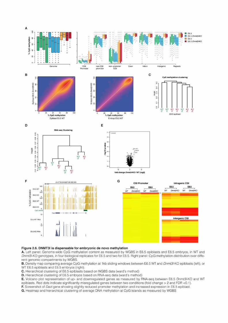
199
contrast, the last decile represents fast acquiring regions that have already attained their final
level at D3. Repartition of the windows of the 10 groups into different genomic compartments
showed that fast acquiring regions were more often found into gene bodies that slow acquiring
ones (Figure 2.5B, right panel). Of note, most CGIs cluster within the lower decile, which
was to be expected since they remain hypomethylated. However, a noticeable proportion of
CGIs appeared in the upper decile, and probably represent islands that gained DNA
methylation during the differentiation, although at a slow rate.
We repeated a similar analysis at D3 of differentiation between Dnmt3l-KO and WT
samples. Most of the genome had reduced methylation in Dnmt3l-KO cells (Figure 2.5C),
allowing again partitioning the genome into 10 groups, based this time upon the differences
between Dnmt3l-KO and WT cells (Figure 2.5D). Top deciles represented regions that
require DNMT3l for enhanced de novo methylation. These showed above 25% of methylation
decrease in Dnmt3l-KO compared to WT cells. By contrast, lower deciles represented
DNMT3l-independent regions that showed no methylation difference between Dnmt3l-KO
and WT cells. Interestingly, DNMT3l-independent regions tended to locate more often in
intragenic sequences than DNMT3l-dependent ones (Figure 2.5D, right panel). To sum up,
this shows that gene bodies have a faster rate of methylation than the rest of the genome and
do not require DNMT3l.
It was recently shown that DNMT3b is recruited to the body of actively transcribed
genes via binding of its PWWP domain to H3K36 methylation (Baubec et al., 2015; Morselli
et al., 2015). To analyze whether fast-acquiring and DNMT3l-independent regions that
localized in gene bodies were affected by the transcriptional status of the underlying gene, we
used the RNA-seq data of WT D3 sample to separate genes into five classes depending on
their transcriptional status: lowest expression, low expression, mid expression, high expression,
highest expression. We then plotted average DNA methylation on and around meta-genes
representing these five classes (Figure 2.5E). As expected, gene bodies were hypermethylated
and promoter regions hypomethylated. By comparing the five transcriptional classes, we could
observe that gene body methylation was systematically higher for highly transcribed genes
than lowly expressed ones, for both WT and Dnmt3l-KO cells, at both D3 and D7. This
showed that highly transcribed regions tend to methylate faster. Moreover, differences
between Dnmt3l-KO and WT cells were minimal for highly transcribed genes (Figure 2.5E,
left panels), whereas lowly expressed genes behave like the rest of the genome, with a delay of
around 20% methylation in absence of DNMT3l (Figure 2.5E, right panels). Screenshots of
representative genomic regions showed indeed that DNA methylation levels were similar
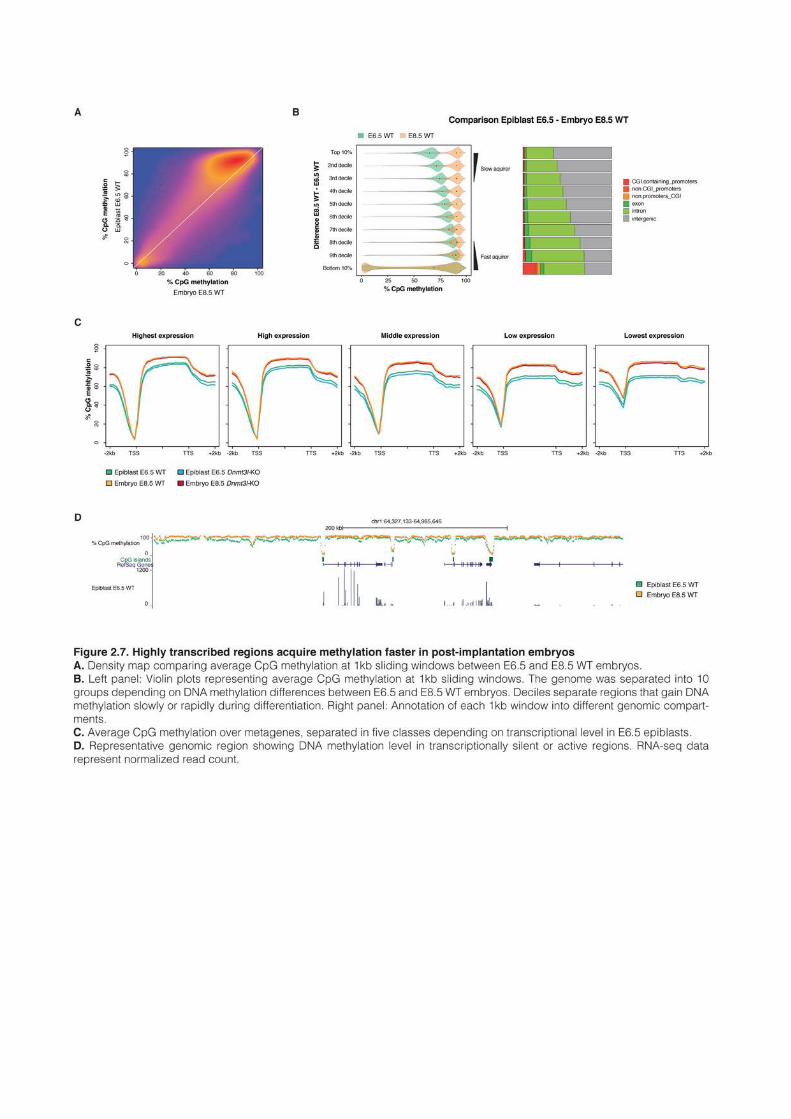
201
between Dnmt3l-KO and WT cells in the body of highly transcribed genes, whereas DNA
methylation acquisition was retarded in the rest of the genome (Figure 2.5F)
Overall, these results suggested that in ES cells, highly transcribed regions tend to
methylate faster than the rest of the genome, and this occurs in a DNMT3l-independent
manner.
DNMT3l is mostly dispensable in vivo for embryonic de novo methylation
The differentiation protocol that we used to follow the dynamic of de novo methylation –
from ES cells to EpiLC – is supposed to recapitulate the transition from ground state
blastocyst cells to committed epiblast cells. In the hope of observing in vivo a similar delayed
methylation in absence of DNMT3l, we performed WGBS in 6.5 days (E6.5) epiblasts and
E8.5 embryos, in WT and Dnmt3l-KO littermates. Two biological replicates were used for
E8.5 embryos, and four for E6.5 epiblasts, separating males and females: this was motivated
by the fact that female embryos have a delayed kinetics of development (Schulz et al., 2014).
Sequencing was carried out at a reduced depth compared to ES cells, with around 10x
coverage for each sample (Table S2.1). In good agreement with other studies (Wang et al.,
2014b), average CpG methylation ranged between 60.0 and 68.9% in the eight E6.5 epiblast
samples, and between 78.0 and 84.5% in the four E8.5 embryos. Importantly, no global
difference could be observed between Dnmt3l-KO and WT samples, either at the level of the
genome or in any genomic compartments (Figure 2.6A and B). Moreover, clustering of E6.5
samples failed to group WT and Dnmt3l-KO datasets together, indicating that the impact of
DNMT3L in embryonic methylation is minimal (Figure 2.6C).
We also carried RNA-seq of E6.5 epiblasts, in four biological replicates for WT and
Dnmt3l-KO contexts, with again in total four males and four females (Table S2.1). As for the
methylation analysis, clustering of E6.5 epiblast RNA-seq datasets failed to separate WT and
Dnmt3l-KO samples (Figure 2.6D). More detailed inspection revealed in fact that Dnmt3l was
the one and only gene that was significantly misregulated between WT and Dnmt3l-KO
embryos (Figure 2.6E). The germline genes Dazl, Mov10l1 and Tuba3a were nonetheless
upregulated around 2-3 fold in average between Dnmt3l-KO and WT epiblasts, but could not
reach our significativity threshold, probably because these genes were very lowly expressed in
both genotypes. The upregulation of these genes correlated with a modest reduction of
promoter CGI methylation in E6.5 epiblasts, as exemplified by Dazl in Figure 2.6F.
In general, among the subset of CGIs that gained DNA methylation, we could observe a
very slight delay in E6.5 Dnmt3l-KO epiblasts, for CGIs localizing either in promoters or in
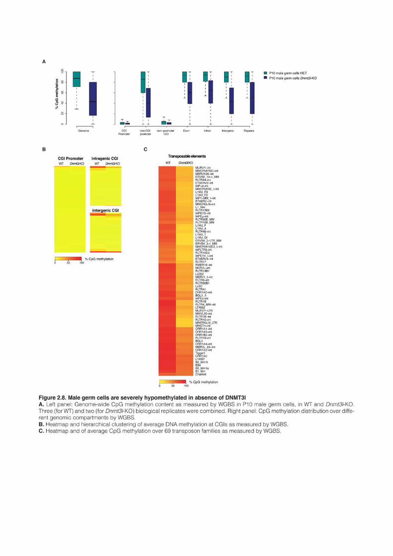
203
inter- and intragenic regions (Figure 2.6G). No difference could be observed for the
methylation of imprinted control regions (around 50% as anticipated) or transposable
elements (Figure 2.6 – supplemental figure 1A, B and C). We found that 225 CGI
promoters had gained significant DNA methylation in E8.5 embryos, a number markedly
smaller than the 431 CGI promoters that we found de novo methylated during ES cell
differentiation. Interestingly in general, the majority of CGIs that gained DNA methylation in
vivo also gained DNA methylation in ES cells, but conversely many methylated CGIs in ES
remained unmethylated in vivo, especially those associated with promoter regions (Figure 2.6 – supplemental figure 1D and E). This indicates that the EpiLC differentiation protocol
that we used did not faithfully reproduce de novo methylation in vivo.
DNA methylation is acquired faster at highly transcribed regions during
embryonic de novo DNA methylation
In E6.5 epiblasts, deposition of DNA methylation is not terminated yet, with an average
level of ~65% of CpG methylation compared to 85% in E8.5 embryos. Levels of methylation
are, however, not homogeneous genome-wide at E6.5, with some regions acquiring DNA
methylation faster than others (Figure 2.7A). To understand what distinguished fast and slow
acquirers of DNA methylation in vivo, we partitioned the genome into 1kb windows and
separated the genome into 10 groups, depending on the DNA methylation difference between
E6.5 and E8.5 embryos (Figure 2.7B). As during ES cell differentiation and in an even more
pronounced manner, we observed that fast acquiring regions tended very often to be in
intragenic position compared to slow acquiring regions. To analyze if the preferential
localization of fast acquiring regions in gene bodies correlated with transcriptional levels, we
used E6.5 RNA-seq data to separate genes into five groups depending on their transcriptional
strength (Figure 2.7C and D). As during ES cell differentiation, highly transcribed genes
tended to gain DNA methylation faster than transcriptionally silent regions. However, in that
case DNMT3l absence had barely any consequences.
This observation confirmed that during embryonic de novo methylation, highly
transcribed regions methylate at a faster rate than the rest of the genome.
Male germ cells are severely hypomethylated in absence of DNMT3l
Male and female Dnmt3l-KO mice are infertile. Dnmt3l-KO oocytes are completely
unmethylated and their progeny dies of imprinting defects around mid-gestation (Bourc’his et
al., 2001; Smallwood et al., 2011). Male germ cells on the other hand cannot complete meiosis
and Dnmt3l-KO males do not produce mature sperm. In particular transposable elements are
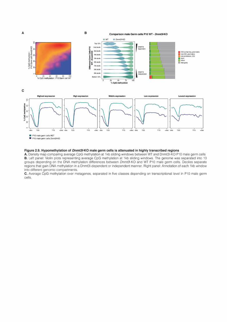
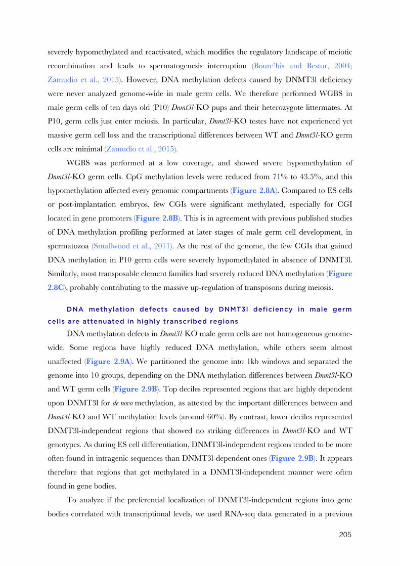
205
severely hypomethylated and reactivated, which modifies the regulatory landscape of meiotic
recombination and leads to spermatogenesis interruption (Bourc’his and Bestor, 2004;
Zamudio et al., 2015). However, DNA methylation defects caused by DNMT3l deficiency
were never analyzed genome-wide in male germ cells. We therefore performed WGBS in
male germ cells of ten days old (P10) Dnmt3l-KO pups and their heterozygote littermates. At
P10, germ cells just enter meiosis. In particular, Dnmt3l-KO testes have not experienced yet
massive germ cell loss and the transcriptional differences between WT and Dnmt3l-KO germ
cells are minimal (Zamudio et al., 2015).
WGBS was performed at a low coverage, and showed severe hypomethylation of
Dnmt3l-KO germ cells. CpG methylation levels were reduced from 71% to 43.5%, and this
hypomethylation affected every genomic compartments (Figure 2.8A). Compared to ES cells
or post-implantation embryos, few CGIs were significant methylated, especially for CGI
located in gene promoters (Figure 2.8B). This is in agreement with previous published studies
of DNA methylation profiling performed at later stages of male germ cell development, in
spermatozoa (Smallwood et al., 2011). As the rest of the genome, the few CGIs that gained
DNA methylation in P10 germ cells were severely hypomethylated in absence of DNMT3l.
Similarly, most transposable element families had severely reduced DNA methylation (Figure 2.8C), probably contributing to the massive up-regulation of transposons during meiosis.
DNA methylation defects caused by DNMT3l deficiency in male germ
cells are attenuated in highly transcribed regions
DNA methylation defects in Dnmt3l-KO male germ cells are not homogeneous genome-
wide. Some regions have highly reduced DNA methylation, while others seem almost
unaffected (Figure 2.9A). We partitioned the genome into 1kb windows and separated the
genome into 10 groups, depending on the DNA methylation differences between Dnmt3l-KO
and WT germ cells (Figure 2.9B). Top deciles represented regions that are highly dependent
upon DNMT3l for de novo methylation, as attested by the important differences between and
Dnmt3l-KO and WT methylation levels (around 60%). By contrast, lower deciles represented
DNMT3l-independent regions that showed no striking differences in Dnmt3l-KO and WT
genotypes. As during ES cell differentiation, DNMT3l-independent regions tended to be more
often found in intragenic sequences than DNMT3l-dependent ones (Figure 2.9B). It appears
therefore that regions that get methylated in a DNMT3l-independent manner were often
found in gene bodies.
To analyze if the preferential localization of DNMT3l-independent regions into gene
bodies correlated with transcriptional levels, we used RNA-seq data generated in a previous
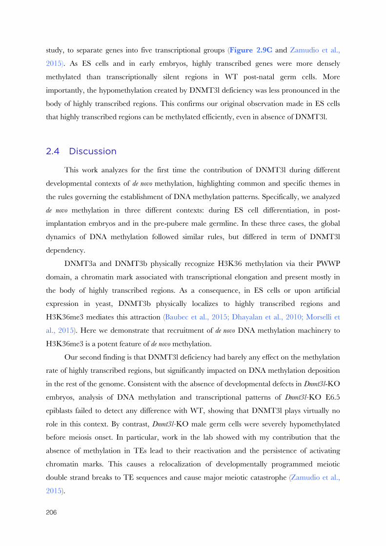
206
study, to separate genes into five transcriptional groups (Figure 2.9C and Zamudio et al.,
2015). As ES cells and in early embryos, highly transcribed genes were more densely
methylated than transcriptionally silent regions in WT post-natal germ cells. More
importantly, the hypomethylation created by DNMT3l deficiency was less pronounced in the
body of highly transcribed regions. This confirms our original observation made in ES cells
that highly transcribed regions can be methylated efficiently, even in absence of DNMT3l.
2.4 Discussion
This work analyzes for the first time the contribution of DNMT3l during different
developmental contexts of de novo methylation, highlighting common and specific themes in
the rules governing the establishment of DNA methylation patterns. Specifically, we analyzed
de novo methylation in three different contexts: during ES cell differentiation, in post-
implantation embryos and in the pre-pubere male germline. In these three cases, the global
dynamics of DNA methylation followed similar rules, but differed in term of DNMT3l
dependency.
DNMT3a and DNMT3b physically recognize H3K36 methylation via their PWWP
domain, a chromatin mark associated with transcriptional elongation and present mostly in
the body of highly transcribed regions. As a consequence, in ES cells or upon artificial
expression in yeast, DNMT3b physically localizes to highly transcribed regions and
H3K36me3 mediates this attraction (Baubec et al., 2015; Dhayalan et al., 2010; Morselli et
al., 2015). Here we demonstrate that recruitment of de novo DNA methylation machinery to
H3K36me3 is a potent feature of de novo methylation.
Our second finding is that DNMT3l deficiency had barely any effect on the methylation
rate of highly transcribed regions, but significantly impacted on DNA methylation deposition
in the rest of the genome. Consistent with the absence of developmental defects in Dnmt3l-KO
embryos, analysis of DNA methylation and transcriptional patterns of Dnmt3l-KO E6.5
epiblasts failed to detect any difference with WT, showing that DNMT3l plays virtually no
role in this context. By contrast, Dnmt3l-KO male germ cells were severely hypomethylated
before meiosis onset. In particular, work in the lab showed with my contribution that the
absence of methylation in TEs lead to their reactivation and the persistence of activating
chromatin marks. This causes a relocalization of developmentally programmed meiotic
double strand breaks to TE sequences and cause major meiotic catastrophe (Zamudio et al.,
2015).
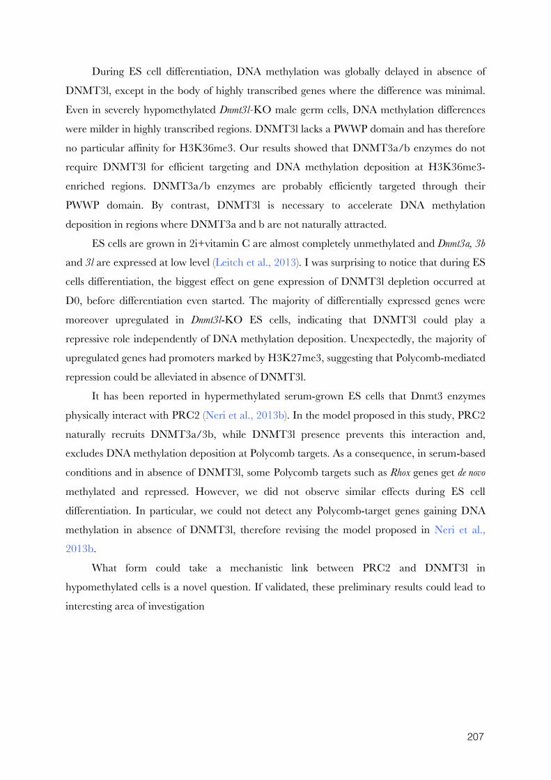
207
During ES cell differentiation, DNA methylation was globally delayed in absence of
DNMT3l, except in the body of highly transcribed genes where the difference was minimal.
Even in severely hypomethylated Dnmt3l-KO male germ cells, DNA methylation differences
were milder in highly transcribed regions. DNMT3l lacks a PWWP domain and has therefore
no particular affinity for H3K36me3. Our results showed that DNMT3a/b enzymes do not
require DNMT3l for efficient targeting and DNA methylation deposition at H3K36me3-
enriched regions. DNMT3a/b enzymes are probably efficiently targeted through their
PWWP domain. By contrast, DNMT3l is necessary to accelerate DNA methylation
deposition in regions where DNMT3a and b are not naturally attracted.
ES cells are grown in 2i+vitamin C are almost completely unmethylated and Dnmt3a, 3b
and 3l are expressed at low level (Leitch et al., 2013). I was surprising to notice that during ES
cells differentiation, the biggest effect on gene expression of DNMT3l depletion occurred at
D0, before differentiation even started. The majority of differentially expressed genes were
moreover upregulated in Dnmt3l-KO ES cells, indicating that DNMT3l could play a
repressive role independently of DNA methylation deposition. Unexpectedly, the majority of
upregulated genes had promoters marked by H3K27me3, suggesting that Polycomb-mediated
repression could be alleviated in absence of DNMT3l.
It has been reported in hypermethylated serum-grown ES cells that Dnmt3 enzymes
physically interact with PRC2 (Neri et al., 2013b). In the model proposed in this study, PRC2
naturally recruits DNMT3a/3b, while DNMT3l presence prevents this interaction and,
excludes DNA methylation deposition at Polycomb targets. As a consequence, in serum-based
conditions and in absence of DNMT3l, some Polycomb targets such as Rhox genes get de novo
methylated and repressed. However, we did not observe similar effects during ES cell
differentiation. In particular, we could not detect any Polycomb-target genes gaining DNA
methylation in absence of DNMT3l, therefore revising the model proposed in Neri et al.,
2013b.
What form could take a mechanistic link between PRC2 and DNMT3l in
hypomethylated cells is a novel question. If validated, these preliminary results could lead to
interesting area of investigation
208
2.5 Experimental procedures
ES cell lines
J1 mouse ES cells, from a male 129S4/SvJae background were used as WT in this
study. Dnmt3l-KO ES cells were generated from J1 ES cells using CRISPR/Cas9 editing.
Briefly, guide-RNAs specific to the target sequences were designed using the online CRISPR
Design Tool (Hsu et al., 2013)(Table S2.?) and incorporated into the X330 backbone (Cong et
al., 2013). Five millions J1 ES cells grown in serum were transfected with 1μg of plasmid using
Amaxa 4d nucleofector (Lonza) and plated at a low density. Individual clones were picked and
screened by PCR; mutated alleles were confirmed by Sanger sequencing. Dnmt3l-KO cells
were obtained by deleting exon 2.
ES cell culture
Undifferentiated ES cells were grown in two different media, serum and 2i, defined as
follow. Serum: Glasgow medium (Sigma), 15% FBS (Gibco), 2mM L-Glutamine, 0.1mM
MEM non essential amino acids (Gibco), 1mM sodium pyruvate (Gibco), 0.1mM β-
mercaptoethanol, 1000U/mL leukemia inhibitory factor (LIF, Miltenyi); 2i: N2B27 medium
(50% neurobasal medium (Gibco), 50% DMEM/F12 (Gibco), 2mM L-glutamine (Gibco),
0.1mM β-mercaptoethanol, Ndiff Neuro2 supplement (Milipore), B27 serum-free supplement
(Gibco)) supplemented with 1000U/mL LIF, 3μM Gsk3 inhibitor CT-99021, 1μM MEK
inhibitor PD0325901. Vitamin C (Sigma) was added at a concentration of 100ug/mL
(Blaschke et al., 2013).
Cells were grown in feeder-free conditions on gelatin-coated plates. ES cells in serum
were passaged with TrypLE Express Enzyme (Life Technologies), whereas. 2i ES cells were
harvested with Accutase (Life Technologies).
To induce differentiation towards EpiLCs, cells were plated at a density of 104 cells/cm2
on Fibronectin (10 µg/ml) coated plates in ground state medium. After one day, cells were
gently washed with 1xPBS, and cultured in N2B27 medium supplemented with 12 ng/ml
Fgf2 (R&D) and 20ng/ml Activin A (R&D). EpiLCs were passaged with Accutase at day 4 of
differentiation.
Mice
Dnmt3l-KO and WT littermates mice were derived and bred in the pathogen-free
Animal Care Facility of the Institut Curie (agreement number: C 75-05-18). All
experimentation was approved by the Institut Curie Animal Care and Use Committee and
209
adhered to European and National Regulation for the Protection of Vertebrate Animals used
for Experimental and other Scientific Purposes (Directive 86/609 and 2010/63). For embryo
collection, females were euthanized by cervical dislocation. Dnmt3l-KO mice were originally
described in (Bourc’his and Bestor, 2004; Bourc’his et al., 2001). E6.5 epiblasts were manually
dissected from extra embryonic tissues.
Dnmt3l-KO and WT P10 germ cells were isolated and purified in the lab of Steve
Jacobsen, as described in Pastor et al., 2014.
DNA methylation analyses
Genomic DNA was isolated using the GenElute Mammalian Genomic DNA Miniprep
Kit (Sigma) with RNase treatment. Global CpG methylation levels were assessed using
LUminometric Methylation Assay (LUMA) as described previously (Karimi et al., 2011a;
Richard Pilsner et al., 2010). Briefly, 500ng of genomic DNA was digested with MspI/EcoRI
and HpaII/EcoRI (NEB) in parallel reactions. HpaII is a methylation-sensitive restriction
enzyme and MspI is its methylation insensitive isoschizomer. EcoRI is included as an internal
reference. The overhangs created by the enzymatic digestion were quantified by
Pyrosequencing (PyroMark Q24, Qiagen) with the dispensation order:
GTGTGTCACACAGTGTGT. Global CpG methylation levels were calculated from the
peak heights at the position 7,8,13,14 as follows: 1-sqrt([p8*p14/p7*p13]HpaII
/[p8*p14/p7*p13]MspI)
Quantification of global cytosine methylation by liquid chromatography and mass
spectrometry was performed by Zymo Research.
Whole-Genome Bisulfite Sequencing libraries from cells and E8.5 days whole embryos
were prepared from 50ng of bisulfite-converted genomic DNA using the EpiGnome/Truseq
DNA Methylation Kit (Illumina) following the manufacturer instructions. Sequencing was
performed in 100pb paired-end reads at a 30X coverage using the Illumina HiSeq2000
platform (Table S1.1C).
Whole-Genome Bisulfite sequencing libraries from E6.5 epiblast were prepared
essentially as described in Smallwood et al., 2014. Whole-Genome Bisulfite sequencing
libraries from P10 germ cells were prepared as described in Pastor et al., 2014.
RNA expression analyses
For cells, total RNA was extracted using Trizol (Life Technologies). Nanostring
nCounter quantification was performed using 100ng of total RNA per sample on a custom
expression Codeset (sequences available upon request). Actin, Ppia, Gapdh and Arp0 were used
210
for normalization. Data are presented as the fold change compared to WT-D0. RNA-seq
libraries were prepared from 500ng of DNase-treated RNA with the TruSeq Stranded mRNA
kit (Illumina). Sequencing was performed in 100pb paired-end reads using the Illumina
HiSeq2000 platform.
For E6.5 epiblast, total RNA was extracted using PicoPure RNA isolation kit (Applied
Biosystem), including DNAse treatment (Qiagen). RNA-seq libraries were prepared from 10-
20ng of DNase-treated RNA with the Ovation RNA-seq system (Nugen). Sequencing was
performed in 100pb paired-end reads using the Illumina HiSeq2000 platform.
Western blotting
To prepare total protein extracts, cells were resuspended in BC250 lysis buffer (25mM
Tris pH 7.9, 0.2mM EDTA, 20% Glycerol, 0.25M KCl and protease inhibitor coktail from
Roche), sonicated and centrifuged to pellet debris. Proteins were quantified by Bradford assay.
Proteins (10-20μg per gel lane) were separated by electrophoresis in 10% poly-acrylamide gels
and transferred onto nitrocellulose membranes using the Trans-Blot turbo transfer system
(Biorad). After incubation with primary antibodies and HRP-conjugated secondary
antibodies, signal was detected using ECL prime kit (Amersham) and ImageQuant Las-4000
mini biomolecular Imager. Antibodies are listed in Table S1.7.
WGBS data analysis
Whole-genome bisulfite sequencing reads generated in this study were treated as follow.
The first eight base pairs of the reads were trimmed using FASTX-Toolkit v0.0.13
(http://hannonlab.cshl.edu/fastx_toolkit/index.html). Adapter sequences were removed with
Cutadapt v1.3 (https://code.google.com/p/cutadapt/) and reads shorter than 16bp were
discarded. Cleaned sequences were aligned onto the Mouse reference genome (mm10) using
Bismark v0.12.5 (Krueger and Andrews, 2011) with Bowtie2-2.1.0 (Langmead and Salzberg,
2012) and default parameters. Only reads mapping uniquely on the genome were conserved.
Methylation calls were extracted after duplicate removal. Only CG dinucleotides covered by a
minimum of 5 reads were conserved for the rest of the analysis.
The R-package Methylkit v0.9.2 (Akalin et al., 2012) was used to provide Pearson’s
correlation scores between samples. To analyze the distribution of CpG methylation in
different genomic compartments, the mouse genome was divided into different partitions. The
RefSeq gene annotation and the RepeatMasker database were downloaded from UCSC table
browser and used for transcript and repeat annotations, respectively. Promoters were defined
as the -1kb to +100pb region around transcription start sites. CGI (CGIs) were defined as in
211
Illingworth et al., 2010. Intergenic partitions were defined as genomic regions that did not
overlap with promoters, CGI, exons, introns or repeats. Whole-genome mapping of CpG
methylation was then intersected with the different genomic compartments using Bedtools
(Quinlan and Hall, 2010).
Average CpG methylation on individual transposons was extracted from
RepeatMasker with Bedtools, average CpG methylation in the different transposon families
was calculated and plotted using R. Heatmap for average CpG methylation in Imprinted
control regions (ICRs) and CGI was generated similarly after retrieving ICR genomic
coordinates from the WAMIDEX database (Schulz et al., 2008).
In order to compare DNA methylation level between conditions, the mouse genome
was partitioned into 1-kb sized windows with an overlap of 500 bp. Windows overlapping
with satellite annotated in the Repeat Masker database were removed. Windows with at least
5 CpGs were kept. Windows with less than 50% of CpGs covered at least 5 times were
removed.
Metaplots showing DNA methylation across gene bodies were created using deepTools
v1.5.11 (Ramírez et al., 2014).
RNA-seq data analysis
In order to quantify gene expression, Paired-end 2x100bp reads were mapped onto
mm10 using Tophat v2.0.6 and RefSeq gene annotation (Kim et al., 2013) allowing five
mismatches. Gene-scaled quantification was performed with HTSeq v0.6.1 (Anders et al.,
2014).
In order to quantify transposon expression, reads mapping to ribosomal RNA (rRNA)
sequences (GenBank identifiers: 18S NR_003278.3, 28S NR_003279.1, 5S D14832.1, 5.8S
K01367.1) were first removed with Bowtie v1.0.0 allowing three mismatches. The rRNA-
depleted libraries were then mapped onto mm10 using Bowtie v1.0.0 allowing zero mismatch
and 10,000 best alignments per read. Exonic reads were removed. In order to count reads
mapping to transposable elements, reads were weighted by the number of mapping sites and
each library was intersected with the reconstructed RepeatMasker annotation, conserving
only reads overlapping at least at 80% with a given transposon.
For each library, read counts for genes and transposons were combined into a single
table. TMM normalization from the edgeR package v3.6.2 (Robinson and Oshlack, 2010)
was first applied. As described in the guideline of limma R-package v3.20.4, normalized
counts were processed by the voom method (Law et al., 2014) to convert them into log2
212
counts per million with associated precision weights. The differential expression was estimated
with the limma package. Genes and transposons were called differentially expressed when two
criteria were met: 1) the fold-change between two conditions was higher than two and 2) the
adjusted p-value using the Benjamini Hochberg procedure was below 0.05.
2.6 Annexes
Supplemental information
Author contribution
M.W. and D.B. conceived the study, analyzed the data and wrote the manuscript. M.W.
performed most of the experiments. Bioinformatics analyses were conducted by A.T. and
M.W. E6.5 and E8.5 embryo dissection were carried out by M. W and J. I. WGBS libraries
for E6.5 embryo were performed in the lab of G.K. by S.S. Germ cells isolation and
subsequent WGBS were performed in the lab of S.J. by W.P.
Acknowledgments
We thank a lot of people
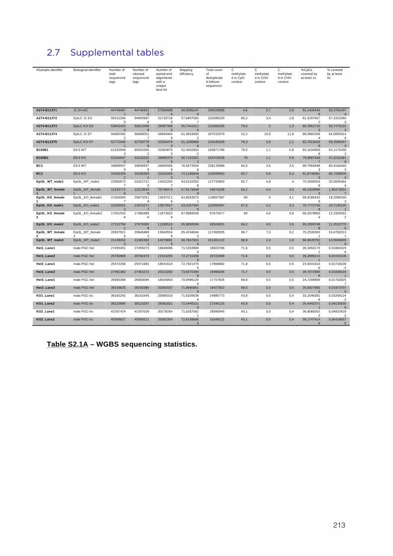
213
2.7 Supplemental tables
#Sample identifier Biological identifier Number of total sequenced tags
Number of cleaned sequenced tags
Number of paired-end alignments with a unique best hit
Mapping efficiency
Total count of deduplicated leftover sequences
C methylated in CpG context
C methylated in CHG context
C methylated in CHH context
%CpGs covered by at least 1x
% covered by at least 5x
A274-B113T1 J1 2i+vitC 447494671
447404318
270945884
60,55951476
190029868 4,6 0,7 0,8 81,14354349
55,37614374
A274-B113T2 EpiLC J1 D3 550122845
549935878
317207185
57,68075837
220289229 65,2 3,4 2,8 81,52976072
57,33153603
A274-B113T3 EpiLC KO D3 538454334
538125998
299973985
55,74419116
212560285 79,6 2 1,3 80,39517103
55,74751023
A274-B113T4 EpiLC J1 D7 566803502
566680527
348404646
61,48166902
247015079 52,2 10,8 11,8 86,08823084
64,08559144
A274-B113T5 EpiLC KO D7 527720401
527587798
323564790
61,32908896
224245029 76,3 2,9 2,1 82,76150438
59,35955473
B100B1 E8.5 WT 610329483
609315652
319648760
52,46029032
183871786 78,5 1,1 0,8 83,16348068
54,11754003
B100B2 E8.5 KO 533246070
532262528
269953708
50,71815012
154703039 78 1,1 0,9 79,90874387
47,32324819
BC1 E8.5 WT 348589379
348589379
266905669
76,56735548
228119988 84,5 3,6 3,5 88,79648483
60,41644453
BC2 E8.5 KO 342063592
342063592
253532693
74,11858465
233939963 82,7 5,9 6,3 91,87460813
66,72885093
Epi3L_WT_male1 Epi3L_WT_male1 225605727
224227120
145322057
64,81020538
127733853 62,7 4,8 4 70,30083246
20,58354644
Epi3L_WT_female1
Epi3L_WT_female1
122437726
122136436
70738474 57,91758489
44074208 64,2 4,4 4,5 46,53280663
1,904735017
Epi3L_KO_female1
Epi3L_KO_female1 210040937
208732518
135374116
64,85530731
119657367 60 5 4,1 69,91864345
18,20863403
Epi3L_KO_male1 Epi3L_KO_male1 220260012
218742717
138739177
63,42573545
122655087 67,6 4,2 3,3 70,77727468
20,71301051
Epi3L_KO_female2
Epi3L_KO_female2 175015536
174604690
118730239
67,9994558 97675677 60 4,6 3,8 66,20796834
12,19263522
Epi3L_KO_male2 Epi3L_KO_male2 171227941
170793658
112665161
65,9656584 92624051 64,2 4,5 3,6 65,29097488
11,35107765
Epi3L_WT_female2
Epi3L_WT_female2
209379216
208464692
136405545
65,43340443
117492835 66,7 7,3 6,2 70,25303031
16,47620131
Epi3L_WT_male2 Epi3L_WT_male2 211493323
210824841
140798915
66,78478415
101361122 68,9 2,4 1,9 66,86457527
14,58466009
Het1_Lane1 male PGC het 27455451 27455073 19640096 71,53539894
18923786 71,6 0,5 0,5 26,34501731
0,018833296
Het1_Lane2 male PGC het 29782808 29782479 21524255 72,27153589
20723098 71,6 0,5 0,5 28,28962131
0,02163136
Het2_Lane1 male PGC het 25472258 25471992 18541610 72,79214755
17868882 71,8 0,5 0,5 24,93416106
0,017160392
Het2_Lane2 male PGC het 27461362 27461072 20213282 73,60703908
19466264 71,7 0,5 0,5 26,70729808
0,019381041
Het3_Lane1 male PGC het 26083368 26083094 18505854 70,94961204
17757826 69,6 0,5 0,5 24,7208806 0,017628257
Het3_Lane2 male PGC het 28193625 28193380 20262037 71,86806619
19427922 69,5 0,5 0,5 26,58278653
0,019737075
KO1_Lane1 male PGC ko 36162242 36161645 25899315 71,62095364
24885773 43,8 0,5 0,4 33,15463916
0,032691242
KO1_Lane2 male PGC ko 39123890 39123297 28381821 72,54455319
27245125 43,8 0,5 0,4 35,43423711
0,041359306
KO2_Lane1 male PGC ko 42267424 42267028 30278284 71,63570621
28990945 43,1 0,5 0,4 36,80892622
0,049226292
KO2_Lane2 male PGC ko 45569627 45569221 33092354 72,61996864
31649121 43,1 0,5 0,4 39,17474149
0,064186575
Table S2.1A – WGBS sequencing statistics.
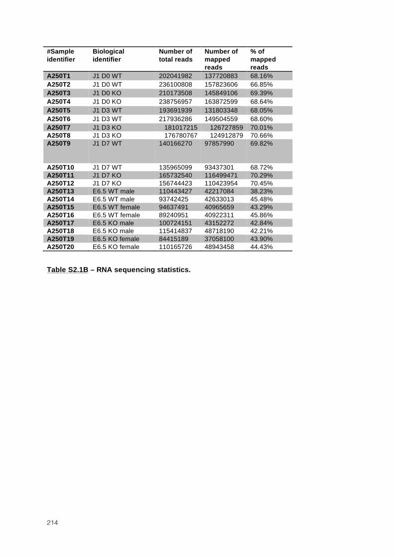
214
#Sample identifier
Biological identifier
Number of total reads
Number of mapped reads
% of mapped reads
A250T1 J1 D0 WT 202041982 137720883 68.16% A250T2 J1 D0 WT 236100808 157823606 66.85% A250T3 J1 D0 KO 210173508 145849106 69.39% A250T4 J1 D0 KO 238756957 163872599 68.64% A250T5 J1 D3 WT 193691939 131803348 68.05% A250T6 J1 D3 WT 217936286 149504559 68.60% A250T7 J1 D3 KO 181017215 126727859 70.01% A250T8 J1 D3 KO 176780767 124912879 70.66% A250T9 J1 D7 WT 140166270 97857990 69.82%
A250T10 J1 D7 WT 135965099 93437301 68.72% A250T11 J1 D7 KO 165732540 116499471 70.29% A250T12 J1 D7 KO 156744423 110423954 70.45% A250T13 E6.5 WT male 110443427 42217084 38.23% A250T14 E6.5 WT male 93742425 42633013 45.48% A250T15 E6.5 WT female 94637491 40965659 43.29% A250T16 E6.5 WT female 89240951 40922311 45.86% A250T17 E6.5 KO male 100724151 43152272 42.84% A250T18 E6.5 KO male 115414837 48718190 42.21% A250T19 E6.5 KO female 84415189 37058100 43.90% A250T20 E6.5 KO female 110165726 48943458 44.43%
Table S2.1B – RNA sequencing statistics.
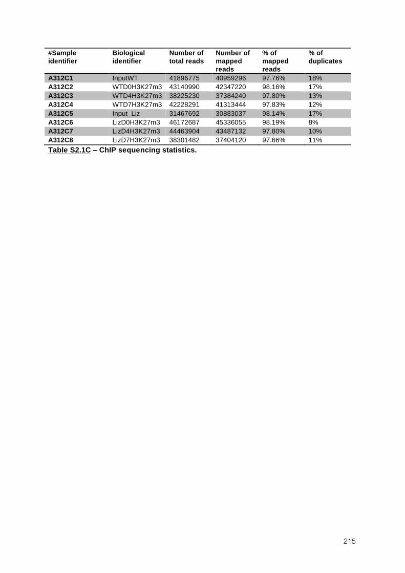
215
#Sample identifier
Biological identifier
Number of total reads
Number of mapped reads
% of mapped reads
% of duplicates
A312C1 InputWT 41896775 40959296 97.76% 18% A312C2 WTD0H3K27m3 43140990 42347220 98.16% 17% A312C3 WTD4H3K27m3 38225230 37384240 97.80% 13% A312C4 WTD7H3K27m3 42228291 41313444 97.83% 12% A312C5 Input_Liz 31467692 30883037 98.14% 17% A312C6 LizD0H3K27m3 46172687 45336055 98.19% 8% A312C7 LizD4H3K27m3 44463904 43487132 97.80% 10% A312C8 LizD7H3K27m3 38301482 37404120 97.66% 11% Table S2.1C – ChIP sequencing statistics.
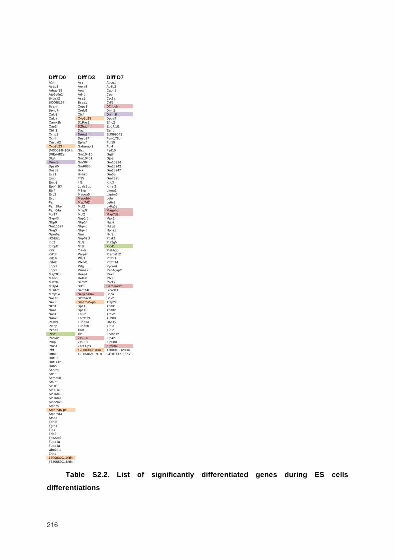
216
Diff D0 Diff D3 Diff D7 A2m Ace Abcg1 Acap3 Anxa8 Ap3b2 Arhgef25 Aoah Capn5 Atp6v0e2 Arl4d Cpe B4galt2 Asz1 Cpt1a BC068157 Bcas1 Crlf2 Bcam Cnpy1 D2hgdh Bend7 Creld1 Dmrt3 Calb2 Ctcfl Dnmt3l Calca Cyp2b23 Dppa4 Camk2b D1Pas1 Efhc2 Cap2 D2hgdh Epb4.1l1 Cbln1 Dazl Esrrb Ccng2 Dnmt3l EU599041 Ccnjl Dusp27 Fam178b Crispld2 Epha4 Fgf15 Cyp2b23 Gabarapl1 Fgf4 D430019H16Rik Glrx Fzd10 D8Ertd82e Gm10416 Ggt7 Dlg4 Gm10451 Gjb3 Dnmt3l Gm364 Gm10324 Dpysl5 Gm6880 Gm13242 Dusp8 Hck Gm13247 Ece1 Hsh2d Gm53 Emb Ifi35 Gm7325 Emp2 Irf2 Kifc3 Epb4.1l3 Lgals3bp Kirrel2 Etv4 M1ap Lama1 Evc2 Magea5 Laptm5 Evc Magohb Ldhc Fah Map7d2 Lefty2 Fam19a4 Mcf2 Ly6g6e Fam64a Mfap5 Magohb Fgf17 Mgl2 Map7d2 Gap43 Nap1l5 Mpc1 Gbp9 Nlrp14 Nab2 Gm11627 Nlrp4c Ndrg3 Gng3 Nlrp4f Nphs1 Gpm6a Nmi Nxf3 H2-Eb1 Nup62cl Pcsk1 Ido2 Nxf2 Pla2g5 Igfbpl1 Nxt2 Plcd1 Kif7 Oasl2 Plekhg5 Krt17 Parp9 Pramef12 Krt19 Plet1 Prdm1 Krt42 Plxnd1 Prdm14 Lppr2 Prtg Pycard Lppr3 Prune2 Rap1gap2 Map3k8 Reep1 Rex2 Mark1 Retsat Rfx2 Mef2b Scml2 Rnf17 Mfap4 Sdc3 Serpina3m Mfsd7c Sema4f Slco3a1 Mmp14 Serpina3m Snca Nacad Slc25a31 Sox2 Neil2 Smarca5-ps Tfap2c Nkd1 Sp110 Triml1 Nnat Sp140 Triml2 Nos1 Taf9b Trpv2 Nuak2 Tnfrsf19 Tubb3 Pcsk9 Tuba3a Uba1y Pianp Tuba3b Xlr5a Pkhd1 Xaf1 Xlr5b Plcd1 Xlr Zcchc12 Podxl2 Zfp936 Zfp42 Prnp Zfp951 Zfp600 Pros1 Znf41-ps Zfp936 Ptrf 1700030C10Rik 1700048O20Rik Rftn1 4930506M07Rik 2410141K09Rik Rnf103 Rnf144b Robo3 Scara5 Sdc2 Sema5b Sft2d2 Slain1 Slc11a1 Slc16a13 Slc16a3 Slc22a23 Smad9 Smarca5-ps Smarcd3 Stac2 Tdrkh Tgm1 Tia1 Trib2 Tsc22d3 Tuba1a Tubb4a Ube2ql1 Zhx1 1700030C10Rik 5730559C18Rik
Table S2.2. List of significantly differentiated genes during ES cells
differentiations
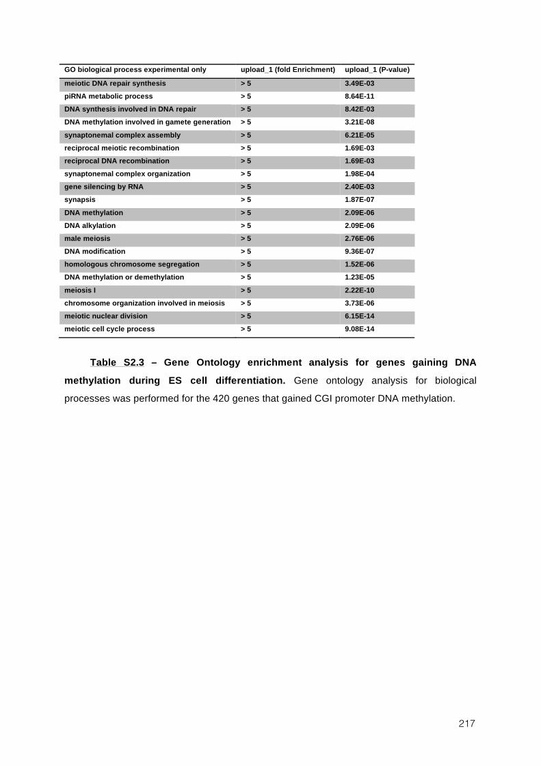
217
GO biological process experimental only upload_1 (fold Enrichment) upload_1 (P-value)
meiotic DNA repair synthesis > 5 3.49E-03 piRNA metabolic process > 5 8.64E-11 DNA synthesis involved in DNA repair > 5 8.42E-03 DNA methylation involved in gamete generation > 5 3.21E-08 synaptonemal complex assembly > 5 6.21E-05 reciprocal meiotic recombination > 5 1.69E-03 reciprocal DNA recombination > 5 1.69E-03 synaptonemal complex organization > 5 1.98E-04 gene silencing by RNA > 5 2.40E-03 synapsis > 5 1.87E-07 DNA methylation > 5 2.09E-06 DNA alkylation > 5 2.09E-06 male meiosis > 5 2.76E-06 DNA modification > 5 9.36E-07 homologous chromosome segregation > 5 1.52E-06 DNA methylation or demethylation > 5 1.23E-05 meiosis I > 5 2.22E-10 chromosome organization involved in meiosis > 5 3.73E-06 meiotic nuclear division > 5 6.15E-14 meiotic cell cycle process > 5 9.08E-14
Table S2.3 – Gene Ontology enrichment analysis for genes gaining DNA
methylation during ES cell differentiation. Gene ontology analysis for biological processes was performed for the 420 genes that gained CGI promoter DNA methylation.

218

219
DISCUSSION
220
3 Discussion
3.1.1 A cellular system to reproduce in vivo DNA methylation reprogramming
Living systems, either single cells or whole organisms, have the amazing and intrinsic
capacity to continuously adjust their biology to respond to internal or external perturbations.
Cells adapt to their environment and maintain an apparent stable state, or homeostasis, that
reflects in fact a balance between countless dynamically regulated processes (Cannon, 1926).
Upon perturbation and given enough time, cells reach a novel dynamically stable state that
reflects the new conditions. Cells in culture are a particularly good example and maintain a
stable state for unlimited periods of time, which can be different depending on the culture
condition or the presence of genetic mutations. By contrast, cells in a developing mammalian
embryo are continuously unbalanced, and cell fate and internal processes are constantly
reacting to changing conditions. As a consequence, cells grown in a steady state in culture
represent a poor model to reproduce the fundamentally dynamic evolution of early
embryogenesis. ES cell differentiation is an easy and efficient way to introduce regulatory
dynamism in cell culture and has been widely used to mimic the evolution of post-
implantation embryos. Reproducing the dynamic changes that occurs in pre-implantation
embryos or developing germ cells is on the other hand more challenging. During these
reprogramming events, cells are dedifferentiating and no good cellular system has yet emerged
to reproduce that event. Generation of iPS cells is, for example, a notoriously long and
inefficient process that fails to mimic the rapid and highly coordinated reprogramming event
of early embryos.
The principal interest of this thesis is to provide a model that reproduces in ES cells the
dynamic evolution of one key aspect of embryonic reprogramming: DNA methylation
erasure. By switching cells from serum to 2i+vitC media, CpG methylation reached 30% in
four days – roughly the level of an E3.5 blastocyst – and ~10% in six days, which is
approximately the time that migrating PGCs take to demethylate from E7.5 to E13.5.
Moreover, upon loss of DNA methylation, we observed that H3K9me2 rapidly disappeared,
that H3K9me3 remained globally stable, whereas H3K27me3 greatly increased, which is
precisely what has been observed in E13.5 PGCs (Hajkova et al., 2008; Seki et al., 2007).
Demethylation upon 2i+vitC conversion occurs at a pace comparable to in vivo events and
appears to reproduce important feature of in vivo chromatin reorganization. This comforted us
221
that our cellular system might represent a valuable tool to study how the genome adapts to
DNA methylation loss.
Our analysis focused both on short and long-term consequences of DNA methylation
loss. In the early embryo and in the mouse male germline, periods of hypomethylation are
very transient and are immediately followed by genome-wide events of de novo methylation. In
humans however, male germ cells remain hypomethylated for several weeks (Gkountela et al.,
2015; Guo et al., 2015; Tang et al., 2015). In addition, female germ cells remain completely
hypomethylated for very long periods of time, several weeks in the mouse and up to 50 years
in humans. Long-term hypomethylation does not exist only in cell culture, but represens a
constitutive feature of germ cell development. Considering how critical is germ cell genetic
integrity, it is likely that germ cells have developed alternative mechanisms that compensate
for DNA methylation loss, especially related to TE regulation. By unraveling how TEs are
regulated in hypomethylated cells, this work therefore provides insight into the mechanisms
that could potentially be at play in germ cells.
Upon loss of DNA methylation in ES cells, we could identify two important phases.
Correlating with DNA methylation disappearance, most of TEs in the genome were first
activated, before being silenced again on the long term. Importantly, H3K27 methylation and
Polycomb proteins become important regulators of TE expression in hypomethylated
contexts. Moreover, some transposon families such as MERVL and probably others operate a
complete switch from H3K9- to H3K27 methylation-based repressive pathways upon loss of
DNA methylation. This observation was unexpected and represents one of the most
interesting finding of this work. It would be of great interest to investigate in male and female
germlines whether what we observed in our cellular system could be also relevant in vivo, for
example by checking the effect of Polycomb disruption specifically in PGCs. A recent study
performed ChIP-seq for H3K9me3 and H3K27me3 in E13.5 PGCs, and observed that
H3K27me3 was precisely localizing in transposons (Liu et al., 2014). However, their data are
intriguing because it appears than all the regions marked by H3K9 methylation are also
enriched for H3K27me3. Reciprocally, when re-analyzing their data we observed that most
regions that are typically exclusively marked by H3K27me3, such as Hox gene clusters,
showed also important enrichment for H3K9me3. This raises questions about the specificity
of antibodies used in this study, and prevents us to draw strong conclusion from this data or to
use it to confirm our results (Figure 3.1A).
Overall, whether the work presented in this thesis has in vivo implications, and especially
whether Polycomb could regulate TEs in fetal germ cells needs to be investigated.
222
3.1.2 DNA methylation has a role in TE repression in ES cells
In differentiated cells, loss of DNA methylation leads to important TE reactivation,
especially of IAP elements (Hutnick et al., 2010; Walsh et al., 1998). By contrast Dnmt-tKO ES
cells show only very modest reactivation of TEs, and this observation had led to the
conclusion that TE repression occurs in a DNA methylation-independent manner in ES cells
(Hutnick et al., 2010; Karimi et al., 2011b). Our work showed that this interpretation is
erroneous. Indeed, upon conversion from serum to 2i+vitamin C, TEs were strongly up-
regulated genome-wide, and this activation involved the majority of TE families. Moreover,
this burst of expression could not be observed upon conversion of Dnmt-tKO ES that are
constitutively unmethylated, showing that medium conversion alone does not cause TE
activation. Importantly also, while the majority of changes in protein-coding gene expression
occurred at the beginning of the conversion, TEs became activated only when DNA
methylation reached a very low level. These observations strongly suggest that the loss of
DNA methylation caused TE reactivation and indicates that DNA methylation is a potent
suppressor of TE expression in ES cells. In our system, the silencing release is, however, very
brief and alternative mechanisms take over rapidly to ensure the stability of TE expression.
The existence of compensatory mechanisms explains why DNA methylation was
considered dispensable for TE repression. ES cells lines used to draw this conclusion, like
Dnmt-tKO ES cells, have been in cultured for extended periods of time, giving enough time to
implement DNA methylation-independent compensation.
One of the most serious limitation of our system is that 2i- and serum-grown ES cells are
transcriptionally very distinct. In consequence, transient activation of TEs could potentially be
essentially a consequence of medium conversion. However, while this work was being
conducted, we became aware that other groups were observing a similar phenomenon, but
using completely different systems. These studies are still unpublished but rely on conditional
depletion of Dnmt1 in serum ES as a way to promote rapid demethylation. In a very
reassuring manner, these groups observed a similar burst of TEs expression followed by a
second phase of silencing. Moreover, and somewhat surprisingly, the timing of TE peak of
expression coincided exactly with our own results, occurring exactly six days after Dnmt1
depletion. These independent results confirm our own experiments and strongly suggest that
DNA methylation rapid loss caused this transient TE reactivation (Rebecca Berrens and Wolf
Reik, 2015 EMBO Mobile Genome Meeting).
223
3.1.3 Pluripotency transcription factors may play a role in transient TE activation
Intriguingly, we observed that the burst of transposon expression coincided with an
increased availability of core pluripotency transcription factors such as OCT4 or NANOG. In
mice and humans, an important proportion of pluripotency binding motifs are localized in
TEs (Kunarso et al., 2010) and at least in human ES cells, pluripotency factors were shown to
drive TE expression (Grow et al., 2015; Wang et al., 2014a). The specific presence of
pluripotent transcription factors therefore probably contributes to the global reactivation of
TEs, but is difficult to explain. Indeed, pluripotent factors are among the only protein-coding
genes that show specifically an increased expression at the time of transposon burst. In theory,
this burst of transcription factor expression could be the sole responsible of the observed TE
transient activation, except that it would then fail to explain why this reactivation is not
observed in Dnmt-tKO ES cells as well.
Testing directly whether the presence of pluripotent factors contribute to TE expression
is difficult to achieve. In serum ES cells, levels of some pluripotency factors such as NANOG
are highly heterogeneous from cells to cells, but we could not detect by immunofluorescence
any correlation between NANOG and IAP or LINE1 protein expression. Moreover,
functional genetic studies of the role of pluripotency factors on TE expression are not really
amenable, as depletion of any of the core pluripotent factors in ES cells would lead to loss of
pluripotency and cell differentiation, changing cell fate too radically to hope drawing any
conclusion. Since loss of function experiments would not be informative, I attempted to
artificially over-express pluripotent transcription factors in ES by using an OSKM construct
originally designed to generate iPS cells20. My hope was that pluripotent factors over-
expression, either in serum or 2i+vitamin C conditions, would lead to TE transient
reactivation. Unfortunately and probably because they are already very highly expressed in
ES cells, I could only induce minimal over-expression of pluripotency factors and did not
observe any effect on transposon expression.
Serum and 2i ES cells maintain pluripotency by targeting different regulatory pathways
(Marks and Stunnenberg, 2014). Serum ES cells represent a metastable and primed
pluripotent state, while 2i ES cells are considered to be in a “ground state” of pluripotency,
which expresses pluripotent transcription factors at an higher and more homogeneous level
(Marks et al., 2012). The transition from serum to 2i+vitamin C is an important perturbation
of the equilibrium of regulatory pathways, and the first days of conversion probably represents
20 In other words, I attempted to reprogram pluripotent cells.
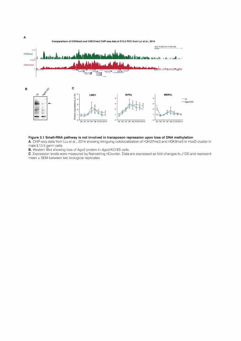
225
an unbalanced cellular state that oscilllates between two distinct equilibria. Speculatively, the
transient abundance of pluripotency factors and the decrease that follows could be seen as an
oscillation of regulatory networks, that would go “too far” before adjusting back to
equilibrium condition. Whether this oscillation is mechanistically linked to DNA methylation
loss, or whether the two events are merely correlative would be difficult to address.
3.1.4 What about the role of small RNA pathways?
Transposon repression upon 2i+vitC-induced loss of DNA methylation correlates with a
reorganization of repressive chromatin marks, a finding that we explored at length. It is
known that small RNA-based mechanisms can provide another layer of transposon
repression, acting at the post-transcriptional level. In the male germline, the piRNA pathway
is critical for TE silencing, but piRNAs are not found in any other cellular contexts including
ES cells. In ES cells and elsewhere, small RNA production relies on the RNAse III
ribonuclease Dicer that cleaves specific RNA precursors into ~21pb small RNAs, either
miRNAs or small interfering RNAs (siRNAs). MiRNAs and siRNAs are loaded into
Argonaute proteins and the complex can recognize complementary RNA targets, causing
either translational inhibition or RNA degradation (Siomi and Siomi, 2009). AGO2 is the
only Argonaute protein that can mediate cleavage of RNA targets and the three other
members of the family lack this specific slicing activity (Liu et al., 2004). Importantly, Ago2 is
absolutely required in the female germline (Kaneda et al., 2009) and associated siRNAs have
been shown to repress TEs in the oocyte (Watanabe et al., 2008, 2011). Whether siRNA-
mediated repression is active in other cellular contexts than the oocyte is under passionate
debate. Small RNA silencing has been shown to silence bona fide viruses in ES cells and is often
proposed to play a role in TE control as well (Maillard et al., 2013).
In order to test whether small RNA-based silencing could be implicated in TE
repression in our system of induced loss of DNA methylation, I generated Ago2-KO ES cells
with CRISPR/Cas9. Because AGO2 slicing activity is not required for the miRNA pathway,
Ago2 deletion would supposedly suppress siRNA silencing without affecting miRNAs. The
knockout was generated by deleting Ago2 second exon in J1 WT ES and was confirmed by
Western Blot. Upon serum to 2i+vitC, I could not detect any significant difference in
transposon expression between Ago2-KO and WT cells, except for MERVLs that were slightly
upregulated in serum. Ago2-KO cells followed the same pattern of activation-repression,
indicating that small RNA pathways (at least siRNAs) are unlikely to play a role in transposon
control in our system (Figure 3.1B and C).
226
3.1.5 H3K9 methylation pathways play a complex role upon loss of DNA methylation
This work improves our comprehension of how H3K9-related pathway maintains TE
silencing. In particular, analysis of H3K9-KMT mutants had until now been conducted
almost exclusively in highly methylated serum ES cells and this work shed important light on
how H3K9me3-silencing adapt to hypomethylated contexts.
Of interest, H3K9me2 is reduced to almost undetectable levels upon conversion from
serum to 2i+vitamin C, and a similar reduction was observed in Dnmt-tKO ES cells, albeit not
at the same extent. It is established from numerous reports that G9a can recruit Dnmt3a and
b and accordingly, DNA methylation is reduced at TEs, major satellite repeats and promoters
in G9a-KO ES cells (Dong et al., 2008; Epsztejn-Litman et al., 2008). Our results indicate
conversely that DNA methylation presence might be necessary for G9a/GLP activity and
H3K9me2 deposition. Interestingly, we observed that upon conversion from serum to
2i+vitamin C, the burst of transposon expression at day 6 was amplified in G9a-KO, even
though H3K9me2 had already disappeared at this time point in WT cells. DNA methylation
recruitment by G9a was shown to be independent of its catalytic activity and our results
suggest similarly that during 2i+vitC conversion, G9a might play a repressive role
independent of H3K9me2 deposition (Dong et al., 2008). How G9a, H3K9me2 and DNA
methylation mechanistically interact would need to be properly investigated.
Upon loss of DNA methylation, H3K9me3 pattern remain globally untouched.
Surprisingly, many TE families with a high enrichment for this repressive mark are
nonetheless transiently reactivated, showing that H3K9me3 itself is not sufficient to maintain
silencing. This suggests that H3K9me3 readers might be destabilized by DNA methylation
disappearance or by the 2i+vitC medium composition . The MBD protein MeCP2 associates
with Suv39h enzymes, binds to methylated DNA and repress transcription of TEs in certain
contexts (Lunyak et al., 2002; Muotri et al., 2010; Nan et al., 1998). Upon loss of DNA
methylation, MeCP2-mediated repression at H3K9me3 sites could be alleviated, potentially
contributing to repression release. It would be also of interest to analyze whether downstream
actors of the H3K9 methylation pathway, such as HP1 or KAP1, remain bound to TEs when
they are activated but still enriched in H3K9me3.
Even thought H3K9 methylation is not sufficient to prevent TE burst of activation, our
results show H3K9-mediated repression is critical for long term re-silencing. KAP1 is critical
for ES cell survival (Rowe et al., 2010); therefore, I could only study Kap1 heterozygous
mutants. Interestingly, half the dose of KAP1 protein was sufficient to maintain IAP
227
repression in serum, likely because these cells are hypermethylated, but not in 2i+vitamin C,
when DNA methylation disappears. Moreover, H3K9me3 levels increased at IAPs in
2i+vitamin C, suggesting that upon loss of DNA methylation, long-term maintenance of
silencing of IAP elements may require a reinforcement of Kap1/H3K9me3-related
repression. To further test that hypothesis, I recently deleted one allele of ESET in ES cells
(complete Eset-KO is also cell lethal, Matsui et al., 2010), which will allow me to compare IAP
repression in serum and 2i+vitamin C and determine if ESET-mediated silencing efficiency is
affected by DNA methylation loss. If validated, this would suggest that in absence of DNA
methylation, IAP silencing is indeed achieved by a reinforcement of H3K9me3-related
silencing. This strategy might be specific to the IAP superfamily. Indeed, IAP and related
MMERVK10C elements are the most strongly reactivated TEs in Eset knockout (Karimi et
al., 2011b). They are also among the only transposons that are covered on their full-length by
H3K9me3 and that never gain any observable H3K27me3, indicating that their dependency
on ESET/KAP1 is particularly strong.
Except for MERVL elements that mostly rely on G9a, the majority of TEs in serum-
grown ES cells are repressed in a KAP1/ESET-dependent manner (Karimi et al., 2011b;
Matsui et al., 2010; Rowe et al., 2010). It was recently proposed that SUV39h enzymes were
adding another layer of repression on TEs to form large H3K9me3 domains spreading
around from KAP1 binding sites (Bulut-Karslioglu et al., 2014). However, we found that
knockout of SUV39h has little effect on TE repression, at the exception of MERVL elements
and of one specific LINE family (L1-A). Upon loss of DNA methylation in Suv39h-dKO, we
were puzzled to observe that IAP expression remained at a constant level and did not go
trough the typical burst of transcription. Of note, the flat curve of IAP expression during
conversion is identical in Suv39h-dKO and Dnmt-tKO cells. In Dnmt-tKO cells, IAP elements
have already adapted to the absence of DNA methylation, which explains that they are not
reactivated. Considering the numerous mechanistic links between SUV39h enzymes and
DNA methylation, it is tempting to speculate that in Suv39h-dKO, IAP have also already
adapted to the lack of SUV39h, and that this adaptation suppresses the dependency on DNA
methylation. In this scenario, lack of SUV39h or DNA methylation would have the same
effect: the implementation of DNA methylation-independent repression.
What form would take these SUV39h- and DNA methylation-independent mechanisms
is an interesting question. One particular challenge of transposon biology is to take into
account the variability of elements inside a single family. It is probably wrong to assume that
every elements are regulated the same way, especially because repressive strategies likely
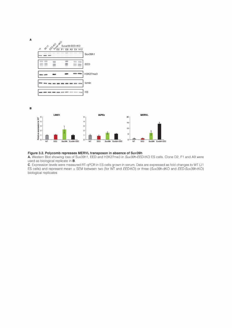
229
depend upon the genomic context. For example, even though H3K27me3 cannot be detected
at IAP sequences by ChIP-seq, IAP elements are strongly upregulated in 2i+vitC in Eed-KO
ES cells, showing that IAPs are at least genetically dependent upon PRC2. IAPs are young
TEs that are almost impossible to distinguish from one to another. In consequence, only a few
elements could rely on Polycomb, which would be impossible to observe by ChIP but would
nonetheless result in strong transcriptional up-regulation. Upon loss of DNA methylation,
MERVL elements operate a complete switch from G9a- and SUV39h-based repression to
Polycomb-mediated silencing. I reasoned that this hidden minority of IAPs could behave like
MERVL family, and that the adaptation to SUV39h loss could involve Polycomb repression.
To test that hypothesis, I very recently generated Eed-Suv39h-tKO ES cells, which
survived in serum-based conditions, albeit growing at a reduced pace. In serum, I found that
MERVL elements were unaltered in Eed-KO, upregulated by five-fold in Suv39h-dKO and
further upregulated by 15-fold in Eed-Suv39h-tKO (Figure 3.2A and B). This shows without
much doubt that Polycomb compensates for SUV39h absence in MERVL elements, even in
globally hypermethylated ES cells, and supports our claim that MERVLs operate a switch
from H3K9 to H3K27-based repression. However, IAP expression was unaffected by single
or joint depletion of Eed and Suv39h1/2 (Figure 3.2 B). This indicates that, in serum at least
and contrary to MERVL, IAP elements do not replace Suv39h by Polycomb.
Upon conversion to 2i+vitamin C, I observed that Eed-Suv39h-tKO ES cells died after
six-eight days: ES cell viability is therefore impaired when DNA methylation, Suv39h and
PRC2 are depleted together, but not when one of these marks is still present. In absence of
DNA methylation or SUV39h, H3K27me3 was shown to relocalize to pericentric
heterochromatic (Cooper et al., 2014; Peters et al., 2003; Saksouk et al., 2014). It would be
very informative to analyze whether Eed-Suv39h-tKO death correlates with massive pericentric
defects, and also to follow transposon expression during the first day of conversion.
Overall, H3K9me3-related pathways are not affected by 2i+vitC-induced loss of DNA
methylation. However, closer analysis reveals a more complex picture: ESET/KAP1-
repression is reinforced, while SUV39h and Polycomb engage in an intricate and complex
crosstalk.
3.1.6 A new function of H3K27me3 in repressing TEs
One of the main findings of my work is the observation that H3K27me3 relocalizes to
TEs upon loss of DNA methylation. In the case of MERVLs at least, it is clear that Polycomb
acts as a potent transcriptional repressor. In MERVL elements, H3K27me3 is localized in
230
promoter regions, which correlate well with its silencing effect. Other families, such as ETn
and VL30, gain H3K27me3 in their 5’-region as well and we can expect that these families
would similarly be reactivated. By contrast, a majority of TE families gain H3K27me3 in their
3’ regions, while their 5’ remained marked by H3K9me3. To investigate whether the presence
of H3K27me3 in TE body could be functionally relevant and be involved in silencing, we are
currently performing RNA-seq for Eed-KO ES cells grown in serum and 2i+vitamin C
conditions.
The presence of Polycomb in TE bodies could affect transcriptional elongation.
H3K27me3 usually recruit PRC1 through the binding of CBX proteins to H3K27me3
residues, and PRC1 subsequently catalyzes local H2Aub deposition. H2A ubiquitination and
PRC1 are thought to block transcriptional initiation or elongation (Brookes et al., 2012; Stock
et al., 2007). It is therefore tempting to speculate that PRC1 could play a similar role in TEs.
Whether PRC1 and H2A ubiquitination are recruited to TEs upon loss of DNA methylation
needs to be investigated.
3.1.7 H3K27me3 is attracted to unmethylated TEs
One of the main finding of my work is the observation that H3K27me3 relocalize to
TEs upon loss of DNA methylation, raising the interesting question of how Polycomb proteins
are targeted there. The question of the determinants of Polycomb recruitment remains largely
unanswered in mammals. In ES cells grown is serum, the majority of H3K27me3-marked
regions nucleate around CGIs and do not usually spread into DNA methylated territories
(Brinkman et al., 2012; Ku et al., 2008; Statham et al., 2012). By contrast, upon loss of DNA
methylation, H3K27me3 leaves CGIs and starts colonizing new genomic compartments, in
particular TEs and pericentric repeats.
Recruitment of PRC2 to pericentric heterochromatin in absence of DNA methylation
was shown to rely on the (putative) DNA binding protein BEND3, probably by recognizing
specific motifs in major satellite repeats (Saksouk et al., 2014). It is therefore unlikely that
BEND3 could be implicated in the targeting of PRC2 to TEs. Polycomb recruitment can also
be mediated by long-non-coding RNAs (Pandey et al., 2008; Plath et al., 2003; Rinn et al.,
2007), and it would be interesting to investigate whether PRC2 physically interacts with TE
RNAs. Intriguingly, TE burst of transcription and H3K27me3 deposition are not
synchronized: H3K27me3 reaches its full potential only after transcription has stopped. This
raises interesting question about the mechanistic links between TE transcription and
H3K27me3 deposition in TE bodies. Several hypotheses are possible, which would need to be
231
tested: 1) H3K27me3 deposition blocks transcription; 2) H3K27me3 can only be laid down
after unrelated mechanisms have suppressed transcription; 3) Transcription recruits
H3K27me3.
The observation that H3K27me3 invades TEs only upon loss of DNA methylation
confirms the general assumption that these two modifications are mutually exclusive in ES
cells. PRC1 complexes associated with RYBP were recently shown to be involved in PRC2
recruitment (Blackledge et al., 2014; Cooper et al., 2014). In particular, the CxxX-protein
KDM2b associates with RYBP-PRC1 and is thought to mediate PRC1 and PRC2
recruitment at unmethylated CpG-rich regions, such as CGIs. Considering the role of CxxX-
domains in recognizing unmethylated CpGs, KDM2b represents therefore a very seducing
model that could explain how H3K27me3 is recruited at unmethylated TEs. Indeed, most
TEs and especially young and active ones tend to be CpG-rich, and KDM2b could mediate
recruitment of PRC1, which in turn would recruit PRC2 and H3K27me3.
In fact, our system represents a very interesting model to study the mechanisms of
Polycomb recruitment. Indeed, thousand of new genomic locations become enriched for
H3K27me3 and could help unraveling how Polycomb machinery is recruited de novo.
Unfortunately, the necessity to write this thesis prevented me so far to generate Kdm2b and
Rybp knockout ES cells and I could not directly test whether depleting KDM2b and RYBP-
PRC1 would affect PRC2 recruitment at TE upon loss of DNA methylation.
From the literature, it appears that depletion of KDM2b in serum-grown ES cells barely
affect H3K27me3 enrichment at CGIs, which indicates that KDM2b is not the unique player
involved in Polycomb recruitment at CGIs (Blackledge et al., 2014). Interestingly, a study
reported that in serum-grown ES cells, numerous germline genes such as Dazl, Mov10l1, Mael
or Ddx4 were bound by RYBP and RING1b and analysis of ChIP-seq data from another
study confirms this observation (Figure 3.3A and Hisada et al., 2012; Morey et al., 2013).
Interestingly, the majority of these germline genes (at the notable exception of Dazl) were
shown to be bound by E2F6, a transcription factor member of one specific RYBP-PRC1
complex (Gao et al., 2012; Velasco et al., 2010), which could explain how PRC1 is recruited
to these regions. These germline genes have methylated CGI promoters, suggesting that
RYBP-PRC1 binding might not be affected by DNA methylation. Interestingly, RYBP-PRC1
presence at germline genes was not associated with PRC2 and CBX-PRC1 (Figure 3.3A),
which is expected considering their high level of DNA methylation. However, we observed
that upon loss of DNA methylation, some of these germline genes such as Dazl or Sycp3 are
gaining H3K27me3, raising the interesting possibility that when DNA methylation
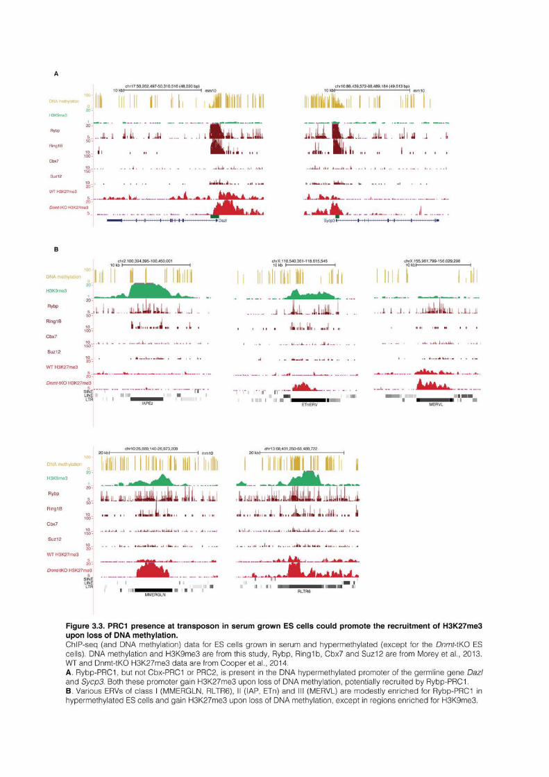
233
disappears, RYBP-PRC1 could trigger the recruitment of PRC2 at these unmethylated targets
(Figure 3.3A).
Remarkably, RYBP was also shown to be present at the LTR of TEs such as IAP,
MERVL or MusD, albeit at comparatively lower levels for the latter,. Moreover, MERVL
elements are activated in Rybp-KO cells (Hisada et al., 2012). The presence of RYBP in
hypermethylated TEs could serve to prepare for the future recruitment of PRC2 and CBX-
PRC1 upon loss of DNA methylation. Inspection of ChIP-seq tracks from available datasets
show that several TEs appear indeed bound by RYBP in serum cells, which we found to,gain
H3K27me3 upon loss of DNA methylation, precisely around RYBP binding site (Figure 3.3B). This observation is very preliminary and would necessitate thorough investigation.
Nonetheless, it can be speculated that Rypb, probably in partner with PRC1 complexes and
DNA binding protein, could act as the seed already in hypermethylated contexts that promote
the recruitment of PRC2 upon loss of DNA methylation.
3.1.8 H3K27 and H3K9 methylation occupy different TE territories
This work is focused on TE regulation, but our detailed analysis of repressive chromatin
reorganization has a broader impact and shed particular light on how H3K9 and H327
repressive pathway interact. In particular both repressive marks were apparently enriched in
the same TE sequences, either in LINEs or class I and II ERVs, but were nonetheless
occupying non-overlapping territories, a pattern that was never observed before. H3K27 and
H3K9 methylation are mutually exclusive in almost all cell types (Hawkins et al., 2010;
Mikkelsen et al., 2007). However, since H3K27me3 antagonizes DNA methylation whereas
H3K9me3 positively associates with, the mutual exclusion of the two marks could be simply
explained by the presence DNA methylation in most contexts.
Since the two marks do not overlap even in 2i+vitamin C medium, our results suggest
that mutual exclusion of H3K9me3 and H3K27me3 is independent of DNA methylation. In
numerous transposon sequences, H3K27 and H3K9 methylation are in very close physical
proximity, probably only a few nucleosomes away from each others. Despite the natural
spreading propensity of these two repressive marks, H3K9me3 and H3K27me3 remain
physically separated, which might suggest that the two repressive compartments are actively
repelling each others. Similarly, in chromocenters that also contain both marks upon loss of
DNA methylation, H3K9me3 and H3K27me3 occupy in fact distinct territories (Cooper et
al., 2014). The two marks appear antagonistic but what mechanistically cause this exclusion is
far for being understood. Binding or catalytic activity of PRC2 and H3K9-KMT is not
234
affected by any of the two chromatin modifications (Bartke et al., 2010; Schmitges et al.,
2011). Antagonism between the marks is therefore probably indirect and it was proposed that
HP1 could prevent the recruitment of PRC1 and PRC2 (Tardat et al., 2015).
Our analysis offers resolution at H3K9me3 and H3K27me3 separation, showing that
they can be immediately adjacent. However, TEs arre repeated sequences and this is
therefore not possible to completely exclude the possibility that this observation is a mapping
artifact. One way to circumvent this issue would be to create artificially an H3K9me3-
H3K27me3 bivalent domain, for example by inserting KAP1-binding sequences inside
H3K27me3-marked CGI or into an Hox cluster. Such a system could be very instrumental to
understand mechanistically what triggers the mutual exclusivity of the two marks.
Overall, the fine analysis of repressive chromatin reorganization at TEs gives very
fundamental insight into the intricate relationship that exists between chromatin
modifications. From a regulatory point of view, the mutual exclusivity of H3K27me3 and
H3K9me3 can probably be explained by the fundamentally different biological roles for these
two repressive pathways. H3K9 methylation is mostly associated with permanent silencing of
the repeated fraction of the genome, whereas Polycomb is highly plastic and involved in genic
regulation. Structurally separating the two repressive domains may ensure a tight control of
genome regulation, for example ensuring that TEs bound by Kap1 and H3K9me3 do not
perturb the regulatory capacity of neighboring Polycomb domains.

235
REFERENCES
236
Aagaard, L., Laible, G., Selenko, P., Schmid, M., Dorn, R., Schotta, G., Kuhfittig, S., Wolf, A., Lebersorger, A., Singh, P.B., et al. (1999). Functional mammalian homologues of the Drosophila PEV-modifier Su(var)3-9 encode centromere-associated proteins which complex with the heterochromatin component M31. EMBO J. 18, 1923–1938.
Aapola, U., Kawasaki, K., Scott, H.S., Ollila, J., Vihinen, M., Heino, M., Shintani, A., Minoshima, S., Krohn, K., Antonarakis, S.E., et al. (2000). Isolation and initial characterization of a novel zinc finger gene, DNMT3L, on 21q22.3, related to the cytosine-5-methyltransferase 3 gene family. Genomics 65, 293–298.
Abad, P., Vaury, C., Pélisson, A., Chaboissier, M.C., Busseau, I., and Bucheton, A. (1989). A long interspersed repetitive element--the I factor of Drosophila teissieri--is able to transpose in different Drosophila species. Proc. Natl. Acad. Sci. U. S. A. 86, 8887–8891.
Adey, N.B., Schichman, S.A., Graham, D.K., Peterson, S.N., Edgell, M.H., and Hutchison, C.A. (1994). Rodent L1 evolution has been driven by a single dominant lineage that has repeatedly acquired new transcriptional regulatory sequences. Mol. Biol. Evol. 11, 778–789.
Akalin, A., Kormaksson, M., Li, S., Garrett-Bakelman, F.E., Figueroa, M.E., Melnick, A., and Mason, C.E. (2012). methylKit: a comprehensive R package for the analysis of genome-wide DNA methylation profiles. Genome Biol. 13, R87.
Alisch, R.S., Garcia-Perez, J.L., Muotri, A.R., Gage, F.H., and Moran, J. V (2006). Unconventional translation of mammalian LINE-1 retrotransposons. Genes Dev. 20, 210–224.
Alkema, M.J., Bronk, M., Verhoeven, E., Otte, A., van ’t Veer, L.J., Berns, A., and van Lohuizen, M. (1997). Identification of Bmi1-interacting proteins as constituents of a multimeric mammalian polycomb complex. Genes Dev. 11, 226–240.
Allis, C.D., Berger, S.L., Cote, J., Dent, S., Jenuwien, T., Kouzarides, T., Pillus, L., Reinberg, D., Shi, Y., Shiekhattar, R., et al. (2007). New nomenclature for chromatin-modifying enzymes. Cell 131, 633–636.
Aloia, L., Di Stefano, B., and Di Croce, L. (2013). Polycomb complexes in stem cells and embryonic development. Development 140, 2525–2534.
Anders, S., Pyl, P.T., and Huber, W. (2014). HTSeq - A Python framework to work with high-throughput sequencing data. Bioinformatics 31, 166–169.
Anderson, R., Copeland, T.K., Schöler, H., Heasman, J., and Wylie, C. (2000). The onset of germ cell migration in the mouse embryo. Mech. Dev. 91, 61–68.
Anxolabehere, D., Kidwell, M., and Periquet, G. (1988). Molecular characteristics of diverse populations are consistent with the hypothesis of a recent invasion of Drosophila melanogaster by mobile P elements. Mol. Biol. Evol. 5, 252–269.
Arabidopsis Genome Initiative (2000). Analysis of the genome sequence of the flowering plant Arabidopsis thaliana. Nature 408, 796–815.
Arand, J., Spieler, D., Karius, T., Branco, M.R., Meilinger, D., Meissner, A., Jenuwein, T., Xu, G., Leonhardt, H., Wolf, V., et al. (2012). In Vivo Control of CpG and Non-CpG DNA Methylation by DNA Methyltransferases. PLoS Genet. 8, e1002750.
Aravin, A.A., Sachidanandam, R., Girard, A., Fejes-Toth, K., and Hannon, G.J. (2007). Developmentally regulated piRNA clusters implicate MILI in transposon control. Science 316, 744–747.
Aravin, A.A., Bourc’his, D., and Bourc’his, D. (2008). Small RNA guides for de novo DNA methylation in mammalian germ cells. Genes Dev. 22, 970–975.
Arita, K., Isogai, S., Oda, T., Unoki, M., Sugita, K., Sekiyama, N., Kuwata, K., Hamamoto, R., Tochio, H., Sato, M., et al. (2012). Recognition of modification status on a histone H3 tail by linked histone reader modules of the epigenetic regulator UHRF1. Proc. Natl. Acad. Sci. U. S. A. 109, 12950–12955.
Arkhipova, I., and Meselson, M. (2000). Transposable elements in sexual and ancient asexual taxa. Proc. Natl. Acad. Sci. U. S. A. 97, 14473–14477.
Arrigoni, R., Alam, S.L., Wamstad, J.A., Bardwell, V.J., Sundquist, W.I., and Schreiber-Agus, N. (2006). The Polycomb-associated protein Rybp is a ubiquitin binding protein. FEBS Lett. 580, 6233–6241.
Athanikar, J.N., Badge, R.M., and Moran, J. V (2004). A YY1-binding site is required for accurate human LINE-1 transcription initiation. Nucleic Acids Res. 32, 3846–3855.
Auclair, G., Guibert, S., Bender, A., and Weber, M. (2014). Ontogeny of CpG island methylation and specificity of DNMT3 methyltransferases during embryonic development in the mouse. Genome Biol. 15, 545.
Aucott, R., Bullwinkel, J., Yu, Y., Shi, W., Billur, M., Brown, J.P., Menzel, U., Kioussis, D., Wang, G., Reisert, I., et al. (2008). HP1-beta is required for development of the cerebral neocortex and neuromuscular junctions. J. Cell Biol. 183, 597–606.
Avery, O.T., Macleod, C.M., and McCarty, M. (1944). Studies on the chemical nature of the substance inducing transformation of pneumococcal types. Induction of transformation by a desoxyribonucleic acid fraction isolated from Pneumococcus type III. J. Exp. Med. 79, 137–158.
Babushok, D. V, Ostertag, E.M., Courtney, C.E., Choi, J.M., and Kazazian, H.H. (2006). L1 integration in a transgenic mouse model. Genome Res. 16, 240–250.
237
Babushok, D. V, Kazazian, H.H., and Jr, Ã. (2007). Progress in understanding the biology of the human mutagen LINE-1. Hum. Mutat. 28, 527–539.
Bailey, J.A., Liu, G., and Eichler, E.E. (2003). An Alu transposition model for the origin and expansion of human segmental duplications. Am. J. Hum. Genet. 73, 823–834.
Baillie, G.J., van de Lagemaat, L.N., Baust, C., and Mager, D.L. (2004). Multiple groups of endogenous betaretroviruses in mice, rats, and other mammals. J. Virol. 78, 5784–5798.
Bailly-Bechet, M., Haudry, A., and Lerat, E. (2014). “One code to find them all”: a perl tool to conveniently parse RepeatMasker output files. Mob. DNA 5, 13.
Bannister, A.J., Zegerman, P., Partridge, J.F., Miska, E.A., Thomas, J.O., Allshire, R.C., and Kouzarides, T. (2001). Selective recognition of methylated lysine 9 on histone H3 by the HP1 chromo domain. Nature 410, 120–124.
Barau, J. (2015). SINEs tether the Synaptonemal Complex to gene-rich regions to prevent harmful recombination. In Proceedings of Late and Unproven Labnight Hypothesis in the Bourc’his Lab, pp. 10–15.
Barski, A., Cuddapah, S., Cui, K., Roh, T.-Y., Schones, D.E., Wang, Z., Wei, G., Chepelev, I., and Zhao, K. (2007). High-resolution profiling of histone methylations in the human genome. Cell 129, 823–837.
Bartke, T., Vermeulen, M., Xhemalce, B., Robson, S.C., Mann, M., and Kouzarides, T. (2010). Nucleosome-interacting proteins regulated by DNA and histone methylation. Cell 143, 470–484.
Bartolome, C., Maside, X., and Charlesworth, B. (2002). On the Abundance and Distribution of Transposable Elements in the Genome of Drosophila melanogaster. Mol. Biol. Evol. 19, 926–937.
Baubec, T., Ivánek, R., Lienert, F., and Schübeler, D. (2013). Methylation-dependent and -independent genomic targeting principles of the MBD protein family. Cell 153, 480–492.
Baubec, T., Colombo, D.F., Wirbelauer, C., Schmidt, J., Burger, L., Krebs, A.R., Akalin, A., and Schübeler, D. (2015). Genomic profiling of DNA methyltransferases reveals a role for DNMT3B in genic methylation. Nature 520, 243–247.
Baust, C., Gagnier, L., Baillie, G.J., Harris, M.J., Juriloff, D.M., and Mager, D.L. (2003). Structure and Expression of Mobile ETnII Retroelements and Their Coding-Competent MusD Relatives in the Mouse. J. Virol. 77, 11448–11458.
Beall, E.L., and Rio, D.C. (1996). Drosophila IRBP/Ku p70 corresponds to the mutagen-sensitive mus309 gene and is involved in P-element excision in vivo. Genes Dev. 10, 921–933.
Beall, E.L., Admon, A., and Rio, D.C. (1994). A Drosophila protein homologous to the human p70 Ku autoimmune antigen interacts with the P transposable element inverted repeats. Proc. Natl. Acad. Sci. U. S. A. 91, 12681–12685.
Beard, C., Li, E., and Jaenisch, R. (1995). Loss of methylation activates Xist in somatic but not in embryonic cells. Genes Dev. 9, 2325–2334.
Becker, K.G., Swergold, G.D., Ozato, K., and Thayer, R.E. (1993). Binding of the ubiquitous nuclear transcription factor YY1 to a cis regulatory sequence in the human LINE-1 transposable element. Hum. Mol. Genet. 2, 1697–1702.
Bellefroid, E.J., Poncelet, D.A., Lecocq, P.J., Revelant, O., and Martial, J.A. (1991). The evolutionarily conserved Krüppel-associated box domain defines a subfamily of eukaryotic multifingered proteins. Proc. Natl. Acad. Sci. U. S. A. 88, 3608–3612.
Van Bemmel, J. (2012). Chromatin Flavors: Chromatin composition and domain organization in Drosophila melanogaster. In Thesis Dissertation, (Erasmus University Rotterdam), pp. 11–23.
Bénit, L., De Parseval, N., Casella, J.F., Callebaut, I., Cordonnier, A., and Heidmann, T. (1997). Cloning of a new murine endogenous retrovirus, MuERV-L, with strong similarity to the human HERV-L element and with a gag coding sequence closely related to the Fv1 restriction gene. J. Virol. 71, 5652–5657.
Bénit, L., Lallemand, J.-B.B., Casella, J.-F.F., Philippe, H., Heidmann, T., Benit, L., Lallemand, J.-B.B., Casella, J.-F.F., Philippe, H., and Heidmann, T. (1999). ERV-L Elements: a Family of Endogenous Retrovirus-Like Elements Active throughout the Evolution of Mammals. J. Virol. 73, 3301–3308.
Bennet, M., and Leicht, I. (2012). Plant DNA C-values database (release 6.0, Dec. 2012). Bennett, M.D. (1971). The Duration of Meiosis. Proc. R. Soc. London. Ser. B, Biol. Sci. 178, 277–299. Bennett, M.D. (1972). Nuclear DNA Content and Minimum Generation Time in Herbaceous Plants. Proc. R.
Soc. B Biol. Sci. 181, 109–135. Bensasson, D., Petrov, D.A., Zhang, D.-X.X., Hartl, D.L., and Hewitt, G.M. (2001). Genomic gigantism: DNA
loss is slow in mountain grasshoppers. Mol. Biol. Evol. 18, 246–253. Berndsen, C.E., and Wolberger, C. (2014). New insights into ubiquitin E3 ligase mechanism. Nat. Struct. Mol.
Biol. 21, 301–307. Bernstein, B.E., Mikkelsen, T.S., Xie, X., Kamal, M., Huebert, D.J., Cuff, J., Fry, B., Meissner, A., Wernig, M.,
Plath, K., et al. (2006a). A bivalent chromatin structure marks key developmental genes in embryonic stem cells. Cell 125, 315–326.
238
Bernstein, B.E., Birney, E., Dunham, I., Green, E.D., Gunter, C., Snyder, M., Kundaje, A., Aldred, S.F., Collins, P.J., Davis, C.A., et al. (2012). An integrated encyclopedia of DNA elements in the human genome. Nature 489, 57–74.
Bernstein, E., Duncan, E.M., Masui, O., Gil, J., Heard, E., and Allis, C.D. (2006b). Mouse polycomb proteins bind differentially to methylated histone H3 and RNA and are enriched in facultative heterochromatin. Mol. Cell. Biol. 26, 2560–2569.
Bestor, T.H., and Ingram, V.M. (1983). Two DNA methyltransferases from murine erythroleukemia cells: purification, sequence specificity, and mode of interaction with DNA. Proc. Natl. Acad. Sci. U. S. A. 80, 5559–5563.
Bestor, T., Laudano, A., Mattaliano, R., and Ingram, V. (1988). Cloning and sequencing of a cDNA encoding DNA methyltransferase of mouse cells. J. Mol. Biol. 203, 971–983.
Bian, C., Chen, Q., and Yu, X. (2015). The zinc finger proteins ZNF644 and WIZ regulate the G9a/GLP complex for gene repression | eLife Lens.
Bickmore, W.A., and van Steensel, B. (2013). Genome architecture: domain organization of interphase chromosomes. Cell 152, 1270–1284.
Biemont, C. (2010). A Brief History of the Status of Transposable Elements: From Junk DNA to Major Players in Evolution. Genetics 186, 1085–1093.
Biémont, C. (1994). Dynamic equilibrium between insertion and excision of P elements in highly inbred lines from an M’ strain of Drosophila melanogaster. J. Mol. Evol. 39, 466–472.
Bird, A.P. (1980). DNA methylation and the frequency of CpG in animal DNA. Nucleic Acids Res. 8, 1499–1504.
Bird, A.P. (1986). CpG-rich islands and the function of DNA methylation. Nature 321, 209–213. Bird, A., Taggart, M., Frommer, M., Miller, O.J., and Macleod, D. (1985). A fraction of the mouse genome that
is derived from islands of nonmethylated, CpG-rich DNA. Cell 40, 91–99. Biterge, B., and Schneider, R. (2014). Histone variants: key players of chromatin. Cell Tissue Res. 356, 457–466. Blackledge, N.P., Zhou, J.C., Tolstorukov, M.Y., Farcas, A.M., Park, P.J., and Klose, R.J. (2010). CpG islands
recruit a histone H3 lysine 36 demethylase. Mol. Cell 38, 179–190. Blackledge, N.P., Farcas, A.M., Kondo, T., King, H.W., McGouran, J.F., Hanssen, L.L.P., Ito, S., Cooper, S.,
Kondo, K., Koseki, Y., et al. (2014). Variant PRC1 complex-dependent H2A ubiquitylation drives PRC2 recruitment and polycomb domain formation. Cell 157, 1445–1459.
Blaschke, K., Ebata, K.T., Karimi, M.M., Zepeda-Martínez, J. a, Goyal, P., Mahapatra, S., Tam, A., Laird, D.J., Hirst, M., Rao, A., et al. (2013). Vitamin C induces Tet-dependent DNA demethylation and a blastocyst-like state in ES cells. Nature 500, 222–226.
Boeke, J.D., and Stoye, J.P. (1997). Retrotransposons, Endogenous Retroviruses, and the Evolution of Retroelements.
Boissinot, S., and Furano, A. V. (2001). Adaptive Evolution in LINE-1 Retrotransposons. Mol. Biol. Evol. 18, 2186–2194.
Borgel, J., Guibert, S., Li, Y., Chiba, H., Schübeler, D., Sasaki, H., Forné, T., and Weber, M. (2010). Targets and dynamics of promoter DNA methylation during early mouse development. Nat. Genet. 42, 1093–1100.
Bostick, M., Kim, J.K., Estève, P.-O., Clark, A., Pradhan, S., and Jacobsen, S.E. (2007). UHRF1 plays a role in maintaining DNA methylation in mammalian cells. Science 317, 1760–1764.
Boulard, M., Edwards, J.R., and Bestor, T.H. (2015). FBXL10 protects Polycomb-bound genes from hypermethylation. Nat. Genet. 47, 479–485.
Bourc’his, D., and Bestor, T.H. (2004). Meiotic catastrophe and retrotransposon reactivation in male germ cells lacking Dnmt3L. Nature 431, 96–99.
Bourc’his, D., Xu, G.L., Lin, C.S., Bollman, B., and Bestor, T.H. (2001). Dnmt3L and the establishment of maternal genomic imprints. Science 294, 2536–2539.
Bowen, N.J., and McDonald, J.F. (2001). Drosophila euchromatic LTR retrotransposons are much younger than the host species in which they reside. Genome Res. 11, 1527–1540.
Boyer, L.A., Plath, K., Zeitlinger, J., Brambrink, T., Medeiros, L.A., Lee, T.I., Levine, S.S., Wernig, M., Tajonar, A., Ray, M.K., et al. (2006). Polycomb complexes repress developmental regulators in murine embryonic stem cells. Nature 441, 349–353.
Brady, T., Lee, Y.N., Ronen, K., Malani, N., Berry, C.C., Bieniasz, P.D., and Bushman, F.D. (2009). Integration target site selection by a resurrected human endogenous retrovirus. Genes Dev. 23, 633–642.
Brind’Amour, J., Liu, S., Hudson, M., Chen, C., Karimi, M.M., and Lorincz, M.C. (2015). An ultra-low-input native ChIP-seq protocol for genome-wide profiling of rare cell populations. Nat. Commun. 6, 6033.
Brinkman, A.B., Gu, H., Bartels, S.J.J., Zhang, Y., Matarese, F., Simmer, F., Marks, H., Bock, C., Gnirke, A., Meissner, A., et al. (2012). Sequential ChIP-bisulfite sequencing enables direct genome-scale investigation of chromatin and DNA methylation cross-talk. Genome Res. 22, 1128–1138.
239
Bromham, L., Clark, F., and McKee, J.J. (2001). Discovery of a novel murine type C retrovirus by data mining. J. Virol. 75, 3053–3057.
Brons, I.G.M., Smithers, L.E., Trotter, M.W.B., Rugg-Gunn, P., Sun, B., Chuva de Sousa Lopes, S.M., Howlett, S.K., Clarkson, A., Ahrlund-Richter, L., Pedersen, R.A., et al. (2007). Derivation of pluripotent epiblast stem cells from mammalian embryos. Nature 448, 191–195.
Brookes, E., de Santiago, I., Hebenstreit, D., Morris, K.J., Carroll, T., Xie, S.Q., Stock, J.K., Heidemann, M., Eick, D., Nozaki, N., et al. (2012). Polycomb associates genome-wide with a specific RNA polymerase II variant, and regulates metabolic genes in ESCs. Cell Stem Cell 10, 157–170.
Brouha, B., Schustak, J., Badge, R.M., Lutz-Prigge, S., Farley, A.H., Moran, J. V, and Kazazian, H.H. (2003). Hot L1s account for the bulk of retrotransposition in the human population. Proc. Natl. Acad. Sci. U. S. A. 100, 5280–5285.
Brown, S.W. (1966). Heterochromatin. Science 151, 417–425. Brown, J.P., Bullwinkel, J., Baron-Lühr, B., Billur, M., Schneider, P., Winking, H., and Singh, P.B. (2010).
HP1gamma function is required for male germ cell survival and spermatogenesis. Epigenetics Chromatin 3, 9.
Brulet, P., Kaghad, M., Xu, Y.S., Croissant, O., and Jacob, F. (1983). Early differential tissue expression of transposon-like repetitive DNA sequences of the mouse. Proc. Natl. Acad. Sci. 80, 5641–5645.
Buchenau, P. (1998). The Distribution of Polycomb-Group Proteins During Cell Division and Development in Drosophila Embryos: Impact on Models for Silencing. J. Cell Biol. 141, 469–481.
Bulut-Karslioglu, A., DeLaRosa-Velázquez, I. a., Ramirez, F., Barenboim, M., Onishi-Seebacher, M., Arand, J., Galán, C., Winter, G.E.E., Engist, B., Gerle, B., et al. (2014). Suv39h-Dependent H3K9me3 Marks Intact Retrotransposons and Silences LINE Elements in Mouse Embryonic Stem Cells. Mol. Cell 55, 277–290.
Burke, W.D., Malik, H.S., Lathe, W.C., and Eickbush, T.H. (1998). Are retrotransposons long-term hitchhikers? Nature 392, 141–142.
Van den Bussche, R.A., Longmire, J.L., and Baker, R.J. (1995). How bats achieve a small C-value: frequency of repetitive DNA in Macrotus. Mamm. Genome 6, 521–525.
Calisher, C.H., Childs, J.E., Field, H.E., Holmes, K. V, and Schountz, T. (2006). Bats: important reservoir hosts of emerging viruses. Clin. Microbiol. Rev. 19, 531–545.
Callinan, P.A., and Batzer, M.A. (2006). Retrotransposable elements and human disease. Genome Dyn. 1, 104–115.
Cammas, F., Mark, M., Dolle, P., Dierich, A., Chambon, P., and Losson, R. (2000). Mice lacking the transcriptional corepressor TIF1beta are defective in early postimplantation development. Development 127, 2955–2963.
Cannon, W.B. (1926). Physiological regulation of normal states: some tentative postulates concerning biological homeostatics. Ses Amis, Ses Coll. Ses Elev.
Canzio, D., Chang, E.Y., Shankar, S., Kuchenbecker, K.M., Simon, M.D., Madhani, H.D., Narlikar, G.J., and Al-Sady, B. (2011). Chromodomain-mediated oligomerization of HP1 suggests a nucleosome-bridging mechanism for heterochromatin assembly. Mol. Cell 41, 67–81.
Cao, R., and Zhang, Y. (2004). SUZ12 is required for both the histone methyltransferase activity and the silencing function of the EED-EZH2 complex. Mol. Cell 15, 57–67.
Cao, R., Wang, L., Wang, H., Xia, L., Erdjument-Bromage, H., Tempst, P., Jones, R.S., and Zhang, Y. (2002). Role of histone H3 lysine 27 methylation in Polycomb-group silencing. Science 298, 1039–1043.
Capy, P., Vitalis, R., Langin, T., Higuet, D., and Bazin, C. (1996). Relationships between transposable elements based upon the integrase-transposase domains: Is there a common ancestor? J. Mol. Evol. 42, 359–368.
Capy, P., Langin, T., Higuet, D., Maurer, P., and Bazin, C. (1997). Do the integrases of LTR-retrotransposons and class II element transposases have a common ancestor? Genetica 100, 63–72.
Capy, P., Gasperi, G., Biémont, C., and Bazin, C. (2000). Stress and transposable elements: co-evolution or useful parasites? Heredity (Edinb). 85, 101–106.
Carrozza, M.J., Li, B., Florens, L., Suganuma, T., Swanson, S.K., Lee, K.K., Shia, W.-J., Anderson, S., Yates, J., Washburn, M.P., et al. (2005). Histone H3 methylation by Set2 directs deacetylation of coding regions by Rpd3S to suppress spurious intragenic transcription. Cell 123, 581–592.
Castro-Diaz, N., Ecco, G., Coluccio, A., Kapopoulou, A., Yazdanpanah, B., Friedli, M., Duc, J., Jang, S.M., Turelli, P., and Trono, D. (2014). Evolutionally dynamic L1 regulation in embryonic stem cells. Genes Dev. 28, 1397–1409.
Cavalier-Smith, T. (1982). Skeletal DNA and the evolution of genome size. Annu. Rev. Biophys. Bioeng. 11, 273–302.
Cerveau, N., Leclercq, S., Bouchon, D., and Cordaux, R. (2011). Evolutionary Dynamics and Genomic Impact of Prokaryote Transposable Elements. In Evolutionary Biology – Concepts, Biodiversity, Macroevolution and Genome Evolution SE - 17, P. Pontarotti, ed. (Springer Berlin Heidelberg), pp. 291–312.
240
Charlesworth, B., and Charlesworth, D. (1983). The population dynamics of transposable elements. Genet. Res. (Camb). 42, 1–27.
Charlesworth, B., Sniegowski, P., and Stephan, W. (1994). The evolutionary dynamics of repetitive DNA in eukaryotes. Nature 371, 215–220.
Chédin, F. (2011). The DNMT3 family of mammalian de novo DNA methyltransferases (Elsevier Inc.). Chen, C., Ara, T., and Gautheret, D. (2009). Using Alu elements as polyadenylation sites: A case of retroposon
exaptation. Mol. Biol. Evol. 26, 327–334. Chen, J., Liu, H., Liu, J., Qi, J., Wei, B., Yang, J., Liang, H., Chen, Y., Chen, J., Wu, Y., et al. (2013). H3K9
methylation is a barrier during somatic cell reprogramming into iPSCs. Nat. Genet. 45, 34–42. Chen, T., Tsujimoto, N., and Li, E. (2004). The PWWP domain of Dnmt3a and Dnmt3b is required for
directing DNA methylation to the major satellite repeats at pericentric heterochromatin. Mol. Cell. Biol. 24, 9048–9058.
Chen, Z.-X., Mann, J.R., Hsieh, C.-L., Riggs, A.D., and Chédin, F. (2005). Physical and functional interactions between the human DNMT3L protein and members of the de novo methyltransferase family. J. Cell. Biochem. 95, 902–917.
Chénais, B. (2013). Transposable elements and human cancer: a causal relationship? Biochim. Biophys. Acta 1835, 28–35.
Cheutin, T., McNairn, A.J., Jenuwein, T., Gilbert, D.M., Singh, P.B., and Misteli, T. (2003). Maintenance of stable heterochromatin domains by dynamic HP1 binding. Science 299, 721–725.
Chin, H.G., Estève, P.-O., Pradhan, M., Benner, J., Patnaik, D., Carey, M.F., and Pradhan, S. (2007). Automethylation of G9a and its implication in wider substrate specificity and HP1 binding. Nucleic Acids Res. 35, 7313–7323.
Chuang, L.S. (1997). Human DNA-(Cytosine-5) Methyltransferase-PCNA Complex as a Target for p21WAF1. Science (80-. ). 277, 1996–2000.
Ciferri, C., Lander, G.C., Maiolica, A., Herzog, F., Aebersold, R., and Nogales, E. (2012). Molecular architecture of human polycomb repressive complex 2. Elife 1, e00005.
Cohen, C.J., Lock, W.M., and Mager, D.L. (2009). Endogenous retroviral LTRs as promoters for human genes: a critical assessment. Gene 448, 105–114.
Collins, R.E., Northrop, J.P., Horton, J.R., Lee, D.Y., Zhang, X., Stallcup, M.R., and Cheng, X. (2008). The ankyrin repeats of G9a and GLP histone methyltransferases are mono- and dimethyllysine binding modules. Nat. Struct. Mol. Biol. 15, 245–250.
Cong, L., Ran, F.A., Cox, D., Lin, S., Barretto, R., Habib, N., Hsu, P.D., Wu, X., Jiang, W., Marraffini, L.A., et al. (2013). Multiplex genome engineering using CRISPR/Cas systems. Science 339, 819–823.
Cooper, S., Dienstbier, M., Hassan, R., Schermelleh, L., Sharif, J., Blackledge, N.P., De Marco, V., Elderkin, S., Koseki, H., Klose, R., et al. (2014). Targeting polycomb to pericentric heterochromatin in embryonic stem cells reveals a role for H2AK119u1 in PRC2 recruitment. Cell Rep. 7, 1456–1470.
Cordaux, R., and Batzer, M.A. (2009). The impact of retrotransposons on human genome evolution. Nat Rev Genet 10, 691–703.
Cordaux, R., Hedges, D.J., Herke, S.W., and Batzer, M.A. (2006). Estimating the retrotransposition rate of human Alu elements. Gene 373, 134–137.
Cordonnier, A., Casella, J.F., and Heidmann, T. (1995). Isolation of novel human endogenous retrovirus-like elements with foamy virus-related pol sequence. J. Virol. 69, 5890–5897.
Corradi, N., Pombert, J.-F., Farinelli, L., Didier, E.S., and Keeling, P.J. (2010). The complete sequence of the smallest known nuclear genome from the microsporidian Encephalitozoon intestinalis. Nat. Commun. 1, 77.
Cortijo, S., Wardenaar, R., Colomé-Tatché, M., Gilly, A., Etcheverry, M., Labadie, K., Caillieux, E., Hospital, F., Aury, J.-M., Wincker, P., et al. (2014). Mapping the epigenetic basis of complex traits. Science 343, 1145–1148.
Cost, G.J., Feng, Q., Jacquier, A., and Boeke, J.D. (2002). Human L1 element target-primed reverse transcription in vitro. EMBO J. 21, 5899–5910.
Craig, N.L., Craigie, R., Gellert, M., and Lambowltz, A. (2002). Mobile DNA II (ASM Pres). Crick, F. (1956). Ideas on Protein Synthesis. Cui, K., Zang, C., Roh, T.-Y., Schones, D.E., Childs, R.W., Peng, W., and Zhao, K. (2009). Chromatin
signatures in multipotent human hematopoietic stem cells indicate the fate of bivalent genes during differentiation. Cell Stem Cell 4, 80–93.
Cui, P., Liu, W., Zhao, Y., Lin, Q., Zhang, D., Ding, F., Xin, C., Zhang, Z., Song, S., Sun, F., et al. (2012). Comparative analyses of H3K4 and H3K27 trimethylations between the mouse cerebrum and testis. Genomics. Proteomics Bioinformatics 10, 82–93.
Dai, Q., Ren, A., Westholm, J.O., Serganov, A.A., Patel, D.J., and Lai, E.C. (2013). The BEN domain is a novel sequence-specific DNA-binding domain conserved in neural transcriptional repressors. Genes Dev. 27, 602–614.
241
Dalton, A.J., Potter, M., and Merwin, R.M. (1961). Some ultrastructural characteristics of a series of primary and transplanted plasma-cell tumors of the mouse. J. Natl. Cancer Inst. 26, 1221–1267.
Daniels, S.B., Peterson, K.R., Strausbaugh, L.D., Kidwell, M.G., and Chovnick, A. (1990). Evidence for horizontal transmission of the P transposable element between Drosophila species. Genetics 124, 339–355.
Darwin, C. (1859). On the Origin of Species by Means of Natural Selection, or the Preservation of Favoured Races in the Struggle for Life (J. Murray).
DeBerardinis, R.J., and Kazazian, H.H. (1999). Analysis of the promoter from an expanding mouse retrotransposon subfamily. Genomics 56, 317–323.
Deininger, P.L., and Batzer, M.A. (1999). Alu Repeats and Human Disease. Mol. Genet. Metab. 67, 183–193. Dekker, J., Marti-Renom, M.A., and Mirny, L.A. (2013). Exploring the three-dimensional organization of
genomes: interpreting chromatin interaction data. Nat. Rev. Genet. 14, 390–403. Devos, K.M., Brown, J.K.M., and Bennetzen, J.L. (2002). Genome size reduction through illegitimate
recombination counteracts genome expansion in Arabidopsis. Genome Res. 12, 1075–1079. Dewannieux, M., Esnault, C., and Heidmann, T. (2003). LINE-mediated retrotransposition of marked Alu
sequences. Nat. Genet. 35, 41–48. Dhayalan, A., Rajavelu, A., Rathert, P., Tamas, R., Jurkowska, R.Z., Ragozin, S., and Jeltsch, A. (2010). The
Dnmt3a PWWP domain reads histone 3 lysine 36 trimethylation and guides DNA methylation. J. Biol. Chem. 285, 26114–26120.
Dixon, J.R., Selvaraj, S., Yue, F., Kim, A., Li, Y., Shen, Y., Hu, M., Liu, J.S., and Ren, B. (2012). Topological domains in mammalian genomes identified by analysis of chromatin interactions. Nature 485, 376–380.
Dodge, J.E., Kang, Y.-K., Beppu, H., Lei, H., and Li, E. (2004). Histone H3-K9 Methyltransferase ESET Is Essential for Early Development. Mol. Cell. Biol. 24, 2478–2486.
Dong, K.B., Maksakova, I.A., Mohn, F., Leung, D., Appanah, R., Lee, S., Yang, H.W., Lam, L.L., Mager, D.L., Sch|[uuml]|beler, D., et al. (2008). DNA methylation in ES cells requires the lysine methyltransferase G9a but not its catalytic activity. EMBO J. 27, 2691–2701.
Donnelly, S.R., Hawkins, T.E., and Moss, S.E. (1999). A conserved nuclear element with a role in mammalian gene regulation. Hum. Mol. Genet. 8, 1723–1728.
Doolittle, W.F., and Sapienza, C. (1980). Selfish genes, the phenotype paradigm and genome evolution. Nature 284, 601–603.
Dorigo, B., Schalch, T., Bystricky, K., and Richmond, T.J. (2003). Chromatin fiber folding: requirement for the histone H4 N-terminal tail. J. Mol. Biol. 327, 85–96.
Drake, J.W., Charlesworth, B., Charlesworth, D., and Crow, J.F. (1998). Rates of Spontaneous Mutation. Genetics 148, 1667–1686.
Druker, R., and Whitelaw, E. (2004). Retrotransposon-derived elements in the mammalian genome: a potential source of disease. J. Inherit. Metab. Dis. 27, 319–330.
Du, J., Johnson, L.M., Jacobsen, S.E., and Patel, D.J. (2015). DNA methylation pathways and their crosstalk with histone methylation. Nat. Rev. Mol. Cell Biol. 16, 519–532.
Dupressoir, A., Marceau, G., Vernochet, C., Bénit, L., Kanellopoulos, C., Sapin, V., and Heidmann, T. (2005). Syncytin-A and syncytin-B, two fusogenic placenta-specific murine envelope genes of retroviral origin conserved in Muridae. Proc. Natl. Acad. Sci. U. S. A. 102, 725–730.
Eickbush, T.H. (1997). Telomerase and retrotransposons: which came first? Science 277, 911–912. Elsässer, S.J., Noh, K.-M., Diaz, N., Allis, C.D., and Banaszynski, L.A. (2015). Histone H3.3 is required for
endogenous retroviral element silencing in embryonic stem cells. Nature 522, 240–244. Emerson, R.O., and Thomas, J.H. (2009). Adaptive evolution in zinc finger transcription factors. PLoS Genet. 5,
e1000325. Endoh, M., Endo, T.A., Endoh, T., Fujimura, Y., Ohara, O., Toyoda, T., Otte, A.P., Okano, M., Brockdorff,
N., Vidal, M., et al. (2008). Polycomb group proteins Ring1A/B are functionally linked to the core transcriptional regulatory circuitry to maintain ES cell identity. Development 135, 1513–1524.
Endoh, M., Endo, T.A., Endoh, T., Isono, K., Sharif, J., Ohara, O., Toyoda, T., Ito, T., Eskeland, R., Bickmore, W.A., et al. (2012). Histone H2A mono-ubiquitination is a crucial step to mediate PRC1-dependent repression of developmental genes to maintain ES cell identity. PLoS Genet. 8, e1002774.
Epsztejn-Litman, S., Feldman, N., Abu-Remaileh, M., Shufaro, Y., Gerson, A., Ueda, J., Deplus, R., Fuks, F.F., Shinkai, Y., Cedar, H., et al. (2008). De novo DNA methylation promoted by G9a prevents reprogramming of embryonically silenced genes. Nat. Struct. Mol. Biol. 15, 1176–1183.
Esnault, C., Maestre, J., and Heidmann, T. (2000). Human LINE retrotransposons generate processed pseudogenes. Nat. Genet. 24, 363–367.
Estève, P.-O., Chin, H.G., Smallwood, A., Feehery, G.R., Gangisetty, O., Karpf, A.R., Carey, M.F., and Pradhan, S. (2006). Direct interaction between DNMT1 and G9a coordinates DNA and histone methylation during replication. Genes Dev. 20, 3089–3103.
242
Eustermann, S., Yang, J.-C., Law, M.J., Amos, R., Chapman, L.M., Jelinska, C., Garrick, D., Clynes, D., Gibbons, R.J., Rhodes, D., et al. (2011). Combinatorial readout of histone H3 modifications specifies localization of ATRX to heterochromatin. Nat. Struct. Mol. Biol. 18, 777–782.
Evans, M.J., and Kaufman, M.H. (1981). Establishment in culture of pluripotential cells from mouse embryos. Nature 292, 154–156.
Evgen’ev, M.B., and Arkhipova, I.R. (2005). Penelope-like elements--a new class of retroelements: distribution, function and possible evolutionary significance. Cytogenet. Genome Res. 110, 510–521.
Ewen, K.A., and Koopman, P. (2010). Mouse germ cell development: from specification to sex determination. Mol. Cell. Endocrinol. 323, 76–93.
Fadloun, A., Le Gras, S., Jost, B., Ziegler-Birling, C., Takahashi, H., Gorab, E., Carninci, P., and Torres-Padilla, M.-E. (2013). Chromatin signatures and retrotransposon profiling in mouse embryos reveal regulation of LINE-1 by RNA. Nat. Struct. Mol. Biol. 20, 332–338.
Falandry, C., Fourel, G., Galy, V., Ristriani, T., Horard, B., Bensimon, E., Salles, G., Gilson, E., and Magdinier, F. (2010). CLLD8/KMT1F is a lysine methyltransferase that is important for chromosome segregation. J. Biol. Chem. 285, 20234–20241.
Fang, J., Feng, Q., Ketel, C.S., Wang, H., Cao, R., Xia, L., Erdjument-Bromage, H., Tempst, P., Simon, J.A., and Zhang, Y. (2002). Purification and Functional Characterization of SET8, a Nucleosomal Histone H4-Lysine 20-Specific Methyltransferase. Curr. Biol. 12, 1086–1099.
Farcas, A.M., Blackledge, N.P., Sudbery, I., Long, H.K., McGouran, J.F., Rose, N.R., Lee, S., Sims, D., Cerase, A., Sheahan, T.W., et al. (2012). KDM2B links the Polycomb Repressive Complex 1 (PRC1) to recognition of CpG islands. Elife 1, e00205.
De Fazio, S., Bartonicek, N., Di Giacomo, M., Abreu-Goodger, C., Sankar, A., Funaya, C., Antony, C., Moreira, P.N., Enright, A.J., and O’Carroll, D. (2011). The endonuclease activity of Mili fuels piRNA amplification that silences LINE1 elements. Nature 480, 259–263.
Fedoroff, N. V. (2012). Transposable Elements, Epigenetics, and Genome Evolution. Science (80-. ). 338, 758–767.
Feng, Q., Wang, H., Ng, H.H., Erdjument-Bromage, H., Tempst, P., Struhl, K., and Zhang, Y. (2002). Methylation of H3-Lysine 79 Is Mediated by a New Family of HMTases without a SET Domain. Curr. Biol. 12, 1052–1058.
Feng, S., Cokus, S.J., Zhang, X., Chen, P.-Y., Bostick, M., Goll, M.G., Hetzel, J., Jain, J., Strauss, S.H., Halpern, M.E., et al. (2010). Conservation and divergence of methylation patterning in plants and animals. Proc. Natl. Acad. Sci. U. S. A. 107, 8689–8694.
Feschotte, C. (2004). Merlin, a new superfamily of DNA transposons identified in diverse animal genomes and related to bacterial IS1016 insertion sequences. Mol. Biol. Evol. 21, 1769–1780.
Feschotte, C., and Pritham, E.J. (2007). DNA transposons and the evolution of eukaryotic genomes. Annu. Rev. Genet. 41, 331–368.
Feschotte, C., Jiang, N., and Wessler, S.R. (2002). Plant transposable elements: where genetics meets genomics. Nat. Rev. Genet. 3, 329–341.
Ficz, G., Hore, T. a., Santos, F., Lee, H.J., Dean, W., Arand, J., Krueger, F., Oxley, D., Paul, Y.L., Walter, J., et al. (2013). FGF signaling inhibition in ESCs drives rapid genome-wide demethylation to the epigenetic ground state of pluripotency. Cell Stem Cell 13, 351–359.
Filion, G.J., van Bemmel, J.G., Braunschweig, U., Talhout, W., Kind, J., Ward, L.D., Brugman, W., de Castro, I.J., Kerkhoven, R.M., Bussemaker, H.J., et al. (2010). Systematic protein location mapping reveals five principal chromatin types in Drosophila cells. Cell 143, 212–224.
Finch, J.T., and Klug, A. (1976). Solenoidal model for superstructure in chromatin. Proc. Natl. Acad. Sci. 73, 1897–1901.
Finnegan, D. (1992). Transposable elements. In The Genome of Drosophila Melanogaster, D. Lindsley, and G. Zimm, eds. (Academic Press, San Diego), pp. 1096–1015.
Finnegan, D.J. (1989). Eukaryotic transposable elements and genome evolution. Trends Genet. 5, 103–107. Finnegan, J.E., and Dennis, E.S. (1993). Isolation and identification by sequence homology of a putative cytosine
methyltransferase from Arabidopsis thaliana. Nucleic Acids Res. 21, 2383–2388. Firestein, R., Cui, X., Huie, P., and Cleary, M.L. (2000). Set Domain-Dependent Regulation of Transcriptional
Silencing and Growth Control by SUV39H1, a Mammalian Ortholog of Drosophila Su(var)3-9. Mol. Cell. Biol. 20, 4900–4909.
Fischle, W., Wang, Y., Jacobs, S.A., Kim, Y., Allis, C.D., and Khorasanizadeh, S. (2003). Molecular basis for the discrimination of repressive methyl-lysine marks in histone H3 by Polycomb and HP1 chromodomains. Genes Dev. 17, 1870–1881.
Fleischmann, A., Michael, T.P., Rivadavia, F., Sousa, A., Wang, W., Temsch, E.M., Greilhuber, J., Müller, K.F., and Heubl, G. (2014). Evolution of genome size and chromosome number in the carnivorous plant genus Genlisea (Lentibulariaceae), with a new estimate of the minimum genome size in angiosperms. Ann. Bot. 114, 1651–1663.
243
Flemming, W. (1882). Zellsubstanz, Kern und Zelltheilung (Leiptiz: F.C.W Vogel). Francis, N.J., Kingston, R.E., and Woodcock, C.L. (2004). Chromatin compaction by a polycomb group protein
complex. Science 306, 1574–1577. Frauer, C., Hoffmann, T., Bultmann, S., Casa, V., Cardoso, M.C., Antes, I., and Leonhardt, H. (2011).
Recognition of 5-hydroxymethylcytosine by the Uhrf1 SRA domain. PLoS One 6, e21306. French, N.S., and Norton, J.D. (1997). Structure and functional properties of mouse VL30 retrotransposons.
Biochim. Biophys. Acta - Gene Struct. Expr. 1352, 33–47. Friedman, J.R., Fredericks, W.J., Jensen, D.E., Speicher, D.W., Huang, X.P., Neilson, E.G., and Rauscher, F.J.
(1996). KAP-1, a novel corepressor for the highly conserved KRAB repression domain. Genes Dev. 10, 2067–2078.
Friend, C. (1957). CELL-FREE TRANSMISSION IN ADULT SWISS MICE OF A DISEASE HAVING THE CHARACTER OF A LEUKEMIA. J. Exp. Med. 105, 307–318.
Friesen, P.D., and Nissen, M.S. (1990). Gene organization and transcription of TED, a lepidopteran retrotransposon integrated within the baculovirus genome. Mol. Cell. Biol. 10, 3067–3077.
Fritsch, L., Robin, P., Mathieu, J.R.R., Souidi, M., Hinaux, H., Rougeulle, C., Harel-Bellan, A., Ameyar-Zazoua, M., and Ait-Si-Ali, S. (2010). A Subset of the Histone H3 Lysine 9 Methyltransferases Suv39h1, G9a, GLP, and SETDB1 Participate in a Multimeric Complex. Mol. Cell 37, 46–56.
Fuks, F. (2003). The DNA methyltransferases associate with HP1 and the SUV39H1 histone methyltransferase. Nucleic Acids Res. 31, 2305–2312.
Gall, J. (1963). Chromosome fibers from an interphase nucleus. Science 139, 120–121. Gao, X., Havecker, E.R., Baranov, P. V, Atkins, J.F., and Voytas, D.F. (2003). Translational recoding signals
between gag and pol in diverse LTR retrotransposons. RNA 9, 1422–1430. Gao, Z., Zhang, J., Bonasio, R., Strino, F., Sawai, A., Parisi, F., Kluger, Y., and Reinberg, D. (2012). PCGF
homologs, CBX proteins, and RYBP define functionally distinct PRC1 family complexes. Mol. Cell 45, 344–356.
Gardiner-Garden, M., and Frommer, M. (1987). CpG Islands in vertebrate genomes. J. Mol. Biol. 196, 261–282.
Georgel, P.T., Horowitz-Scherer, R.A., Adkins, N., Woodcock, C.L., Wade, P.A., and Hansen, J.C. (2003). Chromatin compaction by human MeCP2. Assembly of novel secondary chromatin structures in the absence of DNA methylation. J. Biol. Chem. 278, 32181–32188.
Gibbs, R.A., Weinstock, G.M., Metzker, M.L., Muzny, D.M., Sodergren, E.J., Scherer, S., Scott, G., Steffen, D., Worley, K.C., Burch, P.E., et al. (2004). Genome sequence of the Brown Norway rat yields insights into mammalian evolution. Nature 428, 493–521.
Gifford, R., Kabat, P., Martin, J., Lynch, C., and Tristem, M. (2005). Evolution and distribution of class II-related endogenous retroviruses. J. Virol. 79, 6478–6486.
Gifford, W.D., Pfaff, S.L., and Macfarlan, T.S. (2013). Transposable elements as genetic regulatory substrates in early development. Trends Cell Biol. 23, 218–226.
Gilbert, C., Schaack, S., Pace, J.K., Brindley, P.J., and Feschotte, C. (2010). A role for host-parasite interactions in the horizontal transfer of transposons across phyla. Nature 464, 1347–1350.
Gilbert, N., Lutz-Prigge, S., and Moran, J. V. (2002). Genomic Deletions Created upon LINE-1 Retrotransposition. Cell 110, 315–325.
Ginsburg, M., Snow, M.H., and McLaren, A. (1990). Primordial germ cells in the mouse embryo during gastrulation. Development 110, 521–528.
Gkountela, S., Zhang, K.X., Shafiq, T.A., Liao, W.-W., Hargan-Calvopiña, J., Chen, P.-Y., and Clark, A.T. (2015). DNA Demethylation Dynamics in the Human Prenatal Germline. Cell 161, 1425–1436.
Goll, M.G., and Bestor, T.H. (2005). Eukaryotic cytosine methyltransferases. Annu. Rev. Biochem. 74, 481–514. Goodier, J.L., and Kazazian, H.H. (2008). Retrotransposons revisited: the restraint and rehabilitation of
parasites. Cell 135, 23–35. Goodier, J.L., Ostertag, E.M., and Kazazian, H.H. (2000). Transduction of 3’-flanking sequences is common in
L1 retrotransposition. Hum. Mol. Genet. 9, 653–657. Goodier, J.L., Ostertag, E.M., Du, K., and Kazazian, H.H. (2001). A novel active L1 retrotransposon subfamily
in the mouse. Genome Res. 11, 1677–1685. Gopalakrishnan, S., Sullivan, B.A., Trazzi, S., Della Valle, G., and Robertson, K.D. (2009). DNMT3B interacts
with constitutive centromere protein CENP-C to modulate DNA methylation and the histone code at centromeric regions. Hum. Mol. Genet. 18, 3178–3193.
Gossen, M., and Bujard, H. (1992). Tight control of gene expression in mammalian cells by tetracycline-responsive promoters. Proc. Natl. Acad. Sci. U. S. A. 89, 5547–5551.
Graf, T., and Enver, T. (2009). Forcing cells to change lineages. Nature 462, 587–594. Grau, D.J., Chapman, B.A., Garlick, J.D., Borowsky, M., Francis, N.J., and Kingston, R.E. (2011). Compaction
of chromatin by diverse Polycomb group proteins requires localized regions of high charge. Genes Dev. 25, 2210–2221.
244
Graur, D., Shuali, Y., and Li, W.H. (1989). Deletions in processed pseudogenes accumulate faster in rodents than in humans. J. Mol. Evol. 28, 279–285.
Greenberg, M.V., and Bourc’his, D. (2015). Cultural relativism: maintenance of genomic imprints in pluripotent stem cell culture systems. Curr. Opin. Genet. Dev. 31, 42–49.
Gregory, T.R. (2001). The bigger the C-value, the larger the cell: genome size and red blood cell size in vertebrates. Blood Cells. Mol. Dis. 27, 830–843.
Gregory, T.R. (2015). Animal Genome Size Database http://www.genomesize.com. Gregory, T.R., Andrews, C.B., McGuire, J.A., and Witt, C.C. (2009). The smallest avian genomes are found in
hummingbirds. Proc. Biol. Sci. 276, 3753–3757. Griffiths, A.J.F., Wessler, S.R., Lewontin, R.C., Fixsen, W.D., and Carroll, S.B. (2008). Introduction to Genetic
Analysis, 9th Ed + Solutions Manual (Macmillan Higher Education). Grotkopp, E., Rejmánek, M., Sanderson, M.J., and Rost, T.L. (2004). Evolution of genome size in pines (Pinus)
and its life-history correlates: supertree analyses. Evolution 58, 1705–1729. Grow, E.J., Flynn, R. a., Chavez, S.L., Bayless, N.L., Wossidlo, M., Wesche, D.J., Martin, L., Ware, C.B., Blish,
C. a., Chang, H.Y., et al. (2015). Intrinsic retroviral reactivation in human preimplantation embryos and pluripotent cells. Nature.
Grunstein, M. (1997). Histone acetylation in chromatin structure and transcription. Nature 389, 349–352. Gu, T.-P., Guo, F., Yang, H., Wu, H.-P., Xu, G.-F., Liu, W., Xie, Z.-G., Shi, L., He, X., Jin, S., et al. (2011).
The role of Tet3 DNA dioxygenase in epigenetic reprogramming by oocytes. Nature 477, 606–610. Guelen, L., Pagie, L., Brasset, E., Meuleman, W., Faza, M.B., Talhout, W., Eussen, B.H., de Klein, A., Wessels,
L., de Laat, W., et al. (2008). Domain organization of human chromosomes revealed by mapping of nuclear lamina interactions. Nature 453, 948–951.
Guenatri, M., Bailly, D., Maison, C., and Almouzni, G. (2004). Mouse centric and pericentric satellite repeats form distinct functional heterochromatin. J. Cell Biol. 166, 493–505.
Guenatri, M., Duffié, R., Iranzo, J., Fauque, P., and Bourc’his, D. (2013). Plasticity in Dnmt3L-dependent and -independent modes of de novo methylation in the developing mouse embryo. Development 140, 562–572.
Guibert, S., Forné, T., Weber, M., and Forne, T. (2012). Global profiling of DNA methylation erasure in mouse primordial germ cells. Genome Res. 22, 633–641.
Guo, F., Li, X., Liang, D., Li, T., Zhu, P., Guo, H., Wu, X., Wen, L., Gu, T.-P., Hu, B., et al. (2014). Active and passive demethylation of male and female pronuclear DNA in the mammalian zygote. Cell Stem Cell 15, 447–458.
Guo, F., Yan, L., Guo, H., Li, L., Hu, B., Zhao, Y., Yong, J., Hu, Y., Wang, X., Wei, Y., et al. (2015). The Transcriptome and DNA Methylome Landscapes of Human Primordial Germ Cells. Cell 161, 1437–1452.
Guo, G., Yang, J., Nichols, J., Hall, J.S., Eyres, I., Mansfield, W., and Smith, A. (2009). Klf4 reverts developmentally programmed restriction of ground state pluripotency. Development 136, 1063–1069.
Gurdon, J.B. (1962). The Developmental Capacity of Nuclei taken from Intestinal Epithelium Cells of Feeding Tadpoles. J Embryol Exp Morphol 10, 622–640.
Gurdon, J.B., Elsdale, T.R., and Fischberg, M. (1958). Sexually mature individuals of Xenopus laevis from the transplantation of single somatic nuclei. Nature 182, 64–65.
Habibi, E., Brinkman, A.B., Arand, J., Kroeze, L.I., Kerstens, H.H.D., Matarese, F., Lepikhov, K., Gut, M., Brun-Heath, I., Hubner, N.C., et al. (2013). Whole-genome bisulfite sequencing of two distinct interconvertible DNA methylomes of mouse embryonic stem cells. Cell Stem Cell 13, 360–369.
Hackett, J.A., Sengupta, R., Zylicz, J.J., Murakami, K., Lee, C., Down, T.A., and Surani, M.A. (2013). Germline DNA demethylation dynamics and imprint erasure through 5-hydroxymethylcytosine. Science 339, 448–452.
Hajkova, P., Ancelin, K., Waldmann, T., Lacoste, N., Lange, U.C., Cesari, F., Lee, C., Almouzni, G., Schneider, R., and Surani, M.A. (2008). Chromatin dynamics during epigenetic reprogramming in the mouse germ line. Nature 452, 877–881.
Hajkova, P., Jeffries, S.J., Lee, C., Miller, N., Jackson, S.P., and Surani, M.A. (2010). Genome-wide reprogramming in the mouse germ line entails the base excision repair pathway. Science 329, 78–82.
Hammoud, S.S., Nix, D.A., Zhang, H., Purwar, J., Carrell, D.T., and Cairns, B.R. (2009). Distinctive chromatin in human sperm packages genes for embryo development. Nature 460, 473–478.
Han, K., Lee, J., Meyer, T.J., Remedios, P., Goodwin, L., and Batzer, M.A. (2008). L1 recombination-associated deletions generate human genomic variation. Proc. Natl. Acad. Sci. U. S. A. 105, 19366–19371.
Hansen, J.C. (2002). Conformational Dynamics of the Chromatin Fiber in Solution: Determinants, Mechanisms, and Functions. Annu. Rev. Biophys. Biomol. Struct. 31, 361–392.
De Harven, E., and Friend, C. (1958). Electron microscope study of a cell-free induced leukemia of the mouse: a preliminary report. J. Biophys. Biochem. Cytol. 4, 151–156.
245
Hashimoto, H., Liu, Y., Upadhyay, A.K., Chang, Y., Howerton, S.B., Vertino, P.M., Zhang, X., and Cheng, X. (2012). Recognition and potential mechanisms for replication and erasure of cytosine hydroxymethylation. Nucleic Acids Res. 40, 4841–4849.
Hathaway, N.A., Bell, O., Hodges, C., Miller, E.L., Neel, D.S., and Crabtree, G.R. (2012). Dynamics and Memory of Heterochromatin in Living Cells. Cell.
Havecker, E.R., Gao, X., and Voytas, D.F. (2004). The diversity of LTR retrotransposons. Genome Biol. 5, 225. Hawkins, R.D., Hon, G.C., Lee, L.K., Ngo, Q., Lister, R., Pelizzola, M., Edsall, L.E., Kuan, S., Luu, Y.,
Klugman, S., et al. (2010). Distinct epigenomic landscapes of pluripotent and lineage-committed human cells. Cell Stem Cell 6, 479–491.
Hayashi, K., Lopes, S.M.C. de S., Tang, F., and Surani, M.A. (2008). Dynamic equilibrium and heterogeneity of mouse pluripotent stem cells with distinct functional and epigenetic states. Cell Stem Cell 3, 391–401.
Hayward, B.E., Zavanelli, M., and Furano, A. V (1997). Recombination creates novel L1 (LINE-1) elements in Rattus norvegicus. Genetics 146, 641–654.
He, J., Shen, L., Wan, M., Taranova, O., Wu, H., and Zhang, Y. (2013). Kdm2b maintains murine embryonic stem cell status by recruiting PRC1 complex to CpG islands of developmental genes. Nat. Cell Biol. 15, 373–384.
He, Q., Kim, H., Huang, R., Lu, W., Tang, M., Shi, F., Yang, D., Zhang, X., Huang, J., Liu, D., et al. (2015). The Daxx/Atrx Complex Protects Tandem Repetitive Elements during DNA Hypomethylation by Promoting H3K9 Trimethylation. Cell Stem Cell 17, 273–286.
He, Y.-F., Li, B.-Z., Li, Z., Liu, P., Wang, Y., Tang, Q., Ding, J., Jia, Y., Chen, Z., Li, L., et al. (2011). Tet-mediated formation of 5-carboxylcytosine and its excision by TDG in mammalian DNA. Science 333, 1303–1307.
Heard, E., and Martienssen, R.A. (2014). Transgenerational epigenetic inheritance: myths and mechanisms. Cell 157, 95–109.
Hedges, S.B., Marin, J., Suleski, M., Paymer, M., and Kumar, S. (2015). Tree of life reveals clock-like speciation and diversification. Mol. Biol. Evol. 32, 835–845.
Heinz, S., Benner, C., Spann, N., Bertolino, E., Lin, Y.C., Laslo, P., Cheng, J.X., Murre, C., Singh, H., and Glass, C.K. (2010). Simple combinations of lineage-determining transcription factors prime cis-regulatory elements required for macrophage and B cell identities. Mol. Cell 38, 576–589.
Heitz, E. (1928). Das heterochromatin der Moose. Jahrbuch. Wiss. Bot. 762–818. Heitz, E. (1929). Heterochromatin, chromocentren, chromomeren (Komm. Fischer). Heitz, E. (1932). Geschlechtschromosomen bei einem Laubmoos: vorläufige Mitteilung. Hendrich, B., and Bird, A. (1998). Identification and characterization of a family of mammalian methyl-CpG
binding proteins. Mol. Cell. Biol. 18, 6538–6547. Hirai, H., Karian, P., and Kikyo, N. (2011). Regulation of embryonic stem cell self-renewal and pluripotency by
leukaemia inhibitory factor. Biochem. J. 438, 11–23. Hisada, K., Sánchez, C., Endo, T.A., Endoh, M., Román-Trufero, M., Sharif, J., Koseki, H., and Vidal, M.
(2012). RYBP represses endogenous retroviruses and preimplantation- and germ line-specific genes in mouse embryonic stem cells. Mol. Cell. Biol. 32, 1139–1149.
Holliday, R., and Pugh, J.E. (1975). DNA modification mechanisms and gene activity during development. Science 187, 226–232.
Holz-Schietinger, C., and Reich, N.O. (2010). The inherent processivity of the human de novo methyltransferase 3A (DNMT3A) is enhanced by DNMT3L. J. Biol. Chem. 285, 29091–29100.
Hong, L., Schroth, G., Matthews, H., Yau, P., and Bradbury, E. (1993). Studies of the DNA binding properties of histone H4 amino terminus. Thermal denaturation studies reveal that acetylation markedly reduces the binding constant of the H4 “tail” to DNA. J. Biol. Chem. 268, 305–314.
Houck, M., Clark, J., Peterson, K., and Kidwell, M. (1991). Possible horizontal transfer of Drosophila genes by the mite Proctolaelaps regalis. Science (80-. ). 253, 1125–1128.
Hsu, P.D., Scott, D.A., Weinstein, J.A., Ran, F.A., Konermann, S., Agarwala, V., Li, Y., Fine, E.J., Wu, X., Shalem, O., et al. (2013). DNA targeting specificity of RNA-guided Cas9 nucleases. Nat. Biotechnol. 31, 827–832.
Hu, G., Kim, J., Xu, Q., Leng, Y., Orkin, S.H., and Elledge, S.J. (2009). A genome-wide RNAi screen identifies a new transcriptional module required for self-renewal. Genes Dev. 23, 837–848.
Hu, T.T., Pattyn, P., Bakker, E.G., Cao, J., Cheng, J.-F., Clark, R.M., Fahlgren, N., Fawcett, J.A., Grimwood, J., Gundlach, H., et al. (2011). The Arabidopsis lyrata genome sequence and the basis of rapid genome size change. Nat. Genet. 43, 476–481.
Huff, J.T., and Zilberman, D. (2014). Dnmt1-independent CG methylation contributes to nucleosome positioning in diverse eukaryotes. Cell 156, 1286–1297.
Hughes, A.L., and Hughes, M.K. (1995). Small genomes for better flyers. Nature 377, 391. Hughes, J.F., and Coffin, J.M. (2001). Evidence for genomic rearrangements mediated by human endogenous
retroviruses during primate evolution. Nat. Genet. 29, 487–489.
246
Huntley, S., Baggott, D.M., Hamilton, A.T., Tran-Gyamfi, M., Yang, S., Kim, J., Gordon, L., Branscomb, E., and Stubbs, L. (2006). A comprehensive catalog of human KRAB-associated zinc finger genes: insights into the evolutionary history of a large family of transcriptional repressors. Genome Res. 16, 669–677.
Hurles, M. (2005). How homologous recombination generates a mutable genome. Hum. Genomics 2, 179–186. Hutnick, L.K., Huang, X., Loo, T.-C.T.-C., Ma, Z., and Fan, G. (2010). Repression of Retrotransposal
Elements in Mouse Embryonic Stem Cells Is Primarily Mediated by a DNA Methylation-independent Mechanism. J. Biol. Chem. 285, 21082–21091.
Illingworth, R.S., Gruenewald-Schneider, U., Webb, S., Kerr, A.R.W., James, K.D., Turner, D.J., Smith, C., Harrison, D.J., Andrews, R., and Bird, A.P. (2010). Orphan CpG islands identify numerous conserved promoters in the mammalian genome. PLoS Genet. 6, e1001134.
International Chicken Genome Sequencing Consortium (2004). Sequence and comparative analysis of the chicken genome provide unique perspectives on vertebrate evolution. Nature 432, 695–716.
Isono, K., Endo, T.A., Ku, M., Yamada, D., Suzuki, R., Sharif, J., Ishikura, T., Toyoda, T., Bernstein, B.E., and Koseki, H. (2013). SAM domain polymerization links subnuclear clustering of PRC1 to gene silencing. Dev. Cell 26, 565–577.
Iwase, S., Xiang, B., Ghosh, S., Ren, T., Lewis, P.W., Cochrane, J.C., Allis, C.D., Picketts, D.J., Patel, D.J., Li, H., et al. (2011). ATRX ADD domain links an atypical histone methylation recognition mechanism to human mental-retardation syndrome. Nat. Struct. Mol. Biol. 18, 769–776.
Iyengar, S., and Farnham, P.J. (2011). KAP1 protein: an enigmatic master regulator of the genome. J. Biol. Chem. 286, 26267–26276.
Jacob, F. (1977). Evolution and tinkering. Science 196, 1161–1166. Jacob, F., and Monod, J. (1961). Genetic regulatory mechanisms in the synthesis of proteins. J. Mol. Biol. 3, 318–
356. Jacobs, F.M.J., Greenberg, D., Nguyen, N., Haeussler, M., Ewing, A.D., Katzman, S., Paten, B., Salama, S.R.,
and Haussler, D. (2014). An evolutionary arms race between KRAB zinc-finger genes ZNF91/93 and SVA/L1 retrotransposons. Nature 516, 242–245.
James, T.C., and Elgin, S.C. (1986). Identification of a nonhistone chromosomal protein associated with heterochromatin in Drosophila melanogaster and its gene. Mol. Cell. Biol. 6, 3862–3872.
Jamieson, K., Rountree, M.R., Lewis, Z.A., Stajich, J.E., and Selker, E.U. (2013). Regional control of histone H3 lysine 27 methylation in Neurospora. Proc. Natl. Acad. Sci. U. S. A. 110, 6027–6032.
Jenuwein, T., and Allis, C.D. (2001). Translating the histone code. Science 293, 1074–1080. Jenuwein, T., Laible, G., Dorn, R., and Reuter, G. (1998). SET domain proteins modulate chromatin domains
in eu- and heterochromatin. Cell. Mol. Life Sci. 54, 80–93. Jermann, P., Hoerner, L., Burger, L., Schubeler, D., and Schübeler, D. (2014). Short sequences can efficiently
recruit histone H3 lysine 27 trimethylation in the absence of enhancer activity and DNA methylation. Proc. Natl. Acad. Sci. U. S. A. 111, E3415–E3421.
Jern, P., Sperber, G.O., and Blomberg, J. (2005). Use of endogenous retroviral sequences (ERVs) and structural markers for retroviral phylogenetic inference and taxonomy. Retrovirology 2, 50.
Jia, D., Jurkowska, R.Z., Zhang, X., Jeltsch, A., and Cheng, X. (2007). Structure of Dnmt3a bound to Dnmt3L suggests a model for de novo DNA methylation. Nature 449, 248–251.
Jiang, L., Zhang, J., Wang, J.-J., Wang, L., Zhang, L., Li, G., Yang, X., Ma, X., Sun, X., Cai, J., et al. (2013). Sperm, but not oocyte, DNA methylome is inherited by zebrafish early embryos. Cell 153, 773–784.
Jinek, M., Chylinski, K., Fonfara, I., Hauer, M., Doudna, J.A., and Charpentier, E. (2012). A programmable dual-RNA-guided DNA endonuclease in adaptive bacterial immunity. Science 337, 816–821.
Jirtle, R.L., and Skinner, M.K. (2007). Environmental epigenomics and disease susceptibility. Nat. Rev. Genet. 8, 253–262.
Jobse, C., Buntjer, J.B., Haagsma, N., Breukelman, H.J., Beintema, J.J., and Lenstral, J.A. (1995). Evolution and recombination of bovine DNA repeats. J. Mol. Evol. 41, 277–283.
Jockusch, E.L. (1997). An Evolutionary Correlate of Genome Size Change in Plethodontid Salamanders. Proc. Biol. Sci. 264, 597–604.
Johannes, F., Porcher, E., Teixeira, F.K., Saliba-Colombani, V., Simon, M., Agier, N., Bulski, A., Albuisson, J., Heredia, F., Audigier, P., et al. (2009). Assessing the impact of transgenerational epigenetic variation on complex traits. PLoS Genet. 5, e1000530.
Johnson, T.B., and Coghill, R.D. (1925). RESEARCHES ON PYRIMIDINES. C111. THE DISCOVERY OF 5-METHYL-CYTOSINE IN TUBERCULINIC ACID, THE NUCLEIC ACID OF THE TUBERCLE BACILLUS 1. J. Am. Chem. Soc. 47, 2838–2844.
Johnson, M.E., Rowsey, R.A., Shirley, S., Vandevoort, C., Bailey, J., and Hassold, T. (2013). A specific family of interspersed repeats (SINEs) facilitates meiotic synapsis in mammals. Mol. Cytogenet. 6, 1.
Jolicoeur, P., Rosenberg, N., Cotellessa, A., and Baltimore, D. (1978). Leukemogenicity of clonal isolates of murine leukemia viruses. J. Natl. Cancer Inst. 60, 1473–1476.
Jones, P.A. (2012). Functions of DNA methylation: islands, start sites, gene bodies and beyond. Nat. Rev. Genet.
247
Jørgensen, S., Schotta, G., and Sørensen, C.S. (2013). Histone H4 lysine 20 methylation: key player in epigenetic regulation of genomic integrity. Nucleic Acids Res. 41, 2797–2806.
Jurka, J. (1997). Sequence patterns indicate an enzymatic involvement in integration of mammalian retroposons. Proc. Natl. Acad. Sci. U. S. A. 94, 1872–1877.
Jurka, J., Kapitonov, V. V, Pavlicek, A., Klonowski, P., Kohany, O., and Walichiewicz, J. (2005). Repbase Update, a database of eukaryotic repetitive elements. Cytogenet. Genome Res. 110, 462–467.
Kaer, K., and Speek, M. (2013). Retroelements in human disease. Gene 518, 231–241. Kafri, T., Ariel, M., Brandeis, M., Shemer, R., Urven, L., McCarrey, J., Cedar, H., and Razin, A. (1992).
Developmental pattern of gene-specific DNA methylation in the mouse embryo and germ line. Genes Dev. 6, 705–714.
Kajikawa, M., and Okada, N. (2002). LINEs Mobilize SINEs in the Eel through a Shared 3′ Sequence. Cell 111, 433–444.
Kalmar, T., Lim, C., Hayward, P., Muñoz-Descalzo, S., Nichols, J., Garcia-Ojalvo, J., and Martinez Arias, A. (2009). Regulated fluctuations in nanog expression mediate cell fate decisions in embryonic stem cells. PLoS Biol. 7, e1000149.
Kaneda, M., Okano, M., Hata, K., Sado, T., Tsujimoto, N., Li, E., and Sasaki, H. (2004). Essential role for de novo DNA methyltransferase Dnmt3a in paternal and maternal imprinting. Nature 429, 900–903.
Kaneda, M., Tang, F., O’Carroll, D., Lao, K., and Surani, M.A. (2009). Essential role for Argonaute2 protein in mouse oogenesis. Epigenetics Chromatin 2, 9.
Kapitonov, V. V, and Jurka, J. (1999). Molecular paleontology of transposable elements from Arabidopsis thaliana. Genetica 107, 27–37.
Kapitonov, V. V, and Jurka, J. (2003). Molecular paleontology of transposable elements in the Drosophila melanogaster genome. Proc. Natl. Acad. Sci. U. S. A. 100, 6569–6574.
Kapitonov, V. V, and Jurka, J. (2007). Helitrons on a roll: eukaryotic rolling-circle transposons. Trends Genet. 23, 521–529.
Kapitonov, V. V., and Jurka, J. (2005). RAG1 Core and V(D)J Recombination Signal Sequences Were Derived from Transib Transposons. PLoS Biol. 3, e181.
Kapitonov, V. V, Tempel, S., and Jurka, J. (2009). Simple and fast classification of non-LTR retrotransposons based on phylogeny of their RT domain protein sequences. Gene 448, 207–213.
Karimi, M., Luttropp, K., Ekström, T.J., Mikeska, T., Felsberg, J., Hewitt, C. a, and Dobrovic, A. (2011a). Epigenetics Protocols. 791.
Karimi, M.M., Goyal, P., Maksakova, I.A., Bilenky, M., Leung, D., Tang, J.X., Shinkai, Y., Mager, D.L., Jones, S., Hirst, M., et al. (2011b). DNA Methylation and SETDB1/H3K9me3 Regulate Predominantly Distinct Sets of Genes, Retroelements, and Chimeric Transcripts in mESCs. Cell Stem Cell 8, 676–687.
Kayne, P.S., Kim, U.J., Han, M., Mullen, J.R., Yoshizaki, F., and Grunstein, M. (1988). Extremely conserved histone H4 N terminus is dispensable for growth but essential for repressing the silent mating loci in yeast. Cell 55, 27–39.
Kazazian, H.H. (1998). Mobile elements and disease. Curr. Opin. Genet. Dev. 8, 343–350. Kazazian, H.H. (1999). An estimated frequency of endogenous insertional mutations in humans. Nat. Genet. 22,
130. Keeney, S., Lange, J., and Mohibullah, N. (2014). Self-organization of meiotic recombination initiation: general
principles and molecular pathways. Annu. Rev. Genet. 48, 187–214. Kelley, J.L., Peyton, J.T., Fiston-Lavier, A.-S., Teets, N.M., Yee, M.-C., Johnston, J.S., Bustamante, C.D., Lee,
R.E., and Denlinger, D.L. (2014). Compact genome of the Antarctic midge is likely an adaptation to an extreme environment. Nat. Commun. 5, 4611.
Khan, H., Smit, A., and Boissinot, S. (2006). Molecular evolution and tempo of amplification of human LINE-1 retrotransposons since the origin of primates. Genome Res. 16, 78–87.
Kidwell, M.G., Kidwell, J.F., and Sved, J.A. (1977). Hybrid Dysgenesis in DROSOPHILA MELANOGASTER: A Syndrome of Aberrant Traits Including Mutation, Sterility and Male Recombination. Genetics 86, 813–833.
Kigami, D. (2002). MuERV-L Is One of the Earliest Transcribed Genes in Mouse One-Cell Embryos. Biol. Reprod. 68, 651–654.
Kim, A., Terzian, C., Santamaria, P., Pélisson, A., Purd’homme, N., and Bucheton, A. (1994). Retroviruses in invertebrates: the gypsy retrotransposon is apparently an infectious retrovirus of Drosophila melanogaster. Proc. Natl. Acad. Sci. U. S. A. 91, 1285–1289.
Kim, D., Pertea, G., Trapnell, C., Pimentel, H., Kelley, R., and Salzberg, S.L. (2013). TopHat2: accurate alignment of transcriptomes in the presence of insertions, deletions and gene fusions. Genome Biol. 14, R36.
Kim, F.J., Battini, J.-L., Manel, N., and Sitbon, M. (2004). Emergence of vertebrate retroviruses and envelope capture. Virology 318, 183–191.
248
Kim, I.S., Lee, M., Park, K.C., Jeon, Y., Park, J.H., Hwang, E.J., Jeon, T.I., Ko, S., Lee, H., Baek, S.H., et al. (2012). Roles of Mis18α in epigenetic regulation of centromeric chromatin and CENP-A loading. Mol. Cell 46, 260–273.
Kim, J., Kollhoff, A., Bergmann, A., and Stubbs, L. (2003). Methylation-sensitive binding of transcription factor YY1 to an insulator sequence within the paternally expressed imprinted gene, Peg3. Hum. Mol. Genet. 12, 233–245.
Kirik, A., Salomon, S., and Puchta, H. (2000). Species-specific double-strand break repair and genome evolution in plants. EMBO J. 19, 5562–5566.
Knight, C.A., Clancy, R.B., Götzenberger, L., Dann, L., and Beaulieu, J.M. (2010). On the relationship between pollen size and genome size. J. Bot. 2010.
Kobayashi, H., Sakurai, T., Imai, M., Takahashi, N., Fukuda, A., Yayoi, O., Sato, S., Nakabayashi, K., Hata, K., Sotomaru, Y., et al. (2012). Contribution of Intragenic DNA Methylation in Mouse Gametic DNA Methylomes to Establish Oocyte-Specific Heritable Marks. PLoS Genet 8, e1002440.
Kobayashi, H., Sakurai, T., Miura, F., Imai, M., Mochiduki, K., Yanagisawa, E., Sakashita, A., Wakai, T., Suzuki, Y., Ito, T., et al. (2013). High-resolution DNA methylome analysis of primordial germ cells identifies gender-specific reprogramming in mice. Genome Res. 23, 616–627.
Kohli, R.M., and Zhang, Y. (2013). TET enzymes, TDG and the dynamics of DNA demethylation. Nature 502, 472–479.
Kokura, K., Sun, L., Bedford, M.T., and Fang, J. (2010). Methyl-H3K9-binding protein MPP8 mediates E-cadherin gene silencing and promotes tumour cell motility and invasion. EMBO J. 29, 3673–3687.
De Koning, A.P.J., Gu, W., Castoe, T.A., Batzer, M.A., and Pollock, D.D. (2011). Repetitive elements may comprise over two-thirds of the human genome. PLoS Genet. 7, e1002384.
Kordis, D., and Gubensek, F. (1997). Bov-B long interspersed repeated DNA (LINE) sequences are present in Vipera ammodytes phospholipase A2 genes and in genomes of Viperidae snakes. Eur. J. Biochem. 246, 772–779.
Kordis, D., and Gubensek, F. (1998). Unusual horizontal transfer of a long interspersed nuclear element between distant vertebrate classes. Proc. Natl. Acad. Sci. U. S. A. 95, 10704–10709.
Kornberg, R.D. (1974). Chromatin Structure: A Repeating Unit of Histones and DNA. Science (80-. ). 184, 868–871.
Kossel, A. (1884). Ueber einen peptonartigen Bestandtheil des Zellkerns. Zeitschrift Für Physiol. Chemie 8, 511–515.
Kouzarides, T. (2007). Chromatin modifications and their function. Cell 128, 693–705. Kramerov, D.A., and Vassetzky, N.S. (2001). Structure and origin of a novel dimeric retroposon B1-diD. J. Mol.
Evol. 52, 137–143. Kramerov, D.A., and Vassetzky, N.S. (2005). Short retroposons in eukaryotic genomes. Int. Rev. Cytol. 247,
165–221. Kramerov, D.A., and Vassetzky, N.S. (2011). Origin and evolution of SINEs in eukaryotic genomes. Heredity
(Edinb). 107, 487–495. Kreahling, J., and Graveley, B.R. (2004). The origins and implications of Aluternative splicing. Trends Genet.
20, 1–4. Kress, C., Thomassin, H., and Grange, T. (2006). Active cytosine demethylation triggered by a nuclear receptor
involves DNA strand breaks. Proc. Natl. Acad. Sci. U. S. A. 103, 11112–11117. Krouwels, I.M., Wiesmeijer, K., Abraham, T.E., Molenaar, C., Verwoerd, N.P., Tanke, H.J., and Dirks, R.W.
(2005). A glue for heterochromatin maintenance: stable SUV39H1 binding to heterochromatin is reinforced by the SET domain. J. Cell Biol. 170, 537–549.
Krueger, F., and Andrews, S.R. (2011). Bismark: a flexible aligner and methylation caller for Bisulfite-Seq applications. Bioinformatics 27, 1571–1572.
Ku, M., Koche, R.P., Rheinbay, E., Mendenhall, E.M., Endoh, M., Mikkelsen, T.S., Presser, A., Nusbaum, C., Xie, X., Chi, A.S., et al. (2008). Genomewide analysis of PRC1 and PRC2 occupancy identifies two classes of bivalent domains. PLoS Genet. 4, e1000242.
Kuff, E.L., and Lueders, K.K. (1988). The intracisternal A-particle gene family: structure and functional aspects. Adv. Cancer Res. 51, 183–276.
Kulpa, D.A., and Moran, J. V (2006). Cis-preferential LINE-1 reverse transcriptase activity in ribonucleoprotein particles. Nat. Struct. Mol. Biol. 13, 655–660.
Kunarso, G., Chia, N.-Y., Jeyakani, J., Hwang, C., Lu, X., Chan, Y.-S., Ng, H.-H., and Bourque, G. (2010). Transposable elements have rewired the core regulatory network of human embryonic stem cells. Nat. Genet. 42, 631–634.
Kunath, T., Saba-El-Leil, M.K., Almousailleakh, M., Wray, J., Meloche, S., and Smith, A. (2007). FGF stimulation of the Erk1/2 signalling cascade triggers transition of pluripotent embryonic stem cells from self-renewal to lineage commitment. Development 134, 2895–2902.
249
Lachner, M., O’Carroll, D., Rea, S., Mechtler, K., and Jenuwein, T. (2001). Methylation of histone H3 lysine 9 creates a binding site for HP1 proteins. Nature 410, 116–120.
Lambowitz, A.M., and Zimmerly, S. (2011). Group II introns: mobile ribozymes that invade DNA. Cold Spring Harb. Perspect. Biol. 3, a003616.
Lander, E.S., Linton, L.M., Birren, B., Nusbaum, C., Zody, M.C., Baldwin, J., Devon, K., Dewar, K., Doyle, M., FitzHugh, W., et al. (2001). Initial sequencing and analysis of the human genome. Nature 409, 860–921.
Lane, N., Dean, W., Erhardt, S., Hajkova, P., Surani, A., Walter, J., and Reik, W. (2003). Resistance of IAPs to methylation reprogramming may provide a mechanism for epigenetic inheritance in the mouse. Genes. (New York, N.Y. 2000) 35, 88–93.
Langmead, B., and Salzberg, S.L. (2012). Fast gapped-read alignment with Bowtie 2. Nat. Methods 9, 357–359. Laurent, A.M., Puechberty, J., Prades, C., Gimenez, S., and Roizès, G. (1997). Site-specific retrotransposition of
L1 elements within human alphoid satellite sequences. Genomics 46, 127–132. Law, J.A., and Jacobsen, S.E. (2010). Establishing, maintaining and modifying DNA methylation patterns in
plants and animals. Nat Rev Genet 11, 204–220. Law, C.W., Chen, Y., Shi, W., and Smyth, G.K. (2014). voom: Precision weights unlock linear model analysis
tools for RNA-seq read counts. Genome Biol. 15, R29. Lee, J.Y., and Lee, T.-H. (2012). Effects of DNA methylation on the structure of nucleosomes. J. Am. Chem.
Soc. 134, 173–175. Lee, D.Y., Hayes, J.J., Pruss, D., and Wolffe, A.P. (1993). A positive role for histone acetylation in transcription
factor access to nucleosomal DNA. Cell 72, 73–84. Lee, E., Iskow, R., Yang, L., Gokcumen, O., Haseley, P., Luquette, L.J., Lohr, J.G., Harris, C.C., Ding, L.,
Wilson, R.K., et al. (2012). Landscape of somatic retrotransposition in human cancers. Science 337, 967–971.
Lee, H.J., Hore, T.A., and Reik, W. (2014). Reprogramming the methylome: erasing memory and creating diversity. Cell Stem Cell 14, 710–719.
Lee, J., Han, K., Meyer, T.J., Kim, H.-S., and Batzer, M.A. (2008). Chromosomal inversions between human and chimpanzee lineages caused by retrotransposons. PLoS One 3, e4047.
Leeb, M., Pasini, D., Novatchkova, M., Jaritz, M., Helin, K., and Wutz, A. (2010). Polycomb complexes act redundantly to repress genomic repeats and genes. Genes Dev. 24, 265–276.
Lehnertz, B., Ueda, Y., Derijck, A.A.H.A.H.A., Braunschweig, U., Perez-Burgos, L., Kubicek, S., Chen, T., Li, E., Jenuwein, T., and Peters, A.H.F.M.F.M. (2003). Suv39h-mediated histone H3 lysine 9 methylation directs DNA methylation to major satellite repeats at pericentric heterochromatin. Curr. Biol. 13, 1192–1200.
Lei, H., Oh, S.P., Okano, M., Juttermann, R., Goss, K.A., Jaenisch, R., Li, E., Jüttermann, R., Goss, K.A., Jaenisch, R., et al. (1996). De novo DNA cytosine methyltransferase activities in mouse embryonic stem cells. Development 122, 3195–3205.
Leitch, H.G., McEwen, K.R., Turp, A., Encheva, V., Carroll, T., Grabole, N., Mansfield, W., Nashun, B., Knezovich, J.G., Smith, A., et al. (2013). Naive pluripotency is associated with global DNA hypomethylation. Nat. Struct. Mol. Biol. 20, 311–316.
Lerat, E., Brunet, F., Bazin, C., and Capy, P. (1999). Is the evolution of transposable elements modular? Genetica 107, 15–25.
Leung, D., Du, T., Wagner, U., Xie, W., Lee, A.Y., Goyal, P., Li, Y., Szulwach, K.E., Jin, P., Lorincz, M.C., et al. (2014). Regulation of DNA methylation turnover at LTR retrotransposons and imprinted loci by the histone methyltransferase Setdb1. Proc. Natl. Acad. Sci. U. S. A. 111, 6690–6695.
Lev Maor, G., Yearim, A., and Ast, G. (2015). The alternative role of DNA methylation in splicing regulation. Trends Genet. 31, 274–280.
Levine, S.S., Weiss, A., Erdjument-Bromage, H., Shao, Z., Tempst, P., and Kingston, R.E. (2002). The core of the polycomb repressive complex is compositionally and functionally conserved in flies and humans. Mol. Cell. Biol. 22, 6070–6078.
Levine, S.S., King, I.F.G., and Kingston, R.E. (2004). Division of labor in polycomb group repression. Trends Biochem. Sci. 29, 478–485.
Levinson, G., and Gutman, G.A. (1987). Slipped-strand mispairing: a major mechanism for DNA sequence evolution. Mol. Biol. Evol. 4, 203–221.
Levis, R.W., Ganesan, R., Houtchens, K., Tolar, L.A., and Sheen, F. (1993). Transposons in place of telomeric repeats at a Drosophila telomere. Cell 75, 1083–1093.
Lewis, E.B. (1978). A gene complex controlling segmentation in Drosophila. Nature 276, 565–570. Lewis, P.W., Elsaesser, S.J., Noh, K.-M., Stadler, S.C., and Allis, C.D. (2010). Daxx is an H3.3-specific histone
chaperone and cooperates with ATRX in replication-independent chromatin assembly at telomeres. Proc. Natl. Acad. Sci. U. S. A. 107, 14075–14080.
250
Li, E., Bestor, T.H., and Jaenisch, R. (1992). Targeted mutation of the DNA methyltransferase gene results in embryonic lethality. Cell 69, 915–926.
Li, E., Beard, C., and Jaenisch, R. (1993). Role for DNA methylation in genomic imprinting. Nature 366, 362–365.
Li, G., Margueron, R., Ku, M., Chambon, P., Bernstein, B.E., and Reinberg, D. (2010). Jarid2 and PRC2, partners in regulating gene expression. Genes Dev. 24, 368–380.
Li, H., Rauch, T., Chen, Z.-X.X., Szabó, P.E., Riggs, A.D., and Pfeifer, G.P. (2006a). The histone methyltransferase SETDB1 and the DNA methyltransferase DNMT3A interact directly and localize to promoters silenced in cancer cells. J. Biol. Chem. 281, 19489–19500.
Li, J., Kannan, M., Trivett, A.L., Liao, H., Wu, X., Akagi, K., and Symer, D.E. (2014). An antisense promoter in mouse L1 retrotransposon open reading frame-1 initiates expression of diverse fusion transcripts and limits retrotransposition. Nucleic Acids Res. 42, 4546–4562.
Li, P.W.-L., Li, J., Timmerman, S.L., Krushel, L.A., and Martin, S.L. (2006b). The dicistronic RNA from the mouse LINE-1 retrotransposon contains an internal ribosome entry site upstream of each ORF: implications for retrotransposition. Nucleic Acids Res. 34, 853–864.
Licht, L.E., and Lowcock, L.A. (1991). Genome size and metabolic rate in salamanders. Comp. Biochem. Physiol. Part B Comp. Biochem. 100, 83–92.
Lin, I.G., Han, L., Taghva, A., O’Brien, L.E., and Hsieh, C.-L. (2002). Murine de novo methyltransferase Dnmt3a demonstrates strand asymmetry and site preference in the methylation of DNA in vitro. Mol. Cell. Biol. 22, 704–723.
Lin, X.L., Lin, Y.Z., Koelsch, G., Gustchina, A., Wlodawer, A., and Tang, J. (1992). Enzymic activities of two-chain pepsinogen, two-chain pepsin, and the amino-terminal lobe of pepsinogen. J. Biol. Chem. 267, 17257–17263.
Lisch, D., and Slotkin, R.K. (2011). Strategies for silencing and escape: the ancient struggle between transposable elements and their hosts. Int. Rev. Cell Mol. Biol. 292, 119–152.
Lister, R., Pelizzola, M., Dowen, R.H., Hawkins, R.D., Hon, G., Tonti-Filippini, J., Nery, J.R., Lee, L., Ye, Z., Ngo, Q.-M., et al. (2009). Human DNA methylomes at base resolution show widespread epigenomic differences. Nature 462, 315–322.
Lister, R., Mukamel, E.A., Nery, J.R., Urich, M., Puddifoot, C.A., Johnson, N.D., Lucero, J., Huang, Y., Dwork, A.J., Schultz, M.D., et al. (2013). Global epigenomic reconfiguration during mammalian brain development. Science 341, 1237905.
Liu, J., Carmell, M.A., Rivas, F. V, Marsden, C.G., Thomson, J.M., Song, J.-J., Hammond, S.M., Joshua-Tor, L., and Hannon, G.J. (2004). Argonaute2 is the catalytic engine of mammalian RNAi. Science 305, 1437–1441.
Liu, S., Amour, J.B., Karimi, M.M., Shirane, K., Bogutz, A., Lefebvre, L., Sasaki, H., Shinkai, Y., and Lorincz, M.C. (2014). Setdb1 is required for germline development and silencing of H3K9me3-marked endogenous retroviruses in primordial germ cells. 2041–2055.
Liu, X., Gao, Q., Li, P., Zhao, Q., Zhang, J., Li, J., Koseki, H., and Wong, J. (2013). UHRF1 targets DNMT1 for DNA methylation through cooperative binding of hemi-methylated DNA and methylated H3K9. Nat. Commun. 4, 1563.
Long, H.K., Blackledge, N.P., and Klose, R.J. (2013). ZF-CxxC domain-containing proteins, CpG islands and the chromatin connection. Biochem. Soc. Trans. 41, 727–740.
Loyola, A., Tagami, H., Bonaldi, T., Roche, D., Quivy, J.P., Imhof, A., Nakatani, Y., Dent, S.Y.R., and Almouzni, G. (2009). The HP1alpha-CAF1-SetDB1-containing complex provides H3K9me1 for Suv39-mediated K9me3 in pericentric heterochromatin. EMBO Rep. 10, 769–775.
Lu, X., Simon, M.D., Chodaparambil, J. V, Hansen, J.C., Shokat, K.M., and Luger, K. (2008). The effect of H3K79 dimethylation and H4K20 trimethylation on nucleosome and chromatin structure. Nat. Struct. Mol. Biol. 15, 1122–1124.
Luan, D.D., Korman, M.H., Jakubczak, J.L., and Eickbush, T.H. (1993). Reverse transcription of R2Bm RNA is primed by a nick at the chromosomal target site: a mechanism for non-LTR retrotransposition. Cell 72, 595–605.
Luger, K., Mäder, A.W., Richmond, R.K., Sargent, D.F., and Richmond, T.J. (1997). Crystal structure of the nucleosome core particle at 2.8 A resolution. Nature 389, 251–260.
Luger, K., Dechassa, M.L., and Tremethick, D.J. (2012). New insights into nucleosome and chromatin structure: an ordered state or a disordered affair? Nat. Rev. Mol. Cell Biol. 13, 436–447.
Lunyak, V. V, Burgess, R., Prefontaine, G.G., Nelson, C., Sze, S.-H., Chenoweth, J., Schwartz, P., Pevzner, P.A., Glass, C., Mandel, G., et al. (2002). Corepressor-dependent silencing of chromosomal regions encoding neuronal genes. Science 298, 1747–1752.
Lynch, M. (2010). Evolution of the mutation rate. Trends Genet. 26, 345–352.
251
Lynch, M.D., Smith, A.J.H., De Gobbi, M., Flenley, M., Hughes, J.R., Vernimmen, D., Ayyub, H., Sharpe, J.A., Sloane-Stanley, J.A., Sutherland, L., et al. (2011). An interspecies analysis reveals a key role for unmethylated CpG dinucleotides in vertebrate Polycomb complex recruitment. EMBO J. 31, 317–329.
Maatouk, D.M., Kellam, L.D., Mann, M.R.W., Lei, H., Li, E., Bartolomei, M.S., and Resnick, J.L. (2006). DNA methylation is a primary mechanism for silencing postmigratory primordial germ cell genes in both germ cell and somatic cell lineages. Development 133, 3411–3418.
Macfarlan, T.S., Gifford, W.D., Agarwal, S., Driscoll, S., Lettieri, K., Wang, J., Andrews, S.E., Franco, L., Rosenfeld, M.G., Ren, B., et al. (2011). Endogenous retroviruses and neighboring genes are coordinately repressed by LSD1/KDM1A. Genes Dev. 25, 594–607.
Macfarlan, T.S., Gifford, W.D., Driscoll, S., Lettieri, K., Rowe, H.M., Bonanomi, D., Firth, A., Singer, O., Trono, D., and Pfaff, S.L. (2012). Embryonic stem cell potency fluctuates with endogenous retrovirus activity. Nature 487, 57–63.
Maddison, D.R., Schulz, K.-S., and Maddison, W.P. (2007). The tree of life web project. Zootaxa 1668, 19–40. Maeshima, K., Imai, R., Tamura, S., and Nozaki, T. (2014). Chromatin as dynamic 10-nm fibers. Chromosoma
123, 225–237. Mager, D.L., and Freeman, J.D. (2000). Novel mouse type D endogenous proviruses and ETn elements share
long terminal repeat and internal sequences. J. Virol. 74, 7221–7229. Mager, D.L., and Goodchild, N.L. (1989). Homologous recombination between the LTRs of a human
retrovirus-like element causes a 5-kb deletion in two siblings. Am. J. Hum. Genet. 45, 848–854. Maillard, P. V, Ciaudo, C., Marchais, A., Li, Y., Jay, F., Ding, S.W., and Voinnet, O. (2013). Antiviral RNA
interference in mammalian cells. Science 342, 235–238. Maison, C., and Almouzni, G. (2004). HP1 and the dynamics of heterochromatin maintenance. Nat. Rev. Mol.
Cell Biol. 5, 296–304. Maksakova, I.A., Romanish, M.T., Gagnier, L., Dunn, C.A., van de Lagemaat, L.N., and Mager, D.L. (2006).
Retroviral elements and their hosts: insertional mutagenesis in the mouse germ line. PLoS Genet. 2, e2. Maksakova, I.A., Goyal, P., Bullwinkel, J., Brown, J.P., Bilenky, M., Mager, D.L., Singh, P.B., and Lorincz,
M.C. (2011). H3K9me3-binding proteins are dispensable for SETDB1/H3K9me3-dependent retroviral silencing. Epigenetics Chromatin 4, 12.
Maksakova, I.A., Thompson, P.J., Goyal, P., Jones, S.J.M., Singh, P.B., Karimi, M.M., and Lorincz, M.C. (2013). Distinct roles of KAP1, HP1 and G9a/GLP in silencing of the two-cell-specific retrotransposon MERVL in mouse ES cells. Epigenetics Chromatin 6, 15.
Malik, H.S. (2000). Poised for Contagion: Evolutionary Origins of the Infectious Abilities of Invertebrate Retroviruses. Genome Res. 10, 1307–1318.
Malik, H.S. (2005). Ribonuclease H evolution in retrotransposable elements. Cytogenet. Genome Res. 110, 392–401.
Malik, H.S., and Eickbush, T.H. (2001). Phylogenetic analysis of ribonuclease H domains suggests a late, chimeric origin of LTR retrotransposable elements and retroviruses. Genome Res. 11, 1187–1197.
Malik, H.S., Burke, W.D., and Eickbush, T.H. (1999). The age and evolution of non-LTR retrotransposable elements. Mol. Biol. Evol. 16, 793–805.
Del Mar Lorente, M., Marcos-Gutiérrez, C., Pérez, C., Schoorlemmer, J., Ramírez, A., Magin, T., and Vidal, M. (2000). Loss- and gain-of-function mutations show a polycomb group function for Ring1A in mice. Development 127, 5093–5100.
Margueron, R., and Reinberg, D. (2010). Chromatin structure and the inheritance of epigenetic information. Nat. Rev. Genet. 11, 285–296.
Margueron, R.R., and Reinberg, D. (2011). The Polycomb complex PRC2 and its mark in life. Nature 469, 343–349.
Margueron, R., Li, G., Sarma, K., Blais, A., Zavadil, J., Woodcock, C.L., Dynlacht, B.D., and Reinberg, D. (2008). Ezh1 and Ezh2 maintain repressive chromatin through different mechanisms. Mol. Cell 32, 503–518.
Margueron, R., Justin, N., Ohno, K., Sharpe, M.L., Son, J., Drury 3rd, W.J., Voigt, P., Martin, S.R., Taylor, W.R., De Marco, V., et al. (2009). Role of the polycomb protein EED in the propagation of repressive histone marks. Nature 461, 762–767.
Marí-Ordóñez, A., Marchais, A., Etcheverry, M., Martin, A., Colot, V., and Voinnet, O. (2013). Reconstructing de novo silencing of an active plant retrotransposon. Nat. Genet. 45, 1029–1039.
Marks, H., and Stunnenberg, H.G. (2014). Transcription regulation and chromatin structure in the pluripotent ground state. Biochim. Biophys. Acta 1839, 129–137.
Marks, H., Kalkan, T., Menafra, R., Denissov, S., Jones, K., Hofemeister, H., Nichols, J., Kranz, A., Stewart, a F., Smith, A., et al. (2012). The transcriptional and epigenomic foundations of ground state pluripotency. Cell 149, 590–604.
Marmorstein, R., and Roth, S.Y. (2001). Histone acetyltransferases: function, structure, and catalysis. Curr. Opin. Genet. Dev. 11, 155–161.
252
Marques, A.C., Dupanloup, I., Vinckenbosch, N., Reymond, A., and Kaessmann, H. (2005). Emergence of young human genes after a burst of retroposition in primates. PLoS Biol. 3, e357.
Martin, G.R. (1981). Isolation of a pluripotent cell line from early mouse embryos cultured in medium conditioned by teratocarcinoma stem cells. Proc. Natl. Acad. Sci. U. S. A. 78, 7634–7638.
Martin, S.L. (2006). Mini-Review Article The ORF1 Protein Encoded by LINE-1 : Structure and Function During L1 Retrotransposition. 2006, 1–6.
Martin, S.L., and Bushman, F.D. (2001). Nucleic acid chaperone activity of the ORF1 protein from the mouse LINE-1 retrotransposon. Mol. Cell. Biol. 21, 467–475.
Martin, S.L., Branciforte, D., Keller, D., and Bain, D.L. (2003). Trimeric structure for an essential protein in L1 retrotransposition. Proc. Natl. Acad. Sci. U. S. A. 100, 13815–13820.
Martin, S.L., Cruceanu, M., Branciforte, D., Wai-Lun Li, P., Kwok, S.C., Hodges, R.S., and Williams, M.C. (2005). LINE-1 retrotransposition requires the nucleic acid chaperone activity of the ORF1 protein. J. Mol. Biol. 348, 549–561.
Matsui, T., Leung, D., Miyashita, H., Maksakova, I.A., Miyachi, H., Kimura, H., Tachibana, M., Lorincz, M.C., and Shinkai, Y. (2010). Proviral silencing in embryonic stem cells requires the histone methyltransferase ESET. Nature 464, 927–931.
Maunakea, A.K., Nagarajan, R.P., Bilenky, M., Ballinger, T.J., D’Souza, C., Fouse, S.D., Johnson, B.E., Hong, C., Nielsen, C., Zhao, Y., et al. (2010). Conserved role of intragenic DNA methylation in regulating alternative promoters. Nature 466, 253–257.
McBryant, S.J., Adams, V.H., and Hansen, J.C. (2006). Chromatin architectural proteins. Chromosome Res. 14, 39–51.
McCarthy, E.M., McDonald, J.F., and others (2004). Long terminal repeat retrotransposons of Mus musculus. Genome Biol. 5, R14.
McClintock, B. (1950). The origin and behavior of mutable loci in maize. Proc. Natl. Acad. Sci. U. S. A. 36, 344–355.
McClintock, B. (1983). Nobel Lecture: The Significance of Responses of the Genome to Challenge. Medstrand, P., van de Lagemaat, L.N., and Mager, D.L. (2002). Retroelement distributions in the human
genome: variations associated with age and proximity to genes. Genome Res. 12, 1483–1495. Mendenhall, E.M., Koche, R.P., Truong, T., Zhou, V.W., Issac, B., Chi, A.S., Ku, M., and Bernstein, B.E.
(2010). GC-rich sequence elements recruit PRC2 in mammalian ES cells. PLoS Genet. 6, e1001244. Meshorer, E., Yellajoshula, D., George, E., Scambler, P.J., Brown, D.T., and Misteli, T. (2006). Hyperdynamic
plasticity of chromatin proteins in pluripotent embryonic stem cells. Dev. Cell 10, 105–116. Messerschmidt, D.M., de Vries, W., Ito, M., Solter, D., Ferguson-Smith, A., and Knowles, B.B. (2012). Trim28
is required for epigenetic stability during mouse oocyte to embryo transition. Science 335, 1499–1502. Mi, S., Lee, X., Li, X., Veldman, G.M., Finnerty, H., Racie, L., LaVallie, E., Tang, X.Y., Edouard, P., Howes,
S., et al. (2000). Syncytin is a captive retroviral envelope protein involved in human placental morphogenesis. Nature 403, 785–789.
Miescher, F. (1871). Über die chemische Zusammensetzung der Eiterzellen. Hoppe-Seyler, Med. Chem. Unters 4, 441–460.
Mikkelsen, T.S., Ku, M., Jaffe, D.B., Issac, B., Lieberman, E., Giannoukos, G., Alvarez, P., Brockman, W., Kim, T.-K., Koche, R.P., et al. (2007). Genome-wide maps of chromatin state in pluripotent and lineage-committed cells. Nature 448, 553–560.
Mikkelsen, T.S., Hanna, J., Zhang, X., Ku, M., Wernig, M., Schorderet, P., Bernstein, B.E., Jaenisch, R., Lander, E.S., and Meissner, A. (2008). Dissecting direct reprogramming through integrative genomic analysis. Nature 454, 49–55.
Minc, E., Courvalin, J.C., and Buendia, B. (2000). HP1gamma associates with euchromatin and heterochromatin in mammalian nuclei and chromosomes. Cytogenet. Cell Genet. 90, 279–284.
Mitchell, R.S., Beitzel, B.F., Schroder, A.R.W., Shinn, P., Chen, H., Berry, C.C., Ecker, J.R., and Bushman, F.D. (2004). Retroviral DNA integration: ASLV, HIV, and MLV show distinct target site preferences. PLoS Biol. 2, E234.
Mohn, F., Weber, M., Rebhan, M., Roloff, T.C., Richter, J., Stadler, M.B., Bibel, M., and Schübeler, D. (2008). Lineage-specific polycomb targets and de novo DNA methylation define restriction and potential of neuronal progenitors. Mol. Cell 30, 755–766.
Molaro, A., Falciatori, I., Hodges, E., Aravin, A.A., Marran, K., Rafii, S., McCombie, W.R., Smith, A.D., and Hannon, G.J. (2014). Two waves of de novo methylation during mouse germ cell development. Genes Dev. 28, 1544–1549.
Molyneaux, K.A., Stallock, J., Schaible, K., and Wylie, C. (2001). Time-lapse analysis of living mouse germ cell migration. Dev. Biol. 240, 488–498.
Monk, M., Boubelik, M., and Lehnert, S. (1987). Temporal and regional changes in DNA methylation in the embryonic, extraembryonic and germ cell lineages during mouse embryo development. Development 99, 371–382.
253
Moran, J. V., DeBerardinis, R.J., and Kazazian, H.H. (1999). Exon Shuffling by L1 Retrotransposition. Science (80-. ). 283, 1530–1534.
Morey, L., Pascual, G., Cozzuto, L., Roma, G., Wutz, A., Benitah, S.A., and Di Croce, L. (2012). Nonoverlapping functions of the Polycomb group Cbx family of proteins in embryonic stem cells. Cell Stem Cell 10, 47–62.
Morey, L., Aloia, L., Cozzuto, L., Benitah, S.A., and Di Croce, L. (2013). RYBP and Cbx7 define specific biological functions of polycomb complexes in mouse embryonic stem cells. Cell Rep. 3, 60–69.
Morgan, T.H. (1915). The mechanism of Mendelian heredity,. Morrish, T.A., Gilbert, N., Myers, J.S., Vincent, B.J., Stamato, T.D., Taccioli, G.E., Batzer, M.A., and Moran,
J. V (2002). DNA repair mediated by endonuclease-independent LINE-1 retrotransposition. Nat. Genet. 31, 159–165.
Morselli, M., Pastor, W. a, Montanini, B., Nee, K., Ferrari, R., Fu, K., Bonora, G., Rubbi, L., Clark, A.T., Ottonello, S., et al. (2015). In vivo targeting of de novo DNA methylation by histone modifications in yeast and mouse. Elife 4, 1–21.
Muñoz-López, M., and García-Pérez, J.L. (2010). DNA transposons: nature and applications in genomics. Curr. Genomics 11, 115–128.
Muotri, A.R., Marchetto, M.C.N., Coufal, N.G., Oefner, R., Yeo, G., Nakashima, K., and Gage, F.H. (2010). L1 retrotransposition in neurons is modulated by MeCP2. Nature 468, 443–446.
Myers, S., Bottolo, L., Freeman, C., McVean, G., and Donnelly, P. (2005). A fine-scale map of recombination rates and hotspots across the human genome. Science 310, 321–324.
Najafabadi, H.S., Mnaimneh, S., Schmitges, F.W., Garton, M., Lam, K.N., Yang, A., Albu, M., Weirauch, M.T., Radovani, E., Kim, P.M., et al. (2015). C2H2 zinc finger proteins greatly expand the human regulatory lexicon. Nat. Biotechnol. 33, 555–562.
Nakamura, T., Arai, Y., Umehara, H., Masuhara, M., Kimura, T., Taniguchi, H., Sekimoto, T., Ikawa, M., Yoneda, Y., Okabe, M., et al. (2007). PGC7/Stella protects against DNA demethylation in early embryogenesis. Nat. Cell Biol. 9, 64–71.
Nakamura, T., Liu, Y.-J., Nakashima, H., Umehara, H., Inoue, K., Matoba, S., Tachibana, M., Ogura, A., Shinkai, Y., and Nakano, T. (2012). PGC7 binds histone H3K9me2 to protect against conversion of 5mC to 5hmC in early embryos. Nature 486, 415–419.
Nan, X., Ng, H.H., Johnson, C.A., Laherty, C.D., Turner, B.M., Eisenman, R.N., and Bird, A. (1998). Transcriptional repression by the methyl-CpG-binding protein MeCP2 involves a histone deacetylase complex. Nature 393, 386–389.
Neira, J.L., Román-Trufero, M., Contreras, L.M., Prieto, J., Singh, G., Barrera, F.N., Renart, M.L., and Vidal, M. (2009). The transcriptional repressor RYBP is a natively unfolded protein which folds upon binding to DNA. Biochemistry 48, 1348–1360.
Nekrasov, M., Wild, B., and Müller, J. (2005). Nucleosome binding and histone methyltransferase activity of Drosophila PRC2. EMBO Rep. 6, 348–353.
Nekrutenko, A., and Li, W.H. (2001). Transposable elements are found in a large number of human protein-coding genes. Trends Genet. 17, 619–621.
Neri, F., Incarnato, D., Krepelova, A., Rapelli, S., Pagnani, A., Zecchina, R., Parlato, C., and Oliviero, S. (2013a). Genome-wide analysis identifies a functional association of Tet1 and Polycomb repressive complex 2 in mouse embryonic stem cells. Genome Biol. 14, R91.
Neri, F., Krepelova, A., Incarnato, D., Maldotti, M., Parlato, C., Galvagni, F., Matarese, F., Stunnenberg, H.G., and Oliviero, S. (2013b). Dnmt3L antagonizes DNA methylation at bivalent promoters and favors DNA methylation at gene bodies in ESCs. Cell 155, 121–134.
Neumann, P., Pozárková, D., and Macas, J. (2003). Highly abundant pea LTR retrotransposon Ogre is constitutively transcribed and partially spliced. Plant Mol. Biol. 53, 399–410.
Ng, H.H., Zhang, Y., Hendrich, B., Johnson, C.A., Turner, B.M., Erdjument-Bromage, H., Tempst, P., Reinberg, D., and Bird, A. (1999). MBD2 is a transcriptional repressor belonging to the MeCP1 histone deacetylase complex. Nat. Genet. 23, 58–61.
Ng, R.K., Dean, W., Dawson, C., Lucifero, D., Madeja, Z., Reik, W., and Hemberger, M. (2008). Epigenetic restriction of embryonic cell lineage fate by methylation of Elf5. Nat. Cell Biol. 10, 1280–1290.
Nielsen, A.L., Oulad-Abdelghani, M., Ortiz, J.A., Remboutsika, E., Chambon, P., and Losson, R. (2001). Heterochromatin Formation in Mammalian Cells. Mol. Cell 7, 729–739.
Nielsen, A.L., Sanchez, C., Ichinose, H., Cerviño, M., Lerouge, T., Chambon, P., and Losson, R. (2002a). Selective interaction between the chromatin-remodeling factor BRG1 and the heterochromatin-associated protein HP1alpha. EMBO J. 21, 5797–5806.
Nielsen, P.R., Nietlispach, D., Mott, H.R., Callaghan, J., Bannister, A., Kouzarides, T., Murzin, A.G., Murzina, N. V., and Laue, E.D. (2002b). Structure of the HP1 chromodomain bound to histone H3 methylated at lysine 9. Nature 416, 103–107.
254
Nishino, Y., Eltsov, M., Joti, Y., Ito, K., Takata, H., Takahashi, Y., Hihara, S., Frangakis, A.S., Imamoto, N., Ishikawa, T., et al. (2012). Human mitotic chromosomes consist predominantly of irregularly folded nucleosome fibres without a 30-nm chromatin structure. EMBO J. 31, 1644–1653.
Nishio, H., and Walsh, M.J. (2004). CCAAT displacement protein/cut homolog recruits G9a histone lysine methyltransferase to repress transcription. Proc. Natl. Acad. Sci. U. S. A. 101, 11257–11262.
Nora, E.P., Lajoie, B.R., Schulz, E.G., Giorgetti, L., Okamoto, I., Servant, N., Piolot, T., van Berkum, N.L., Meisig, J., Sedat, J., et al. (2012). Spatial partitioning of the regulatory landscape of the X-inactivation centre. Nature 485, 381–385.
Norton, V.G., Imai, B.S., Yau, P., and Bradbury, E.M. (1989). Histone acetylation reduces nucleosome core particle linking number change. Cell 57, 449–457.
Nusse, R., and Varmus, H.E. (1982). Many tumors induced by the mouse mammary tumor virus contain a provirus integrated in the same region of the host genome. Cell 31, 99–109.
O’brochta, D.A., and Handler, A.M. (1988). Mobility of P elements in drosophilids and nondrosophilids. Proc. Natl. Acad. Sci. U. S. A. 85, 6052–6056.
O’Carroll, D., Scherthan, H., Peters, A.H.F.M., Opravil, S., Haynes, A.R., Laible, G., Rea, S., Schmid, M., Lebersorger, A., Jerratsch, M., et al. (2000). Isolation and Characterization of Suv39h2, a Second Histone H3 Methyltransferase Gene That Displays Testis-Specific Expression. Mol. Cell. Biol. 20, 9423–9433.
Ogawa, H., Ishiguro, K.-I., Gaubatz, S., Livingston, D.M., and Nakatani, Y. (2002). A complex with chromatin modifiers that occupies E2F- and Myc-responsive genes in G0 cells. Science 296, 1132–1136.
Ohm, J.E., McGarvey, K.M., Yu, X., Cheng, L., Schuebel, K.E., Cope, L., Mohammad, H.P., Chen, W., Daniel, V.C., Yu, W., et al. (2007). A stem cell-like chromatin pattern may predispose tumor suppressor genes to DNA hypermethylation and heritable silencing. Nat. Genet. 39, 237–242.
Ohno, S. (1972). So much “junk” DNA in our genome. Evol. Genet. Syst. Brookhaven Sympoisa Biol. 23, 366–370.
Okada, N., Hamada, M., Ogiwara, I., and Ohshima, K. (1997). SINEs and LINEs share common 3′ sequences: a review. Gene 205, 229–243.
Okano, M., Xie, S., and Li, E. (1998). Cloning and characterization of a family of novel mammalian DNA (cytosine-5) methyltransferases. Nat. Genet. 19, 219–220.
Okano, M., Bell, D.W., Haber, D.A., and Li, E. (1999). DNA Methyltransferases Dnmt3a and Dnmt3b Are Essential for De Novo Methylation and Mammalian Development. Cell 99, 247–257.
Olins, A.L., and Olins, D.E. (1974). Spheroid chromatin units (v bodies). Science 183, 330–332. Olins, D.E., and Olins, A.L. (2003). Chromatin history: our view from the bridge. Nat. Rev. Mol. Cell Biol. 4,
809–814. Ono, M., Yasunaga, T., Miyata, T., and Ushikubo, H. (1986). Nucleotide sequence of human endogenous
retrovirus genome related to the mouse mammary tumor virus genome. J. Virol. 60, 589–598. Ooi, S.K., Wolf, D., Hartung, O., Agarwal, S., Daley, G.Q., Goff, S.P., and Bestor, T.H. (2010). Dynamic
instability of genomic methylation patterns in pluripotent stem cells. Epigenetics Chromatin 3, 17. Ooi, S.K.T., Qiu, C., Bernstein, E., Li, K., Jia, D., Yang, Z., Erdjument-Bromage, H., Tempst, P., Lin, S.-P.,
Allis, C.D., et al. (2007). DNMT3L connects unmethylated lysine 4 of histone H3 to de novo methylation of DNA. Nature 448, 714–717.
Organ, C.L., Shedlock, A.M., Meade, A., Pagel, M., and Edwards, S. V (2007). Origin of avian genome size and structure in non-avian dinosaurs. Nature 446, 180–184.
Orgel, L.E., and Crick, F.H. (1980). Selfish DNA: the ultimate parasite. Nature 284, 604–607. Ostertag, E.M., Goodier, J.L., Zhang, Y., and Kazazian, H.H. (2003). SVA elements are nonautonomous
retrotransposons that cause disease in humans. Am. J. Hum. Genet. 73, 1444–1451. Otani, J., Nankumo, T., Arita, K., Inamoto, S., Ariyoshi, M., and Shirakawa, M. (2009). Structural basis for
recognition of H3K4 methylation status by the DNA methyltransferase 3A ATRX-DNMT3-DNMT3L domain. EMBO Rep. 10, 1235–1241.
Ovchinnikov, I., Troxel, A.B., and Swergold, G.D. (2001). Genomic characterization of recent human LINE-1 insertions: evidence supporting random insertion. Genome Res. 11, 2050–2058.
Pace, J.K., and Feschotte, C. (2007). The evolutionary history of human DNA transposons: Evidence for intense activity in the primate lineage. Genome Res. 17, 422–432.
Pace, J.K., Gilbert, C., Clark, M.S., and Feschotte, C. (2008). Repeated horizontal transfer of a DNA transposon in mammals and other tetrapods. Proc. Natl. Acad. Sci. U. S. A. 105, 17023–17028.
Pandey, R.R., Mondal, T., Mohammad, F., Enroth, S., Redrup, L., Komorowski, J., Nagano, T., Mancini-Dinardo, D., and Kanduri, C. (2008). Kcnq1ot1 antisense noncoding RNA mediates lineage-specific transcriptional silencing through chromatin-level regulation. Mol. Cell 32, 232–246.
Pascale, E., Valle, E., and Furano, A. V. (1990). Amplification of an ancestral mammalian L1 family of long interspersed repeated DNA occurred just before the murine radiation. Proc. Natl. Acad. Sci. 87, 9481–9485.
255
Pasini, D., Bracken, A.P., Hansen, J.B., Capillo, M., and Helin, K. (2007). The polycomb group protein Suz12 is required for embryonic stem cell differentiation. Mol. Cell. Biol. 27, 3769–3779.
Pasini, D., Malatesta, M., Jung, H.R., Walfridsson, J., Willer, A., Olsson, L., Skotte, J., Wutz, A., Porse, B., Jensen, O.N., et al. (2010). Characterization of an antagonistic switch between histone H3 lysine 27 methylation and acetylation in the transcriptional regulation of Polycomb group target genes. Nucleic Acids Res. 38, 4958–4969.
Pastor, W.A., Stroud, H., Nee, K., Liu, W., Pezic, D., Manakov, S., Lee, S.A., Moissiard, G., Zamudio, N., Bourc’his, D., et al. (2014). MORC1 represses transposable elements in the mouse male germline. Nat. Commun. 5, 5795.
Pauler, F.M., Sloane, M.A., Huang, R., Regha, K., Koerner, M. V, Tamir, I., Sommer, A., Aszodi, A., Jenuwein, T., and Barlow, D.P. (2009). H3K27me3 forms BLOCs over silent genes and intergenic regions and specifies a histone banding pattern on a mouse autosomal chromosome. Genome Res. 19, 221–233.
Pearson, M.N., and Rohrmann, G.F. (2002). Transfer, Incorporation, and Substitution of Envelope Fusion Proteins among Members of the Baculoviridae, Orthomyxoviridae, and Metaviridae (Insect Retrovirus) Families. J. Virol. 76, 5301–5304.
Peaston, A.E., Evsikov, A. V, Graber, J.H., de Vries, W.N., Holbrook, A.E., Solter, D., and Knowles, B.B. (2004). Retrotransposons regulate host genes in mouse oocytes and preimplantation embryos. Dev. Cell 7, 597–606.
Peat, J.R., Dean, W., Clark, S.J., Krueger, F., Smallwood, S.A., Ficz, G., Kim, J.K., Marioni, J.C., Hore, T.A., and Reik, W. (2014). Genome-wide bisulfite sequencing in zygotes identifies demethylation targets and maps the contribution of TET3 oxidation. Cell Rep. 9, 1990–2000.
Pellicer, J., Fay, M., and Leitch, I. (2010). The largest eukaryotic genome of them all? Bot. J. Linn. Soc. 164, 10–15.
Peng, J.C., Valouev, A., Swigut, T., Zhang, J., Zhao, Y., Sidow, A., and Wysocka, J. (2009). Jarid2/Jumonji coordinates control of PRC2 enzymatic activity and target gene occupancy in pluripotent cells. Cell 139, 1290–1302.
Perepelitsa-Belancio, V., and Deininger, P. (2003). RNA truncation by premature polyadenylation attenuates human mobile element activity. Nat. Genet. 35, 363–366.
Perez-Pinera, P., Ousterout, D.G., Brown, M.T., and Gersbach, C.A. (2012). Gene targeting to the ROSA26 locus directed by engineered zinc finger nucleases. Nucleic Acids Res. 40, 3741–3752.
Peters, J. (2014). The role of genomic imprinting in biology and disease: an expanding view. Nat. Rev. Genet. 15, 517–530.
Peters, G., and Glover, C. (1980). tRNA’s and priming of RNA-directed DNA synthesis in mouse mammary tumor virus. J. Virol. 35, 31–40.
Peters, A.H.F.M.A.H.F.M., Kubicek, S., Mechtler, K., O’Sullivan, R.J., Derijck, A.A.H.A.A.A.H.A., Perez-Burgos, L., Kohlmaier, A., Opravil, S., Tachibana, M., Shinkai, Y., et al. (2003). Partitioning and plasticity of repressive histone methylation states in mammalian chromatin. Mol. Cell 12, 1577–1589.
Peters, A.H.F.M.F.M., Carroll, O., Scherthan, H., Mechtler, K., Sauer, S., Scho, C., Weipoltshammer, K., Pagani, M., Lachner, M., Kohlmaier, A., et al. (2001). Loss of the Suv39h Histone Methyltransferases Impairs Mammalian Heterochromatin and Genome Stability. Cell 107, 323–337.
Petrov, D.A. (2001). Evolution of genome size: new approaches to an old problem. Trends Genet. 17, 23–28. Petrov, D.A. (2002). DNA loss and evolution of genome size in Drosophila. Genetica 115, 81–91. Petrov, D.A., and Hartl, D.L. (1998). High rate of DNA loss in the Drosophila melanogaster and Drosophila
virilis species groups. Mol. Biol. Evol. 15, 293–302. Petrov, D.A., and Hartl, D.L. (1999). Patterns of nucleotide substitution in Drosophila and mammalian genomes.
Proc. Natl. Acad. Sci. U. S. A. 96, 1475–1479. Petrov, D.A., and Hartl, D.L. (2000). Pseudogene evolution and natural selection for a compact genome. J.
Hered. 91, 221–227. Petrov, D.A., Lozovskaya, E.R., and Hartl, D.L. (1996). High intrinsic rate of DNA loss in Drosophila. Nature
384, 346–349. Petrov, D.A., Sangster, T.A., Johnston, J.S., Hartl, D.L., and Shaw, K.L. (2000). Evidence for DNA loss as a
determinant of genome size. Science 287, 1060–1062. Pezic, D., Manakov, S.A., Sachidanandam, R., and Aravin, A.A. (2014). piRNA pathway targets active LINE1
elements to establish the repressive H3K9me3 mark in germ cells. Genes Dev. 28, 1410–1428. Pickeral, O.K., Makałowski, W., Boguski, M.S., and Boeke, J.D. (2000). Frequent Human Genomic DNA
Transduction Driven by LINE-1 Retrotransposition. Genome Res. 10, 411–415. Pickersgill, H., Kalverda, B., de Wit, E., Talhout, W., Fornerod, M., and van Steensel, B. (2006).
Characterization of the Drosophila melanogaster genome at the nuclear lamina. Nat. Genet. 38, 1005–1014.
256
Pinheiro, I., Margueron, R., Shukeir, N., Eisold, M., Fritzsch, C., Richter, F.M.M., Mittler, G., Genoud, C., Goyama, S., Kurokawa, M., et al. (2012). Prdm3 and Prdm16 are H3K9me1 Methyltransferases Required for Mammalian Heterochromatin Integrity. Cell 150, 948–960.
Piskurek, O., and Okada, N. (2007). Poxviruses as possible vectors for horizontal transfer of retroposons from reptiles to mammals. Proc. Natl. Acad. Sci. U. S. A. 104, 12046–12051.
Plasterk, R.H.., Izsvák, Z., and Ivics, Z. (1999). Resident aliens: the Tc1/mariner superfamily of transposable elements. Trends Genet. 15, 326–332.
Plath, K., Fang, J., Mlynarczyk-Evans, S.K., Cao, R., Worringer, K.A., Wang, H., de la Cruz, C.C., Otte, A.P., Panning, B., and Zhang, Y. (2003). Role of histone H3 lysine 27 methylation in X inactivation. Science 300, 131–135.
Ponger, L., and Li, W.-H. (2005). Evolutionary diversification of DNA methyltransferases in eukaryotic genomes. Mol. Biol. Evol. 22, 1119–1128.
Potok, M.E., Nix, D.A., Parnell, T.J., and Cairns, B.R. (2013). Reprogramming the maternal zebrafish genome after fertilization to match the paternal methylation pattern. Cell 153, 759–772.
Poulter, R.T.M., and Goodwin, T.J.D. (2005). DIRS-1 and the other tyrosine recombinase retrotransposons. Cytogenet. Genome Res. 110, 575–588.
Pritham, E.J., Putliwala, T., and Feschotte, C. (2007). Mavericks, a novel class of giant transposable elements widespread in eukaryotes and related to DNA viruses. Gene 390, 3–17.
Promislow, D.E., Jordan, I.K., and McDonald, J.F. (1999). Genomic demography: a life-history analysis of transposable element evolution. Proc. Biol. Sci. 266, 1555–1560.
Puschendorf, M., Terranova, R., Boutsma, E., Mao, X., Isono, K., Brykczynska, U., Kolb, C., Otte, A.P., Koseki, H., Orkin, S.H., et al. (2008). PRC1 and Suv39h specify parental asymmetry at constitutive heterochromatin in early mouse embryos. Nat. Genet. 40, 411–420.
Quenneville, S., Verde, G., Corsinotti, A., Kapopoulou, A., Jakobsson, J., Offner, S., Baglivo, I., Pedone, P.V. V, Grimaldi, G., Riccio, A., et al. (2011). In Embryonic Stem Cells, ZFP57/KAP1 Recognize a Methylated Hexanucleotide to Affect Chromatin and DNA Methylation of Imprinting Control Regions. Mol. Cell 44, 361–372.
Quenneville, S., Turelli, P., Bojkowska, K., Raclot, C., Offner, S., Kapopoulou, A., and Trono, D. (2012). The KRAB-ZFP/KAP1 system contributes to the early embryonic establishment of site-specific DNA methylation patterns maintained during development. Cell Rep. 2, 766–773.
Quentin, Y. (1994). A master sequence related to a free left Alu monomer (FLAM) at the origin of the B1 family in rodent genomes. Nucleic Acids Res. 22, 2222–2227.
Quinlan, A.R., and Hall, I.M. (2010). BEDTools: a flexible suite of utilities for comparing genomic features. Bioinformatics 26, 841–842.
Ramírez, F., Dündar, F., Diehl, S., Grüning, B.A., and Manke, T. (2014). deepTools: a flexible platform for exploring deep-sequencing data. Nucleic Acids Res. 42, W187–W191.
Rando, O.J. (2012). Combinatorial complexity in chromatin structure and function: revisiting the histone code. Curr. Opin. Genet. Dev. 22, 148–155.
Rao, S.S.P., Huntley, M.H., Durand, N.C., Stamenova, E.K., Bochkov, I.D., Robinson, J.T., Sanborn, A.L., Machol, I., Omer, A.D., Lander, E.S., et al. (2014). A 3D Map of the Human Genome at Kilobase Resolution Reveals Principles of Chromatin Looping. Cell 1–16.
Ray, D.A., Pagan, H.J.T., Thompson, M.L., and Stevens, R.D. (2007). Bats with hATs: evidence for recent DNA transposon activity in genus Myotis. Mol. Biol. Evol. 24, 632–639.
Ray, D.A., Feschotte, C., Pagan, H.J.T., Smith, J.D., Pritham, E.J., Arensburger, P., Atkinson, P.W., and Craig, N.L. (2008). Multiple waves of recent DNA transposon activity in the bat, Myotis lucifugus. Genome Res. 18, 717–728.
Rea, S., Eisenhaber, F., O’Carroll, D., Strahl, B.D., Sun, Z.W., Schmid, M., Opravil, S., Mechtler, K., Ponting, C.P., Allis, C.D., et al. (2000). Regulation of chromatin structure by site-specific histone H3 methyltransferases. Nature 406, 593–599.
Reichmann, J., Crichton, J.H., Madej, M.J., Taggart, M., Gautier, P., Garcia-Perez, J.L., Meehan, R.R., and Adams, I.R. (2012). Microarray analysis of LTR retrotransposon silencing identifies Hdac1 as a regulator of retrotransposon expression in mouse embryonic stem cells. PLoS Comput. Biol. 8.
Ribet, D., Louvet-Vallée, S., Harper, F., de Parseval, N., Dewannieux, M., Heidmann, O., Pierron, G., Maro, B., and Heidmann, T. (2008). Murine endogenous retrovirus MuERV-L is the progenitor of the “orphan” epsilon viruslike particles of the early mouse embryo. J. Virol. 82, 1622–1625.
Rice, J.C., Briggs, S.D., Ueberheide, B., Barber, C.M., Shabanowitz, J., Hunt, D.F., Shinkai, Y., and Allis, C.D. (2003). Histone Methyltransferases Direct Different Degrees of Methylation to Define Distinct Chromatin Domains. Mol. Cell 12, 1591–1598.
Richard Pilsner, J., Lazarus, A.L., Nam, D.H., Letcher, R.J., Sonne, C., Dietz, R., and Basu, N. (2010). Mercury-associated DNA hypomethylation in polar bear brains via the LUminometric Methylation Assay: A sensitive method to study epigenetics in wildlife. Mol. Ecol. 19, 307–314.
257
Riggs, A.D. (1975). X inactivation, differentiation, and DNA methylation. Cytogenet. Genome Res. 14, 9–25. Rinn, J.L., Kertesz, M., Wang, J.K., Squazzo, S.L., Xu, X., Brugmann, S.A., Goodnough, L.H., Helms, J.A.,
Farnham, P.J., Segal, E., et al. (2007). Functional demarcation of active and silent chromatin domains in human HOX loci by noncoding RNAs. Cell 129, 1311–1323.
Robertson, H.M. (1993). The mariner transposable element is widespread in insects. Nature 362, 241–245. Robertson, H.M. (2000). The Large srh Family of Chemoreceptor Genes in Caenorhabditis Nematodes Reveals
Processes of Genome Evolution Involving Large Duplications and Deletions and Intron Gains and Losses. Genome Res. 10, 192–203.
Robinson, M.D., and Oshlack, A. (2010). A scaling normalization method for differential expression analysis of RNA-seq data. Genome Biol. 11, R25.
Robinson, P.J.J., and Rhodes, D. (2006). Structure of the “30 nm” chromatin fibre: a key role for the linker histone. Curr. Opin. Struct. Biol. 16, 336–343.
Robinson, P.J.J., Fairall, L., Huynh, V.A.T., and Rhodes, D. (2006). EM measurements define the dimensions of the “30-nm” chromatin fiber: evidence for a compact, interdigitated structure. Proc. Natl. Acad. Sci. U. S. A. 103, 6506–6511.
Da Rocha, S.T., Boeva, V., Escamilla-Del-Arenal, M., Ancelin, K., Granier, C., Matias, N.R., Sanulli, S., Chow, J., Schulz, E., Picard, C., et al. (2014). Jarid2 Is Implicated in the Initial Xist-Induced Targeting of PRC2 to the Inactive X Chromosome. Mol. Cell 53, 301–316.
Rodriguez-Paredes, M., and Esteller, M. (2011). Cancer epigenetics reaches mainstream oncology. Nat Med 330–339.
Rollins, R. a, Haghighi, F., Edwards, J.R., Das, R., Zhang, M.Q., Ju, J., and Bestor, T.H. (2006). Large-scale structure of genomic methylation patterns. Genome Res. 16, 157–163.
Rose, N.R., and Klose, R.J. (2014). Understanding the relationship between DNA methylation and histone lysine methylation. Biochim. Biophys. Acta - Gene Regul. Mech. 1839, 1362–1372.
Ross, J.P., Rand, K.N., and Molloy, P.L. (2010). Hypomethylation of repeated DNA sequences in cancer. Epigenomics 2, 245–269.
Rothbart, S.B., Dickson, B.M., Ong, M.S., Krajewski, K., Houliston, S., Kireev, D.B., Arrowsmith, C.H., and Strahl, B.D. (2013). Multivalent histone engagement by the linked tandem Tudor and PHD domains of UHRF1 is required for the epigenetic inheritance of DNA methylation. Genes Dev. 27, 1288–1298.
Roudier, F., Ahmed, I., Bérard, C., Sarazin, A., Mary-Huard, T., Cortijo, S., Bouyer, D., Caillieux, E., Duvernois-Berthet, E., Al-Shikhley, L., et al. (2011). Integrative epigenomic mapping defines four main chromatin states in Arabidopsis. EMBO J. 30, 1928–1938.
Le Rouzic, A., and Capy, P. (2005). The first steps of transposable elements invasion: parasitic strategy vs. genetic drift. Genetics 169, 1033–1043.
Le Rouzic, A., and Capy, P. (2009). Theoretical approaches to the dynamics of transposable elements in Genomes, Populations, and species. In Transposons and the Dynamic Genome, D. Lankenau, and J.-N. Volff, eds. (Springer Science & Business Media), pp. 1–14.
Le Rouzic, A., Boutin, T.S., and Capy, P. (2007a). Long-term evolution of transposable elements. Proc. Natl. Acad. Sci. U. S. A. 104, 19375–19380.
Le Rouzic, A., Dupas, S., and Capy, P. (2007b). Genome ecosystem and transposable elements species. Gene 390, 214–220.
Rowe, H.M., and Trono, D. (2011). Dynamic control of endogenous retroviruses during development. Virology 411, 273–287.
Rowe, H.M., Jakobsson, J., Mesnard, D., Rougemont, J., Reynard, S., Aktas, T., Maillard, P. V, Layard-Liesching, H., Verp, S., Marquis, J., et al. (2010). KAP1 controls endogenous retroviruses in embryonic stem cells. Nature 463, 237–240.
Rowe, H.M., Friedli, M., Offner, S., Verp, S., Mesnard, D., Marquis, J., Aktas, T., and Trono, D. (2013). De novo DNA methylation of endogenous retroviruses is shaped by KRAB-ZFPs/KAP1 and ESET. Development 140, 519–529.
Rubin, G.M., Kidwell, M.G., and Bingham, P.M. (1982). The molecular basis of P-M hybrid dysgenesis: The nature of induced mutations. Cell 29, 987–994.
Rush, M., Appanah, R., Lee, S., Lam, L.L., Goyal, P., and Lorincz, M.C. (2009). Targeting of EZH2 to a defined genomic site is sufficient for recruitment of Dnmt3a but not de novo DNA methylation. Epigenetics 4, 404–414.
Ryan, R.F., Schultz, D.C., Ayyanathan, K., Singh, P.B., Friedman, J.R., Fredericks, W.J., and Rauscher, F.J. (1999). KAP-1 corepressor protein interacts and colocalizes with heterochromatic and euchromatic HP1 proteins: a potential role for Krüppel-associated box-zinc finger proteins in heterochromatin-mediated gene silencing. Mol. Cell. Biol. 19, 4366–4378.
Sachs, M., Onodera, C., Blaschke, K., Ebata, K.T., Song, J.S., and Ramalho-Santos, M. (2013). Bivalent chromatin marks developmental regulatory genes in the mouse embryonic germline in vivo. Cell Rep. 3, 1777–1784.
258
Sadic, D., Schmidt, K., Groh, S., Kondofersky, I., Ellwart, J., Fuchs, C., Theis, F.J., and Schotta, G. (2015). Atrx promotes heterochromatin formation at retrotransposons. EMBO Rep. 16, 836–850.
Saksouk, N., Barth, T.K.K., Ziegler-Birling, C., Olova, N., Nowak, A., Rey, E., Mateos-Langerak, J., Urbach, S., Reik, W., Torres-Padilla, M.-E., et al. (2014). Redundant Mechanisms to Form Silent Chromatin at Pericentromeric Regions Rely on BEND3 and DNA Methylation. Mol. Cell 2, 580–594.
Sandmeyer, S.B., and Clemens, K.A. (2010). Function of a retrotransposon nucleocapsid protein. RNA Biol. 7, 642–654.
SanMiguel, P., Gaut, B.S., Tikhonov, A., Nakajima, Y., and Bennetzen, J.L. (1998). The paleontology of intergene retrotransposons of maize. Nat. Genet. 20, 43–45.
Santos, F., Peat, J., Burgess, H., Rada, C., Reik, W., and Dean, W. (2013). Active demethylation in mouse zygotes involves cytosine deamination and base excision repair. Epigenetics Chromatin 6, 39.
Sarma, K., Margueron, R., Ivanov, A., Pirrotta, V., and Reinberg, D. (2008). Ezh2 requires PHF1 to efficiently catalyze H3 lysine 27 trimethylation in vivo. Mol. Cell. Biol. 28, 2718–2731.
Sarraf, S.A., and Stancheva, I. (2004). Methyl-CpG binding protein MBD1 couples histone H3 methylation at lysine 9 by SETDB1 to DNA replication and chromatin assembly. Mol. Cell 15, 595–605.
Sasaki, M., Lange, J., and Keeney, S. (2010). Genome destabilization by homologous recombination in the germ line. Nat. Rev. Mol. Cell Biol. 11, 182–195.
Sauvageau, M., and Sauvageau, G. (2010). Polycomb group proteins: multi-faceted regulators of somatic stem cells and cancer. Cell Stem Cell 7, 299–313.
Saxonov, S., Berg, P., and Brutlag, D.L. (2006). A genome-wide analysis of CpG dinucleotides in the human genome distinguishes two distinct classes of promoters. Proc. Natl. Acad. Sci. 103, 1412–1417.
Saxton, J.A., and Martin, S.L. (1998). Recombination between subtypes creates a mosaic lineage of LINE-1 that is expressed and actively retrotransposing in the mouse genome. J. Mol. Biol. 280, 611–622.
Schalch, T., Duda, S., Sargent, D.F., and Richmond, T.J. (2005). X-ray structure of a tetranucleosome and its implications for the chromatin fibre. Nature 436, 138–141.
Schliehe, C., Flynn, E.K., Vilagos, B., Richson, U., Swaminathan, S., Bosnjak, B., Bauer, L., Kandasamy, R.K., Griesshammer, I.M., Kosack, L., et al. (2015). The methyltransferase Setdb2 mediates virus-induced susceptibility to bacterial superinfection. Nat. Immunol. 16, 67–74.
Schmitges, F.W., Prusty, A.B., Faty, M., Stützer, A., Lingaraju, G.M., Aiwazian, J., Sack, R., Hess, D., Li, L., Zhou, S., et al. (2011). Histone Methylation by PRC2 Is Inhibited by Active Chromatin Marks. Mol. Cell 42, 330–341.
Schnable, P.S., Ware, D., Fulton, R.S., Stein, J.C., Wei, F., Pasternak, S., Liang, C., Zhang, J., Fulton, L., Graves, T.A., et al. (2009). The B73 maize genome: complexity, diversity, and dynamics. Science 326, 1112–1115.
Schoeftner, S., Sengupta, A.K., Kubicek, S., Mechtler, K., Spahn, L., Koseki, H., Jenuwein, T., and Wutz, A. (2006). Recruitment of PRC1 function at the initiation of X inactivation independent of PRC2 and silencing. EMBO J 25, 3110–3122.
Schotta, G., Lachner, M., Sarma, K., Ebert, A., Sengupta, R., Reuter, G., Reinberg, D., and Jenuwein, T. (2004). A silencing pathway to induce H3-K9 and H4-K20 trimethylation at constitutive heterochromatin. Genes Dev. 18, 1251–1262.
Schotta, G., Sengupta, R., Kubicek, S., Malin, S., Kauer, M., Callén, E., Celeste, A., Pagani, M., Opravil, S., De La Rosa-Velazquez, I.A., et al. (2008). A chromatin-wide transition to H4K20 monomethylation impairs genome integrity and programmed DNA rearrangements in the mouse. Genes Dev. 22, 2048–2061.
Schübeler, D. (2015). Function and information content of DNA methylation. Nature 517, 321–326. Schueler, M.G., Higgins, A.W., Rudd, M.K., Gustashaw, K., and Willard, H.F. (2001). Genomic and genetic
definition of a functional human centromere. Science 294, 109–115. Schuettengruber, B., and Cavalli, G. (2009). Recruitment of polycomb group complexes and their role in the
dynamic regulation of cell fate choice. Development 136, 3531–3542. Schultz, J. (1936). Variegation in Drosophila and the Inert Chromosome Regions. Proc. Natl. Acad. Sci. U. S. A.
22, 27–33. Schultz, J. (1941). The evidence of the nucleoprotein nature of the gene. Cold Spring Harb. Symp. Quant. Biol.
9, 55–65. Schultz, D.C., Friedman, J.R., and Rauscher, F.J. (2001). Targeting histone deacetylase complexes via KRAB-
zinc finger proteins: the PHD and bromodomains of KAP-1 form a cooperative unit that recruits a novel isoform of the Mi-2alpha subunit of NuRD. Genes Dev. 15, 428–443.
Schultz, D.C., Ayyanathan, K., Negorev, D., Maul, G.G., and Rauscher, F.J. (2002). SETDB1: a novel KAP-1-associated histone H3, lysine 9-specific methyltransferase that contributes to HP1-mediated silencing of euchromatic genes by KRAB zinc-finger proteins. Genes Dev. 16, 919–932.
259
Schulz, E.G., Meisig, J., Nakamura, T., Okamoto, I., Sieber, A., Picard, C., Borensztein, M., Saitou, M., Blüthgen, N., and Heard, E. (2014). The two active X chromosomes in female ESCs block exit from the pluripotent state by modulating the ESC signaling network. Cell Stem Cell 14, 203–216.
Schulz, R., Woodfine, K., Menheniott, T.R., Bourc’his, D., Bestor, T., and Oakey, R.J. (2008). WAMIDEX: a web atlas of murine genomic imprinting and differential expression. Epigenetics 3, 89–96.
Schulz, W.A., Steinhoff, C., and Florl, A.R. (2006). Methylation of endogenous human retroelements in health and disease. Curr. Top. Microbiol. Immunol. 310, 211–250.
Seisenberger, S., Andrews, S., Krueger, F., Arand, J., Walter, J., Santos, F., Popp, C., Thienpont, B., Dean, W., and Reik, W. (2012). The Dynamics of Genome-wide DNA Methylation Reprogramming in Mouse Primordial Germ Cells. Mol. Cell 48.
Seisenberger, S., Peat, J.R., Hore, T.A., Dean, W., and Reik, W. (2013). Reprogramming DNA methylation in the mammalian life cycle : building and breaking epigenetic barriers. 95, 1–11.
Seki, Y., Hayashi, K., Itoh, K., Mizugaki, M., Saitou, M., and Matsui, Y. (2005). Extensive and orderly reprogramming of genome-wide chromatin modifications associated with specification and early development of germ cells in mice. Dev. Biol. 278, 440–458.
Seki, Y., Yamaji, M., Yabuta, Y., Sano, M., Shigeta, M., Matsui, Y., Saga, Y., Tachibana, M., Shinkai, Y., and Saitou, M. (2007). Cellular dynamics associated with the genome-wide epigenetic reprogramming in migrating primordial germ cells in mice. Development 134, 2627–2638.
Sen, S.K., Han, K., Wang, J., Lee, J., Wang, H., Callinan, P.A., Dyer, M., Cordaux, R., Liang, P., and Batzer, M.A. (2006). Human genomic deletions mediated by recombination between Alu elements. Am. J. Hum. Genet. 79, 41–53.
Sen, S.K., Huang, C.T., Han, K., and Batzer, M.A. (2007). Endonuclease-independent insertion provides an alternative pathway for L1 retrotransposition in the human genome. Nucleic Acids Res. 35, 3741–3751.
Sexton, T., Yaffe, E., Kenigsberg, E., Bantignies, F., Leblanc, B., Hoichman, M., Parrinello, H., Tanay, A., and Cavalli, G. (2012). Three-dimensional folding and functional organization principles of the Drosophila genome. Cell 148, 458–472.
Shao, Z., Raible, F., Mollaaghababa, R., Guyon, J.R., Wu, C.T., Bender, W., and Kingston, R.E. (1999). Stabilization of chromatin structure by PRC1, a Polycomb complex. Cell 98, 37–46.
Shapiro, J.A. (1969). Mutations caused by the insertion of genetic material into the galactose operon of Escherichia coli. J. Mol. Biol. 40, 93–105.
Sharif, J., Muto, M., Takebayashi, S., Suetake, I., Iwamatsu, A., Endo, T.A., Shinga, J., Mizutani-Koseki, Y., Toyoda, T., Okamura, K., et al. (2007). The SRA protein Np95 mediates epigenetic inheritance by recruiting Dnmt1 to methylated DNA. Nature 450, 908–912.
Shaver, S., Casas-Mollano, J.A., Cerny, R.L., and Cerutti, H. (2014). Origin of the polycomb repressive complex 2 and gene silencing by an E(z) homolog in the unicellular alga Chlamydomonas. Epigenetics 5, 301–312.
Shinkai, Y., and Tachibana, M. (2011). H3K9 methyltransferase G9a and the related molecule GLP. Genes Dev. 25, 781–788.
Shirane, K., Toh, H., Kobayashi, H., Miura, F., Chiba, H., Ito, T., Kono, T., and Sasaki, H. (2013). Mouse oocyte methylomes at base resolution reveal genome-wide accumulation of non-CpG methylation and role of DNA methyltransferases. PLoS Genet. 9, e1003439.
Shogren-Knaak, M., Ishii, H., Sun, J.-M., Pazin, M.J., Davie, J.R., and Peterson, C.L. (2006). Histone H4-K16 acetylation controls chromatin structure and protein interactions. Science 311, 844–847.
Silva, J.C., Loreto, E.L., and Clark, J.B. (2004). Factors that affect the horizontal transfer of transposable elements. Curr. Issues Mol. Biol. 6, 57–71.
Simon, J.A., and Kingston, R.E. (2013). Occupying chromatin: Polycomb mechanisms for getting to genomic targets, stopping transcriptional traffic, and staying put. Mol. Cell 49, 808–824.
Siomi, H., and Siomi, M.C. (2009). On the road to reading the RNA-interference code. Nature 457, 396–404. Slotkin, R.K., and Martienssen, R. (2007). Transposable elements and the epigenetic regulation of the genome.
Nat. Rev. Genet. 8, 272–285. Smallwood, A., Black, J.C., Tanese, N., Pradhan, S., and Carey, M. (2008). HP1-mediated silencing targets Pol
II coactivator complexes. Nat. Struct. Mol. Biol. 15, 318–320. Smallwood, A., Hon, G.C., Jin, F., Henry, R.E., Espinosa, J.M., and Ren, B. (2012). CBX3 regulates efficient
RNA processing genome-wide. Genome Res. 22, 1426–1436. Smallwood, S. a, Lee, H.J., Angermueller, C., Krueger, F., Saadeh, H., Peat, J., Andrews, S.R., Stegle, O., Reik,
W., and Kelsey, G. (2014). Single-cell genome-wide bisulfite sequencing for assessing epigenetic heterogeneity. Nat. Methods 11, 817–820.
Smallwood, S. a S.A., Tomizawa, S., Krueger, F., Ruf, N., Carli, N., Segonds-Pichon, A., Sato, S., Hata, K., Andrews, S.R., and Kelsey, G. (2011). Dynamic CpG island methylation landscape in oocytes and preimplantation embryos. Nat Genet advance on, 811–814.
260
Smit, A.F. (1993). Identification of a new, abundant superfamily of mammalian LTR-transposons. Nucleic Acids Res. 21, 1863–1872.
Smit, A.F. (1999). Interspersed repeats and other mementos of transposable elements in mammalian genomes. Curr. Opin. Genet. Dev. 9, 657–663.
Smit, A., Hubley, R., and Green, P. (2013). RepeatMasker Open-4.0. Smit, A.F., Tóth, G., Riggs, A.D., and Jurka, J. (1995). Ancestral, mammalian-wide subfamilies of LINE-1
repetitive sequences. J. Mol. Biol. 246, 401–417. Smith, Z.D., and Meissner, A. (2013). DNA methylation: roles in mammalian development. Nat. Rev. Genet.
14, 204–220. Smith, A.G., Heath, J.K., Donaldson, D.D., Wong, G.G., Moreau, J., Stahl, M., and Rogers, D. (1988).
Inhibition of pluripotential embryonic stem cell differentiation by purified polypeptides. Nature 336, 688–690.
Smith, Z.D., Chan, M.M., Mikkelsen, T.S., Gu, H., Gnirke, A., Regev, A., and Meissner, A. (2012). A unique regulatory phase of DNA methylation in the early mammalian embryo. Nature.
Solomon, M.J., Larsen, P.L., and Varshavsky, A. (1988). Mapping protein-DNA interactions in vivo with formaldehyde: evidence that histone H4 is retained on a highly transcribed gene. Cell 53, 937–947.
Song, J., Rechkoblit, O., Bestor, T.H., and Patel, D.J. (2011). Structure of DNMT1-DNA complex reveals a role for autoinhibition in maintenance DNA methylation. Science 331, 1036–1040.
Song, Q., Decato, B., Hong, E.E., Zhou, M., Fang, F., Qu, J., Garvin, T., Kessler, M., Zhou, J., and Smith, A.D. (2013). A reference methylome database and analysis pipeline to facilitate integrative and comparative epigenomics. PLoS One 8, e81148.
Sookdeo, A., Hepp, C.M., McClure, M. a, and Boissinot, S. (2013). Revisiting the evolution of mouse LINE-1 in the genomic era. Mob. DNA 4, 3.
Soriano, P., Meunier-Rotival, M., and Bernardi, G. (1983). The distribution of interspersed repeats is nonuniform and conserved in the mouse and human genomes. Proc. Natl. Acad. Sci. U. S. A. 80, 1816–1820.
Speek, M. (2001). Antisense promoter of human L1 retrotransposon drives transcription of adjacent cellular genes. Mol. Cell. Biol. 21, 1973–1985.
Srikanta, D., Sen, S.K., Huang, C.T., Conlin, E.M., Rhodes, R.M., and Batzer, M.A. (2009). An alternative pathway for Alu retrotransposition suggests a role in DNA double-strand break repair. Genomics 93, 205–212.
Sripathy, S.P., Stevens, J., and Schultz, D.C. (2006). The KAP1 corepressor functions to coordinate the assembly of de novo HP1-demarcated microenvironments of heterochromatin required for KRAB zinc finger protein-mediated transcriptional repression. Mol. Cell. Biol. 26, 8623–8638.
Stadler, M.B., Murr, R., Burger, L., Ivanek, R., Lienert, F., Schöler, A., van Nimwegen, E., Wirbelauer, C., Oakeley, E.J., Gaidatzis, D., et al. (2011). DNA-binding factors shape the mouse methylome at distal regulatory regions. Nature 480, 490–495.
Stancheva, I., El-Maarri, O., Walter, J., Niveleau, A., and Meehan, R.R. (2002). DNA methylation at promoter regions regulates the timing of gene activation in Xenopus laevis embryos. Dev. Biol. 243, 155–165.
Statham, A.L., Robinson, M.D., Song, J.Z., Coolen, M.W., Stirzaker, C., and Clark, S.J. (2012). Bisulphite-sequencing of chromatin immunoprecipitated DNA (BisChIP-seq) directly informs methylation status of histone-modified DNA. Genome Res. 22, 1120–1127.
Van Steensel, B. (2011). Chromatin: constructing the big picture. EMBO J. 30, 1885–1895. Stein, R., Razin, A., and Cedar, H. (1982). In vitro methylation of the hamster adenine
phosphoribosyltransferase gene inhibits its expression in mouse L cells. Proc. Natl. Acad. Sci. U. S. A. 79, 3418–3422.
Stewart, C.L., Gadi, I., and Bhatt, H. (1994). Stem cells from primordial germ cells can reenter the germ line. Dev. Biol. 161, 626–628.
Stewart, M.D., Li, J., and Wong, J. (2005). Relationship between histone H3 lysine 9 methylation, transcription repression, and heterochromatin protein 1 recruitment. Mol. Cell. Biol. 25, 2525–2538.
Stock, J.K., Giadrossi, S., Casanova, M., Brookes, E., Vidal, M., Koseki, H., Brockdorff, N., Fisher, A.G., and Pombo, A. (2007). Ring1-mediated ubiquitination of H2A restrains poised RNA polymerase II at bivalent genes in mouse ES cells. Nat. Cell Biol. 9, 1428–1435.
Stocking, C., and Kozak, C. a (2008). Murine endogenous retroviruses. Cell. Mol. Life Sci. 65, 3383–3398. Strahl, B.D., and Allis, C.D. (2000). The language of covalent histone modifications. Nature 403, 41–45. Subramanian, R.P., Wildschutte, J.H., Russo, C., and Coffin, J.M. (2011). Identification, characterization, and
comparative genomic distribution of the HERV-K (HML-2) group of human endogenous retroviruses. Retrovirology 8, 90.
Suetake, I., Shinozaki, F., Miyagawa, J., Takeshima, H., and Tajima, S. (2004). DNMT3L stimulates the DNA methylation activity of Dnmt3a and Dnmt3b through a direct interaction. J. Biol. Chem. 279, 27816–27823.
261
Suh, N., and Blelloch, R. (2011). Small RNAs in early mammalian development: from gametes to gastrulation. Development 138, 1653–1661.
Sun, C., Shepard, D.B., Chong, R.A., López Arriaza, J., Hall, K., Castoe, T.A., Feschotte, C., Pollock, D.D., and Mueller, R.L. (2012a). LTR retrotransposons contribute to genomic gigantism in plethodontid salamanders. Genome Biol. Evol. 4, 168–183.
Sun, C., López Arriaza, J.R., and Mueller, R.L. (2012b). Slow DNA loss in the gigantic genomes of salamanders. Genome Biol. Evol. 4, 1340–1348.
Sun, Y.-B., Xiong, Z.-J., Xiang, X.-Y., Liu, S.-P., Zhou, W.-W., Tu, X.-L., Zhong, L., Wang, L., Wu, D.-D., Zhang, B.-L., et al. (2015). Whole-genome sequence of the Tibetan frog Nanorana parkeri and the comparative evolution of tetrapod genomes. Proc. Natl. Acad. Sci. U. S. A. 112, E1257–E1262.
Suzuki, M.M., and Bird, A. (2008). DNA methylation landscapes: provocative insights from epigenomics. Nat. Rev. Genet. 9, 465–476.
Suzuki, M.M., Kerr, A.R.W., De Sousa, D., and Bird, A. (2007). CpG methylation is targeted to transcription units in an invertebrate genome. Genome Res. 17, 625–631.
Sverdlov, E.D. (1998). Perpetually mobile footprints of ancient infections in human genome. FEBS Lett. 428, 1–6.
Swartz, M.N., Trautner, T.A., and Kornberg, A. (1962). Enzymatic synthesis of deoxyribonucleic acid. XI. Further studies on nearest neighbor base sequences in deoxyribonucleic acids. J. Biol. Chem. 237, 1961–1967.
Symer, D.E., Connelly, C., Szak, S.T., Caputo, E.M., Cost, G.J., Parmigiani, G., and Boeke, J.D. (2002). Human L1 Retrotransposition Is Associated with Genetic Instability In Vivo. Cell 110, 327–338.
Szak, S.T., Pickeral, O.K., Makalowski, W., Boguski, M.S., Landsman, D., and Boeke, J.D. (2002). Molecular archeology of L1 insertions in the human genome. Genome Biol. 3, research0052.1–research0052.18.
Szarski, H. (1970). Changes in the Amount of DNA in Cell Nuclei during Vertebrate Evolution. Nature 226, 651–652.
Szarski, H. (1983). Cell size and the concept of wasteful and frugal evolutionary strategies. J. Theor. Biol. 105, 201–209.
Tachibana, M., Sugimoto, K., Fukushima, T., and Shinkai, Y. (2001). Set domain-containing protein, G9a, is a novel lysine-preferring mammalian histone methyltransferase with hyperactivity and specific selectivity to lysines 9 and 27 of histone H3. J. Biol. Chem. 276, 25309–25317.
Tachibana, M., Sugimoto, K., Nozaki, M., Ueda, J., Ohta, T., Ohki, M., Fukuda, M., Takeda, N., Niida, H., Kato, H., et al. (2002). G9a histone methyltransferase plays a dominant role in euchromatic histone H3 lysine 9 methylation and is essential for early embryogenesis. Genes Dev. 16, 1779–1791.
Tachibana, M., Ueda, J., Fukuda, M., Takeda, N., Ohta, T., Iwanari, H., Sakihama, T., Kodama, T., Hamakubo, T., and Shinkai, Y. (2005). Histone methyltransferases G9a and GLP form heteromeric complexes and are both crucial for methylation of euchromatin at H3-K9. Genes Dev. 19, 815–826.
Tachibana, M., Nozaki, M., Takeda, N., and Shinkai, Y. (2007). Functional dynamics of H3K9 methylation during meiotic prophase progression. EMBO J. 26, 3346–3359.
Tahiliani, M., Koh, K.P., Shen, Y., Pastor, W.A., Bandukwala, H., Brudno, Y., Agarwal, S., Iyer, L.M., Liu, D.R., Aravind, L., et al. (2009). Conversion of 5-methylcytosine to 5-hydroxymethylcytosine in mammalian DNA by MLL partner TET1. Science 324, 930–935.
Takahashi, K., and Yamanaka, S. (2006). Induction of pluripotent stem cells from mouse embryonic and adult fibroblast cultures by defined factors. Cell 126, 663–676.
Takeshima, H., Suetake, I., and Tajima, S. (2008). Mouse Dnmt3a preferentially methylates linker DNA and is inhibited by histone H1. J. Mol. Biol. 383, 810–821.
Takeshita, K., Suetake, I., Yamashita, E., Suga, M., Narita, H., Nakagawa, A., and Tajima, S. (2011). Structural insight into maintenance methylation by mouse DNA methyltransferase 1 (Dnmt1). Proc. Natl. Acad. Sci. U. S. A. 108, 9055–9059.
Tamaru, H., and Selker, E.U. (2001). A histone H3 methyltransferase controls DNA methylation in Neurospora crassa. Nature 414, 277–283.
Tanay, A., O’Donnell, A.H., Damelin, M., and Bestor, T.H. (2007). Hyperconserved CpG domains underlie Polycomb-binding sites. Proc. Natl. Acad. Sci. U. S. A. 104, 5521–5526.
Tang, W.W.C., Dietmann, S., Irie, N., Leitch, H.G., Floros, V.I., Bradshaw, C.R., Hackett, J.A., Chinnery, P.F., and Surani, M.A. (2015). A Unique Gene Regulatory Network Resets the Human Germline Epigenome for Development. Cell 161, 1453–1467.
Tardat, M., Albert, M., Kunzmann, R., Liu, Z., Kaustov, L., Thierry, R., Duan, S., Brykczynska, U., Arrowsmith, C.H., and Peters, A.H.F.M. (2015). Cbx2 targets PRC1 to constitutive heterochromatin in mouse zygotes in a parent-of-origin-dependent manner. Mol. Cell 58, 157–171.
Tarlinton, R.E., Meers, J., and Young, P.R. (2006). Retroviral invasion of the koala genome. Nature 442, 79–81. Tate, P.H., and Bird, A.P. (1993). Effects of DNA methylation on DNA-binding proteins and gene expression.
Curr. Opin. Genet. Dev. 3, 226–231.

262
Tavares, L., Dimitrova, E., Oxley, D., Webster, J., Poot, R., Demmers, J., Bezstarosti, K., Taylor, S., Ura, H., Koide, H., et al. (2012). RYBP-PRC1 complexes mediate H2A ubiquitylation at polycomb target sites independently of PRC2 and H3K27me3. Cell 148, 664–678.
Tchenio, T. (2000). Members of the SRY family regulate the human LINE retrotransposons. Nucleic Acids Res. 28, 411–415.
Tesar, P.J., Chenoweth, J.G., Brook, F.A., Davies, T.J., Evans, E.P., Mack, D.L., Gardner, R.L., and McKay, R.D.G. (2007). New cell lines from mouse epiblast share defining features with human embryonic stem cells. Nature 448, 196–199.
Thiagalingam, S., Cheng, K.-H., Lee, H.J., Mineva, N., Thiagalingam, A., and Ponte, J.F. (2003). Histone deacetylases: unique players in shaping the epigenetic histone code. Ann. N. Y. Acad. Sci. 983, 84–100.
Thoma, F., Koller, T., and Klug, A. (1979). Involvement of histone H1 in the organization of the nucleosome and of the salt-dependent superstructures of chromatin. J. Cell Biol. 83, 403–427.
Thomas, C.A. (1971). The genetic organization of chromosomes. Annu. Rev. Genet. 5, 237–256. Thomas, J.H., and Schneider, S. (2011). Coevolution of retroelements and tandem zinc finger genes. Genome
Res. 21, 1800–1812. Thompson, C.B. (1995). New insights into V(D)J recombination and its role in the evolution of the immune
system. Immunity 3, 531–539. Thomson, J.P., Skene, P.J., Selfridge, J., Clouaire, T., Guy, J., Webb, S., Kerr, A.R.W., Deaton, A., Andrews,
R., James, K.D., et al. (2010). CpG islands influence chromatin structure via the CpG-binding protein Cfp1. Nature 464, 1082–1086.
Toyooka, Y., Shimosato, D., Murakami, K., Takahashi, K., and Niwa, H. (2008). Identification and characterization of subpopulations in undifferentiated ES cell culture. Development 135, 909–918.
Trapnell, C., Williams, B.A., Pertea, G., Mortazavi, A., Kwan, G., van Baren, M.J., Salzberg, S.L., Wold, B.J., and Pachter, L. (2010). Transcript assembly and quantification by RNA-Seq reveals unannotated transcripts and isoform switching during cell differentiation. Nat. Biotechnol. 28, 511–515.
Tremblay, K.D., Duran, K.L., and Bartolomei, M.S. (1997). A 5’ 2-kilobase-pair region of the imprinted mouse H19 gene exhibits exclusive paternal methylation throughout development. Mol. Cell. Biol. 17, 4322–4329.
Tremethick, D.J. (2007). Higher-Order Structures of Chromatin: The Elusive 30 nm Fiber. Cell 128, 651–654. Trojer, P., Cao, A.R., Gao, Z., Li, Y., Zhang, J., Xu, X., Li, G., Losson, R., Erdjument-Bromage, H., Tempst,
P., et al. (2011). L3MBTL2 protein acts in concert with PcG protein-mediated monoubiquitination of H2A to establish a repressive chromatin structure. Mol. Cell 42, 438–450.
Tse, C., and Hansen, J.C. (1997). Hybrid trypsinized nucleosomal arrays: identification of multiple functional roles of the H2A/H2B and H3/H4 N-termini in chromatin fiber compaction. Biochemistry 36, 11381–11388.
Tsukahara, S., Kobayashi, A., Kawabe, A., Mathieu, O., Miura, A., and Kakutani, T. (2009). Bursts of retrotransposition reproduced in Arabidopsis. Nature 461, 423–426.
Tsumura, A., Hayakawa, T., Kumaki, Y., Takebayashi, S., Sakaue, M., Matsuoka, C., Shimotohno, K., Ishikawa, F., Li, E., Ueda, H.R., et al. (2006). Maintenance of self-renewal ability of mouse embryonic stem cells in the absence of DNA methyltransferases Dnmt1, Dnmt3a and Dnmt3b. Genes to Cells 11, 805–814.
Ueda, J., Tachibana, M., Ikura, T., and Shinkai, Y. (2006). Zinc finger protein Wiz links G9a/GLP histone methyltransferases to the co-repressor molecule CtBP. J. Biol. Chem. 281, 20120–20128.
Vakoc, C.R., Mandat, S.A., Olenchock, B.A., and Blobel, G.A. (2005). Histone H3 lysine 9 methylation and HP1gamma are associated with transcription elongation through mammalian chromatin. Mol. Cell 19, 381–391.
Vardimon, L., Kressmann, A., Cedar, H., Maechler, M., and Doerfler, W. (1982). Expression of a cloned adenovirus gene is inhibited by in vitro methylation. Proc. Natl. Acad. Sci. U. S. A. 79, 1073–1077.
Vassetzky, N.S., and Kramerov, D.A. (2013). SINEBase: a database and tool for SINE analysis. Nucleic Acids Res. 41, D83–D89.
Velasco, G., Hubé, F., Rollin, J., Neuillet, D., Philippe, C., Bouzinba-Segard, H., Galvani, A., Viegas-Péquignot, E., and Francastel, C. (2010). Dnmt3b recruitment through E2F6 transcriptional repressor mediates germ-line gene silencing in murine somatic tissues. Proc. Natl. Acad. Sci. U. S. A. 107, 9281–9286.
Venkatarama, T., Lai, F., Luo, X., Zhou, Y., Newman, K., and King, M. Lou (2010). Repression of zygotic gene expression in the Xenopus germline. Development 137, 651–660.
Vinckenbosch, N., Dupanloup, I., and Kaessmann, H. (2006). Evolutionary fate of retroposed gene copies in the human genome. Proc. Natl. Acad. Sci. U. S. A. 103, 3220–3225.
Viré, E., Brenner, C., Deplus, R., Blanchon, L., Fraga, M., Didelot, C., Morey, L., Van Eynde, A., Bernard, D., Vanderwinden, J.-M., et al. (2006). The Polycomb group protein EZH2 directly controls DNA methylation. Nature 439, 871–874.
263
Vlachogiannis, G., Niederhuth, C.E., Tuna, S., Stathopoulou, A., Viiri, K., de Rooij, D.G., Jenner, R.G., Schmitz, R.J., and Ooi, S.K.T. (2015). The Dnmt3L ADD Domain Controls Cytosine Methylation Establishment during Spermatogenesis. Cell Rep. 10, 944–956.
Voigt, P., LeRoy, G., Drury, W.J., Zee, B.M., Son, J., Beck, D.B., Young, N.L., Garcia, B.A., and Reinberg, D. (2012). Asymmetrically modified nucleosomes. Cell 151, 181–193.
Volff, J.-N. (2006). Turning junk into gold: domestication of transposable elements and the creation of new genes in eukaryotes. Bioessays 28, 913–922.
Voncken, J.W., Roelen, B.A.J., Roefs, M., de Vries, S., Verhoeven, E., Marino, S., Deschamps, J., and van Lohuizen, M. (2003). Rnf2 (Ring1b) deficiency causes gastrulation arrest and cell cycle inhibition. Proc. Natl. Acad. Sci. U. S. A. 100, 2468–2473.
Vos, J.C., De Baere, I., and Plasterk, R.H. (1996). Transposase is the only nematode protein required for in vitro transposition of Tc1. Genes Dev. 10, 755–761.
Wallau, G.L., Ortiz, M.F., and Loreto, E.L.S. (2012). Horizontal transposon transfer in eukarya: detection, bias, and perspectives. Genome Biol. Evol. 4, 689–699.
Walsh, C.P., and Xu, G.L. (2006). Cytosine methylation and DNA repair. Curr. Top. Microbiol. Immunol. 301, 283–315.
Walsh, C.P., Chaillet, J.R., Bestor, T.H., Dev, M.M.A., Exp, G.M.A., Med, B., Natl, R.J.P., Res, R.J.M., Walsh, C.P., Chaillet, J.R., et al. (1998). Transcription of IAP endogenous retroviruses is constrained by cytosine methylation. Nat. Genet. 20, 116–117.
Wang, H., An, W., Cao, R., Xia, L., Erdjument-Bromage, H., Chatton, B., Tempst, P., Roeder, R.G., and Zhang, Y. (2003). mAM Facilitates Conversion by ESET of Dimethyl to Trimethyl Lysine 9 of Histone H3 to Cause Transcriptional Repression. Mol. Cell 12, 475–487.
Wang, H., Wang, L., Erdjument-Bromage, H., Vidal, M., Tempst, P., Jones, R.S., and Zhang, Y. (2004). Role of histone H2A ubiquitination in Polycomb silencing. Nature 431, 873–878.
Wang, H., Xing, J., Grover, D., Hedges, D.J., Han, K., Walker, J.A., and Batzer, M.A. (2005). SVA Elements: A Hominid-specific Retroposon Family. J. Mol. Biol. 354, 994–1007.
Wang, J., Xie, G., Singh, M., Ghanbarian, A.T., Raskó, T., Szvetnik, A., Cai, H., Besser, D., Prigione, A., Fuchs, N. V., et al. (2014a). Primate-specific endogenous retrovirus-driven transcription defines naive-like stem cells. Nature.
Wang, L., Zhang, J., Duan, J., Gao, X., Zhu, W., Lu, X., Yang, L., Zhang, J., Li, G., Ci, W., et al. (2014b). Programming and inheritance of parental DNA methylomes in mammals. Cell 157, 979–991.
Wang, R., Taylor, A.B., Leal, B.Z., Chadwell, L. V, Ilangovan, U., Robinson, A.K., Schirf, V., Hart, P.J., Lafer, E.M., Demeler, B., et al. (2010). Polycomb group targeting through different binding partners of RING1B C-terminal domain. Structure 18, 966–975.
Watanabe, T., Totoki, Y., Toyoda, A., Kaneda, M., Kuramochi-Miyagawa, S., Obata, Y., Chiba, H., Kohara, Y., Kono, T., Nakano, T., et al. (2008). Endogenous siRNAs from naturally formed dsRNAs regulate transcripts in mouse oocytes. Nature 453, 539–543.
Watanabe, T., Tomizawa, S. -i. S., Mitsuya, K., Totoki, Y., Yamamoto, Y., Kuramochi-Miyagawa, S., Iida, N., Hoki, Y., Murphy, P.J., Toyoda, A., et al. (2011). Role for piRNAs and Noncoding RNA in de Novo DNA Methylation of the Imprinted Mouse Rasgrf1 Locus. Science (80-. ). 332, 848–852.
Waterston, R.H., Lindblad-Toh, K., Birney, E., Rogers, J., Abril, J.F., Agarwal, P., Agarwala, R., Ainscough, R., Alexandersson, M., An, P., et al. (2002). Initial sequencing and comparative analysis of the mouse genome. Nature 420, 520–562.
Watson, J.D., and Crick, F.H. (1953). Molecular structure of nucleic acids; a structure for deoxyribose nucleic acid. Nature 171, 737–738.
Weber, M., Hellmann, I., Stadler, M.B., Ramos, L., Pääbo, S., Rebhan, M., and Schübeler, D. (2007). Distribution, silencing potential and evolutionary impact of promoter DNA methylation in the human genome. Nat. Genet. 39, 457–466.
Weichenrieder, O., Wild, K., Strub, K., and Cusack, S. (2000). Structure and assembly of the Alu domain of the mammalian signal recognition particle. Nature 408, 167–173.
Wen, B., Wu, H., Shinkai, Y., Irizarry, R.A., and Feinberg, A.P. (2009). Large histone H3 lysine 9 dimethylated chromatin blocks distinguish differentiated from embryonic stem cells. Nat. Genet. 41, 246–250.
Whitcomb, S.J., Basu, A., Allis, C.D., and Bernstein, E. (2007). Polycomb Group proteins: an evolutionary perspective. Trends Genet. 23, 494–502.
Wicker, T., Sabot, F., Hua-Van, A., Bennetzen, J.L., Capy, P., Chalhoub, B., Flavell, A., Leroy, P., Morgante, M., Panaud, O., et al. (2007). A unified classification system for eukaryotic transposable elements. Nat. Rev. Genet. 8, 973–982.
Wiederschain, D., Susan, W., Chen, L., Loo, A., Yang, G., Huang, A., Chen, Y., Caponigro, G., Yao, Y., and Lengauer, C. (2009). Single-vector inducible lentiviral RNAi system for oncology target validation. Cell Cycle 8, 498–504.
264
Wienholz, B.L., Kareta, M.S., Moarefi, A.H., Gordon, C.A., Ginno, P.A., and Chédin, F. (2010). DNMT3L modulates significant and distinct flanking sequence preference for DNA methylation by DNMT3A and DNMT3B in vivo. PLoS Genet. 6, e1001106.
Williams, R.L., Hilton, D.J., Pease, S., Willson, T.A., Stewart, C.L., Gearing, D.P., Wagner, E.F., Metcalf, D., Nicola, N.A., and Gough, N.M. (1988). Myeloid leukaemia inhibitory factor maintains the developmental potential of embryonic stem cells. Nature 336, 684–687.
Wolf, D., and Goff, S.P. (2007). TRIM28 mediates primer binding site-targeted silencing of murine leukemia virus in embryonic cells. Cell 131, 46–57.
Wolf, D., and Goff, S.P. (2009). Embryonic stem cells use ZFP809 to silence retroviral DNAs. Nature 458, 1201–1204.
Wolf, G., Yang, P., Füchtbauer, A.C., Füchtbauer, E.-M., Silva, A.M., Park, C., Wu, W., Nielsen, A.L., Pedersen, F.S., and Macfarlan, T.S. (2015). The KRAB zinc finger protein ZFP809 is required to initiate epigenetic silencing of endogenous retroviruses. Genes Dev. 29, 538–554.
Woodcock, C.L., Skoultchi, A.I., and Fan, Y. (2006). Role of linker histone in chromatin structure and function: H1 stoichiometry and nucleosome repeat length. Chromosome Res. 14, 17–25.
Wray, J., Kalkan, T., and Smith, A.G. (2010). The ground state of pluripotency. Biochem. Soc. Trans. 38, 1027–1032.
Wright, S.I., Agrawal, N., and Bureau, T.E. (2003). Effects of recombination rate and gene density on transposable element distributions in Arabidopsis thaliana. Genome Res. 13, 1897–1903.
Wu, H., D’Alessio, A.C., Ito, S., Xia, K., Wang, Z., Cui, K., Zhao, K., Sun, Y.E., and Zhang, Y. (2011a). Dual functions of Tet1 in transcriptional regulation in mouse embryonic stem cells. Nature 473, 389–393.
Wu, H., Chen, X., Xiong, J., Li, Y., Li, H., Ding, X., Liu, S., Chen, S., Gao, S., and Zhu, B. (2011b). Histone methyltransferase G9a contributes to H3K27 methylation in vivo. Cell Res. 21, 365–367.
Wu, X., Johansen, J.V., and Helin, K. (2013). Fbxl10/Kdm2b recruits polycomb repressive complex 1 to CpG islands and regulates H2A ubiquitylation. Mol. Cell 49, 1134–1146.
Wyatt, G. (1950). Occurrence of 5-Methyl-Cytosine in Nucleic Acids. Nature 166, 237–238. Xing, J., Wang, H., Belancio, V.P., Cordaux, R., Deininger, P.L., and Batzer, M.A. (2006). Emergence of
primate genes by retrotransposon-mediated sequence transduction. Proc. Natl. Acad. Sci. U. S. A. 103, 17608–17613.
Xing, J., Zhang, Y., Han, K., Salem, A.H., Sen, S.K., Huff, C.D., Zhou, Q., Kirkness, E.F., Levy, S., Batzer, M.A., et al. (2009). Mobile elements create structural variation: analysis of a complete human genome. Genome Res. 19, 1516–1526.
Xiong, Y., and Eickbush, T.H. (1988). Similarity of reverse transcriptase-like sequences of viruses, transposable elements, and mitochondrial introns. Mol. Biol. Evol. 5, 675–690.
Xiong, Y., and Eickbush, T.H. (1990). Origin and evolution of retroelements based upon their reverse transcriptase sequences. EMBO J. 9, 3353–3362.
Xu, P.-F., Zhu, K.-Y., Jin, Y., Chen, Y., Sun, X.-J., Deng, M., Chen, S.-J., Chen, Z., and Liu, T.X. (2010). Setdb2 restricts dorsal organizer territory and regulates left-right asymmetry through suppressing fgf8 activity. Proc. Natl. Acad. Sci. U. S. A. 107, 2521–2526.
Ying, Q.-L., Wray, J., Nichols, J., Batlle-Morera, L., Doble, B., Woodgett, J., Cohen, P., and Smith, A. (2008). The ground state of embryonic stem cell self-renewal. Nature 453, 519–523.
Ying, Q.L., Nichols, J., Chambers, I., and Smith, A. (2003). BMP induction of Id proteins suppresses differentiation and sustains embryonic stem cell self-renewal in collaboration with STAT3. Cell 115, 281–292.
Yoder, J.A., Walsh, C.P., and Bestor, T.H. (1997). Cytosine methylation and the ecology of intragenomic parasites. Trends Genet. 13, 335–340.
Yokochi, T., Poduch, K., Ryba, T., Lu, J., Hiratani, I., Tachibana, M., Shinkai, Y., and Gilbert, D.M. (2009). G9a selectively represses a class of late-replicating genes at the nuclear periphery. Proc. Natl. Acad. Sci. U. S. A. 106, 19363–19368.
Yoshiyama, M., Tu, Z., Kainoh, Y., Honda, H., Shono, T., and Kimura, K. (2001). Possible horizontal transfer of a transposable element from host to parasitoid. Mol. Biol. Evol. 18, 1952–1958.
You, J.S., and Jones, P.A. (2012). Cancer genetics and epigenetics: two sides of the same coin? Cancer Cell 22, 9–20.
Youngson, N.A., Kocialkowski, S., Peel, N., and Ferguson-Smith, A.C. (2005). A small family of sushi-class retrotransposon-derived genes in mammals and their relation to genomic imprinting. J. Mol. Evol. 61, 481–490.
Yu, M., Mazor, T., Huang, H., Huang, H.-T., Kathrein, K.L., Woo, A.J., Chouinard, C.R., Labadorf, A., Akie, T.E., Moran, T.B., et al. (2012). Direct recruitment of polycomb repressive complex 1 to chromatin by core binding transcription factors. Mol. Cell 45, 330–343.
Zamudio, N., and Bourc’his, D. (2010). Transposable elements in the mammalian germline: a comfortable niche or a deadly trap&quest. Heredity (Edinb). 105, 92–104.
265
Zamudio, N., Barau, J., Teissandier, A., Walter, M., Borsos, M., Servant, N., and Bourc’his, D. (2015). DNA methylation restrains transposons from adopting a chromatin signature permissive for meiotic recombination. Genes Dev. 29, 1256–1270.
Zemach, A., McDaniel, I.E., Silva, P., and Zilberman, D. (2010). Genome-wide evolutionary analysis of eukaryotic DNA methylation. Science 328, 916–919.
Zhang, J. (2003). Evolution by gene duplication: an update. Trends Ecol. Evol. 18, 292–298. Zhang, K., Mosch, K., Fischle, W., and Grewal, S.I.S. (2008a). Roles of the Clr4 methyltransferase complex in
nucleation, spreading and maintenance of heterochromatin. Nat. Struct. Mol. Biol. 15, 381–388. Zhang, Y., Liu, T., Meyer, C.A., Eeckhoute, J., Johnson, D.S., Bernstein, B.E., Nusbaum, C., Myers, R.M.,
Brown, M., Li, W., et al. (2008b). Model-based analysis of ChIP-Seq (MACS). Genome Biol. 9, R137. Zhang, Z., Harrison, P.M., Liu, Y., and Gerstein, M. (2003). Millions of years of evolution preserved: a
comprehensive catalog of the processed pseudogenes in the human genome. Genome Res. 13, 2541–2558.
Zhao, J., Ohsumi, T.K., Kung, J.T., Ogawa, Y., Grau, D.J., Sarma, K., Song, J.J., Kingston, R.E., Borowsky, M., and Lee, J.T. (2010). Genome-wide identification of polycomb-associated RNAs by RIP-seq. Mol. Cell 40, 939–953.
Zhao, X.D., Han, X., Chew, J.L., Liu, J., Chiu, K.P., Choo, A., Orlov, Y.L., Sung, W.-K., Shahab, A., Kuznetsov, V.A., et al. (2007). Whole-genome mapping of histone H3 Lys4 and 27 trimethylations reveals distinct genomic compartments in human embryonic stem cells. Cell Stem Cell 1, 286–298.
Zhu, J., Adli, M., Zou, J.Y., Verstappen, G., Coyne, M., Zhang, X., Durham, T., Miri, M., Deshpande, V., De Jager, P.L., et al. (2013). Genome-wide chromatin state transitions associated with developmental and environmental cues. Cell 152, 642–654.
Zuo, X., Sheng, J., Lau, H.-T., McDonald, C.M., Andrade, M., Cullen, D.E., Bell, F.T., Iacovino, M., Kyba, M., Xu, G., et al. (2012). Zinc finger protein ZFP57 requires its co-factor to recruit DNA methyltransferases and maintains DNA methylation imprint in embryonic stem cells via its transcriptional repression domain. J. Biol. Chem. 287, 2107–2118.
Related Documents