Transport of the Pathogenic Prion Protein through Soils Kurt H. Jacobson[Graduate Student], Civil & Environmental Engineering, University of Wisconsin, Madison, WI 53706, USA Seunghak Lee[Manager], Research and Development Planning Department, Technology & Innovation Development Office, Hyundai Engineering Company Ltd., Hyundai 41 Tower 917-9, Mok-dong, Yangcheon-gu, Seoul 158-723, Korea Robert A. Somerville[Senior Research Scientist], Neuropathogenesis Division, The Roslin Institute and Royal (Dick) School of Veterinary Studies, The University of Edinburgh, Edinburgh EH9 3JF, Scotland UK Debbie McKenzie[Associate Professor], Centre for Prions and Protein Folding Diseases, Department of Biological Sciences, University of Alberta, Edmonton, Alberta, T6G 2M8, Canada Craig H. Benson[Wisconsin Distinguished Professor], and Geological Engineering, University of Wisconsin, Madison, WI 53706, USA Joel A. Pedersen * [Associate Professor] Departments of Soil Science and Civil & Environmental Engineering, Environmental Chemistry and Technology Program, University of Wisconsin, Madison, WI 53706, USA Abstract Transmissible spongiform encephalopathies (TSEs) are progressive neurodegenerative diseases and include bovine spongiform encephalopathy of cattle, chronic wasting disease (CWD) of deer and elk, scrapie in sheep and goats, and Creutzfeldt-Jakob disease in humans. An abnormally folded form of the prion protein (designated PrP TSE ) is typically associated with TSE infectivity and may constitute the major, if not sole, component of the infectious agent. Transmission of CWD and scrapie is mediated in part by an environmental reservoir of infectivity. Soil appears to be a plausible candidate for this reservoir. TSE agent transport through soil is expected to influence the accessibility of the pathogen to animals after deposition and must be understood to assess the risks associated with burial of infected carcasses. We report results of saturated column experiments designed to evaluate PrP TSE transport through five soils with relatively high sand or silt contents. Protease-treated TSE-infected brain homogenate was used as a model for PrP TSE present in decomposing infected tissue. Synthetic rainwater was used as the eluent. PrP TSE was retained by all five soils; no detectable PrP TSE was eluted over more than 40 pore volumes of flow. Lower bound apparent attachment coefficients were estimated for each soil. Our results suggest that TSE agent released from decomposing tissues would remain near the site of initial deposition. In the case of infected carcasses deposited on the land surface, this may result in local sources of infectivity to other animals. Transmissible spongiform encephalopathies (TSEs) are a family of fatal neurodegenerative disorders affecting a variety of mammalian species. Examples include Creutzfeldt-Jakob disease (CJD) and kuru in humans, bovine spongiform encephalopathy (BSE), scrapie in * Tel: (608) 263-4971, Fax: (608) 265-2595, [email protected]. NIH Public Access Author Manuscript J Environ Qual. Author manuscript; available in PMC 2011 April 11. Published in final edited form as: J Environ Qual. 2010 ; 39(4): 1145–1152. NIH-PA Author Manuscript NIH-PA Author Manuscript NIH-PA Author Manuscript

Welcome message from author
This document is posted to help you gain knowledge. Please leave a comment to let me know what you think about it! Share it to your friends and learn new things together.
Transcript

Transport of the Pathogenic Prion Protein through Soils
Kurt H. Jacobson[Graduate Student],Civil & Environmental Engineering, University of Wisconsin, Madison, WI 53706, USA
Seunghak Lee[Manager],Research and Development Planning Department, Technology & Innovation Development Office,Hyundai Engineering Company Ltd., Hyundai 41 Tower 917-9, Mok-dong, Yangcheon-gu, Seoul158-723, Korea
Robert A. Somerville[Senior Research Scientist],Neuropathogenesis Division, The Roslin Institute and Royal (Dick) School of Veterinary Studies,The University of Edinburgh, Edinburgh EH9 3JF, Scotland UK
Debbie McKenzie[Associate Professor],Centre for Prions and Protein Folding Diseases, Department of Biological Sciences, University ofAlberta, Edmonton, Alberta, T6G 2M8, Canada
Craig H. Benson[Wisconsin Distinguished Professor], andGeological Engineering, University of Wisconsin, Madison, WI 53706, USA
Joel A. Pedersen*[Associate Professor]Departments of Soil Science and Civil & Environmental Engineering, Environmental Chemistryand Technology Program, University of Wisconsin, Madison, WI 53706, USA
AbstractTransmissible spongiform encephalopathies (TSEs) are progressive neurodegenerative diseasesand include bovine spongiform encephalopathy of cattle, chronic wasting disease (CWD) of deerand elk, scrapie in sheep and goats, and Creutzfeldt-Jakob disease in humans. An abnormallyfolded form of the prion protein (designated PrPTSE) is typically associated with TSE infectivityand may constitute the major, if not sole, component of the infectious agent. Transmission ofCWD and scrapie is mediated in part by an environmental reservoir of infectivity. Soil appears tobe a plausible candidate for this reservoir. TSE agent transport through soil is expected toinfluence the accessibility of the pathogen to animals after deposition and must be understood toassess the risks associated with burial of infected carcasses. We report results of saturated columnexperiments designed to evaluate PrPTSE transport through five soils with relatively high sand orsilt contents. Protease-treated TSE-infected brain homogenate was used as a model for PrPTSE
present in decomposing infected tissue. Synthetic rainwater was used as the eluent. PrPTSE wasretained by all five soils; no detectable PrPTSE was eluted over more than 40 pore volumes offlow. Lower bound apparent attachment coefficients were estimated for each soil. Our resultssuggest that TSE agent released from decomposing tissues would remain near the site of initialdeposition. In the case of infected carcasses deposited on the land surface, this may result in localsources of infectivity to other animals.
Transmissible spongiform encephalopathies (TSEs) are a family of fatal neurodegenerativedisorders affecting a variety of mammalian species. Examples include Creutzfeldt-Jakobdisease (CJD) and kuru in humans, bovine spongiform encephalopathy (BSE), scrapie in
*Tel: (608) 263-4971, Fax: (608) 265-2595, [email protected].
NIH Public AccessAuthor ManuscriptJ Environ Qual. Author manuscript; available in PMC 2011 April 11.
Published in final edited form as:J Environ Qual. 2010 ; 39(4): 1145–1152.
NIH
-PA Author Manuscript
NIH
-PA Author Manuscript
NIH
-PA Author Manuscript

goats and sheep, and chronic wasting disease in deer, moose, and elk (cervids). AlthoughTSE agents have not been fully characterized at the molecular level, a major component ofthe infectious agent is the prion protein, PrP. In infected tissues, the normal cellular PrP(PrPC) has assumed abnormal conformations (referred to here as PrPTSE) that are oftenfound in protease-resistant sedimentable aggregates (Prusiner 1998). After infection, TSEinfectivity accumulates slowly, mainly in central nervous system and lymphoid tissues. Dueto the extreme resistance of TSEs to common physical, chemical and enzymatic methods ofinactivation (Taylor 2000), the likelihood is high that TSE infectivity entering theenvironment through disposal of infected tissues, in situ decay of infected carcasses, andexcreta from infected animals will persist for long periods of time (years) serving as a poolof infectivity (Schramm et al., 2006). Environmental transmission of CWD to captivecervids has been demonstrated (Miller et al., 2004; Mathiason et al., 2009).
Among the potential environmental reservoirs of TSE infectivity, soil appears highlyplausible due to the deposition of TSE agent to this medium, the persistence of TSE agent insoils (Brown and Gajdusek, 1991; Seidel et al., 2007), maintenance of infectivity by soil-particle bound prions (Johnson et al., 2006; Johnson et al., 2007a; Seidel et al., 2007),potentially enhanced transmission of soil-bound TSE agent (Johnson et al., 2007a), and theconsumption of soil by herbivores (Beyer et al., 1994; Atwood and Weeks, 2003).
Several prior studies examined TSE agent survival in soil and the interaction of PrPTSE withsoil components. Brown and Gadjusek (1991) recovered infectivity from TSE-infectedhamster brain material buried in garden soil for 3 years. The experiment was not designed toassess migration of infectivity in soils; however, the authors noted little vertical movementof the infectious agent. More recent studies have investigated attachment and persistence inthe environment in more detail. Johnson et al. (2006) found that TSE infectivity adhered tocommon soil minerals and that soil mineral particle-prion complexes were infectious by theintracerebral route of exposure. Subsequently, enhanced oral TSE transmission wasdemonstrated for prions bound to montmorillonite and to some whole soils (Johnson et al.,2007a). Ma et al. (2007) measured PrPTSE aggregate attachment to quartz sand and foundattachment to be maximal near the average isoelectric point (pHIEP) of the PrPTSE
aggregates (i.e., the pH at which the ensemble of PrPTSE aggregates exhibited zero averageelectrophoretic mobility; see the Supplemental Information for further discussion) and athigh solution ionic strength (I). Infectivity was shown to persist in the soil for at least 29months when brain tissue from hamsters infected with the 263K strain of hamster-adaptedscrapie was buried outdoors in soil lysimeters (Seidel et al., 2007). Although these burialexperiments were not specifically designed to investigate transport of the pathogenic prionprotein, movement of PrPTSE into the underlying soil was noted to be minimal. Neither thisstudy nor the earlier work by Brown and Gajdusek characterized the saturation state of thesoils or the flux of liquid moving through the area containing infectious material.
We recently evaluated the potential for PrPTSE to migrate through different landfill materials(Jacobson et al., 2009). All landfill materials examined retained PrPTSE near the point ofinitial loading. A fraction of the loaded PrPTSE migrated through columns packed withshredded municipal solid waste and green waste residual material (a potential burial materialderived from composted yard waste); detectable PrPTSE did not break through columnspacked with natural soils used for daily cover (viz. a silty loam and a sandy clay loam) over40 pore volumes of flow. The study focused on landfill disposal of infected carcasses and,therefore, employed landfill leachate as the eluent. Jacobson et al. (2009) used pathogenicprion protein enriched from the brain tissue of infected animals as the PrPTSE source (Boltonet al., 1987; McKenzie et al., 1998). The enrichment procedure concentrates PrPTSE, butpromotes formation of protein aggregates that are likely larger than those formed in vivo(released from decomposing carcasses or present in excreta or secretions from infected
Jacobson et al. Page 2
J Environ Qual. Author manuscript; available in PMC 2011 April 11.
NIH
-PA Author Manuscript
NIH
-PA Author Manuscript
NIH
-PA Author Manuscript

organisms). The enrichment procedure also eliminates many of the biomacromoleculespresent with PrPTSE in disposed tissue. The need exists to investigate the mobility of PrPTSE
in a form more closely corresponding to that released from decomposing carcasses. Studiesexamining PrPTSE transport resulting from on-farm carcass burial in soil and deposition tosurface soils (e.g., from infected carcasses, hunter-deposited “gut piles”, urine, feces) havenot been reported.
We hypothesized that PrPTSE in partially decomposed infected brain homogenate wouldexhibit limited migration in natural soils. The disposal scenario we chose to examine wason-farm burial in soil, and the experiments were designed to parallel an on-going field studyin the UK. The soils chosen were those used in the UK study. The field study used disturbedsoils; we, therefore, employed packed columns. Protease-treated, infectious brainhomogenate was used as the PrPTSE source. This matrix includes cellular debris (e.g.,membrane fragments) that would be found in infectious material released from decayingtissue, and protease treatment was used to simulate the breakdown of PrPTSE (Saunders etal., 2008) and its surrounding tissue matrix as infected carcasses decay. We chose syntheticnorthern European rainwater as the eluent to represent the liquid phase in contact with thePrPTSE and soil. Boardman silt loam was included in the current study to provide a link withprevious work (Johnson et al., 2009).
MATERIALS AND METHODSSoils
Transport of PrPTSE was investigated in five soils: Site S Topsoil (silt loam), Site S Subsoil(sandy loam), Site C Topsoil (sandy clay loam), Site C Subsoil (sandy clay loam), andBoardman (silt loam). The first four soils originated in central Scotland and represent thoseused in ongoing field-scale study of survival and migration of TSE infectivity from buriedcattle heads. These soils were selected for the field study to represent areas of the UK whereBSE occurred at high incidence and to incorporate properties assumed to influencemovement or retention of infectivity. Cooke et al. (2006) detail the origins of these soils.Boardman silt loam, originating from eolian deposits in eastern Oregon, USA, is used as amunicipal solid waste landfill cover material and was employed by Jacobson et al. (2009) tostudy transport of purified PrPTSE. Properties of the soils are summarized in Table 1.
Source of PrPTSE
Brain homogenate (BH, 10% (w/v)) was prepared in 10 mM 3-(N-morpholino)propanesulfonic acid (MOPS) (pH 7) from the brain tissue of Syrian goldenhamsters (Mesocricetus auratus) clinically affected with the HY strain of hamster-adaptedtransmissible mink encephalopathy. To simulate the effect of endogenous proteases duringthe decay of tissues and to eliminate the non-infectious, cellular form of the prion protein(PrPC), BH was treated for 2 h with 200 μg·mL−1 (final concentration) proteinase K (PK) at37°C. The protease was then inhibited by addition of 5 mM phenylmethylsulphonyl fluoride(PMSF). To control for potential PrPTSE degradation by residual PK activity in the PK-treated brain homogenate, infected BH was treated with PK and inhibited with PMSF asabove, and then maintained on the benchtop for a time period corresponding to the durationof the column experiments. Immunoblot analysis (vide infra) of this control indicated nofurther loss of PrPTSE (data not shown). The size distribution of particles in PK-digestedinfected BH was determined by dynamic light scattering (ZetaSizer Nano ZS, MalvernInstruments, Worcestershire, UK). These analyses were preformed on aliquots of the samePK-digested infected BH used in transport experiments.
Jacobson et al. Page 3
J Environ Qual. Author manuscript; available in PMC 2011 April 11.
NIH
-PA Author Manuscript
NIH
-PA Author Manuscript
NIH
-PA Author Manuscript

PrPTSE Transport ExperimentsAll column experiments were conducted in custom fabricated, 10-mm internal diameter, 24-mm high, poly(tetrafluoroethylene) (PTFE) columns containing a 1-mm-thick perforatedPTFE frit at the bottom (Jacobson et al., 2009). All other components (ferrules, fittings, sealsand tubing) were constructed from PTFE, PTFE-coated Viton, or fluorinated ethylenepolypropylene, materials shown to minimize PrPTSE binding (data not shown).
The eluent used in all transport experiments was a solution simulating northern Europeanrainwater (Boyd, 2000) composed of 89.2 μM (2.05 mg·L−1) Na+, 9.0 μM (0.35 mg·L−1)K+, 35.4 μM (1.42 mg·L−1) Ca2+, 16 μM (0.39 mg·L−1) Mg2+, 174 μM (6.16 mg·L−1) Cl−,34.2 μM (2.19 mg·L−1) SO4
2−, and 4.4 μM (0.27 mg·L−1) NO3− in distilled deionized water
(ddH20; 0.18MΩ-m resistivity). The pH of the synthetic rainwater was 5.7, and I was 0.3mM.
Prior to packing columns, soil water contents were adjusted to field conditions (cf. Table 1)by addition of ddH2O and overnight equilibration. Following equilibration, soils werepacked into columns by gentle tamping to dry densities representative of collection siteconditions (cf. Table 1). The soil was then saturated by pumping >20 pore volumes (PV) ofsimulated rainwater through the soil using a syringe pump (KD Scientific, Holliston, MA).Hydrodynamic properties of the soils were determined by pumping simulated rainwaterspiked with 1.2 mM KBr through the columns. Bromide concentrations in the effluent weredetermined using a microplate-based colormetric method (Lepore et al., 2009). Theadvection-dispersion-reaction equation (ADRE) was fit to the Br− data by adjusting theeffective porosity and dispersion coefficient following the procedure in Lee and Benson(2004). Tailing in the Br− data was not observed, and excellent fits (R2 > 0.9999) wereobtained with the ADRE, suggesting that the dominant transport mechanisms in the columnswere well modeled and represented by advection-dispersion theory.
Following the Br− tracer, the columns were flushed with >20 PV of simulated rainwater.The top plate of the column was then removed, and 50 μL of PK-digested 10% BH waspipetted directly onto the top of the soil column. The column was resealed, and simulatedrainwater was pumped through the column at a seepage velocity of ~0.2 m·d−1. Effluentsamples were collected from the columns in ~0.5 PV increments in Protein Lo-Bindmicrocentrifuge tubes (Eppendorf AG, Hamburg, Germany) using a Retriever IIautosampler (Teledyne Isco, Lincoln, NE) and stored at −80°C until analysis.
Prior to analysis, PrPTSE in the effluent samples was concentrated by methanol precipitation.Samples were mixed with 4× their volume of ice-cold methanol and 10 μL of 10 mg·mL−1
solution of porcine thyroglobulin (to facilitate formation of a visible pellet). Samples werestored overnight at −20°C and centrifuged for 30 min at 24,400 g at 0°C. After thesupernatant was aspirated and discarded, the pellet was dried in a vacuum centrifuge (SpeedVac SC110, Thermo Savant, Waltham, MA). Dry pellets were re-suspended in a 1:1 mixtureof 5× SDS-PAGE sample buffer (100 mM Tris, 7.5 mM EDTA, 100 mM DTT, 350 mMSDS, pH 8.0) and ddH2O.
After collecting 40 PV of flow, columns were frozen for 1 h at −80°C and sectioned into 3-mm slices using a razor blade (except for the Site C subsoil which was frozen and sectionedafter 20 PV of flow due to the large pressure drop encountered with this material). PrPTSE
was extracted from the sectioned column portions for 10 min in 100°C 5× SDS-PAGEsample buffer (Johnson et al., 2006).
Jacobson et al. Page 4
J Environ Qual. Author manuscript; available in PMC 2011 April 11.
NIH
-PA Author Manuscript
NIH
-PA Author Manuscript
NIH
-PA Author Manuscript
SDS-PAGE and Immunoblot AnalysisSamples in SDS-PAGE buffer were fractionated by SDS-PAGE, electrotransferred to apolyvinylidene fluoride membrane, and immunoblotted with a 1:40,000 dilution ofmonoclonal antibody (mAb) 3F4 (Signet, Dedham, MA) that targets residues 109-112 ofPrP (hamster numbering). The secondary antibody was a 1:10,000 dilution of goat anti-mouse IgG (Bio-Rad, Hercules, CA) conjugated to horseradish peroxidase. Detection wasachieved by exposing the membrane to West Pico peroxidase detection substrate (Pierce,Rockford, IL). As in our previous study (Jacobson et al., 2009), samples were run on thesame gel as serial dilutions of the starting material to facilitate semiquantitation of PrP (datanot shown). The amount of PrP in samples was determined by interpolation of immunoblotband intensities to those from the standard curve generated from serial dilutions of PK-treated BH.
Data AnalysisWe previously modeled PrPTSE transport in saturated porous media successfully using theADRE equation with instantaneous linear adsorption and first-order attachment anddetachment (Eq. 1) (Jacobson et al., 2009):
(1)
where C is the PrPTSE concentration in the aqueous phase, t is time, z is distance in verticaldirection, q is the Darcy velocity, D is the dispersion coefficient, ρd is the dry density ofmedia in column, Kd is distribution coefficient describing linear, instantaneous andreversible sorption, katt is first-order attachment coefficient, kdet is first-order detachmentcoefficient, and S is the solid-phase (attached) PrPTSE concentration.
PrP was not detected in any effluent samples in the current study. In such cases, we haveshown that retention of PrPTSE during steady, saturated flow can be described by first-orderattachment alone (Jacobson et al. 2009). For this case, Eq. 1 becomes:
(2)
Solving Eq 2 for an initial slug input of mass at z = 0 (the top of the column) to yields:
(3)
where A is the cross sectional area of the column. A lower bound estimate of katt can beobtained by solving Eq. 3 for katt assuming the concentration of PrPTSE in the first porevolume of effluent is at the detection limit of the immunoblotting assay. In this formulation,the attachment coefficient must be considered an apparent value (katt
app) because a singlevalue is used to describe PrPTSE attachment to soil particle surfaces with a distribution ofproperties. The resolution of the experimental data does not justify use of a probabilitydensity function for the attachment coefficient distribution (Bradford et al., 2006). Strainingwas not explicitly included in our analysis (vide infra). To the extent that straining occurredin any of the porous media investigated, it would be captured in the katt
app .
Jacobson et al. Page 5
J Environ Qual. Author manuscript; available in PMC 2011 April 11.
NIH
-PA Author Manuscript
NIH
-PA Author Manuscript
NIH
-PA Author Manuscript

RESULTS AND DISCUSSIONPrPTSE Transport Experiments
Effluent samples from columns analyzed up to pore volume 40 (pore volume 20 for the SiteC subsoil column) did not contain PrP detectable by immunoblot analysis. Detection limitsfor immunoblotting were 0.1 μL of 10% BH or 0.2% of the total BH spiked into the columns(approximately 0.1 ng PrPTSE). Extraction of PrPTSE from the column sections demonstratedthat detectable levels of PrPTSE were confined to the upper 3 to 6 mm of the soil column(Fig. 1). Penetration of detectable PrPTSE beyond 3 mm occurred only in the Site S Topsoil(detectable PrPTSE in upper 6 mm, Figure 1). Compared to the other soils used, the Site STopsoil had the largest porosity and organic carbon fraction (Table 1), two properties thatmay increase the propensity for PrPTSE transport (Jacobson et al., 2009). The similarity inmigration in the other columns prevented an assessment of the effect of soil properties ontransport (e.g., textural class, CEC, mineralogy). The minimal migration of detectablePrPTSE in PK-digested BH suggests, however, that transport of PrPTSE from decomposingtissue will be limited in many soils, especially those that having finer texture than the soilsused here. We note, however, that the organic carbon contents of the soils employed werelow (foc = 0.0024 – 0.0049). Previous work suggests that attachment of PrPTSE to soilparticles diminishes as foc increases (Jacobson et al., 2009), but the interaction of thepathogenic prion protein with natural organic matter in soils warrants more thoroughinvestigation.
We previously examined transport of purified PrPTSE through Boardman silt loam undersaturated conditions and reported penetration of detectable PrPTSE to a depth of 12 mmwhen landfill leachate was used as the eluent (Jacobson et al., 2009). In the present study,we used N-terminally truncated PrPTSE in a less aggregated state and in the presence ofcellular debris, and used synthetic rainwater as the eluent. The more limited mobility ofPrPTSE in the present study may have been due to differences in the form of PrPTSE, solutionchemistry, or both. The PrPTSE aggregates in the BH, although presumably smaller thanthose in purified preparations, were likely associated with larger cellular fragments that mayhave been retained in the porous medium by straining, thus limiting their movement (videinfra). PK digestion of PrPTSE results in the removal of the N-terminal 67 amino acids in theHY strain of TME (Bessen and Marsh, 1994), six of which are basic residues. This N-terminal truncation is expected to lower the pHIEP of the protein. As of the time of writing,the pHIEP of N-terminally truncated PrPTSE has not been reported. The reduction in pHIEPwould be expected to favor migration. Eluent chemistry differed between the two sets ofexperiments: the present study used synthetic rainwater with pH 5.7, I = 0.3 mM, and adissolved organic carbon concentration ([DOC]) of 0 mg·L−1; the landfill leachate was pH7.7, I = 37 mM, and [DOC] = 175 ± 2 mg·L−1 (Jacobson et al., 2009). PrPTSE aggregate sizeis maximal near the average isoelectric point of aggregates of full-length PrPTSE (pHIEP ≈4.6) and tends to increase with increasing I (Ma et al., 2007). The differences in eluentchemistry in the two sets of experiments, the lack of electrophoretic mobility measurementsfor N-terminally truncated PrPTSE, and the association of PrPTSE with membrane fragmentsand other cellular debris in brain tissue homogenate complicates comparison of expectedPrPTSE aggregate size based on the results of Ma et al. (2007). The relatively high DOCconcentration in the landfill leachate may have facilitated PrPTSE transport in the Boardmansilt loam.
The only other study explicitly designed to examine movement of PrP in soil usedrecombinant ovine (sheep) prion protein (α-recPrP) expressed in Escherichia coli rather thanthe disease-associated prion protein (Cooke and Shaw, 2007). Penetration of α-recPrP was,at most, 10 mm in loamy sand and sandy clay loam (i.e., the Site C Topsoil used in ourstudy) under unsaturated conditions. The biophysical properties of this surrogate differ
Jacobson et al. Page 6
J Environ Qual. Author manuscript; available in PMC 2011 April 11.
NIH
-PA Author Manuscript
NIH
-PA Author Manuscript
NIH
-PA Author Manuscript

substantially from those of the purified and BH PrPTSE used in our studies. In contrast toPrPTSE, α-recPrP has low β-sheet content, lacks glycosylation, and has a much higherisoelectric point (pHIEP = 9.2 for α-recPrP [Rao et al., 2007]). Although both PrPTSE and α-recPrP exhibited limited mobility in the soils examined, the pronounced biophysicaldifferences between these proteins limits the degree to which results from experiments usingα-recPrP results can be extrapolated.
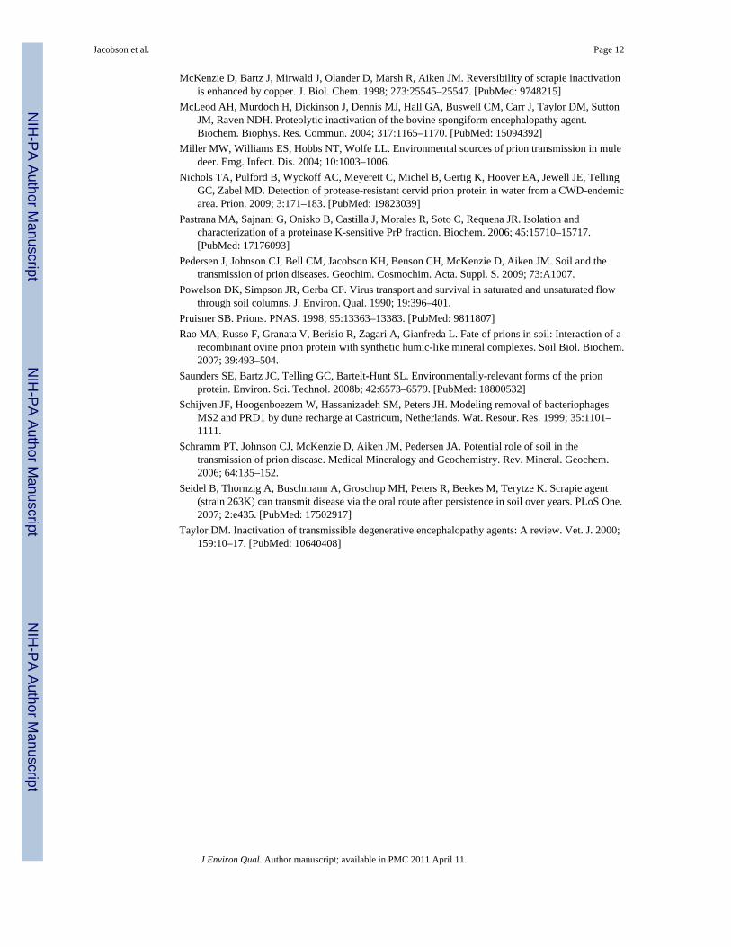
PrPTSE Recovery in Transport ExperimentsRecovery of PrPTSE loaded onto the columns by extraction of soils with 100°C SDS-PAGEsample buffer was incomplete. Recoveries ranged from 23% for the Boardman silt loamcolumn to 76% from the Site C Subsoil column and averaged near 50% (Table 2). If PrPwas present at the detection limit of immunoblotting for sections containing no observablePrP, recoveries would increase by ~3% (Table 2).
The PrPTSE recoveries observed in the present study, while incomplete, are consistent withthose reported previously (Johnson et al., 2006; Leita et al., 2006; Cooke et al., 2007; Seidelet al., 2007; Jacobson et al., 2009), and higher than those reported for α-recPrP after 1 monthin the column (Cooke and Shaw, 2007). Possible sources of loss in the present study thatwere considered and discounted or controlled for include (1) sorption to experimentalequipment, (2) degradation, and (3) interference of the leachate matrix and soil extracts withdetection. First, poly(tetrafluoroethylene) (PTFE, Teflon®) or comparable fluoropolymerswere used for column components in contact with PrPTSE during transport experiments (videsupra) based on trials that indicated minimal PrPTSE binding to PTFE relative to otherpotential column materials (viz. glass, polyvinyl chloride, polymethyl methacrylate).Furthermore, the surface area of the soil particles (23-28 m2) greatly exceeded that availablefor attachment to the column (~8 × 10⁻⁴ m2). All collection tubes used were manufacturedto minimize protein binding. Second, degradation of PrPTSE by residual, active PK in theBH could not account for the incomplete recoveries. The BH used was digested with PKprior to use. This treatment was expected to eliminate the majority of labile PrP in the brainhomogenate (PrPC and protease-sensitive PrPTSE), leaving protease-resistant PrPTSE
(resPrPTSE) (Pastrana et al., 2006). Significant degradation of resPrPTSE by soilmicroorganisms over the time course of experiments would not be anticipated; PrPTSE isresistant to most proteases tested (McLeod et al., 2004). Prior to addition to the column, PKwas inhibited with PMSF. Control experiments demonstrated that further degradation ofPrPTSE did not occur after PMSF addition. Third, all standards used in immunoblottingexperiments were treated in the same manner as the experimental samples to eliminatematrix effects on the detection of PrP. The standards were exposed to and loaded onto thepolyacrylamide gel in effluent liquid from the relevant column collected immediately priorto introducing PrPTSE to the column.
A possible explanation for the incomplete recovery is a time-dependent decline inextractability caused by a strengthening of the attachment between PrPTSE and componentsof the porous material. Time-dependent declines in extractability from soil have been well-documented of small organic molecules (Hatzinger and Alexander, 1995; Alexander, 2000),noted in previous laboratory-scale experiments analyzing PrPTSE in soils conducted in ourlaboratory, and observed or suspected in experiments carried out by others (Cooke et al.,2007; Seidel et al., 2007). Mechanisms governing declines in PrPTSE extractability warrantfurther investigation.
Estimated Attachment CoefficientsPrP was not detected in any effluent samples. Thus, Eq. 3 was used to estimate a lower-bound katt
app that would yield PrPTSE at the detection limit of the immunoblot assay in the
Jacobson et al. Page 7
J Environ Qual. Author manuscript; available in PMC 2011 April 11.
NIH
-PA Author Manuscript
NIH
-PA Author Manuscript
NIH
-PA Author Manuscript

first pore volume of effluent. The actual kattapp of PrP in these soils is expected to be higher.
In this analysis, all possible retention mechanisms (e.g., attachment, straining) are collapsedinto a single first-order kinetic parameter (see discussion of straining below). Thissimplification of the factors controlling PrP-media interactions is consistent with the level ofdetail in our dataset. Of the five soils tested in this study, the Site C Subsoil had the lowestkatt
app, 1.8 h−1, while the Boardman silt loam had the highest kattapp, 2.8 h−1. The Site S
Topsoil, the Site S Subsoil, and the Site C Topsoil had intermediate lower-bound kattapp
values (2.2, 2.1, and 2.0 h−1, respectively).
These kattapp are lower than the lower-bound katt
app values (2.6 to 3.3 h−1) we previouslyreported (Jacobson et al., 2009). This difference is, however, due at least in part todifferences in the detection limits of the immunoblot for PK-treated infected BH versusenriched PrPTSE, and does not imply an actual difference in attachment. The katt
app for prionproteins are at least one order of magnitude larger than katt
app reported for virus particles inporous media (Jin et al., 2000; Schijven et al., 1999), the most comparable colloidal entities.
Straining and wedging were not explicitly included in the analysis; to the extent thatstraining and wedging occurred in the porous media investigated, they would be captured inthe apparent attachment coefficients. Straining can occur regardless of the presence orabsence of an energy barrier to attachment (Johnson et al., 2007b). The transportexperiments were conducted under solution conditions favoring attachment (Johnson et al.,2006; Cooke et al., 2007). However, we have not identified whether the initial contactbetween PrPTSE and soil particles resulted from direct attatchment or straining.
If physical straining was the sole mechanism for retention of PrPTSE, we would expect asubstantial proportion of PrPTSE to be removed by a simple water extraction. We tested thispossibility using a procedure outlined by Bradford et al. (2006). At the conclusion of atransport experiment, rather than freeze the column, we excavated the soil from the columnand added it to microcentrifuge tubes containing an excess of eluent. After gentle mixingand centrifugation, all detectable PrPTSE remained associated with the soil particles (data notshown). When PK-digested BH was subjected to the same centrifugation conditions, themajority of PrPTSE was in the supernatant fraction. These results indicate that PrPTSE in BHwas attached to the soil particles, regardless of the mechanism responsible for initial contact.Detachment of PrPTSE from soil particles requires extraction more aggressive thanresuspension in simulated rainwater. The most effective extractants for PrPTSE to date aresolutions of anionic surfactants, and even these are not fully effective (Johnson et al., 2006;Cooke et al., 2007). The work on PrPTSE extraction suggests that hydrophobic andelectrostatic interactions must be disrupted to release PrPTSE from soil particles.
In well-sorted, uniform porous media, straining can be an important colloid retentionmechanism for colloid-to-median grain (or “collector”) diameter ratios (dc/d50) ≥ 0.005(Johnson et al., 2007).. In the present study, dc/d50 ratios were 0.0027 to 0.0031 for theScottish soils and 0.006 for the Boardman silt loam using dc set equal to the Z-averagehydrodynamic diameter of particles in PK-treated BH suspended in synthetic rainwater (240nm, determined by dynamic light scattering). These dc/d50 ratios suggest that theexperiments conducted with one soil (Boardman silt loam) may have been at the very edgeof the straining regime. However, the applicability of such dc/d50 thresholds for the onset ofstraining is unclear for natural soils containing a broad range of particle sizes and surfacechemistries, and for colloids exhibiting substantial polydispersity. Straining of colloidsexhibiting substantial polydispersity in natural soils with a broader range of pore sizes mayoccur at dc/d50 ratios less than 0.005.
Jacobson et al. Page 8
J Environ Qual. Author manuscript; available in PMC 2011 April 11.
NIH
-PA Author Manuscript
NIH
-PA Author Manuscript
NIH
-PA Author Manuscript

Environmental ImplicationsThe present study examined migration of N-terminally truncated PrPTSE in several soils withrelatively high sand and silt contents; retention of full-length PrPTSE is expected to besimilar or more limited. The PrPTSE in BH decomposed in a laboratory setting was reportedto be primarily N-terminally truncated (Saunders et al., 2008), and this is likely true ofPrPTSE released from decomposing carcasses. We therefore digested infected BH with PK toN-terminally cleave PrPTSE. Proteinase K treatment had the added benefits of eliminatingPrPC and partially degrading the BH matrix. Since the N-terminus is implicated in the strongbinding of PrPTSE to clay particles (Johnson et al., 2006; Cooke et al., 2007), and it isexpected that full-length PrPTSE would be retained more strongly than N-terminallytruncated PrPTSE, at least by clay minerals.
The persistence of TSE infectivity in the environment (Brown and Gajdusek, 1991; Miller etal., 2004; Georgsson et al., 2006; Seidel et al., 2007) may lead to release and migration ofPrPTSE over time scales considerably longer than those simulated. Furthermore, the slowdecay of the tissue of an infected carcass, but not the associated PrPTSE, might also slowrelease of the infectious agent. Other factors that warrant investigation include the influenceof natural organic matter (dissolved and particle-associated), the effects of pore size anddistribution, and transport through preferential flow paths formed by biota and physicalpedogenic processes (Kung et al., 2000; Cey et al., 2009). The present study was notdesigned to investigate the potential for macropore transport of PrPTSE. Transport viapreferential flow paths may allow more extensive migration of PrPTSE in soils than observedhere.
Although the present study was designed to provide insight into the potential for PrPTSE tomigrate soil in the context of on-farm burial of infected carcasses, implications forenvironmental transmission of CWD and scrapie are apparent. TSE agent in infected tissuesdeposited at the soil surface (e.g., carcasses of deceased animals, gut piles, placenta) mayremain near the soil surface in a bioavailable form (Johnson et al., 2007a, Seidel et al., 2007)and contribute to the spread of CWD and scrapie through relatively small, contaminatedpatches of soils serving as “hotspots” of infectivity. The strong binding of PrPTSE to somesoil components and maintenance of the agent near the soil surface also raises the possibilitythat disease-associated prion protein could be delivered to surface waters attached toparticles during runoff-producing rainfall and snow melt events (Nichols et al., 2009).
CONCLUSIONSOur results suggest that migration of PrPTSE released from decomposing infected tissueswill be minimal in natural soils with textures similar to silt loam, loamy sand, sandy loam,and sandy clay loam. The majority of PrPTSE released into soil will remain at or near thelocation of deposition. The experiments in the present study were conducted under saturatedconditions; transport under unsaturated conditions is expected to be equally if not morelimited (Powelson et al., 1990). Migration of PrPTSE (and most likely infectivity) deeper intothe soil column is expected to be restricted in finely textured soils with relatively loworganic carbon contents.
Supplementary MaterialRefer to Web version on PubMed Central for supplementary material.
Jacobson et al. Page 9
J Environ Qual. Author manuscript; available in PMC 2011 April 11.
NIH
-PA Author Manuscript
NIH
-PA Author Manuscript
NIH
-PA Author Manuscript

AcknowledgmentsThis research was supported by grants from the Wisconsin Department of Natural Resources, the National ScienceFoundation (CBET-0547484 (CAREER) and CBET-0826204), and the U.S. Environmental Protection Agency (4C-R070-NAEX). We thank Erin Shanle, Kartik Kumar, Xiaodong Wang and Letitia Wong for laboratory assistance.K.H.J. was supported by a National Institutes of Health training grant (NIH 5 T32 GM08349). C.H.B. was partiallysupported by his Wisconsin Distinguished Professorship. Endorsement by the sponsors is not implied and shouldnot be assumed. We thank two anonymous reviewers for their constructive comments.
ABBREVIATIONS
ADRE advection-dispersion-reaction equation
BH brain homogenate
BSE bovine spongiform encephalopathy
CEC cation exchange capacity
CJD Creutzfeldt-Jakob disease
CWD chronic wasting disease
DOC dissolved organic carbon
DTT dithiothreitol
EDTA ethylenedinitrilotetraacetic acid
HY Hyper strain of hamster-adapted transmissible mink encephalopathy
I ionic strength
IgG immunoglobulin G
mAb monoclonal antibody
MOPS 3-(N-morpholino)propanesulfonic acid
PAGE polyacrylamide gel electrophoresis
PFTE poly(tetrafluoroethylene)
pHIEP isoelectric point
PK proteinase K
PMSF phenylmethylsulphonyl fluoride
PrP prion protein
PrPC cellular prion protein
PrPTSE disease-associated prion protein
PV pore volume
recPrP recombinant prion protein
resPrPTSE proteinase K-resistant PrPTSE
SDS sodium dodecyl sulfate
TME transmissible mink encephalopathy
Tris Tris-(hydroxymethyl) aminomethane HCl
TSE transmissible spongiform encephalopathy
Jacobson et al. Page 10
J Environ Qual. Author manuscript; available in PMC 2011 April 11.
NIH
-PA Author Manuscript
NIH
-PA Author Manuscript
NIH
-PA Author Manuscript

REFERENCESAlexander M. Aging, bioavailability, and overestimation of risk from environmental pollutants.
Environ. Sci. Technol. 2000; 34:4259–4265.Atwood TC, Weeks HP. Sex-specific patterns of mineral lick preference in white-tailed deer.
Northeastern Naturalist. 2003; 10:409–414.Bessen RA, Marsh RF. Distinct PrP properties suggest the molecular basis of strain variation in
transmissible mink encephalopathy. J. Virol. 1994; 68:7859–7868. [PubMed: 7966576]Beyer WN, Connor EE, Gerould S. Estimates of soil ingestion by wildlife. J. Wildl. Manage. 1994;
58:375–382.Bolton DC, Bendheim PE, Marmorstein AD, Potempska A. Isolation and structural studies of the
intact scrapie agent protein. Arch. Biochem. Biophys. 1987; 258:579–590. [PubMed: 2890330]Boyd, Claude E. Water quality: An introduction. Springer; NY: 2000.Bradford SA, Simunek J, Bettahar M, van Genuchten MT, Yates SR. Significance of straining in
colloid deposition: Evidence and implications. Wat. Resour. Res. 2006; 42(12)Brown P, Gadjusek DC. Survival of scrapie virus after 3 years internment. Lancet. 1991; 337:269–270.
[PubMed: 1671114]Cey EE, Rudolph DL, Passmore J. Influence of macroporosity on preferential solute and colloid
transport in unsaturated field soils. J. Contam. Hydrol. 2009; 107:45–57. [PubMed: 19435645]Cooke CM, Rodgers J, Smith A, Fernie K, Shaw G, Sommerville RA. Fate of prions in soil: Detergent
extraction of PrP from soils. Environ. Sci. Technol. 2007; 41:811–817. [PubMed: 17328187]Cooke CM, Shaw G. Fate of prions in soil: Longevity and migration of recPrP in soil columns. Soil
Biol. Biochem. 2007; 39:1181–1191.Georgsson G, Sigurdarson S, Brown P. Infectious agent of sheep scrapie may persist in the
environment for at least 16 years. J. Gen. Virol. 2006; 87:3737–3740. [PubMed: 17098992]Hatzinger PB, Alexander M. Effect of aging of chemicals in soil on their biodegradability and
extractability. Environ. Sci. Technol. 1995; 29:537–545.Jacobson KJ, Lee S, McKenzie D, Benson CH, Pedersen JA. Transport of the pathogenic prion protein
through landfill materials. Environ. Sci. Technol. 2009; 43:2022–2028. [PubMed: 19368208]Jin Y, Chu Y, Li Y. Virus removal and transport in saturated and unsaturated sand columns. J.
Contam. Hydrol. 2000; 43:111–128.Johnson CJ, Pedersen JA, Chappell RJ, McKenzie D, Aiken JM. Oral transmissibility of prion disease
is enhanced by binding to soil particles. PLoS Pathog. 2007a; 3:e93. [PubMed: 17616973]Johnson CJ, Phillips KE, Schramm PT, McKenzie D, Aiken JM, Pedersen JA. Prions adhere to soil
minerals and remain infectious. PLoS Pathog. 2006; 2:296–302.Johnson WP, Tong M, Li X. On colloid retention in saturated porous media in the presence of energy
barriers: The failure of alpha, and opportunities to predict eta. Wat. Resour. Res. 2007b; 43(12)Kung K-JS, Kladivko EJ, Gish TJ, Steenhuis TS, Bubenzer G, Helling CS. Quantifying preferential
flow by breakthrough of sequentially applied tracers: Silt loam soil. Soil Sci. Soc. Am. J. 2000;64:1296–1304.
Lee T, Benson CH. Using waste green sands for treating alachlor and metolachlor in groundwater. J.Environ. Qual. 2004; 33:1682–1693. [PubMed: 15356228]
Leita L, Fornasier F, De Nobili M, Bertoli A, Genovesi S, Sequi P. Interactions of prion proteins withsoil. Soil Biol. Biochem. 2006; 38:1638–1644.
Lepore BJ, Barak P. Colormetric microwell method for determining bromide concentrations. Soil Sci.Soc. Am. J. 2009; 73:1130–1136.
Ma X, Benson CH, McKenzie D, Aiken JM, Pedersen JA. Adsorption of pathogenic prion protein toquartz sand. Environ. Sci. Technol. 2007; 41:2324–2330. [PubMed: 17438782]
Mathiason CK, Hays SA, Powers J, Hayes-Klug J, Langenberg J, Dahmes SJ, Osborn DA, Miller KV,Warren RJ, Mason GL, Hoover EA. Infectious prions in pre-clinical deer and transmission ofchronic wasting disease solely by environmental exposure. PLoS ONE. 2009; 4(6):e5916.[PubMed: 19529769]
Jacobson et al. Page 11
J Environ Qual. Author manuscript; available in PMC 2011 April 11.
NIH
-PA Author Manuscript
NIH
-PA Author Manuscript
NIH
-PA Author Manuscript
McKenzie D, Bartz J, Mirwald J, Olander D, Marsh R, Aiken JM. Reversibility of scrapie inactivationis enhanced by copper. J. Biol. Chem. 1998; 273:25545–25547. [PubMed: 9748215]
McLeod AH, Murdoch H, Dickinson J, Dennis MJ, Hall GA, Buswell CM, Carr J, Taylor DM, SuttonJM, Raven NDH. Proteolytic inactivation of the bovine spongiform encephalopathy agent.Biochem. Biophys. Res. Commun. 2004; 317:1165–1170. [PubMed: 15094392]
Miller MW, Williams ES, Hobbs NT, Wolfe LL. Environmental sources of prion transmission in muledeer. Emg. Infect. Dis. 2004; 10:1003–1006.
Nichols TA, Pulford B, Wyckoff AC, Meyerett C, Michel B, Gertig K, Hoover EA, Jewell JE, TellingGC, Zabel MD. Detection of protease-resistant cervid prion protein in water from a CWD-endemicarea. Prion. 2009; 3:171–183. [PubMed: 19823039]
Pastrana MA, Sajnani G, Onisko B, Castilla J, Morales R, Soto C, Requena JR. Isolation andcharacterization of a proteinase K-sensitive PrP fraction. Biochem. 2006; 45:15710–15717.[PubMed: 17176093]
Pedersen J, Johnson CJ, Bell CM, Jacobson KH, Benson CH, McKenzie D, Aiken JM. Soil and thetransmission of prion diseases. Geochim. Cosmochim. Acta. Suppl. S. 2009; 73:A1007.
Powelson DK, Simpson JR, Gerba CP. Virus transport and survival in saturated and unsaturated flowthrough soil columns. J. Environ. Qual. 1990; 19:396–401.
Pruisner SB. Prions. PNAS. 1998; 95:13363–13383. [PubMed: 9811807]Rao MA, Russo F, Granata V, Berisio R, Zagari A, Gianfreda L. Fate of prions in soil: Interaction of a
recombinant ovine prion protein with synthetic humic-like mineral complexes. Soil Biol. Biochem.2007; 39:493–504.
Saunders SE, Bartz JC, Telling GC, Bartelt-Hunt SL. Environmentally-relevant forms of the prionprotein. Environ. Sci. Technol. 2008b; 42:6573–6579. [PubMed: 18800532]
Schijven JF, Hoogenboezem W, Hassanizadeh SM, Peters JH. Modeling removal of bacteriophagesMS2 and PRD1 by dune recharge at Castricum, Netherlands. Wat. Resour. Res. 1999; 35:1101–1111.
Schramm PT, Johnson CJ, McKenzie D, Aiken JM, Pedersen JA. Potential role of soil in thetransmission of prion disease. Medical Mineralogy and Geochemistry. Rev. Mineral. Geochem.2006; 64:135–152.
Seidel B, Thornzig A, Buschmann A, Groschup MH, Peters R, Beekes M, Terytze K. Scrapie agent(strain 263K) can transmit disease via the oral route after persistence in soil over years. PLoS One.2007; 2:e435. [PubMed: 17502917]
Taylor DM. Inactivation of transmissible degenerative encephalopathy agents: A review. Vet. J. 2000;159:10–17. [PubMed: 10640408]
Jacobson et al. Page 12
J Environ Qual. Author manuscript; available in PMC 2011 April 11.
NIH
-PA Author Manuscript
NIH
-PA Author Manuscript
NIH
-PA Author Manuscript

FIGURE 1.PrPTSE extracted from (a) Boardman silt loam, (b) Site S Topsoil (c) Site S Subsoil (d) SiteC Topsoil and (e) Site C Subsoil. Each section represents ~3 mm or 12.5% of the columnheight. Section 1 is the topmost section of each column. 50 μL of 10% brain homogenate(BH, ~50 ng PrPTSE) was applied to the top of the column. Positive controls are given as apercentage of the total BH volume initially applied to the column. PrPTSE is detected usingthe monoclonal antibody, 3F4. Protein molecular mass is indicated at the left as determinedby use of pre-stained protein standards (Bio-Rad Precision Plus, Hercules, CA).
Jacobson et al. Page 13
J Environ Qual. Author manuscript; available in PMC 2011 April 11.
NIH
-PA Author Manuscript
NIH
-PA Author Manuscript
NIH
-PA Author Manuscript

NIH
-PA Author Manuscript
NIH
-PA Author Manuscript
NIH
-PA Author Manuscript
Jacobson et al. Page 14
Tabl
e 1
Soil
Prop
ertie
s
Para
met
erSi
te S
Top
soil
Site
S S
ubso
ilSi
te C
Top
soil
Site
C S
ubso
ilB
oard
man
silt
spec
ific
surf
ace
area
(m2 ·g
−1 )
†9.
414
.610
.612
.713
.0
frac
tion
orga
nic
carb
on (f
oc)‡
0.00
490.
0024
0.00
240.
0032
0.00
47
dry
dens
ity (M
g·m−
3 )1.
311.
391.
611.
541.
47
grav
imet
ric w
ater
con
tent
0.14
0.16
0.12
0.18
0.14
effe
ctiv
e po
rosi
ty0.
400.
340.
390.
280.
32
pore
vol
ume
(mm
3 )75
464
173
552
860
3
disp
ersi
vity
(mm
)1.
211.
621.
151.
1312
.5
CEC
(cm
ol+ ·
kg−
1 )16
.715
.016
.319
.017
.0
Min
eral
ogy
(%) §
Qua
rtz56
4467
5334
K-F
elds
par
411
44
6
Plag
iocl
ase
2626
58
35
Pyrit
e0
01
10
Hem
atite
12
00
1
Tota
l Phy
llosi
licat
es¶
1317
2334
24
Part
icle
Siz
e D
istr
ibut
ion
††
% sa
nd82
6563
6437
% si
lt12
1914
1555
% c
lay
616
2321
8
† Spec
ific
surf
ace
area
det
erm
ined
by
N2
adso
rptio
n
‡ f oc,
frac
tion
of o
rgan
ic c
arbo
n, d
eter
min
ed b
y th
e or
gani
c ca
rbon
dry
com
bust
ion
met
hod
usin
g a
CN
S-20
00 in
stru
men
t (Le
co, S
t. Jo
seph
, MO
)
§ Min
eral
ogy
dete
rmin
ed b
y X
-ray
diff
ract
ion
(K/T
Geo
scie
nces
, Arg
yle,
TX
)
¶ Full
phyl
losi
licat
e da
ta c
an b
e fo
und
in th
e Su
pple
men
tal I
nfor
mat
ion
††Pa
rticl
e si
ze d
istri
butio
n de
term
ined
by
wet
sedi
men
tatio
n (U
nive
rsité
Mon
tpel
lier)
.
J Environ Qual. Author manuscript; available in PMC 2011 April 11.
NIH
-PA Author Manuscript
NIH
-PA Author Manuscript
NIH
-PA Author Manuscript
Jacobson et al. Page 15
Table 2
Lower-bound attachment coefficients and recoveries
Soil
Apparentattachmentcoefficient,katt
app (h−1)
% PrPTSE
recovered †
Site S Topsoil > 2.2 40 (42)
Site S Subsoil > 2.1 54 (57)
Site C Topsoil > 2.0 68 (71)
Site C Subsoil > 1.8 76 (78)
Boardman Silt > 2.8 23 (26)
†Recoveries of PrPTSE determined by comparison of immunoblots of dilutions of soil column extracts to known amounts of PrPTSE. The
numbers in parentheses indicate PrPTSE recovery if samples with no detectable PrPTSE are assumed to have an amount of PrPTSE equivalent tothe detection limit.
J Environ Qual. Author manuscript; available in PMC 2011 April 11.
Related Documents