Page 1/32 Risk Factors Associated with Coronavirus Disease 2019 (COVID-19) Symptoms and Potential Vertical Transmission During Pregnancy: A Retrospective Cohort Study Bibita Peter John H. Stroger, Jr. Hospital of Cook County Nicholas Ree John H. Stroger, Jr. Hospital of Cook County Karen Ferrer John H. Stroger, Jr. Hospital of Cook County Laila Younes AMITA Health Saints Mary and Elizabeth Medical Center Barbara Lepe AMITA Health Saints Mary and Elizabeth Medical Center Khilfeh Manhal John H. Stroger, Jr. Hospital of Cook County Janardhan Mydam ( [email protected] ) John H. Stroger, Jr. Hospital of Cook County Research Article Keywords: COVID-19, BMI, obesity, overweight, potassium, hypokalemic, pregnancy, SARS-CoV-2 Posted Date: October 1st, 2021 DOI: https://doi.org/10.21203/rs.3.rs-916723/v1 License: This work is licensed under a Creative Commons Attribution 4.0 International License. Read Full License

Welcome message from author
This document is posted to help you gain knowledge. Please leave a comment to let me know what you think about it! Share it to your friends and learn new things together.
Transcript
Page 1/32
Risk Factors Associated with Coronavirus Disease2019 (COVID-19) Symptoms and Potential VerticalTransmission During Pregnancy: A RetrospectiveCohort StudyBibita Peter
John H. Stroger, Jr. Hospital of Cook CountyNicholas Ree
John H. Stroger, Jr. Hospital of Cook CountyKaren Ferrer
John H. Stroger, Jr. Hospital of Cook CountyLaila Younes
AMITA Health Saints Mary and Elizabeth Medical CenterBarbara Lepe
AMITA Health Saints Mary and Elizabeth Medical CenterKhilfeh Manhal
John H. Stroger, Jr. Hospital of Cook CountyJanardhan Mydam ( [email protected] )
John H. Stroger, Jr. Hospital of Cook County
Research Article
Keywords: COVID-19, BMI, obesity, overweight, potassium, hypokalemic, pregnancy, SARS-CoV-2
Posted Date: October 1st, 2021
DOI: https://doi.org/10.21203/rs.3.rs-916723/v1
License: This work is licensed under a Creative Commons Attribution 4.0 International License. ReadFull License
Page 2/32
AbstractObjective: The COVID-19 pandemic is of special concern for pregnant women. A growing body of evidencesuggests the virus can have a deleterious impact upon outcomes related to birth and newborn health.There is a paucity of published research demonstrating the factors that in�uence disease severity amongthose who are pregnant, while a growing body of evidence demonstrates that vertical transmission occurs.Our study investigated the impact of maternal characteristics upon COVID-19 outcomes, as well aswhether disease severity impacted pregnancy outcomes.
Methods: We conducted a retrospective cohort study of pregnant women with COVID-19 who wereadmitted to two public hospitals in our state between April-August, 2020. Pregnancy outcomes and clinical,laboratory, and placental data were collected.
Results: Thirty-four pregnant women tested positive for SARS-CoV-2. Among them, 55% (19/34) weresymptomatic. Of those who were symptomatic, 68% (13/19) presented with fever and cough. Those withsymptoms had a statistically signi�cant higher pregestational mean BMI compared with asymptomaticwomen (35.7±7.9 vs 26.7±6.9, P=0.004). Screening of biochemical records demonstrated thatsymptomatic women had lower potassium levels compared with those who were asymptomatic (median:3.70 mEq/L vs 4.30 mEq/L, P=0.009). High BMI (42.4 kg/m2) and low potassium levels (3.0 mEq/L) wereobserved in the only case of postpartum mortality among the symptomatic women. We did not observeany in�uence of maternal COVID-19 severity on placental histopathology/infant health or evidence ofvertical transmission, regardless of preterm status or duration of fetal exposure.
Conclusion: High pregestational BMI and lower potassium levels were associated with the presence ofCOVID-19 symptoms among pregnant women.
BackgroundAs the global impact of coronavirus disease 2019 (COVID-19) has grown, so has our understanding of theplethora of presenting symptoms. Known symptoms now include anosmia, myalgia, and gastrointestinalsymptoms in addition to respiratory symptoms of fever, dry cough, and dyspnea.1–3 It is also establishedthat older age, underlying medical conditions (immunosuppression, hypertension and diabetes), andrace/ethnicity (Black, Hispanic and Asian) increase risk of severe disease.4–6 The impact of COVID-19 andassociated risk factors on pregnancy and newborn health remains less certain.
Published research demonstrates an increased risk of premature labor and worse pregnancy outcomes inwomen infected with SARS CoV-2.7,8 Most often, they show no clinical symptoms and absence oftransmission of infection to the newborn.1,9 However, a recent case series reported seven maternal deathsfollowing severe COVID-19 infection.10 With recent implementation of aggressive COVID-19 testing, severalreports have emerged associating factors such as pregestational BMI, lymphocyte count, heart rate, andrespiratory rate with disease symptoms in SARS CoV-2-infected pregnant women.11–14 Another studydemonstrated an increased risk of preterm delivery in symptomatic women, suggesting a need to identify

Page 3/32
high-risk pregnant women.15 One of the potential reasons for the increased risk could be damage toplacental tissue.16
Our study sought to investigate the impact of maternal demographic, clinical and biochemicalcharacteristics on disease severity in pregnant women infected with SARS-Cov-2. We further investigatedthe in�uence of disease severity on pregnancy outcomes, vertical transmission of infection,histopathological evaluation of the placenta, and outcomes of the newborn.
Materials And Methods
Study design and study populationDuring the �rst-wave peak of COVID-19 infections in the US, testing for COVID-19 became universal for allpregnancy admissions from April 2020 in Illinois, USA. We conducted a retrospective cohort study betweenApril 1 and August 15, 2020 that included all consecutive pregnant women who were admitted at anygestational age and had laboratory-con�rmed COVID-19. Admissions were from two Chicago maternityhospitals— John H. Stroger, Jr. Hospital of Cook County and AMITA St. Mary’s and Elizabeth Hospital. Ourstudy was possible because of the April 2020 implementation of universal COVID-19 testing for allpregnant women admitted to hospitals in Illinois. We followed the World Health Organization (WHO)guidelines for diagnosis, which de�ne positive real-time reverse transcriptase-polymerase chain reaction(RT-PCR) assay of nasal or pharyngeal swabs as laboratory-con�rmed SARS-CoV-2.17 Patients with lack ofdata on symptoms and an inconsiderable number of other clinical characteristics, including risk factorsand maternal and perinatal outcomes, were excluded. We further included their newborns in the study, whowere also tested for SARS-CoV-2 infection using throat swabs.
Ethical approval and data collection
The study's protocol was expeditiously approved by the institutional research ethics committeesassociated with John H. Stroger, Jr. Hospital of Cook County (approval number: 20–098) and AMITAHealth Saints Mary and Elizabeth Medical Center (approval number: 2021-0193-02). The requirement ofinformed consent was waived due to the retrospective study design.
The following demographic and baseline maternal data were collected: age, race and ethnicity,anthropometric characteristics (e.g., BMI), lifestyle habits including substance abuse, comorbiditiesincluding pregestational obesity, hypertension, and gestational diabetes; whether the patient receivedantepartum therapy including hydroxychloroquine treatment. All participants underwent clinical evaluationof presenting signs and symptoms, detailed laboratory assessment of blood and urine samples, andradiologic chest assessment, if needed.
Maternal blood sample assessment included hemoglobin, blood cell counts, in�ammatory markers (e.g., C-reactive protein [CRP]), serum concentration of electrolytes (sodium, potassium, calcium, and chloride),liver function (alanine aminotransferase [ALT], aspartate aminotransferase [AST]), and renal function

Page 4/32
(blood urea nitrogen [BUN] and creatinine). Data on pregnancy outcomes (including mode of delivery,gestational age (GA), and preeclampsia), and neonatal outcomes (including symptoms, APGAR scores,and birth weight) were recorded. A preterm birth or premature birth was de�ned as one occurring at < 37weeks. We used speci�c cut-off values to de�ne blood dyscrasias for pregnant women according to theirtrimester: leukopenia as white cell count < 5.9 x 10⁹/L, neutropenia as neutrophil count < 3.9 x 10⁹/L, andlymphopenia as lymphocyte count < 1.0 x 10⁹/L 18 for the third trimester.
We also performed the gross and histopathological evaluation of placentas according to the Amsterdamconsensus statement guidelines.19
We assessed the effect of length of duration of fetal exposure to maternal SARS-CoV-2 and the possibilityof increased risk of vertical transmission, morbidity, and mortality speci�cally among preterm infants (< 37weeks of gestation). We sub-analyzed women who were infected prior to 37 weeks of gestation, focusingon evidence of SARS-CoV-2 infection among preterm infants born to these women, including positive RT-PCR testing and any clinical signs/symptoms attributable to infection.
We further performed laboratory assessment of blood samples taken from the newborns and followed upboth mother and infant until six weeks after delivery. We de�ned the infant's speci�c blood dyscrasiasaccording to their age of life: leukopenia as white cell count < 13.0 x 10⁹/L for term infants at 1–12 hoursof life and < 9.0 x 10⁹/L for preterm infants at birth; neutropenia as neutrophil count < 6.0 x 10⁹/L for terminfants at 1–12 hours of life and < 6.0 x 10⁹/L for preterm infants at birth; and lymphopenia as lymphocytecount < 2.0 x 10⁹/L for both term infants at 1–12 hours of life and preterm infants at birth.21 Evidence ofvertical transmission was further evaluated for the presence of SARS-CoV-2 according to CDC guidelines.22
As, and when, appropriate we evaluated infants for immunoglobulin [Ig] G and IgM levels.
All the data collected was curated using a customized data collection form, and two study investigators(JM and BP) independently reviewed the data collection forms for any errors. The data was locked andsecured appropriately according to rules and principles laid down in the Health Insurance Portability andAccountability Act of 1996 (HIPAA). The data collected from both sites was synchronized and anyinaccuracies were veri�ed with the concerned representative of the speci�c center.
Study OutcomeStudy outcomeThe patients were further classi�ed into two groups: symptomatic and asymptomatic, according to theexistence of any of known signs and symptoms of COVID-19 infection.
Statistical AnalysesStatistical analyses were conducted using STATA/IC 16 (Stata-Corp LP, TX, SA) and SAS 9.4 (SAS Institute,Inc., Cary, NC). Continuous variables were expressed as mean and standard deviation (SD) for normally
Page 5/32
distributed data, median and range for non-normally distributed data, and categorical variables asfrequency and percentage. The continuous variables were compared between symptomatic andasymptomatic individuals using a t-test or Mann-Whitney-U test and categorical variables were comparedusing the Pearson chi-square test or Fisher's-exact test, as appropriate. We calculated the unadjusted oddsratio (OR) using the simple logistic regression model. A p-value of < 0.05 was considered statisticallysigni�cant.
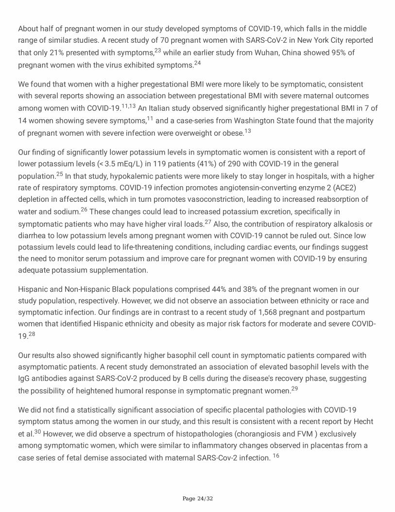
ResultsA total of 37 pregnant women with COVID-19 were admitted to the study centers during the study period.One patient was excluded for lack of information on the existence of symptoms, while two were excludeddue to lack of data related to maternal and perinatal outcomes. In total, 34 women were included in thestudy–19 were symptomatic (55%) for COVID-19 (Fig. 1).
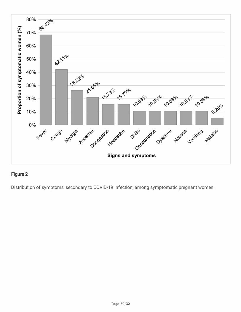
Baseline characteristics of pregnant womenThe baseline characteristics of the study population are shown in Table 1. Among the 19 symptomaticpatients, fever (n = 13; 68%), cough (n = 8; 42%), and myalgia (n = 5; 26%) were the most commonlyobserved symptoms (Fig. 2). In consent with their distribution, fever, cough, and myalgia was the mostfrequently observed combination of coexisting symptoms, seen in three patients (15.79%; data not shown).The average age of women in our study population was 26. The largest proportion were of Hispanicancestry (44%), followed by non-Hispanic Black (38%). The distribution of age and ethnicity did not differsigni�cantly between symptomatic and asymptomatic patients.

Page 6/32
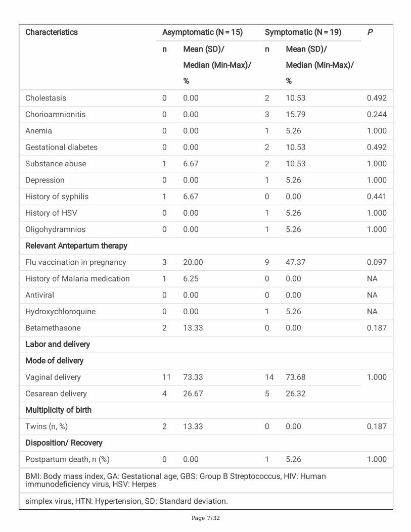
Table 1Clinical and Demographic Characteristics of Pregnant Women with COVID-19 by Symptom Status
Characteristics Asymptomatic (N = 15) Symptomatic (N = 19) P
n Mean (SD)/
Median (Min-Max)/
%
n Mean (SD)/
Median (Min-Max)/
%
Age at diagnosis 15 24.93 (5.09) 19 27.32 (5.96) 0.227
Gravidity
> 1 9 60.00 15 78.95 0.276
Parity
> 1 5 33.33 8 42.11 0.601
GA at diagnosis (weeks) 15 39.00 (26.20–41.00)
18 35.90 (24.00–41.00)
0.173
GA at delivery (weeks) 14 39.05 (27.00-41.40) 19 39.10 (37.10–41.20)
1.000
Diagnosis to delivery interval(weeks)
14 0.05 (0.00-1.80) 19 2.10 (0.00–15.00) 0.035
Ethnicity
Hispanic 7 46.67 8 42.11 1.000
Non-Hispanic black 6 40.00 7 36.84
Non-Hispanic white 0 0.00 1 5.26
Other 2 13.33 3 15.79
Complications during pregnancy
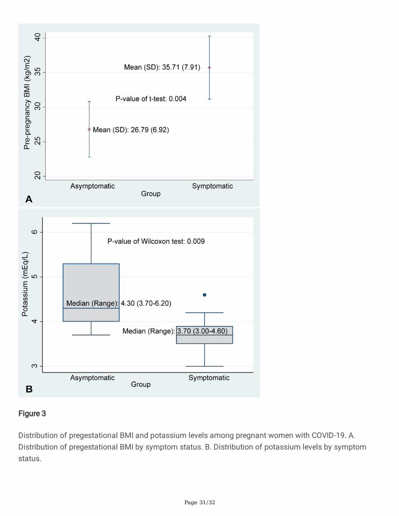
Pre-pregnancy BMI (kg/m2) 14 26.79 (6.92) 14 35.71 (7.91) 0.004
Obesity (BMI > 30) 0 0.00 5 26.32 0.032
GBS positive 4 26.67 7 36.84 0.715
HIV positive 0 0.0 2 10.53 0.492
Preeclampsia 2 13.33 2 10.53 1.000
HTN 1 6.67 5 26.32 0.196
BMI: Body mass index, GA: Gestational age, GBS: Group B Streptococcus, HIV: Humanimmunode�ciency virus, HSV: Herpes
simplex virus, HTN: Hypertension, SD: Standard deviation.
Page 7/32
Characteristics Asymptomatic (N = 15) Symptomatic (N = 19) P
n Mean (SD)/
Median (Min-Max)/
%
n Mean (SD)/
Median (Min-Max)/
%
Cholestasis 0 0.00 2 10.53 0.492
Chorioamnionitis 0 0.00 3 15.79 0.244
Anemia 0 0.00 1 5.26 1.000
Gestational diabetes 0 0.00 2 10.53 0.492
Substance abuse 1 6.67 2 10.53 1.000
Depression 0 0.00 1 5.26 1.000
History of syphilis 1 6.67 0 0.00 0.441
History of HSV 0 0.00 1 5.26 1.000
Oligohydramnios 0 0.00 1 5.26 1.000
Relevant Antepartum therapy
Flu vaccination in pregnancy 3 20.00 9 47.37 0.097
History of Malaria medication 1 6.25 0 0.00 NA
Antiviral 0 0.00 0 0.00 NA
Hydroxychloroquine 0 0.00 1 5.26 NA
Betamethasone 2 13.33 0 0.00 0.187
Labor and delivery
Mode of delivery
Vaginal delivery 11 73.33 14 73.68 1.000
Cesarean delivery 4 26.67 5 26.32
Multiplicity of birth
Twins (n, %) 2 13.33 0 0.00 0.187
Disposition/ Recovery
Postpartum death, n (%) 0 0.00 1 5.26 1.000
BMI: Body mass index, GA: Gestational age, GBS: Group B Streptococcus, HIV: Humanimmunode�ciency virus, HSV: Herpes
simplex virus, HTN: Hypertension, SD: Standard deviation.
Page 8/32
Obesity (n = 5; 26.32%), gestational diabetes (n = 2; 10.53%), and hypertension (n = 5; 26.32%) were themost commonly occurring comorbid conditions. All three were overrepresented in symptomatic patients.We also observed that the symptomatic women had signi�cantly higher pregestational BMI compared toasymptomatic women (35.71 vs 26.79, P = 0.004; Fig. 3A). Two of our symptomatic patients were alsopositive for HIV. Another three symptomatic women were diagnosed with chorioamnionitis. Acomparatively large proportion of symptomatic women in our cohort had received the �u vaccination in therecent past (47.37% of symptomatic women vs 20.00% of asymptomatic women). We further observedthat every third patient in our enrolled population was a Group B streptococcus (GBS) carrier; however, wefailed to detect any in�uence of GBS status on the absence or presence of symptoms. Our cohort also hadtwo women with a history with substance abuse, one symptomatic and one asymptomatic. We alsodetected syphilis in one of the asymptomatic patients.
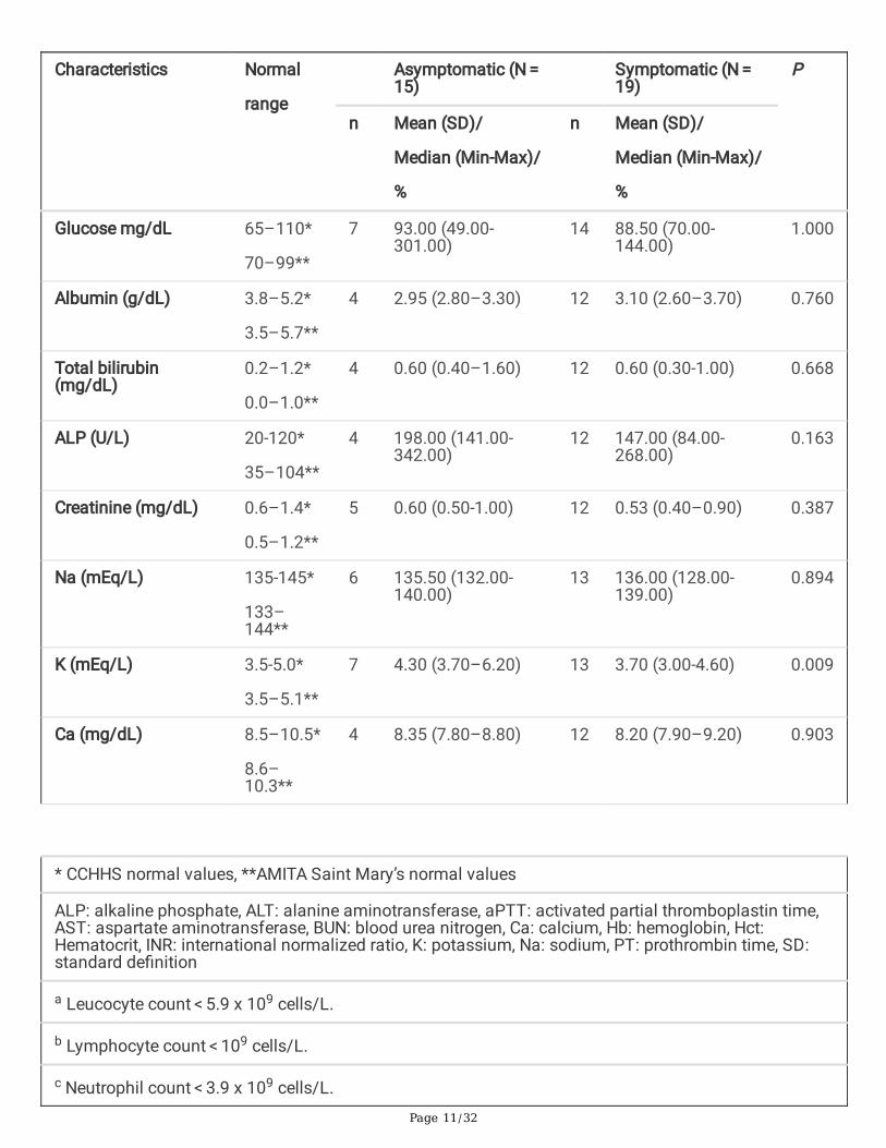
Laboratory characteristics of pregnant womenInitial evaluation of laboratory characteristics of the study population are shown in Table 2. Hematologicalanalyses suggested signi�cantly elevated basophil counts in symptomatic, compared with asymptomatic,women (P = 0.035). However, we failed to observe any signi�cant difference in blood dyscrasias, includingleukopenia, neutropenia, and lymphopenia. No differences in kidney or liver function were observed.Symptomatic women had signi�cantly lower potassium levels compared to asymptomatic women(median: 3.70 mEq/L [range 3.00-4.60] vs median 4.30 mEq/L [range 3.70–6.20]; P = 0.009; Fig. 3B). Threewomen, all of them symptomatic, showed evidence of secondary infection, with positive cell (n = 1) andurine (n = 2) cultures.

Page 9/32
Table 2Laboratory Characteristics of Pregnant Women with COVID-19 by Symptom Status
Characteristics Normal
range
Asymptomatic (N = 15)
Symptomatic (N = 19)
P
n Mean (SD)/
Median (Min-Max)/
%
n Mean (SD)/
Median (Min-Max)/
%
Hematological pro�le
Blood group
A nd 4 26.67 5 26.32 0.928
AB nd 1 6.67 0 0.00
B nd 1 6.67 2 10.53
O nd 9 60.00 12 63.16
Rhesus status
Negative nd 0 0.00 2 10.53 0.492
Positive nd 15 100 17 89.47
Hct (%) 34.9–44.3*
34.7–45.1**
15 34.00 (24.60–40.40)
17 33.90 (27.00-40.70) 0.940
Leukocyte count(K/uL)
4.4–10.6*
4.0–11.0**
15 8.80(4.40–14.20) 16 9.70 (4.50–12.20) 0.621
Leukopeniaa
Yes nd 2 13.33 5 31.25 0.394
* CCHHS normal values, **AMITA Saint Mary’s normal values
ALP: alkaline phosphate, ALT: alanine aminotransferase, aPTT: activated partial thromboplastin time,AST: aspartate aminotransferase, BUN: blood urea nitrogen, Ca: calcium, Hb: hemoglobin, Hct:Hematocrit, INR: international normalized ratio, K: potassium, Na: sodium, PT: prothrombin time, SD:standard de�nition
a Leucocyte count < 5.9 x 10⁹ cells/L.
b Lymphocyte count < 109 cells/L.
c Neutrophil count < 3.9 x 10⁹ cells/L.

Page 10/32
Characteristics Normal
range
Asymptomatic (N = 15)
Symptomatic (N = 19)
P
n Mean (SD)/
Median (Min-Max)/
%
n Mean (SD)/
Median (Min-Max)/
%
Lymphocyte count(K/uL)
1.2–3.4*
0.6–3.4**
10 16.75 (9.50–31.00) 9 15.40 (5.00–33.00) 0.902
Lymphopeniab
Yes nd 0 0.00 0 0.00 NA
Basophil count(K/uL)
0-0.1*
0-0.2**
10 0.05 (0.00-0.40) 9 0.30 (0.00–5.00) 0.035
Neutrophil count(K/uL)
2.2–6.9*
1.7–7.7**
10 72.70 (40.40–86.10)
7 72.80 (65.60–79.00)
1.000
Neutropeniac
Yes nd 0 0.00 0 0.00 NA
Platelet count (K/uL) 161–369*
150–450**
14 216.00 (93.00-377.00)
16 209.50 (113.00-290.00)
0.950
Blood biochemistry pro�le
ALT (U/L) 5-35*
0–50**
5 12.00 (6.00–20.00) 13 12.00 (7.00–39.00) 0.519
AST (U/L) 0-40*
0–40**
5 20.00 (19.00–28.00)
13 17.00 (12.00–35.00)
0.236
* CCHHS normal values, **AMITA Saint Mary’s normal values
ALP: alkaline phosphate, ALT: alanine aminotransferase, aPTT: activated partial thromboplastin time,AST: aspartate aminotransferase, BUN: blood urea nitrogen, Ca: calcium, Hb: hemoglobin, Hct:Hematocrit, INR: international normalized ratio, K: potassium, Na: sodium, PT: prothrombin time, SD:standard de�nition
a Leucocyte count < 5.9 x 10⁹ cells/L.
b Lymphocyte count < 109 cells/L.
c Neutrophil count < 3.9 x 10⁹ cells/L.
Page 11/32
Characteristics Normal
range
Asymptomatic (N = 15)
Symptomatic (N = 19)
P
n Mean (SD)/
Median (Min-Max)/
%
n Mean (SD)/
Median (Min-Max)/
%
Glucose mg/dL 65–110*
70–99**
7 93.00 (49.00-301.00)
14 88.50 (70.00-144.00)
1.000
Albumin (g/dL) 3.8–5.2*
3.5–5.7**
4 2.95 (2.80–3.30) 12 3.10 (2.60–3.70) 0.760
Total bilirubin(mg/dL)
0.2–1.2*
0.0–1.0**
4 0.60 (0.40–1.60) 12 0.60 (0.30-1.00) 0.668
ALP (U/L) 20-120*
35–104**
4 198.00 (141.00-342.00)
12 147.00 (84.00-268.00)
0.163
Creatinine (mg/dL) 0.6–1.4*
0.5–1.2**
5 0.60 (0.50-1.00) 12 0.53 (0.40–0.90) 0.387
Na (mEq/L) 135-145*
133–144**
6 135.50 (132.00-140.00)
13 136.00 (128.00-139.00)
0.894
K (mEq/L) 3.5-5.0*
3.5–5.1**
7 4.30 (3.70–6.20) 13 3.70 (3.00-4.60) 0.009
Ca (mg/dL) 8.5–10.5*
8.6–10.3**
4 8.35 (7.80–8.80) 12 8.20 (7.90–9.20) 0.903
* CCHHS normal values, **AMITA Saint Mary’s normal values
ALP: alkaline phosphate, ALT: alanine aminotransferase, aPTT: activated partial thromboplastin time,AST: aspartate aminotransferase, BUN: blood urea nitrogen, Ca: calcium, Hb: hemoglobin, Hct:Hematocrit, INR: international normalized ratio, K: potassium, Na: sodium, PT: prothrombin time, SD:standard de�nition
a Leucocyte count < 5.9 x 10⁹ cells/L.
b Lymphocyte count < 109 cells/L.
c Neutrophil count < 3.9 x 10⁹ cells/L.

Page 12/32
Characteristics Normal
range
Asymptomatic (N = 15)
Symptomatic (N = 19)
P
n Mean (SD)/
Median (Min-Max)/
%
n Mean (SD)/
Median (Min-Max)/
%
BUN (mg/dL) 8–20*
7–25**
5 5.00 (3.00–18.00) 12 5.00 (2.00–9.00) 0.631
Protein (g/dL) 6.4–8.3*
6.4–8.9**
4 5.50 (4.80-7.00) 12 5.75 (4.70–6.70) 0.670
Coagulation pro�le
PT (s) 11.9–14.1*
7 12.70 (11.30–16.00)
7 13.10 (10.70–13.50)
0.848
aPTT (s) 25.1-36.5* 7 31.70 (26.00-35.10) 6 30.45 (27.00-34.70) 0.943
INR 0.9–1.1** 7 1.00 (0.89–1.31) 7 1.01 (0.49–1.05) 0.654
Fibrinogen (mg/dL) 178–454*
163–463**
7 419.00 (283.00-674.00)
6 456.00 (354.00-542.00)
1.000
Microbiological pro�le
Blood culture
Negative 2 100.00 4 80.00 1.000
Positive 0 0.00 1 20.00
Urine culture
Negative 2 100 3 60.00 1.000
Positive 0 0.00 2 4.00
* CCHHS normal values, **AMITA Saint Mary’s normal values
ALP: alkaline phosphate, ALT: alanine aminotransferase, aPTT: activated partial thromboplastin time,AST: aspartate aminotransferase, BUN: blood urea nitrogen, Ca: calcium, Hb: hemoglobin, Hct:Hematocrit, INR: international normalized ratio, K: potassium, Na: sodium, PT: prothrombin time, SD:standard de�nition
a Leucocyte count < 5.9 x 10⁹ cells/L.
b Lymphocyte count < 109 cells/L.
c Neutrophil count < 3.9 x 10⁹ cells/L.

Page 13/32
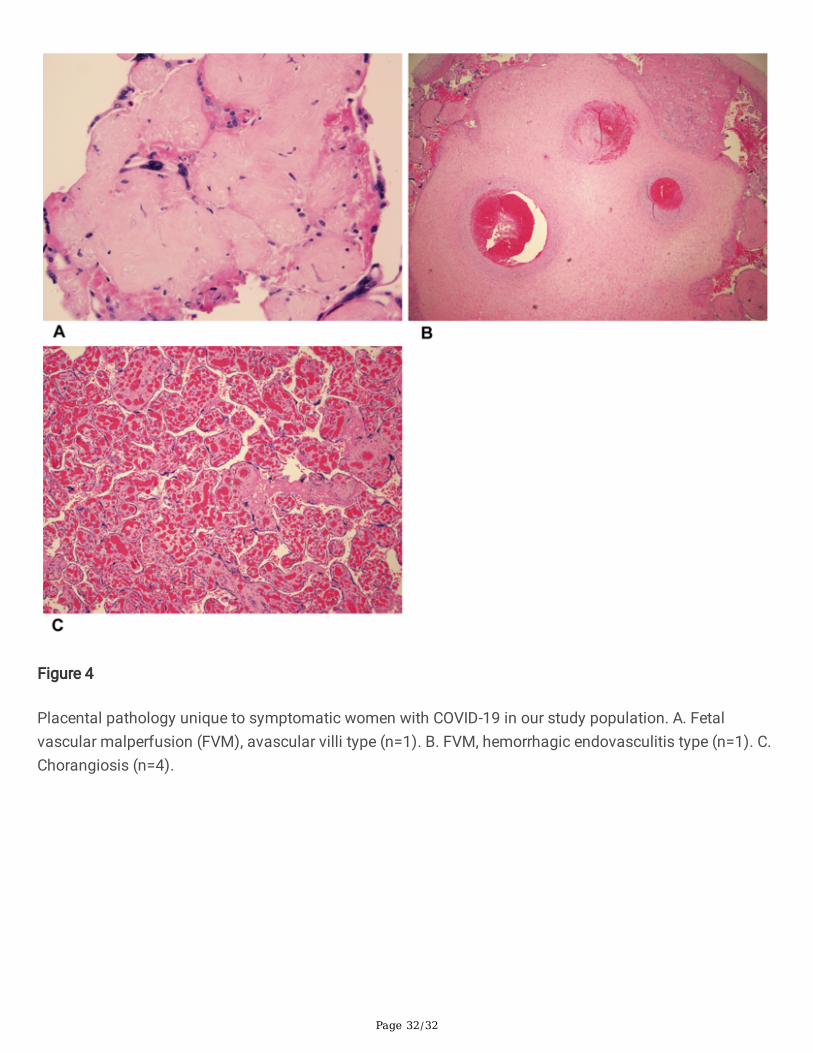
Placental histopathological featuresWe found no association of speci�c histopathological placental features with signs and symptoms ofCOVID-19 (Table 3). However, we did observe a higher prevalence of FVM, immunological or in�ammatoryprocesses, and chorangiosis in symptomatic women compared to asymptomatic women. Fetal vascularmalperfusion (n = 2; one avascular villi type and one hemorrhagic endovasculitis type) and chorangiosis (n = 4) were observed exclusively in the placentas of symptomatic patients (Fig. 4).

Page 14/32
Table 3Placental Pathology of Pregnant Women with COVID-19 by Symptom Status
Characteristics Asymptomatic (N = 15) Symptomatic (N = 19) P
n Mean (SD)/
Median (Min-Max)/
%
n Mean (SD)/
Median (Min-Max)/
%
MVM
No 3 25.00 6 31.58 1.000
Yes 9 75.00 13 68.42
FVM
No 12 100 17 89.47 0.510
Yes 0 0.00 2* 10.53
IM processes
No 7 58.33 7 36.84 0.242
Yes 5 41.67 12 63.16
Chorangiosis
No 12 100 15 78.95 0.139
Yes 0 0.00 4 21.05
Intervillous thrombus
No 9 75.00 17 89.47 0.350
Yes 3 25.00 2 10.53
Placental weight 11 489.00 (122.87) 16 506.13 (96.44) 0.689
IM: in�ammatory or immune process, FVM: fetal vascular malperfusion, MVM: maternal
vascular malperfusion, SD: standard deviation
*Avascular villi type (n = 1), hemorrhagic endovasculitis type (n = 1)
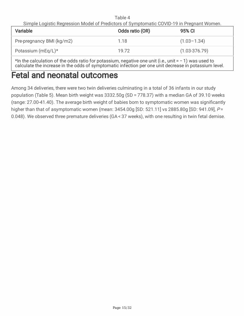
Logistic regression analysis of signi�cant maternal variablesSimple logistic regression analysis showed that the odds of severe COVID-19 increased as pregestationalBMI increased and pre-delivery potassium levels decreased (Table 4). For each unit increase inpregestational BMI, the odds of being symptomatic due to SARS Cov-2 infection increased by 18% (OR:1.18; 95% CI: 1.03–1.34), while a one-unit decrease in serum potassium levels among pregnant womenbefore delivery increased the odds of symptomatic infection by 19.72 (OR: 19.72; 95% CI: 1.03–376.79).
Page 15/32
Table 4Simple Logistic Regression Model of Predictors of Symptomatic COVID-19 in Pregnant Women.
Variable Odds ratio (OR) 95% CI
Pre-pregnancy BMI (kg/m2) 1.18 (1.03–1.34)
Potassium (mEq/L)* 19.72 (1.03-376.79)
*In the calculation of the odds ratio for potassium, negative one unit (i.e., unit = − 1) was used tocalculate the increase in the odds of symptomatic infection per one unit decrease in potassium level.
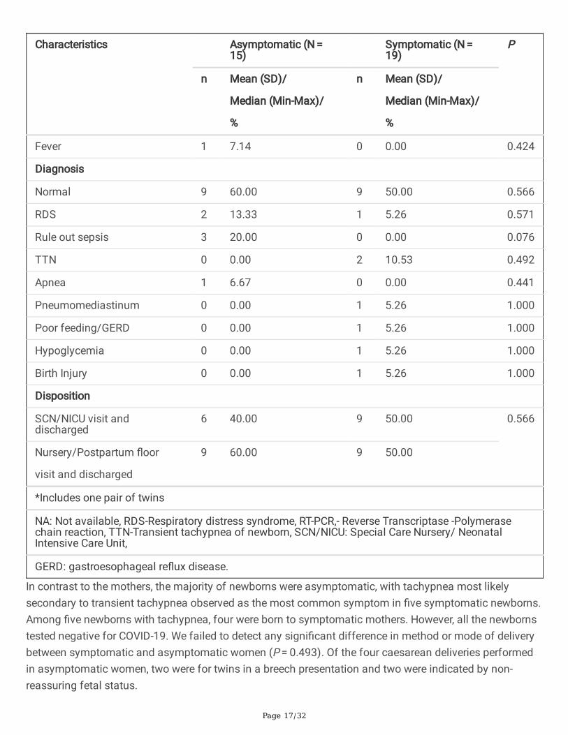
Fetal and neonatal outcomesAmong 34 deliveries, there were two twin deliveries culminating in a total of 36 infants in our studypopulation (Table 5). Mean birth weight was 3332.50g (SD = 778.37) with a median GA of 39.10 weeks(range: 27.00-41.40). The average birth weight of babies born to symptomatic women was signi�cantlyhigher than that of asymptomatic women (mean: 3454.00g [SD: 521.11] vs 2885.80g [SD: 941.09], P = 0.048). We observed three premature deliveries (GA < 37 weeks), with one resulting in twin fetal demise.

Page 16/32
Table 5Clinical and Demographic Characteristics of Infants Born to Women with COVID-19, by Maternal Symptom
StatusCharacteristics Asymptomatic (N =
15) Symptomatic (N =
19)P
n Mean (SD)/
Median (Min-Max)/
%
n Mean (SD)/
Median (Min-Max)/
%
Weight of newborns, g 15 2885.80 (941.09) 19 3454.00 (521.11) 0.048
Premature birth (< 37 weeks) 3 21.43 0 0.00 0.067
Stillbirth* 2 13.33 0 0.00
Sex
Male 9 60.00 9 47.37 0.464
Female 6 40.00 10 52.63
Apgar scores at 1 minute 15 9.00 (4.00–9.00) 19 9.00 (3.00–9.00) 0.790
Apgar scores at 5 minutes 15 9.00 (5.00–9.00) 19 9.00 (6.00–9.00) 0.724
RT PCR for SARS-Cov2
Positive 0 0.00 0 0.00 NA
Negative 15 100.00 13 68.42
Not done 1 0.00 6 31.58
Initial temperature 15 36.80 (35.80–37.20) 18 36.70 (36.40–37.60) 0.971
Symptoms
Apnea 1 7.14 0 0.00 0.424
Increased work of breathing 2 14.29 3 15.79 1.000
Desaturations 2 14.29 4 21.05 0.618
Hypoglycemia 0 0.00 1 5.26 1.000
Poor feeding 0 0.00 2 10.53 0.496
*Includes one pair of twins
NA: Not available, RDS-Respiratory distress syndrome, RT-PCR,- Reverse Transcriptase -Polymerasechain reaction, TTN-Transient tachypnea of newborn, SCN/NICU: Special Care Nursery/ NeonatalIntensive Care Unit,
GERD: gastroesophageal re�ux disease.
Page 17/32
Characteristics Asymptomatic (N = 15)
Symptomatic (N = 19)
P
n Mean (SD)/
Median (Min-Max)/
%
n Mean (SD)/
Median (Min-Max)/
%
Fever 1 7.14 0 0.00 0.424
Diagnosis
Normal 9 60.00 9 50.00 0.566
RDS 2 13.33 1 5.26 0.571
Rule out sepsis 3 20.00 0 0.00 0.076
TTN 0 0.00 2 10.53 0.492
Apnea 1 6.67 0 0.00 0.441
Pneumomediastinum 0 0.00 1 5.26 1.000
Poor feeding/GERD 0 0.00 1 5.26 1.000
Hypoglycemia 0 0.00 1 5.26 1.000
Birth Injury 0 0.00 1 5.26 1.000
Disposition
SCN/NICU visit anddischarged
6 40.00 9 50.00 0.566
Nursery/Postpartum �oor
visit and discharged
9 60.00 9 50.00
*Includes one pair of twins
NA: Not available, RDS-Respiratory distress syndrome, RT-PCR,- Reverse Transcriptase -Polymerasechain reaction, TTN-Transient tachypnea of newborn, SCN/NICU: Special Care Nursery/ NeonatalIntensive Care Unit,
GERD: gastroesophageal re�ux disease.
In contrast to the mothers, the majority of newborns were asymptomatic, with tachypnea most likelysecondary to transient tachypnea observed as the most common symptom in �ve symptomatic newborns.Among �ve newborns with tachypnea, four were born to symptomatic mothers. However, all the newbornstested negative for COVID-19. We failed to detect any signi�cant difference in method or mode of deliverybetween symptomatic and asymptomatic women (P = 0.493). Of the four caesarean deliveries performedin asymptomatic women, two were for twins in a breech presentation and two were indicated by non-reassuring fetal status.
Page 18/32
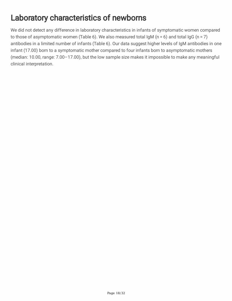
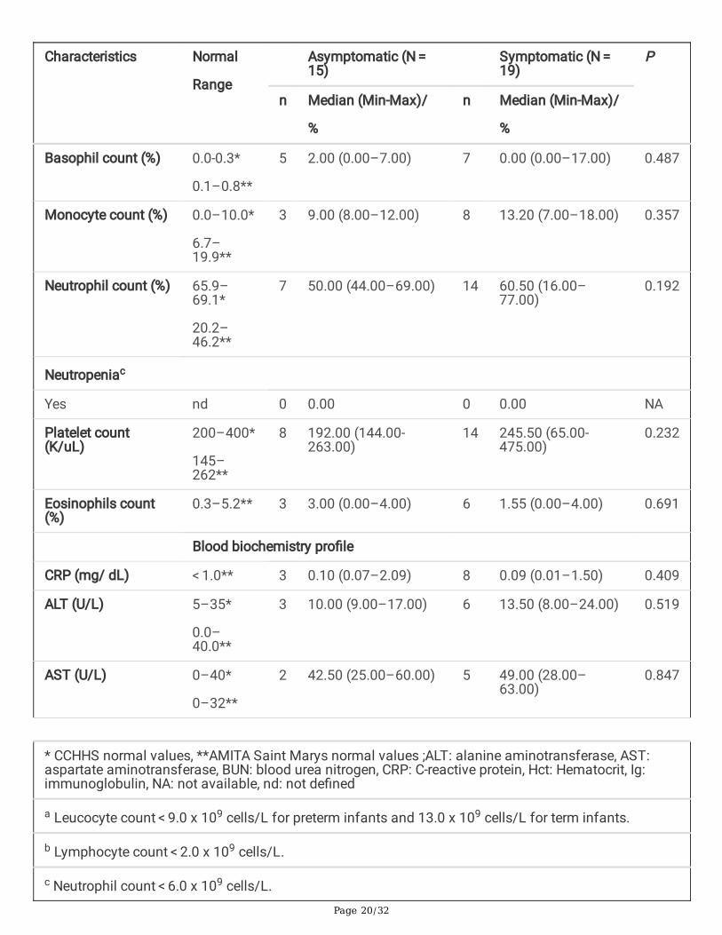
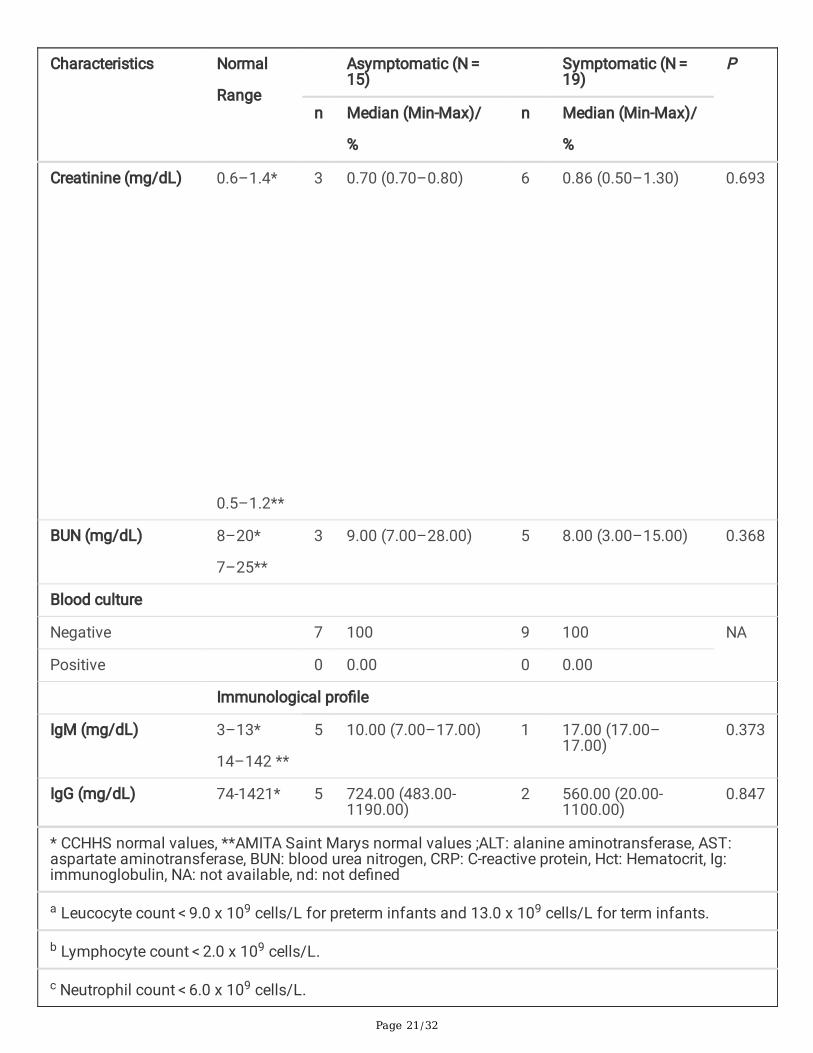
Laboratory characteristics of newbornsWe did not detect any difference in laboratory characteristics in infants of symptomatic women comparedto those of asymptomatic women (Table 6). We also measured total IgM (n = 6) and total IgG (n = 7)antibodies in a limited number of infants (Table 6). Our data suggest higher levels of IgM antibodies in oneinfant (17.00) born to a symptomatic mother compared to four infants born to asymptomatic mothers(median: 10.00, range: 7.00–17.00), but the low sample size makes it impossible to make any meaningfulclinical interpretation.

Page 19/32
Table 6Laboratory Characteristics of Infants Born to Women with COVID-19, by Maternal Symptom Status
Characteristics Normal
Range
Asymptomatic (N = 15)
Symptomatic (N = 19)
P
n Median (Min-Max)/
%
n Median (Min-Max)/
%
Hematological pro�le
Hct (%) 42–60.0*
42.0–54.0**
6 46.40 (38.70–56.40) 14 50.90 (45.20–61.80)
0.322
Leucocyte count(K/uL)
9.1–34.0*
8.0-15.4**
8 13.00 (6.90–24.80) 14 13.85 (9.10–23.70) 0.657
Leukopeniaa
Yes nd 2 28.57 5 35.71 1.000
Lymphocyte count(%)
11.0-30.9*
33.7–67.6**
7 26.00 (19.00–41.00) 14 22.00 (5.00–49.00) 0.501
Lymphopeniab
Yes nd 0 0.00 0 0.00 NA
* CCHHS normal values, **AMITA Saint Marys normal values ;ALT: alanine aminotransferase, AST:aspartate aminotransferase, BUN: blood urea nitrogen, CRP: C-reactive protein, Hct: Hematocrit, Ig:immunoglobulin, NA: not available, nd: not de�ned
a Leucocyte count < 9.0 x 10⁹ cells/L for preterm infants and 13.0 x 10⁹ cells/L for term infants.
b Lymphocyte count < 2.0 x 10⁹ cells/L.
c Neutrophil count < 6.0 x 10⁹ cells/L.
Page 20/32
Characteristics Normal
Range
Asymptomatic (N = 15)
Symptomatic (N = 19)
P
n Median (Min-Max)/
%
n Median (Min-Max)/
%
Basophil count (%) 0.0-0.3*
0.1–0.8**
5 2.00 (0.00–7.00) 7 0.00 (0.00–17.00) 0.487
Monocyte count (%) 0.0–10.0*
6.7–19.9**
3 9.00 (8.00–12.00) 8 13.20 (7.00–18.00) 0.357
Neutrophil count (%) 65.9–69.1*
20.2–46.2**
7 50.00 (44.00–69.00) 14 60.50 (16.00–77.00)
0.192
Neutropeniac
Yes nd 0 0.00 0 0.00 NA
Platelet count(K/uL)
200–400*
145–262**
8 192.00 (144.00-263.00)
14 245.50 (65.00-475.00)
0.232
Eosinophils count(%)
0.3–5.2** 3 3.00 (0.00–4.00) 6 1.55 (0.00–4.00) 0.691
Blood biochemistry pro�le
CRP (mg/ dL) < 1.0** 3 0.10 (0.07–2.09) 8 0.09 (0.01–1.50) 0.409
ALT (U/L) 5–35*
0.0–40.0**
3 10.00 (9.00–17.00) 6 13.50 (8.00–24.00) 0.519
AST (U/L) 0–40*
0–32**
2 42.50 (25.00–60.00) 5 49.00 (28.00–63.00)
0.847
* CCHHS normal values, **AMITA Saint Marys normal values ;ALT: alanine aminotransferase, AST:aspartate aminotransferase, BUN: blood urea nitrogen, CRP: C-reactive protein, Hct: Hematocrit, Ig:immunoglobulin, NA: not available, nd: not de�ned
a Leucocyte count < 9.0 x 10⁹ cells/L for preterm infants and 13.0 x 10⁹ cells/L for term infants.
b Lymphocyte count < 2.0 x 10⁹ cells/L.
c Neutrophil count < 6.0 x 10⁹ cells/L.
Page 21/32
Characteristics Normal
Range
Asymptomatic (N = 15)
Symptomatic (N = 19)
P
n Median (Min-Max)/
%
n Median (Min-Max)/
%
Creatinine (mg/dL) 0.6–1.4*
0.5–1.2**
3 0.70 (0.70–0.80) 6 0.86 (0.50–1.30) 0.693
BUN (mg/dL) 8–20*
7–25**
3 9.00 (7.00–28.00) 5 8.00 (3.00–15.00) 0.368
Blood culture
Negative 7 100 9 100 NA
Positive 0 0.00 0 0.00
Immunological pro�le
IgM (mg/dL) 3–13*
14–142 **
5 10.00 (7.00–17.00) 1 17.00 (17.00–17.00)
0.373
IgG (mg/dL) 74-1421* 5 724.00 (483.00-1190.00)
2 560.00 (20.00-1100.00)
0.847
* CCHHS normal values, **AMITA Saint Marys normal values ;ALT: alanine aminotransferase, AST:aspartate aminotransferase, BUN: blood urea nitrogen, CRP: C-reactive protein, Hct: Hematocrit, Ig:immunoglobulin, NA: not available, nd: not de�ned
a Leucocyte count < 9.0 x 10⁹ cells/L for preterm infants and 13.0 x 10⁹ cells/L for term infants.
b Lymphocyte count < 2.0 x 10⁹ cells/L.
c Neutrophil count < 6.0 x 10⁹ cells/L.

Page 22/32
Length of preterm fetal exposure and risk of verticaltransmissionFourteen of 33 pregnant women (42%) were infected prior to GA of 37 weeks, the cutoff for preterm birth(one woman had missing data on GA at the onset of infection). For the women who were diagnosed priorto 37 weeks (n = 14), the gestational age of the fetus at maternal COVID diagnosis ranged from 24 to 36.6weeks, with the longest duration of exposure 14.5 weeks (Table 7). The average duration of fetal exposure,which is the time interval between the diagnosis of maternal COVID-19 infection and birth, was two weeks.The mean duration of fetal exposure was longer for symptomatic than asymptomatic women (2.10 weeksversus 0.05 weeks (P = 0.035). However, none of the infants tested positive for COVID-19, despite fetalprematurity and a relatively long duration of exposure. Moreover none of the infants demonstrated anysigns or symptoms speci�c to COVID-19 .

Page 23/32
Table 7Neonatal outcomes and duration of fetal exposure among women infected with SARS-CoV-2 prior to 37
weeks gestation.*S.No.
GA at
maternal
COVID-19
diagnosis
GA of
fetus/
new-born
ondelivery
Durationbetweeninfectionanddeliveryin days
COVID-19
test onthe
newborn
(24hrs)
COVID-19
test onthe
newborn
(48hrs)
Total
IgM
(mg/dL)
Total
IgG
(mg/dL)
Signs andsymptoms
exclusivelyattributed
to COVID-19
infections
1 26.2 28 12 Negative Negative 10 483 None
2 33.3 38.3 35 Negative Negative NA NA None
3 36.6 37.2 03 Negative Negative NA NA None
4 34.6 38 22 NA NA NA NA None
5 35.2 37.3 14 Negative Negative NA NA None
6 32.1 32.6** 05 Negative,
Negative
Negative,
Negative
9, 17 706,724
None
7 33.1 39.6 60 Negative Negative NA NA None
8 28.4 39.4 76 NA NA NA NA None
9 36.6 37.1 02 Negative Negative NA < 20 None
10 32.6 40 50 Negative Negative NA NA None
11 24 38.5 102 NA NA NA NA None
12 28 39.3 80 NA NA NA NA None
13 32 39.5 53 NA NA NA NA None
*Among 14 women infected prior to 37 weeks gestation, one mother had an infant with fetal demise.
**Twins; GA: gestational age, Ig: immunoglobulin, NA: not available
DiscussionTo the best of our knowledge, this is the largest cohort of mother-baby dyads (SARS-Cov-2–positivewomen and their newborn infants) to undergo detailed clinical and biochemical investigation in the stateof Illinois. In this study, high BMI and low potassium levels were associated with symptomatic disease inmothers.
Page 24/32
About half of pregnant women in our study developed symptoms of COVID-19, which falls in the middlerange of similar studies. A recent study of 70 pregnant women with SARS-CoV-2 in New York City reportedthat only 21% presented with symptoms,23 while an earlier study from Wuhan, China showed 95% ofpregnant women with the virus exhibited symptoms.24
We found that women with a higher pregestational BMI were more likely to be symptomatic, consistentwith several reports showing an association between pregestational BMI with severe maternal outcomesamong women with COVID-19.11,13 An Italian study observed signi�cantly higher pregestational BMI in 7 of14 women showing severe symptoms,11 and a case-series from Washington State found that the majorityof pregnant women with severe infection were overweight or obese.13
Our �nding of signi�cantly lower potassium levels in symptomatic women is consistent with a report oflower potassium levels (< 3.5 mEq/L) in 119 patients (41%) of 290 with COVID-19 in the generalpopulation.25 In that study, hypokalemic patients were more likely to stay longer in hospitals, with a higherrate of respiratory symptoms. COVID-19 infection promotes angiotensin-converting enzyme 2 (ACE2)depletion in affected cells, which in turn promotes vasoconstriction, leading to increased reabsorption ofwater and sodium.26 These changes could lead to increased potassium excretion, speci�cally insymptomatic patients who may have higher viral loads.27 Also, the contribution of respiratory alkalosis ordiarrhea to low potassium levels among pregnant women with COVID-19 cannot be ruled out. Since lowpotassium levels could lead to life-threatening conditions, including cardiac events, our �ndings suggestthe need to monitor serum potassium and improve care for pregnant women with COVID-19 by ensuringadequate potassium supplementation.
Hispanic and Non-Hispanic Black populations comprised 44% and 38% of the pregnant women in ourstudy population, respectively. However, we did not observe an association between ethnicity or race andsymptomatic infection. Our �ndings are in contrast to a recent study of 1,568 pregnant and postpartumwomen that identi�ed Hispanic ethnicity and obesity as major risk factors for moderate and severe COVID-19.28
Our results also showed signi�cantly higher basophil cell count in symptomatic patients compared withasymptomatic patients. A recent study demonstrated an association of elevated basophil levels with theIgG antibodies against SARS-CoV-2 produced by B cells during the disease's recovery phase, suggestingthe possibility of heightened humoral response in symptomatic pregnant women.29
We did not �nd a statistically signi�cant association of speci�c placental pathologies with COVID-19symptom status among the women in our study, and this result is consistent with a recent report by Hechtet al.30 However, we did observe a spectrum of histopathologies (chorangiosis and FVM ) exclusivelyamong symptomatic women, which were similar to in�ammatory changes observed in placentas from acase series of fetal demise associated with maternal SARS-Cov-2 infection. 16

Page 25/32
Our data showed absence of perinatal transmission of COVID-19, regardless of the duration the fetus wasexposed to the virus prior to delivery (Table 7). Our �ndings are remarkable because they underscore thedistinctly different perinatal transmission dynamics of SARS-CoV-2 compared to other viruses that causecongenital infections, 20 where timing and duration of exposure may affect likelihood of transmissionand/or severity of disease in the newborn, and effects may be even greater for preterm infants.20 In ourstudy, none of the preterm infants were affected, and even the infant exposed for over 3 months in uterodid not test positive or show symptoms of COVID-19. Our results are in line with current literature showinglow rates of vertical transmission and symptomatic infection among infants of women with COVID-19during pregnancy.31–33
Our study has several limitations. The small sample size limits generalizability to other states and citiesacross US, and we may be underpowered due to missing values for several laboratory characteristics.Additionally, our retrospective data collection may contribute to ascertainment bias.
Even so, our �ndings have important implications for clinical management of pregnant women withCOVID-19. Our study suggests this group should be closely monitored for a rapid progression ofsymptoms, especially women with a high pregestational BMI and/or low potassium levels. However, itremains to be seen whether low potassium levels are a cause or outcome of symptoms observed inpregnant women with COVID-19.
DeclarationsEthics approval and consent to participate
The study's protocol was expeditiously approved by the institutional research ethics committeesassociated with John H. Stroger, Jr. Hospital of Cook County (approval number: 20-098) and AMITA HealthSaints Mary and Elizabeth Medical Center (approval number: 2021-0193-02). The institutional researchethics committees associated with John H. Stroger, Jr. Hospital of Cook County (approval number: 20-098)and AMITA Health Saints Mary and Elizabeth Medical Center (approval number: 2021-0193-02) waived therequirement of informed consent.
Guidelines
All methods were carried out in accordance with relevant guidelines and regulations.
Consent for publication
Not applicable.
Availability of data and materials
The datasets used and/or analysed during the current study are available from the corresponding authoron reasonable request, with removal of identifying information.
Page 26/32
Competing interests
The author(s) report no con�ict(s) of interest.
Funding
No funding was received for this study.
Authors' contributions
Author contributions were as follows: JM—conception, design, data analysis, and �rst draft; BP—datacollection, case follow-up, and �rst-draft edit; NR and KF—pathologic analysis of placentas and review/editof related content; KM—review and substantial edit; BL and LY data collection and IRB permissions fromAMITA St. Mary’s and Elizabeth Hospital.
All authors have approved the manuscript and have agreed to be personally accountable for their owncontributions and to ensure that questions related to the accuracy or integrity of any part of the work, evenones in which the author was not personally involved, are appropriately investigated, resolved, and theresolution documented in the literature.
Acknowledgements
The authors would like to thank Mohammed Siddiqui, BS, for his assistance in editing the slides for thepresentation of this study at the meeting of the Pediatric Academic Societies (PAS), and Helen Atkinson,MD for her assistance with manuscript preparation.
References1. Chen H, Guo J, Wang C, et al. Clinical characteristics and intrauterine vertical transmission potential of
COVID-19 infection in nine pregnant women: a retrospective review of medical records. Lancet2020;395:809-15. doi: 10.1016/S0140-6736(20)30360-3 2020/03/11.
2. Dell'Era V, Farri F, Garzaro G, Gatto M, Alu� Valletti P, Garzaro M. Smell and taste disorders duringCOVID-19 outbreak: cross-sectional study on 355 patients. Head Neck 2020;42:1591-96. doi:10.1002/hed.26288
3. Kumar A, Arora A, Sharma P, et al. Gastrointestinal and hepatic manifestations of Corona VirusDisease-19 and their relationship to severe clinical course: A systematic review and meta-analysis.Indian Journal of Gastroenterology 2020; 39:268–84. doi: 10.1007/s12664-020-01058-3
4. Li J, Huang DQ, Zou B, et al. Epidemiology of COVID-19: A Systematic Review and Meta-analysis ofClinical Characteristics, Risk factors and Outcomes. J Med Virol 2020:1‐10. doi: 10.1002/jmv.26424
5. Chin-Hong P, Alexander KM, Haynes N, Albert MA, Association of Black C. Pulling at the heart: COVID-19, race/ethnicity and ongoing disparities. Nat Rev Cardiol 2020;17:533-35. doi: 10.1038/s41569-020-0416-6
Page 27/32
�. Dai CL, Kornilov SA, Roper RT, et al. Characteristics and Factors Associated with COVID-19 Infection,Hospitalization, and Mortality Across Race and Ethnicity. medRxiv 2021. doi:10.1101/2020.10.14.20212803
7. Kourtis AP, Read JS, Jamieson DJ. Pregnancy and infection. N Engl J Med 2014;370:2211-18. doi:10.1056/NEJMra1213566.
�. Maródi L. Neonatal innate immunity to infectious agents. Infect Immun 2006;74:1999-2006. doi:10.1128/IAI.74.4.1999-2006.2006.
9. Zhu H, Wang L, Fang C, et al. Clinical analysis of 10 neonates born to mothers with 2019-nCoVpneumonia. Transl Pediatr 2020;9:51-60. doi: 10.21037/tp.2020.02.06
10. Hantoushzadeh S, Shamshirsaz AA, Aleyasin A, et al. Maternal death due to COVID-19. Am J ObstetGynecol 2020;223:109.e1-09.e16. doi: 10.1016/j.ajog.2020.04.030
11. Savasi VM, Parisi F, Patane L, et al. Clinical Findings and Disease Severity in Hospitalized PregnantWomen With Coronavirus Disease 2019 (COVID-19). Obstet Gynecol 2020;136:252-58. doi:10.1097/AOG.0000000000003979
12. Sentilhes L, De Marcillac F, Jouffrieau C, et al. Coronavirus disease 2019 in pregnancy was associatedwith maternal morbidity and preterm birth. Am J Obstet Gynecol 2020;223:914.e1-14.e15. doi:10.1016/j.ajog.2020.06.022
13. Lokken EM, Walker CL, Delaney S, et al. Clinical characteristics of 46 pregnant women with a severeacute respiratory syndrome coronavirus 2 infection in Washington State. Am J Obstet Gynecol2020;223:911.e1-11.e14. doi: 10.1016/j.ajog.2020.05.031
14. Vivanti AJ, Mattern J, Vauloup-Fellous C, et al. Retrospective Description of Pregnant Women Infectedwith Severe Acute Respiratory Syndrome Coronavirus 2, France. Emerg Infect Dis 2020;26:2069–76.doi: 10.3201/eid2609.202144
15. London V, McLaren R, Jr., Atallah F, et al. The Relationship between Status at Presentation andOutcomes among Pregnant Women with COVID-19. Am J Perinatol 2020;37:991-94. doi: 10.1055/s-0040-1712164
1�. Richtmann R, Torloni MR, Oyamada Otani AR, et al. Fetal deaths in pregnancies with SARS-CoV-2infection in Brazil: A case series. Case Rep Womens Health 2020;27:e00243. doi:10.1016/j.crwh.2020.e00243
17. World Health Organization. Laboratory testing for coronavirus disease 2019 (COVID-19) in suspectedhuman cases: interim guidance, 2 March 2020. Accessed June 28, 2021. https://apps.who.int/iris/handle/10665/331329.
1�. Abbassi-Ghanavati M, Greer LG, Cunningham FG. Pregnancy and laboratory studies: a reference tablefor clinicians. Obstet Gynecol 2009;114:1326-31. doi: 10.1097/AOG.0b013e3181c2bde8
19. Khong TY, Mooney EE, Ariel I, et al. Sampling and De�nitions of Placental Lesions: AmsterdamPlacental Workshop Group Consensus Statement. Arch Pathol Lab Med 2016;140:698-713. doi:10.5858/arpa.2015-0225-CC

Page 28/32
20. Leung K, Hon K, Yeung A, Leung A, Man E. Congenital infections in Hong Kong: an overview of TORCH.Hong Kong medical journal 2020;26:127-38. doi: 10.12809/hkmj198287.
21. Rennie JM, Roberton NRC. Textbook of Neonatology. Edinburgh: Churchill Livingstone; 1999.
22. McCartney SA, Kachikis A, Huebner EM, Walker CL, Chandrasekaran S, Adams Waldorf KM. Obesity asa contributor to immunopathology in pregnant and non-pregnant adults with COVID-19. Am J ReprodImmunol 2020:e13320. doi: 10.1111/aji.13320
23. Prabhu M, Cagino K, Matthews KC, et al. Pregnancy and postpartum outcomes in a universally testedpopulation for SARS-CoV-2 in New York City: a prospective cohort study. BJOG 2020;127:1548-56. doi:10.1111/1471-0528.16403
24. Chen L, Li Q, Zheng D, et al. Clinical Characteristics of Pregnant Women with Covid-19 in Wuhan,China. N Engl J Med 2020;382:e100(1)-e00(3). doi: 10.1056/NEJMc2009226
25. Alfano G, Ferrari A, Fontana F, et al. Hypokalemia in Patients with COVID-19. Clin Exp Nephrol2020;25:401–09. doi: 10.1007/s10157-020-01996-4.
2�. Guo J, Huang Z, Lin L, Lv J. Coronavirus Disease 2019 (COVID-19) and Cardiovascular Disease: AViewpoint on the Potential In�uence of Angiotensin-Converting Enzyme Inhibitors/AngiotensinReceptor Blockers on Onset and Severity of Severe Acute Respiratory Syndrome Coronavirus 2Infection. J Am Heart Assoc 2020;9:e016219. doi: 10.1161/jaha.120.016219
27. Weir MR, Rolfe M. Potassium homeostasis and renin-angiotensin-aldosterone system inhibitors. Clin JAm Soc Nephrol 2010;5:531-48. doi: 10.2215/cjn.07821109
2�. Grechukhina O, Greenberg V, Lundsberg LS, et al. Coronavirus disease 2019 pregnancy outcomes in aracially and ethnically diverse population. Am J Obstet Gynecol MFM 2020;2:100246. doi:10.1016/j.ajogmf.2020.100246
29. Rodriguez L, Pekkarinen PT, Lakshmikanth T, et al. Systems-Level Immunomonitoring from Acute toRecovery Phase of Severe COVID-19. Cell Rep Med 2020;1:100078-78. doi:10.1016/j.xcrm.2020.100078
30. Hecht JL, Quade B, Deshpande V, et al. SARS-CoV-2 can infect the placenta and is not associated withspeci�c placental histopathology: a series of 19 placentas from COVID-19-positive mothers. ModPathol 2020;33:2092-103. doi: 10.1038/s41379-020-0639-4
31. Kotlyar AM, Grechukhina O, Chen A, et al. Vertical transmission of coronavirus disease 2019: asystematic review and meta-analysis. Am J Obstet Gynecol 2021;224:35-53.e3. doi:10.1016/j.ajog.2020.07.049
32. Tolu LB, Ezeh A, Feyissa GT. Vertical transmission of severe acute respiratory syndrome coronavirus 2:a scoping review. PLOS ONE 2021;16:e0250196. doi: 10.1371/journal.pone.0250196.
33. Salvatore CM, Han J-Y, Acker KP, et al. Neonatal management and outcomes during the COVID-19pandemic: an observation cohort study. Lancet Child Adolesc Health 2020;4:721-27. doi:10.1016/S2352-4642(20)30235-2
Figures
Page 29/32
Figure 1
Flowchart of the study population selection and maternal and infant outcomes. Hospital 1 is John HStoger, Jr Hospital of Cook County; Hospital 2 is Amita St. Mary’s and Elizabeth Hospital.
Page 30/32
Figure 2
Distribution of symptoms, secondary to COVID-19 infection, among symptomatic pregnant women.
Page 31/32
Figure 3
Distribution of pregestational BMI and potassium levels among pregnant women with COVID-19. A.Distribution of pregestational BMI by symptom status. B. Distribution of potassium levels by symptomstatus.
Page 32/32
Figure 4
Placental pathology unique to symptomatic women with COVID-19 in our study population. A. Fetalvascular malperfusion (FVM), avascular villi type (n=1). B. FVM, hemorrhagic endovasculitis type (n=1). C.Chorangiosis (n=4).
Related Documents


![Review Article Hemoglobin Concentration and Pregnancy ...fetal gender Ren et al., [ ] Retrospective cohort , 25.8±2.9 China First PTB, LBW, and SGA Maternal age, gravidity, education,](https://static.cupdf.com/doc/110x72/60daea2ee634211738593b12/review-article-hemoglobin-concentration-and-pregnancy-fetal-gender-ren-et-al.jpg)