Transient Inhibition of Transforming Growth Factor-β1 in Human Diabetic CD 34 + Cells Enhances Vascular Reparative Functions Running Title: TGF-β1 PMO enhance vascular repair by CD34 + cells Ashay D. Bhatwadekar 1A* , E.P. Guerin 1A,2* , Yagna P.R. Jarajapu 1A , Sergio Caballero 1A , Carl Sheridan 2 , David Kent 3 , Laurence Kennedy 1B , M. Cecilia Lansang 1B , Frank W. Ruscetti 4 , Carl J. Pepine 1C , Paul J. Higgins 5 , Stephen H Bartelmez 6 , Maria B. Grant 1A *Equal Contributions 1A Pharmacology and Therapeutics, 1B Division of Endocrinology, Diabetes, and Metabolism, 1C Division of Cardiology, University of Florida Gainesville, FL; 2 The Vision Clinic, Circular Road, Kilkenny, Ireland; 3 School of Clinical Sciences University of Liverpool, Liverpool, United Kingdom; 4 Laboratory of Experimental Immunology, Center for Cancer Research, National Cancer Institute-Frederick, Frederick, MD; 5 Center for Cell Biology & Cancer Research Albany Medical College, Albany, NY; 6 BetaStem Therapeutics Inc., San Francisco, CA Correspondence: Maria B. Grant, MD E-mail:[email protected] Stephen H. Bartelmez, PhD Email: [email protected] Additional information for this article can be found in an online appendix at http://diabetes.diabetesjournals.org Submitted 28 February 2010 and accepted 27 April 2010. This is an uncopyedited electronic version of an article accepted for publication in Diabetes. The American Diabetes Association, publisher of Diabetes, is not responsible for any errors or omissions in this version of the manuscript or any version derived from it by third parties. The definitive publisher-authenticated version will be available in a future issue of Diabetes in print and online at http://diabetes.diabetesjournals.org. Diabetes Publish Ahead of Print, published online May 11, 2010 Copyright American Diabetes Association, Inc., 2010

Welcome message from author
This document is posted to help you gain knowledge. Please leave a comment to let me know what you think about it! Share it to your friends and learn new things together.
Transcript

Transient Inhibition of Transforming Growth Factor-β1 in Human Diabetic CD 34+ Cells Enhances Vascular Reparative Functions
Running Title: TGF-β1 PMO enhance vascular repair by CD34+ cells
Ashay D. Bhatwadekar1A*, E.P. Guerin1A,2*, Yagna P.R. Jarajapu1A, Sergio Caballero1A, Carl Sheridan 2, David Kent 3, Laurence Kennedy 1B, M. Cecilia Lansang 1B, Frank W. Ruscetti4, Carl J.
Pepine1C, Paul J. Higgins5, Stephen H Bartelmez6, Maria B. Grant1A
*Equal Contributions
1APharmacology and Therapeutics, 1BDivision of Endocrinology, Diabetes, and Metabolism, 1C Division of Cardiology, University of Florida Gainesville, FL; 2The Vision Clinic, Circular
Road, Kilkenny, Ireland; 3School of Clinical Sciences University of Liverpool, Liverpool, United Kingdom; 4 Laboratory of Experimental Immunology, Center for Cancer Research, National
Cancer Institute-Frederick, Frederick, MD; 5Center for Cell Biology & Cancer Research Albany Medical College, Albany, NY; 6BetaStem Therapeutics Inc., San Francisco, CA
Correspondence: Maria B. Grant, MD
E-mail:[email protected]
Stephen H. Bartelmez, PhD Email: [email protected]
Additional information for this article can be found in an online appendix at http://diabetes.diabetesjournals.org
Submitted 28 February 2010 and accepted 27 April 2010.
This is an uncopyedited electronic version of an article accepted for publication in Diabetes. The American Diabetes Association, publisher of Diabetes, is not responsible for any errors or omissions in this version of the manuscript or any version derived from it by third parties. The definitive publisher-authenticated version will be available in a future issue of Diabetes in print and online at http://diabetes.diabetesjournals.org.
Diabetes Publish Ahead of Print, published online May 11, 2010
Copyright American Diabetes Association, Inc., 2010

TGF-β1 PMO enhance vascular repair by CD34+ cells
2
Objective: Peripheral blood CD34+ cells from diabetic patients demonstrate reduced vascular reparative function due to decreased proliferation and diminished migratory prowess, largely resulting from decreased nitric oxide (NO) bioavailability. The levels of TGF-β, a key factor that modulates stem cell quiescence, are increased in the serum of type 2 diabetics. We asked whether transient TGF-β1 inhibition in CD34+ cells would improve their reparative ability. Research Design and Methods: To inhibit TGF-β1 protein expression, CD34+ cells were treated ex vivo with antisense phosphorodiamidate morpholino oligomers (TGF-β1-PMO) and analyzed for cell-surface CXCR4 expression, cell survival in the absence of added growth factors, SDF-1-induced migration, NO release, and in vivo retinal vascular reparative ability. Results: TGF-β1-PMO treatment of diabetic CD34+ cells resulted in increased expression of CXCR4, enhanced survival in the absence of growth factors, and increased migration and NO release as compared to cells treated with control PMO. Using a retinal ischemia reperfusion injury model in mice, we observed that recruitment of diabetic CD34+ cells to injured acellular retinal capillaries was greater following TGF-β1-PMO treatment compared to control-PMO treated cells. Conclusion: Transient inhibition of TGF-β1 may represent a promising therapeutic strategy for restoring the reparative capacity of dysfunctional diabetic CD34+ cells.

TGF-β1 PMO enhance vascular repair by CD34+ cells
3
one marrow derived progenitor cells (BMPCs) support vascular repair postnatally by direct integration into
blood vessels and by the release of paracrine factors such as vascular endothelial cell growth factor (VEGF), matrix metalloproteases, and angiopoietins to the neovessels (1; 2). BMPCs possess dramatic ability to revascularize areas within 6-12 hrs following the injury (3) accounting for total 1 – 12% of the endothelial cells present in blood vessels (4). Lineage negative (lin-) cells from mice that express the cell-surface antigens Sca-1 (Ly-6A/E) and c-kit can develop into endothelium, as can human lin- cells expressing surface CD34 (1; 5). Treatment with CD34+ cells presents an important therapeutic option for revascularization of ischemic vascular areas (6) and has been successful in numerous clinical trials (7; 8).
However, diabetes significantly impairs the vasoreparative ability of CD34+ cells. Diabetic patients with peripheral vascular disease have decreased levels of CD34+ cells and suffer poor vessel growth in response to ischemia (9), this defect is linked to reduced precursor cell function (10). The widespread vasodegeneration seen in diabetic retinopathy (DR) may be attributed to the inability of BMPCs to compensate for the increased endothelial injury associated with diabetes. In particular, the diabetic BMPCs are unable to repair retinal vasculature (11), thus the total rate of retinal cell loss greatly exceeds the reparative function these cells. We showed that diabetic CD34+ cells fail to revascularize areas of retinal vascular injury (11) likely due to reduced migration. Diabetic peripheral neuropathy further hampers repair due to defects of circadian release of BMPCs from the bone marrow (BM), creating an imbalance between the demand and supply of BMPCs during the vasodegenerative stage of DR (12). Pharmacological manipulation of diabetic CD34+ cells (13) can serve as an
important therapeutic strategy for their use as autologous cell therapy to facilitate vascular repair.
Transforming growth factor-β1 (TGF-β1) is a pleiotropic factor that regulates the balance between proliferation, differentiation, and quiescence of hematopoietic stem cells (HSCs), both as an extracellular and intracellular ligand (14; 15). TGF-β1 is elevated in the serum of diabetics and possibly intracellularly in CD34+cell (16). Enhanced levels of endogenous TGF-β1 have been reported in peripheral blood (PB) mononuclear cells of patients with diabetic nephropathy (17) and its increase provides a novel mechanism of cellular injury related to elevated glucose levels (18). Increased levels of TGF-β1 induce cellular senescence and growth arrest (19). Using blocking antibodies, we showed that transiently inhibiting TGF-β1 in murine HSCs promoted survival of these cells in the absence of growth factors. (20).
In this study, we investigated the effect of transient inhibition of endogenous TGF-β1 in peripheral blood (PB) diabetic CD34+ cells using ex vivo treatment with PMOs. PMOs act by stearic inhibition of protein synthesis by high affinity binding to 14 to 15 contiguous bases. PMOs are highly stable both intra- and extra-cellularly but are degraded after binding with a half-life of ~ 2– 4 days in cells (21). We report here that transient inhibition of TGF-β1 using TGF-β1-PMO may represent a promising therapeutic strategy for restoring vascular reparative function in dysfunctional diabetic CD34+ cells. RESEARCH DESIGN AND METHODS Animal Studies. All animal studies were approved by the institutional animal care and use committee and studies were conducted in accordance with The Guiding Principles in the Care and Use of Animals (NIH) as well as the Association of Research in Vision and Ophthalmology (ARVO) Statement for the
B

TGF-β1 PMO enhance vascular repair by CD34+ cells
4
Use of Animals in Ophthalmic and Vision Research. Isolation of Murine HSCs. HSCs were harvested from the BM obtained from femurs and tibiae of C57BL/6-Tg (UBC-GFP) 30Scha/J mice homozygous for GFP. Fluorescently labelled c-kit (CD117) and Sca-1 (BD PharMingen, San Diego, CA) were used to enrich HSCs from mononuclear cell fractions using a BD cell sorter (FACS Calibur, BD Bioscience, San Jose, CA). We have previously shown that this technique yields a 95% pure hemangioblast cell/HSC population (5). Isolation of long term repopulating HSCs (LTR-HSC). Detailed protocol for the isolation of LTR-HSCs is previously described (22). Briefly, a dose response of LTR-HSCs (30-1000 cells) pre-treated for 1 hr with anti-TGF-β1 neutralizing antibody (clone 1D11.16) or IgG1K isotype control antibody, both at 20 ug/ml (R & D Systems, Minneapolis, MN, USA) were transplanted i.v. into lethally irradiated mice and survival was examined over time. Acute Vascular Injury: Ischemia Reperfusion (I/R) injury Model. Acute vascular injury was induced in mice using the I/R model, as detailed in the Online Supplement. Briefly, the anterior chamber of the eye was then subjected to 2 h of increased hydrostatic pressure resulting in retinal ischemia. This model results in the generation of acellular capillaries similar to what is observed in longstanding DR but occurs after 7 days of injury (23). At this time, the animals received injections of isolated human CD34+ cells pre-treated with either TGF-β1-PMO or control-PMO into their vitreous. TGF-β1 inhibition using PMO. TGF-β1-PMOs or control-PMOs (AVI-Biopharma, Portland, OR) were synthesized lyophilized, then dissolved in sterile distilled water, and serially diluted. CD34+ cells were incubated for 16 h at 37°C in Iscove's Modified Dulbecco's Medium (IMDM) containing 10%
FBS, 10% selected horse serum, and 40 μg/mL PMO. The detailed protocol for PMO treatment is described in the Online Supplement. Endogenous TGF-β1 expression using flow cytometry. Lin-Sca-1+ cells (described above) were treated with anti-TGF-β1 antibodies or TGFβ1-PMO. Endogenous expression of TGF-β1 was then studied after fixation and permeabilization using PE-labelled TGFβ1 antibodies (IQ Products, Groningen, Holland) and analyzed by flow cytometry (FACS Vantage DiVa I flow cytometer, San Jose, CA). Anti-actin antibodies (BD PharMingen) served as control. Patient Selection. PB samples (50 mL) were collected from type 2 diabetic patients and healthy age-matched volunteers. The study was approved by the University of Florida Institutional Review Board. All participants gave written informed consent to participate. Criteria for inclusion and exclusion of participants are provided in the Online Supplement and detailed patient characteristic are described in Tables 1 and 2. Isolation of Human CD34+ Cells. Mononuclear cells were isolated from PB using Lymphoprep then subjected to magnetic selection of lin+ cells yielding lin- cells (Stem Cell Technologies, Vancouver, BC, Canada), which were then incubated with anti-CD34-PE and anti-CD45-PE-Cy7 (BD PharMingen) and other select antibodies. Lin- CD34+ CD45mid cells were clearly resolved from lymphocytes and other myeloid cells using a FACS Vantage DiVa flow cytometer. CD34+ cells enriched by Miltenyi positive selection from human cord blood and BM obtained from AllCells (Emeryville, CA) and were > 95% pure as assessed by FACS analysis. Viability was determined by 7AAD exclusion and was routinely > 95% after washings (detailed protocol in online supplement). CD34+ Survival in the Absence of Added Growth Factors. Lin- CD34+CD45mid cells

TGF-β1 PMO enhance vascular repair by CD34+ cells
5
from cord blood or PB were directly sorted into 96-well plates (10 ± 3 cells/well) containing plain medium, control-PMO or TGF-β1-PMO. Cell viability and proliferation were assessed daily using direct light microscopy (×200), both before growth factors and after their addition. Recombinant hematopoietic growth factor cocktail was added on day 5 to each well which contained human Tpo (20 ng/mL), Stem cell factor (SCF) (50 ng/mL), IL-3 (50 ng/mL), and IL-6 (20 ng/mL) (PeproTech, NJ). Cells were scored that survived after the initial 5-day period in the absence of growth factors. Migration Assay. Cell migration following overnight inhibition with TGF-β1-PMOs or control-PMOs was analyzed using a modified Boyden chamber assay as described previously (24). CXCR4 expression on CD34+ cells. At T=0 and following treatment with TGF-β1-PMO for 16 hours, healthy or diabetic CD34+ cells were incubated with CD34, CD45 and CXCR4 antibodies (BD Bisociences) to analyse CXCR4 expression using FACS (BD Bioscience). Determination of NO Production by DAF-FM Fluorescence ImagingNO production was quantified in PB CD34+ cells using NO-sensitive DAF-FM fluorescence, as described previously (25). Statistical Analyses. Data are presented as mean values ±SEM. Statistical analyses were performed using Graphpad InStat 3.0 (GraphPad Software, San Diego, CA, USA). Statistical difference in the mean were assessed using one way analysis of variance (ANOVA) followed by Tukey Carmer post-hoc test or unpaired t test. RESULTS TGF-β1 inhibition in vitro promotes engraftment of murine long term repopulating HSCs. To study the effect of TGF-β1 inhibition on survival and engraftment of LTR-HSCs, mice were lethally
irradiated with 950 rads and anti-TGF-β1 antibody treated LTR-HSCs were transplanted. As shown in Figure 1 A, isotype antibody treated LTR-HSC did not mediate survival of lethally irradiate mice over a range of 30-500 cells per mouse and even at 1000 LTR-HSC per mouse the six month survival was only 10%. In striking contrast, anti TGF-β1 treatment for 1 hour prior to transplant resulted in survival of ~70% of mice at 60 cells per mouse. However, if 250 LTR-HSC were treated with anti-TGF-β1 antibody prior to transplant, essentially 100 % of mice survived for >6 months post transplant.
Neutralizing antibody targets only cell surface TGF-β1. We also wanted to evaluate the pharmacological effect of both neutralization of cell surface TGF-β1 and intracellular TGF-β1 and used PMOs. FACS analysis on murine lin- Sca1+ c-kit+ cells treated with either TGF-β1-PMO or TGF-β1 neutralizing antibody resulted in specific inhibition of endogenous TGF-β1 (Figure 1B). A functional assay that we used is survival of enriched progenitor cells in the absence of growth factors. Both murine LTR-HSC and human CD34+ cells exhibit a rapid death curve in the absence of stem cell growth factors. We observed that inhibition of endogenous TGF-β1 in LTR-HSC resulted in prolonged survival of the cells even in single cell per well cultures (data not shown). Human CD34+ cells also survive in the absence of growth factors if TGF-β1-PMO was present (Supplemental Figure 1). Unexpectedly, human CD34+ cells did not survive in the absence of growth factors when TGF-β1 neutralizing antibody was present (in contrast to murine LTR-HSC) (Supplemental Figure 1) but did so when cultured with TGF-β1-PMO. Human CD34+ cells not only proliferated in response to the added growth factors after 5 days but generated large numbers of progeny during the subsequent 7 day growth period with growth factors present. In contrast, control antibody, control PMO and TGF-β1

TGF-β1 PMO enhance vascular repair by CD34+ cells
6
neutralizing antibody did not promote survival of CD34+ cells in the absence of growth factors nor did they respond to added growth factors at day 5. At this point, we chose to exclusively use TGF-β1-PMO and not TGF-β1 neutralizing antibody for subsequent studies with human cells.
The PMOs used in these studies were 18-21 oligonucliotides in length which showed excellent cellular uptake without the aid of any transfection reagent or peptide conjugation. To further establish kinetics PMO uptake by CD34+ cells, FITC conjugated PMO were developed (FITC-PMO kindly provided by Pat Iversen, AVI-Biopharma). FITC-PMOs were rapidly taken up by PB CD34+ cells in an exponential manner, at 40 μg/ml FITC-PMO a plateau was observed after overnight ex vivo incubation, while at a concentration of 160 μg/ml saturation occurred within a 1-3 hours (Supplemental Figure 2). TGF-β1 inhibition in murine HSCs increases homing to areas of ocular injury. We pre-treated murine HSCs (lin-c-kit+Sca1+) with TGF-β1 PMO or control PMO and tested cell homing to areas of laser injury. As shown in Supplemental Figure 3, pre-treatment with TGF-β1-PMO substantially increased the number of green fluorescent protein (GFP+) HSCs that were recruited to the sites of laser rupture of Bruch’s membrane compared to control-PMO treated cells (p < 0.05). Inhibition of Endogenous TGF-β1 in CD34+ Cells Increases Vascular Repair in the Retinal I/R Injury Model. We next examined the reparative function of diabetic CD34+ cells following pre-treatment with TGF-β1 PMO or control PMO in the I/R model which recapitulates key aspects the vasodegenerate phase (acellular capillaries) of DR. Analysis of retinal flat mounts showed that pre-treatment of both diabetic and healthy CD34+ cells with TGF-β1-PMO increased homing to areas of retinal injury to a greater degree than control-PMO (Figure 2A).
Diabetic CD34+ cells showed a 2.2-fold lower co-localization to injured retinal areas than healthy CD34+ cells (Figure 2B). Pre-treatment of healthy CD34+ cells with TGF-β1-PMO resulted in a 2.51-fold (p< 0.01) increase in the numbers of CD34+ cells homing to injured vessels. On the other hand, treatment of diabetic CD34+ cells with TGF-β1-PMO produced a robust reparative response (p< 0.01), as seen by a 5.4-fold (p< 0.01) increase in numbers of CD34+ cells colocalized with vessels supporting the restored ability of these cells to home to acellular capillaries (Figure 2B). Inhibition of TGF-β1 using PMO increases survival of Diabetic CD34+ cells. Our animal studies showed that inhibition of TGF-β1 improved homing to areas of injury, the critical first step in repair. To evaluate the mechanism responsible for the improvement in CD34+ cell homing following transient inhibition of endogenous TGF-β1, we first tested the impact of PMO treatment on survival of human CD34+ cells. For these studies, we used CD34+ cells from sources that would be typically used for human revascularization trials, such as cord blood, BM and peripheral blood. Cord blood and BM CD34+ cells were cultured in medium with or without PMOs for 5 days. In the absence of growth factors, 95% of human lin- CD34+ cells died after 2 – 3 days, even though they were cultured in a rich medium (IMDM with 10% FBS and 10% horse serum). In contrast, TGF-β1-PMO-treated normal cord blood and BM cells survived (p<0.05) in the absence of growth factors and continued their growth over the course of an additional 7 days (Figure 3A). Next, PB CD34+ cells from diabetics and age- and sex-matched controls were tested. As observed for cord blood and BM CD34+ cells, the addition of growth factors elicited greater proliferation in TGF-β1-PMO-treated PB cells than in control-PMO-treated cells (p<0.05) (Figure 3B).

TGF-β1 PMO enhance vascular repair by CD34+ cells
7
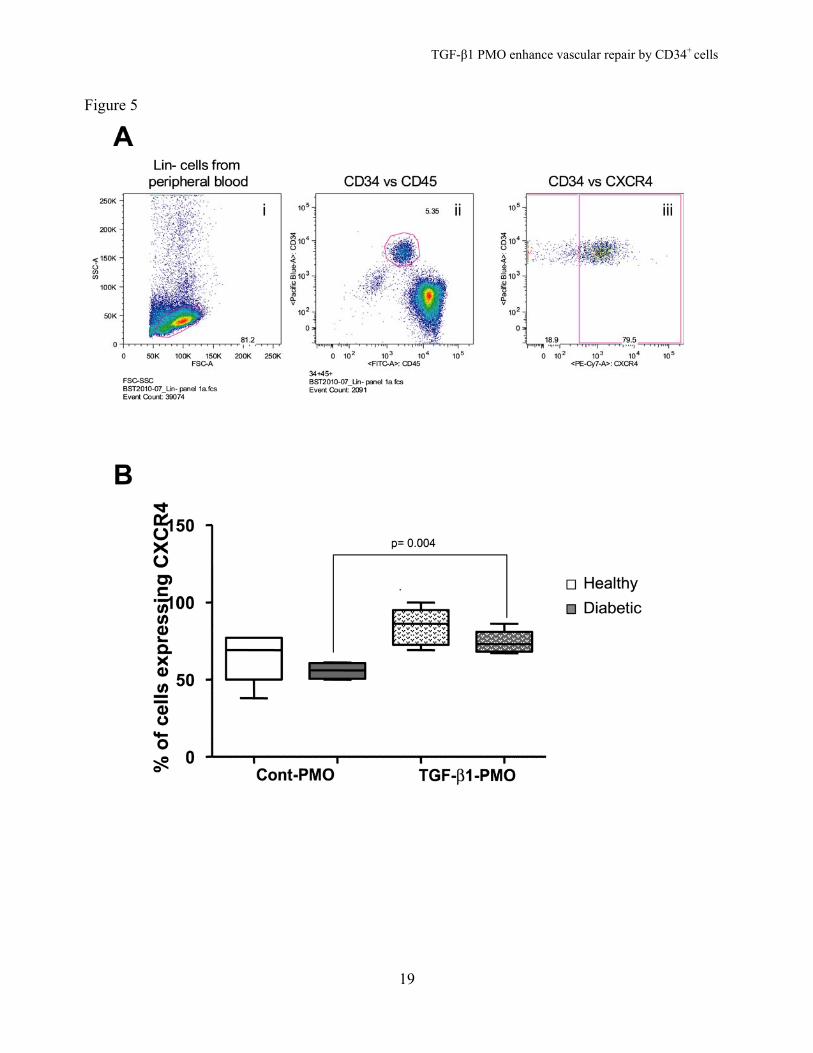
TGF1-β1-PMO Improved Migration of Diabetic CD34+ Cells to SDF-1 and Increased CXCR4 Expression. Inhibition of endogenous TGF-β1 expression enhanced migration of both diabetic (p< 0.05 vs. control-PMO) and healthy cells (p< 0.01 vs. control-PMO) (Figure 4). To understand the mechanisms involved in this enhanced migratory function of CD34+ cells, we analyzed expression of the SDF-1 receptor, CXCR4. Enriched CD34+ cells were gated for the CD34+ CD45mid population (Figure 5A) and CXCR4 expression was analyzed (Figure 5B). We found that CXCR4 surface expression was greater in TGF-β1-PMO-treated diabetic cells than in control-PMO-treated cells (p=0.004). A similar trend was observed in healthy cells; however it did not achieve statistical significance. Inhibition of Endogenous TGF-β1 Increases SDF-1-induced NO generation in Diabetic CD34+ Cells. Bioavailable NO is reduced in diabetic progenitors and this contributes to the migratory dysfunction observed in these cells (13; 26). Since inhibition of TGF-β1 in healthy and diabetic CD34+ cells increased migration and homing in both animal models, we asked whether this effect was coupled to enhanced NO generation. In healthy CD34+ cells, SDF-1 (100 nmol/L) increased NO generation by 45 ± 4% (Figure 6). The signal transduction pathway involved in SDF-1-induced NO release in CD34+ cells has not been elucidated. To delineate this pathway we tested different pharmacological inhibitors. SDF-1-induced NO release was significantly decreased when cells were pre-treated with one of the following: 10 μmol/L AMD3100, a specific non-peptide CXCR4 antagonist (3 ± 2%, p < 0.001); 100 ng/mL pertussis toxin, a Gi protein inhibitor (20 ± 9%, p < 0.01); 20 µmol/L LY294002, a phosphoinositide 3-kinase (PI3K) inhibitor (12 ± 3%, p < 0.001); or 30 µM triciribin, an Akt inhibitor (12 ± 3%, p < 0.01) (Figure 6). These results suggest
that in CD34+ cells SDF-1-induced NO release involves the CXCR4/Gi-protein/PI3K/Akt pathway.
Next, we determined whether pre-treatment with control or TGF-β1-PMO influenced NO release in these cells. We found that SDF-1 elicited a comparable degree of NO release in healthy, PB CD34+ cells that had been pre-treated with either control-PMO (60 ± 5%) or TGF-β1-PMO (54 ± 4%,) (Figure 7A, B). In contrast, SDF-1-induced marked NO generation in diabetic CD34+ cells treated with TGF-β1-PMO (69 ± 7%, p < 0.0001) but not in cells treated with control-PMO (9.2%) (Figure 7A, B). This ability of TGF-β1-PMO to restore NO release in diabetic CD34+ cells was diminished by the selective CXCR4 inhibitor, AMD3100 (28 ± 2%, p < 0.01) supporting that this effect was mediated by CXCR4 activation. DISCUSSION
Therapeutic revascularization with endothelial progenitor cells holds promise as a treatment modality to prevent tissue damage and restore blood flow in individuals such as diabetics who are not ideal candidates for standard revascularization procedures due to their small vessel disease. However, while cell therapy is needed in diabetic patients, this approach has limited utility due to endothelial progenitor cell dysfunction (27; 28). Specifically, endothelial progenitors isolated from diabetic individuals demonstrate reduced proliferation, migration, and differentiation into endothelial cells (1). Exposure to high concentrations of glucose reduces endothelial nitric oxide synthase (eNOS) expression in these cells. HIF-1α modification by methylglyoxal, a consequence of increased reactive oxygen species formed during high glucose exposure is likely responsible (27). BMPCs from db/db mice show reduced expression of eNOS and phospho-eNOS (29). Consistent with this finding, we have shown that human diabetic CD34+ cells have reduced

TGF-β1 PMO enhance vascular repair by CD34+ cells
8
NO bioavailability, which is associated with decreased migration that can be restored through exposure to NO donors (13).
In this study, we tested whether the reparative function of diabetic progenitors could be enhanced through inhibition of TGF-β1, a novel approach that we hypothesized could correct multiple functional defects in these cells. We found that pre-treating CD34+ progenitors with TGF-β1-PMO enhanced proliferation and migration in vitro as well as homing in two preclinical in vivo models of injury. Endogenous TGF-β1 has been shown to be a primary factor maintaining HSCs in G0/G1. However, exogenous TGF-β1 (in plasma and tissue), SDF-1/CXCR4 signaling, and circulating growth factors could also regulate HSC cell cycle status. Although the majority of TGF-β1 is expressed as a latent, inactive form, HSCs and BMPCs express transglutaminase and other proteolytic enzymes that can activate endogenous TGF-β1 (SHB, unpublished data). Accordingly, active intracellular TGF-β1 has been identified in HSCs and endothelial progenitors (30). HSC quiescence, division, and daughter cell fate decisions have been shown to be mediated by the TGF-β1/hematopoietic growth factor axis (22; 31).
Previously, it has been reported that inhibition of TGF-β1 induces G0-to-G1 transition (32). Here we find that it promotes cell migration and proliferation and improves homing of CD34+ cells to the vasculature. If intracellular or extracellular TGF-β1 is withdrawn from CD34+ cells in the presence of optimal hematopoietic growth factors, then G0 cells rapidly shift to G1 and the G2/M/S interface. TGF-β1 inhibition drives CD34+cells into G1 (33) and has been shown to increase the engraftment potential of HSCs (34). In contrast to TGF-β1 inhibition, withdrawal of hematopoietic growth factors results in CD34+ cell death, some of which is mediated by TGF-β1 (35).
Here we show that if TGF-β1 is inhibited intracellularly in the absence of hematopoietic growth factors, human CD34+ cells can survive for greater than a week ex vivo. In vivo we observe an enhanced reparative response that is likely due to not only increased migration of the TGF-β1-PMO-treated human CD34+ cells, but also to increased proliferation at the site of vascular injury. Similar to engraftment into the BM of LTR-HSC following TGF-β1-PMO treatment, we observed correction of defective homing in the diabetic CD34+ cells treated with TGF-β1-PMO. To better understand the mechanisms of this beneficial effect, we first focused on the SDF-1/CXCR4 pathway that has been show to be central to mediating tissue repair following injury. SDF-1 is highly expressed by ischemic tissue and can also increase progenitor cell homing back to the BM (36; 37). SDF-1 plays an important role in vascular development (38) and ablation of the SDF-1/CXCR4 gene in mice produces defects in blood vessel formation. As shown in Figure 1A, anti-TGF-β treated LTR-HSC demonstrate increased homing to the BM and as shown in Figure 2 and supplementary Figure 3, anti-TGF-β treated cells migrate better to injured tissue. This can be due to increased CXCR-4 expression, increased proliferative and migratory potential. Our results revealed that the migration of human CD34+ cells to SDF-1 was reduced in diabetic cells and that this migratory defect was corrected by endogenous TGF-β1 inhibition, suggesting that efficient signal transduction of the SDF-1/CXCR4 pathway was restored.
To further understand the influence of TGF-β1 inhibition on this pathway, we evaluated NO release in response to SDF-1 and used different pharmacological inhibitors to delineate the involvement of CXCR4 receptor signaling pathway in SDF-1-induced NO release. Importantly, our studies also indicate that SDF-1-induced NO release occurred via activation of CXCR4 and this

TGF-β1 PMO enhance vascular repair by CD34+ cells
9
involved Gi protein, PI3K and subsequent Akt activation. Akt activates eNOS by phosphorylation at Ser1177, resulting in NO production (39; 40) Our findings are consistent with earlier studies showing that SDF-1-induced chemotaxis of T lymphocytes involves Gi protein-coupled PI3K activation and Akt phosphorylation (41; 42).
We found that healthy CD34+ cells demonstrated robust NO release in response to SDF-1 and TGF-β1 inhibition did not increase NO release further. This suggests that CXCR-4 activation required for NO release in response to SDF-1 was already maximal in the nondiabetic cells, before TGF-β1 inhibition using PMOs. In contrast, TGF-β1 inhibition in diabetic CD34+ cells corrected defective NO release in response to SDF-1, in part by significantly increasing the expression of CXCR-4 on the diabetic cells, which was not the case for the nondiabetic cells (Figure 5B). This implies that in healthy nondiabetic CD34+ cells activation of CXCR-4 triggers NO independent signaling pathways, such as tyrosine phosphorylation of Wiskott-Aldrich syndrome protein (WASp). WASp is a critical regulator of actin cytoskeleton remodeling following CXCR4 activation. SDF-1 exposure results in Cdc42 activation and WASp tyrosine phosphorylation as well as in WASp association with Fyn and Pyk-2 tyrosine kinases (43). Here, we observed that cells were recruited to areas of vascular injury in two distinct but complementary injury models. Systemic administration of TGF-β1-PMO-treated murine LTR-HSC’s increased the homing of these cells to sites of neovascularization (supplemental Figure 3) and intravitreal administration of TGF-β1-PMO-treated CD34+ cells enhanced homing to injured retinal vessels (Figure 2). Both the laser injury model and the I/R model result in release of angiogenic growth factors (44). We have previously shown that laser injury alone is sufficient to induce stem cell recruitment as early as 1 week following injury (45). In this
model, SDF-1 antibodies reduce the recruitment of HSCs to the CNV lesion, suggesting that SDF-1/CXCR4 participates in the recruitment of cells to sites of laser injury (46). Our current study suggests that treatment of the murine HSCs and human CD34+cells with TGF-β1-PMO increases their homing to areas of injury by enhancing their migratory potential. This is likely due to increased surface expression of CXCR4 mediated by TGF-β1-PMO exposure. Activation of CXCR-4 generates NO which is critical to CD34+ cell migration (13; 47).
Retinal and sub retinal ischemia contributes to visual impairment and blindness
in diseases as diverse as retinopathy of pre-maturity, DR, and age-related macular degeneration. The I/R model mimics many aspects of the pathophysiology of retinal ischemia and leads to the development of acellular capillaries. We have previously shown that, in this model, healthy CD34+/endothelial precursors re-endothelialize ischemic capillaries; however, diabetic CD34+/endothelial precursors cells do not (11). In the present study, inhibition of TGF-β1 enhanced the recruitment of diabetic as well as healthy CD34+ cells to sites of retinal injury and corrected this defect in diabetic CD34+ cells.
In conclusion, our studies show that transient inhibition of TGF-β1 in CD34+ cells ex vivo enhances repair following vascular damage. This finding may have a profound impact on disease states associated with vascular dysfunction such as ischemic heart disease and diabetic vascular complications. While an attempt is being made to replace traditional approaches for alleviating tissue ischemia (e.g., stents, angioplasty, or vessel grafts) with cell therapy, autologous cellular therapy has not been feasible in diabetic patients because of dysfunctional cells. Transient inhibition of TGF-β1 may represent a promising therapeutic strategy for restoring vascular reparative function in diabetic CD34+

TGF-β1 PMO enhance vascular repair by CD34+ cells
10
cells and may increase the likelihood of successful cellular therapy in diabetic individuals.
Author Contributions. A.B., E.G., Y.J., S.C. generated and analyzed data. C.S., D.K., F.R., C.P., P.H contributed to discussion. C.L. and L.K. provided access to diabetic clinic at University of Florida. S.B. and M.G. analyzed data and wrote manuscript. ACKNOWLEDGMENTS
The authors would like to thank Dr. Martin Friedlander (The Scripps Research Institute, La Jolla, CA) and Dr. George King
(Joslin Diabetes Center, Boston, MA) for their helpful discussions during the completion of these studies and the preparation of this manuscript. We also thank Dr. Patrick L. Iversen (AVI Biopharma Inc, Portland, OR) for many helpful discussions concerning the approaches used in these studies and for his ability to design PMOs that are effective. This work was supported by grants to MBG from NIH (EY007739 and EY012601), U01 HL087366, and Juvenile Diabetes Research Foundation and to ADB from the American Heart Association and to SB from BetaStem Therapeutics. Disclosures: SB, BetaStem Therapeutics Inc
REFERENCES
1. Asahara T, Murohara T, Sullivan A, Silver M, van der Zee R, Li T, Witzenbichler B, Schatteman G, Isner JM: Isolation of putative progenitor endothelial cells for angiogenesis. Science 275:964-967, 1997 2. Hristov M, Weber C: Progenitor cell trafficking in the vascular wall. J Thromb Haemost 7 Suppl 1:31-34, 2009 3. Gill M, Dias S, Hattori K, Rivera ML, Hicklin D, Witte L, Girardi L, Yurt R, Himel H, Rafii S: Vascular trauma induces rapid but transient mobilization of VEGFR2(+)AC133(+) endothelial precursor cells. Circ Res 88:167-174, 2001 4. Schatteman GC: Adult bone marrow-derived hemangioblasts, endothelial cell progenitors, and EPCs. Curr Top Dev Biol 64:141-180, 2004 5. Grant MB, May WS, Caballero S, Brown GA, Guthrie SM, Mames RN, Byrne BJ, Vaught T, Spoerri PE, Peck AB, Scott EW: Adult hematopoietic stem cells provide functional hemangioblast activity during retinal neovascularization. Nat Med 8:607-612, 2002 6. Schatteman GC, Hanlon HD, Jiao C, Dodds SG, Christy BA: Blood-derived angioblasts accelerate blood-flow restoration in diabetic mice. J Clin Invest 106:571-578, 2000 7. Kawamoto A, Katayama M, Handa N, Kinoshita M, Takano H, Horii M, Sadamoto K, Yokoyama A, Yamanaka T, Onodera R, Kuroda A, Baba R, Kaneko Y, Tsukie T, Kurimoto Y, Okada Y, Kihara Y, Morioka S, Fukushima M, Asahara T: Intramuscular transplantation of G-CSF-mobilized CD34(+) cells in patients with critical limb ischemia: a phase I/IIa, multicenter, single-blinded, dose-escalation clinical trial. Stem Cells 27:2857-2864, 2009 8. Sekiguchi H, Ii M, Losordo DW: The relative potency and safety of endothelial progenitor cells and unselected mononuclear cells for recovery from myocardial infarction and ischemia. J Cell Physiol 219:235-242, 2009 9. Fadini GP, Sartore S, Schiavon M, Albiero M, Baesso I, Cabrelle A, Agostini C, Avogaro A: Diabetes impairs progenitor cell mobilisation after hindlimb ischaemia-reperfusion injury in rats. Diabetologia 49:3075-3084, 2006 10. Fadini GP, Miorin M, Facco M, Bonamico S, Baesso I, Grego F, Menegolo M, de Kreutzenberg SV, Tiengo A, Agostini C, Avogaro A: Circulating endothelial progenitor cells are

TGF-β1 PMO enhance vascular repair by CD34+ cells
11
reduced in peripheral vascular complications of type 2 diabetes mellitus. J Am Coll Cardiol 45:1449-1457, 2005 11. Caballero S, Sengupta N, Afzal A, Chang KH, Li Calzi S, Guberski DL, Kern TS, Grant MB: Ischemic vascular damage can be repaired by healthy, but not diabetic, endothelial progenitor cells. Diabetes 56:960-967, 2007 12. Busik JV, Tikhonenko M, Bhatwadekar A, Opreanu M, Yakubova N, Caballero S, Player D, Nakagawa T, Afzal A, Kielczewski J, Sochacki A, Hasty S, Li Calzi S, Kim S, Duclas SK, Segal MS, Guberski DL, Esselman WJ, Boulton ME, Grant MB: Diabetic retinopathy is associated with bone marrow neuropathy and a depressed peripheral clock. J Exp Med 206:2897-2906, 2009 13. Segal MS, Shah R, Afzal A, Perrault CM, Chang K, Schuler A, Beem E, Shaw LC, Li Calzi S, Harrison JK, Tran-Son-Tay R, Grant MB: Nitric oxide cytoskeletal-induced alterations reverse the endothelial progenitor cell migratory defect associated with diabetes. Diabetes 55:102-109, 2006 14. Ruscetti FW, Bartelmez SH: Transforming growth factor beta, pleiotropic regulator of hematopoietic stem cells: potential physiological and clinical relevance. Int J Hematol 74:18-25, 2001 15. Keller JR, Mantel C, Sing GK, Ellingsworth LR, Ruscetti SK, Ruscetti FW: Transforming growth factor beta 1 selectively regulates early murine hematopoietic progenitors and inhibits the growth of IL-3-dependent myeloid leukemia cell lines. J Exp Med 168:737-750, 1988 16. Herder C, Zierer A, Koenig W, Roden M, Meisinger C, Thorand B: Transforming growth factor-beta1 and incident type 2 diabetes: results from the MONICA/KORA case-cohort study, 1984-2002. Diabetes Care 32:1921-1923, 2009 17. Nam JS, Cho MH, Lee GT, Park JS, Ahn CW, Cha BS, Lim SK, Kim KR, Ha HJ, Lee HC: The activation of NF-kappaB and AP-1 in peripheral blood mononuclear cells isolated from patients with diabetic nephropathy. Diabetes Res Clin Pract 81:25-32, 2008 18. Anjaneyulu M, Berent-Spillson A, Inoue T, Choi J, Cherian K, Russell JW: Transforming growth factor-beta induces cellular injury in experimental diabetic neuropathy. Exp Neurol 211:469-479, 2008 19. Untergasser G, Gander R, Rumpold H, Heinrich E, Plas E, Berger P: TGF-beta cytokines increase senescence-associated beta-galactosidase activity in human prostate basal cells by supporting differentiation processes, but not cellular senescence. Exp Gerontol 38:1179-1188, 2003 20. Ruscetti FW, Akel S, Bartelmez SH: Autocrine transforming growth factor-beta regulation of hematopoiesis: many outcomes that depend on the context. Oncogene 24:5751-5763, 2005 21. Summerton JE: Morpholino, siRNA, and S-DNA compared: impact of structure and mechanism of action on off-target effects and sequence specificity. Curr Top Med Chem 7:651-660, 2007 22. Sitnicka E, Ruscetti FW, Priestley GV, Wolf NS, Bartelmez SH: Transforming growth factor beta 1 directly and reversibly inhibits the initial cell divisions of long-term repopulating hematopoietic stem cells. Blood 88:82-88, 1996 23. Zheng L, Gong B, Hatala DA, Kern TS: Retinal ischemia and reperfusion causes capillary degeneration: similarities to diabetes. Invest Ophthalmol Vis Sci 48:361-367, 2007 24. Chang KH, Chan-Ling T, McFarland EL, Afzal A, Pan H, Baxter LC, Shaw LC, Caballero S, Sengupta N, Li Calzi S, Sullivan SM, Grant MB: IGF binding protein-3 regulates hematopoietic

TGF-β1 PMO enhance vascular repair by CD34+ cells
12
stem cell and endothelial precursor cell function during vascular development. Proc Natl Acad Sci U S A 104:10595-10600, 2007 25. Kielczewski JL, Jarajapu YP, McFarland EL, Cai J, Afzal A, Calzi SL, Chang KH, Lydic T, Shaw LC, Busik J, Hughes J, Cardounel AJ, Wilson K, Lyons TJ, Boulton ME, Mames RN, Chan-Ling T, Grant MB: Insulin-like growth factor binding protein-3 mediates vascular repair by enhancing nitric oxide generation. Circ Res 105:897-905, 2009 26. Aicher A, Heeschen C, Mildner-Rihm C, Urbich C, Ihling C, Technau-Ihling K, Zeiher AM, Dimmeler S: Essential role of endothelial nitric oxide synthase for mobilization of stem and progenitor cells. Nat Med 9:1370-1376, 2003 27. Ceradini DJ, Yao D, Grogan RH, Callaghan MJ, Edelstein D, Brownlee M, Gurtner GC: Decreasing intracellular superoxide corrects defective ischemia-induced new vessel formation in diabetic mice. J Biol Chem 283:10930-10938, 2008 28. Egan CG, Lavery R, Caporali F, Fondelli C, Laghi-Pasini F, Dotta F, Sorrentino V: Generalised reduction of putative endothelial progenitors and CXCR4-positive peripheral blood cells in type 2 diabetes. Diabetologia 51:1296-1305, 2008 29. Yan J, Tie G, Park B, Yan Y, Nowicki PT, Messina LM: Recovery from hind limb ischemia is less effective in type 2 than in type 1 diabetic mice: Roles of endothelial nitric oxide synthase and endothelial progenitor cells. J Vasc Surg, 2009 30. Fernandez T, Amoroso S, Sharpe S, Jones GM, Bliskovski V, Kovalchuk A, Wakefield LM, Kim SJ, Potter M, Letterio JJ: Disruption of transforming growth factor beta signaling by a novel ligand-dependent mechanism. J Exp Med 195:1247-1255, 2002 31. Ploemacher RE, van Soest PL, Boudewijn A: Autocrine transforming growth factor beta 1 blocks colony formation and progenitor cell generation by hemopoietic stem cells stimulated with steel factor. Stem Cells 11:336-347, 1993 32. Dao MA, Hwa J, Nolta JA: Molecular mechanism of transforming growth factor beta-mediated cell-cycle modulation in primary human CD34(+) progenitors. Blood 99:499-506, 2002 33. Morita N, Yamamoto M, Tanizawa T: Correlation of c-kit expression and cell cycle regulation by transforming growth factor-beta in CD34+ CD38- human bone marrow cells. Eur J Haematol 71:351-358, 2003 34. Dao MA, Taylor N, Nolta JA: Reduction in levels of the cyclin-dependent kinase inhibitor p27(kip-1) coupled with transforming growth factor beta neutralization induces cell-cycle entry and increases retroviral transduction of primitive human hematopoietic cells. Proc Natl Acad Sci U S A 95:13006-13011, 1998 35. Xiao M, Oppenlander BK, Dooley DC: Transforming growth factor-beta(1) induces apoptosis in CD34(+)CD38(-/low) cells that express Bcl-2 at a low level. Exp Hematol 29:1098-1108, 2001 36. Glimm H, Tang P, Clark-Lewis I, von Kalle C, Eaves C: Ex vivo treatment of proliferating human cord blood stem cells with stroma-derived factor-1 enhances their ability to engraft NOD/SCID mice. Blood 99:3454-3457, 2002 37. Voermans C, Kooi ML, Rodenhuis S, van der Lelie H, van der Schoot CE, Gerritsen WR: In vitro migratory capacity of CD34+ cells is related to hematopoietic recovery after autologous stem cell transplantation. Blood 97:799-804, 2001 38. Lima e Silva R, Shen J, Hackett SF, Kachi S, Akiyama H, Kiuchi K, Yokoi K, Hatara MC, Lauer T, Aslam S, Gong YY, Xiao WH, Khu NH, Thut C, Campochiaro PA: The SDF-1/CXCR4 ligand/receptor pair is an important contributor to several types of ocular neovascularization. Faseb J 21:3219-3230, 2007

TGF-β1 PMO enhance vascular repair by CD34+ cells
13
39. Dimmeler S, Fleming I, Fisslthaler B, Hermann C, Busse R, Zeiher AM: Activation of nitric oxide synthase in endothelial cells by Akt-dependent phosphorylation. Nature 399:601-605, 1999 40. Fulton D, Gratton JP, McCabe TJ, Fontana J, Fujio Y, Walsh K, Franke TF, Papapetropoulos A, Sessa WC: Regulation of endothelium-derived nitric oxide production by the protein kinase Akt. Nature 399:597-601, 1999 41. Cherla RP, Ganju RK: Stromal cell-derived factor 1 alpha-induced chemotaxis in T cells is mediated by nitric oxide signaling pathways. J Immunol 166:3067-3074, 2001 42. Sotsios Y, Whittaker GC, Westwick J, Ward SG: The CXC chemokine stromal cell-derived factor activates a Gi-coupled phosphoinositide 3-kinase in T lymphocytes. J Immunol 163:5954-5963, 1999 43. Stabile H, Carlino C, Mazza C, Giliani S, Morrone S, Notarangelo LD, Notarangelo LD, Santoni A, Gismondi A: Impaired NK-cell migration in WAS/XLT patients: role of Cdc42/WASp pathway in the control of chemokine-induced beta2 integrin high-affinity state. Blood 115:2818-2826 44. Boyd SR, Zachary I, Chakravarthy U, Allen GJ, Wisdom GB, Cree IA, Martin JF, Hykin PG: Correlation of increased vascular endothelial growth factor with neovascularization and permeability in ischemic central vein occlusion. Arch Ophthalmol 120:1644-1650, 2002 45. Sengupta N, Caballero S, Mames RN, Butler JM, Scott EW, Grant MB: The role of adult bone marrow-derived stem cells in choroidal neovascularization. Invest Ophthalmol Vis Sci 44:4908-4913, 2003 46. Sengupta N, Caballero S, Mames RN, Timmers AM, Saban D, Grant MB: Preventing stem cell incorporation into choroidal neovascularization by targeting homing and attachment factors. Invest Ophthalmol Vis Sci 46:343-348, 2005 47. Li Calzi S, Purich DL, Chang KH, Afzal A, Nakagawa T, Busik JV, Agarwal A, Segal MS, Grant MB: Carbon monoxide and nitric oxide mediate cytoskeletal reorganization in microvascular cells via vasodilator-stimulated phosphoprotein phosphorylation: evidence for blunted responsiveness in diabetes. Diabetes 57:2488-2494, 2008
Figure Legends Figure 1. Inhibition of endogenous TGF-β1 in murine LTR-HSC accelerates engraftment and enhances repopulating efficiency. (A) When low numbers of highly enriched bone marrow LTR-HSCs (see methods) were treated with anti-TGF-β1 neutralizing antibody just prior to transplant into lethally irradiated mice (950 rads) without the support of helper bone marrow, a large proportion of mice survived irradiation death. The proportion of mice that survived for six months after transplantation are shown. The datum is expressed as mean survival of 10-20 mice/group. (B) Both anti-TGF-β1 PMO and TGF-β1 neutralizing cell surface antibodies inhibit endogenous expression of TGF-β1 in primitive hematopoietic cells (lin- Sca-1+-cells). Enriched bone marrow cells were treated with anti-TGF-β1 PMOs or antibodies overnight, cells were fixed and permeabilized and PE-labelled TGF-β1 antibodies were used to detect endogenous TGF-β1. Anti-actin antibodies served as the permeabilization control. Flow cytometry dot plots divided in 4 quadrants showing TGF-β1 expression on y axis while actin expression on X axis. Values in upper right quadrant of each individual plot indicates the percent of TGF-β1 expressing cells. The bottom two plots show that use of either TGF-β1 blocking antibody or TGF-β1 PMO reduced the

TGF-β1 PMO enhance vascular repair by CD34+ cells
14
percentage of cells expressing TGF-β1. For the TGF-β1 blocking antibody studies, the percentage of cells expressing TGF- β1 fell from 90.1 to 23.8 percent while for the TGF-β1 PMO studies the percentage fell from 51.2 to 14.6 percent, (n=10-20 mice/group). Figure 2. TGF-β1-PMO-treated HSCs integrate into degenerating capillaries in the I/R-damaged retina. (A) Representative immunofluorescence images showing incorporation of intravitreally injected CD34+cells (labelled green) into the retinal ischemic vasculature (red). Note that TGF-β1-PMO-treated CD34+ cells show a substantial increase in incorporation into the vasculature. (B) Morphometric quantification of blood vessel-colocalized CD34+ cells expressed as the percentage of the vascular area (n=5 mice each group). Figure 3. TGF-β1 inhibition improves survival of CD34+ cells from healthy and diabetic subjects. (A) Effect of TGF-β1-PMO on the survival of CD34+ cells sorted from healthy human bone marrow or umbilical cord blood or (B) the survival of CD34+ cells sorted from healthy and diabetic human PB. Cells (10 cells per well, 8 replicate wells) were treated with either TGF-β1-PMO or control-PMO. All cells were cultured in the absence of added growth factors for 5 days after which growth factors were added to detect surviving (live) cells (final concentrations 20 ng/mL TPO, 50 ng/mL SCF, 50 ng/mL IL-3, 20 ng/mL IL-6). Results are expressed as percentage of wells containing, growth-factor responsive cells that attained >5000 cells per well at day 12. Figure 4. Endogenous TGF-β1 inhibition in CD34+ cells from peripheral blood of healthy and diabetic subjects improves their migration. Boyden chamber assay showing migration of cells to 100 nmol/L SDF-1 (representative of 5 separate experiments). Figure 5. Anti-TGF-β1-PMO upregulates CXCR4 expression in healthy and diabetic CD34+ cells. (A) Flow cytometry dots plots of lin- PB cells showing selection of (i) low forward and low side scatter cells then (ii) selective gating for the CD34+ and CD45+ population. CXCR4 expression (iii) of the CD34+CD45+ population was studied using multicolored antibody labelling. (B) Box plot showing increase CXCR4 expression by anti-TGF-β1-PMO in both healthy and diabetic CD34+ cells (n=5 for each group). Figure 6. SDF-1 induces NO release via activation of CXCR4/Gi-protein/PI3K/Akt pathway. (A) Representative images of DAF-FM fluorescence in the absence of any treatment (time-control) (i) or following treatment with 100 nmol/L SDF-1 in the absence (ii) or presence of 10 μmol/L AMD3100 (iii), 100 ng/mL pertussis toxin (PTX) (iv), 20 μmol/L LY294002 (v), or 20 μmol/L triciribin (vi). The color scale for DAF-FM fluorescence is shown to the right. (B) Effects of different pharmacological treatments on SDF-1-mediated NO release expressed as a percent increase over the time-control (n=4 patients and up to 75 cells imaged/donor). Figure 7. SDF-1 induced NO release via activation of CXCR4 expression. (A) Representative DAF-FM fluorescence images of healthy and diabetic CD34 cells treated with control or TGF-β1-PMO with or without CXCR4 inhibitor (10 μmol/L AMD3100) and stimulated with 100 nmol/L SDF-1, the DAF-FM fluorescence color scale is shown to the right. (B) Quantification of the effects of PMOs on NO release in CD34+ cells expressed as a percent of NO release upon stimulation by SDF-1 (100nM) as compared to respective time control for the individual group, (n=4 patients and up to 75 cells imaged/donor).

TGF-β1 PMO enhance vascular repair by CD34+ cells
15
Figure 1

TGF-β1 PMO enhance vascular repair by CD34+ cells
16
Figure 2

TGF-β1 PMO enhance vascular repair by CD34+ cells
17
Figure 3

TGF-β1 PMO enhance vascular repair by CD34+ cells
18
Figure 4
TGF-β1 PMO enhance vascular repair by CD34+ cells
19
Figure 5

TGF-β1 PMO enhance vascular repair by CD34+ cells
20
Figure 6

TGF-β1 PMO enhance vascular repair by CD34+ cells
21
Figure 7
Related Documents