Transcriptome Profiling of a Toxic Dinoflagellate Reveals a Gene-Rich Protist and a Potential Impact on Gene Expression Due to Bacterial Presence Ahmed Moustafa 1 , Andrew N. Evans 2 , David M. Kulis 3 , Jeremiah D. Hackett 4 , Deana L. Erdner 2 , Donald M. Anderson 3 , Debashish Bhattacharya 1 * 1 Ecology, Evolution and Natural Resources, Institute of Marine and Coastal Sciences, Rutgers, The State University of New Jersey, New Brunswick, New Jersey, United States of America, 2 Marine Science Institute, University of Texas at Austin, Port Aransas, Texas, United States of America, 3 Woods Hole Oceanographic Institution, Woods Hole, Massachusetts, United States of America, 4 Ecology and Evolutionary Biology Department, University of Arizona, Tucson, Arizona, United States of America Abstract Background: Dinoflagellates are unicellular, often photosynthetic protists that play a major role in the dynamics of the Earth’s oceans and climate. Sequencing of dinoflagellate nuclear DNA is thwarted by their massive genome sizes that are often several times that in humans. However, modern transcriptomic methods offer promising approaches to tackle this challenging system. Here, we used massively parallel signature sequencing (MPSS) to understand global transcriptional regulation patterns in Alexandrium tamarense cultures that were grown under four different conditions. Methodology/Principal Findings: We generated more than 40,000 unique short expression signatures gathered from the four conditions. Of these, about 11,000 signatures did not display detectable differential expression patterns. At a p-value , 1E-10, 1,124 signatures were differentially expressed in the three treatments, xenic, nitrogen-limited, and phosphorus- limited, compared to the nutrient-replete control, with the presence of bacteria explaining the largest set of these differentially expressed signatures. Conclusions/Significance: Among microbial eukaryotes, dinoflagellates contain the largest number of genes in their nuclear genomes. These genes occur in complex families, many of which have evolved via recent gene duplication events. Our expression data suggest that about 73% of the Alexandrium transcriptome shows no significant change in gene expression under the experimental conditions used here and may comprise a ‘‘core’’ component for this species. We report a fundamental shift in expression patterns in response to the presence of bacteria, highlighting the impact of biotic interaction on gene expression in dinoflagellates. Citation: Moustafa A, Evans AN, Kulis DM, Hackett JD, Erdner DL, et al. (2010) Transcriptome Profiling of a Toxic Dinoflagellate Reveals a Gene-Rich Protist and a Potential Impact on Gene Expression Due to Bacterial Presence. PLoS ONE 5(3): e9688. doi:10.1371/journal.pone.0009688 Editor: Ramy K. Aziz, Cairo University, Egypt Received November 13, 2009; Accepted February 22, 2010; Published March 12, 2010 Copyright: ß 2010 Moustafa et al. This is an open-access article distributed under the terms of the Creative Commons Attribution License, which permits unrestricted use, distribution, and reproduction in any medium, provided the original author and source are credited. Funding: This work was primarily funded by a collaborative grant from the National Institutes of Health (R01 ES 013679-01A2) awarded to DB, DMA, and M. Bento Soares. Funding support for DMA and DLE was also provided from the Woods Hole Center for Oceans and Human Health from the NSF/NIEHS Centers for Oceans and Human Health program, NIEHS (P50 ES 012742) and (NSF OCE-043072). Additional support came from the National Science Foundation (EF-0732440) in a grant awarded to F. Gerald Plumley, DB, JDH, and DMA. AM was supported by an Institutional NRSA (T 32 GM98629). The funders had no role in study design, data collection and analysis, decision to publish, or preparation of the manuscript. Competing Interests: The authors have declared that no competing interests exist. * E-mail: [email protected] Introduction Dinoflagellates (Phylum Alveolata, Supergroup Chromalveolata) are unicellular protists that are among the most abundant phytoplankton in marine and freshwater ecosystems. Dinoflagellates display a range of lifestyles that together make these organisms of central ecological and economic importance. On the one hand, as oxygenic photosynthesizers, about 50% of the known species play a vital role in oxygen evolution and ocean primary production. On the other hand, some dinoflagellate species form massive toxic or non-toxic harmful algal blooms (commonly known as ‘‘red tides’’) in the oceans, leading to negative impacts on human health, fisheries, and many other coastal resources. Dinoflagellates can exhibit different trophic states, of which some are obligatory and others reflect rapid and transient responses to cellular or environmental conditions. Many dinofla- gellates are able to exist autotrophically via photosynthesis in some stages of their lifecycle. However, there are also strict cases of heterotrophy due to the absence of plastids, as in Protoperidinium that feeds on other dinoflagellates [1] and Paulsenella that parasitizes diatoms [2]. In addition, alternation between autotro- phy and heterotrophy; i.e., mixotrophy, exists in many dinofla- gellates and is supported by the presence of food vacuoles and plastids in these taxa (e.g., Alexandrium ostenfeldii [3,4]). In dinoflagellates, sexuality and subsequent encystment play a key role in bloom dynamics [5]. Encystment allows dinoflagellates to survive unfavorable environmental conditions in the form of resistant cysts, which remain dormant for a mandatory period of several months and then germinate when conditions become favorable. The exponential proliferation of germinated cells results PLoS ONE | www.plosone.org 1 March 2010 | Volume 5 | Issue 3 | e9688

Welcome message from author
This document is posted to help you gain knowledge. Please leave a comment to let me know what you think about it! Share it to your friends and learn new things together.
Transcript
Transcriptome Profiling of a Toxic Dinoflagellate Revealsa Gene-Rich Protist and a Potential Impact on GeneExpression Due to Bacterial PresenceAhmed Moustafa1, Andrew N. Evans2, David M. Kulis3, Jeremiah D. Hackett4, Deana L. Erdner2,
Donald M. Anderson3, Debashish Bhattacharya1*
1 Ecology, Evolution and Natural Resources, Institute of Marine and Coastal Sciences, Rutgers, The State University of New Jersey, New Brunswick, New Jersey, United
States of America, 2 Marine Science Institute, University of Texas at Austin, Port Aransas, Texas, United States of America, 3 Woods Hole Oceanographic Institution, Woods
Hole, Massachusetts, United States of America, 4 Ecology and Evolutionary Biology Department, University of Arizona, Tucson, Arizona, United States of America
Abstract
Background: Dinoflagellates are unicellular, often photosynthetic protists that play a major role in the dynamics of theEarth’s oceans and climate. Sequencing of dinoflagellate nuclear DNA is thwarted by their massive genome sizes that areoften several times that in humans. However, modern transcriptomic methods offer promising approaches to tackle thischallenging system. Here, we used massively parallel signature sequencing (MPSS) to understand global transcriptionalregulation patterns in Alexandrium tamarense cultures that were grown under four different conditions.
Methodology/Principal Findings: We generated more than 40,000 unique short expression signatures gathered from thefour conditions. Of these, about 11,000 signatures did not display detectable differential expression patterns. At a p-value ,1E-10, 1,124 signatures were differentially expressed in the three treatments, xenic, nitrogen-limited, and phosphorus-limited, compared to the nutrient-replete control, with the presence of bacteria explaining the largest set of thesedifferentially expressed signatures.
Conclusions/Significance: Among microbial eukaryotes, dinoflagellates contain the largest number of genes in theirnuclear genomes. These genes occur in complex families, many of which have evolved via recent gene duplication events.Our expression data suggest that about 73% of the Alexandrium transcriptome shows no significant change in geneexpression under the experimental conditions used here and may comprise a ‘‘core’’ component for this species. We reporta fundamental shift in expression patterns in response to the presence of bacteria, highlighting the impact of bioticinteraction on gene expression in dinoflagellates.
Citation: Moustafa A, Evans AN, Kulis DM, Hackett JD, Erdner DL, et al. (2010) Transcriptome Profiling of a Toxic Dinoflagellate Reveals a Gene-Rich Protist and aPotential Impact on Gene Expression Due to Bacterial Presence. PLoS ONE 5(3): e9688. doi:10.1371/journal.pone.0009688
Editor: Ramy K. Aziz, Cairo University, Egypt
Received November 13, 2009; Accepted February 22, 2010; Published March 12, 2010
Copyright: � 2010 Moustafa et al. This is an open-access article distributed under the terms of the Creative Commons Attribution License, which permitsunrestricted use, distribution, and reproduction in any medium, provided the original author and source are credited.
Funding: This work was primarily funded by a collaborative grant from the National Institutes of Health (R01 ES 013679-01A2) awarded to DB, DMA, and M.Bento Soares. Funding support for DMA and DLE was also provided from the Woods Hole Center for Oceans and Human Health from the NSF/NIEHS Centers forOceans and Human Health program, NIEHS (P50 ES 012742) and (NSF OCE-043072). Additional support came from the National Science Foundation (EF-0732440)in a grant awarded to F. Gerald Plumley, DB, JDH, and DMA. AM was supported by an Institutional NRSA (T 32 GM98629). The funders had no role in study design,data collection and analysis, decision to publish, or preparation of the manuscript.
Competing Interests: The authors have declared that no competing interests exist.
* E-mail: [email protected]
Introduction
Dinoflagellates (Phylum Alveolata, Supergroup Chromalveolata)
are unicellular protists that are among the most abundant
phytoplankton in marine and freshwater ecosystems. Dinoflagellates
display a range of lifestyles that together make these organisms of
central ecological and economic importance. On the one hand, as
oxygenic photosynthesizers, about 50% of the known species play a
vital role in oxygen evolution and ocean primary production. On
the other hand, some dinoflagellate species form massive toxic or
non-toxic harmful algal blooms (commonly known as ‘‘red tides’’) in
the oceans, leading to negative impacts on human health, fisheries,
and many other coastal resources.
Dinoflagellates can exhibit different trophic states, of which
some are obligatory and others reflect rapid and transient
responses to cellular or environmental conditions. Many dinofla-
gellates are able to exist autotrophically via photosynthesis in some
stages of their lifecycle. However, there are also strict cases of
heterotrophy due to the absence of plastids, as in Protoperidinium
that feeds on other dinoflagellates [1] and Paulsenella that
parasitizes diatoms [2]. In addition, alternation between autotro-
phy and heterotrophy; i.e., mixotrophy, exists in many dinofla-
gellates and is supported by the presence of food vacuoles and
plastids in these taxa (e.g., Alexandrium ostenfeldii [3,4]).
In dinoflagellates, sexuality and subsequent encystment play a
key role in bloom dynamics [5]. Encystment allows dinoflagellates
to survive unfavorable environmental conditions in the form of
resistant cysts, which remain dormant for a mandatory period of
several months and then germinate when conditions become
favorable. The exponential proliferation of germinated cells results
PLoS ONE | www.plosone.org 1 March 2010 | Volume 5 | Issue 3 | e9688
in blooms, which terminate through induction of encystment.
Cysts can also be geographically dispersed, giving rise to blooms in
regions with no previous history of that species [6,7,8,9].
Although dinoflagellates follow a typical eukaryotic G1-S-G2-M
cell cycle [10], they have genetic and cytological properties that
distinguish them starkly from other eukaryotes. One of the most
remarkable characteristics of dinoflagellates is the large amount of
nuclear DNA. On average, algal and plant nuclei contain 0.5 pg/
cell, however, in dinoflagellates, DNA content varies from 2.0 pg/
cell as in Amphidinium carterae [11] to up to 200.0 pg/cell in
Lingulodinium polyedrum (formerly Gonyaulax polyedra) [12], corre-
sponding to ca. 200,000 Mb. Such a massive amount of DNA has
made dinoflagellates a challenging system for complete genome
sequencing approaches. However, modern transcriptomic meth-
ods provide promising strategies to gene discovery in dinoflagel-
lates and an opportunity to address key questions about their
ecology and life cycles.
Bacterial assemblages were shown to be associated with and
attached to dinoflagellates [13] where their availability markedly
affects different aspects of dinoflagellate life cycles such as the
quantity of toxin that is produced [14,15], level of motility [16],
growth rate [15,17], and bloom formation and termination [18].
To investigate the influence of the biotic interaction between
dinoflagellates and associated bacterial communities, we prepared
RNA from a xenic (X) strain of Alexandrium tamarense (hereafter,
Alexandrium) and compared its expression profile to that of the
nutrient-replete control condition (F) and nutrient-stressed cells
under nitrogen (N) and phosphorus (P) limitation. A previous study
[19] validated the utilization of ‘‘massively parallel signature
sequencing’’ (MPSS) [20] to analyze transcriptional regulation in a
closely related dinoflagellate (Alexandrium fundyense) and provided
evidence for the complexity of the transcriptome, the presence of
gene families, and the extent of transcriptional regulation. Here,
we report the results of a comprehensive profiling of Alexandrium
transcriptome using MPSS. Our results provide novel insights into
the extent of gene richness, the dynamics of gene family evolution,
the magnitude of transcriptional regulation, and the impact of the
presence of bacteria on global gene expression patterns in
dinoflagellates.
Results and Discussion
Using MPSS, each sample resulted in a library of ,3,000,000
short signature sequences, containing an average of 290,941
unique sequences (hereafter, simply signatures) with 1,073,382
signatures from all treatments. After screening for deterministic
(i.e., absence of nucleotide ambiguities) and significantly expressed
signatures (i.e., $4 signatures per million [TPM] in at least one
library), we found between 38,000 – 39,000 usable signatures per
culture treatment (Table 1). We identified 40,029 unique
signatures when the data from all treatments were combined. In
agreement with earlier findings [21], our data show that the most
abundant transcripts among the examined conditions belong to
families that encode chlorophyll a-b binding protein, histone family
protein, S-adenosylmethionine synthetase, and S-adenosylhomo-
cysteine hydrolase. Of a total of 40,029, only 18, 2, and 12
signatures were found exclusively in the nutrient-replete (control),
N-depleted, and P-depleted cultures, respectively. In contrast, 487
signatures were found exclusively in the xenic culture, suggesting
the presence of bacteria had the most significant impact on the
transcriptome of Alexandrium under the conditions used here; i.e.,
exclusive transcription of 1.3% of the total number of transcribed
genes. Our data also showed the expected transcriptional
responses to nutrient limitation, in particular the up-regulation
of genes involved in the pathways of cell-death and gamete
formation, which will be discussed in detail elsewhere. Here, we
focus on genome-wide aspects of dinoflagellate gene expression
with a specific focus on the impact of associated bacteria on gene
expression.
Gene Content and Gene FamiliesPrevious MPSS analyses using well-annotated genomes have
shown a strong correlation between the number of transcribed
signatures and the total number of nuclear genes. In addition,
these studies have demonstrated that as more libraries and
conditions are examined, the number of unique signatures more
closely represents the total number of predicted gene models in a
genome. For example, in Arabidopsis, the number of annotated
genes is 27,165 [22] and the number of unique MPSS signatures
associated with protein coding regions gathered from 17 libraries is
at least 29,569 [23]. Based on this correlation, we postulate that
there are about 40,000 transcribed genes in Alexandrium, making it
the most complex protist transcriptome yet described. It should be
remembered, however, that although this number is relatively
large compared to other free-living protists (e.g., 27,000 genes in
the ciliate Tetrahymena thermophila [24] and 12,000 genes in the
diatom Phaeodactylum tricornutum [25]), it does not account for the
massive amount of nuclear DNA (ca. 150 Gb, estimated using
pulse-field gel electrophoresis) in haploid Alexandrium cells. Clearly,
gene number and genome size are uncoupled in these taxa. It is
worth pointing out that in a recent genome size versus gene
content regression study, dinoflagellates were predicted to contain
40,086 genes in the smallest genome and 92,013 genes in the
largest [26].
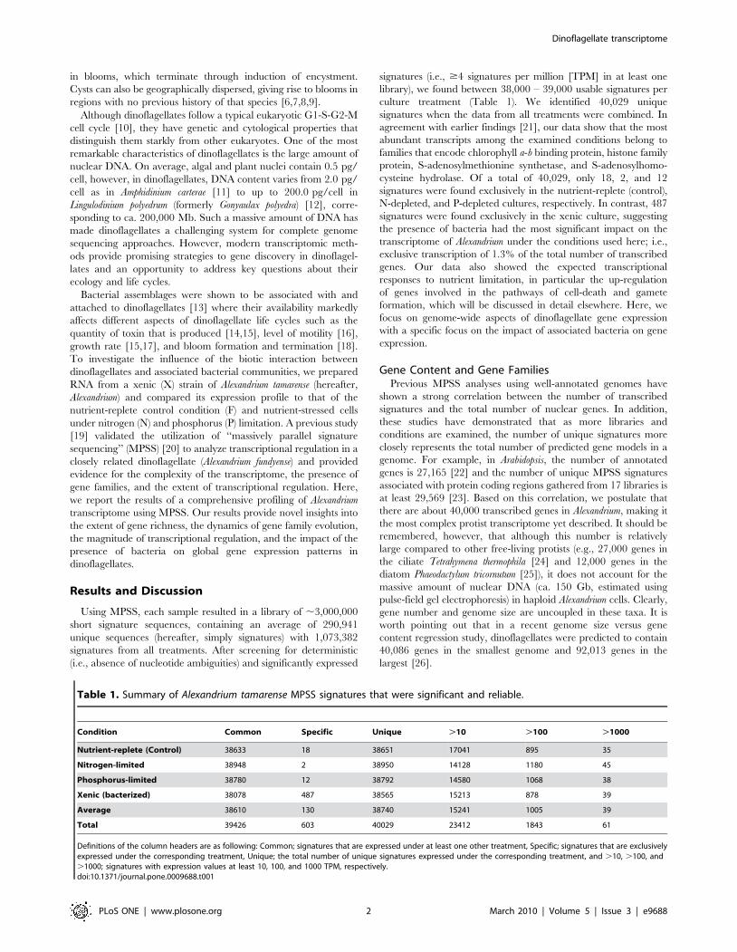
Table 1. Summary of Alexandrium tamarense MPSS signatures that were significant and reliable.
Condition Common Specific Unique .10 .100 .1000
Nutrient-replete (Control) 38633 18 38651 17041 895 35
Nitrogen-limited 38948 2 38950 14128 1180 45
Phosphorus-limited 38780 12 38792 14580 1068 38
Xenic (bacterized) 38078 487 38565 15213 878 39
Average 38610 130 38740 15241 1005 39
Total 39426 603 40029 23412 1843 61
Definitions of the column headers are as following: Common; signatures that are expressed under at least one other treatment, Specific; signatures that are exclusivelyexpressed under the corresponding treatment, Unique; the total number of unique signatures expressed under the corresponding treatment, and .10, .100, and.1000; signatures with expression values at least 10, 100, and 1000 TPM, respectively.doi:10.1371/journal.pone.0009688.t001
Dinoflagellate transcriptome
PLoS ONE | www.plosone.org 2 March 2010 | Volume 5 | Issue 3 | e9688
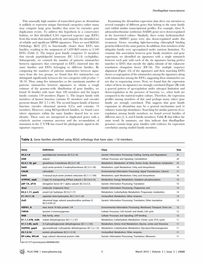
This unusually high number of transcribed genes in Alexandrium
is unlikely to represent unique functional categories; rather many
may comprise large gene families that arose by extensive gene
duplication events. To address this hypothesis in a conservative
fashion, we first identified 4,341 expressed sequence tags (ESTs)
from this strain that match perfectly and uniquely the identified set
of reliable and significant MPSS signatures. Then we used KEGG
Orthology (KO) [27] to functionally cluster these ESTs into
families, resulting in the assignment of 1,020 KO entries to 2,169
ESTs (Table 2). The largest gene family comprises 31 members
that encode peptidylprolyl isomerase (EC 5.2.1.8; cyclophilin).
Subsequently, we counted the number of pairwise mismatches
between signatures that correspond to ESTs clustered into the
same families and ESTs belonging to different families. By
comparing the numbers of pairwise mismatches between signa-
tures from the two groups, we found that five mismatches can
distinguish significantly between the two categories with p-value ,
1E-10. Thus, using five mismatches as the maximum number of
pairwise mismatches between signatures to obtain a rough
estimate of the genome-wide distribution of gene families, we
found 56 families with more than 100 members and the largest
family contains 139 members (Figure 1). The largest family with
members of known function contains 81 members and encodes
pyruvate kinase (EC 2.7.1.40). The second largest family of known
function encodes ribosomal protein L27a and contains 74
members. However, using KO-predicted families, we found cases
where signatures within the same families shared low to zero
identity. These cases are interpreted as duplicated genes with a
relatively ancient common ancestor and the accumulation of
mutations in the 39 UTR has erased the phylogenetic signal in the
signature sequences.
Examining the Alexandrium expression data drew our attention to
several examples of different genes that belong to the same family
and exhibit similar transcriptional profiles. For example, three S-
adenosylmethionine synthetase (SAMS) genes were down-regulated
in the bacterized culture. Similarly, three serine hydroxymethyl-
transferase (SHMT) genes were also down-regulated under this
treatment. Genes encoding light-harvesting chlorophyll binding
proteins followed the same pattern. In addition, four members of the
ubiquitin family were up-regulated under nutrient limitation. To
examine this association between gene family members and gene
expression, we identified six signatures with a single mismatch
between each pair with each of the six signatures having perfect
matches to ESTs that encode the alpha subunit of the eukaryote
translation elongation factor (EF-1a). The multiple sequence
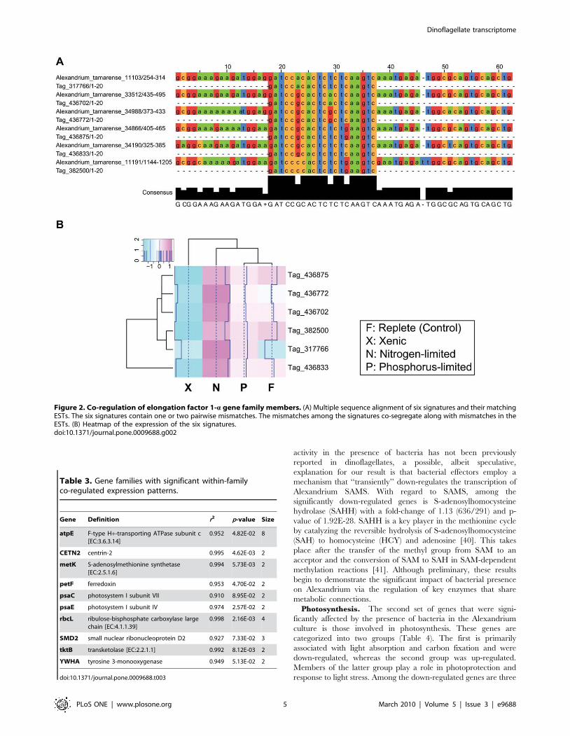
alignment (Figure 2A) of the signatures and their matching ESTs
shows co-segregation of the mismatches among the signatures along
with mismatches among the ESTs, suggesting these mismatches are
not due to sequencing errors. Next, we found that the expression
values of these six signatures are strongly correlated (Figure 2B) with
a general pattern of up-regulation under nitrogen limitation and
down-regulation in the presence of bacteria; i.e., when both are
compared to the nutrient-replete culture. Therefore, the expression
profiles among members of this (and perhaps many other) gene
family are strongly correlated. This suggests that gene family
expansion in Alexandrium may be a general mechanism used to
enhance transcript abundance. Searching for similar patterns of co-
regulation among family members, we found several families of
different sizes (2, 4, and 8 family members; Table 3) that follow the
same trend. In summary, our data indicate that dinoflagellate
genomes contain large gene families with evidence for expression
correlation among studied family members.
Table 2. Gene families identified using KEGG orthology that have sizes .10 members.
Gene Definition Class Size
E5.2.1.8 peptidylprolyl isomerase [EC:5.2.1.8] Genetic Information Processing; Folding, Sorting and Degradation 31
ANK ankyrin Cellular Processes and Signaling; Cytoskeleton 29
E2.5.1.18, gst glutathione S-transferase [EC:2.5.1.18] Metabolism; Metabolism of Other Amino Acids; Glutathione metabolism 23
fabD [acyl-carrier-protein] S-malonyltransferase [EC:2.3.1.39] Metabolism; Lipid Metabolism; Fatty acid biosynthesis 19
CALM calmodulin Environmental Information Processing; Signal Transduction; Calcium 17
fabG 3-oxoacyl-[acyl-carrier protein] reductase [EC:1.1.1.100] Metabolism; Lipid Metabolism; Fatty acid biosynthesis 16
ATPF0C, atpE F-type H+-transporting ATPase subunit c [EC:3.6.3.14] Metabolism; Energy Metabolism; Oxidative phosphorylation 15
eEF-1A, ef1A elongation factor EF-1 alpha subunit [EC:3.6.5.3] Genetic Information Processing; Translation 15
dnaJ molecular chaperone DnaJ Genetic Information Processing; Chaperones and 15
E4.2.1.17, paaG enoyl-CoA hydratase [EC:4.2.1.17] Metabolism; Carbohydrate Metabolism; Propanoate metabolism 14
E1.14.11.16 aspartate beta-hydroxylase [EC:1.14.11.16] Unclassified; Metabolism; Other enzymes 13
rluD ribosomal large subunit pseudouridine synthase D[EC:5.4.99.12]
Genetic Information Processing; Translation; Other translation 13
HSPA1_8 heat shock 70 kDa protein 1/8 Environmental Information Processing; Membrane Transport; Pores ion 12
YWHA tyrosine 3-monooxygenase Cellular Processes; Cell Growth and Death; Cell cycle 12
RAB Rab family, other Cellular Processes and Signaling; GTP-binding 12
E1.1.1.37B, mdh malate dehydrogenase [EC:1.1.1.37] Metabolism; Carbohydrate Metabolism; Citrate cycle (TCA cycle) 11
E1.1.1.95, serA D-3-phosphoglycerate dehydrogenase [EC:1.1.1.95] Metabolism; Amino Acid Metabolism; Glycine, serine and threonine 11
GAPDH, gapA glyceraldehyde 3-phosphate dehydrogenase [EC:1.2.1.12] Metabolism; Carbohydrate Metabolism; Glycolysis/Gluconeogenesis 11
E3.1.3.16 protein phosphatase [EC:3.1.3.16] Unclassified; Metabolism; Other enzymes 11
RP-L40e, RPL40 large subunit ribosomal protein L40e Genetic Information Processing; Translation; Ribosome 11
doi:10.1371/journal.pone.0009688.t002
Dinoflagellate transcriptome
PLoS ONE | www.plosone.org 3 March 2010 | Volume 5 | Issue 3 | e9688
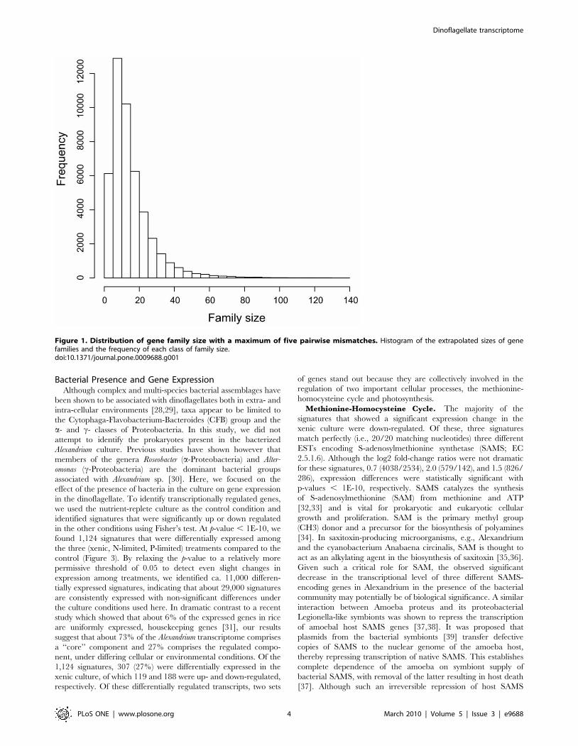
Bacterial Presence and Gene ExpressionAlthough complex and multi-species bacterial assemblages have
been shown to be associated with dinoflagellates both in extra- and
intra-cellular environments [28,29], taxa appear to be limited to
the Cytophaga-Flavobacterium-Bacteroides (CFB) group and the
a- and c- classes of Proteobacteria. In this study, we did not
attempt to identify the prokaryotes present in the bacterized
Alexandrium culture. Previous studies have shown however that
members of the genera Roseobacter (a-Proteobacteria) and Alter-
omonas (c-Proteobacteria) are the dominant bacterial groups
associated with Alexandrium sp. [30]. Here, we focused on the
effect of the presence of bacteria in the culture on gene expression
in the dinoflagellate. To identify transcriptionally regulated genes,
we used the nutrient-replete culture as the control condition and
identified signatures that were significantly up or down regulated
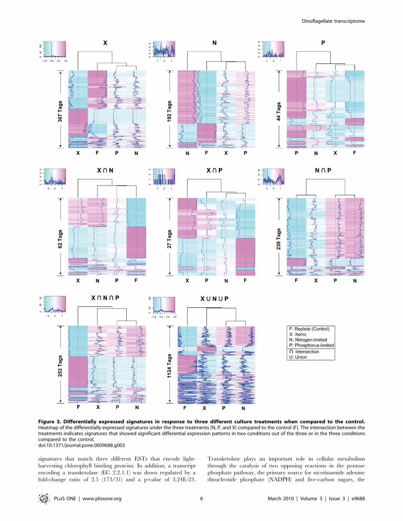
in the other conditions using Fisher’s test. At p-value , 1E-10, we
found 1,124 signatures that were differentially expressed among
the three (xenic, N-limited, P-limited) treatments compared to the
control (Figure 3). By relaxing the p-value to a relatively more
permissive threshold of 0.05 to detect even slight changes in
expression among treatments, we identified ca. 11,000 differen-
tially expressed signatures, indicating that about 29,000 signatures
are consistently expressed with non-significant differences under
the culture conditions used here. In dramatic contrast to a recent
study which showed that about 6% of the expressed genes in rice
are uniformly expressed, housekeeping genes [31], our results
suggest that about 73% of the Alexandrium transcriptome comprises
a ‘‘core’’ component and 27% comprises the regulated compo-
nent, under differing cellular or environmental conditions. Of the
1,124 signatures, 307 (27%) were differentially expressed in the
xenic culture, of which 119 and 188 were up- and down-regulated,
respectively. Of these differentially regulated transcripts, two sets
of genes stand out because they are collectively involved in the
regulation of two important cellular processes, the methionine-
homocysteine cycle and photosynthesis.
Methionine-Homocysteine Cycle. The majority of the
signatures that showed a significant expression change in the
xenic culture were down-regulated. Of these, three signatures
match perfectly (i.e., 20/20 matching nucleotides) three different
ESTs encoding S-adenosylmethionine synthetase (SAMS; EC
2.5.1.6). Although the log2 fold-change ratios were not dramatic
for these signatures, 0.7 (4038/2534), 2.0 (579/142), and 1.5 (826/
286), expression differences were statistically significant with
p-values , 1E-10, respectively. SAMS catalyzes the synthesis
of S-adenosylmethionine (SAM) from methionine and ATP
[32,33] and is vital for prokaryotic and eukaryotic cellular
growth and proliferation. SAM is the primary methyl group
(CH3) donor and a precursor for the biosynthesis of polyamines
[34]. In saxitoxin-producing microorganisms, e.g., Alexandrium
and the cyanobacterium Anabaena circinalis, SAM is thought to
act as an alkylating agent in the biosynthesis of saxitoxin [35,36].
Given such a critical role for SAM, the observed significant
decrease in the transcriptional level of three different SAMS-
encoding genes in Alexandrium in the presence of the bacterial
community may potentially be of biological significance. A similar
interaction between Amoeba proteus and its proteobacterial
Legionella-like symbionts was shown to repress the transcription
of amoebal host SAMS genes [37,38]. It was proposed that
plasmids from the bacterial symbionts [39] transfer defective
copies of SAMS to the nuclear genome of the amoeba host,
thereby repressing transcription of native SAMS. This establishes
complete dependence of the amoeba on symbiont supply of
bacterial SAMS, with removal of the latter resulting in host death
[37]. Although such an irreversible repression of host SAMS
Figure 1. Distribution of gene family size with a maximum of five pairwise mismatches. Histogram of the extrapolated sizes of genefamilies and the frequency of each class of family size.doi:10.1371/journal.pone.0009688.g001
Dinoflagellate transcriptome
PLoS ONE | www.plosone.org 4 March 2010 | Volume 5 | Issue 3 | e9688
activity in the presence of bacteria has not been previously
reported in dinoflagellates, a possible, albeit speculative,
explanation for our result is that bacterial effectors employ a
mechanism that ‘‘transiently’’ down-regulates the transcription of
Alexandrium SAMS. With regard to SAMS, among the
significantly down-regulated genes is S-adenosylhomocysteine
hydrolase (SAHH) with a fold-change of 1.13 (636/291) and p-
value of 1.92E-28. SAHH is a key player in the methionine cycle
by catalyzing the reversible hydrolysis of S-adenosylhomocysteine
(SAH) to homocysteine (HCY) and adenosine [40]. This takes
place after the transfer of the methyl group from SAM to an
acceptor and the conversion of SAM to SAH in SAM-dependent
methylation reactions [41]. Although preliminary, these results
begin to demonstrate the significant impact of bacterial presence
on Alexandrium via the regulation of key enzymes that share
metabolic connections.
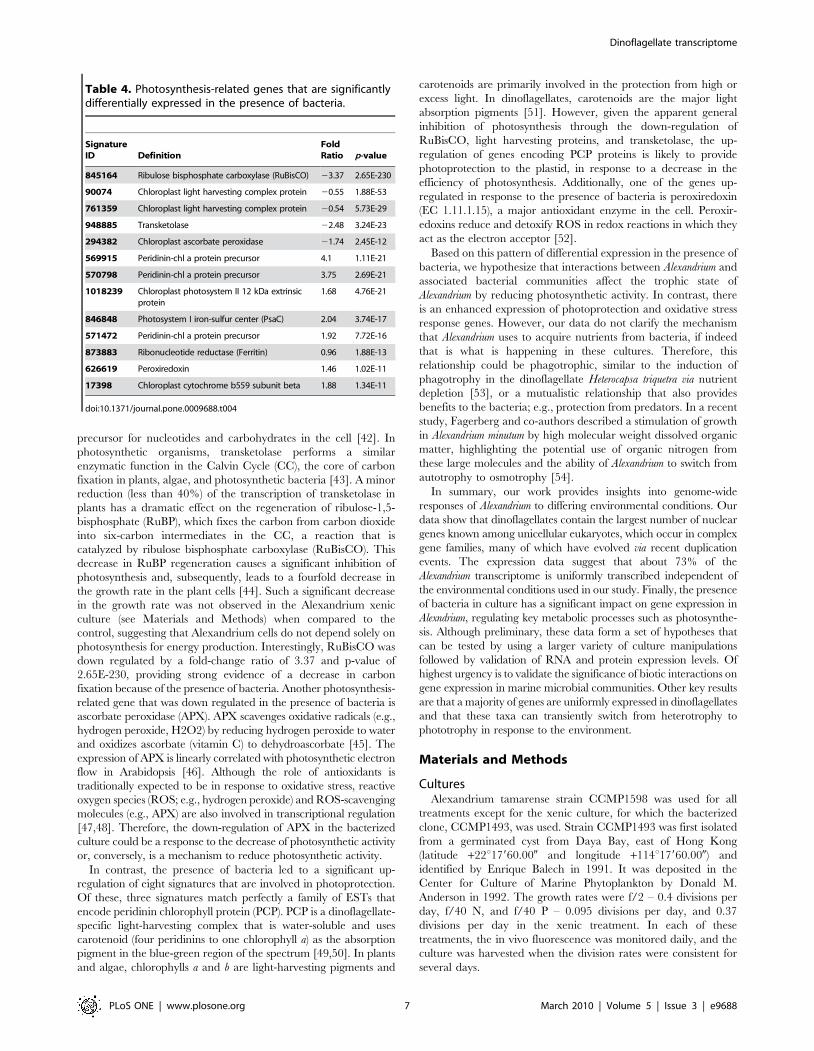
Photosynthesis. The second set of genes that were signi-
ficantly affected by the presence of bacteria in the Alexandrium
culture is those involved in photosynthesis. These genes are
categorized into two groups (Table 4). The first is primarily
associated with light absorption and carbon fixation and were
down-regulated, whereas the second group was up-regulated.
Members of the latter group play a role in photoprotection and
response to light stress. Among the down-regulated genes are three
Figure 2. Co-regulation of elongation factor 1-a gene family members. (A) Multiple sequence alignment of six signatures and their matchingESTs. The six signatures contain one or two pairwise mismatches. The mismatches among the signatures co-segregate along with mismatches in theESTs. (B) Heatmap of the expression of the six signatures.doi:10.1371/journal.pone.0009688.g002
Table 3. Gene families with significant within-familyco-regulated expression patterns.
Gene Definition r2 p-value Size
atpE F-type H+-transporting ATPase subunit c[EC:3.6.3.14]
0.952 4.82E-02 8
CETN2 centrin-2 0.995 4.62E-03 2
metK S-adenosylmethionine synthetase[EC:2.5.1.6]
0.994 5.73E-03 2
petF ferredoxin 0.953 4.70E-02 2
psaC photosystem I subunit VII 0.910 8.95E-02 2
psaE photosystem I subunit IV 0.974 2.57E-02 2
rbcL ribulose-bisphosphate carboxylase largechain [EC:4.1.1.39]
0.998 2.16E-03 4
SMD2 small nuclear ribonucleoprotein D2 0.927 7.33E-02 3
tktB transketolase [EC:2.2.1.1] 0.992 8.12E-03 2
YWHA tyrosine 3-monooxygenase 0.949 5.13E-02 2
doi:10.1371/journal.pone.0009688.t003
Dinoflagellate transcriptome
PLoS ONE | www.plosone.org 5 March 2010 | Volume 5 | Issue 3 | e9688
signatures that match three different ESTs that encode light-
harvesting chlorophyll binding proteins. In addition, a transcript
encoding a transketolase (EC 2.2.1.1) was down regulated by a
fold-change ratio of 2.5 (173/31) and a p-value of 3.24E-23.
Transketolase plays an important role in cellular metabolism
through the catalysis of two opposing reactions in the pentose
phosphate pathway, the primary source for nicotinamide adenine
dinucleotide phosphate (NADPH) and five-carbon sugars, the
Figure 3. Differentially expressed signatures in response to three different culture treatments when compared to the control.Heatmap of the differentially expressed signatures under the three treatments (N, P, and X) compared to the control (F). The intersection between thetreatments indicates signatures that showed significant differential expression patterns in two conditions out of the three or in the three conditionscompared to the control.doi:10.1371/journal.pone.0009688.g003
Dinoflagellate transcriptome
PLoS ONE | www.plosone.org 6 March 2010 | Volume 5 | Issue 3 | e9688
precursor for nucleotides and carbohydrates in the cell [42]. In
photosynthetic organisms, transketolase performs a similar
enzymatic function in the Calvin Cycle (CC), the core of carbon
fixation in plants, algae, and photosynthetic bacteria [43]. A minor
reduction (less than 40%) of the transcription of transketolase in
plants has a dramatic effect on the regeneration of ribulose-1,5-
bisphosphate (RuBP), which fixes the carbon from carbon dioxide
into six-carbon intermediates in the CC, a reaction that is
catalyzed by ribulose bisphosphate carboxylase (RuBisCO). This
decrease in RuBP regeneration causes a significant inhibition of
photosynthesis and, subsequently, leads to a fourfold decrease in
the growth rate in the plant cells [44]. Such a significant decrease
in the growth rate was not observed in the Alexandrium xenic
culture (see Materials and Methods) when compared to the
control, suggesting that Alexandrium cells do not depend solely on
photosynthesis for energy production. Interestingly, RuBisCO was
down regulated by a fold-change ratio of 3.37 and p-value of
2.65E-230, providing strong evidence of a decrease in carbon
fixation because of the presence of bacteria. Another photosynthesis-
related gene that was down regulated in the presence of bacteria is
ascorbate peroxidase (APX). APX scavenges oxidative radicals (e.g.,
hydrogen peroxide, H2O2) by reducing hydrogen peroxide to water
and oxidizes ascorbate (vitamin C) to dehydroascorbate [45]. The
expression of APX is linearly correlated with photosynthetic electron
flow in Arabidopsis [46]. Although the role of antioxidants is
traditionally expected to be in response to oxidative stress, reactive
oxygen species (ROS; e.g., hydrogen peroxide) and ROS-scavenging
molecules (e.g., APX) are also involved in transcriptional regulation
[47,48]. Therefore, the down-regulation of APX in the bacterized
culture could be a response to the decrease of photosynthetic activity
or, conversely, is a mechanism to reduce photosynthetic activity.
In contrast, the presence of bacteria led to a significant up-
regulation of eight signatures that are involved in photoprotection.
Of these, three signatures match perfectly a family of ESTs that
encode peridinin chlorophyll protein (PCP). PCP is a dinoflagellate-
specific light-harvesting complex that is water-soluble and uses
carotenoid (four peridinins to one chlorophyll a) as the absorption
pigment in the blue-green region of the spectrum [49,50]. In plants
and algae, chlorophylls a and b are light-harvesting pigments and
carotenoids are primarily involved in the protection from high or
excess light. In dinoflagellates, carotenoids are the major light
absorption pigments [51]. However, given the apparent general
inhibition of photosynthesis through the down-regulation of
RuBisCO, light harvesting proteins, and transketolase, the up-
regulation of genes encoding PCP proteins is likely to provide
photoprotection to the plastid, in response to a decrease in the
efficiency of photosynthesis. Additionally, one of the genes up-
regulated in response to the presence of bacteria is peroxiredoxin
(EC 1.11.1.15), a major antioxidant enzyme in the cell. Peroxir-
edoxins reduce and detoxify ROS in redox reactions in which they
act as the electron acceptor [52].
Based on this pattern of differential expression in the presence of
bacteria, we hypothesize that interactions between Alexandrium and
associated bacterial communities affect the trophic state of
Alexandrium by reducing photosynthetic activity. In contrast, there
is an enhanced expression of photoprotection and oxidative stress
response genes. However, our data do not clarify the mechanism
that Alexandrium uses to acquire nutrients from bacteria, if indeed
that is what is happening in these cultures. Therefore, this
relationship could be phagotrophic, similar to the induction of
phagotrophy in the dinoflagellate Heterocapsa triquetra via nutrient
depletion [53], or a mutualistic relationship that also provides
benefits to the bacteria; e.g., protection from predators. In a recent
study, Fagerberg and co-authors described a stimulation of growth
in Alexandrium minutum by high molecular weight dissolved organic
matter, highlighting the potential use of organic nitrogen from
these large molecules and the ability of Alexandrium to switch from
autotrophy to osmotrophy [54].
In summary, our work provides insights into genome-wide
responses of Alexandrium to differing environmental conditions. Our
data show that dinoflagellates contain the largest number of nuclear
genes known among unicellular eukaryotes, which occur in complex
gene families, many of which have evolved via recent duplication
events. The expression data suggest that about 73% of the
Alexandrium transcriptome is uniformly transcribed independent of
the environmental conditions used in our study. Finally, the presence
of bacteria in culture has a significant impact on gene expression in
Alexndrium, regulating key metabolic processes such as photosynthe-
sis. Although preliminary, these data form a set of hypotheses that
can be tested by using a larger variety of culture manipulations
followed by validation of RNA and protein expression levels. Of
highest urgency is to validate the significance of biotic interactions on
gene expression in marine microbial communities. Other key results
are that a majority of genes are uniformly expressed in dinoflagellates
and that these taxa can transiently switch from heterotrophy to
phototrophy in response to the environment.
Materials and Methods
CulturesAlexandrium tamarense strain CCMP1598 was used for all
treatments except for the xenic culture, for which the bacterized
clone, CCMP1493, was used. Strain CCMP1493 was first isolated
from a germinated cyst from Daya Bay, east of Hong Kong
(latitude +22u17960.000 and longitude +114u17960.000) and
identified by Enrique Balech in 1991. It was deposited in the
Center for Culture of Marine Phytoplankton by Donald M.
Anderson in 1992. The growth rates were f/2 – 0.4 divisions per
day, f/40 N, and f/40 P – 0.095 divisions per day, and 0.37
divisions per day in the xenic treatment. In each of these
treatments, the in vivo fluorescence was monitored daily, and the
culture was harvested when the division rates were consistent for
several days.
Table 4. Photosynthesis-related genes that are significantlydifferentially expressed in the presence of bacteria.
SignatureID Definition
FoldRatio p-value
845164 Ribulose bisphosphate carboxylase (RuBisCO) 23.37 2.65E-230
90074 Chloroplast light harvesting complex protein 20.55 1.88E-53
761359 Chloroplast light harvesting complex protein 20.54 5.73E-29
948885 Transketolase 22.48 3.24E-23
294382 Chloroplast ascorbate peroxidase 21.74 2.45E-12
569915 Peridinin-chl a protein precursor 4.1 1.11E-21
570798 Peridinin-chl a protein precursor 3.75 2.69E-21
1018239 Chloroplast photosystem II 12 kDa extrinsicprotein
1.68 4.76E-21
846848 Photosystem I iron-sulfur center (PsaC) 2.04 3.74E-17
571472 Peridinin-chl a protein precursor 1.92 7.72E-16
873883 Ribonucleotide reductase (Ferritin) 0.96 1.88E-13
626619 Peroxiredoxin 1.46 1.02E-11
17398 Chloroplast cytochrome b559 subunit beta 1.88 1.34E-11
doi:10.1371/journal.pone.0009688.t004
Dinoflagellate transcriptome
PLoS ONE | www.plosone.org 7 March 2010 | Volume 5 | Issue 3 | e9688
Expressed Sequence Tag and 454 Transcript SequencingTotal RNA was extracted from cultures of CCMP 1598 grown
under replete (f/2), nitrogen-limited (f/40 N), and phosphorus-
limited (f/40 P) conditions as described above, using the Nucleospin
RNA II purification kit (Clontech Laboratories, Mountain View,
CA, USA) according to the manufacturer’s protocol. A start and a
normalized directionally cloned (39 NotI-59EcoR1) cDNA library was
constructed from the pooled RNA as previously described [55]. The
complete set of existing EST clones derived from a previous study of
Alexandrium tamarense CCMP1598 [21] was then used in a DNA
hybridization protocol with the normalized library [56] to generate
a subtracted cDNA library for single-pass 39 EST sequencing. We
generated a total of 11,171 ESTs using Sanger sequencing of the
subtracted library which were processed as previously described
[57]. The clustering, which relied on the 39 UTR regions to
facilitate accuracy, was done using UIcluster v3.0.5 [58]. This
procedure resulted in a total non-redundant ‘‘unigene’’ set of 6,723
unique clusters. These data were combined with the existing
unigenes described by Hackett et al. [21] and clustered using CAP3
[59] with a 95% cutoff identity between overlapping reads to avoid
over-assembly that could mask biologically significant differences
among closely related gene families. This second round of clustering
resulted in a Sanger-based database of 12,329 unigenes from
Alexandrium. We also generated EST data from Alexandrium using
‘454’ pyrosequencing. For this procedure, equimolar amounts of
total RNA from each condition were pooled and cDNA was
synthesized from 1 mg of total RNA using the Clontech Super
SMART PCR cDNA synthesis kit following the manufacturer’s
instructions with the following modifications. Second-strand cDNA
synthesis was done with a single round of primer extension using a
59 trans-spliced leader primer conjugated to Clontech’s primer IIA
sequence to select for full-length dinoflagellate transcripts. All
dinoflagellate transcripts contain an identical 59 trans-spliced leader
sequence on mature mRNAs [60]. The product of this single round
of primer extension was purified using the Qiagen PCR purification
kit to remove the spliced-leader primers and the cDNA was
amplified by PCR using the Clontech primer IIA according to the
Clontech cDNA synthesis protocol. A single microtitre plate of 454
Titanium sequencing was done at the Arizona Genome Institute
(Tucson, AZ, USA) using 5 mg of amplified cDNA. These data were
assembled using gsAssembler (Roche NimbleGen, Inc., Madison,
WI, USA) into contigs representing Alexandrium cDNAs. The 12,329
unigenes generated using Sanger sequencing were co-assembled
with the 454-derived contigs under Seqman (DNASTAR, Madison,
WI, USA) using the default settings into a total of 35,431
dinoflagellate unigenes. The combined Sanger and 454 EST data
were annotated using a best-hit approach against Pfam (version
23.0) [61] with blastx.
Massively Parallel Signature Sequencing (MPSS)The same sources of mRNA used to construct the cDNA
libraries were also used to generate the MPSS libraries to ensure
comparability between the EST sequences and MPSS data.
Additionally, mRNA was extracted from cultures of CCMP1493
grown under replete (f/2) for the xenic condition. The cDNAs
were captured according to Illumina’s protocols as described in
Erdner and Anderson [19] and Brenner et al. [20]. Briefly, the
cDNA was digested with DpnII and then amplified using PCR.
Each cDNA was tagged by a 32-base synthetic oligonucleotide.
The tagged cDNAs were then hybridized to their complementary
32-base tags that were covalently attached to microbeads. Each
bead has only a single type of tag, but it is present in excess and
generally, about 100,000 copies of a cDNA can be bound to a
single bead. The result was a library of microbeads, in which each
bead contained about 100,000 identical copies of a cDNA
fragment that was derived from a particular mRNA. Libraries of
approximately 2 million microbeads were loaded into flow cells for
sequencing, which was performed simultaneously on each
microbead in a cell. The result was a 21 bp signature sequence
for every bead (hence every mRNA species) in the sample. The
sequences from one or more flow cells (each containing a portion
of the sample) were combined to form a set of 350,000 signatures.
All of the signature sequences in a data set were then identified
and compared to all other signature sequences, and identical
sequences were grouped and counted.
Expression Data AnalysisUsing blastn, MPSS signatures were matched to the assembled
unigenes. To increase the sensitivity of the blastn search, the option
of filtering low complexity regions was disabled, the word size was
set to two nucleotides, and the expected e-value was relaxed to
1E+3. The search results were validated such that no more than
three mismatches between a signature and a matching EST were
allowed and a perfect match within the four nucleotides of the DpnII
site (GATC) was necessary. For each signature, the matching ESTs
were ordered by the identity scores rather than original e-value-
based by blast. A matching EST with the maximum identity score
and minimum e-value was designated as the most likely signature-
matching EST. The annotations of the matching ESTs were
directly transferred to the signatures. The unigene set generated
by this study, including all previous EST data available from
Alexandrium can be accessed from the public project web site: http://
dbdata.rutgers.edu/alexbase. This web site also provides the MPSS
expression data and matching ESTs. The combined Sanger/454
sequencing derived unigene set and the MPSS tag expression data
over the different culture conditions are also available as
supplementary files Figure S1 and S2, respectively.
Differential Expression AnalysesSignature frequencies were transformed to transcript per million
(TPM) normalized values, where a signature-normalized value
equals the signature frequency divided by the sum of the
frequencies of all signatures in a library. Signatures with
ambiguous nucleotides (i.e., other than A, C, T, and G) or repeats
of sizes (string of the same nucleotide) .7 nucleotides were
excluded. Additionally, signatures with frequencies less than 4
TPM under all conditions were also discarded. Pairwise Fisher’s
exact tests [62] were performed to determine the statistical
significance of the differential expression patterns between the
different treatments. Considering two libraries X and Y of n
signatures with frequencies for signature k is xk and yk
respectively, then the 2|2 matrix (i.e., the contingency table)
for the Fisher’s test was prepared as following,
n11 ~ xk
n12 ~ yk
n21 ~Xn
i~1
xi { n11
n22 ~Xn
i~1
yi { n12
Then, the probability p was calculated according to the formula,
p~n11 z n12ð Þ!x n21 z n22ð Þ!x n11 z n21ð Þ!x n12 z n22ð Þ!
n! x n11! x n12! x n21!x n22!
Dinoflagellate transcriptome
PLoS ONE | www.plosone.org 8 March 2010 | Volume 5 | Issue 3 | e9688
Finally, the probability was adjusted using the BH method [63] to
control the false discovery rate. To determine the differentially
expressed signatures, we used p-value , 1E-10 as a consistent
threshold between all pairwise comparisons.
Supporting Information
Figure S1 The set of unigenes derived from the dinoflagellate
Alexandrium tamarense CCMP1598 using Sanger and 454
sequencing of cDNA.
Found at: doi:10.1371/journal.pone.0009688.s001 (19.86 MB
PDF)
Figure S2 The unique set of Alexandrium MPSS signatures
derived from this work and their expression levels under the
different culture conditions that were studied.
Found at: doi:10.1371/journal.pone.0009688.s002 (71.88 MB
PDF)
Author Contributions
Conceived and designed the experiments: AM JH DLE DMA DB.
Performed the experiments: ANE DMK JH DLE. Analyzed the data: AM.
Contributed reagents/materials/analysis tools: AM ANE DMK JH. Wrote
the paper: AM DB.
References
1. Jeong HJ, Latz MI (1994) Growth and grazing rates of the heterotrophicdinoflagellates Protoperidinium spp. on red tide dinoflagellates. Mar Ecol Prog Ser
106: 173–185.
2. Drebes G, Schnepf E (1982) Phagotrophy and development of Paulsenella cfchaetoceratis (Dinophyta), an ectoparasite of the diatom Streptotheca-thamesis. Helgol
Meeresunters 35: 501–515.
3. Jacobson DM, Anderson DM (1996) Widespread phagocytosis of ciliates and
other protists by marine mixotrophic and heterotrophic thecate dinoflagellates.J Phycol 32: 279–285.
4. Jeong HJ, Du Yoo Y, Park JY, Song JY, Kim ST, et al. (2005) Feeding by
phototrophic red-tide dinoflagellates: five species newly revealed and six speciespreviously known to be mixotrophic. Aquat Microb Ecol 40: 133–150.
5. Pfiester LA, Anderson DM (1987) Dinoflagellate reproduction. In: Taylor FJR,
ed. The Biology of Dinoflagellates: Blackwell Science Inc. pp 611–648.
6. Anderson DM, Coats DW, Tyler MA (1985) Encystment of the dinoflagellateGyrodinium uncatenum: temperature and nutrient effects. J Phycol 21: 200–206.
7. Anderson DM, Kulis DM, Binder BJ (1984) Sexuality and cyst formation in the
dinoflagellate Gonyaulax tamarensis: cyst yield in batch cultures. J Phycol 20: 418–425.
8. Anderson DM, Lively JJ, Reardon EM, Price CA (1985) Sinking characteristics
of dinoflagellate cysts. Limnol Oceanogr 30: 1000–1009.
9. Anderson DM, Stock CA, Keafer BA, Nelson AB, Thompson B, et al. (2005)Alexandrium fundyense cyst dynamics in the Gulf of Maine. Deep Sea Res II 52:
2522–2542.
10. Bhaud Y, Guillebault D, Lennon J, Defacque H, Soyer-Gobillard MO, et al.(2000) Morphology and behaviour of dinoflagellate chromosomes during the cell
cycle and mitosis. J Cell Sci 113 (Pt 7): 1231–1239.
11. Galleron C, Durrand AM (1978) Characterization of a dinoflagellate
(Amphidinium-carterae) DNA. Biochimie 60: 1235–1242.
12. Holm-Hansen O (1969) Algae: amounts of DNA and organic carbon in single
cells. Science 163: 87–88.
13. Alavi M, Miller T, Erlandson K, Schneider R, Belas R (2001) Bacterial
community associated with Pfiesteria-like dinoflagellate cultures. EnvironMicrobiol 3: 380–396.
14. Hold GL, Smith EA, Birkbeck TH, Gallacher S (2001) Comparison of paralytic
shellfish toxin (PST) production by the dinoflagellates Alexandrium lusitanicum
NEPCC 253 and Alexandrium tamarense NEPCC 407 in the presence and absence
of bacteria. FEMS Microbiol Ecol 36: 223–234.
15. Doucette GJ (1995) Interactions between bacteria and harmful algae: a review.
Nat Toxins 3: 65–74.
16. Mayali X, Franks PJS, Tanaka Y, Azam F (2008) Bacteria-induced motilityreduction in Lingulodinium polyedrum (Dinophyceae). J Phycol 44: 923–928.
17. Fukami K, Yuzawa A, Nishijima T, Hata Y (1992) Isolation and properties of a
bacterium inhibiting the growth of Gymnodinium-nagasakiense. Nippon Suisan Gakk58: 1073–1077.
18. Mayali X, Franks PJS, Azarn F (2008) Cultivation and ecosystem role of a
marine Roseobacter clade-affiliated cluster bacterium. Appl Environ Microbiol 74:2595–2603.
19. Erdner DL, Anderson DM (2006) Global transcriptional profiling of the toxic
dinoflagellate Alexandrium fundyense using Massively Parallel Signature Sequenc-
ing. BMC Genomics 7: 88.
20. Brenner S, Johnson M, Bridgham J, Golda G, Lloyd DH, et al. (2000) Geneexpression analysis by massively parallel signature sequencing (MPSS) on
microbead arrays. Nat Biotechnol 18: 630–634.
21. Hackett JD, Scheetz TE, Yoon HS, Soares MB, Bonaldo MF, et al. (2005)Insights into a dinoflagellate genome through expressed sequence tag analysis.
BMC Genomics 6: 80.
22. Pruitt KD, Tatusova T, Maglott DR (2007) NCBI reference sequences (RefSeq):a curated non-redundant sequence database of genomes, transcripts and
proteins. Nucleic Acids Res 35: D61–65.
23. Meyers BC, Tej SS, Vu TH, Haudenschild CD, Agrawal V, et al. (2004) The
use of MPSS for whole-genome transcriptional analysis in Arabidopsis. GenomeRes 14: 1641–1653.
24. Eisen JA, Coyne RS, Wu M, Wu DY, Thiagarajan M, et al. (2006)
Macronuclear genome sequence of the ciliate Tetrahymena thermophila, a modeleukaryote. PloS Biol 4: 1620–1642.
25. Bowler C, Allen AE, Badger JH, Grimwood J, Jabbari K, et al. (2008) The
Phaeodactylum genome reveals the evolutionary history of diatom genomes.
Nature 456: 239–244.
26. Hou Y, Lin S (2009) Distinct gene number-genome size relationships for
eukaryotes and non-eukaryotes: gene content estimation for dinoflagellate
genomes. PLoS One 4: e6978.
27. Kanehisa M, Goto S (2000) KEGG: Kyoto Encyclopedia of Genes and
Genomes. Nucleic Acids Res 28: 27–30.
28. Delong EF, Franks DG, Alldredge AL (1993) Phylogenetic diversity of
aggregated-attached vs. free-living marine bacterial assemblages. Limnol
Oceanogr 38: 924–934.
29. Kodama M, Doucette GJ, Green DH (2006) Relationships between bacteria and
harmful algae. Ecol Harmful Algae 189: 243–255.
30. Gallacher S, Flynn KJ, Franco JM, Brueggemann EE, Hines HB (1997)
Evidence for production of paralytic shellfish toxins by bacteria associated with
Alexandrium spp. (Dinophyta) in culture. Appl Environ Microbiol 63: 239–245.
31. Jiao YL, Tausta SL, Gandotra N, Sun N, Liu T, et al. (2009) A transcriptome
atlas of rice cell types uncovers cellular, functional and developmental
hierarchies. Nat Genet 41: 258–263.
32. Catoni GL (1953) S-Adenosylmethionine; a new intermediate formed enzymat-
ically from L-methionine and adenosinetriphosphate. J Biol Chem 204:
403–416.
33. Mato JM, Alvarez L, Ortiz P, Pajares MA (1997) S-adenosylmethionine
synthesis: molecular mechanisms and clinical implications. Pharmacol Ther 73:
265–280.
34. Roje S (2006) S-Adenosyl-L-methionine: beyond the universal methyl group
donor. Phytochem 67: 1686–1698.
35. Shimizu Y (1986) Chemistry and biochemistry of saxitoxin analogues and
tetrodotoxin. Ann N Y Acad Sci 479: 24–31.
36. Shimizu Y, Norte M, Hori A, Genenah A, Kobayashi M (1984) Biosynthesis of
saxitoxin analogs: the unexpected pathway. J Am Chem Soc 106: 6433–6434.
37. Choi JY, Lee TW, Jeon KW, Ahn TI (1997) Evidence for symbiont-induced
alteration of a host’s gene expression: irreversible loss of SAM synthetase from
Amoeba proteus. J Eukaryot Microbiol 44: 412–419.
38. Jeon KW, Lorch IJ (1967) Unusual intra-cellular bacterial infection in large,
free-living amoebae. Exp Cell Res 48: 236–240.
39. Han JH, Jeon KW (1980) Isolation and partial characterization of two plasmid
deoxyribonucleic acids from endosymbiotic bacteria of Amoeba proteus. J Bacteriol
141: 1466–1469.
40. De La Haba G, Cantoni GL (1959) The enzymatic synthesis of S-adenosyl-L-
homocysteine from adenosine and homocysteine. J Biol Chem 234: 603–608.
41. Chiang PK, Gordon RK, Tal J, Zeng GC, Doctor BP, et al. (1996) S-
Adenosylmethionine and methylation. FASEB J 10: 471–480.
42. Berg JM, Tymoczko JL, Stryer L (2007) Biochemistry. New York: W.H.
Freeman. 1120 p.
43. Calvin M, Benson AA (1948) The path of carbon in photosynthesis. Science 107:
476–480.
44. Henkes S, Sonnewald U, Badur R, Flachmann R, Stitt M (2001) A small
decrease of plastid transketolase activity in antisense tobacco transformants has
dramatic effects on photosynthesis and phenylpropanoid metabolism. Plant Cell
13: 535–551.
45. Smirnoff N (2000) Ascorbate biosynthesis and function in photoprotection.
Philos Trans R Soc Lond B Biol Sci 355: 1455–1464.
46. Karpinski S, Escobar C, Karpinska B, Creissen G, Mullineaux PM (1997)
Photosynthetic electron transport regulates the expression of cytosolic ascorbate
peroxidase genes in Arabidopsis during excess light stress. Plant Cell 9: 627–640.
47. Danon A, Mayfield SP (1994) Light-regulated translation of chloroplast
messenger RNAs through redox potential. Science 266: 1717–1719.
48. Pfannschmidt T, Brautigam K, Wagner R, Dietzel L, Schroter Y, et al. (2009)
Potential regulation of gene expression in photosynthetic cells by redox and
energy state: approaches towards better understanding. Ann Bot (Lond) 103:
599–607.
49. Haidak DJ, Mathews CK, Sweeney BM (1966) Pigment protein complex from
Gonyaulax. Science 152: 212–213.
Dinoflagellate transcriptome
PLoS ONE | www.plosone.org 9 March 2010 | Volume 5 | Issue 3 | e9688
50. Haxo FT, Kycia JH, Somers GF, Bennett A, Siegelman HW (1976) Peridinin-
chlorophyll a proteins of the dinoflagellate Amphidinium carterae (Plymouth 450).Plant Physiol 57: 297–303.
51. Green BR, Durnford DG (1996) The chlorophyll-carotenoid proteins of
oxygenic photosynthesis. Annu Rev Plant Physiol Plant Mol Biol 47: 685–714.52. Wood ZA, Schroder E, Robin Harris J, Poole LB (2003) Structure, mechanism
and regulation of peroxiredoxins. Trends Biochem Sci 28: 32–40.53. Legrand C, Graneli E, Carlsson P (1998) Induced phagotrophy in the
photosynthetic dinoflagellate Heterocapsa triquetra. Aquat Microb Ecol 15: 65–75.
54. Fagerberg T, Carlsson P, Lundgren M (2009) A large molecular size fraction ofriverine high molecular weight dissolved organic matter (HMW DOM)
stimulates growth of the harmful dinoflagellate Alexandrium minutum. HarmfulAlgae 8: 823–831.
55. Bonaldo MDF, Lennon G, Soares MB (1996) Normalization and subtraction:Two approaches to facilitate gene discovery. Genome Res 6: 791–806.
56. Soares MB, Bonaldo MdF, Hackett JD, Bhattacharya D (2009) Expressed
sequence tags: normalization and subtraction of cDNA libraries. Methods MolBiol. pp 109–123.
57. Scheetz TE, Laffin JJ, Berger B, Holte S, Baumes SA, et al. (2004) High-
throughput gene discovery in the rat. Genome Res 14: 733–741.
58. Trivedi N, Bischof J, Davis S, Pedretti K, Scheetz TE, et al. (2002) Parallel
creation of non-redundant gene indices from partial mRNA transcripts. Future
Generat Comput Syst 18: 863–870.
59. Huang X, Madan A (1999) CAP3: A DNA sequence assembly program.
Genome Res 9: 868–877.
60. Zhang H, Hou YB, Miranda L, Campbell DA, Sturm NR, et al. (2007) Spliced
leader RNA trans-splicing in dinoflagellates. Proc Natl Acad Sci U S A 104:
4618–4623.
61. Finn RD, Tate J, Mistry J, Coggill PC, Sammut SJ, et al. (2008) The Pfam
protein families database. Nucleic Acids Res 36: D281–288.
62. Fisher RA (1935) The logic of inductive inference. J Royal Stat Soc 98: 39–82.
63. Benjamini Y, Hochberg Y (1995) controlling the false discovery rate - a practical
and powerful approach to multiple testing. J Royal Stat Soc Ser B Method 57:
289–300.
Dinoflagellate transcriptome
PLoS ONE | www.plosone.org 10 March 2010 | Volume 5 | Issue 3 | e9688
Related Documents