Genome Biology 2007, 8:R217 Open Access 2007 Mata et al. Volume 8, Issue 10, Article R217 Research Transcriptional regulatory network for sexual differentiation in fission yeast Juan Mata *† , Anna Wilbrey *‡ and Jürg Bähler * Addresses: * Cancer Research UK Fission Yeast Functional Genomics Group, Wellcome Trust Sanger Institute, Wellcome Trust Genome Campus, Cambridge CB10 1HH, UK. † Current address: Department of Biochemistry, University of Cambridge, Downing Site, Cambridge CB2 1QW, UK. ‡ Current address: The Babraham Institute, B501, Babraham Research Campus, Cambridge CB22 3AT, UK. Correspondence: Juan Mata. Email: [email protected] © 2007 Mata et al; licensee BioMed Central Ltd. This is an open access article distributed under the terms of the Creative Commons Attribution License (http://creativecommons.org/licenses/by/2.0), which permits unrestricted use, distribution, and reproduction in any medium, provided the original work is properly cited. Sexual differentiation in fission yeast <p>Microarray analysis of the transcriptome of fission yeast after genetic perturbation of 6 genes known to have a role in sexual differen- tiation reveals insights into the regulatory principles controlling the gene expression program driving this process.</p> Abstract Background: Changes in gene expression are hallmarks of cellular differentiation. Sexual differentiation in fission yeast (Schizosaccharomyces pombe) provides a model system for gene expression programs accompanying and driving cellular specialization. The expression of hundreds of genes is modulated in successive waves during meiosis and sporulation in S. pombe, and several known transcription factors are critical for these processes. Results: We used DNA microarrays to investigate meiotic gene regulation by examining transcriptomes after genetic perturbations (gene deletion and/or overexpression) of rep1, mei4, atf21 and atf31, which encode known transcription factors controlling sexual differentiation. This analysis reveals target genes at a genome-wide scale and uncovers combinatorial control by Atf21p and Atf31p. We also studied two transcription factors not previously implicated in sexual differentiation whose meiotic induction depended on Mei4p: Rsv2p induces stress-related genes during spore formation, while Rsv1p represses glucose-metabolism genes. Our data further reveal negative feedback interactions: both Rep1p and Mei4p not only activate specific gene expression waves (early and middle genes, respectively) but are also required for repression of genes induced in the previous waves (Ste11p-dependent and early genes, respectively). Conclusion: These data give insight into regulatory principles controlling the extensive gene expression program driving sexual differentiation and highlight sophisticated interactions and combinatorial control among transcription factors. Besides triggering simultaneous expression of gene waves, transcription factors also repress genes in the previous wave and induce other factors that in turn regulate a subsequent wave. These dependencies ensure an ordered and timely succession of transcriptional waves during cellular differentiation. Background Meiosis and the formation of specialized gametes are funda- mental processes of sexual reproduction. Diploid cells of the fission yeast Schizosaccharomyces pombe undergo two mei- otic nuclear divisions to produce four stress-resistant spores in response to environmental stimuli [1,2]. This sexual differ- entiation is accompanied and driven by an extensive gene expression program, during which a large proportion of all Published: 10 October 2007 Genome Biology 2007, 8:R217 (doi:10.1186/gb-2007-8-10-r217) Received: 17 July 2007 Revised: 11 September 2007 Accepted: 10 October 2007 The electronic version of this article is the complete one and can be found online at http://genomebiology.com/2007/8/10/R217

Welcome message from author
This document is posted to help you gain knowledge. Please leave a comment to let me know what you think about it! Share it to your friends and learn new things together.
Transcript
Open Access2007Mataet al.Volume 8, Issue 10, Article R217ResearchTranscriptional regulatory network for sexual differentiation in fission yeastJuan Mata*†, Anna Wilbrey*‡ and Jürg Bähler*
Addresses: *Cancer Research UK Fission Yeast Functional Genomics Group, Wellcome Trust Sanger Institute, Wellcome Trust Genome Campus, Cambridge CB10 1HH, UK. †Current address: Department of Biochemistry, University of Cambridge, Downing Site, Cambridge CB2 1QW, UK. ‡Current address: The Babraham Institute, B501, Babraham Research Campus, Cambridge CB22 3AT, UK.
Correspondence: Juan Mata. Email: [email protected]
© 2007 Mata et al; licensee BioMed Central Ltd. This is an open access article distributed under the terms of the Creative Commons Attribution License (http://creativecommons.org/licenses/by/2.0), which permits unrestricted use, distribution, and reproduction in any medium, provided the original work is properly cited.Sexual differentiation in fission yeast<p>Microarray analysis of the transcriptome of fission yeast after genetic perturbation of 6 genes known to have a role in sexual differen-tiation reveals insights into the regulatory principles controlling the gene expression program driving this process.</p>
Abstract
Background: Changes in gene expression are hallmarks of cellular differentiation. Sexualdifferentiation in fission yeast (Schizosaccharomyces pombe) provides a model system for geneexpression programs accompanying and driving cellular specialization. The expression of hundredsof genes is modulated in successive waves during meiosis and sporulation in S. pombe, and severalknown transcription factors are critical for these processes.
Results: We used DNA microarrays to investigate meiotic gene regulation by examiningtranscriptomes after genetic perturbations (gene deletion and/or overexpression) of rep1, mei4,atf21 and atf31, which encode known transcription factors controlling sexual differentiation. Thisanalysis reveals target genes at a genome-wide scale and uncovers combinatorial control by Atf21pand Atf31p. We also studied two transcription factors not previously implicated in sexualdifferentiation whose meiotic induction depended on Mei4p: Rsv2p induces stress-related genesduring spore formation, while Rsv1p represses glucose-metabolism genes. Our data further revealnegative feedback interactions: both Rep1p and Mei4p not only activate specific gene expressionwaves (early and middle genes, respectively) but are also required for repression of genes inducedin the previous waves (Ste11p-dependent and early genes, respectively).
Conclusion: These data give insight into regulatory principles controlling the extensive geneexpression program driving sexual differentiation and highlight sophisticated interactions andcombinatorial control among transcription factors. Besides triggering simultaneous expression ofgene waves, transcription factors also repress genes in the previous wave and induce other factorsthat in turn regulate a subsequent wave. These dependencies ensure an ordered and timelysuccession of transcriptional waves during cellular differentiation.
BackgroundMeiosis and the formation of specialized gametes are funda-mental processes of sexual reproduction. Diploid cells of thefission yeast Schizosaccharomyces pombe undergo two mei-
otic nuclear divisions to produce four stress-resistant sporesin response to environmental stimuli [1,2]. This sexual differ-entiation is accompanied and driven by an extensive geneexpression program, during which a large proportion of all
Published: 10 October 2007
Genome Biology 2007, 8:R217 (doi:10.1186/gb-2007-8-10-r217)
Received: 17 July 2007Revised: 11 September 2007Accepted: 10 October 2007
The electronic version of this article is the complete one and can be found online at http://genomebiology.com/2007/8/10/R217
Genome Biology 2007, 8:R217

http://genomebiology.com/2007/8/10/R217 Genome Biology 2007, Volume 8, Issue 10, Article R217 Mata et al. R217.2
genes are either induced or repressed [3-5]. We have previ-ously classified the genes that are up-regulated at least four-fold into four major clusters, which represent successiveexpression waves coinciding with the main biological eventsof the differentiation process: genes induced in response toenvironmental changes (starvation and pheromone-inducedgenes), early genes (pre-meiotic S phase and recombination),middle genes (meiotic divisions and early steps of spore for-mation), and late genes (spore maturation) [4].
Posttranscriptional control is involved in regulating mRNAlevels during meiosis [6-8]. In addition, transcriptional con-trol is of fundamental importance for sexual differentiation,and several transcription factors are essential for successfulmeiosis and spore formation. A subset of the genes induced inresponse to nutritional changes is controlled by the transcrip-tion factor Ste11p [3,9], while some early genes are under thecontrol of the Rep1p transcriptional regulator [10-12]. Theforkhead-family protein Mei4p controls the expression ofseveral middle genes [13-15], and the basic leucine zipper(bZIP) transcription factors Atf21p and Atf31p control a sub-set of late genes [4]. It is not known, however, if other tran-scription factors are involved in this process, and how theactivity of the different factors is regulated and coordinated tobring about the orderly succession of transcriptional waves.
Here, we investigate the regulation of meiotic genes by exam-ining the transcriptome of cells deleted for or overexpressinggenes encoding transcription factors whose expression isinduced during sexual differentiation. Our data highlight theimportance of combinatorial control in transcriptional regu-lation and indicate that the progression of the gene expres-sion waves is achieved by transcriptional cascades andfeedback interactions between transcription factors. We alsoidentify two new transcriptional regulators involved in con-trolling late genes.
Results and discussionRep1p activates a subset of the early genesThe Rep1p transcription factor is involved in the regulation ofseveral early genes required for premeiotic S phase and mei-otic recombination [10-12]. However, the expression of someearly genes is independent of Rep1p [11]. To better under-stand the role of Rep1p, we sought to systematically identifyits target genes by using DNA microarrays to follow geneexpression in rep1Δ mutant cells undergoing meiosis.
Because good synchrony is important to obtain gene expres-sion profiles of high temporal resolution, we used cells carry-ing a temperature-sensitive mutation in the meiotic inhibitorPat1p [16,17]. We arrested cells in G1 by removing the nitro-gen source from the medium and induced meiosis by shiftingthe cells to the restrictive temperature. Although pat1-induced meiosis is not identical to wild-type meiosis in somerespects [18], we have previously shown that the gene expres-
sion of early, middle, and late genes is similar in both types ofexperiments [4].
Deletion of rep1 had complex effects on the expression ofearly genes (Figure 1a). About 47% of early genes were notfully induced in rep1Δ cells (full induction was defined as anincrease within two-fold of that of wild-type cells at everytime point between 1 and 3 hours; see Additional data file forcomplete lists of Rep1p-dependent and Rep1p-independentgenes). However, the effects of rep1Δ on gene expression werevaried, with some genes being partially induced but often in adelayed fashion compared to wild type. Rep1p-dependent andRep1p-independent genes showed no clear functional distinc-tion. Both groups were significantly enriched in genesinvolved in meiotic recombination and meiosis I (GO:0007131 and GO: 0007127, P < 2 × 10⁻⁶), while Rep1p-dependent genes were uniquely enriched in genes involved inthe mitotic cell-cycle (GO: 0000279, P < 7 × 10-13). Control ofgene expression during early meiosis involves several tran-scription factors that also function during the mitotic cell-cycle (Cdc10p, Res2p and Rep2p), which are likely to cooper-ate with Rep1p [10]. The large fraction of genes whose expres-sion seems to be independent of Rep1p suggests thatadditional transcription factors are important for the controlof the early genes.
Mei4p regulates the induction of middle genesThe forkhead-family transcription factor Mei4p is essentialfor progression through the first meiotic division and hasbeen reported to activate the expression of some middle genes[14,15]. It is unclear, however, whether all middle genes areregulated by Mei4p or whether additional transcription fac-tors are involved in their induction. To address this question,we used DNA microarrays to systematically identify Mei4ptargets. A dual strategy was applied: first, we compared mei-otic time courses of mei4Δ cells with wild-type cells; and sec-ond, we studied the effect of Mei4p overproduction invegetative cells.
While deletion of mei4 did not impair the up-regulation ofstarvation-induced or early genes, the induction of almost allmiddle and late genes was strongly affected (Figure 1b,c).Lists of Mei4p-independent and Mei4p-dependent middlegenes are presented in Additional data file 9 (Mei4p depend-ency was defined as an induction at least two-fold lower thanthat of wild-type cells at any time point between 3 and 7hours). It is likely that the effect on late genes is indirect, asthe expression of some transcription factors regulating lategenes is dependent on Mei4p(see below). To distinguishbetween direct and indirect effects, we took advantage of thefinding that overproduction of Mei4p leads to induction of itstargets even in vegetative cells [14,15]. We cloned the mei4gene under the control of the regulatable nmt1 promoter [19]and overexpressed mei4 in vegetative cells, in which endog-enous mei4 mRNA is almost absent [15]. Mei4p-overproduc-ing cells looked normal 18 hours after induction of the nmt1
Genome Biology 2007, 8:R217
http://genomebiology.com/2007/8/10/R217 Genome Biology 2007, Volume 8, Issue 10, Article R217 Mata et al. R217.3
promoter (the time point used for microarray analysis). Atlater time points (24 hours), they appeared smaller than wild-type cells and many cells had lysed. A total of 454 genes wereinduced at least two-fold following induction of mei4 expres-sion (Figure 1c). This group consisted mostly of middle genes(306), as well 35 genes of the nitrogen response, 19 earlygenes, and 13 late genes. The remaining genes had not beenclassified in the original study because they did not pass thefour-fold cut-off for gene expression changes relative to vege-tative cells [4]. The Mei4p-induced genes included 55% of allmiddle genes, but only 16% of the nitrogen response genes,19% of the early genes, and 9% of the late genes. We noticedthat many non-middle genes induced by Mei4p overproduc-tion had complex expression profiles, with several peaks ofexpression or unusually broad peaks (data not shown). Theregulation of these genes is probably under the control of sev-eral transcription factors, with Mei4p being responsible fortheir expression at the time of the meiotic divisions. Of theremaining 81 genes that were induced by Mei4p overproduc-tion but had not been classified, 36 depended on Mei4p fortheir full induction (as defined above for the mei4Δ timecourse; Additional data file 6). Several of the latter showedmiddle gene-like expression profiles. It is also possible thatMei4p overproduction leads to the artifactual induction oftargets of other forkhead transcription factors (Sep1p andFkh2p) that normally control the periodic expression of asubset of cell cycle-regulated genes [20]. We looked atwhether the expression of periodic genes in cluster 1 (which isenriched in Sep1p targets and forkhead-binding sequences)[21] was increased in Mei4p-overproducing cells. The expres-sion of 35 genes of this cluster (out of 94) was induced byMei4p overproduction. However, 29 of these 35 had beenclassified as middle genes. Similarly, 9 out of 41 Sep1p-dependent periodic targets were induced by Mei4p overex-pression, but 8 of them are also middle genes. Therefore, itseems unlikely that Mei4p induces the ectopic expression ofSep1p targets.
Together, the results are consistent with the view thatMei4pdirectly controls the expression of most middle genes,and that the low expression of late genes in a mei4 mutant isan indirect effect. Given that some middle genes (220 out ofthe 504 Mei4p-dependent middle genes) are not induced byMei4p overproduction, we cannot rule out the existence of asecond factor that activates the expression of middle genes.However, because the expression of almost all middle genes isreduced in a mei4Δ strain (504 out of 549 show at least a two-fold reduction in induction; Figure 1c), we believe this possi-bility to be unlikely. The strong induction of at least somemiddle genes, however, may be supported by posttranscrip-tional mechanisms [7,22].
Control of late genes by Atf21p and Atf31pWe have previously shown that deletion of either atf21 oratf31 affects the expression of approximately 55% of the lategenes [4]. To better understand how these transcription fac-
Meiotic gene expression program in rep1Δ and mei4Δ mutantsFigure 1Meiotic gene expression program in rep1Δ and mei4Δ mutants. (a) Left: hierarchical cluster analysis with columns representing experimental time points and rows representing early genes. Vegetatively growing cells (V) are synchronized in G1 by nitrogen removal and enter meiosis by temperature shift at time 0. The mRNA levels at each time point of pat1(from [4]) and rep1Δ pat1 time courses relative to the levels in vegetative cells are color-coded as indicated at the bottom with missing data in gray. Rep1p-independent and -dependent genes are indicated at right as drawn-out or dashed blue lines, respectively. Right: average expression profiles of Rep1p-dependent and Rep1p-independent genes in wild-type (pat1-synchronized cells from [4]) and rep1Δ cells. (b) Average expression profiles of the four main gene clusters upregulated during pat1-induced meiosis: nitrogen-starvation response (black), early (blue), middle (red) and late (green), as defined in [4]. Experimental details are as in (a). The y-axis shows the expression level at the corresponding time point relative to expression in vegetative cells. Graphs from left to right: pat1 cells (from [4]) and mei4Δ pat1 cells. (c) Hierarchical cluster analysis with columns representing experimental time points and rows representing genes. The mRNA levels at each time point of pat1 time courses relative to the levels in vegetative cells are color-coded as indicated in (a). The last column (OE) shows the expression level in cells overproducing Mei4p relative to an empty vector control. The four gene clusters are indicated on the left.
V 0 1 2 3 4 5 6 7 V 0 1 2 3 4 5 6 7Time (hours)
mei4 pat1
Mea
n ex
pres
sion
rat
io
10
1
Late
Mid
dle
Ear
ly
V 0 1 2 3 4 5 6 7 V 0 1 2 3 4 5 6 7
-N
pat1
mei4 pat1pat1
(b)
(c)
V 0 1 2 3 V 0 1 2 3 pat1 rep1 pat1(a)
+8x 1 -8x
Ear
ly
V 0 1 2 3 V 0 1 2 3 Time (hours)
Mea
n ex
pres
sion
rat
io
10
1
pat1 rep1 pat1
Rep1p dep.
Rep1p indep.
OE
Genome Biology 2007, 8:R217

http://genomebiology.com/2007/8/10/R217 Genome Biology 2007, Volume 8, Issue 10, Article R217 Mata et al. R217.4
tors function, we overexpressed them in vegetative cells,where they are normally present at low levels [4]. Overpro-duction of Atf21p resulted in the induction of approximately25 genes, some of them related to stress processes [23] (Fig-ure 2a, and a complete list is in Additional data file 7). Whenwe took samples for microarray analysis (18 hours afterinduction of the nmt1 promoter), Atf21p-overproducing cellsappeared slightly elongated. At later time points (24 hours),most cells were highly elongated and contained one or moresepta (data not shown). Overproduction of Atf31p resulted inthe induction of approximately 12 genes, many of which arealso induced in response to heat shock [23] (Figure 2b, and acomplete list is in Additional data file 7). Note that heat-shockrelated genes show a highly specific expression pattern dur-ing pat1-induced meiosis, with a sharp induction followingthe initial temperature shift and a quick down-regulationafter the meiotic divisions [4]. Despite this finding, atf31Δcells respond normally to heat shock (JM, AW and JB, unpub-lished observation). Atf31p-overproducing cells appearednormal after 18 hours of induction but became elongatedafter 24 hours (data not shown). However, neither group ofgenes induced by Atf21p or Atf31p overexpression showedany large overlap with the potential meiotic targets of Atf21p/Atf31p that we have previously identified [4] (Figure 2a,b, leftpanels).
We then simultaneously overexpressed both Atf21p andAtf31p. This caused a phenotype similar to that of cells over-producing Atf21p alone, but led to much stronger geneexpression changes (325 genes induced at least two-fold),which also showed a highly significant enrichment in the mei-otic targets of Atf21p/Atf31p (Figure 2c, and a complete list isin Additional data file 7). Many of the genes that were inducedshow expression profiles typical of late genes, but had notpreviously been defined as Atf21p/Atf31p targets (Figure 2c,left). There are two reasons for this difference. First, the defi-nition of Atf21p/Atf31p targets was based on experimentsusing asynchronous cells, which are less sensitive. Most of thegenes not originally defined as Atf21p/Atf31p targets, how-ever, were expressed at slightly lower levels in atf21Δ andatf31Δ cells (mean expression ratios of 0.8 and 0.91, respec-tively) [4]. Second, the four-fold cut-off relative to vegetativecells that we used in the original study means that many ofthese genes were excluded from the analysis. Therefore, thereal number of Atf21p/Atf31p targets is probably considera-bly higher than the conservative estimate from our previousstudy [4].
This experiment suggests that Atf21p and Atf31p cooperate toinduce the expression of late genes during meiosis, probablyby forming a heterodimer. These results highlight the impor-tance of combinatorial control in the regulation of transcrip-tional programs. The fission yeast genome encodes threeadditional bZIP family transcription factors besides Atf21pand Atf31p [24-26]. These transcription factors can work ashomodimers or heterodimers, creating numerous regulatory
possibilities. It is likely that control by different combinationsof bZIP transcription factors is used by fission yeast to launchspecific gene expression programs in response to differentenvironmental or developmental conditions. Combinatorialcontrol by bZIP transcription factors is well known from stud-ies in other organisms (for example, [27,28]).
Two novel transcription factors regulate the induction of late genesThe expression of many late genes is independent of theAtf21p/Atf31p system [4]. We have previously reported thatseveral genes that potentially encode transcription factors arespecifically induced at various stages of sexual differentiation,
Atf21p and Atf31p cooperate to induce expression of meiotic genesFigure 2Atf21p and Atf31p cooperate to induce expression of meiotic genes. Effects of overproduction of (a) Atf21p, (b) Atf31p, and (c) both Atf21p and Atf31p. The Venn diagrams on the left show the overlaps between genes induced by overproduction of the different transcription factors (OE) and potential meiotic Atf21p target genes (obtained by analysis of atf21Δ cells [4]). The numbers in brackets show the overlap between the two lists expected by chance given the sizes of the gene sets considered and the total number of genes. The overlap between Atf31p-induced genes and meiotic Atf21p targets is not significant, while the overlaps between Atf21p and Atf21p/Atf31p-induced genes and Atf21p targets are both significant (P = 2.3 × 10⁻³ and 1.5 × 10-62, respectively; note, however, that only four Atf21p targets are induced by Atf21p overexpression). The panels on the right show the meiotic expression profiles of genes induced by the overproduction of the corresponding transcription factors (pat1-synchronized cells from [4], experiment and labeling as in Figure 1). The profiles of atf21 and atf31 are highlighted in blue and green, respectively.
Time (hours)
Exp
ress
ion
ratioAtf21p meiotic
targetsatf21 OE
214
(0.5) 117
atf21
V 0 1 2 3 4 5 6 7 8 10 12
1
10
100
V 0 1 2 3 4 5 6 7 8 10 12
1
10
100
Exp
ress
ion
ratio
atf31Atf21p meiotic
targetsatf31 OE
111
(0.3) 120
V 0 1 2 3 4 5 6 7 8 10 12
1
10
100
Exp
ress
ion
ratio
0.1
Atf21p meiotictargets
atf21/atf31OE
25471(7) 50
(a)
(b)
(c)
Genome Biology 2007, 8:R217

http://genomebiology.com/2007/8/10/R217 Genome Biology 2007, Volume 8, Issue 10, Article R217 Mata et al. R217.5
including spore formation, raising the possibility that some ofthese factors regulate the expression of late genes [4].
We explored the function of rsv1 and rsv2, encoding potentialtranscription factors with C2H2-type zinc fingers that areinduced in middle/late meiosis (Figures 3a and 4a). Rsv1phas previously been reported to be required for maintainingviability in stationary phase cells; in particular, Rsv1p isrequired to prevent the loss of viability associated with glu-cose depletion [29]. Rsv2p has a similar function during sta-tionary phase (L López-Maury, personal communication) andduring nitrogen depletion [30]. Deletion of either rsv1 or rsv2did not affect cell viability or growth of vegetative cells, andrsv1Δ and rsv2Δ cells were able to mate and form spores ofnormal morphology that germinated with the same efficiencyas that of wild-type spores (data not shown). Despite the lackof a clear meiotic phenotype, we reasoned that the deletionsmight cause subtle effects on gene expression.
We therefore used DNA microarrays to compare the tran-scriptome of mutant cells undergoing sexual differentiationwith that of wild-type cells. For this experiment, we usedhomothallic haploid cells without pat1 mutation, which mateto form diploid cells before undergoing sexual differentiation.Because sexual differentiation is less synchronous in thesewild-type cells, different meiotic stages can be studied in asingle sample [4]. Despite the relative lack of synchrony,these experiments are highly reproducible: only 1.3% and1.4% of the genes showed differences of more than 2-foldbetween two independent biological replicates of rsv2 andrsv1 experiments, respectively. Most genes were expressed atsimilar levels in wild-type and rsv2Δ cells, showing that mei-otic progression and gene expression were not disrupted.However, 39 genes were expressed at lower levels in rsv2Δmutants compared to wild-type cells (reduced at least two-fold in two independent repeats; Figure 3b, and a completelist of genes is in Additional data file 11). Of these genes, 18had been classified as late genes, and most of the additional21 genes also showed a late peak of expression (Figure 3c).The latter genes have not been classified as late genes, eitherbecause they did not pass the threshold of induction used forthe classification, or because they had complex expressionpatterns containing multiple peaks [4]. A large fraction of thepotential Rsv2p targets (17/39) are also induced in severaldifferent stress conditions [23]. We conclude that Rsv2p acti-vates the expression of a subset of genes during late meiosisand may contribute to the acquisition of stress resistance bythe spore. No obvious regulatory motifs were significantlyenriched in the promoters of Rsv2-regulated genes.
Deletion of rsv1 did not cause major changes in gene expres-sion, indicating that meiotic progression and gene expressionwere normal in the mutant. However, a group of 24 geneswere consistently expressed at higher levels compared towild-type cells (increased at least two-fold in two independ-ent repeats; see Figure 4b and Additional data file 10 for a
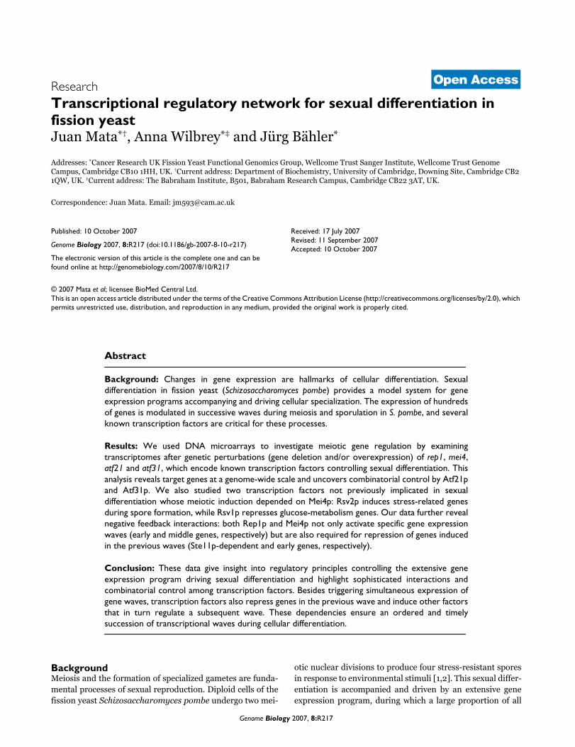
Identification of Rsv2p-dependent genesFigure 3Identification of Rsv2p-dependent genes. (a) Expression profile of rsv2 during meiosis and sporulation (from [4], experiment and labeling as in Figure 1). (b) Comparison of gene expression levels between wild-type and rsv2Δ meiotic cells. Genes outside the blue lines differ by more than two-fold in expression levels. rsv2 is indicated by an arrow. (c) Expression profiles of the potential Rsv2p-dependent genes identified in (b) (pat1-synchronized cells from [4]).
Time (hours)
Exp
ress
ion
ratio
rsv2
V 0 1 2 3 4 5 6 7 8 10 12
(a)
(c)
1
10
V 0 1 2 3 4 5 6 7 8 10 12
1
10
100
Exp
ress
ion
ratio
Time (hours)
10 1e2 1e3 1e4 1e510
1e2
1e3
1e4
(b)
Exp
ress
ion
sign
al r
sv2
Expression signal wild-type
Genome Biology 2007, 8:R217
http://genomebiology.com/2007/8/10/R217 Genome Biology 2007, Volume 8, Issue 10, Article R217 Mata et al. R217.6
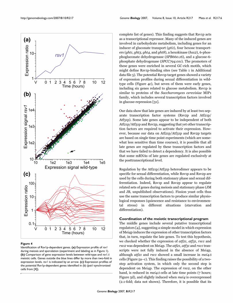
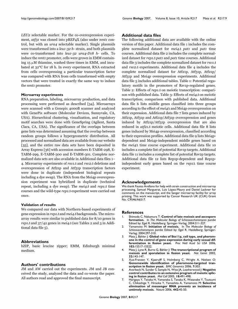
complete list of genes). This finding suggests that Rsv1p actsas a transcriptional repressor. Many of the induced genes areinvolved in carbohydrate metabolism, including genes for aninducer of gluconate transport (gti1), four hexose transport-ers (ght1, ght3, ght4, and ght8), a hexokinase (hsx2), 6-phos-phogluconate dehydrogenase (SPB660.16), and a glucose-6-phosphate dehydrogenase (SPCC794.01c). The promoters ofthese genes were enriched in several GC-rich motifs, whichmight define Rsv1p-binding sites (see Table 1 in Additionaldata file 5). The potential Rsv1p target genes showed a varietyof expression profiles during sexual differentiation in wild-type cells (Figure 4c), but seven of them were early genes,including six genes related to glucose metabolism. Rsv1p issimilar to proteins of the Saccharomyces cerevisiae MIP1family, which includes several transcription factors involvedin glucose-repression [31].
Our data show that late genes are induced by at least two sep-arate transcription factor systems (Rsv2p and Atf21p/Atf31p). Some late genes appear to be independent of bothAtf21p/Atf31p and Rsv2p, suggesting that yet other transcrip-tion factors are required to activate their expression. How-ever, because our data on Atf21p/Atf31p and Rsv2p targetsare based on single time point experiments (which are some-what less sensitive than time courses), it is possible that alllate genes are regulated by these transcription factors andthat we have failed to detect a dependency. It is also possiblethat some mRNAs of late genes are regulated exclusively atthe posttranscriptional level.
Regulation by the Atf21p/Atf31p heterodimer appears to bespecific for sexual differentiation, while Rsv1p and Rsv2p areused by the cells during both stationary phase and sexual dif-ferentiation. Indeed, Rsv1p and Rsv2p appear to regulaterelated sets of genes during meiosis and stationary phase (JMand JB, unpublished observations). Fission yeast cells thususe the same transcription factors to produce similar physio-logical responses (quiescence and resistance to environmen-tal stress) in different situations (starvation anddifferentiation).
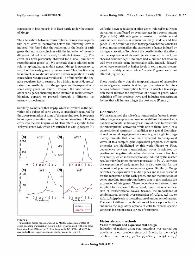
Coordination of the meiotic transcriptional programThe middle genes include several putative transcriptionalregulators [4], suggesting a simple model in which expressionof Mei4p induces the expression of other transcription factorsthat, in turn, regulate the late genes. To test this hypothesis,we checked whether the expression of atf21, atf31, rsv1 andrsv2 was dependent on Mei4p. The atf21, atf31 and rsv1 tran-scripts were not fully induced in the absence of Mei4p,although atf21 and rsv1 showed a small increase in mei4Δcells (Figure 5a–c). This finding raises the possibility of a two-step activation system, in which only the second step isdependent on Mei4p. The expression of rsv2, on the otherhand, is reduced in mei4Δ cells at late time points (7 hours;Figure 5d), and slightly induced when mei4 is overexpressed(2.1-fold; data not shown). Therefore, it is possible that its
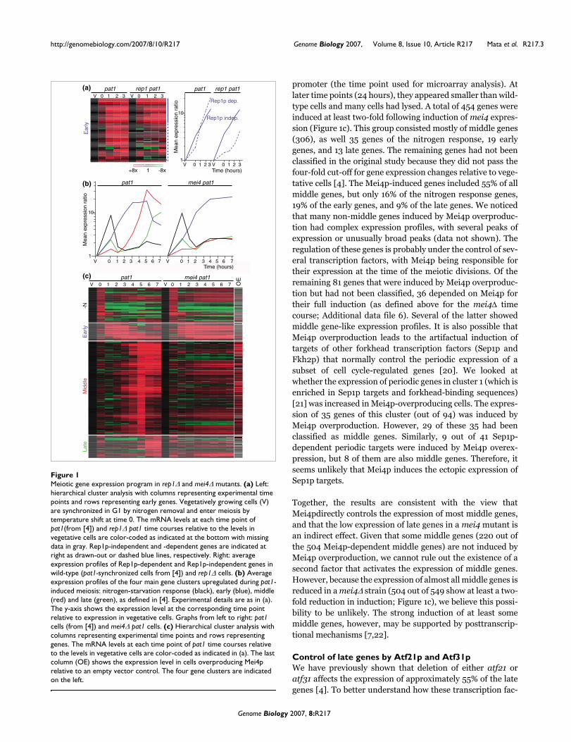
Identification of Rsv1p-dependent genesFigure 4Identification of Rsv1p-dependent genes. (a) Expression profile of rsv1 during meiosis and sporulation (experiment and labeling as in Figure 1). (b) Comparison of gene expression levels between wild-type and rsv1Δ meiotic cells. Genes outside the blue lines differ by more than two-fold in expression levels. rsv1 is indicated by an arrow. (c) Expression profiles of the potential Rsv1p-dependent genes identified in (b) (pat1-synchronized cells from [4]).
10 1e2 1e3 1e4 1e510
1e2
1e3
1e4
Time (hours)
Exp
ress
ion
ratio
rsv1
V 0 1 2 3 4 5 6 7 8 10 12
(a)
(b)
(c)
1
10
Exp
ress
ion
sign
al rs
v1
V 0 1 2 3 4 5 6 7 8 10 12
1
10
100
Exp
ress
ion
ratio
Time (hours)
0.1
Expression signal wild-type
Genome Biology 2007, 8:R217
http://genomebiology.com/2007/8/10/R217 Genome Biology 2007, Volume 8, Issue 10, Article R217 Mata et al. R217.7
expression in late meiosis is at least partly under the controlof Mei4p.
The alternation between transcriptional waves also requiresthat each wave is inactivated before the following wave isinduced. We found that the reduction in the levels of earlygenes that normally coincides with the induction of the mid-dle genes did not occur in mei4Δ mutants (Figure 1b,c). Thiseffect has been previously observed for a small number ofrecombination genes [14]. We conclude that in addition to itsrole in up-regulating middle genes, Mei4p is necessary toswitch off the early gene expression wave. This function maybe indirect, as we did not observe a down-regulation of earlygenes when Mei4p is overproduced. The finding that the neg-ative regulator Rsv1p seems to be a Mei4p target (Figure 5c)raises the possibility that Mei4p represses the expression ofsome early genes via Rsv1p. However, the inactivation ofother early genes, including those involved in meiotic recom-bination, appears to proceed through a different, yetunknown, mechanism.
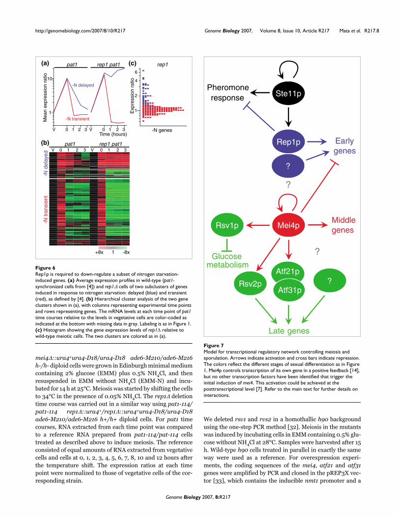
Similarly, we noticed that Rep1p, which is involved in the acti-vation of a subset of early genes, is specifically required forthe down-regulation of some of the genes induced in responseto nitrogen starvation and pheromone signaling followingentry into meiosis (Figure 6a,b). This effect is specific to the'delayed' genes [4], which are enriched in Ste11p targets [3],
while the down-regulation of other genes induced by nitrogenstarvation is unaffected or even stronger in a rep1Δ mutant(Figure 6a,b). Although gene expression in wild-type andpat1-induced meiosis is similar for early, middle and lategenes [4], the conditions used to trigger synchronous meiosisin pat1 mutants can affect the expression of genes induced bynitrogen starvation. To rule out the possibility that the effectson the expression of delayed genes were an artifact, wechecked whether rep1Δ mutants had a similar behavior inwild-type meiosis using homothallic cells. Indeed, 'delayed'genes were expressed at higher levels in rep1Δ mutants com-pared to wild-type cells, while 'transient' genes were notaffected (Figure 6c).
These results show that the temporal pattern of successivewaves of gene expression is at least partly controlled by inter-actions between transcription factors, in which a transcrip-tion factor induces the expression of a wave of genes, whileswitching off the previous wave and inducing transcriptionfactors that will in turn trigger the next wave (Figure 7).
ConclusionWe have analyzed the role of six transcription factors in regu-lating the gene expression program of different stages of sex-ual developmental in fission yeast. Five of these factors workas transcriptional activators, while one of them (Rsv1p) is atranscriptional repressor. In addition to a global identifica-tion of potential target genes, our results give insight into reg-ulatory circuits that coordinate different transcriptionalwaves of this complex gene expression program. Two mainprinciples are highlighted by this work (Figure 7). First,dependence between transcriptional waves is achieved bypositive and negative interactions between transcription fac-tors. Rep1p, which is transcriptionally induced by the masterregulator for the pheromone response Ste11p [3,12], activatesthe expression of early genes but is also essential for therepression of pheromone-response genes. Similarly, Mei4pactivates the expression of middle genes and is also essentialfor the repression of the early genes, and for the induction ofgenes encoding transcription factors that in turn activate theexpression of late genes. These dependencies between tran-scription factors ensure the ordered, uni-directional succes-sion of transcriptional waves. Second, the importance ofcombinatorial control: overexpression of Atf21p, Atf31p, orAtf21p/Atf31p leads to the activation of unique sets of targets.The use of different combinations of transcription factorsenhances the regulatory options of cells to express specificgene sets in response to a variety of situations.
Materials and methodsYeast methods and experimental designInduction of meiosis using pat1 mutations was carried outexactly as in our previous study [4]. Briefly, for the mei4Δdeletion time course, pat1-114/pat1-114 mei4Δ::ura4+/
Transcription factor genes regulated by Mei4pFigure 5Transcription factor genes regulated by Mei4p. Expression profiles of genes encoding transcription factors in pat1-synchronized wild-type (blue lines, data from [4]) and mei4Δ (red lines) cells: (a) atf21, (b) atf31, (c) rsv1 and (d) rsv2. Experiments and labeling are as in Figure 1.
Exp
ress
ion
ratio
atf21
V 0 1 2 3 4 5 6 7
(a)200
Time (hours)
100
atf31
V 0 1 2 3 4 5 6 7
200
100
(b)
Exp
ress
ion
ratio
rsv1
V 0 1 2 3 4 5 6 7
(c)20
10
rsv2
V 0 1 2 3 4 5 6 7
10
(d)
Time (hours)
5
mei4Δmei4+
Genome Biology 2007, 8:R217
http://genomebiology.com/2007/8/10/R217 Genome Biology 2007, Volume 8, Issue 10, Article R217 Mata et al. R217.8
mei4Δ::ura4+ura4-D18/ura4-D18 ade6-M210/ade6-M216h-/h- diploid cells were grown in Edinburgh minimal mediumcontaining 2% glucose (EMM) plus 0.5% NH4Cl, and thenresuspended in EMM without NH4Cl (EMM-N) and incu-bated for 14 h at 25°C. Meiosis was started by shifting the cellsto 34°C in the presence of 0.05% NH4Cl. The rep1Δ deletiontime course was carried out in a similar way using pat1-114/pat1-114 rep1Δ::ura4+/rep1Δ::ura4+ura4-D18/ura4-D18ade6-M210/ade6-M216 h+/h+ diploid cells. For pat1 timecourses, RNA extracted from each time point was comparedto a reference RNA prepared from pat1-114/pat-114 cellstreated as described above to induce meiosis. The referenceconsisted of equal amounts of RNA extracted from vegetativecells and cells at 0, 1, 2, 3, 4, 5, 6, 7, 8, 10 and 12 hours afterthe temperature shift. The expression ratios at each timepoint were normalized to those of vegetative cells of the cor-responding strain.
We deleted rsv1 and rvs2 in a homothallic h90 backgroundusing the one-step PCR method [32]. Meiosis in the mutantswas induced by incubating cells in EMM containing 0.5% glu-cose without NH4Cl at 28°C. Samples were harvested after 15h. Wild-type h90 cells treated in parallel in exactly the sameway were used as a reference. For overexpression experi-ments, the coding sequences of the mei4, atf21 and atf31genes were amplified by PCR and cloned in the pREP3X vec-tor [33], which contains the inducible nmt1 promoter and a
Rep1p is required to down-regulate a subset of nitrogen starvation-induced genesFigure 6Rep1p is required to down-regulate a subset of nitrogen starvation-induced genes. (a) Average expression profiles in wild-type (pat1-synchronized cells from [4]) and rep1Δ cells of two subclusters of genes induced in response to nitrogen starvation: delayed (blue) and transient (red), as defined by [4]. (b) Hierarchical cluster analysis of the two gene clusters shown in (a), with columns representing experimental time points and rows representing genes. The mRNA levels at each time point of pat1 time courses relative to the levels in vegetative cells are color-coded as indicated at the bottom with missing data in gray. Labeling is as in Figure 1. (c) Histogram showing the gene expression levels of rep1Δ relative to wild-type meiotic cells. The two clusters are colored as in (a).
V 0 1 2 3 V 0 1 2 3 Time (hours)
pat1
Mea
n ex
pres
sion
rat
io
10
1
rep1 pat1
+8x 1 -8x
V 0 1 2 3 V 0 1 2 3
-N delayed
-N transient
1
2
4
6
rep1
-N genes
pat1 rep1 pat1
-N tr
ansi
ent
-N d
elay
ed
(a) (c)
(b)
Exp
ress
ion
ratio
Model for transcriptional regulatory network controlling meiosis and sporulationFigure 7Model for transcriptional regulatory network controlling meiosis and sporulation. Arrows indicate activation and cross bars indicate repression. The colors reflect the different stages of sexual differentiation as in Figure 1. Mei4p controls transcription of its own gene in a positive feedback [14], but no other transcription factors have been identified that trigger the initial induction of mei4. This activation could be achieved at the posttranscriptional level [7]. Refer to the main text for further details on interactions.
Atf21p
Ste11p
Earlygenes
Middlegenes
Late genes
?
Glucosemetabolism
Rep1p
Mei4p
Atf31pRsv2p
Rsv1p
?
?
Pheromoneresponse
?
Genome Biology 2007, 8:R217
http://genomebiology.com/2007/8/10/R217 Genome Biology 2007, Volume 8, Issue 10, Article R217 Mata et al. R217.9
LEU2 selectable marker. For the co-overexpression experi-ment, atf31 was cloned into pREP4X (also under nmt1 con-trol, but with an ura4 selectable marker). Single plasmidswere transformed into a leu1-32 h- strain, and both plasmidswere co-transformed into leu1-32 ura4-D18 h- cells. Toinduce the nmt1 promoter, cells were grown in EMM contain-ing 15 μM thiamine, washed three times in EMM, and incu-bated at 32°C for 18 h. In every experiment, RNA extractedfrom cells overexpressing a particular transcription factorwas compared with RNA from cells transformed with emptyvectors that were treated in exactly the same way to inducethe nmt1 promoter.
Microarray experimentsRNA preparation, labeling, microarray production, and dataprocessing were performed as described [34]. Microarrayswere scanned with a Genepix 4000B scanner and analyzedwith GenePix software (Molecular Devices, Sunnyvale, CA,USA). Hierarchical clustering, visualization, and regulatorymotif searches were done with GeneSpring (Agilent, SantaClara, CA, USA). The significance of the overlaps betweengene lists was determined assuming that the overlap betweenrandom groups follows a hypergeometric distribution. Allprocessed and normalized data are available from our website[35], and the entire raw data sets have been deposited inArray Express [36] with accession numbers E-TABM-298, E-TABM-299, E-TABM-300 and E-TABM-301. Complete nor-malized data sets are also available in Additional data files 1–4. Microarray experiments of rsv1Δ and rsv2Δ deletions andoverexpression of Atf21p and Atf31p transcription factorswere done in duplicate (independent biological repeatsincluding a dye swap). The RNA from the Mei4p overexpres-sion experiment was hybridized in duplicate (technicalrepeat, including a dye swap). The mei4Δ and rep1Δ timecourses and the wild-type rep1Δ experiment were carried outonce.
Validation of resultsWe compared our data with Northern-based experiments ofgene expression in rep1Δ and mei4Δ backgrounds. The micro-array results were similar to published data for 8/10 genes inrep1Δ and 37/42 genes in mei4Δ (see Tables 2 and 3 in Addi-tional data file 5).
AbbreviationsbZIP, basic leucine zipper; EMM, Edinburgh minimalmedium.
Authors' contributionsJM and AW carried out the experiments. JM and JB con-ceived the study, analyzed the data and co-wrote the paper.All authors read and approved the final manuscript.
Additional data filesThe following additional data are available with the onlineversion of this paper. Additional data file 1 includes the com-plete normalized dataset for mei4Δ pat1 and pat1 timecourses. Additional data file 2 includes the complete normal-ized dataset for rep1Δ pat1 and pat1 time courses. Additionaldata file 3 includes the complete normalized dataset for rsv1Δand rsv2Δ experiments. Additional data file 4 includes thecomplete normalized dataset for Atf21p, Atf31p, Atf21p/Atf31p and Mei4p overexpression experiments. Additionaldata file 5 includes additional tables. Table 1: Potential regu-latory motifs in the promoters of Rsv1p-regulated genes.Table 2: Effects of rep1Δ on meiotic transcription: compari-son with published data. Table 3: Effects of mei4Δ on meiotictranscription; comparison with published data. Additionaldata file 6 lists middle genes classified into three groupsaccording to the effect of mei4Δ and Mei4p overexpression ontheir expression. Additional data file 7 lists genes induced byAtf21p, Atf31p and Atf21p/Atf31p overexpression and genesinduced by Atf21p/Atf31p overexpression that are alsoreduced in atf21Δ meiotic cells. Additional data file 8 listsgenes induced by Mei4p overexpression, classified accordingto their expression profiles. Additional data file 9 lists Mei4p-dependent and Mei4p-independent middle genes based onthe mei4Δ time course experiment. Additional data file 10includes a complete list of potential Rsv1p targets. Additionaldata file 11 includes a complete list of potential Rsv2p targets.Additional data file 12 lists Rep1p-dependent and Rep1p-independent early genes based on the rep1Δ time courseexperiment.Additional data file 1Complete normalized dataset for mei4Δ pat1 and pat1 time coursesComplete normalized dataset for mei4Δ pat1 and pat1 time courses.Click here for fileAdditional data file 2Complete normalized dataset for rep1Δ pat1 and pat1 time coursesComplete normalized dataset for rep1Δ pat1 and pat1 time courses.Click here for fileAdditional data file 3Complete normalized dataset for rsv1Δ and rsv2Δ experimentsComplete normalized dataset for rsv1Δ and rsv2Δ experiments.Click here for fileAdditional data file 4Complete normalized dataset for Atf21p, Atf31p, Atf21p/Atf31p and Mei4p overexpression experimentsComplete normalized dataset for Atf21p, Atf31p, Atf21p/Atf31p and Mei4p overexpression experiments.Click here for fileAdditional data file 5Additional tablesTable 1: Potential regulatory motifs in the promoters of Rsv1p-reg-ulated genes. Table 2: Effects of rep1Δ on meiotic transcription: comparison with published data. Table 3: Effects of mei4Δ on mei-otic transcription; comparison with published data.Click here for fileAdditional data file 6Middle genes classified into three groups according to the effect of mei4Δ and Mei4p overexpression on their expressionMiddle genes classified into three groups according to the effect of mei4Δ and Mei4p overexpression on their expression.Click here for fileAdditional data file 7Genes induced by Atf21p, Atf31p and Atf21p/Atf31p overexpression and genes induced by Atf21p/Atf31p overexpression that are also reduced in atf21Δ meiotic cellsGenes induced by Atf21p, Atf31p and Atf21p/Atf31p overexpression and genes induced by Atf21p/Atf31p overexpression that are also reduced in atf21Δ meiotic cells.Click here for fileAdditional data file 8Genes induced by Mei4p overexpression, classified according to their expression profilesGenes induced by Mei4p overexpression, classified according to their expression profiles.Click here for fileAdditional data file 9Mei4p-dependent and Mei4p-independent middle genes based on the mei4Δ time course experimentMei4p-dependent and Mei4p-independent middle genes based on the mei4Δ time course experiment.Click here for fileAdditional data file 10Complete list of potential Rsv1p targetsComplete list of potential Rsv1p targets.Click here for fileAdditional data file 11Complete list of potential Rsv2p targetsComplete list of potential Rsv2p targets.Click here for fileAdditional data file 12Rep1p-dependent and Rep1p-independent early genes based on the rep1Δ time course experimentRep1p-dependent and Rep1p-independent early genes based on the rep1Δ time course experiment.Click here for file
AcknowledgementsWe thank Fausto Arellano for help with strain construction and microarrayprocessing, Samuel Marguerat, Luis López-Maury and Daniel Lackner forcomments on the manuscript, and the Sanger microarray facility for arrayprinting. This work was supported by Cancer Research UK (CUK) GrantNo. C9546/A6517.
References1. Shimoda C, Nakamura T: Control of late meiosis and ascospore
formation. In The Molecular Biology of Schizosaccharomyces pombeEdited by: Egel R. Heidelberg: Springer-Verlag; 2004:311-327.
2. Yamamoto M: Initiation of meiosis. In The Molecular Biology ofSchizosaccharomyces pombe Edited by: Egel R. Heidelberg: Springer-Verlag; 2004:297-310.
3. Mata J, Bähler J: Global roles of Ste11p, cell type, and pherom-one in the control of gene expression during early sexual dif-ferentiation in fission yeast. Proc Natl Acad Sci USA 2006,103:15517-15522.
4. Mata J, Lyne R, Burns G, Bähler J: The transcriptional program ofmeiosis and sporulation in fission yeast. Nat Genet 2002,32:143-147.
5. Xue-Franzen Y, Kjaerulff S, Holmberg C, Wright A, Nielsen O:Genomewide identification of pheromone-targeted tran-scription in fission yeast. BMC Genomics 2006, 7:303.
6. Averbeck N, Sunder S, Sample N, Wise JA, Leatherwood J: Negativecontrol contributes to an extensive program of meiotic splic-ing in fission yeast. Mol Cell 2005, 18:491-498.
7. Harigaya Y, Tanaka H, Yamanaka S, Tanaka K, Watanabe Y, TsutsumiC, Chikashige Y, Hiraoka Y, Yamashita A, Yamamoto M: Selectiveelimination of messenger RNA prevents an incidence ofuntimely meiosis. Nature 2006, 442:45-50.
Genome Biology 2007, 8:R217
http://genomebiology.com/2007/8/10/R217 Genome Biology 2007, Volume 8, Issue 10, Article R217 Mata et al. R217.10
8. Watanabe Y, Yamamoto M: S. pombe mei2+ encodes an RNA-binding protein essential for premeiotic DNA synthesis andmeiosis I, which cooperates with a novel RNA speciesmeiRNA. Cell 1994, 78:487-498.
9. Sugimoto A, Iino Y, Maeda T, Watanabe Y, Yamamoto M: Schizosac-charomyces pombe ste11+ encodes a transcription factor withan HMG motif that is a critical regulator of sexual develop-ment. Genes Dev 1991, 5:1990-1999.
10. Cunliffe L, White S, McInerny CJ: DSC1-MCB regulation of mei-otic transcription in Schizosaccharomyces pombe. Mol GenetGenomics 2004, 271:60-71.
11. Ding R, Smith GR: Global control of meiotic recombinationgenes by Schizosaccharomyces pombe rec16 (rep1). Mol GenGenet 1998, 258:663-670.
12. Sugiyama A, Tanaka K, Okazaki K, Nojima H, Okayama H: A zinc fin-ger protein controls the onset of premeiotic DNA synthesisof fission yeast in a Mei2-independent cascade. EMBO J 1994,13:1881-1887.
13. Watanabe T, Miyashita K, Saito TT, Yoneki T, Kakihara Y, NabeshimaK, Kishi YA, Shimoda C, Nojima H: Comprehensive isolation ofmeiosis-specific genes identifies novel proteins and unusualnon-coding transcripts in Schizosaccharomyces pombe. NucleicAcids Res 2001, 29:2327-2337.
14. Abe H, Shimoda C: Autoregulated expression of Schizosaccha-romyces pombe meiosis-specific transcription factor Mei4and a genome-wide search for its target genes. Genetics 2000,154:1497-1508.
15. Horie S, Watanabe Y, Tanaka K, Nishiwaki S, Fujioka H, Abe H,Yamamoto M, Shimoda C: The Schizosaccharomyces pombemei4+ gene encodes a meiosis-specific transcription factorcontaining a forkhead DNA-binding domain. Mol Cell Biol 1998,18:2118-2129.
16. Iino Y, Yamamoto M: Negative control for the initiation of mei-osis in Schizosaccharomyces pombe. Proc Natl Acad Sci USA 1985,82:2447-2451.
17. McLeod M, Beach D: Homology between the ran1+ gene of fis-sion yeast and protein kinases. EMBO J 1986, 5:3665-3671.
18. Yamamoto A, Hiraoka Y: Monopolar spindle attachment of sis-ter chromatids is ensured by two distinct mechanisms at thefirst meiotic division in fission yeast. EMBO J 2003,22:2284-2296.
19. Maundrell K: Thiamine-repressible expression vectors pREPand pRIP for fission yeast. Gene 1993, 123:127-130.
20. Bähler J: Cell-cycle control of gene expression in budding andfission yeast. Annu Rev Genet 2005, 39:69-94.
21. Rustici G, Mata J, Kivinen K, Lio P, Penkett CJ, Burns G, Hayles J,Brazma A, Nurse P, Bähler J: Periodic gene expression programof the fission yeast cell cycle. Nat Genet 2004, 36:809-817.
22. Mata J, Marguerat S, Bähler J: Post-transcriptional control ofgene expression: a genome-wide perspective. Trends BiochemSci 2005, 30:506-514.
23. Chen D, Toone WM, Mata J, Lyne R, Burns G, Kivinen K, Brazma A,Jones N, Bähler J: Global transcriptional responses of fissionyeast to environmental stress. Mol Biol Cell 2003, 14:214-229.
24. Harrison C, Katayama S, Dhut S, Chen D, Jones N, Bähler J, Toda T:SCF(Pof1)-ubiquitin and its target Zip1 transcription factormediate cadmium response in fission yeast. EMBO J 2005,24:599-610.
25. Takeda T, Toda T, Kominami K, Kohnosu A, Yanagida M, Jones N:Schizosaccharomyces pombe atf1+ encodes a transcriptionfactor required for sexual development and entry into sta-tionary phase. EMBO J 1995, 14:6193-6208.
26. Watanabe Y, Yamamoto M: Schizosaccharomyces pombe pcr1+encodes a CREB/ATF protein involved in regulation of geneexpression for sexual development. Mol Cell Biol 1996,16:704-711.
27. Newman JR, Keating AE: Comprehensive identification ofhuman bZIP interactions with coiled-coil arrays. Science 2003,300:2097-2101.
28. Weltmeier F, Ehlert A, Mayer CS, Dietrich K, Wang X, Schutze K,Alonso R, Harter K, Vicente-Carbajosa J, Droge-Laser W: Combina-torial control of Arabidopsis proline dehydrogenase tran-scription by specific heterodimerisation of bZIPtranscription factors. EMBO J 2006, 25:3133-3143.
29. Hao Z, Furunobu A, Nagata A, Okayama H: A zinc finger proteinrequired for stationary phase viability in fission yeast. J Cell Sci1997, 110:2557-2566.
30. Shimanuki M, Chung SY, Chikashige Y, Kawasaki Y, Uehara L, Tsut-
sumi C, Hatanaka M, Hiraoka Y, Nagao K, Yanagida M: Two-step,extensive alterations in the transcriptome from G0 arrest tocell division in Schizosaccharomyces pombe. Genes Cells 2007,12:677-692.
31. Lutfiyya LL, Iyer VR, DeRisi J, DeVit MJ, Brown PO, Johnston M:Characterization of three related glucose repressors andgenes they regulate in Saccharomyces cerevisiae. Genetics 1998,150:1377-1391.
32. Bähler J, Wu JQ, Longtine MS, Shah NG, McKenzie A 3rd, Steever AB,Wach A, Philippsen P, Pringle JR: Heterologous modules for effi-cient and versatile PCR-based gene targeting in Schizosac-charomyces pombe. Yeast 1998, 14:943-951.
33. Forsburg SL, Sherman DA: General purpose tagging vectors forfission yeast. Gene 1997, 191:191-195.
34. Lyne R, Burns G, Mata J, Penkett CJ, Rustici G, Chen D, Langford C,Vetrie D, Bähler J: Whole-genome microarrays of fission yeast:characteristics, accuracy, reproducibility, and processing ofarray data. BMC Genomics 2003, 4:27.
35. Fission Yeast Functional Genomics [http://www.sanger.ac.uk/PostGenomics/S_pombe/]
36. Array Express [http://www.ebi.ac.uk/arrayexpress/]
Genome Biology 2007, 8:R217
http://www.ncbi.nlm.nih.gov/entrez/query.fcgi?cmd=Retrieve&db=PubMed&dopt=Abstract&list_uids=7520368
http://www.ncbi.nlm.nih.gov/entrez/query.fcgi?cmd=Retrieve&db=PubMed&dopt=Abstract&list_uids=1657709
http://www.ncbi.nlm.nih.gov/entrez/query.fcgi?cmd=Retrieve&db=PubMed&dopt=Abstract&list_uids=1657709
http://www.ncbi.nlm.nih.gov/entrez/query.fcgi?cmd=Retrieve&db=PubMed&dopt=Abstract&list_uids=1657709
http://www.ncbi.nlm.nih.gov/entrez/query.fcgi?cmd=Retrieve&db=PubMed&dopt=Abstract&list_uids=9671035
http://www.ncbi.nlm.nih.gov/entrez/query.fcgi?cmd=Retrieve&db=PubMed&dopt=Abstract&list_uids=8168486
http://www.ncbi.nlm.nih.gov/entrez/query.fcgi?cmd=Retrieve&db=PubMed&dopt=Abstract&list_uids=8168486
http://www.ncbi.nlm.nih.gov/entrez/query.fcgi?cmd=Retrieve&db=PubMed&dopt=Abstract&list_uids=8168486
http://www.ncbi.nlm.nih.gov/entrez/query.fcgi?cmd=Retrieve&db=PubMed&dopt=Abstract&list_uids=9528784
http://www.ncbi.nlm.nih.gov/entrez/query.fcgi?cmd=Retrieve&db=PubMed&dopt=Abstract&list_uids=9528784
http://www.ncbi.nlm.nih.gov/entrez/query.fcgi?cmd=Retrieve&db=PubMed&dopt=Abstract&list_uids=3830131
http://www.ncbi.nlm.nih.gov/entrez/query.fcgi?cmd=Retrieve&db=PubMed&dopt=Abstract&list_uids=3830131
http://www.ncbi.nlm.nih.gov/entrez/query.fcgi?cmd=Retrieve&db=PubMed&dopt=Abstract&list_uids=8422996
http://www.ncbi.nlm.nih.gov/entrez/query.fcgi?cmd=Retrieve&db=PubMed&dopt=Abstract&list_uids=8422996
http://www.ncbi.nlm.nih.gov/entrez/query.fcgi?cmd=Retrieve&db=PubMed&dopt=Abstract&list_uids=8557039
http://www.ncbi.nlm.nih.gov/entrez/query.fcgi?cmd=Retrieve&db=PubMed&dopt=Abstract&list_uids=8557039
http://www.ncbi.nlm.nih.gov/entrez/query.fcgi?cmd=Retrieve&db=PubMed&dopt=Abstract&list_uids=8557039
http://www.ncbi.nlm.nih.gov/entrez/query.fcgi?cmd=Retrieve&db=PubMed&dopt=Abstract&list_uids=8552099
http://www.ncbi.nlm.nih.gov/entrez/query.fcgi?cmd=Retrieve&db=PubMed&dopt=Abstract&list_uids=8552099
http://www.ncbi.nlm.nih.gov/entrez/query.fcgi?cmd=Retrieve&db=PubMed&dopt=Abstract&list_uids=8552099
http://www.ncbi.nlm.nih.gov/entrez/query.fcgi?cmd=Retrieve&db=PubMed&dopt=Abstract&list_uids=9372444
http://www.ncbi.nlm.nih.gov/entrez/query.fcgi?cmd=Retrieve&db=PubMed&dopt=Abstract&list_uids=9372444
http://www.ncbi.nlm.nih.gov/entrez/query.fcgi?cmd=Retrieve&db=PubMed&dopt=Abstract&list_uids=9832517
http://www.ncbi.nlm.nih.gov/entrez/query.fcgi?cmd=Retrieve&db=PubMed&dopt=Abstract&list_uids=9717240
http://www.ncbi.nlm.nih.gov/entrez/query.fcgi?cmd=Retrieve&db=PubMed&dopt=Abstract&list_uids=9218719
Related Documents