TRANSCRIPTIONAL REGULATION OF THYROID DEVELOPMENT POSSIBLE INTERPLAY OF ENDODERM- AND MESODERM-DERIVED MORPHOGENETIC SIGNALS JESSICA WESTERLUND Institute of Biomedicine Department of Medical Chemistry and Cell biology The Sahlgrenska Academy at University of Gothenburg 2008

Welcome message from author
This document is posted to help you gain knowledge. Please leave a comment to let me know what you think about it! Share it to your friends and learn new things together.
Transcript

TRANSCRIPTIONAL REGULATION OF THYROID DEVELOPMENT
POSSIBLE INTERPLAY OF ENDODERM- AND MESODERM-DERIVED MORPHOGENETIC SIGNALS
JESSICA WESTERLUND
Institute of Biomedicine
Department of Medical Chemistry and Cell biology
The Sahlgrenska Academy at University of Gothenburg
2008

ISBN: 978-91-628-7558-9 © Jessica Westerlund, October 2008 Institute of Biomedicine The Sahlgrenska Academy at University of Gothenburg Printed by Geson Sverige AB Göteborg, Sweden, 2008
ABSTRACT
TRANSCRIPTIONAL REGULATION OF THYROID DEVELOPMENT: POSSIBLE INTERPLAY OF ENDODERM- AND MESODERM-DERIVED MORPHOGENETIC SIGNALS
Jessica Westerlund
Institute of Biomedicine, Sahlgrenska Academy, University of Gothenburg, Göteborg, Sweden
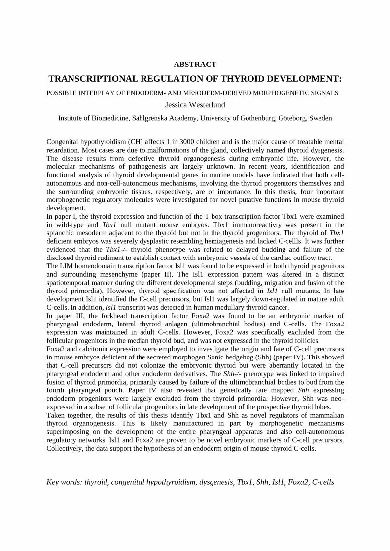
Congenital hypothyroidism (CH) affects 1 in 3000 children and is the major cause of treatable mental retardation. Most cases are due to malformations of the gland, collectively named thyroid dysgenesis. The disease results from defective thyroid organogenesis during embryonic life. However, the molecular mechanisms of pathogenesis are largely unknown. In recent years, identification and functional analysis of thyroid developmental genes in murine models have indicated that both cell-autonomous and non-cell-autonomous mechanisms, involving the thyroid progenitors themselves and the surrounding embryonic tissues, respectively, are of importance. In this thesis, four important morphogenetic regulatory molecules were investigated for novel putative functions in mouse thyroid development. In paper I, the thyroid expression and function of the T-box transcription factor Tbx1 were examined in wild-type and Tbx1 null mutant mouse embryos. Tbx1 immunoreactivty was present in the splanchic mesoderm adjacent to the thyroid but not in the thyroid progenitors. The thyroid of Tbx1 deficient embryos was severely dysplastic resembling hemiagenesis and lacked C-cellls. It was further evidenced that the Tbx1-/- thyroid phenotype was related to delayed budding and failure of the disclosed thyroid rudiment to establish contact with embryonic vessels of the cardiac outflow tract. The LIM homeodomain transcription factor Isl1 was found to be expressed in both thyroid progenitors and surrounding mesenchyme (paper II). The Isl1 expression pattern was altered in a distinct spatiotemporal manner during the different developmental steps (budding, migration and fusion of the thyroid primordia). However, thyroid specification was not affected in Isl1 null mutants. In late development Isl1 identified the C-cell precursors, but Isl1 was largely down-regulated in mature adult C-cells. In addition, Isl1 transcript was detected in human medullary thyroid cancer. In paper III, the forkhead transcription factor Foxa2 was found to be an embryonic marker of pharyngeal endoderm, lateral thyroid anlagen (ultimobranchial bodies) and C-cells. The Foxa2 expression was maintained in adult C-cells. However, Foxa2 was specifically excluded from the follicular progenitors in the median thyroid bud, and was not expressed in the thyroid follicles. Foxa2 and calcitonin expression were employed to investigate the origin and fate of C-cell precursors in mouse embryos deficient of the secreted morphogen Sonic hedgehog (Shh) (paper IV). This showed that C-cell precursors did not colonize the embryonic thyroid but were aberrantly located in the pharyngeal endoderm and other endoderm derivatives. The Shh-/- phenotype was linked to impaired fusion of thyroid primordia, primarily caused by failure of the ultimobranchial bodies to bud from the fourth pharyngeal pouch. Paper IV also revealed that genetically fate mapped Shh expressing endoderm progenitors were largely excluded from the thyroid primordia. However, Shh was neo-expressed in a subset of follicular progenitors in late development of the prospective thyroid lobes. Taken together, the results of this thesis identify Tbx1 and Shh as novel regulators of mammalian thyroid organogenesis. This is likely manufactured in part by morphogenetic mechanisms superimposing on the development of the entire pharyngeal apparatus and also cell-autonomous regulatory networks. Isl1 and Foxa2 are proven to be novel embryonic markers of C-cell precursors. Collectively, the data support the hypothesis of an endoderm origin of mouse thyroid C-cells.
Key words: thyroid, congenital hypothyroidism, dysgenesis, Tbx1, Shh, Isl1, Foxa2, C-cells

LIST OF PUBLICATIONS
The thesis is based on the following papers, referred to in the text by their roman numerals.
I. Fagman H*, Liao J*, Westerlund J*, Andersson L, Morrow BE, Nilsson M. The 22q11 deletion syndrome candidate gene Tbx1 determines thyroid size and positioning. Human Molecular Genetics. 2007 Feb1; 16(3):276-85. * Contributed equally as joint First Authors
II. Westerlund J, Andersson L, Carlsson T, Zoppoli P, Fagman H, Nilsson M. Expression of Islet1 in thyroid development related to budding, migration and fusion of primordia. Developmental Dynamics (in press)
III. Westerlund J*, Andersson L*, Carlsson T, Fagman H, Nilsson M. Foxa family members mark embryonic progenitor cells differentiating into C-cells in the developing thyroid gland. Manuscript * Contributed equally as joint First Authors
IV. Westerlund J, Andersson L, Carlsson T, Fagman H, Nilsson M. Sonic hedgehog regulates the fusion of midline and lateral embryonic thyroid primordia and entry of C-cell precursors to the thyroid gland. Manuscript

TABLE OF CONTENTS
ABBREVATIONS ………………………………………………….…… 7
INTRODUCTION
The thyroid
Importance of thyroid hormone………………………………………..… 8
Structure and function of the thyroid gland…………………….…..……. 8
Regulation of thyroid hormone production…………………………..….. 9
Congenital hypothyroidism………………………………………..…….. 10
Thyroid Development
Specification, placode stage…………………………………...……….… 11
Budding and detachment………………………………………..…..….... 12
Migration…………………………………………………………….…… 13
Fusion……………………………………………………………….……. 13
Growth and differentiation………………………………………….……. 14
Thyroid progenitor cells
The follicular lineage……………………………………………...……… 15
C-cell precursors……………….…………………………………...…….. 15
Thyroid transcription factors……………………………………………… 16
Foregut development
Patterning of the endoderm ……………………………………...………. 19
Early regionalisation the first step in controlling endodermal cell fate
Folding events change the original anterioposterior position of the cells
Foregut and the pharyngeal apparatus…………………………….…...…. 21
Key molecules in pharyngeal arch and pouch development ……………... 21
Endodermal-mesodermal interactions………………………………....….. 22
Regulation of the A-P position
Regulation of morphogenetic events forming the foregut
Regulation of specific domains with organ forming potency
Key steps and general traits in organogenesis
Possible interactions regulating thyroid specification…………………….. 24
Possible interactions regulating thyroid budding…………………………. 25
Possible interactions in thyroid migration………………………………… 26
Thyroid and cardiac development…………………………………. 27
Key molecules in endoderm organogenesis
Tbx1…………………………………………………………………..…... 28
Isl1…………………………………………………………………..……. 28
Foxa………………………………………………………………..……… 28
Shh…………………………………………………………………..…….. 29
AIMS ……………………………………………………...……..………... 30
RESULTS
Role of Tbx1, a downstream target of Shh signalling, in thyroid organogenesis…..…. 31
(paper I)
Expression pattern of Isl1 in the developing thyroid and surrounding embryonic tissues 32
(paper II)
Foxa2 is a novel C-cell precursor marker…………………………………..……. 33
(paper III)
Role of Sonic hedgehog in embryonic C-cell differentiation and migration………..... 34
(paper IV)
DISCUSSION
Thyroid morphogenesis: interactions from surrounding tissues………... 36
Cellular expression pattern during thyroid development……………….. 37
Possible implications in thyroid disease: dysgenesis and cancer…..……. 38
CONCLUSIONS……………………………………………….…… 40
ACKNOWLEDGEMENTS ……………………………………… 41
REFERENCES………………………………………………………… 43
7
ABBREVIATIONS
C-cells Calcitonin producing cells
CH Congenital hypothyroidism
dpc Days postcoitum
E Embryonic day
Fgf Follicular growth factor
Isl1 Islet 1
MTC Medullary thyroid cancer
NC Neural crest
NCC Neural crest cells
NIS Sodium-iodide symporter
Ptc Patched
PTCF Papillary thyroid cancer (follicular type)
RA Retinoic acid
Shh Sonic hedgehog
Smo Smoothend
T3 Tri-iodo-thyronine
T4 Thyroxine
TG Thyroglobulin
TPO Thyroperoxidase
TSH Thyroid stimulating hormone
TSHR Thyroid stimulating hormone receptor
TTF-1 Thyroid transcription factor 1
TTF-2 Thyroid transcription factor 2
UB Ultimobranchial bodies

8
INTRODUCTION
In this thesis novel thyroid progenitor markers and mechanisms of embryonic thyroid development are presented. It is based on four separate studies (papers I-IV). Before report and discussion of data I will briefly introduce the background and theories on which the investigation is based. In the first part, the importance of the thyroid as an endocrine organ, especially during early life, and the regulated mechanism of thyroid hormone biosynthesis are outlined. In the second part, key developmental steps of the thyroid and relevant aspects of the formation of the definitive endoderm and other embryonic organs associated to the gut tube are highlighted.
The thyroid
Importance of thyroid hormone Thyroid hormones are crucial for life at all stages. In its active form thyroid hormone affects in principal every cell in the body and a functioning thyroid gland is thus necessary to the foetus, in childhood and during adult life. In addition, maternal supply of thyroid hormone before the onset of hormone synthesis is mandatory for normal intrauterine development. Whereas tissues in the adult body require thyroid hormone for metabolic processes immature tissues in the foetus and the child are dependent on thyroid hormone for development, differentiation and growth (Moreno et al., 2008). One important example of such a tissue is the central nervous system (Cao et al., 1994; Oppenheimer and Schwartz, 1997). The growth and maturation of the brain cells will not proceed normally without thyroid hormones and the child who had insufficient hormone production during its foetal life risks to become mentally retarded. Ahead there will be more about congenital hypothyroidism, the most common preventable cause of mental retardation among children.
Structure and function of the thyroid gland The bi-lobed thyroid is positioned in the neck, inferior of the cricoid cartilage. The thyroid lobes are interconnected by the isthmus, seen in front of the proximal trachea. The gross anatomy of the thyroid is similar between human and mice, although in humans the isthmus bridges the midsection of the lobes making the gland resemble the shape of a butterfly whereas in mice the gland is truly horseshoe-shaped with the isthmus being the most caudal portion.
The thyroid has the same endocrine cell types and histoarchitecture in all mammals. The thyroid follicular cells (thyrocytes) that produce the pro-hormone thyroglobulin (TG) constitute single-layered epithelia organised into delimited numerous spherical follicles. They are the functional units of the thyroid gland and actively take up iodide from the circulation by the sodium iodide symporter (NIS). Iodide is stored in the lumina covalently bound to TG during thyroid hormone synthesis. The other endocrine cell type in the thyroid is C-cells that produce calcitonin, a hormone implicated in calcium homeostasis. The C-cells do not participate in the follicular epithelium but adopt a parafollicular position and, moreover, they are mostly distributed in the centre of each thyroid lobe. This is reminiscent of the fact that the follicular and the C-cell progenitors emerge from different regions along the axis of the pharyngeal endoderm, which forms anatomically distinct thyroid primordia that later undergo fusion to form the composite organ. The development of the two thyroid anlagen will be outlined in detail later.

9
Regulation of thyroid hormone production As already emphasized, the thyroid hormone supply to the foetus is first achieved by passage of maternal hormones across the placenta (Dickhoff and Swanson, 1990; van Tuyl et al., 2004). By the 10th week of gestation thyroid hormone biosynthesis is initiated in humans (Gruters et al., 2003) corresponding to embryonic day 14.5 in mice (De Felice et al., 2004). The synthesis and release of thyroid hormones into the circulation is regulated by negative feed back of a hormonal circuit (Shupnik et al., 1989), already working in foetal life. In short, the hypothalamus stimulates secretion of thyroid stimulating hormone (TSH) from the pituitary gland, and TSH in its turn positively regulates the thyroid and thus increases both TG production and thyroid hormone synthesis and release. The thyroid hormones, tri- and tetraiodotyronines (T3 and T4), negatively regulates hypothalamus and the pituitary gland, thus closing the autoregulatory loop. Disturbances of hormone supply ay any level eventually leads to impaired thyroid function, which can be diagnosed by monitoring the circulating levels of TSH, T3 and T4.
TSH stimulates the thyroid by binding to the G-protein-coupled TSH receptor located in the basolateral membrane of the follicular cells. Upon ligand-receptor binding mainly the cyclic AMP signalling pathway is activated and increases the expression of genes, e.g. NIS, TG, and thyroid peroxidase (TPO), involved in thyroid hormone biosynthesis. Also a number of events take place inside the follicular cells in order to produce and release thyroid hormones. This includes organification of iodide, which becomes oxidised once it enters the follicular lumen. The oxidation process requires hydrogen peroxide (H2O2) produced by the thyrocytes and TPO present in the apical membrane. TSH also increases the endocytosis of colloid present in the follicular lumen. The colloid in the lumen contains stored thyroid hormones in the form of iodinated and coupled tyrosins bound to the backbone of TG, waiting to be endocytosed by the thyrocytes and released into the circulation.
The thyrocyte is a polarized cell with the apical membrane directed towards the follicular lumen and the basolateral membrane facing the outside of the follicles, in close contact with the circulation. The polarisation is absolutely necessary for normal thyroid function as it forms the basis of vectorial transport e.g. of iodide, manifested by the restricted distribution of membrane proteins. Accordingly, NIS is only expressed in the basolateral membrane and pendrin, putatively involved in iodide efflux to the follicular lumen, is only present in the apical membrane.
Figure 1. Thyroid parenchyma histoarchitecture. Note the parafollicular positions of C-cells (Cc),
not reaching into to the follicular lumina (L)).

10
Abnormal circulating thyroid hormone levels can either be caused by an impaired hypothalamic/pituitary regulation of the thyroid or by defective function of the thyrocytes themselves. Both hypo- and hyperthyroidism can be due to an impaired regulation, however in hypothyroidism a dysfunctional thyroid gland is the most common cause for the disease. Of greater relevance to this thesis, in congenital hypothyroidism insufficient thyroid hormone supply to the infant is most often caused by dysgenesis of the thyroid gland, which significantly reduces the size of thyroid tissue developed during embryogenesis. The next section will therefore describe the features of such developmental defects and their classification.
Congenital hypothyroidism Congenital hypothyroidism (CH) is the most common congenital endocrine disorder affecting approximately 1 of 3500 newborns (Toublanc, 1992). Due to the prerequisite of thyroid hormones for normal brain and skeletal development untreated children with CH will suffer from mental retardation and dwarfism, also known as cretinism.
However, by systemically biochemical screening infants born with hypothyroidism are now detected in due time and in most cases irreversible brain damage can be prevented. Thus, if synthetic thyroid hormones are supplied within days after birth post-natal brain development is rescued and the CH cohort will achieve no measurable IQ reduction later in life.
CH is due to a defect at any of three levels: systemic regulation of the hypothalamic-pituitary-thyroid axis, de novo synthesis of thyroid hormone in the thyrocytes, and thyroid organogenesis. In very rare cases CH is due to pituitary or hypothalamic developmental defects. More commonly is CH caused by mutations in one of the genes implicated in thyroid hormone production, which collectively is referred to as thyroid dyshormonogenesis (Vliet, 2003). One of the most severe forms of this condition is recognized in infants with TPO mutations leading to a nearly complete loss of endogenous thyroid hormone. In all, congenial defects of hormone synthesis constitute 15% of diagnosed cases. This means that morphogenetic defects of the thyroid gland itself are the most frequent (85%). The phenotype of thyroid dysgenesis differs much, the heterogeneity depending on at which developmental stage during thyroid organogenesis the defect has occurred. A defect in specification of midline thyroid precursors results in a absent thyroid (agenesis), a defect in migration of precursors committed to a thyroid fate results in aberrant localization of thyroid tissue usually detected cranial of the normal position of the gland (ectopia), and a defect in proliferation and bilateral growth typically results in a smaller size of the gland (hypoplasia) that occasionally presents as a single lobe (hemiagenesis) (Grant et al., 1992). Accordingly, thyroid dysgenesis represents a spectrum of disorders which have different pathogenesis and clinical severity.
Thyroid development Insights into factors regulating normal thyroid development have largely evolved from studies on genetically modified mice, the relevance of which to humans has been confirmed by identification of mutations in the corresponding genes in patients with thyroid dysgenesis. This has shown that the transcription factors known to regulate the expression of genes implicated in thyroid hormone synthesis (e.g. NIS, TG and TPO) also play important roles in early thyroid development. Specifically, four transcription factors, Nkx2.1, Foxe1, Pax8 and Hhex, are co-expressed in thyroid progenitors already at the placode stage coinciding with the timing of thyroid specification (De Felice and Di Lauro, 2004). Although thyroid differentiation designated by the onset of thyroid-specific genes is not initiated until later, these transcription factors continue to be expressed and are presumably active throughout further development of the thyroid designated cells. Knockout studies in mice have shown

11
that each of the transcription factors are important for the survival, proliferation and migration of the midline thyroid progenitors arising in the pharyngeal endoderm (De Felice and Di Lauro, 2004). However, in search for genetic causes of thyroid dysgenesis in humans only a minority of the patients have mutations in the corresponding genes (shown for Nkx2.1, Foxe1, and Pax8). Thus, further studies on animal models need to be carried out in order to identify novel candidate genes involved in thyroid development and malformation during embryogenesis. Before going into detail on the molecules of interest investigated in this thesis, key steps of thyroid organogenesis and hitherto defined modes of developmental regulation will be described in the following paragraphs.
As mentioned, at the developmental stage thyroid hormone synthesis starts the final shape of the gland is already decided. However, it is important to consider that the thyroid consists of fused anlagen, one medial and two lateral, which have formed and detached from different regions of the pharyngeal endoderm in earlier stages. For clarification the development of each anlage will be described separately until they fuse. For a more detailed overview of thyroid development, there are several recent reviews that can be recommended, for example (De Felice and Di Lauro, 2004). The morphogenesis of mouse thyroid organogenesis was recently stepwise mapped in our laboratory (Fagman et al., 2006); this forms the basis of the presentation here. As mouse thyroid development follows essentially the same pattern as in the human foetus (Trueba et al., 2005), the approximate corresponding embryonic age for each developmental stage in mice and men are indicated in the subheadings.
1. Specification, placode stage (Mouse: E8.5-9.5, Human: 4th week) The process leading to the establishment of the thyroid midline anlage is termed specification. The first visible specified thyroid progenitors can be identified as a subpopulation of cells forming a thickening in the anterior pharyngeal floor at the base of the tongue between the first and second pharyngeal arch (Missero et al., 1998). These cells are designated a follicular fate. The midline thyroid precursors can furthermore be distinguished from the rest of the endoderm by their co-expression of four transcription factors: Nkx2.1 (formerly known as TTF-1) (Lazzaro et al., 1991), Foxe1 (formerly TTF-2) (Zannini et al., 1997), Hhex (Thomas et al., 1998) and Pax8 (Plachov et al., 1990). Although each of these transcription factors fulfils functions in other embryonic tissues as well, their combined expression is only found in the midline thyroid progenitor lineage. The thickening of the endoderm enlarges by recruitment of more progenitors and the thyroid placode transit into the thyroid bud. This takes place in close apposition to the aortic sac.
To date no factors responsible for initiation of thyroid specification in mouse or other higher vertebrates are known, although recently factors derived from the surrounding presumably cardiac mesoderm is inferred an inductive role in the development of the thyroid homologue in zebrafish (Wendl et al., 2007). Knockout studies in mice have shown that the combined thyroid-specific proteins are crucial for thyroid progenitors to survive, proliferate, assemble into a bud and thereafter delaminate from the pharyngeal endoderm. However, the thyroid placode can be detected also in embryos deficient of any of Nkx2.1, Foxe1, Pax8 or Hhex, expressing all the other non-mutated regulatory genes. Master developmental genes turning on the coordinated expression of Nkx2.1, foxe1, Pax8 and Hhex in endoderm progenitors committed to a thyroid fate thus await to be discovered.
The lateral thyroid anlage, known as the ultimobranchial bodies (UB), constitutes bilateral structures that can be identified in the embryo approximately one day later than the midline thyroid primordium. The general believe is that the UB serve as transporting vehicles and bring the C-cell precursors to the
12
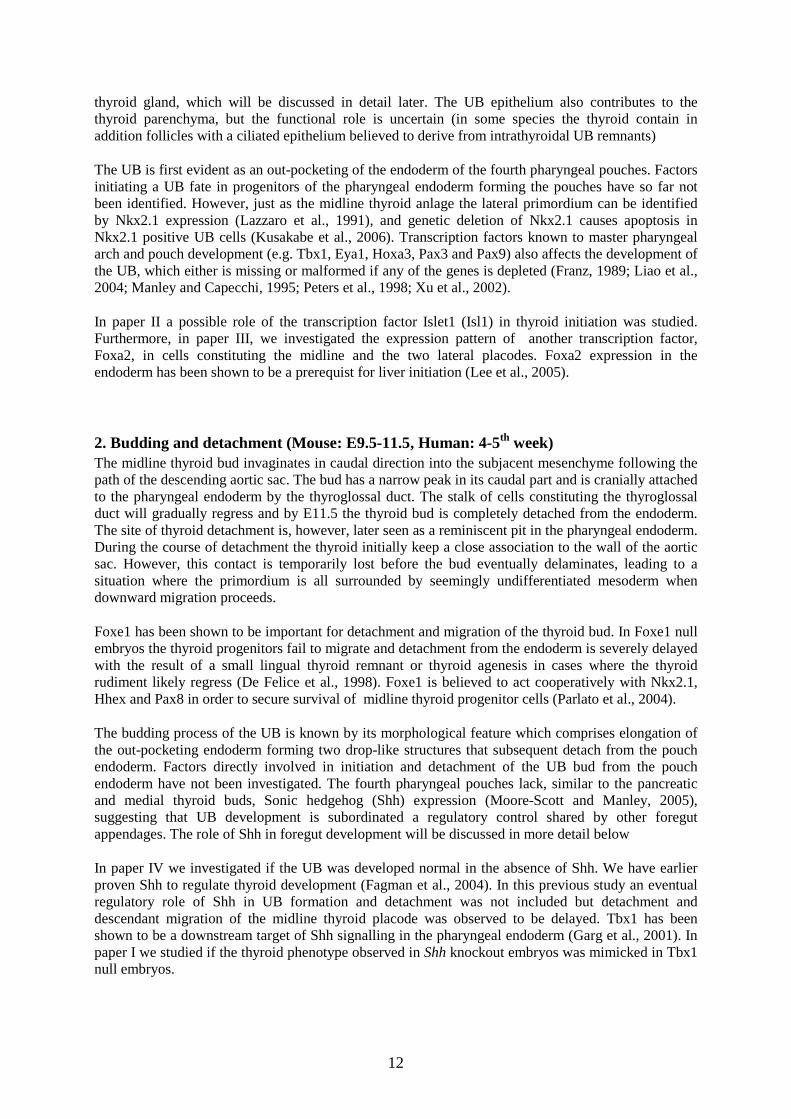
thyroid gland, which will be discussed in detail later. The UB epithelium also contributes to the thyroid parenchyma, but the functional role is uncertain (in some species the thyroid contain in addition follicles with a ciliated epithelium believed to derive from intrathyroidal UB remnants)
The UB is first evident as an out-pocketing of the endoderm of the fourth pharyngeal pouches. Factors initiating a UB fate in progenitors of the pharyngeal endoderm forming the pouches have so far not been identified. However, just as the midline thyroid anlage the lateral primordium can be identified by Nkx2.1 expression (Lazzaro et al., 1991), and genetic deletion of Nkx2.1 causes apoptosis in Nkx2.1 positive UB cells (Kusakabe et al., 2006). Transcription factors known to master pharyngeal arch and pouch development (e.g. Tbx1, Eya1, Hoxa3, Pax3 and Pax9) also affects the development of the UB, which either is missing or malformed if any of the genes is depleted (Franz, 1989; Liao et al., 2004; Manley and Capecchi, 1995; Peters et al., 1998; Xu et al., 2002).
In paper II a possible role of the transcription factor Islet1 (Isl1) in thyroid initiation was studied. Furthermore, in paper III, we investigated the expression pattern of another transcription factor, Foxa2, in cells constituting the midline and the two lateral placodes. Foxa2 expression in the endoderm has been shown to be a prerequist for liver initiation (Lee et al., 2005).
2. Budding and detachment (Mouse: E9.5-11.5, Human: 4-5th week) The midline thyroid bud invaginates in caudal direction into the subjacent mesenchyme following the path of the descending aortic sac. The bud has a narrow peak in its caudal part and is cranially attached to the pharyngeal endoderm by the thyroglossal duct. The stalk of cells constituting the thyroglossal duct will gradually regress and by E11.5 the thyroid bud is completely detached from the endoderm. The site of thyroid detachment is, however, later seen as a reminiscent pit in the pharyngeal endoderm. During the course of detachment the thyroid initially keep a close association to the wall of the aortic sac. However, this contact is temporarily lost before the bud eventually delaminates, leading to a situation where the primordium is all surrounded by seemingly undifferentiated mesoderm when downward migration proceeds.
Foxe1 has been shown to be important for detachment and migration of the thyroid bud. In Foxe1 null embryos the thyroid progenitors fail to migrate and detachment from the endoderm is severely delayed with the result of a small lingual thyroid remnant or thyroid agenesis in cases where the thyroid rudiment likely regress (De Felice et al., 1998). Foxe1 is believed to act cooperatively with Nkx2.1, Hhex and Pax8 in order to secure survival of midline thyroid progenitor cells (Parlato et al., 2004).
The budding process of the UB is known by its morphological feature which comprises elongation of the out-pocketing endoderm forming two drop-like structures that subsequent detach from the pouch endoderm. Factors directly involved in initiation and detachment of the UB bud from the pouch endoderm have not been investigated. The fourth pharyngeal pouches lack, similar to the pancreatic and medial thyroid buds, Sonic hedgehog (Shh) expression (Moore-Scott and Manley, 2005), suggesting that UB development is subordinated a regulatory control shared by other foregut appendages. The role of Shh in foregut development will be discussed in more detail below
In paper IV we investigated if the UB was developed normal in the absence of Shh. We have earlier proven Shh to regulate thyroid development (Fagman et al., 2004). In this previous study an eventual regulatory role of Shh in UB formation and detachment was not included but detachment and descendant migration of the midline thyroid placode was observed to be delayed. Tbx1 has been shown to be a downstream target of Shh signalling in the pharyngeal endoderm (Garg et al., 2001). In paper I we studied if the thyroid phenotype observed in Shh knockout embryos was mimicked in Tbx1 null embryos.

13
3. Migration (Mouse: E11.5-12.5, Human: 5-7th week) After detachment thyroid primordium translocate in a caudal direction in the midline and by E12 it can be identified as a group of cells again located on top of the aortic sac. Thus, the midline thyroid progenitors seem to re-establish contact with the vessel wall. The possible role this re-localisation might have for the thyroid to develop normally guided by the course of embryonic vessels leading from the prospective aortic arch will be discussed later. The length of the distance that the thyroid precursors move from origin to final position can be demonstrated by the fact that the migration requires almost 4 weeks in human embryos (the movement of the bud incorporated). How the migration of the midline thyroid progenitors is regulated still needs to be elucidated. It appears reasonable to assume, however, that interactions with surrounding embryonic tissues, which are not static but move and expand in different directions, are of importance. Input regulatory signals from cells present outside the primordium (e.g. mesenchyme) are defined as non-cell-autonomous.
Also the paired UB migrate from their site of origin in the pharyngeal endoderm approaching the expanding midline thyroid. This process is completely unknown regarding regulatory signals acting as driving force and compass for the movement, although it can be hypothesized that the approach of the two primordia is facilitated by reciprocal attraction signals.
4. Fusion (Mouse: E12.5- 13.5, Human: 7-8th week) In level with the third pharyngeal arch arteries the midline thyroid progenitors have reached the final position of the thyroid gland and begin to grow bilateral. During the elongation of the midline anlage the cells proliferate and the thyroid midline parenchyma increases as it follows the route of the third pharyngeal arch arteries. When the midline thyroid anlage has reached the lateral aspects in the embryo each tip face one of the paired ultimobranchial bodies. At E 13 the proliferating midline thyroid parenchyma engulf the UB and the two anlagen fuse forming primitive thyroid lobes. Shortly after fusion cells derived from the UB can be detected as a spheroid structure within each thyroid lobe.
Although errors during this developmental stage results in the most common thyroid phenotype in CH factors regulating the midline progenitor mass to form into two interconnected lobes remain to be elucidated. There are examples of knockout animals were the two thyroid anlagen fail to fuse, however the mechanisms responsible have not been determined.
In paper I we investigated if the thyroid develops normal when the UB is missing. Furthermore, Shh signalling as a potential regulator of the fusion process was investigated in paper IV.
5. Growth and differentiation (Mouse: E13.5-E17.5, Human: 10-12th week) Interestingly, after cells derived from the UB have been incorporated in the thyroid lobes they no longer proliferate. Instead the increased size of thyroid parenchyma and formation of a normal sized gland seems to be due to proliferation of the designated thyroid follicular cells.
During this stage thyroid midline progenitors are beginning to organize into the typical thyroid histoarchitecture and cords of follicles can be identified in a star like pattern in each immature thyroid lobe. Cells derived from the UB initiate to spread from their central position out in the lobe parenchyma following the cords. Thus, a rough organisation of the thyroid progenitors is initiated immediately after fusion of the two anlagen establishing the histoarchitecture within the gland. Shortly after organisation of follicles with parafollicular positioned cells detectable amounts of TG and calcitonin is produced. Thus, the cells are beginning to attain a differentiated state and the midline derived cells are now able to produce and release thyroid hormones. In similarity calcitonin is produced by differentiated C-cells.

14
It would be convincingly if the differentiation of thyrocytes was initiated by signals secreted from the lateral primordia. However this is most likely not the case since also ectopic thyroid gland that has not fused with the ultimobranchial bodies produce TG. Factors regulating thyrocyte differentiation and C-cell differentiation are to date unknown.
In paper I we studied if the thyroid hypoplasia observed in Shh null embryos was also caused by loss of the transcription factor Tbx1.
Figur 2. The keysteps during thyroid gland development. Impaired regulation at any of the crucial stages will cause thyroid dysgenesis and result in different thyroid phenotypes depending on at which development step the abnormal regulation has occurred. Ph. endoderm – Pharyngeal endoderm; as – Aortic sac; ub –Ultimobranchial bodies.

15
Thyroid progenitors
The follicular lineage That the midline thyroid progenitors originate in the anterior endoderm is indisputable. Also, the thyroid placode located in the pharyngeal floor is the site in the endoderm were cells are committed to a thyroid fate. However, BrdU labelling in vivo experiments on mouse embryos revealed that the initial placode and later the thyroid bud consist of cells which do not proliferate, yet both the placode and bud gradually enlarge between E9.5-10.5 (Fagman et al., 2006). In fact, DNA synthesis in midline progenitors was recognized first at E11.5, i.e. after the bud was detached from the endoderm (Fagman et al., 2006). This indicates that thyroid progenitors likely are recruited from the neighbouring parts of the pharyngeal endoderm during the entire budding process. To date, in lack of reliable markers of distinct domains of the definitive endoderm (Tremblay and Zaret, 2005), there are no published studies intending to identify which progenitor cells outside the placode may acquire a thyroid fate before the thyroid-specific constellation of transcription factors is expressed.
C-cell precursors The C-cells were named in1962 after the discovery of their ability to produce calcitonin (Copp et al., 1962). In mouse the calcitonin production starts in late development after fusion of the two thyroid anlagen. Thus, calcitonin is not possible to use as a marker for C-cell identification during early development. Fluorescent amine tracing experiment has been conducted in order to identify the whereabouts of the C-cells before the onset of calcitonin production (Pearse and Carvalheira, 1967). The study strengthened earlier views by showing that the mammalian C-cells are brought to the thyroid by the UB.
Even though the means by which C-cell precursors eventually are transported to the thyroid have been known for almost half a century the embryonic origin of the C-cells is still controversial. In the late 1960’s Pearse introduced the APUD (amine precursor uptake and decarboxylation) concept where the C-cells together with around 40 other cell types were grouped into one entity, all recognised as neural crest derivatives (Pearse, 1968). A neural crest origin of avian C-cells was subsequently strengthen by fate mapping experiments using quail-chick transplantation chimeras (Le Douarin and Le Lievre, 1970; Polak et al., 1974). In this classical study, restricted regions of the neural tube from quail embryo were transplanted into chick embryos, and the fate of the quail cells recognized by a characteristic large nucleus with dense chromatin mass was followed in the microscope. However, in recent years the long accepted neural crest origin of the C-cells has been questioned. Taking the advantage of Wnt1-Cre previously proven to mark the progeny of neural crest cells (Jiang et al., 2000), genetic fate mapping experiments were conducted by crossing Wnt1-Cre heterozygous with the Rosa26 Reporter mouse and the embryonic thyroid was examined (Kameda et al., 2007). Surprisingly, Wnt1 expressing descendants colonized the thyroid stroma but did not mark the C-cells (Kameda et al., 2007). Based on the finding that the C-cells expressed E-cadherin it was suggested that the C-cell precursors in mice derived from the endoderm (Kameda et al., 2007). This hypothesis has yet not been tested by genetic marking of descendants to pluripotent endoderm. The embryonic origin of mammalian C-cells thus remains to be established. Moreover, presently no embryonic markers of C-cell precursors, which could be employed tracing them before thyroid entry and differentiation into C-cells, are known.

16
Thyroid transcription factors As mentioned, the specified thyroid progenitor cells are distinguished from the rest of the pharyngeal endoderm by the expression of a unique combination of transcription factors. That is, even though expression of e.g. Nkx2.1 is identified in the lung co-expression of Nkx2.1, Foxe1, Pax8 and Hhex can only be detected in cells committed to the thyroid follicular fate. The genes are actively transcribed throughout development and also in adult thyrocytes, in which the function switch to maintain the differentiated state. In fact, Nkx2.1 and Pax8 were first discovered as regulators of thyroid-specific genes (Damante et al., 2001; Pellizzari et al., 2000). Mice knockout studies have demonstrated crucial roles for each of the transcription factors during early thyroid development (De Felice et al., 1998; Kimura et al., 1996; Mansouri et al., 1998; Martinez Barbera et al., 2000) and also revealed molecular interactions in an intricate regulatory network controlling the survival and migration of the thyroid progenitors (Parlato et al., 2004). As the interplay between the transcription factors is absolutely necessary for normal thyroid development, their expression pattern and major actions in the embryo will be highlighted below.
Nkx2.1 Nkx2.1 (formerly known as TTF-1 for thyroid transcription factor-1) is a homeobox domain transcription factor member of the Nkx2 family. The protein was first identified because of its binding ability to the promoters of TG and TPO in thyroid cells. Initially, the name for Nkx2.1 was therefore thyroid-specific enhancer binding protein (T/ebp). Later, embryonic studies revealed Nkx2.1 expression in both thyroid anlagen, the trachea, lungs, pituitary and the forebrain (Lazzaro et al., 1991). In the endoderm in the pharyngeal floor Nkx2.1 expression is restricted to the cells within the boundaries of the thyroid placode (Fig. 3).
Mice lacking expression of Nkx2.1 die at birth presumably due to respiratory failure since the lungs are hypoplastic (Kimura et al., 1996). In these animals the thyroid is absent but study of early stage embryos (E8.5) has shown that initiation of the thyroid developing program is not disturbed since the thyroid placode is detectable in the pharyngeal endoderm. However,,the Nkx2.1-deficient thyroid rudiment regresses most likely through apoptosis of progenitor cells. Also the UB are correctly formed at first but degenerate after budding (Kusakabe et al., 2006). Therefore, both UB and C-cells are missing in Nkx2.1 knockout mice. Together, this indicates that Nkx2.1 is dispensable for the initial specification of both thyroid primordia but is absolutely required for the survival of all known cell types involved. How Nkx2.1 keeps thyroid progenitors alive during embryogenesis is not clear but a hypothesis including a role for fibroblast growth factors (Fgfs) has been suggested (Wendl et al., 2007).
In humans, heterozygous loss-of-function mutations in Nkx2.1 results in mild hypothyroidism and neurological disturbances and respiratory distress reminiscent of the knockout mouse phenotype (Krude et al., 2002; Pohlenz et al., 2002). This means that haploinsufficiency for Nkx2.1 does not challenge thyroid cell survival. Screenings of patients with CH has shown that only very few cases of thyroid dysgenesis is due to mutations in Nkx2.1. Homozygous Nkx2.1 mutations have not been found and are therefore likely not compatible with survival of the human foetus.
Foxe1 The forkhead/winged-helix domain transcription factor Foxe1 (formerly called TTF-2 for thyroid transcription factor-2) is expressed besides the thyroid midline progenitors in most of the foregut endoderm. Foxe1 thus exhibits a less restricted expression domain than Nkx2.1 in the endoderm of the pharyngeal floor (Fig. 3).

17
Mice depleted of Foxe1 die soon after birth (De Felice et al., 1998). The death is most likely due to failure to thrive caused by a severe cleft palate. In the homozygote mice no thyroid gland can be detected in its normal position and is occasionally missing (agenesis). However, the common phenotype is a small lingual thyroid remnant. Foxe1 -/- early stage mouse embryos resembles wild-type embryos with a normally specified thyroid placode in the pharyngeal floor. However, the thyroid rudiment in the Foxe1 mutants fails to bud but maintain a position in or close to the endoderm. Thus, Foxe1 is necessary during thyroid development in order to govern a normal migration of the midline thyroid bud and furthermore cooperates in the control of thyroid cell survival. Albeit the changed position and size the lingual thyroid in Foxe1 null embryos is differentiated and synthesise TG (De Felice et al., 1998).
In humans homozygous defects in Foxe1 result in a rare disease featured of athyreosis, cleft palate and spiky hair (Clifton-Bligh et al., 1998).
Pax8 Pax8 is a paired domain transcription factor involved in thyroid development and gene expression after follicular differentiation. Similar to Nkx2.1 Pax8 recognises specific sequences in the promoter region regulating the thyroid specific genes TPO and TG. Interestingly, in differentiated thyroid follicular cells Pax8 directly interacts with Nkx2.1. Whether the transcription factors cooperate also during early thyroid gland organogenesis has not been shown. However, such collaboration could only take part in the midline thyroid progenitors since Pax8 is not expressed in the UB. In fact, Pax8 is the only of the four transcription factors that has an endoderm expression limited to the median thyroid primordium (Fig. 3 ). Other embryonic mouse tissues that express Pax8 are a part of forebrain (myelencephalon) and the kidneys (Plachov et al., 1990).
Pax8 null mice are born alive but die a few weeks after birth (Mansouri et al., 1998). In mutant embryos the midline thyroid anlage is correctly specified and forms a normal bud that detaches and migrates. However, soon thereafter the thyroid regresses completely (Mansouri et al., 1998). Thus, like Nkx2.1 Pax8 is crucial for the survival of follicular progenitor cells. In contrast, C-cells are present in Pax8 -/- mice, reflecting the fact that Pax8 is not expressed in the UB which develop normally. In fact newborn mice depleted of Pax8 have an undeveloped gland that almost completely consists of C-cells indicating that it likely is devoid of thyroid midline parenchyma and consists only of UB derived cells.
In humans mono-allelic mutation of the Pax8 gene causes CH with either hypoplasia or ectopic thyroid tissue (Macchia et al., 1998). It is also a rare disease.
Hhex This is a homeodomain-containing transcription factor named after its first identification in multipotent hematopoietic cells (hematopoietically expressed homeobox). Since then Hhex expression has been identified in embryonic primitive and definitive endoderm and is important for regionalization of the anterior endoderm. Consequently, Hhex is identified in the primordium of several foregut derived organs, such as thyroid, lungs, liver, and pancreas (Bogue et al., 2000; Thomas et al., 1998). Expression of Hhex can also be detected in endothelial cell precursors (Thomas et al., 1998). In the pharyngeal floor endoderm Hhex expression resembles the broad expression pattern of Foxe1 (Fig. 3).
Hhex depleted embryos die at mid-gestation and have either absent or hypoplastic thyroid (Martinez Barbera et al., 2000). Notably, the thyroid is initially formed also in Hhex mutants. However, in similarity with mutants lacking the other transcription factors the thyroid follicular progenitors fail to

18
survive in Hhex -/- mouse embryos and shortly after specification only a few non-migrating cells constitute the thyroid primordium. Interestingly, the remaining thyroid progenitors do not express Nkx2.1, Pax8 or Foxe1 before they regress. In accordance with this Hhex has been shown to be required for the maintenance of Nkx2.1, Pax8, and Foxe1 expression in the developing thyroid (Parlato et al., 2004). Thus, the thyroid phenotype observed in Hhex mutants might be caused by the loss of any of the other participating transcription factors.
The cause of death of the Hhex knockout mouse embryo is not established, but may be related to the fact that Hhex plays a critical role also for liver, forebrain, and heart development (Martinez Barbera et al., 2000). The severity of the phenotype found in Hhex null mice might explain why no Hhex mutations have been identified in humans.
Figur 3. Unique co-expression of four transcription factors marks the position for thyroid initiation in
the floor of the pharyngeal endoderm. Note the different restriction of expression domains. Black
marks protein expression.

19
Foregut development
Patterning of the endoderm The cells that will become a part of the thyroid parenchyma start as members of the definitive endoderm. But how is it possible for a small subpopulation of cells to distinguish themselves from their cell neighbours and start the expression of tissue-specific genes? In order to understand this, insight into the earliest stages of mouse development with the formation of the definitive endoderm and the primitive gut tube is necessary and will therefore be outlined here.
Early regionalisation is the first step in controlling endodermal cell fate
Already when the newly specified definitive endoderm exits the primitive streak during gastrulation the fate of the cells are roughly determined. The apparently homogenous endoderm sheet are formed so that cells which are recruited first to the primitive streak will end up more anterior and cells entering later will be positioned in posterior regions of the endoderm (Lawson et al., 1986; Lawson and Pedersen, 1987; Tam and Beddington, 1992). The very early regionalisation along the anterior-posterior (AP) axis can be identified by restricted gene expression, e.g. the anterior definitive endoderm express Cerberus (Cerl) (Belo et al., 1997), Otx2 (Rhinn et al., 1998), Hesx1 (Thomas and Beddington, 1996) and Hhex (Thomas et al., 1998). Sox17 is first expressed in the anterior endoderm but is down-regulated as the expression shifts progressively to more posterior regions (Kanai-Azuma et al., 2002). In contrast, Cdx2 expression can be identified solely in posterior endoderm (F. Beck, 1995). Thus, even though the approximately 500-1000 cells constituting the definitive endoderm sheet seems homogenous anatomically the cells have an early onset of molecular heterogeneity along the A-P axis, governing cell competence and their fates as development proceeds. The A-P patterning thus constitutes a first broad regionalisation of the endoderm, which likely pre-patterns the foregut for the specification of the different appendages evolving from it.
Folding events change the original anterioposterior position of the cells
The anterior part of the definitive endoderm is fated to become the epithelial internal lining of the foregut (Tremblay and Zaret, 2005), the embryonic region of the gut tube that gives rise to the thyroid (both anlagen), lungs, liver, pancreas, thymus, esophagus and stomach. The formation of a tube from the flat endodermal sheet begins anterior in the embryo by a ventral folding of the anterior-most endoderm (embryonic gut tube formation is reviewed in (Grapin-Botton and Melton, 2000)). This fold generates a pocket, the anterior intestinal portal (AIP), and shortly after the formation of AIP a similar pocket is formed by ventral folding of the posterior endoderm creating the caudal intestinal portal (CIP) (Fig. 4). The cavities form the prospective foregut (AIP) and prospective hindgut (CIP) and their formation leads to the establishment of a ventral-dorsal axis in the embryonic gut tube (Wells and Melton, 1999). As the folded tissues move towards each other the pockets elongate and the former most anterior and most posterior positioned endodermal cells meet at the stalk of the yolk sac were the forming tube closes ventrally. Simultaneously to the closure the lateral endoderm folds ventrally and the embryonic gut tube is formed. The folding of the endodermal sheet thus alters the anterior-posterior position of cells originally incorporated in the definitive endoderm. This suggests a possibility that the original position of cells later participating in organogenesis might be a site for reception of morphogenetic signals that determine cell fate. Knowledge of this might provide important insight on the mechanisms of pre-patterning endoderm-derived tissues and how pluripotent progenitors become competent to respond to primordium-specific signals.

20
Recent fate mapping studies show that there is a broad subdivision in the anterior endoderm and that the programming of the precursors to the respective cell fate has already been initiated before the formation of the embryonic gut tube (Franklin et al., 2008; Tremblay and Zaret, 2005). In reference of thyroid development, the original position for the follicular progenitors in the ventral pharyngeal floor is believed to be the midline anterior-most portion of the endoderm (Franklin et al., 2008). Thus, the thyrocyte progenitors are pre-patterned in and share competence with the most rostrally part of the endoderm. In contrast, the UB constituting the lateral thyroid anlagen are derivates of the fourth pharyngeal pouches. Cells of the transient pharyngeal pouches have been traced to have their origin in lateral endoderm in more caudal regions of the foregut as compared to the origin of progenitor cells destined to the median thyroid anlage (Franklin et al., 2008). The implication of this is that, after fusion of the two primordia, the thyroid consists of cells that have evolved from distant regions of the anterior endoderm. Interestingly, the liver was recently found to be composed of cells derived from multiple sites in the lateral and midline definitive endoderm (Tremblay and Zaret, 2005).
Figure 4. Folding of the endoderm into the embryonic gut tube. The formation of a tube from the sheet
of definitive endoderm is initiated by ventral folding of the anterior and posterior ends, forming two
pockets: the anterior intestinal portal (AIP) and the caudal intestinal portal (CIP). The AIP constitutes
the prospective foregut. A – Anterior; P – Posterior; Fp – Floor plate. Adapted from (Lewis and Tam,
2006).

21
Foregut and the pharyngeal apparatus
The most cranial part of the newly formed foregut constitutes the prospective pharynx and is thus termed the pharyngeal endoderm. In this region the transient pharyngeal apparatus is formed. This early embryonic structure is to a large extent evolutionary conserved giving rise to craniofacial structures, the pharynx and the glands (thyroid, parathyroid and thymus) appending from the anterior foregut. The pharyngeal apparatus consists of paired lateral out-bulging arches with intervening pouches that are named in roman numerical order as they develop. The development requires the interplay between all three germ layers and is achieved in a cranial-to-caudal direction giving that the arches and pouches with number I are positioned most cranially. The external of the arches is covered by ectoderm, the internal is lined by endoderm and in the middle of each arch is a core of mesenchyme. The mesenchyme consists of centrally located mesoderm surrounded by neural crest-derived cells. The neural crest cells have migrated into the arches from the rhombomeres and are considered to be responsible for the typical patterning of the pharyngeal apparatus. Notably, it is from this cell population thyroidal C-cells are believed to emanate. Interestingly, relatively recent studies have implied an important regulatory role of the endoderm in pharyngeal arch and pouch development (Graham, 2003; Lindsay et al., 2001). In fact, the endoderm is now favoured as the key regulating tissue, illustrated by the severe malformations of the pharyngeal apparatus resulting from genetic deletion of endoderm-expressed transcription factors and morphogens.
Each pharyngeal arch is also intervened by a pharyngeal artery arising from the aortic sac. The arch arteries run through the arches to be connected to the right and left dorsal aortae. The midline thyroid grows bilaterally towards the UB following the course of the third pharyngeal arch arteries (Fagman et al., 2006). It is suggested that the vicinity to the embryonic vessels might implicate morphogenetic guidance preceding the fusion of primordia and thyroid lobe formation. Of interest, in adults the segments of the carotid arteries located closest to the thyroid gland are in fact the transposed third pharyngeal arteries.
Key molecules in pharyngeal arch and pouch development
The pharyngeal arches and pouches exhibit individual patterning along the A-P axis giving each pair their own identity. This is of importance for the fate decision of progenitors populating the individual arches and pouches. Since the normal pharyngeal arch formation is dependent on evaginations of the endoderm to form pharyngeal pouches signals regulating the development of the embryonic pharynx are believed to be relayed from the endoderm (Piotrowski and Nüsslein-Volhard, 2000). A key regulatory role is inferred the transcription factor Tbx1 expressed in both the pharyngeal endoderm and mesoderm (Garg et al., 2001; Lindsay et al., 2001). In the absence of Tbx1 the caudal pharyngeal pouches and pharyngeal arch arteries fail to develop (Lindsay et al., 2001) resulting in a lack of derivatives from the third (thymus, parathyroid) and fourth pouches (UB) (Liao et al., 2004). The mechanism by which Tbx1 govern development of the inferior pharyngeal apparatus is probably to initiate the evaginations of the lateral endoderm forming pharyngeal pouches (Baldini, 2005). The resulting segmentation along the pharyngeal endoderm will provide the structural basis of arch-specific invasion of streams of neural crest cells migrating from the rhombomeres (Anthony, 2008). Due to the lack of pharyngeal arch arteries in Tbx1 knockout embryos it has also been suggested that Tbx1 regulates arch artery formation in a non-cell-autonomous manner (Baldini, 2005). The effector molecule for the suggested intercellular signalling could be Fgf8, since Fgf8 and Tbx1 expression is overlapping in the primitive pharynx (Abu-Issa et al., 2002) and deletion of Fgf8 reproduces the Tbx1 mutant phenotype (Abu-Issa et al., 2002; Jerome and Papaioannou, 2001).
22
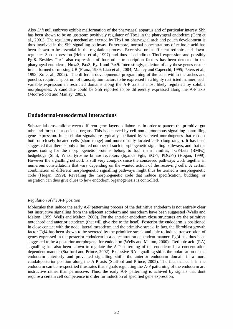
Also Shh null embryos exhibit malformation of the pharyngeal appartus and of particular interest Shh has been shown to be an upstream positively regulator of Tbx1 in the pharyngeal endoderm (Garg et al., 2001). The regulatory mechanism exerted by Tbx1 on pharyngeal arch and pouch development is thus involved in the Shh signalling pathway. Furtermore, normal concentrations of retinoic acid has been shown to be essential in the regulation process. Excessive or insufficient retinoic acid down-regulates Shh expression (Helms et al., 1997) and thus also indirect Tbx1 expression and possibly Fgf8. Besides Tbx1 also expression of four other transcription factors has been detected in the pharyngeal endoderm; Hoxa3, Pax3, Eya1 and Pax9. Interestingly, deletion of any these genes results in malformed or missing UB (Franz, 1989; Liao et al., 2004; Manley and Capecchi, 1995; Peters et al., 1998; Xu et al., 2002). The different developmental programming of the cells within the arches and pouches require a spectrum of transcription factors to be expressed in a highly restricted manner, such variable expression in restricted domains along the A-P axis is most likely regulated by soluble morphogenes. A candidate could be Shh reported to be differently expressed along the A-P axis (Moore-Scott and Manley, 2005).
Endodermal-mesodermal interactions
Substantial cross-talk between different germ layers collaborates in order to pattern the primitive gut tube and form the associated organs. This is achieved by cell non-autonomous signalling controlling gene expression. Inter-cellular signals are typically mediated by secreted morphogenes that can act both on closely located cells (short range) and more distally located cells (long range). It has been suggested that there is only a limited number of such morphogenetic signalling pathways, and that the genes coding for the morphogenetic proteins belong to four main families; TGF-beta (BMPs), hedgehogs (Shh), Wnts, tyrosine kinase receptors (ligands Fgfs, EGFs, PDGFs) (Hogan, 1999). However the signalling network is still very complex since the conserved pathways work together in numerous constellations that vary depending on the wanted action of the receiving cells. A certain combination of different morphogenetic signalling pathways might thus be termed a morphogenetic code (Hogan, 1999). Revealing the morphogenetic code that induce specification, budding, or migration can thus give clues to how endoderm organogenesis is controlled.
Regulation of the A-P position
Molecules that induce the early A-P patterning process of the definitive endoderm is not entirely clear but instructive signalling from the adjacent ectoderm and mesoderm have been suggested (Wells and Melton, 1999; Wells and Melton, 2000). For the anterior endoderm close structures are the primitive notochord and anterior ectoderm (that will give rise to the head). Posterior the endoderm is positioned in close contact with the node, lateral mesoderm and the primitive streak. In fact, the fibroblast growth factor Fgf4 has been shown to be secreted by the primitive streak and able to induce transcription of genes expressed in the posterior endoderm in a concentration dependent manner. Fgf4 has thus been suggested to be a posterior morphogene for endoderm (Wells and Melton, 2000). Retinoic acid (RA) signalling has also been shown to regulate the A-P patterning of the endoderm in a concentration dependent manner (Stafford and Prince, 2002). Excessive RA signalling shifts the polarisation of the endoderm anteriorly and prevented signalling shifts the anterior endoderm domain in a more caudal/posterior position along the A-P axis (Stafford and Prince, 2002). The fact that cells in the endoderm can be re-specified illustrates that signals regulating the A-P patterning of the endoderm are instructive rather than permissive. Thus, the early A-P patterning is achived by signals that dont require a certain cell competence in order for induction of specified gene expression.

23
Regulation of morphogenetic events forming the foregut
Signals involved in folding of the flat endodermal sheet into a tube with regions expressing tissue specific genes still need to be detected. It is not clear what mechanisms govern this crucial process during embryogenesis. However, interactions between mesoderm and endoderm appear to be required for foregut formation and regionalisation along the gut tube. Simultaneously with gut closure the mouse embryo turns. Turning of the embryo results in an inside-out flip which brings the previous externally endoderm internally invested by the mesoderm. In both drosophila and chick signals from the overlying mesoderm have been proven to initiate endoderm morphogenesis (Immerglück et al., 1990). The patterning signals results in specific restricted domains along the A-P axis of the primitive gut tube which loosely marks the positions for subsequent organ development. Thus substantial cross-talk between different germ layers collaborates in order to pattern the primitive gut tube and form the associated organs. As mentioned, this effect is achieved by cell non-autonomous signalling. In general, information of specialised domains within a polarized tissue as the foregut is often established by members of bone morphogenetic protein (Bmp) and wingless (Wnt) families, which oppose either the activity or expression Shh (Lee and Treisman, 2001; Marcelle et al., 1997; Zhang et al., 2002).
Regulation of specific domains with organ forming potency
As mentioned earlier the regionalisation of organ specific domains within the anterior definitive endoderm is first rough to later be progressively refined. During this event the early ubiquitous gene expression, such as Cerl in the entire anterior region, will be down regulated and instead expression of more detailed region specific genes is identified. One such gene is Hhex expressed in the ventral-lateral endoderm giving rise to the liver, ventral pancreas, lungs and thyroid (Martinez Barbera et al., 2000; Thomas et al., 1998). Besides Hhex several genes become expressed in well-defined restricted domains of the endoderm. Some of the genes expressed in the initial organ progenitors remain actively transcribed during development and will later regulate organ-specific gene expression (Grapin-Botton and Melton, 2000). At thyroid initiation Hhex, pax8, Foxe1, Nkx2.1 are expressed in the thyroid follicular progenitors (De Felice and Di Lauro, 2004). The genes are continuously actively transcribed during thyroid organogenesis and later regulate thyroid specific gene expression such as NIS, TPO, and TG which take part in thyroid hormone production. The specification of restricted domains within the endoderm can be considered to be the first morphogenetic event forming the primitive gut tube into anatomically defined compartments in which cells are committed to undertake a defined developmental program. For the midline thyroid anlage molecular mechanisms regulating the boundaries of the placode and induce transcription of thyroid specific genes are largely unknown, although Shh is specifically excluded from the progenitor cells similar to that of pancreatic development.

24
Key steps and general traits in endoderm organogenesis
The key steps of early thyroid development, specification, budding and migration (or expansion), are common steps in organogenesis. Clues in the search for regulatory candidates of thyroid development might be given from the more extensively studied liver, lung and pancreas. The next section will therefore briefly cover what is known about developmental regulators of these organs.
Specification Along the foregut endoderm all organ primordia can be identified as regionalised domains delineated from the endoderm by the expression of their organ-specific genes. wThe specification and development of these domains are dependent on the organ-specific transcription regulators expressed within each domain as well as inductive or inhibitory signals from surrounding tissues.
Lungs
Initiation of pulmonary cell fate was shown to occur by Fgf signalling (Serls et al., 2005). The source of secreted Fgf 1 and 2 was localised to the cardiac mesoderm and depending on the concentration either liver or lung specification was initiated in the ventral foregut endoderm. It was shown that high concentrations of Fgf 1 and Fgf2 induced Nkx2.1 expression in pulmonary fated cells (Serls et al., 2005).
Liver
Fgf 1, 2 and 8 secreted by the cardiac mesoderm have been shown to be able to induce a hepatic fate in cells in the adjacent endoderm (Jung et al., 1999). For the induction to result in expression of hepatocyte-specific genes pre-patterning of the endodermal cells involving expression of Foxa1 and Foxa2 is required (Lee et al., 2005). Thus, a hepatic cell fate is induced by secreted morphogenes but specification will only occur if the endoderm domain is pre-patterned correctly.
Pancreas
The pancreas is formed from ventral and dorsal restricted domains (Slack, 1995). For the dorsal pancreas primordium the position of initiation is controlled by repression of Shh (Hebrok et al., 1998). Fgf 2 and activin-βB are secreted from the notochord that lies adjacent to the dorsal pancreas initiation site and repress Shh in the endoderm which allows pancreatic specific gene expression (Hebrok et al., 1998). Initiation of the ventral primordium is unlike liver and lung specification not induced by Fgf signalling. Instead the absence of Fgf signalling leads to expression of ventral pancreas specific genes (Deutsch et al., 2001). Moreover, Fgf signalling from the cardiac mesoderm is believed to positively regulate Shh expression in the endoderm and in that way suppress a pancreatic fate in cells anterior to the ventral pancreas primordium.
Possible interactions regulating thyroid specification
Taken together organ fate initiation in the foregut endoderm seems to be dependent on morphogenetic signals secreted by neighbouring tissues. Even though the responsible signalling molecules for thyroid initiation is not yet known it seems likely that also a thyroid follicular fate is induced by signals

25
secreted by adjacent structures at the embryonic age of specification. The thyroid initiation site is positioned close to the aortic sac and the cardiac mesoderm, implying a role of Fgf signalling also in thyroid commitment. Also, the transcription factor Nkx2.1 expressed in both pulmonary and thyroid precursors was shown to be induced by Fgfs (Serls et al., 2005). Notably thyroid specification has been shown to be dependent on Fgf signalling in zebrafish (Wendl et al., 2007) and in mouse explant experiments (Ohuchi et al., 2000).
Placode formation and budding Typically the specified cells in the endoderm form a placode, seen as a thickening of the endoderm, which subsequently forms an evagination and creates a bud. The process of the outgrowing bud is usually achieved by proliferation of the specified cells. This accounts for the liver, lungs and pancreas which are all formed due to proliferation of their respective specified cells (Ahlgren et al., 1996; Gualdi et al., 1996; Guz et al., 1995; Teitelman et al., 1993). Interestingly, at this step the thyroid parenchyma is dissimilar from the other endoderm derived organs since the midline thyroid precursors forms an outgrowth without signs of DNA synthesis (Fagman et al., 2006).
Lungs
During pulmonary morphogenesis Shh, Bmp4 and Wnt7b are expressed at high levels in the distal bud endoderm (Bellusci et al., 1997b). Also a type of Fgf receptor (Fgf IIIb) was detected in the prospective lung bud. Fgf ligands with high affinity to the receptor, i.e. Fgf 1, Fgf 7 and Fgf10, have been shown essential for patterning and morphogenesis of the pulmonary parenchyma (Bellusci et al., 1997a; Bellusci et al., 1997b; Celli et al., 1998).
Liver
The hepatic bud proliferates and evaginates into the septum transversum (mesoderm that give rise to the diaphragm) which express hepatocyte growth factor (Liver specification and morphogenesis is reviewed in (Zaret, 2000)). Proliferation studies on hepatocytic progenitors showed that the rate of proliferation is not different from that of the nearby gut endoderm cells (Bort et al., 2006).
Pancreas
Pancreas precursors form a ventral and a dorsal bud that eventually fuse. Interestingly, the transcription factor Islet1 (Isl1) has been shown to be crucial for dorsal pancreas bud formation (Ahlgren et al., 1997). Furthermore Isl1 is essential for differentiation of pancreatic precursors into endocrine and exocrine pancreatic cells.
Possible interactions regulating thyroid budding
Regulatoy molecules involved in this process are completely unknown. The thyroid follicular progenitors could interact with endothelia or mesenchyme or both during the budding process. The interactions could hypothetically involve both the underlying and subsequent surrounding mesenchyme. Taken into account that early embryonic gene expression that persists during development to later regulate tissue specific genes often are developmental regulators as well (e.g. Nkx2.1, Foxe1, Pax8 and Hhex for the thyroid) if Isl1 is expressed in thyroid progenitors it could possibly also regulate thyroid budding in addition to pancreas budding.
26
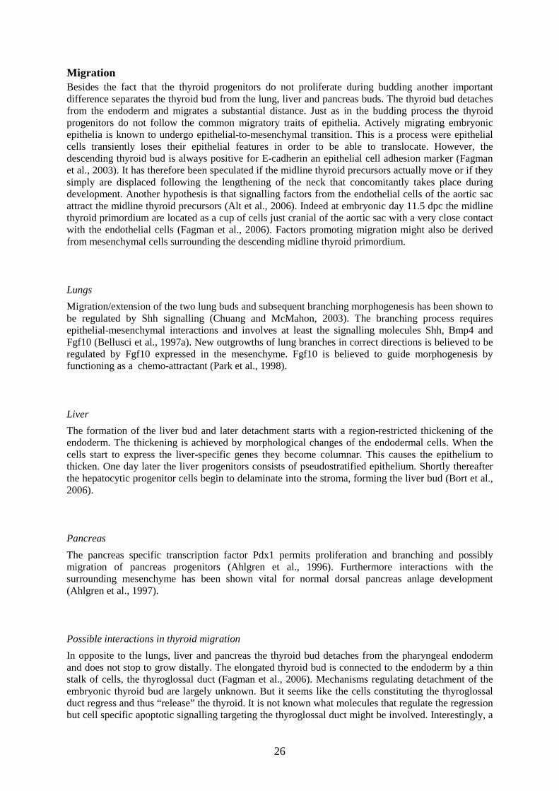
Migration Besides the fact that the thyroid progenitors do not proliferate during budding another important difference separates the thyroid bud from the lung, liver and pancreas buds. The thyroid bud detaches from the endoderm and migrates a substantial distance. Just as in the budding process the thyroid progenitors do not follow the common migratory traits of epithelia. Actively migrating embryonic epithelia is known to undergo epithelial-to-mesenchymal transition. This is a process were epithelial cells transiently loses their epithelial features in order to be able to translocate. However, the descending thyroid bud is always positive for E-cadherin an epithelial cell adhesion marker (Fagman et al., 2003). It has therefore been speculated if the midline thyroid precursors actually move or if they simply are displaced following the lengthening of the neck that concomitantly takes place during development. Another hypothesis is that signalling factors from the endothelial cells of the aortic sac attract the midline thyroid precursors (Alt et al., 2006). Indeed at embryonic day 11.5 dpc the midline thyroid primordium are located as a cup of cells just cranial of the aortic sac with a very close contact with the endothelial cells (Fagman et al., 2006). Factors promoting migration might also be derived from mesenchymal cells surrounding the descending midline thyroid primordium.
Lungs
Migration/extension of the two lung buds and subsequent branching morphogenesis has been shown to be regulated by Shh signalling (Chuang and McMahon, 2003). The branching process requires epithelial-mesenchymal interactions and involves at least the signalling molecules Shh, Bmp4 and Fgf10 (Bellusci et al., 1997a). New outgrowths of lung branches in correct directions is believed to be regulated by Fgf10 expressed in the mesenchyme. Fgf10 is believed to guide morphogenesis by functioning as a chemo-attractant (Park et al., 1998).
Liver
The formation of the liver bud and later detachment starts with a region-restricted thickening of the endoderm. The thickening is achieved by morphological changes of the endodermal cells. When the cells start to express the liver-specific genes they become columnar. This causes the epithelium to thicken. One day later the liver progenitors consists of pseudostratified epithelium. Shortly thereafter the hepatocytic progenitor cells begin to delaminate into the stroma, forming the liver bud (Bort et al., 2006).
Pancreas
The pancreas specific transcription factor Pdx1 permits proliferation and branching and possibly migration of pancreas progenitors (Ahlgren et al., 1996). Furthermore interactions with the surrounding mesenchyme has been shown vital for normal dorsal pancreas anlage development (Ahlgren et al., 1997).
Possible interactions in thyroid migration
In opposite to the lungs, liver and pancreas the thyroid bud detaches from the pharyngeal endoderm and does not stop to grow distally. The elongated thyroid bud is connected to the endoderm by a thin stalk of cells, the thyroglossal duct (Fagman et al., 2006). Mechanisms regulating detachment of the embryonic thyroid bud are largely unknown. But it seems like the cells constituting the thyroglossal duct regress and thus “release” the thyroid. It is not known what molecules that regulate the regression but cell specific apoptotic signalling targeting the thyroglossal duct might be involved. Interestingly, a

27
loss of Shh signalling has been shown to cause a delayed detachment of the thyroid parenchyma from the pharyngeal endoderm (Fagman et al., 2004).
Thyroid and cardiac development
Children with CH have been reported to have a higher frequency than the general population for other congenital malformations, in most cases cardiovascular (Olivieri et al., 2002; Roberts et al., 1997). It is therefore reasonable to suggest that genes involved in cardiac organogenesis might also participate in thyroid development. Thus, genes expressed during embryogenesis in both thyroid and heart progenitors are promising candidates to be investigated for their involvement in the pathogenesis of thyroid dysgenesis.
In early embryogenesis, at the time when the thyroid progenitors are seen as a placode in the pharyngeal endoderm, a subpopulation of cardiac progenitors has been identified in the underlying mesenchyme. These cardiac progenitors are known to belong to the secondary heart field which gives rise to the outflow tract, right ventricle and atria. This population of cells will thus first be located close to the initiation site of the midline thyroid placode and second be close again incorporated in the outflow tract. It was therefore of great interest that the transcription factor Islet 1 (Isl1), originally identified in pancreatic endocrine precursors, regulates the survival of both the cardiac progenitors and the overlaying pharyngeal endoderm (Cai et al., 2003).
Also, initiations of the other endoderm derived organs (lungs, liver, and pancreas) have been shown to depend on signals secreted from the primitive cardiac mesoderm. The initiation was showed to occur in a concentration dependent manner where high expression of Fgf 1 and 2 led to pulmonary cell fate, low concentration induced hepatic cell fate and an absence of Fgf signalling initiated pancreatic cell fate in the ventral foregut endoderm (Deutsch et al., 2001; Serls et al., 2005). In turn, the endoderm tissue has been shown to be able to specify cardiac myoblasts (Marvin et al., 2001). Thus, there seem to be reciprocal inductive signalling between the embryonic endoderm and the adjacent cardiac mesoderm, suggesting that thyroid and cardiac organogenesis may be interdependently regulated during early development.
Children with DiGeorge syndrome harbouring the 22q11 deletion (Lindsay et al., 2001) suffer from cardiovascular malformations and in addition a spectrum of developmental defects of derivatives of the pharyngeal apparatus (Liao et al., 2004). The candidate 22q11 deletion gene is Tbx1 (Lindsay et al., 2001), which as mentioned earlier plays a critical role in regulating pharyngeal arch development. Interestingly, one of the features observed in homozygote mutation of Tbx1 is a lack of caudal pharyngeal pouches and pharyngeal arch arteries. Considering that the thyroid is formed from a region close to the aortic sac and follows the route of the third pharyngeal arch arteries that arise from the aortic sac during the development it would be very interesting to study if the thyroid develops normal in the absence of Tbx1.

28
Key molecules in endoderm organogenesis
From the above described regulatory mechanisms impacting on foregut development, three transcription factors (Tbx1, Isl1 and Fox2) and one morphogen (Shh) were found particularly attractive to investigate for putative roles in mouse thyroid development. The rationales are outlined in detail in the respective papers I-IV of the thesis. Before summarizing the results of the studies, some additional features of the investigated factors of relevance for the understanding and interpretation of data will be briefly highlighted.
Tbx1 Expression of the T-box transcription factor Tbx1 is required for a normal development of the pharyngeal apparatus and its derivates (Baldini, 2005). In the pharyngeal region Tbx1 transcripts are found both in the endoderm and the mesoderm core of the arches but not in the neural crest derived cell population (Garg et al., 2001). In 2001, Tbx1 was recognised as the gene responsible for the phenotype observed in patients with DiGeorge syndrome, exhibiting cardiac outflow tract malformations (Garg et al., 2001). Subsequent studies showed Sonic hedgehog (Shh) to be the upstream activator of Tbx1 in the pharyngeal endoderm (Garg et al., 2001; Yamagishi et al., 2003). As Shh has previously been shown to be regulate thyroid development in our lab (Fagman et al., 2004), Tbx1 became a number one putative candidate as mediator of the thyroid phenotype in Shh mutants. There are previously occasional reports of hypothyroidism and also thyroid dysgenesis in children with DiGeorge syndrome, further supporting this possibility.
Isl1 Islet1 (Isl1) is a transcription factor of the LIM homeodomain family and was first recognised for its ability to bind to the insulin enhancer promoter in pancreatic rat cells (Karlsson et al., 1990). Since then Isl1 transcripts have been identified in numerous tissues during early embryonic development, and the protein have been discovered to control cell fates (Ahlgren et al., 1997; Pfaff et al., 1996). Of particular interest, Isl1 has been found to be crucial for both pancreas (Ahlgren et al., 1997) and cardiac (Cai et al., 2003) organogenesis. During cardiac development Isl1 has been identified in precursors of the second heart field giving rise to the outflow tract. Considering the close spatial relationship between the thyroid midline progenitors and the outflow tract Isl1 was to us a very interesting candidate molecule for taking part in thyroid development.
Foxa2 Foxa2 was first named hepatocyte nuclear factor 3β (HNF-3β) on the basis for its regulatory role of liver specific genes (Lai et al., 1990). The transcription factor belongs to the Forkhead family with three related members: Foxa1 (HNF-3α), Foxa2 (HNF-3β), and Foxa3 (HNF-3γ) (Ang et al., 1993; Monaghan et al., 1993; Sasaki and Hogan, 1993). Both Foxa1 and Foxa2 are expressed early in the definitive endoderm and are later in development important regulators of tissue specific genes in endoderm derived organs such as the lungs, liver and pancreas (Bohinski et al., 1994; Cereghini, 1996; Wu et al., 1997). Interestingly, for initiation of a hepatic cell fate Foxa2 expression in the endoderm governs cell competence and is required for a proper response to the inducing signals (Lee et al., 2005). The thyroid have been reported to express Foxa2 transcript (Monaghan et al., 1993; Sato and Di Lauro, 1996). However, the previous data were limited and did not cover expression patterns in both thyroid anlagen. Since Foxa2 has been shown to regulate calcitonin gene expression (Viney et al., 2004) it was of interest to re-evaluate the expression pattern of Foxa proteins in the thyroid anlagen and their progressive development.
29
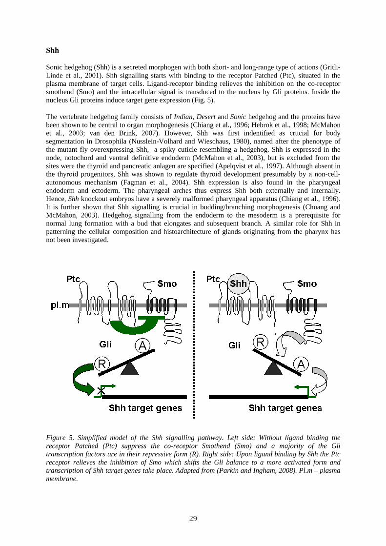
Shh
Sonic hedgehog (Shh) is a secreted morphogen with both short- and long-range type of actions (Gritli-Linde et al., 2001). Shh signalling starts with binding to the receptor Patched (Ptc), situated in the plasma membrane of target cells. Ligand-receptor binding relieves the inhibition on the co-receptor smothend (Smo) and the intracellular signal is transduced to the nucleus by Gli proteins. Inside the nucleus Gli proteins induce target gene expression (Fig. 5).
The vertebrate hedgehog family consists of Indian, Desert and Sonic hedgehog and the proteins have been shown to be central to organ morphogenesis (Chiang et al., 1996; Hebrok et al., 1998; McMahon et al., 2003; van den Brink, 2007). However, Shh was first indentified as crucial for body segmentation in Drosophila (Nusslein-Volhard and Wieschaus, 1980), named after the phenotype of the mutant fly overexpressing Shh, a spiky cuticle resembling a hedgehog. Shh is expressed in the node, notochord and ventral definitive endoderm (McMahon et al., 2003), but is excluded from the sites were the thyroid and pancreatic anlagen are specified (Apelqvist et al., 1997). Although absent in the thyroid progenitors, Shh was shown to regulate thyroid development presumably by a non-cell-autonomous mechanism (Fagman et al., 2004). Shh expression is also found in the pharyngeal endoderm and ectoderm. The pharyngeal arches thus express Shh both externally and internally. Hence, Shh knockout embryos have a severely malformed pharyngeal apparatus (Chiang et al., 1996). It is further shown that Shh signalling is crucial in budding/branching morphogenesis (Chuang and McMahon, 2003). Hedgehog signalling from the endoderm to the mesoderm is a prerequisite for normal lung formation with a bud that elongates and subsequent branch. A similar role for Shh in patterning the cellular composition and histoarchitecture of glands originating from the pharynx has not been investigated.
Figure 5. Simplified model of the Shh signalling pathway. Left side: Without ligand binding the receptor Patched (Ptc) suppress the co-receptor Smothend (Smo) and a majority of the Gli transcription factors are in their repressive form (R). Right side: Upon ligand binding by Shh the Ptc receptor relieves the inhibition of Smo which shifts the Gli balance to a more activated form and transcription of Shh target genes take place. Adapted from (Parkin and Ingham, 2008). Pl.m – plasma membrane.

30
AIMS OF THE STUDY
The overall aim of this thesis was to further elucidate how the organogenesis of the thyroid gland is accomplished during embryonic life. Focus was on known morphogenetic regulatory molecules that might exert novel hitherto undiscovered effects on the multistep process of mouse thyroid development. More specifically, the following molecules with associated main question of interest have been studied in the four papers (I-IV):
I. Tbx1: is the thyroid phenotype observed in Shh homozygous mutants caused by defective Tbx1 signalling?
II. Isl1: does the expression pattern of Isl1 in the pharyngeal endoderm and mesoderm suggest a role for this transcription factor in thyroid development and progenitor differentiation?
III. Foxa2: is this endoderm marker expressed by embryonic C-cells refecting an endoderm origin of the C-cell precursor?
IV. Shh: are Shh deletion and Shh genetic fate mapping instrumental in deciphering the contribution of pharyngeal progenitors to the thyroid endocrine cell lineages?

31
RESULTS In order to identify novel morphogenetic mechanisms involved in thyroid organogenesis the expression pattern and regulatory roles of molecules implicated in the development of endoderm derivates were investigated, both at the initiation/specification of thyroid progenitor cells early and throughout the developmental stages leading to the differentiated, composite gland.
The studies were to a large extent conducted on mouse embryos. Offspring’s of both normal (wild-type) and genetically modified animals (detailed below) were investigated. The morning a vaginal plug was detected in mated animals the age of the embryos was estimated to 0.5 days postcoitum (dpc). Embryos with embryonic ages ranging between 9.5-19.0 dpc were collected to be investigated, variably between the four papers. After fixation and freezing the embryos were cryosectioned and immunolabeled with antibodies against proteins of interest. Immunofluorescence was evaluated in a confocal microscope and images were captured and analysed. Serial images of cryosectioned embryos were used to make 3D-reconstructions of a thyroid phenotype observed in Tbx1 -/- embryos. Microarray data obtained from a database were analysed in the search for marker genes of medullary thyroid carcinoma, this was done in collaboration with the genetic institution at the Biogem s.c.a.r.l. laboratory in Ariano Irpino, Italy.
Role of Tbx1, a downstream target of Shh signalling, in thyroid organogenesis (paper I)
Since we previously reported that mouse embryos depleted of Shh failed to develop a normal thyroid gland (Fagman et al., 2004) it was of great interest to find downstream targets of Shh that could be involved mediating the effect. The T-box transcription factor Tbx1 was shown to be positively regulated by Shh during pharyngeal arch development (Garg et al., 2001). Both anlagen of the thyroid gland evolve from endoderm within the pharyngeal apparatus in the neck. In order to elucidate if Shh regulated thyroid development via Tbx1 the thyroid phenotype was investigated in mice deficient of Tbx1. This was done in paper I.
In paper I Tbx1 knockout embryos were observed to have only a single thyroid lobe which was similar to the thyroid phenotype detected earlier in Shh -/- embryos (Fagman et al., 2004). The thyroid gland of wild type littermates to the Tbx1 homozygous had at this stage in development undergone bilobation and consisted of two lobes seen on the left- and right-hand side of the trachea and ventrally connected by an isthmus. The single thyroid lobe detected in the Tbx1 null embryos had no preference for the left- or right-hand side but was identified in a ventrolateral region of the trachea, close to one of the carotid arteries. In comparison, the abnormal thyroid was severely hypoplastic with a volume of one fifth of the total normal gland. Besides the changed morphological structure the thyroid primordium was also found to be located more cranial in the neck at the site of the laryngeotracheal groove.
During normal development the midline thyroid primordium grows laterally at the level of the third pharyngeal arch arteries. It has been suggested that the route of the arteries might guide the midline thyroid progenitors on their way. The close location of the site were thyroid progenitor cells are specified to the endothelium of the aortic sac might also reflect signalling between the two cell types. We therefore questioned if the impaired bilobation and reduced size of the thyroid gland could be due to abnormalities in early steps of thyroid development.

32
Indeed, we discovered that the detachment and downward translocation of the thyroid primordia was delayed during early development in the Tbx1 knockout embryos. Whereas the thyroid primordium of wild-type embryos was assembled cranial of the aortic sac and completely detached the thyroid diverticulum in the Tbx1 -/- embryos was still connected to the pharyngeal endoderm and apparently failed to maintain contact with the aortic sac. The postponed detachment might be the explanation to the thyroid phenotype observed in Tbx1 null embryos, possibly related to altered influence from signals generated in tissues surrounding the developing thyroid. Of interest, Tbx1 was not expressed in the thyroid progenitors themselves but in the mesenchyme underlying the endoderm close to the migrating thyroid. The possible role of epithelial-mesenchymal interactions will be further elaborated in the discussion section.
Although the parenchyma of the thyroid gland was smaller in size, asymmetrically distributed and misplaced, the thyroid follicular cells expressed the pro-hormone thyroglobulin (TG). Thus, the terminal differentiation of the thyroid progenitors was not disturbed in Tbx1 depleted embryos. It was also found that the single thyroid lobe in the knockout embryos lacked calcitonin producing C-cells. In fact calcitonin production was not identified in any cells in the Tbx1 depleted embryos. This is in agreement with the notion that the ultimobranchial bodies bringing the C-cells to the thyroid parenchyma fail to develop in Tbx1-/- mice (Liao et al., 2004).
Expression pattern of Isl1 in the developing thyroid and surrounding embryonic tissues (paper II)
Another molecule suggested to influence endoderm-mesoderm interactions is the LIM homeodomain transcription factor Islet1 (Isl1) proven to be important for crucial steps in pancreas organogenesis (Ahlgren et al., 1997). Of interest, Isl1 expressing heart progenitors are located in the mesenchyme underlying the pharyngeal endoderm at the site of thyroid specification (Cai et al., 2003; Missero et al., 1998). It is also known that Isl1 is expressed in the anterior endoderm coinciding with the primordial stage of endoderm appendages. To get insight in the possibility that Isl1 might be a novel regulator of thyroid organogenesis we mapped in paper II the expression pattern of Isl1 in the midline and lateral thyroid anlagen and also in their respective surrounding mesenchyme at the different developmental stages. Furthermore a possible role of Isl1 in thyroid progenitor specification was investigated.
The midline thyroid primordium uniformly expressed Isl1 as did the entire pharyngeal endoderm. The Isl1 expression in thyroid progenitor cells continued throughout the developmental stages (budding, detachment and bilateral growth) preceding fusion with the UB. During budding the midline thyroid anlage was in close contact with Isl1 positive mesenchyme. Also cells constituting the lateral thyroid anlage, the UB, were found to express Isl1. Isl1 immunoreactivity was not limited to the fourth pharyngeal pouches but was observed throughout the pharyngeal endoderm. A core of Isl1 positive mesenchyme was observed ventral of the laryngotracheal groove enveloping the presumptive trachea. Notably, the Isl1 positive mesenchyme did not expand laterally and mesenchyme surrounding the ultimobranchial bodies was Isl1 negative during the entire development.
Interestingly, fusion of the two thyroid anlagen dramatically changed the expression pattern of Isl1. Prior to fusion both anlagen expressed Isl1, but soon thereafter Isl1 immunoreactivity was barely detectable in cells derived from the midline thyroid primordium. Cells derived from the lateral thyroid primordia (UB) were found to express Isl1 intensively also after the fusion.
Comparison of the locations of Isl1 positive cells and calcitonin positive cells within the presumptive thyroid lobes revealed that they overlapped. Therefore UB cells that maintained their Isl1 expression
33
were suggested to be immature C-cells. In human medullary thyroid cancer analysis of microarray expression profiles showed that Isl1 transcripts were significantly above the mean expression level. Since this type of cancer arises from transformed C-cells within the thyroid Isl1 may also be a marker of human embryonic C-cells.
Of particular note, when examining the fourth pharyngeal pouches we detected that expression of Isl1 preceded the expression of Nkx2.1. This led us to speculate that Isl1 might in fact be a regulator of Nkx2.1 implicated in the specification of thyroid progenitors. In Isl1-/- embryos the midline thyroid placode identifed by expression of Nkx2.1 and Pax8 in the progenitor cells was found to develop normally in the pharyngeal floor, arguing against this possibility. However, although specified correctly the thyroid placode in mutants was smaller compared to that of wild-type littermates, suggesting a role of Isl1 in early thyroid bud development.
Foxa2 is a novel C-cell precursor marker (paper III)
The true origin of the calcitonin producing C-cells scattered within the thyroid parenchyma has been a question for debate since the 1960´s. Experiments performed in different species and lack of reliable C-cell markers before the onset of calcitonin production have added to the difficulties in resolving the question. In paper III we investigated the expression of Foxa1 and Foxa2 in both developing thyroid primordia. The two forkhead transcription factors are known to be expressed in the definitive endoderm (Kaestner et al., 1994) and have so far not been reported detected in neural crest cells. It thus seemed as expression of Foxa1 and Foxa2 could be used as endodermal markers in the search for the origin of the C-cells.
Foxa2 positive cells were observed in adult mouse thyroid glands. The cells were found in parafollicular positions, a distribution pattern known to correspond to the C-cells. Parallel immunostaining accordingly showed that the Foxa2 expressing cells in fact also expressed calcitonin. This was surprising and of high interest to us since Foxa2 is a known endodermal marker and the C-cells are widely accepted as derivates of the cranial neural crest.
Cryosectioning and subsequent immunolabeling of embryos showed that the Foxa2 positive cells found in the adult gland was brought there by the ultimobranchial bodies, the lateral thyroid primordium. From the budding stage and up to the point of fusion no cells within the midline thyroid anlage expressed Foxa2. In contrast,, the ultimobranchial bodies contained an increasing number of Foxa2 positive cells, starting already before they delaminated from the pouch endoderm. Prior to fusion with the midline thyroid anlage most ultimobranchial cells expressed Foxa2. Analysis of Ki67 expression revealed that most of these cells had exited the cell cycle, indicating that the increased Foxa2 expression was likely not the result of propagation of a subpopulation of cells but gradually acquired in the epithelium as the ultimobranchial body evolved . This suggested that the Foxa2 positive population constituted cells originally encountered in the budding endoderm of the fourth pharyngeal pouch.
The Foxa2 positive ultimobranchial cells were shown to express Foxa1 as well, compatible with the notion that Foxa1 and Foxa2 often is co-expressed and exert redundant functions in endoderm derivatives. Depending on different species hosts of the primary antibodies used for immunostaning, this allowed us to directly compare the expression of Foxa1 and calcitonin in the same tissue sections. This unequivocally showed that in late development Foxa1 and calcitonin are co-expressed in C-cells present in the prospective thyroid lobes. Taken together, these data is in the line with an endodermal origin of the thyroidal C-cells.

34
Role of Sonic hedgehog in embryonic C-cell differentiation and migration (paper IV)
Even though the embryonic origin of the C-cells is controversial (at least in mice is the NCC origin questioned) little doubt lies in that the C-cells are transported to the thyroid by the ultimobranchial bodies. This is convincingly proven by the fact that embryonic malformations of the ultimobranchial bodies lead to aberrant localization or even loss of C-cells. However, the process how C-cell precursors enter or perhaps develop within the epithelia of the ultimobranchial bodies and later disseminate in the thyroid parenchyma are yet unsolved issues. Due to the known influence of Shh on both pharyngeal arch development and neural crest cell fate (Ahlgren and Bronner-Fraser, 1999; Testaz et al., 2001) this morphogen is a putative candidate for the embryonic regulation of C-cell whereabouts. In paper IV, we therefore investigated whether Shh regulates the formation of ultimobranchial bodies and thyroid entering of C-cell precursors in Shh deficient mouse embryos. We speculated that if C-cells are neural crest derivates we would not be able to detect any calcitonin producing cells in the mutant ultimobrachial body and thyroid.
The single thyroid lobe formed in Shh null embryos (Fagman et al., 2004) did not contain calcitonin producing cells at the start of normal calcitonin production. It was also obvious that in these mutant animals the two thyroid anlagen failed to fuse, suggesting that the C-cell precursors did not colonize the developing thyroid to attain the normal parafollicular location due to a migration defect. This possibility was supported by the finding that numerous calcitonin positive cells were encounted in the early pharyngeal endoderm and later in development also in the wall of the presumptive trachea and in a large midline epithelial structure. This abnormal epithelial structure was (based on E-cadherin staining pattern) suggested to constitute the thymic rudiment that aberrantly connected to the pharyngeal endoderm also in late development.
Paired ultimobranchial bodies were not present in the Shh-deficient embryos. Instead a single ultimobranchial body rudiment still attached to the pharyngeal endoderm was identified. Instead of the normal lateral localization the ultimobranchial body rudiment was facing the dorsal aspect of the embryo. Serial sectioning of mutants revealed that the altered unpaired ultimobranchial body in fact was in contact with another more caudal positioned structure. We presumed this to be paired thymic rudiments that later likely fuse with the ultimobranchial body epithelia, forming a compound ultimobracnhial body/thymus. Cells of the normal ultimobranchial bodies are known to express Nkx2.1 (Lazzaro et al., 1991) and also Foxa2 (paper III in this thesis). Thus by examining the localisation of cells expressing both Nkx2.1 and Foxa2 progenitors of the ultimobranchial bodies could be identified. The recognised ultimobranchial body homologue was integrated in the pharyngeal endoderm and was detected to encompass calcitonin producing cells. Thus, although obviously malformed the ultimobranchial body homologue in Shh depleted embryos was discovered to harbour C-cells. In view of the finding in Paper III, that Foxa2 is a reliable marker of C-cell precursors, these observations in the Shh mutant collectively provides strong evidence favouring the hypothesis that mouse C-cells originates within the anterior endoderm.
In order to try to understand the mechanisms of action that Shh exert on thyroid development we performed genetic fate mapping studies of Shh expressing endoderm using ShhGFP-Cre/Rosa26R recombinant mice. This approach, which indelibly labels Shh expressing cells and their progeny, has previously successfully been used to trace cell fates in other embryonic tissues. Shh expressing cells were detected by immunostaining of β-galactosidase (β-gal) generated by the activated reporter gene. Shh was found to be expressed at the expected locations: the floor plate of the neural tube, notochord and the ventral domain of the anterior endoderm. The midline thyroid primordium contained essentially no β-gal/+ cells, indicating that the progenitors likely derived from the dorsal, Shh negative, endoderm and not the immediately adjacent ventral endoderm. This is an important side observation, which may give clues on the controversial issue when and where follicular progenitors are specified before they express thyroid-specific developmental genes.
35
A limited number of endoderm cells present in the ventral portion of the fourth pharyngeal pouch and later gathered ventromedially in the ultimobranchial bodies expressed β-gal. At the primordia fusion stage β-gal/+ cells appeared in the midline thyroid and later on scattered throughout the whole immature thyroid lobes. Thus, Shh was detected in cells derived from both anlagen after fusion. As many of the β-gal/+ cells expressed Pax8, which expression is lacking in the ultimobranchial epithelium, this suggests that Shh was de novo expressed in a subset of progenitors derived from the midline anlage. Comparision of parallel immunolabeled sections showed that β-gal/+ did not match the localization of calcitonin, indicating that Shh was not expressed in C-cells. The Shh positive cells detected within the thyroid parenchyma might therefore constitute a new embryonic thyroid cell phenotype. The function of these cells might speculatively be involved in the fusion of the midline and lateral thyroid primordia. As the thyroid is hypoplastic in the Shh knockout, it is also possible that Shh expressed in the thyroid in mid-late development might contribute to thyroid embryonic growth.

36
DISCUSSION
Thyroid morphogenesis: interactions from surrounding tissues
In order to understand how the morphogenesis of the thyroid gland is regulated it may be helpful to study the spatial relationship between the thyroid tissue(s) and surrounding structures at different time points during their development. In paper I we showed that intermediate steps in thyroid development are regulated in a non-cell-autonomous way by Tbx1. Such tissue interactions likely require normal positioning of the primordium taking part in the communication. In order to achieve proper interactions between neighbouring structures the timing is crucial, referred to as a “developmental window” and means that during a certain limited time, at a certain position in the embryo, certain molecules are secreted. Depending on pre-patterning of cells in the receiving tissue different responses can be emitted from the same input. Tissue receiving secreted signalling molecules might also hold a dose dependent response further refining the signalling pathways. One example is the pharyngeal apparatus with differently expressed molecules along the A-P axis of the pharyngeal arches and pouches, making a unique identity for each pair of arches or pouches. Since the pharyngeal apparatus is a transient structure it can be considered to be one developmental window.
Shh has been shown to be crucial during regulation of the pharyngeal apparatus and the signalling is in a dose-dependent manner further strengthening the unique identities of the arches and pouches (Litingtung et al., 1998; Moore-Scott and Manley, 2005). The thyroid phenotype observed in Shh null embryos despite lack of Shh expression in thyroid precursors showed that the thyroid to some extent is regulated non-cell-autonomously (Fagman et al., 2004). In paper I we observed a similar phenotype as in Shh embryo mutants indicating that the regulatory mechanism of Shh signalling on thyroid development is via Tbx1 activation. However, similar to Shh expression Tbx1 was not identified in thyroid follicular progenitors. Thus, the regulation was achieved by interactions between the endoderm/epithelia and neighbouring structures. During the time of development neighbouring structures varies at different embryonic ages but for the thyroid gland a close relationship to vessels is maintained almost throughout the entire development. An anomaly in Tbx1 null embryos is also the complete lack of pharyngeal arch arteries (Lindsay et al., 2001). The altered cardiovascular structure with missing guiding tracks for the thyroid bi-lobation may be the basis to the thyroid hemiagenesis we observed in Tbx1 -/- embryos. Indeed signalling from adjacent vessels have been implicated a role in thyroid organogenesis (Alt et al., 2006). However, it can not be sad with certainty weather the bi-lobation defect noted occurs because the guiding tracks in form of the third pharyngeal arch arteries are missing or because the delayed detachment of the thyroid bud from the endoderm causes an altered surrounding/environment with failure to re-establish a contact with the aortic sac. Even though the shape and positioning of the thyroid varies over species the close relationship to the aortic sac for a second time is an evolutionary conserved event and is thus most likely of relevance for a normal thyroid development (Alt et al., 2006).
Another possible tissue interaction that is lost in the absence of Tbx1 is the one between the midline and lateral thyroid primordia. Tbx1 is a key regulator of the pharyngeal apparatus (Jerome and Papaioannou, 2001; Lindsay et al., 2001) and caudal derivatives are not formed in the lack of Tbx1 (Liao et al., 2004). Subsequently, in Tbx1 mutant embryos the UB are missing and again the normal structural relationship between the thyroid parenchyma and the surrounding is altered. During thyroid research the main interest has been focused on the midline anlage and less attention has been given the lateral analge (UB). From the point of their detachment until the time of fusion with the midline anlage the UB are surrounded by mesenchyme. Signalling between the mesenchyme and the UB epithelia has not been studied. Instead the thyroid phenotype in embryos lacking UB has been described. Interestingly, Hoxa3 and Eya1 depleted embryos are observed with similar thyroid phenotypes as the hemiagenesis we detected in Tbx1 null embryos (Manley and Capecchi, 1995; Xu et al., 2002). When examining the UB they were missing or failed to fuse with the midline anlage in all mutants. Thus, besides Tbx1 also active transcription of Hoxa3 and Eya1 is required for a normal formation of the UB and absence of any of these genes results in thyroid hemiagenesis and/or hypoplasi. Of note, in paper
37
IV we show that also Shh depleted embryos have a failed fusion between the two thyroid anlagen. And earlier we demonstrated thyroid hemiagenesis in Shh null embryos (Fagman et al., 2004).
In normal embryos the detached UB are positioned bi-laterally of the midline thyroid anlage. Considering the spatial organisation between the two thyroid anlagen and the fact that the midline thyroid primordium grows bi-laterally in direction towards the UB it is tempting to speculate that the UB have a chemo-attractant effect on the midline thyroid tissue. However, Pax9 -/- embryos lack UB but in these animals the thyroid nevertheless has its normal bi-lobed shape (Peters et al., 1998).
Cellular expression pattern during thyroid development
Studying protein expression patterns at different developmental stages might give insight on molecular regulation taking place at that moment. In this thesis we have monitored the expression of four proteins during the development of the two thyroid anlagen in search for new candidate molecules taking part in thyroid development regulation. Many of the developmental stages during normal gland formation still need to be elucidated. One of them is what initiates a thyroid fate in the pharyngeal endoderm. In contrast the initiation of lung, liver and pancreas fate has been shown to be dependent on different concentrations of Fgf signalling (Serls et al., 2005). In paper I, we observed a delayed budding and detachment in Tbx1 mutant embryos. Immunohistochemistry of wild-type embryos revealed that Tbx1 was excluded from the thyroid progenitor cells but abundant in the underlying and subsequent surrounding mesenchyme. This suggests a regulatory role of molecules expressed in adjacent tissues during thyroid development.
Since Tbx1 is a transcription factor the effector molecule/secreted signal from the mesenchyme to the endodermal thyroid progenitors still need to be identified. If Fgfs are in fact the molecules regulating the bi-lobation process during thyroid development remains to be elucidated. That we detected strong Tbx1 immunoreactivity in the close underlying mesenchyme at specification and also subsequent surrounding the thyroid bud indicates that the thyroid budding process might be regulated by epithelial - mesenchymal interactions. Such interactions has been shown to regulate dorsal pancreas budding and involves expression of Isl1 (Ahlgren et al., 1997).
Interestingly, cells in the thyroid placode were detected to express Isl1 (showed in paper II). The expression pattern was not limited within the boundaries of the thyroid placode but was detected throughout the endoderm in the pharyngeal floor, similar to the expression pattern for Hhex and Foxe1 known to belong to the thyroid specific combination of transcription factors. If Isl1 cooperates in the known regulatory network of Nkx2.1, Pax8, Hhex and Foxe1 remain to be elucidated. However the detected expression pattern of Isl1 in both thyroid progenitors and adjacent mesenchyme strongly resembles the expression pattern of the Isl1 regulated dorsal pancreas parenchyma with investing mesenchyme (Ahlgren et al., 1997). Since Isl1 null embryos are lethal before thyroid budding take place a regulatory role of Isl1 during thyroid budding could not be established. But the molecule is a candidate in regulating the budding and detachment of the thyroid considering its expression pattern during early thyroid development. Also in the cells constituting the lateral thyroid primordia Isl1 expression was detected during budding. However, the mesenchyme surrounding the fourth pharyngeal pouches was not at any time during development detected to express Isl1. Thus if Isl1 expression in the mesenchyme regulates thyroid midline precursor budding the regulation of UB outgrowth seems to be achieved in a different manner.
The thyroid phenotypes observed in embryos with malformed or missing UB indicates that the fusion process is important for the development of a normal gland. Interestingly, mapping of the protein expression pattern from initiation of cell fate to fusion of primordia revealed an astonishing switch in protein expression shortly after fusion. Both lateral and midline thyroid primordia continuously expressed Isl1 from specification until fusion. But, shortly after fushion cells derived from the midline

38
anlage (designated thyrocytes) did no longer express detectable amounts of Isl1. This was in contrast to the cells constituting the UB which remained Isl1 positive also after the fusion process. If the transcription is switched off in one cell population but not in another within the same organ this likely indicates a functional difference between the cells. In the thyroid gland cells with a midline origin are the only cells giving rise to the thyrocytes. Initial differentiation of the midline progenitors start shortly after fusion, identified by TG production. Thus Isl1 could be a marker of embryonic not yet fully differentiated cells. This would be in agreement with that Isl1 has been identified in cardiac progenitors but not later in differentiated cardiomyocytes (Cai et al., 2003). However, Isl1 expression has been detected during the entire development of neurons in the dorsal and sympathetic ganglia contradicting Isl1 expression to be limited to undifferentiated cells (Avivi and Goldstein, 1999). But in favour for being a marker of embryonic precursors at least within the thyroid parenchyma the number of Isl1 positive cells decreased as the number of calcitonin producing cells increased. Thus when the C-cells became more differentiated and switched on calcitonin production they no longer expressed Isl1.
As for Isl1 expression the expression of Shh was detected to become altered after fusion of the midline and lateral thyroid primordia (paper IV). But instead of switching off gene transcription in the entire midline cell population a subset of midline derived cells were detected to gain Shh expression. Thus, after the fusion of primordia into two primitive thyroid lobes a subset of cells that was almost totally lacking Shh expression prior fusion will differentiate into thyrocytes and initiate Shh expression. Similarly, far from all cells in the UB expressed Shh. Shh is generally believed to function as a signalling molecule from epithelia to mesenchyme (McMahon et al., 2003). When Shh was absent we observed that the two thyroid anlagen failed to fuse. Weather the fusion normally takes place by Shh signalling from one epithelial structure to another or via the intervening mesenchyme is presently not known. It should also be noted that the pharyngeal apparatus is severely misshaped in Shh knockouts and also in other mutants with a malformed pharyngeal apparatus the two thyroid anlagen fail to fuse (Manley and Capecchi, 1995).
Possible implications in thyroid disease: dysgenesis and cancer
Knockout mouse models have been useful in investigating protein functions and molecules regulating thyroid development are beginning to be elucidated. In paper I we detected Tbx1 to be a molecule regulating thyroid development. In the absence of Tbx1 the thyroid gland undergoes developmental defects that results in hypothyroidism. The parenchyma of the thyroid constitutes only one, considerably smaller, lobe compared to wild-type thyroid lobes (hemiagenesis and hypoplasia). This observation is in agreement with earlier studies were we showed a similar thyroid dysgenesis to occur in Shh depleted mouse embryos (Fagman et al., 2004). Thus, the hemiagenesis and hypoplasia observed for the thyroid gland in Shh knockout embryos might be due to disruption of Tbx1 signalling. Tbx1 has indeed been demonstrated to be a downstream target of Shh signalling in the pharyngeal arches (Garg et al., 2001) and has previously been shown to be mutated in patients with DiGeorge syndrome (Jerome and Papaioannou, 2001; Lindsay et al., 2001; Merscher et al., 2001). Interestingly, a search for thyroid dysgenesis among children with Digeorge syndrome revealed developmental defects of the thyroid gland similar to what we discovered in Tbx1 -/- mouse embryos to be overrepresented (e.g. hemiagenesis and hypoplasia) (Ryan et al., 1997). This is in agreement with the fact that children born with cardiovascular anomalies often also are represented with other organ malformations and thyroid dysgenesis being the most frequent. For that reason it is worth extra attention that Isl1 expression was identified in progenitors of both the heart and the thyroid, suggesting that the organs are regulated interdependently.
In paper III, we found that known endodermal markers (Foxa1 and Foxa2) are co-expressed with calcitonin in mature C-cells in the mouse embryo or foetus. The observation, corroborated by the

39
Shh-/- phenotype investigated in paper IV, suggests that the current opinion of a neuroectodermal (neural crest) origin of thyroidal C-cells, proposed from avian studies in the 1970s (Le Douarin and Le Lievre, 1970; Le Lievre and Le Douarin, 1975; Polak et al., 1974), needs to be reassessed for mammalian species. The ancestor giving rise to a more differentiated cell type is important to elucidate not only for the interest of developmental biologists but the knowledge can be useful in understanding the behaviour of cancer cells (Hendrix et al., 2007). Tumour progression typically involves regression of cancer cells to a less differentiated state characterized by a phenotype more prone to migrate and invade the surroundings of the tumour. Hence, knowledge of certain embryonic features related to growth and migration may be applied to the behaviour of malignant tumour cells. Insight into factors determining cell fate during embryogenesis may thus give clues elucidating the molecular mechanisms involved in tumour progression. In the thyroid, distinct types of tumours arise from the respective endocrine cells: follicular and papillary carcinomas originate from transformed follicular cells whereas medullary thyroid cancer is a C-cell tumour. In paper II, we show that Isl1 is expressed in C-cell precursors and not yet fully differentiated C-cells, but not in the majority of C-cells in the adult mouse thyroid. By analysis of published microarray data on thyroid tumours we were also able to show that Isl1 is expressed in human medullary thyroid carcinoma but not in papillary tumours, confirming that Isl1 is a genuine C-cell marker. These findings suggest a possibility that embryonic features is adopted by medullary thyroid cancer cells, and that this might influence the tumour phenotype.

40
CONCLUSIONS
Paper I
The T-box transcription factor Tbx1 regulates mouse thyroid development in a non-cell-autonomous manner. Tbx1 deficiency leads to severe thyroid hypoplasia. The mechanism might be related to delayed budding and impaired association of the migrating thyroid to embryonic vessels of the cardiac outflow tract.
Paper II
The LIM homeodomain transcription factor Isl1 is expressed in a distinct spatiotemporal manner in both median and lateral primordia during mouse thyroid development. However, thyroid specification does not require Isl1. Isl1 is a novel C-cell precursor marker.
Paper III
The forkhead transcription factor Foxa2 (and Foxa1) is expressed in the pharyngeal endoderm, ultimobranchial bodies and mature C-cells. The data support an endoderm origin of mouse C-cells.
Paper IV
The morphogen Sonic hedgehog (Shh) is required for budding of the ultimobranchial bodies and fusion of the thyroid primordia to form a composite gland. Shh further restricts the endoderm domain of C-cell differentiation and migration. Shh fate mapped endoderm is excluded from the thyroid bud. However, Shh expression is induced in a subset of Pax8+ progenitors when the prospective thyroid lobes are formed.

41
ACKNOWLEDGEMENTS
As the endoderm and the mesoderm are dependent on signals from each other we humans are also dependent on our relationships to each other. I would like to take the opportunity to express my gratitude to the people that in different ways have contributed and helped me during the work with this thesis:
Present and former members of the thyroid research laboratory
Mikael Nilsson my supervisor, for careful guidance and having an open mind leaving much space for others to explore. Also excellent at giving medical advice about ruptured Achilles and all sorts of complaints. Thank you for introducing me to thyroid developmental biology and sharing many hours in front of the microscope.
Henrik Fagman my co-supervisor, for patiently explaining intricate developmental processes and still have a great sense of humour. It is never boring when you are around. Also much thanks for rapid communication even though now living in Italy.
Louise Andersson my college and booth pal, it is not granted for all to be able to choose who to work with and I could not have made a better chose. Thank you for sharing the excitement over interesting new discoveries and also for sharing disappointments in between.
Therese Carlsson our lab technician, and my good friend. Thank you for endless cryosectioning and great skills in making nice mouse embryo sections. I am also grateful for your advice in a wide range of matters.
Madeleine Nordén my college, for shared interest in training even though a bit more enthusiastic than me. Also nice with shared memories from the Medical Biology studies in Linköping. Thank you for giving me technical assistance with computer programs and massages every now and then.
Lars E Ericson our head of chief, for interesting conversations which undoubtedly leads to discussions. Also thanks for enrolling me in the phd-studies.
Camilla Ingeson our newest member, and the most live-full. For having a sparkling personality and strong opinions, I have enjoyed your company.
Mats Grände, Sofia Tedelind, Abeba Demelash, Nirmal Bohgal, Fredrik Larsson and Daniel Löfgren former colleges, for being there when I started my studies in the thyroid research group and showing me how it is all done. Especially Fredrik for good computer problem solving and Sofia keeping track of dates and forms to be filled in. Also Daniel for chatting and entertaining us during coffee and lunch breaks.
Neighbouring lab groups and collaborates
Ernst Nyström at the Department of Endocrinology, Sahlgrenska University Hospital, and a member of planeringsgruppen för thyroideaforskning, for teaching me the clinical aspects of thyroid dysfunction at various conferences and meetings. And also for shared passion for writing.
The EM facility, with Bengt R Johansson, Yvonne Josefsson, and Gunnel Bokhede. For nice times during department events, with Yvonnes great laugh making us all in a good mood.
42
Jan-Olof Karlsson lab group, especially Ann Kling-Petersen for shared interest in cats and nice lunch company. Pekna and Pekny lab groups, especially Peter Smith the most polite guy in the corridor, and possibly the funniest. Thank you for nice bus company and giving me the opportunity to practise my english. Anne Uv lab group, in particular Erika Tång and Tina Chavoski and the lab group of Stina Simonsson; Helena Johansson, Djeneta Vizlin Hodzic, for nice collaboration during the phd-course in Developmental Biology. Also for nice times teaching together at student labs. Thanks also to Yalda Bogestål the newest member in Stina Simonsson lab group, for shared memories from the Medical Biology studies in Linköping, and for being a source of inspiration. Keiko Funa lab group, especially Daniel Wetterskog and Sven Enerbäck lab group, especially Daniel Nilsson, Zahra Arani, Mikael Hegelind, and Gunnar Hansson lab group, especially Lena Jansson, and Dan Bäckström lab group, especially Gisela Nilsson, and Per Elias lab group, especially Monica Ohlsson, and also Fredrik Kartberg in Tommy Nilsson lab group, for nice collaboration and excellent guidance during teaching at student labs.
Pietro Zoppoli from the genetic institution at the Biogem s.c.a.r.l. laboratory in Ariano Irpino, Italy. For analyzing microarray data and collaboration.
All co-authors, for contributing to this thesis.
Laila Falk, Eva Lyche, Carina Ejdeholm, Minette Henriksson, for administrative assistance
The CCI facility, many thanks to Julia Fernandez-Rodriguez and Maria Smeds for impeccable knowledge on how to capture amazing images using the 2000 Biorad microscope.
Friends and family
Journalist friends from JMG, especially Ellen and Emma for their interest in my work and taking a great pride in presenting the most interesting parts to others.
Ulrich my former houses-sharing mate, for interesting conversations, advice, and good problem solving ability.
Eva and Lars my parents, for encouraging my curiousness and bringing me up in the spirit that everything is possible.
Fredrik my brother and Cecilia his girlfriend, for being over-nice and always holding out a helping hand when needed. Also thanks to Cecilia for her warm personality.
Eivor and Isidor my grandparents, for always being totally understanding and very hospitable. It is nice to know that my grandmother will be on my side no matter what.
Kornelia, for shared adventures at Astra Zeneca and for always being up for some fun activities.
Jennie my childhood best friend, for interesting conversations and being a genuine true friend.
Goofy, Marcus, Johan, Dennis, Malle, Anna (R + S), Johanna (E + S), Fia and Tove friends from north, for sharing many fun times together. Everyone have a special place in my heart.
Mette, for making the student time in Linköping so much fun and also for showing me new ways to study immunohistochemically stained tissue samples without using a microscope (!)
I would also like to thank all of my friends for nice company and willingness to follow me on various excursions as nice breaks from the work in the dark microscope room.

43
REFERENCES
Abu-Issa, R., Smyth, G., Smoak, I., Yamamura, K., and Meyers, E. N. (2002). Fgf8 is required for pharyngeal arch and cardiovascular development in the mouse. Development 129, 4613-4625. Ahlgren, S. C., and Bronner-Fraser, M. (1999). Inhibition of sonic hedgehog signaling in vivo results in craniofacial neural crest cell death. Curr Biol 9, 1304-1314. Ahlgren, U., Jonsson, J., and Edlund, H. (1996). The morphogenesis of the pancreatic mesenchyme is uncoupled from that of the pancreatic epithelium in IPF1/PDX1-deficient mice. Development 122, 1409-1416. Ahlgren, U., Pfaff, S. L., Jessell, T. M., Edlund, T., and Edlund, H. (1997). Independent requirement for ISL1 in formation of pancreatic mesenchyme and islet cells. Nature 385, 257-260. Alt, B., Elsalini, O. A., Schrumpf, P., Haufs, N., Lawson, N. D., Schwabe, G. C., Mundlos, S., Gruters, A., Krude, H., and Rohr, K. B. (2006). Arteries define the position of the thyroid gland during its developmental relocalisation. Development 133, 3797-3804. Ang, S. L., Wierda, A., Wong, D., Stevens, K. A., Cascio, S., Rossant, J., and Zaret, K. S. (1993). The formation and maintenance of the definitive endoderm lineage in the mouse: involvement of HNF3/forkhead proteins. Development 119, 1301-1315. Anthony, G. (2008). Deconstructing the pharyngeal metamere. Journal of Experimental Zoology Part B: Molecular and Developmental Evolution 310B, 336-344. Apelqvist, Å., Ahlgren, U., and Edlund, H. (1997). Sonic hedgehog directs specialised mesoderm differentiation in the intestine and pancreas. Current Biology 7, 801-804. Avivi, C., and Goldstein, R. S. (1999). Differential expression of Islet-1 in neural crest-derived ganglia: Islet-1+ dorsal root ganglion cells are post-mitotic and Islet-1+ sympathetic ganglion cells are still cycling. Developmental Brain Research 115, 89-92. Baldini, A. (2005). Dissecting contiguous gene defects: TBX1. Curr Opin Genet Dev 15, 279-284. Bellusci, S., Furuta, Y., Rush, M. G., Henderson, R., Winnier, G., and Hogan, B. L. (1997a). Involvement of Sonic hedgehog (Shh) in mouse embryonic lung growth and morphogenesis. Development 124, 53-63. Bellusci, S., Grindley, J., Emoto, H., Itoh, N., and Hogan, B. L. (1997b). Fibroblast growth factor 10 (FGF10) and branching morphogenesis in the embryonic mouse lung. Development 124, 4867-4878. Belo, J. A., Bouwmeester, T., Leyns, L., Kertesz, N., Gallo, M., Follettie, M., and De Robertis, E. M. (1997). Cerberus-like is a secreted factor with neuralizing activity expressed in the anterior primitive endoderm of the mouse gastrula. Mechanisms of Development 68, 45-57. Bogue, C. W., Ganea, G. R., Sturm, E., Ianucci, R., and Jacobs, H. C. (2000). Hex expression suggests a role in the development and function of organs derived from foregut endoderm. Dev Dyn 219, 84-89. Bohinski, R. J., Di Lauro, R., and Whitsett, J. A. (1994). The lung-specific surfactant protein B gene promoter is a target for thyroid transcription factor 1 and hepatocyte nuclear factor 3, indicating common factors for organ-specific gene expression along the foregut axis. Mol Cell Biol 14, 5671-5681. Bort, R., Signore, M., Tremblay, K., Barbera, J. P. M., and Zaret, K. S. (2006). Hex homeobox gene controls the transition of the endoderm to a pseudostratified, cell emergent epithelium for liver bud development. Developmental Biology 290, 44-56.

44
Cai, C. L., Liang, X., Shi, Y., Chu, P. H., Pfaff, S. L., Chen, J., and Evans, S. (2003). Isl1 identifies a cardiac progenitor population that proliferates prior to differentiation and contributes a majority of cells to the heart. Dev Cell 5, 877-889. Cao, X. Y., Jiang, X. M., Dou, Z. H., Rakeman, M. A., Zhang, M. L., O'Donnell, K., Ma, T., Amette, K., DeLong, N., and DeLong, G. R. (1994). Timing of vulnerability of the brain to iodine deficiency in endemic cretinism. N Engl J Med 331, 1739-1744. Celli, G., LaRochelle, W. J., Mackem, S., Sharp, R., and Merlino, G. (1998). Soluble dominant-negative receptor uncovers essential roles for fibroblast growth factors in multi-organ induction and patterning. Embo J 17, 1642-1655. Cereghini, S. (1996). Liver-enriched transcription factors and hepatocyte differentiation. FASEB J 10, 267-282. Chiang, C., Litingtung, Y., Lee, E., Young, K. E., Corden, J. L., Westphal, H., and Beachy, P. A. (1996). Cyclopia and defective axial patterning in mice lacking Sonic hedgehog gene function. Nature 383, 407-413. Chuang, P. T., and McMahon, A. P. (2003). Branching morphogenesis of the lung: new molecular insights into an old problem. Trends Cell Biol 13, 86-91. Clifton-Bligh, R. J., Wentworth, J. M., Heinz, P., Crisp, M. S., John, R., Lazarus, J. H., Ludgate, M., and Chatterjee, V. K. (1998). Mutation of the gene encoding human TTF-2 associated with thyroid agenesis, cleft palate and choanal atresia. Nat Genet 19, 399-401. Copp, D. H., Cameron, E. C., Cheney, B. A., Davidson, A. G., and Henze, K. G. (1962). Evidence for calcitonin--a new hormone from the parathyroid that lowers blood calcium. Endocrinology 70, 638-649. Damante, G., Tell, G., and Di Lauro, R. (2001). A unique combination of transcription factors controls differentiation of thyroid cells. Prog Nucleic Acid Res Mol Biol 66, 307-356. De Felice, M., and Di Lauro, R. (2004). Thyroid Development and Its Disorders: Genetics and Molecular Mechanisms. Endocr Rev 25, 722-746. De Felice, M., Ovitt, C., Biffali, E., Rodriguez-Mallon, A., Arra, C., Anastassiadis, K., Macchia, P. E., Mattei, M.-G., Mariano, A., Scholer, H., et al. (1998). A mouse model for hereditary thyroid dysgenesis and cleft palate. Nat Genet 19, 395-398. De Felice, M., Postiglione, M. P., and Di Lauro, R. (2004). Minireview: Thyrotropin Receptor Signaling in Development and Differentiation of the Thyroid Gland: Insights from Mouse Models and Human Diseases. Endocrinology 145, 4062-4067. Deutsch, G., Jung, J., Zheng, M., Lora, J., and Zaret, K. S. (2001). A bipotential precursor population for pancreas and liver within the embryonic endoderm. Development 128, 871-881. Dickhoff, W. W., and Swanson, P. (1990). Functions of salmon pituitary glycoprotein hormones: "the maturational surge hypothesis". Prog Clin Biol Res 342, 349-356. F. Beck, T. E. A. R. R. J. (1995). Expression of <I>Cdx-2</I> in the mouse embryo and placenta: Possible role in patterning of the extra-embryonic membranes. Developmental Dynamics 204, 219-227. Fagman, H., Andersson, L., and Nilsson, M. (2006). The developing mouse thyroid: embryonic vessel contacts and parenchymal growth pattern during specification, budding, migration, and lobulation. Dev Dyn 235, 444-455. Fagman, H., Grande, M., Edsbagge, J., Semb, H., and Nilsson, M. (2003). Expression of classical cadherins in thyroid development: maintenance of an epithelial phenotype throughout organogenesis. Endocrinology 144, 3618-3624. Fagman, H., Grande, M., Gritli-Linde, A., and Nilsson, M. (2004). Genetic Deletion of Sonic Hedgehog Causes Hemiagenesis and Ectopic Development of the Thyroid in Mouse. Am J Pathol 164, 1865-1872.

45
Franklin, V., Khoo, P. L., Bildsoe, H., Wong, N., Lewis, S., and Tam, P. P. L. (2008). Regionalisation of the endoderm progenitors and morphogenesis of the gut portals of the mouse embryo. Mechanisms of Development 125, 587-600. Franz, T. (1989). Persistent truncus arteriosus in the Splotch mutant mouse. Anat Embryol (Berl) 180, 457-464. Garg, V., Yamagishi, C., Hu, T., Kathiriya, I. S., Yamagishi, H., and Srivastava, D. (2001). Tbx1, a DiGeorge Syndrome Candidate Gene, Is Regulated by Sonic Hedgehog during Pharyngeal Arch Development. Developmental Biology 235, 62-73. Graham, A. (2003). Development of the pharyngeal arches. Am J Med Genet A 119A, 251-256. Grant, D. B., Smith, I., Fuggle, P. W., Tokar, S., and Chapple, J. (1992). Congenital hypothyroidism detected by neonatal screening: relationship between biochemical severity and early clinical features. Arch Dis Child 67, 87-90. Grapin-Botton, A., and Melton, D. A. (2000). Endoderm development: from patterning to organogenesis. Trends in Genetics 16, 124-130. Gritli-Linde, A., Lewis, P., McMahon, A. P., and Linde, A. (2001). The whereabouts of a morphogen: direct evidence for short- and graded long-range activity of hedgehog signaling peptides. Dev Biol 236, 364-386. Gruters, A., Biebermann, H., and Krude, H. (2003). Neonatal thyroid disorders. Horm Res 59 Suppl 1, 24-29. Gualdi, R., Bossard, P., Zheng, M., Hamada, Y., Coleman, J. R., and Zaret, K. S. (1996). Hepatic specification of the gut endoderm in vitro: cell signaling and transcriptional control. Genes Dev 10, 1670-1682. Guz, Y., Montminy, M. R., Stein, R., Leonard, J., Gamer, L. W., Wright, C. V., and Teitelman, G. (1995). Expression of murine STF-1, a putative insulin gene transcription factor, in beta cells of pancreas, duodenal epithelium and pancreatic exocrine and endocrine progenitors during ontogeny. Development 121, 11-18. Hebrok, M., Kim, S. K., and Melton, D. A. (1998). Notochord repression of endodermal Sonic hedgehog permits pancreas development. Genes Dev 12, 1705-1713. Helms, J. A., Kim, C. H., Hu, D., Minkoff, R., Thaller, C., and Eichele, G. (1997). Sonic hedgehogParticipates in Craniofacial Morphogenesis and Is Down-regulated by Teratogenic Doses of Retinoic Acid. Developmental Biology 187, 25-35. Hendrix, M. J., Seftor, E. A., Seftor, R. E., Kasemeier-Kulesa, J., Kulesa, P. M., and Postovit, L. M. (2007). Reprogramming metastatic tumour cells with embryonic microenvironments. Nat Rev Cancer 7, 246-255. Hogan, B. L. M. (1999). Morphogenesis. Cell 96, 225-233. Immerglück, K., Lawrence, P. A., and Bienz, M. (1990). Induction across germ layers in Drosophila mediated by a genetic cascade. Cell 62, 261-268. Jerome, L. A., and Papaioannou, V. E. (2001). DiGeorge syndrome phenotype in mice mutant for the T-box gene, Tbx1. Nat Genet 27, 286-291. Jiang, X., Rowitch, D. H., Soriano, P., McMahon, A. P., and Sucov, H. M. (2000). Fate of the mammalian cardiac neural crest. Development 127, 1607-1616. Jung, J., Zheng, M., Goldfarb, M., and Zaret, K. S. (1999). Initiation of Mammalian Liver Development from Endoderm by Fibroblast Growth Factors. Science 284, 1998-2003. Kaestner, K. H., Hiemisch, H., Luckow, B., and Schütz, G. (1994). The HNF-3 Gene Family of Transcription Factors in Mice: Gene Structure, cDNA Sequence, and mRNA Distribution. Genomics 20, 377-385. Kameda, Y., Nishimaki, T., Chisaka, O., Iseki, S., and Sucov, H. M. (2007). Expression of the Epithelial Marker E-Cadherin by Thyroid C Cells and Their Precursors During Murine Development. J Histochem Cytochem 55, 1075-1088.

46
Kanai-Azuma, M., Kanai, Y., Gad, J. M., Tajima, Y., Taya, C., Kurohmaru, M., Sanai, Y., Yonekawa, H., Yazaki, K., Tam, P. P., and Hayashi, Y. (2002). Depletion of definitive gut endoderm in Sox17-null mutant mice. Development 129, 2367-2379. Karlsson, O., Thor, S., Norberg, T., Ohlsson, H., and Edlund, T. (1990). Insulin gene enhancer binding protein Isl-1 is a member of a novel class of proteins containing both a homeo-and a Cys–His domain. Nature 344, 879-882. Kimura, S., Hara, Y., Pineau, T., Fernandez-Salguero, P., Fox, C. H., Ward, J. M., and Gonzalez, F. J. (1996). The T/ebp null mouse: thyroid-specific enhancer-binding protein is essential for the organogenesis of the thyroid, lung, ventral forebrain, and pituitary. Genes Dev 10, 60-69. Krude, H., Schutz, B., Biebermann, H., von Moers, A., Schnabel, D., Neitzel, H., Tonnies, H., Weise, D., Lafferty, A., Schwarz, S., et al. (2002). Choreoathetosis, hypothyroidism, and pulmonary alterations due to human NKX2-1 haploinsufficiency. J Clin Invest 109, 475-480. Kusakabe, T., Hoshi, N., and Kimura, S. (2006). Origin of the ultimobranchial body cyst: T/ebp/Nkx2.1 expression is required for development and fusion of the ultimobranchial body to the thyroid. Dev Dyn 235, 1300-1309. Lai, E., Prezioso, V. R., Smith, E., Litvin, O., Costa, R. H., and Darnell, J. E., Jr. (1990). HNF-3A, a hepatocyte-enriched transcription factor of novel structure is regulated transcriptionally. Genes Dev 4, 1427-1436. Lawson, K. A., Meneses, J. J., and Pedersen, R. A. (1986). Cell fate and cell lineage in the endoderm of the presomite mouse embryo, studied with an intracellular tracer. Developmental Biology 115, 325-339. Lawson, K. A., and Pedersen, R. A. (1987). Cell fate, morphogenetic movement and population kinetics of embryonic endoderm at the time of germ layer formation in the mouse. Development 101, 627-652. Lazzaro, D., Price, M., de Felice, M., and Di Lauro, R. (1991). The transcription factor TTF-1 is expressed at the onset of thyroid and lung morphogenesis and in restricted regions of the foetal brain. Development 113, 1093-1104. Le Douarin, N., and Le Lievre, C. (1970). [Demonstration of neural origin of calcitonin cells of ultimobranchial body of chick embryo]. C R Acad Sci Hebd Seances Acad Sci D 270, 2857-2860. Le Lievre, C. S., and Le Douarin, N. M. (1975). Mesenchymal derivatives of the neural crest: analysis of chimaeric quail and chick embryos. J Embryol Exp Morphol 34, 125-154. Lee, C. S., Friedman, J. R., Fulmer, J. T., and Kaestner, K. H. (2005). The initiation of liver development is dependent on Foxa transcription factors. Nature 435, 944-947. Lee, J. D., and Treisman, J. E. (2001). The role of Wingless signaling in establishing the anteroposterior and dorsoventral axes of the eye disc. Development 128, 1519-1529. Lewis, S. L., and Tam, P. P. (2006). Definitive endoderm of the mouse embryo: formation, cell fates, and morphogenetic function. Dev Dyn 235, 2315-2329. Liao, J., Kochilas, L., Nowotschin, S., Arnold, J. S., Aggarwal, V. S., Epstein, J. A., Brown, M. C., Adams, J., and Morrow, B. E. (2004). Full spectrum of malformations in velo-cardio-facial syndrome/DiGeorge syndrome mouse models by altering Tbx1 dosage. Hum Mol Genet 13, 1577-1585. Lindsay, E. A., Vitelli, F., Su, H., Morishima, M., Huynh, T., Pramparo, T., Jurecic, V., Ogunrinu, G., Sutherland, H. F., Scambler, P. J., et al. (2001). Tbx1 haploinsufficieny in the DiGeorge syndrome region causes aortic arch defects in mice. Nature 410, 97-101. Litingtung, Y., Lei, L., Westphal, H., and Chiang, C. (1998). Sonic hedgehog is essential to foregut development. Nat Genet 20, 58-61.
47
Macchia, P. E., Lapi, P., Krude, H., Pirro, M. T., Missero, C., Chiovato, L., Souabni, A., Baserga, M., Tassi, V., Pinchera, A., et al. (1998). PAX8 mutations associated with congenital hypothyroidism caused by thyroid dysgenesis. Nat Genet 19, 83-86. Manley, N. R., and Capecchi, M. R. (1995). The role of Hoxa-3 in mouse thymus and thyroid development. Development 121, 1989-2003. Mansouri, A., Chowdhury, K., and Gruss, P. (1998). Follicular cells of the thyroid gland require Pax8 gene function. Nat Genet 19, 87-90. Marcelle, C., Stark, M. R., and Bronner-Fraser, M. (1997). Coordinate actions of BMPs, Wnts, Shh and noggin mediate patterning of the dorsal somite. Development 124, 3955-3963. Martinez Barbera, J. P., Clements, M., Thomas, P., Rodriguez, T., Meloy, D., Kioussis, D., and Beddington, R. S. (2000). The homeobox gene Hex is required in definitive endodermal tissues for normal forebrain, liver and thyroid formation. Development 127, 2433-2445. Marvin, M. J., Di Rocco, G., Gardiner, A., Bush, S. M., and Lassar, A. B. (2001). Inhibition of Wnt activity induces heart formation from posterior mesoderm. Genes Dev 15, 316-327. McMahon, A. P., Ingham, P. W., and Tabin, C. J. (2003). Developmental roles and clinical significance of hedgehog signaling. Curr Top Dev Biol 53, 1-114. Merscher, S., Funke, B., Epstein, J. A., Heyer, J., Puech, A., Lu, M. M., Xavier, R. J., Demay, M. B., Russell, R. G., Factor, S., et al. (2001). TBX1 is responsible for cardiovascular defects in velo-cardio-facial/DiGeorge syndrome. Cell 104, 619-629. Missero, C., Cobellis, G., De Felice, M., and Di Lauro, R. (1998). Molecular events involved in differentiation of thyroid follicular cells. Mol Cell Endocrinol 140, 37-43. Monaghan, A. P., Kaestner, K. H., Grau, E., and Schutz, G. (1993). Postimplantation expression patterns indicate a role for the mouse forkhead/HNF-3 alpha, beta and gamma genes in determination of the definitive endoderm, chordamesoderm and neuroectoderm. Development 119, 567-578. Moore-Scott, B. A., and Manley, N. R. (2005). Differential expression of Sonic hedgehog along the anterior-posterior axis regulates patterning of pharyngeal pouch endoderm and pharyngeal endoderm-derived organs. Developmental Biology 278, 323-335. Moreno, M., de Lange, P., Lombardi, A., Silvestri, E., Lanni, A., and Goglia, F. (2008). Metabolic effects of thyroid hormone derivatives. Thyroid 18, 239-253. Nusslein-Volhard, C., and Wieschaus, E. (1980). Mutations affecting segment number and polarity in Drosophila. Nature 287, 795-801. Ohuchi, H., Hori, Y., Yamasaki, M., Harada, H., Sekine, K., Kato, S., and Itoh, N. (2000). FGF10 Acts as a Major Ligand for FGF Receptor 2 IIIb in Mouse Multi-Organ Development. Biochemical and Biophysical Research Communications 277, 643-649. Olivieri, A., Stazi, M. A., Mastroiacovo, P., Fazzini, C., Medda, E., Spagnolo, A., De Angelis, S., Grandolfo, M. E., Taruscio, D., Cordeddu, V., and Sorcini, M. (2002). A population-based study on the frequency of additional congenital malformations in infants with congenital hypothyroidism: data from the Italian Registry for Congenital Hypothyroidism (1991-1998). J Clin Endocrinol Metab 87, 557-562. Oppenheimer, J. H., and Schwartz, H. L. (1997). Molecular basis of thyroid hormone-dependent brain development. Endocr Rev 18, 462-475. Park, W. Y., Miranda, B., Lebeche, D., Hashimoto, G., and Cardoso, W. V. (1998). FGF-10 is a chemotactic factor for distal epithelial buds during lung development. Dev Biol 201, 125-134. Parkin, C. A., and Ingham, P. W. (2008). The adventures of Sonic Hedgehog in development and repair. I. Hedgehog signaling in gastrointestinal development and disease. Am J Physiol Gastrointest Liver Physiol 294, G363-367. Parlato, R., Rosica, A., Rodriguez-Mallon, A., Affuso, A., Postiglione, M. P., Arra, C., Mansouri, A., Kimura, S., Di Lauro, R., and De Felice, M. (2004). An integrated regulatory

48
network controlling survival and migration in thyroid organogenesis. Developmental Biology 276, 464-475. Pearse, A. G. (1968). Common cytochemical and ultrastructural characteristics of cells producing polypeptide hormones (the APUD series) and their relevance to thyroid and ultimobranchial C cells and calcitonin. Proc R Soc Lond B Biol Sci 170, 71-80. Pearse, A. G. E., and Carvalheira, A. F. (1967). Cytochemical Evidence for an Ultimobranchial Origin of Rodent Thyroid C Cells. Nature 214, 929-930. Pellizzari, L., D'Elia, A., Rustighi, A., Manfioletti, G., Tell, G., and Damante, G. (2000). Expression and function of the homeodomain-containing protein Hex in thyroid cells. Nucleic Acids Res 28, 2503-2511. Peters, H., Neubuser, A., Kratochwil, K., and Balling, R. (1998). Pax9-deficient mice lack pharyngeal pouch derivatives and teeth and exhibit craniofacial and limb abnormalities. Genes Dev 12, 2735-2747. Pfaff, S. L., Mendelsohn, M., Stewart, C. L., Edlund, T., and Jessell, T. M. (1996). Requirement for LIM Homeobox Gene Isl1 in Motor Neuron Generation Reveals a Motor Neuron- Dependent Step in Interneuron Differentiation. Cell 84, 309-320. Piotrowski, T., and Nüsslein-Volhard, C. (2000). The Endoderm Plays an Important Role in Patterning the Segmented Pharyngeal Region in Zebrafish (Danio rerio). Developmental Biology 225, 339-356. Plachov, D., Chowdhury, K., Walther, C., Simon, D., Guenet, J. L., and Gruss, P. (1990). Pax8, a murine paired box gene expressed in the developing excretory system and thyroid gland. Development 110, 643-651. Pohlenz, J., Dumitrescu, A., Zundel, D., Martine, U., Schonberger, W., Koo, E., Weiss, R. E., Cohen, R. N., Kimura, S., and Refetoff, S. (2002). Partial deficiency of thyroid transcription factor 1 produces predominantly neurological defects in humans and mice. J Clin Invest 109, 469-473. Polak, J. M., Pearse, A. G., Le Lievre, C., Fontaine, J., and Le Douarin, N. M. (1974). Immunocytochemical confirmation of the neural crest origin of avian calcitonin-producing cells. Histochemistry 40, 209-214. Rhinn, M., Dierich, A., Shawlot, W., Behringer, R. R., Le Meur, M., and Ang, S. L. (1998). Sequential roles for Otx2 in visceral endoderm and neuroectoderm for forebrain and midbrain induction and specification. Development 125, 845-856. Roberts, H. E., Moore, C. A., Fernhoff, P. M., Brown, A. L., and Khoury, M. J. (1997). Population study of congenital hypothyroidism and associated birth defects, Atlanta, 1979-1992. Am J Med Genet 71, 29-32. Ryan, A. K., Goodship, J. A., Wilson, D. I., Philip, N., Levy, A., Seidel, H., Schuffenhauer, S., Oechsler, H., Belohradsky, B., Prieur, M., et al. (1997). Spectrum of clinical features associated with interstitial chromosome 22q11 deletions: a European collaborative study. J Med Genet 34, 798-804. Sasaki, H., and Hogan, B. L. (1993). Differential expression of multiple fork head related genes during gastrulation and axial pattern formation in the mouse embryo. Development 118, 47-59. Sato, K., and Di Lauro, R. (1996). Hepatocyte Nuclear Factor 3[beta] Participates in the Transcriptional Regulation of the Thyroperoxidase Promoter. Biochemical and Biophysical Research Communications 220, 86-93. Serls, A. E., Doherty, S., Parvatiyar, P., Wells, J. M., and Deutsch, G. H. (2005). Different thresholds of fibroblast growth factors pattern the ventral foregut into liver and lung. Development 132, 35-47. Shupnik, M. A., Ridgway, E. C., and Chin, W. W. (1989). Molecular biology of thyrotropin. Endocr Rev 10, 459-475.
49
Slack, J. M. (1995). Developmental biology of the pancreas. Development 121, 1569-1580. Stafford, D., and Prince, V. E. (2002). Retinoic acid signaling is required for a critical early step in zebrafish pancreatic development. Curr Biol 12, 1215-1220. Tam, P. P., and Beddington, R. S. (1992). Establishment and organization of germ layers in the gastrulating mouse embryo. Ciba Found Symp 165, 27-41; discussion 42-29. Teitelman, G., Alpert, S., Polak, J. M., Martinez, A., and Hanahan, D. (1993). Precursor cells of mouse endocrine pancreas coexpress insulin, glucagon and the neuronal proteins tyrosine hydroxylase and neuropeptide Y, but not pancreatic polypeptide. Development 118, 1031-1039. Testaz, S., Jarov, A., Williams, K. P., Ling, L. E., Koteliansky, V. E., Fournier-Thibault, C., and Duband, J.-L. (2001). Sonic hedgehog restricts adhesion and migration of neural crest cells independently of the Patched- Smoothened-Gli signaling pathway. Proceedings of the National Academy of Sciences of the United States of America 98, 12521-12526. Thomas, P., and Beddington, R. (1996). Anterior primitive endoderm may be responsible for patterning the anterior neural plate in the mouse embryo. Curr Biol 6, 1487-1496. Thomas, P. Q., Brown, A., and Beddington, R. S. (1998). Hex: a homeobox gene revealing peri-implantation asymmetry in the mouse embryo and an early transient marker of endothelial cell precursors. Development 125, 85-94. Toublanc, J. E. (1992). Comparison of epidemiological data on congenital hypothyroidism in Europe with those of other parts in the world. Horm Res 38, 230-235. Tremblay, K. D., and Zaret, K. S. (2005). Distinct populations of endoderm cells converge to generate the embryonic liver bud and ventral foregut tissues. Dev Biol 280, 87-99. Trueba, S. S., Auge, J., Mattei, G., Etchevers, H., Martinovic, J., Czernichow, P., Vekemans, M., Polak, M., and Attie-Bitach, T. (2005). PAX8, TITF1, and FOXE1 gene expression patterns during human development: new insights into human thyroid development and thyroid dysgenesis-associated malformations. J Clin Endocrinol Metab 90, 455-462. van den Brink, G. R. (2007). Hedgehog signaling in development and homeostasis of the gastrointestinal tract. Physiol Rev 87, 1343-1375. van Tuyl, M., Blommaart, P. E., de Boer, P. A., Wert, S. E., Ruijter, J. M., Islam, S., Schnitzer, J., Ellison, A. R., Tibboel, D., Moorman, A. F., and Lamers, W. H. (2004). Prenatal exposure to thyroid hormone is necessary for normal postnatal development of murine heart and lungs. Dev Biol 272, 104-117. Wells, J. M., and Melton, D. A. (1999). VERTEBRATE ENDODERM DEVELOPMENT. Annual Review of Cell and Developmental Biology 15, 393-410. Wells, J. M., and Melton, D. A. (2000). Early mouse endoderm is patterned by soluble factors from adjacent germ layers. Development 127, 1563-1572. Wendl, T., Adzic, D., Schoenebeck, J. J., Scholpp, S., Brand, M., Yelon, D., and Rohr, K. B. (2007). Early developmental specification of the thyroid gland depends on han-expressing surrounding tissue and on FGF signals. Development 134, 2871-2879. Viney, T. J., Schmidt, T. W., Gierasch, W., Sattar, A. W., Yaggie, R. E., Kuburas, A., Quinn, J. P., Coulson, J. M., and Russo, A. F. (2004). Regulation of the cell-specific calcitonin/calcitonin gene-related peptide enhancer by USF and the Foxa2 forkhead protein. J Biol Chem 279, 49948-49955. Vliet, G. V. (2003). Development of the thyroid gland: lessons from congenitally hypothyroid mice and men. Clinical Genetics 63, 445-455. Wu, K. L., Gannon, M., Peshavaria, M., Offield, M. F., Henderson, E., Ray, M., Marks, A., Gamer, L. W., Wright, C. V., and Stein, R. (1997). Hepatocyte nuclear factor 3beta is involved in pancreatic beta-cell- specific transcription of the pdx-1 gene. Mol Cell Biol 17, 6002-6013.
50
Xu, P.-X., Zheng, W., Laclef, C., Maire, P., Maas, R. L., Peters, H., and Xu, X. (2002). Eya1 is required for the morphogenesis of mammalian thymus, parathyroid and thyroid. Development 129, 3033-3044. Yamagishi, H., Maeda, J., Hu, T., McAnally, J., Conway, S. J., Kume, T., Meyers, E. N., Yamagishi, C., and Srivastava, D. (2003). Tbx1 is regulated by tissue-specific forkhead proteins through a common Sonic hedgehog-responsive enhancer. Genes Dev 17, 269-281. Zannini, M., Avantaggiato, V., Biffali, E., Arnone, M. I., Sato, K., Pischetola, M., Taylor, B. A., Phillips, S. J., Simeone, A., and Di Lauro, R. (1997). TTF-2, a new forkhead protein, shows a temporal expression in the developing thyroid which is consistent with a role in controlling the onset of differentiation. Embo J 16, 3185-3197. Zaret, K. S. (2000). Liver specification and early morphogenesis. Mechanisms of Development 92, 83-88. Zhang, Z., Song, Y., Zhao, X., Zhang, X., Fermin, C., and Chen, Y. (2002). Rescue of cleft palate in Msx1-deficient mice by transgenic Bmp4 reveals a network of BMP and Shh signaling in the regulation of mammalian palatogenesis. Development 129, 4135-4146.
Related Documents