TRAFFICKING AND ACTIVITY DEPENDENT FUNCTION OF VESICULAR TRANSPORTERS by Lesley Anne Colgan B.S. int., Biology, Yale University, 2002 Submitted to the Graduate Faculty of The University of Pittsburgh School of Medicine in partial fulfillment of the requirements for the degree of Doctor of Philosophy University of Pittsburgh 2009

Welcome message from author
This document is posted to help you gain knowledge. Please leave a comment to let me know what you think about it! Share it to your friends and learn new things together.
Transcript
i
TRAFFICKING AND ACTIVITY DEPENDENT FUNCTION OF VESICULAR TRANSPORTERS
by
Lesley Anne Colgan
B.S. int., Biology, Yale University, 2002
Submitted to the Graduate Faculty of
The University of Pittsburgh School of Medicine in partial fulfillment
of the requirements for the degree of
Doctor of Philosophy
University of Pittsburgh
2009
UNIVERSITY OF PITTSBURGH
SCHOOL OF MEDICINE
This dissertation was presented
by
Lesley Anne Colgan
It was defended on
February 23rd, 2009
and approved by
Thomas Martin, Professor, Dept. of Biochemistry, University of Wisconsin Madison
Susan Amara, Detre Professor and Chair, Dept. of Neurobiology, University of Pittsburgh
John Horn, Professor, Dept. of Neurobiology, University of Pittsburgh
Adrian Michael, Associate Professor, Dept. of Chemistry, University of Pittsburgh
Gonzalo Torres, Assistant Professor, Dept. of Neurobiology, University of Pittsburgh
Yongjian Liu, Research Assistant Professor, Dept. of Neurobiology, University of Pittsburgh
Edwin Levitan, Professor, Dept. of Pharmacology and Chemical Biology, University of Pittsburgh
ii

Copyright © by Lesley Anne Colgan
2009
iii
TRAFFICKING AND ACTIVITY DEPENDENT FUNCTION OF
VESICULAR TRANSPORTERS
Lesley Anne Colgan, PhD
University of Pittsburgh, 2009
Vesicular neurotransmitter transporters (VNTs) are a small family of proteins responsible
for packaging neurotransmitter into secretory vesicles. Their presence and function are required
for regulated secretion from neuronal and neuroendocrine cells. During both the biogenesis and
the activity-dependent recycling of secretory vesicles, VNTs undergo trafficking that can
determine the quality, quantity, and location of packaged neurotransmitter. Thus understanding
the signals and mechanisms of VNT trafficking is essential to understanding the regulation of
neurotransmission.
Here, the synaptic vesicle specific trafficking of Vesicular Acetylcholine Transporter
(VAChT) is investigated. A dileucine containing targeting motif, with dual properties for
internalization and synaptic vesicle targeting, is identified in the C-terminus of VAChT.
Chimeras between this motif and an unrelated plasma membrane protein localize to synaptic-
vesicle-like vesicles in a neuroendocrine cell line. The specificity and generalization of this
motif is assessed. Next, sorting nexin 5 (SNX5), implicated in the regulation of membrane
traffic, is identified as a novel regulator of VAChT targeting to synaptic vesicles. Disruption of
SNX5 function leads to a decrease in VAChT-directed synaptic vesicle targeting and a
iv
concomitant increase in targeting to large dense core vesicles. This shift between secretory
granules suggests an important mechanism of VNT regulation with the potential to shape
properties of neurotransmission.
In order to understand the physiologic importance of VNT regulation, vesicular transport
and its influence on activity-dependent release must be assessed in living neurons. However, this
has not been possible. Therefore, a live cell assay was established to measure vesicular transport
and its contributions to release in brain slice. Using a pH sensitive, fluorescent serotonin analog
visualized by two-photon microscopy, activity dependent somatic release and vesicular
monoamine transporter (VMAT) activity were measured in the dorsal raphe nucleus.
Interestingly, while a portion of monoamine packaged at rest was held in reserve, monoamine
packaged during stimulation was released efficiently. The work presented in this thesis provides
a greater understanding of VNT trafficking and activity-dependent function. Furthermore, it
provides the foundation for the comprehensive study of the active role of VNTs in shaping the
properties of neurotransmission.
v
TABLE OF CONTENTS
PREFACE.................................................................................................................................... xii
Acknowledgements..................................................................................................... xii
List of Abbreviations ................................................................................................ xiv
1.0 INTRODUCTION........................................................................................................ 1
1.1 VESICULAR NEUROTRANSMITTER TRANSPORTERS AND
NEUROTRANSMISSION................................................................................................... 1
1.1.1 Neurotransmitter and Secretory Vesicle Cycles ........................................ 1
1.1.2 Vesicular Neurotransmitter Transporter (VNT) Function....................... 4
1.1.3 VNT Discovery and Characterization......................................................... 5
1.1.4 Biophysical Properties of Vesicular Transport.......................................... 6
1.1.5 Cloning and Molecular Characterization of VNTs.................................... 8
1.1.6 Genetic Alteration and Knockdown Studies ............................................ 10
1.2 REGULATION OF VNTS................................................................................ 11
1.2.1 Quantal Size................................................................................................. 11
1.2.2 Presynaptic Regulation of Quantal Size ................................................... 12
1.2.3 Regulation of VNTs..................................................................................... 14
1.3 VESICULAR NEUROTRANSMITTER TRANSPORTER TRAFFICKING
.........................……………………………………………………………………………..18
vi
1.3.1 Secretory Vesicle Types.............................................................................. 18
1.3.2 Vesicle Specific Targeting of VATs........................................................... 20
1.3.3 Synaptic Vesicle Biogenesis and Recycling............................................... 23
1.3.4 Synaptic Vesicle Targeting of VATs ......................................................... 27
1.4 CURRENT ASSAYS OF VNT FUNCTION................................................... 28
1.5 THESIS GOALS................................................................................................ 30
2.0 DILEUCINE MOTIF IS SUFFICIENT FOR INTERNALIZATION AND SYNAPTIC VESICLE
TARGETING OF VESICULAR ACETYCHOLINE TRANSPORTER................................................. 32
2.1 ABSTRACT........................................................................................................ 32
2.2 INTRODUCTION ............................................................................................. 33
2.3 RESULTS ........................................................................................................... 35
2.3.1 The C-terminus of VAChT is sufficient for SVLV targeting.................. 35
2.3.2 Dileucine containing motif is required for SVLV targeting.................... 38
2.3.3 Dileucine containing motif is sufficient for SVLV targeting................... 40
2.3.4 The dileucine containing motif serves as an internalization motif as well
as a SVTM................................................................................................................... 41
2.3.5 Specificity of the dileucine containing motif for SVLV targeting .......... 46
2.4 DISCUSSION..................................................................................................... 48
2.5 MATERIALS AND METHODS...................................................................... 53
2.5.1 Chemicals and antibodies........................................................................... 53
2.5.2 Plasmid construction and mutagenesis ..................................................... 53
2.5.3 Cell culture and transfection...................................................................... 54
2.5.4 Immunofluorescence and confocal microscopy........................................ 55
vii
2.5.5 Fractionation analysis................................................................................. 56
2.5.6 Western blot analysis.................................................................................. 56
2.5.7 Internalization assay................................................................................... 57
3.0 SORTING NEXIN 5 REGULATES THE SYNAPTIC VESICLE SPECIFIC
TARGETING OF VESICULAR ACETYLCHOLINE TRANSPORTER ........................... 58
3.1 ABSRACT .......................................................................................................... 58
3.2 RESULTS ........................................................................................................... 59
3.2.1 Sorting Nexin 5 associates with VAChT ................................................... 59
3.2.2 SNX5 regulates SV Trafficking ................................................................. 62
4.0 COUPLING OF VESICULAR TRANSPORT AND SOMATIC RELEASE IN
SEROTONIN NEURONS.......................................................................................................... 65
4.1 ABSTRACT........................................................................................................ 65
4.2 INTRODUCTION ............................................................................................. 66
4.3 RESULTS ........................................................................................................... 68
4.3.1 Two-photon excitation and pH sensitivity of dHT................................... 68
4.3.2 Cellular uptake and vesicular packaging of dHT by SERT and VMAT70
4.3.3 Two-photon imaging of somatic vesicular release in the raphe nucleus 73
4.3.4 Autoreceptor-mediated inhibition of somatic release in the presence of
an antidepressant ....................................................................................................... 74
4.3.5 Detection of activity-dependent vesicular transport................................ 76
4.3.6 Quantification of VMAT-mediated packaging and release during
stimulation .................................................................................................................. 79
4.3.7 Efficient release of monoamine packaged during stimulation................ 82
viii
4.4 DISCUSSION..................................................................................................... 85
4.5 METHODS......................................................................................................... 87
4.5.1 PC12 cell experiments................................................................................. 87
4.5.2 Slice Experiments........................................................................................ 88
4.5.3 Optical Setups.............................................................................................. 89
4.5.4 Image Analysis ............................................................................................ 89
4.5.5 Arithmetic Analysis .................................................................................... 90
5.0 DISCUSSION ............................................................................................................. 93
5.1 SUMMARY AND SIGNIFICANCE OF FINDINGS..................................... 93
5.2 REGULATION OF VACHT TRAFFICKING............................................... 96
5.2.1 Multiplicity of Pathways............................................................................. 96
5.2.2 Multiplicity of Signals............................................................................... 100
5.2.3 Potential mechanisms of SNX5 regulation ............................................. 102
5.2.4 Future Studies of VNT trafficking .......................................................... 106
5.3 PHYSIOLOGIC RELEVANCE OF VNT REGULATION ........................ 107
5.4 CONCLUDING REMARKS .......................................................................... 110
APPENDIX................................................................................................................................ 112
SUPPLEMENTAL INFORMATION..................................................................................... 112
BIBLIOGRAPHY..................................................................................................................... 118
ix
LIST OF FIGURES
Figure 1. Neurotransmitter and Vesicle Cycles ............................................................................. 3
Figure 2. Vesicular transport relies on a H+ electrochemical gradient. .......................................... 7
Figure 3. Secretory Vesicle Biogenesis ........................................................................................ 22
Figure 4. Synaptic vesicle recycling pathways ............................................................................ 26
Figure 5. VAChT C-terminus is sufficient for SVLV targeting in PC12 cells............................. 37
Figure 6. Dileucine containing motif is necessary for SVLV targeting. ...................................... 39
Figure 7. Dileucine containing motif is sufficient for SVLV targeting in PC12 cells.................. 41
Figure 8. Dileucine containing motif is essential for the internalization of TacA....................... 43
Figure 9 Dileucine containing motif of VAChT C-terminus serves as a SVTM. ........................ 45
Figure 10. Vesicular targeting of membrane proteins that contain dileucine containing motifs.. 48
Figure 11. SNX5 associates with VAChT .................................................................................... 61
Figure 12. SNX5 regulates trafficking of VAChT to SVs........................................................... 63
Figure 13. pH and two-photon characteristics of dHT. ............................................................... 69
Figure 14. SERT-mediated dHT loading detected by two-photon microscopy. .......................... 70
Figure 15. VMAT-mediated loading of dHT into release competent secretory vesicles. ........... 72
Figure 16. AMPA-induced somatic vesicular release from DR serotonin neurons..................... 74
Figure 17. Autoreceptors inhibit somatic release in the presence of an antidepressant. ............. 76
Figure 18. VMAT-mediated depletion of extra-vesicular dHT from the nucleus. ...................... 78
x
Figure 19. Vesicular content before and after stimulated release. ................................................ 80
Figure 20. Activity-dependent contribution of vesicular transport to somatic release. ................ 84
xi
PREFACE
Acknowledgements
The work presented in this thesis would not have been possible without the guidance and
assistance of mentors, colleagues, family and friends. I have been fortunate to be surrounded by
so many generous people who have helped me to reach this milestone. I would first like to thank
my two wonderful mentors that have guided me throughout my graduate work. The work
presented in Chapters 2 and 3 was done under the guidance of Dr. Yongjian Liu. Yongjian spent
a tremendous amount of time training me as I entered the graduate program. For this, I am
extremely grateful. He taught me both hands on and intellectual skills that have been invaluable
throughout my graduate career. In addition to Yongjian’s guidance, the work presented could
not have been completed without the help of my colleague Dr. Hao Liu. Hao completed the
siRNA experiments and pulldown assays presented in Chapter 3. In addition, he has always
been willing to share his expertise, advice, and friendship.
The work presented in Chapter 4 was completed under my current mentor, Dr. Edwin
Levitan. Ed has been a wonderful source of support and inspiration. Since entering his lab he
has provided me with an ideal balance of guidance and freedom. He has challenged me to
question my assumptions and to let my data guide my research. I admire his enthusiasm, clarity
of thought and scientific intellect greatly, and hope to reflect some of these qualities in my own
scientific career. The work presented in Chapter 4 was greatly assisted by Dr. Ilva Putzier, who
not only provided me with the brain slices for many of my experiments, but who also took the
time and patience to train me in this technique.
I would like to sincerely thank my thesis committee, which has dedicated significant time
and effort to my training. In particular, I would like to thank my outside examiner Dr. Tom
Martin for offering his guidance. I admire his work greatly and it was an honor to have had an
xii
xiii
opportunity to meet and discuss my work with him. My thesis chair, Dr. Susan Amara has been
incredibly supportive throughout my graduate career, particularly as I transitioned between
projects and labs. She has offered invaluable advice for both my research as well as my career.
Her personal commitment to my training reflects her dedication to training students as well as
her generosity. Dr. John Horn has opened up his lab to me for animal work. He has been very
welcoming and has offered critical suggestions that have shaped my work along the way. Dr.
Adrian Michael has introduced me to the world of electrochemistry. I have enjoyed
collaborating with him and especially appreciate his supportive and open nature. Dr. Gonzalo
Torres has always been willing to assist me when I needed guidance. His enthusiasm is catching
and it has been a pleasure to have him on my committee.
Finally, I would like to thank my loved ones. My wonderful husband, whom I admire so
much, has been a constant source of love, support and encouragement. His eagerness to share in
my daily successes and failures makes me feel so blessed. Thus, as I reach this milestone, we
share in it together. Perhaps the two people that have done more than any other to allow me to
reach this stage in my life are my loving parents. They have inspired in me the self-confidence
and freedom to explore my passions in life. If I ever lost track of my enthusiasm for science in
the daily grind, I only had to call my Dad who expresses his continual amazement by scientific
progress. His excitement and interest is contagious, and it doesn’t take long for me to regain my
own sense of awe, which attracted me to science in the first place. My mother is a constant
source of reassurance and love. She has always encouraged me to strive for my best, while
reassuring me that I will achieve what I set my mind to. Moreover, my parents remind me to
always appreciate all that I have been blessed with. My brother and sisters have wholeheartedly
offered their enthusiasm and encouragement and have provided me with a support system on
which I can always rely. My dear friends have also been wonderful. Not only have they filled
the last five years with laughter and fun, but also they have taught me so much. As I reach this
milestone, I am so grateful for all of those who have helped me make it here. I am even more
grateful that I will be able to take the advice, guidance and support that those around me have so
generously offered with me as I move forward in my career.
List of Abbreviations
5-HT- 5- hydroxytryptamine, serotonin
Ach- acetylcholine
DA- dopamine
DAT- dopamine transporter
DHT- 5,7-dihydroxytryptamine
E- epinephrine
Glu- glutamate
ISG- immature secretory granule
LDCV- large dense core vesicle
NE- norepinephrine
NT- neurotransmitter
PMT- plasma membrane transporter
SDCV- small dense core vesicles
SGII- secretogranin II
SV- synaptic vesicle
SVLV- synaptic vesicle like vesicle
SVTM- synaptic vesicle targeting motif
Syn- synaptophysin
v-ATPase- vacuolar type ATPase
VAT- vesicular amine transporter
VAChT- vesicular acetylcholine transporter
VGluT- vesicular glutamate transporter
VIAAT-vesicular inhibitory amino acid
VMAT- vesicular monoamine transporter
VNT- vesicular neurotransmitter transporter
VNUT- vesicular nucleotide transporter
xiv
1.0 INTRODUCTION
1.1 VESICULAR NEUROTRANSMITTER TRANSPORTERS AND
NEUROTRANSMISSION
Vesicular neurotransmitter transporters (VNTs) are required for regulated secretion from
neuronal and neuroendocrine cells. Described as the ‘gate keeper’ (Eiden, 2000) for secretory
vesicles, this small family of proteins is responsible for the vesicular concentration and
packaging of neurotransmitters. Classically thought to be an invariant passageway, current
evidence described below suggests that VNTs determine the quality, quantity, and location of the
neurotransmitter packaged and consequently the parameters of neuronal signaling. Thus, VNTs
are not only required for the maintenance of neurotransmission, but likely play a more active role
in its regulation.
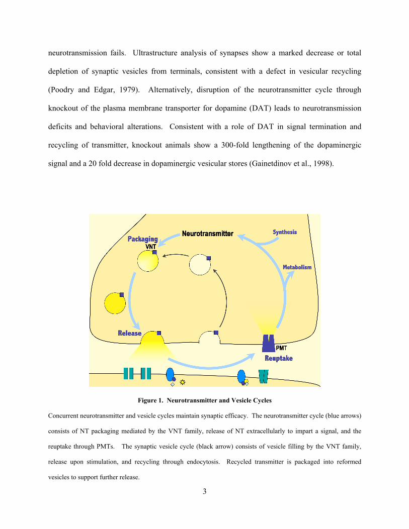
1.1.1 Neurotransmitter and Secretory Vesicle Cycles
The maintenance of neurotransmission is reliant on concurrent cycles of neurotransmitter and
secretory vesicles through packaging, release, and recycling (Figure 1). The convergence of
these cycles is the packaging of transmitter into secretory vesicles mediated by the VNT family.
This active transport highly concentrates transmitter, while sequestering it from degradative
enzymes. Furthermore, it provides efficient storage of the chemical prior to stimulation-induced
release. Upon the generation of a nerve impulse, local calcium entry triggers the fusion of
1
secretory vesicles with the cell membrane to release their neurotransmitter content. This can
occur via complete fusion, in which the vesicles collapse into the cellular plasma membrane
releasing their content, or via ‘kiss and run’, in which a transient fusion pore connecting the
vesicle to the plasma membrane opens, allowing the release of vesicular content while
maintaining vesicle identity. Once in the extracellular milieu, neurotransmitter interacts with
receptors in nearby cells to impart a molecular response, thereby mediating ‘information
transfer’ from the releasing neuron to neighboring cells. Signal termination in most cases is
mediated by reuptake of the released transmitter by specific plasma membrane transporters
(PMT) into the releasing neuron and/or nearby cells. PMT mediated uptake not only removes
transmitter from the extracellular space, but also allows for transmitter recycling for future
release. For acetylcholine (Ach), signal termination is mediated by the degradation of
extracellular transmitter. However, the metabolite choline undergoes specific reuptake for reuse
in cholinergic transmission. In addition to transmitter, secretory vesicles and associated proteins
involved in regulated release, including VNTs, are also recycled to maintain synaptic efficacy.
During ‘kiss and run’ modes of release, reformation of secretory vesicles is through direct
closing of the fusion pore. However, during full fusion transmission, the lipid and protein
content of secretory vesicles must be sorted and retrieved through a slower, clathrin-dependent
endocytosis. The concomitant recycling of transmitter and secretory vesicles allow for
neurotransmitter to be efficiently repackaged by the VNT family into secretory vesicles to
support further neurotransmission.
Disruption of individual steps in either the neurotransmitter or vesicle cycles leads to
alterations in neurotransmission. For example, genetic disruption of the Drosophila isoform of
dynamin, which is involved in vesicle recycling, leads to rapid paralysis as sustained
2
neurotransmission fails. Ultrastructure analysis of synapses show a marked decrease or total
depletion of synaptic vesicles from terminals, consistent with a defect in vesicular recycling
(Poodry and Edgar, 1979). Alternatively, disruption of the neurotransmitter cycle through
knockout of the plasma membrane transporter for dopamine (DAT) leads to neurotransmission
deficits and behavioral alterations. Consistent with a role of DAT in signal termination and
recycling of transmitter, knockout animals show a 300-fold lengthening of the dopaminergic
signal and a 20 fold decrease in dopaminergic vesicular stores (Gainetdinov et al., 1998).
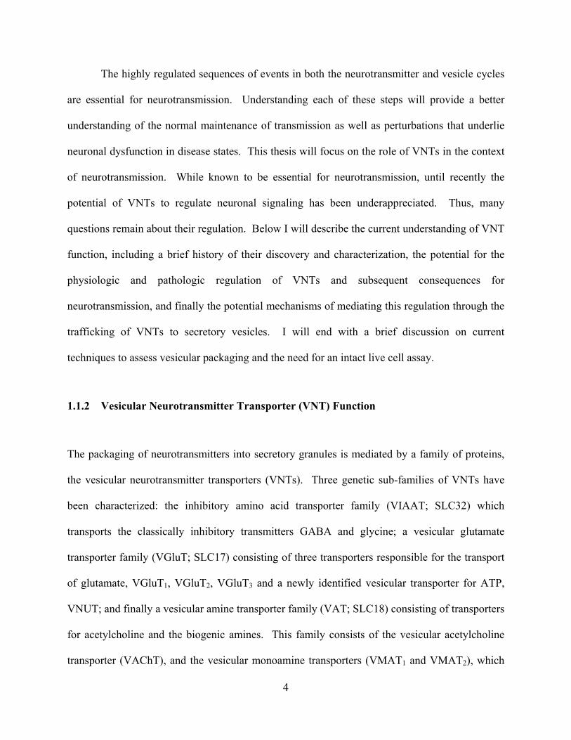
Figure 1. Neurotransmitter and Vesicle Cycles
Concurrent neurotransmitter and vesicle cycles maintain synaptic efficacy. The neurotransmitter cycle (blue arrows)
consists of NT packaging mediated by the VNT family, release of NT extracellularly to impart a signal, and the
reuptake through PMTs. The synaptic vesicle cycle (black arrow) consists of vesicle filling by the VNT family,
release upon stimulation, and recycling through endocytosis. Recycled transmitter is packaged into reformed
vesicles to support further release.
3
The highly regulated sequences of events in both the neurotransmitter and vesicle cycles
are essential for neurotransmission. Understanding each of these steps will provide a better
understanding of the normal maintenance of transmission as well as perturbations that underlie
neuronal dysfunction in disease states. This thesis will focus on the role of VNTs in the context
of neurotransmission. While known to be essential for neurotransmission, until recently the
potential of VNTs to regulate neuronal signaling has been underappreciated. Thus, many
questions remain about their regulation. Below I will describe the current understanding of VNT
function, including a brief history of their discovery and characterization, the potential for the
physiologic and pathologic regulation of VNTs and subsequent consequences for
neurotransmission, and finally the potential mechanisms of mediating this regulation through the
trafficking of VNTs to secretory vesicles. I will end with a brief discussion on current
techniques to assess vesicular packaging and the need for an intact live cell assay.
1.1.2 Vesicular Neurotransmitter Transporter (VNT) Function
The packaging of neurotransmitters into secretory granules is mediated by a family of proteins,
the vesicular neurotransmitter transporters (VNTs). Three genetic sub-families of VNTs have
been characterized: the inhibitory amino acid transporter family (VIAAT; SLC32) which
transports the classically inhibitory transmitters GABA and glycine; a vesicular glutamate
transporter family (VGluT; SLC17) consisting of three transporters responsible for the transport
of glutamate, VGluT1, VGluT2, VGluT3 and a newly identified vesicular transporter for ATP,
VNUT; and finally a vesicular amine transporter family (VAT; SLC18) consisting of transporters
for acetylcholine and the biogenic amines. This family consists of the vesicular acetylcholine
transporter (VAChT), and the vesicular monoamine transporters (VMAT1 and VMAT2), which
4
package dopamine (DA), serotonin (5-HT), norepinephrine (NE), epinephrine (E), and
histamine. The VAT family is the most widely studied of the vesicular transporters due to the
central role of the cholinergic and aminergic transmitter systems in mental health and
neurodegenerative disorders, and as such, it will be the focus of this thesis.
1.1.3 VNT Discovery and Characterization
The discovery of VNTs stemmed from the identification of secretory granules as the storage sites
for neurotransmitters and the subsequent isolation of these granules. The abundance of
chromaffin granules in the adrenal medulla and the high concentrations of monoamine stored in
them provided an ideal model system for studying neurotransmitter transport and storage. It is
not surprising, therefore, that the earliest isolation and characterization of secretory vesicles was
from bovine adrenal medulla in work by Hillarp and colleagues during the mid to late 1950’s
(Hillarp, 1958a, b). These studies used density gradient centrifugation to isolate secretory
granules and characterize their neurotransmitter content as well associated ATPase activity. This
work, along with the transformational work of Katz and colleagues on quantal acetylcholine
release at the neuromuscular junction (Del Castillo and Katz, 1954) and the advancements in
electron microscopy, which provided the first visual images of uniformly sized membrane
compartments within the nerve terminal (Robertson, 1956), helped to solidify the concept of
vesicular transmitter release (Del Castillo and Katz, 1956). The early characterization of VNTs,
and in particular, VATs, was aided greatly by the use of specific drugs that disrupted storage of
neurotransmitters in secretory vesicles. Particularly the use of reserpine which inhibits
monoamine transport (Hillarp, 1960; Jonsson and Sachs, 1969), and later vesicamol which
blocks cholinergic transport (Marshall, 1970). Interestingly, reserpine, a widely used treatment
5
for hypertension at the time, showed severe side-effects of depressive symptoms in humans, an
early indication of the centrality of monoamine transport to mental health (Freis, 1954).
1.1.4 Biophysical Properties of Vesicular Transport
In the 70’s and 80’s considerable work was done on the biophysical properties of transport.
Studies examining the vesicular concentrations of transmitters made it clear that vesicular
transport was an active process requiring energy to package neurotransmitters against their
concentration gradient. By 1979, adrenergic transport function had been reconstituted after
solubilization of chromaffin granules and transfer of isolated protein to liposomes. Transport
function was activated with the addition of an artificial pH gradient. As with endogenous
transport, the reconstituted transport was inhibited by reserpine and required ATP as an energy
source (Maron et al., 1979).
The requirement of ATP in the accumulation and storage of secretory vesicle content
suggested that ATP hydrolysis might provide the transport energy for the movement of
transmitter into the vesicle. Studies in the early 80’s identified the dependence of the transport
on a proton gradient, in which energy was derived from the transport of H+ ions down their
electrochemical gradient (Anderson et al., 1982; Toll and Howard, 1980). The Mg+2-dependent
vacuolar-type H+-ATPase (v-ATPase), the same protein known to mediate the acidification of
lysosomes, was soon identified as the source of the proton gradient (Cidon and Sihra, 1989).
The v-ATPase hydrolyzes ATP to translocate protons into secretory vesicles generating an
electrochemical gradient that can be used to drive the active packaging of neurotransmitter. A
measurable granule chemical potential (pH~ -1.4) and electrical potential ( ~ +39 mV) is
harnessed by the VNT family to exchange movement of neurotransmitter into the vesicle with
6
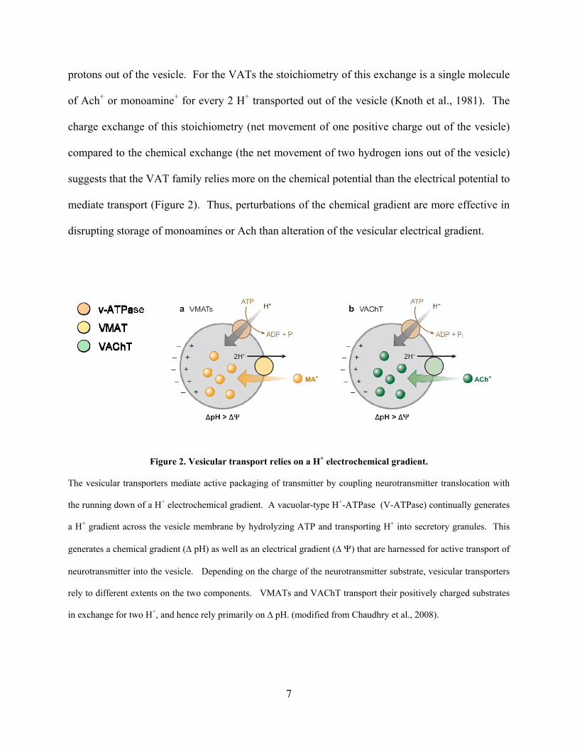
protons out of the vesicle. For the VATs the stoichiometry of this exchange is a single molecule
of Ach+ or monoamine+ for every 2 H+ transported out of the vesicle (Knoth et al., 1981). The
charge exchange of this stoichiometry (net movement of one positive charge out of the vesicle)
compared to the chemical exchange (the net movement of two hydrogen ions out of the vesicle)
suggests that the VAT family relies more on the chemical potential than the electrical potential to
mediate transport (Figure 2). Thus, perturbations of the chemical gradient are more effective in
disrupting storage of monoamines or Ach than alteration of the vesicular electrical gradient.
Figure 2. Vesicular transport relies on a H+ electrochemical gradient.
The vesicular transporters mediate active packaging of transmitter by coupling neurotransmitter translocation with
the running down of a H+ electrochemical gradient. A vacuolar-type H+-ATPase (V-ATPase) continually generates
a H+ gradient across the vesicle membrane by hydrolyzing ATP and transporting H+ into secretory granules. This
generates a chemical gradient ( pH) as well as an electrical gradient () that are harnessed for active transport of
neurotransmitter into the vesicle. Depending on the charge of the neurotransmitter substrate, vesicular transporters
rely to different extents on the two components. VMATs and VAChT transport their positively charged substrates
in exchange for two H+, and hence rely primarily on ∆ pH. (modified from Chaudhry et al., 2008).
7
The generation of the electrochemical gradient described above predicts energy to
support a concentration gradient of vesicular NT on the order of ~104 relative to cytoplasmic NT.
While vesicular concentration gradients vary greatly in-vivo, monoamines gradients have been
reported to reach concentrations upwards of 105. Thus, predicted concentrations of NT are an
order of magnitude less than can be measured in vivo. One explanation of this difference is that
monoamines form insoluble aggregates through intermolecular interactions in vesicles. This
reduces their ‘effective’ concentration and allows for the further concentration of transmitter.
Surprisingly, concentration gradients of Ach are often lower than that predicted by the gradient
potential. Thus, VAChT transport seems to be less efficient than VMAT transport. Furthermore,
the affinity of VAChT for Ach (mM range) and its transport rate (~1/s) is lower than those of
VMAT (km ~ M range; turnover ~10/s). In addition to H+ as a counterion, a role for chloride
in vesicular transport has been described. The transport of the negatively charged chloride ion
into the vesicle would allow dissipation of the proton electric gradient and thus further increase
the concentration of H+ inside the vesicle. The role for chloride transport is not well understood,
but is thought to be particularly relevant for VGluT function (Moriyama and Yamamoto, 1995).
1.1.5 Cloning and Molecular Characterization of VNTs
In the early 90’s, the vesicular transporters were molecularly characterized through their cloning
and sequencing. The first vesicular transporters cloned were the VMAT family, independently
cloned by two groups using different strategies. Liu and colleagues assayed resistance to the
active neurotoxic metabolite MPP+, implicated in a Parkinsonian-like disease phenotype.
Chromaffin cells and the neuroendocrine PC12 cell line showed resistance to MPP+ toxicity,
however a non-aminergic cell line, Chinese hamster ovary (CHO) cells, showed sensitivity.
8
Thus, to investigate the protein responsible for MPP+ resistance, a cDNA library from PC12 cells
was transformed into CHO cells and clones were selected for viability upon MPP+ exposure.
Selection and subsequent screening of clones resistant to toxicity led to the identification of the
protein that conferred resistance to MPP+, later understood to mediate resistance by the
sequestering of the toxin in secretory vesicles. The expression of the identified clone shifted
dopamine from a cytoplasmic to a punctate distribution. These characteristics were found to be
reserpine dependent, suggesting the identification of the protein as the putative vesicular
transporter. Finally, the putative transporter was shown to transport dopamine in a reconstituted
system with comparable biophysical and pharmacologic properties as previously characterized
(Liu et al., 1992a; Liu et al., 1992b). In addition to the isolated gene product, vesicular
monoamine transporter 1 (VMAT1), the group identified through homology an additional closely
related family member (VMAT2). Around the same time, VMAT2 was independently cloned
and verified by another group by assaying serotonin uptake. DNA derived from RBL cells,
which transport and store serotonin in secretory granules, was transfected into a non-aminergic
host cell and clones demonstrating high levels of serotonin uptake were selected (Erickson et al.,
1992). The two independently verified VMAT proteins share high sequence homology,
encoding polypeptides with twelve transmembrane domains, a large luminal loop, and
cytoplasmic N and C termini domains (Liu et al., 1992a). Sequence differences between the two
proteins were found primarily in the N and C termini domains and the luminal loop. Distribution
analysis revealed complementary expression patterns with VMAT1 expressed primarily in the
peripheral nervous system and VMAT2 in the brain (Peter et al., 1995). Finally, functional
analysis of the two proteins showed similar transport of all of the biogenic amines with the
exception of histamine, for which VMAT2 has higher affinity (Liu et al., 1996).
9
The cloning of VAChT soon followed (Alfonso et al., 1993; Erickson et al., 1994;
Roghani et al., 1994; Varoqui et al., 1994). It was first cloned by analysis of a previously
characterized mutation in c. elegans, UNC-17, that showed paralysis and resistance to aldicarb,
and inhibitor of synaptic acetylcholine metabolism. The mutated gene in UNC-17 was cloned
and sequenced and showed high homology (~ 40% identity and 65% similarity) to the VMAT
family and a similar predicted structure of twelve transmembrane domains and cytosolic N and C
termini tails. Together with the functional analysis of the mutant, UNC-17 was identified as a
putative vesicular acetylcholine transporter. Since the cloning of the VAT family, the
transporters for glutamate (VGluT 1-3), and GABA/ glycine (VIAAT) have been identified and
characterized (Bellocchio et al., 2000; McIntire et al., 1997; Takamori et al., 2000). Recently,
the vesicular nucleotide transporter (VNUT), a member of the VGluT gene family, has been
cloned and identified as the vesicular ATP transporter (Sawada et al., 2008).
1.1.6 Genetic Alteration and Knockdown Studies
The vesicular hypothesis of transmission, suggested that vesicular transporter function was
required for neurotransmission. In fact, the presumed centrality of this family to proper neuronal
function led to the widespread use of markers of VNT density as indicators of neurodegenerative
diseases such as Parkinson’s disease and Alzheimer’s disease. The essential role of these
proteins to neurotransmission was confirmed directly by the genetic disruption of the vesicular
transporters. The mammalian knockout of VMAT2 was completed in the late 90’s by several
groups of investigators who found the knockout to be perinatal lethal. Knockout mice showed
drastically reduced monoamine levels and disruption of vesicular release (Fon et al., 1997;
Takahashi et al., 1997; Wang et al., 1997). Null mutants of the VAChT gene in c. elegans were
10
also lethal (Alfonso et al., 1993). Interestingly, cellular analysis of drug and knockout studies
revealed that while disruption of transporter function blocked the loading of synaptic vesicles
with transmitter and thus neurotransmitter signaling, the biogenesis of secretory vesicles and the
vesicle cycle were not disrupted. Thus empty vesicles were found to cycle normally, suggesting
that the vesicle cycle is ‘blind’ to vesicular neurotransmitter content (Croft et al., 2005; Parsons
et al., 1999). This finding indicates that the amount of transmitter release is determined by
vesicular transport, including the expression and localization of transporters to vesicles, rather
than a checkpoint for the selective release of filled vesicles. This, together with other emerging
evidence discussed below, suggests an active role of VNTs in shaping the properties of synaptic
transmission. Thus in the last decade focus in the field has shifted toward understanding the
potential regulation of the VNTs.
1.2 REGULATION OF VNTS
1.2.1 Quantal Size
Quantal size is defined as the neuronal electrical response to the release of a single packet, or
quantum, of neurotransmitter. The term was introduced in studies by Bernard Katz on the
postsynaptic responses to spontaneous and stimulated release at the neuromuscular junction (For
review see Augustine and Kasai, 2007). As implied by the name, quanta were widely considered
invariant; thus, when changes in quantal size began to be appreciated the mechanisms were
believed to be postsynaptic in nature. The prevalent dogma was that the amount of
neurotransmitter released per synaptic vesicle was fixed and that only changes in the receptor
11
response to that transmitter could underlie changes in quantal size. This was further supported
by an assumption that the amount of transmitter released by a single synaptic vesicle would
saturate postsynaptic receptors (Frerking and Wilson, 1996). If this were the case, any increase
in the amount of transmitter released per vesicle would have no physiologic effect on signal
transmission. However, close examination of the spontaneous release of single vesicles (mPSPs)
or stimulated release (PSPs) has shown that under physiologic conditions significant variations in
quantal size exist. More recent studies have determined that even at the high-affinity NMDA
glutamate receptor this variation is present and due to alterations in the amount of
neurotransmitter released (Liu et al., 1999; Mainen et al., 1999; McAllister and Stevens, 2000).
Thus, at least most of the time receptors are not saturated and the amount of transmitter released
per vesicle shows significant variation. At aminergic terminals the majority of released
transmitter interacts with slower, metabotropic receptors and is paracrine in nature, often not
confined to highly local synapses. In this case, alterations in quantal size are likely to have a
large effect on signal transduction, both by altering the number of receptors activated as well as
the duration of their activation. Thus, understanding presynaptic changes in quantal size is
relevant to understanding neurotransmission.
1.2.2 Presynaptic Regulation of Quantal Size
In addition to the in-vivo variation of quantal size at many synapses, a growing body of work
suggests that these presynaptic changes are regulated. Perhaps the most physiologic of these
regulations is seen in-vivo with the movement of Drosophila from a plate containing food to one
without. Concurrent with an increase in foraging and crawl speed, an increase in quantal size is
12
seen within 35 minutes. This increase is due to changes in presynaptic mechanisms, namely the
amount of transmitter stored in synaptic vesicles (Steinert et al., 2006).
Moreover, experimental paradigms demonstrate activity-dependent modulation of quantal
size by presynaptic mechanisms. For example, disruption of neural activity of cholinergic
neurons leads to a concomitant increase in quantal size (Van der Kloot and Molgo, 1994; Wang
et al., 2005). On the other hand, high frequency stimulation of the neuromuscular junction
reduced quantal size, independent of postsynaptic changes (Doherty et al., 1984). The molecular
mechanisms that mediate these changes are not well understood, although broad perturbation of
signaling molecules, such as activation of protein kinase A (PKA) and inhibition of protein
kinase C have been shown to increase and decrease quantal size respectively, suggesting that
these changes are regulated (Staal et al., 2008; Van der Kloot and Branisteanu, 1992).
Presynaptic alterations in quantal size reflect changes in the amount of NT released from
a single synaptic vesicle. This could be modulated by one or both of the following mechanisms:
(1) changing the amount of NT contained in the vesicle or (2) the amount of NT released from
the vesicle upon exocytosis. In the case of the latter, the amount of NT released per vesicle
could be modulated through regulation of the size or stability of the fusion pore during ‘kiss and
run’ modes of release (Jackson and Chapman, 2008). However, studies suggest that for small
(non-peptidergic) transmitters, the fast rise times of spontaneous release events (< 100 μs) and
the size of secretory vesicles demonstrate that even for unstable fusion pores all NT would be
released (Klyachko and Jackson, 2002). Thus, changes in fusion pore stability would be unlikely
to influence quantal size. During full fusion modes of release, the content of each vesicle is
completely released, eliminating this possibility for this type of variation.
13
Thus, presynaptic alterations of quantal size most likely reflect changes in the amount of
neurotransmitter stored in secretory vesicles. Biophysical understanding of vesicular transport
suggests that this could be regulated by changes in the driving force of transport (i.e. the H+
electrochemical gradient), the concentration of cytosolic neurotransmitter, or regulation of the
transporter itself. In fact, increasing the cytosolic concentration of transmitter leads to increases
in the vesicular storage of transmitter. This is seen most clearly as the basis for the therapeutic
efficacy of leva-dopa, a widely proscribed treatment in Parkinson’s disease. Administration of
leva-dopa, the synthetic precursor of dopamine, alleviates symptoms associated with a loss of
dopaminergic terminals in PD. The efficacy of this treatment is due to an elevation of cytosolic
levels of dopamine (synthesized intracellularly from leva-dopa), and a subsequent increase in the
vesicular storage and quantal size of DA release (Emmanuel Pothos, 1996). The ability to alter
the vesicular content of secretory vesicles however, relies most directly on the VNTs. The copy
number, location and activity of VNTs all have the potential to regulate quantal size. In the
following section, the growing evidence for the regulation of VNTs will be presented.
1.2.3 Regulation of VNTs
Because, quanta were largely considered invariant, the relevance of VNT regulation to
neurotransmission had to first be demonstrated. The most direct and convincing evidence to
show that alterations of VNT function led to changes in quantal size and neurotransmission were
experimental manipulations of VNT copy number. Overexpression of VAChT or VMAT2 was
demonstrated to increase quantal size (Pothos et al., 2000; Song et al., 1997). Interestingly,
expression of VMAT2 in cells or neurons that do not normally store or release catecholamines
was able to induce quantal release of dopamine (Li et al., 2005; Pothos et al., 2000). On the
14
other hand, knock-down of VMAT2 led to reductions in monoamine transmission. Animals in
which VMAT2 expression was greatly reduced but not eliminated were viable but showed
behavioral deficits including motor deficits and a depressive–like phenotype (Fukui et al., 2007;
Mooslehner et al., 2001). Another study examining VMAT2 deficient animals demonstrated age-
dependent progressive loss of substantia nigra dopaminergic neurons, characteristic of
Parkinson’s disease (PD), and an increased accumulation of the PD related protein alpha-
synuclein (Colebrooke et al., 2006). Knockdown of VAChT in mice led to impairments of
cholinergic transmission and deficits in learning and memory tasks (de Castro et al., 2008; Prado
et al., 2006). These studies demonstrated that behaviorally relevant changes in
neurotransmission were induced by alterations of vesicular transport levels, thus identifying
VATs as an important potential site of regulation.
Not surprisingly, recent studies have begun to implicate alterations of VATs in disease
pathogenesis. In Huntington’s disease (HD) a downregulation of VAChT protein independent of
neuronal death is seen in post-mortem HD human tissue as well as in a mouse model of HD
(Smith et al., 2006). Consistently, changes in neurotransmission have been implicated as some
of the earliest changes in HD pathogenesis (Smith et al., 2005). Alpha-synuclein, a protein
implicated in familial PD and seen to accumulate in neurons during sporadic PD, can interact
directly with VMAT2 (Guo et al., 2008). In addition, in-vitro overexpression of the wildtype or
mutant alpha-synuclein protein leads to a decrease in VMAT2 protein levels and an increase in
cytosolic dopamine levels (Mosharov et al., 2006). Strikingly, an increase in cytoplasmic
dopamine is thought to contribute greatly to neurodegeneration of neurons in PD (Hastings and
Zigmond, 1997).
15
Regulation of VNTs have also been seen after acute application of drugs of abuse (For
review see Fleckenstein and Hanson, 2003). In particular, alteration of VMAT2 function is
known to play a major role in the psychotropic effects of drugs of abuse, including
amphetamines. Amphetamines induce the rapid depletion of monoamines from synaptic vesicles
by direct interaction with VMAT (Partilla et al., 2006) as well as their actions as a weak base
(Sulzer et al., 1992). Moreover, amphetamines have been recently linked to changes in VMAT
localization. Purified cytoplasmic vesicle preparations from synaptosomes of animals exposed to
a single high dose of methamphetamine showed decreased binding of VMAT2 markers as well as
decreased monoamine uptake (Hogan et al., 2000). This was consistent with studies that showed
that methamphetamine decreased VMAT2 protein levels in a cytoplasmic vesicle preparation
without a change in total homogenate. This indicated a redistribution of VMAT2 protein away
from vesicles (Riddle et al., 2002). On the other hand, cocaine, an inhibitor of the plasma
membrane DAT, led to a shift in VMAT2 localization to cytoplasmic vesicle-enriched fractions
and a concomitant increase in vesicular dopaminergic uptake. While the mechanisms of this
drug-induced regulation of VMAT2 are not clear, the cocaine effect is blocked by antagonists to
the D2 dopamine receptor suggesting that it is regulated through a metabotropic signaling
cascade (Brown et al., 2001; Riddle et al., 2002).
The above studies suggest that regulation of VNTs may be a relevant mechanism for
changes in neurotransmission related to disease and drugs of abuse. Recent studies have begun
to identify other potential mechanisms of VNT regulation that may play a role in more
physiologic regulation of neurotransmission. In a series of studies done in both cell lines and
primary neurons, Ahnert-Hilger and colleagues have shown that VMAT1 and VMAT2 as well as
VGluT can be regulated by heterotrimeric G proteins that associate with the secretory vesicle.
16
Using transport assays from isolated vesicle preparations of neuroendocrine cells and primary
neurons, uptake of radiolabelled monoamines was inhibited by Go2. The electrochemical
gradient of vesicles is not altered, suggesting direct modulation of VMAT2 function.
Interestingly, the intralumenal loop of the transporter and packaged transmitter are implicated in
the G protein inhibition as a means to sense and regulate vesicular content. Although the role of
G protein mediated VAT regulation is not yet understood, it is the first identification of
regulatory machinery that directly alters VAT function (Ahnert-Hilger et al., 1998; Brunk et al.,
2006; Holtje et al., 2000; Holtje et al., 2003).
Direct demonstration of physiologic regulation of VNTs comes from recent studies that
examined the periodicity of VGluT expression on synaptic vesicles (Darna et al., 2008;
Yelamanchili et al., 2006). Studies found that while the total protein level of VGluT in mouse
brain was constant throughout the day, the amount present on synaptic vesicles was strongly
regulated by circadian rhythm. Using a pronase assay, the authors demonstrated that levels of
plasma membrane localized VGluT, but not other synaptic vesicle proteins such as
synaptotagmin, fluctuated depending of the time of day. In a complementary fashion, isolated
secretory granules showed shifting levels of the vesicular transport of glutamate, suggesting a
diurnal translocation of VGluT from the plasma membrane to synaptic vesicles. This regulation
required the Per2 gene, implicated in light adaptations of the biological clock as animals lacking
the gene did not show this VGluT regulation. These studies show that the VNTs are a target of
physiologic regulation. Moreover, this regulation is capable of defining quantal size and
neuronal signaling properties and is relevant to disease pathogenesis, drug effects and plasticity
of neurotransmission.
17
1.3 VESICULAR NEUROTRANSMITTER TRANSPORTER TRAFFICKING
Regulation of protein trafficking has recently been identified as a primary mechanism of acute
regulation of transmission. For example the membrane trafficking of AMPA receptors are
believed to underlie early changes in synaptic plasticity (Malenka, 2003). Recent studies suggest
that the regulation of VNT trafficking is also involved in defining characteristics of
neurotransmitter release, neuronal function and behavior. During both the biogenesis and the
activity-dependent recycling of secretory vesicles, VNTs undergo trafficking that has the
potential to determine both the type of secretory vesicle into which neurotransmitter is packaged
as well as the amount of neurotransmitter packaged into vesicles. Thus, VNT trafficking is
amenable to regulation as a means of defining characteristics of synaptic transmission.
Understanding the signals, mechanisms and regulation of VNT trafficking are therefore essential
to understanding neurotransmission.
1.3.1 Secretory Vesicle Types
Multiple types of secretory vesicles with distinctive release properties underlie regulated release
from neurons. The targeting of vesicular transporters to distinct types of vesicles defines their
neurotransmitter content and thus characteristics of neurotransmitter release. The primary types
of secretory granules that mediate neurotransmission are small clear synaptic vesicles (SVs) and
large dense core vesicles (LDCVs). In addition small dense core vesicles (SDCVs) are often
categorized as a unique vesicle class although they are poorly characterized. Many
characteristics including size, origin and content of these classes of vesicles are unique, as are
their release properties and role in neuronal signaling (Edwards, 1998; Martin, 2003). SVs,
18
morphologically characterized as small (~ 40-50 nm) are typically found in clusters at
specialized regions of membrane termed active zones and mediate the fast synaptic transmission
of classical transmitters. The properties of activity-dependent SV release have been
characterized extensively at the frog neuromuscular junction with the release of Ach and in
central hippocampal synapses with the release of glutamate. Classically, these vesicles show
high Ca+2 sensitivity, synchronous release and short release latency (< 1 ms). These vesicles
recycle locally at release sites, rapidly refill and can undergo multiple rounds of release.
LDCVs, on the other hand, are characterized by their larger size (> 80 nm) and electron dense
neuropeptide content. In addition to neuropeptides, LDCVs often contain monoaminergic
transmitter. The release properties of LDCVs, classically studied using chromaffin granules,
show lower sensitivity for Ca+2, release more slowly (> 10 ms) and show asynchrony of release.
Moreover, LDCVs are not thought to recycle locally, but rather form at the TGN where they are
filled with neuropeptide content.
The subcellular distribution of VATs on secretory vesicles has been studied in some
systems, however characterization is far from complete. Although cholinergic terminals contain
both SVs and LDCVs, VAChT preferentially localizes to SVs (Gilmor et al., 1996; Weihe et al.,
1996). This preferential localization to SVs makes it an ideal candidate protein to study the
signals and machinery that regulate SV-specific trafficking. On the other hand, the VMAT
transporter has been localized to multiple vesicles types including SVs, LDCVs, and SDCVs
(Nirenberg et al., 1996). The localization of VMAT to multiple vesicle populations suggests that
its trafficking may be regulated between vesicle types.
19
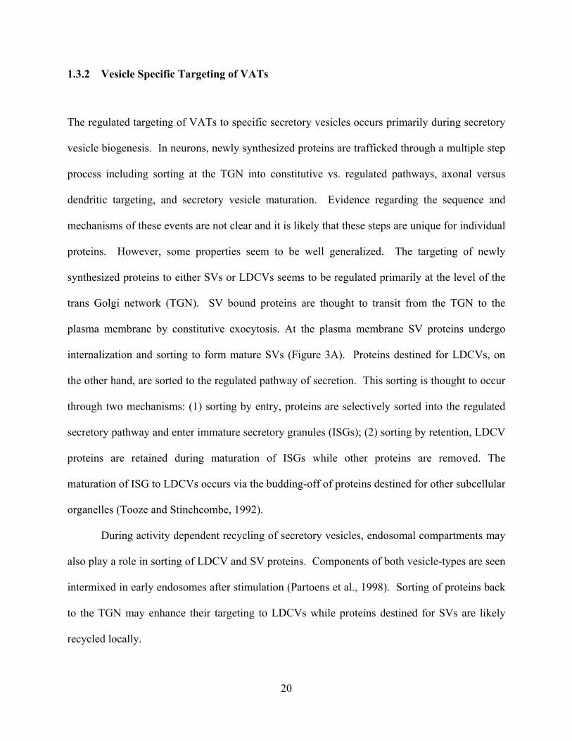
1.3.2 Vesicle Specific Targeting of VATs
The regulated targeting of VATs to specific secretory vesicles occurs primarily during secretory
vesicle biogenesis. In neurons, newly synthesized proteins are trafficked through a multiple step
process including sorting at the TGN into constitutive vs. regulated pathways, axonal versus
dendritic targeting, and secretory vesicle maturation. Evidence regarding the sequence and
mechanisms of these events are not clear and it is likely that these steps are unique for individual
proteins. However, some properties seem to be well generalized. The targeting of newly
synthesized proteins to either SVs or LDCVs seems to be regulated primarily at the level of the
trans Golgi network (TGN). SV bound proteins are thought to transit from the TGN to the
plasma membrane by constitutive exocytosis. At the plasma membrane SV proteins undergo
internalization and sorting to form mature SVs (Figure 3A). Proteins destined for LDCVs, on
the other hand, are sorted to the regulated pathway of secretion. This sorting is thought to occur
through two mechanisms: (1) sorting by entry, proteins are selectively sorted into the regulated
secretory pathway and enter immature secretory granules (ISGs); (2) sorting by retention, LDCV
proteins are retained during maturation of ISGs while other proteins are removed. The
maturation of ISG to LDCVs occurs via the budding-off of proteins destined for other subcellular
organelles (Tooze and Stinchcombe, 1992).
During activity dependent recycling of secretory vesicles, endosomal compartments may
also play a role in sorting of LDCV and SV proteins. Components of both vesicle-types are seen
intermixed in early endosomes after stimulation (Partoens et al., 1998). Sorting of proteins back
to the TGN may enhance their targeting to LDCVs while proteins destined for SVs are likely
recycled locally.
20
Much of what is known about the molecular signals that mediate the trafficking of VATs
has come from biochemical analysis of nascent proteins in cell lines, namely the neuroendocrine
PC12 cell line. This cell line, expresses low levels of both VAChT and VMAT1. In PC12 cells
VAChT localizes strongly to synaptic vesicle like vesicles (SVLVs), whereas exogenous
expression of VMAT2 or endogenous VMAT1 preferentially localize to LDCVs. The
preferential localization of these two similar proteins to unique secretory vesicle types has made
this an advantageous system for studying the signals and machinery that regulate vesicle-specific
traffic. Studies investigating the trafficking of these proteins have used chimera between
VMAT2 and VAChT in order to identify regions of the protein important for vesicle specific
targeting. Chimera in which the cytoplasmic tail of VAChT and VMAT were switched
demonstrated the importance of the C-terminus to its localization. Mutation analysis within
these regions identified classic dileucine motifs that were essential to internalization of both
VAChT and VMAT (Tan et al., 1998). Upstream glutamate residues of the VMAT2 dileucine
motif (KEEKMAIL) were shown to be involved in the specific localization of VMAT2 to
LDCVs. Mutation of these residues to alanines reduced VMAT2 targeting to LDCVs without
altering its endocytosis. Interestingly, phosphorylation of a serine residue upstream of the
VAChT dileucine motif (RSERDVLL), which mimics the negative charges of the VMAT2
upstream residues, has been shown to promote trafficking of VAChT to LDCVs (Krantz et al.,
2000). Furthermore, an acidic patch in the C terminus of VMAT2 has been identified as a
retention sequence for localization to LDCVs. The deletion of these residues or the
phosphorylation of two serine residues within this patch by casein kinase 2 promotes the removal
of VMAT2 from immature granules during maturation and thus reduces its expression on LDCVs
(Waites et al., 2001). These mechanisms suggest that changes in phosphorylation states of VATs
21
may play an important role in their regulated targeting. The physiological regulation and
relevance of this potential regulation remains to be investigated in neurons. Moreover, the
cytosolic machinery that may regulate these trafficking events has not been identified.
Figure 3. Secretory Vesicle Biogenesis
A. Vesicle Specific Biogenesis. Large dense core vesicles (LV) are sorted by selection at the TGN (1) or retention
(2) during immature granule (im. LV) maturation. Synaptic vesicles (SV) are formed after internalization from
components that have undergone constitutive secretion (CS). B. Synaptic Vesicle Formation. At the terminal SVs
are formed by internalization through an endosomal intermediate (EE) in an AP-3 dependent manner and/or via
direct internalization though an AP-2 dependent mechanism. (Modified from Fei et al., 2008).
22
1.3.3 Synaptic Vesicle Biogenesis and Recycling
Data examining axonal transport of tubulovesicular structures carrying SV proteins suggest that
SV destined proteins are transported individually or in subsets through constitutive exocytosis or
in specialized synaptic vesicle precursors (Okada et al., 1995). Regardless, the formation of the
mature SV, containing the required complement of SV proteins and characteristic morphology,
occurs through internalization at the nerve terminal. Internalization may be mediated by direct
budding of the SV from the plasma membrane in an AP-2, clathrin dependent manner or budding
from an endosomal intermediate requiring AP-3 (Figure 3B; for review see Hannah et al., 1999).
The central role of clathrin-mediated internalization in the formation of SVs suggests that SV
biogenesis may share common mechanisms of protein targeting with those of activity-dependent
SV recycling.
Mechanisms of SV recycling were first characterized through the publication of several
seminal papers in the 70’s (For review see Heuser, 1989). These experiments used electron
microscopy to visualize neuromuscular junction synapses that had been stimulated in the
presence of a fluid phase marker to label recycling vesicles. After stimulation labeled vesicles
were detected, indicating the vesicle recycling. However, the mechanisms of recycling proposed
by different group were quite distinct (Figure 4). In one case, Ceccarelli and colleagues reported
the appearance of labeled vesicles with no change in the number of vesicles or the size of the
membrane even after hours of stimulation. Moreover, clathrin intermediates were not identified,
suggesting a clathrin-independent form of recycling (Ceccarelli et al., 1973). Heuser and Reese,
however reported that after just minutes of stimulation, a rapid depletion in the number of
synaptic vesicles was seen, concomitant with an increase in the size of the plasma membrane.
Moreover the appearance of many labeled cisternae and clathrin coated vesicles were evident.
23
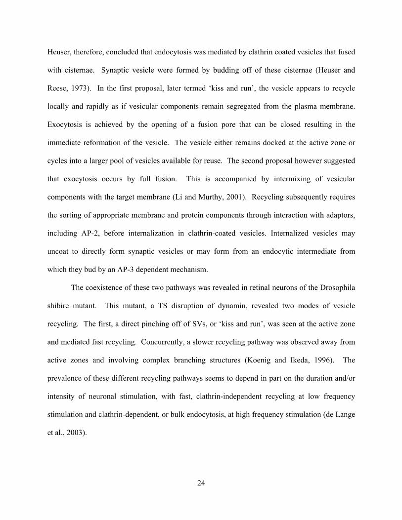
Heuser, therefore, concluded that endocytosis was mediated by clathrin coated vesicles that fused
with cisternae. Synaptic vesicle were formed by budding off of these cisternae (Heuser and
Reese, 1973). In the first proposal, later termed ‘kiss and run’, the vesicle appears to recycle
locally and rapidly as if vesicular components remain segregated from the plasma membrane.
Exocytosis is achieved by the opening of a fusion pore that can be closed resulting in the
immediate reformation of the vesicle. The vesicle either remains docked at the active zone or
cycles into a larger pool of vesicles available for reuse. The second proposal however suggested
that exocytosis occurs by full fusion. This is accompanied by intermixing of vesicular
components with the target membrane (Li and Murthy, 2001). Recycling subsequently requires
the sorting of appropriate membrane and protein components through interaction with adaptors,
including AP-2, before internalization in clathrin-coated vesicles. Internalized vesicles may
uncoat to directly form synaptic vesicles or may form from an endocytic intermediate from
which they bud by an AP-3 dependent mechanism.
The coexistence of these two pathways was revealed in retinal neurons of the Drosophila
shibire mutant. This mutant, a TS disruption of dynamin, revealed two modes of vesicle
recycling. The first, a direct pinching off of SVs, or ‘kiss and run’, was seen at the active zone
and mediated fast recycling. Concurrently, a slower recycling pathway was observed away from
active zones and involving complex branching structures (Koenig and Ikeda, 1996). The
prevalence of these different recycling pathways seems to depend in part on the duration and/or
intensity of neuronal stimulation, with fast, clathrin-independent recycling at low frequency
stimulation and clathrin-dependent, or bulk endocytosis, at high frequency stimulation (de Lange
et al., 2003).
24
The regulation between and physiologic relevance of multiple mechanisms of vesicle
recycling is not clear. However several hypotheses have been suggested. The activity
dependence of the different pathways may suggest that bulk endocytosis occurs only under
strong stimulus conditions that may saturate fast, clathrin independent modes of recycling. An
alternative suggestion is that different recycling pathways may form distinct vesicle pools and/or
distinct synaptic vesicles. In this case, vesicles undergoing rapid endocytosis recycle locally to
the ready-releasable pool of vesicles, whereas slower endosomal mediated pathways recycle to a
reserve pool of vesicles (Richards et al., 2000). A third consideration is the apparent preference
of certain SV proteins to recycle through specific pathways. The mocha mouse, which displays a
mutation in the AP-3 adaptor protein, involved in the endosomal formation of SVs, shows
selective mislocalization of certain SV proteins (including the VIAAT and the Zn transporter)
with no apparent defects in other SV proteins. These results suggest that trafficking of selective
SV proteins is mediated in an AP-3 dependent manner (Kantheti et al., 1998; Nakatsu et al.,
2004). Although many questions remain as to their precise physiologic relevance, it is clear that
the presence of multiple trafficking pathways allows for regulation of protein trafficking during
SV recycling.
25
Figure 4. Synaptic vesicle recycling pathways
Multiple pathways are proposed: (1) kiss-and-run- a pathway in which vesicles endocytose by closure of the fusion
pore and are refilled with neurotransmitters either while remaining docked to the active zone or via a local recycling
pathway that is clathrin independent but results in mixing vesicles with the reserve pool after endocytosis; (2)
clathrin mediated endocytosis- a pathway whereby vesicles undergo clathrin-mediated endocytosis and recycle
either by direct uncoating or via an endosomal intermediate. (Modified from Sudhof, 2004).
26
1.3.4 Synaptic Vesicle Targeting of VATs
The selective localization of VAChT to SVs has made it an ideal protein for identifying synaptic
vesicle specific targeting sequences. The central role of internalization in the biogenesis and
recycling of synaptic vesicles suggests that signals that mediate the internalization of SV proteins
should be important in their SV targeting. Consistently, the dileucine motif in VAChT is
required for its SV targeting (Tan et al., 1998). Furthermore, VAChT has been shown to interact
with clathrin adaptor protein AP-2, involved in clathrin mediated endocytosis. Moreover
disruption of endocytic machinery such as dynamin and clathrin leads to accumulation of
VAChT on the plasma membrane (Barbosa et al., 2002; Ferreira et al., 2005) and thus a
reduction in its SV targeting. In addition to interaction with AP-2, an interaction with the
adaptor protein AP-1, normally associated with trafficking at the TGN, has been indicated,
although the functional significance of this interaction is not yet known. Furthermore, a non-
traditional tyrosine motif has also been implicated in the internalization of VAChT, although it is
not required in neuroendocrine cells. Interestingly, an interaction between VAChT and AP-3 has
not been detected, thus alternative machinery may be involved in the endosomal trafficking of
VAChT to synaptic vesicles (Ferreira et al., 2005). The requirement of endocytosis for the SV
targeting of VAChT is clear, however the presence of a sufficient SV targeting motif is
unknown. Moreover, the proteins that regulate the SV specific targeting of VAChT are
unknown.
27
1.4 CURRENT ASSAYS OF VNT FUNCTION
Recent evidence suggests that vesicular storage may be regulated under physiologic conditions,
during exposure to drugs and during the pathogenesis of disease. Because alterations of
vesicular storage lead to changes in synaptic transmission, understanding this regulation is
essential. However, the inability to directly monitor vesicular transport in living neurons has
hindered understanding of VNT function and regulation during neurotransmission.
Biophysical measurements of VNT mediated transport have relied on in-vitro transport
assays. These studies have been invaluable for understanding mechanisms of transport including
molecular and chemical properties. However the necessity of isolating vesicular fractions in
these assays unavoidably isolates the transport process from the cellular environment as well as
separates vesicle transport from vesicular release. Thus, while in-vitro transport assays are a
direct measurement of VNT function, they do not allow the acute study of physiologic regulation
of transport or the relationship of alterations in transport with neurotransmission.
Electrophysiological assays, on the other hand, are able to dynamically assess changes in
quantal size but are not able to directly attribute these changes to alterations in vesicular
transport. Electrophysiology measures postsynaptic cellular responses to stimulation. Therefore
changes in quantal size can be indicative of changes in presynaptic or postsynaptic mechanisms.
Furthermore as the readout is the summation of all inputs into the selected cell, this technique
provides poor spatial information and localized changes (i.e. at a single or subset of terminals)
may not be detected.
Recently, imaging techniques have allowed the exploration of subcellular dynamics of
vesicular proteins and membranes. The use of fluorescently tagged proteins and lipophilic dyes
has provided a means to study the trafficking of SV proteins and vesicles with good spatial and
28
temporal resolution. However, while providing valuable information on the vesicle cycle they
are unable to provide information on vesicular content or the function of vesicular transporters.
Perhaps the most informative studies thus far have relied on perturbation of transporter
function through drugs or genetic techniques followed by one of the readouts assays described
above. While these approaches stress the essential role of vesicular transport in
neurotransmission and are able to identify the potential for VNT regulation to modulate neuronal
function, they preclude the understanding of normal VNT function and regulation during
neurotransmission.
A live assay of vesicular transport in neurons would aid in our understanding of the role
of vesicular packaging in neurotransmission. By taking advantage of optical techniques to
visualize neurotransmitter, vesicular transport can be studied with good spatial resolution and
minimal perturbation of the system. An assay that monitors neurotransmitter would allow for
measurements of both vesicular transport as well as release. This would provide insight into
both the normal function of VNTs in the context of neurotransmission, allowing for the first
measurements of the contribution of activity dependent vesicular transport to release. Moreover,
this would provide a means of assaying VNT regulation and the role of this regulation in shaping
properties of neurotransmission.
29
1.5 THESIS GOALS
Until recently, the regulation of VNTs as a means to modulating synaptic transmission had been
underappreciated. Thus, many questions remain about the regulation of VNTs as well the
consequences of VNT regulation for neurotransmission. To understand complex mechanisms of
VNT regulation an understanding of basic properties, such as VNT trafficking and the
contribution of vesicular transport to release are necessary. Thus, the goals of this thesis were
(1) to better understand the signals and machinery that mediate the SV-specific trafficking of
vesicular transporters and (2) to establish a live-cell optical assay to measure vesicular transport
and its contributions to release in neurons.
The results described in Chapter 2 rely on the synaptic vesicle specific targeting of
VAChT in a neuroendocrine cell line to identify a sufficient synaptic vesicle targeting motif.
The identified motif contains a classical dileucine motif that shows duality of function as an
internalization and synaptic vesicle targeting sequence. The specificity of this motif as a SVTM
is discussed. This work has been previously published (Colgan et al., 2007).
In Chapter 3 Sorting nexin 5 (SNX5) is identified as a novel regulatory protein that
directs the SV trafficking of VAChT. SNX5 is characterized and the functional interaction of
SNX5 and VAChT is tested. Disruption of SNX5 leads to the mistargeting of protein from SVs
to LDCVs. This chapter has been written in the format of a ‘Brief Communication’ in
preparation for publication.
In Chapter 4 a novel assay is established that allows for the first measurements of
vesicular transport in live neurons. Concomitant measurements of vesicular packaging and
release allow for the contributions of vesicular transport to release to be assessed. This work is
in preparation for publication.
30
Chapter 5 presents a summary of the work presented and a broad discussion of its
implications for the field of VNTs and the larger field of neurotransmission. The incorporation
of the findings within a broader view of the literature and future directions for study are
addressed. Throughout, I focus on the active role of VNTs in shaping the properties of
neurotransmission.
31
2.0 DILEUCINE MOTIF IS SUFFICIENT FOR INTERNALIZATION AND SYNAPTIC VESICLE
TARGETING OF VESICULAR ACETYCHOLINE TRANSPORTER
2.1 ABSTRACT
Efficient cholinergic transmission requires accurate targeting of vesicular acetylcholine
transporter (VAChT) to synaptic vesicles (SVs). However, the signals that regulate this vesicular
targeting are not well characterized. Although previous studies suggest that the C-terminus of the
transporter is required for its SV targeting, it is not clear whether this region is sufficient for this
process. Furthermore a synaptic vesicle targeting motif (SVTM) within this sequence remains to
be identified. Here we use a chimeric protein, TacA, between an unrelated plasma membrane
protein, Tac, and the C-terminus of VAChT to demonstrate the sufficiency of the C-terminus for
targeting to synaptic vesicle-like vesicles (SVLVs) in PC12 cells. TacA shows colocalization and
cosedimentation with the SV marker synaptophysin. Deletion mutation analysis of TacA
demonstrates that a short, dileucine-motif containing sequence is required and sufficient to direct
this targeting. Di-alanine mutation analysis within this sequence suggests indistinguishable
signals for both internalization and synaptic vesicle sorting. Using additional chimeras as
controls, we confirm the specificity of this region for SVLVs targeting. Therefore, we suggest
that the dileucine containing motif is sufficient as a dual signal for both internalization and SV
targeting during VAChT trafficking.
32
2.2 INTRODUCTION
Synaptic transmission requires the efficient vesicular packaging of neurotransmitters, a process
mediated by a group of vesicular neurotransmitter transport proteins (VNT) (Liu and Edwards,
1997). Thus, targeting of these functional transport proteins to synaptic vesicles (SVs) is highly
regulated in order to maintain synaptic efficacy. Despite this, the molecular mechanisms
underlying the SV targeting of VNTs remain elusive. Evidence suggests that clathrin mediated
internalization plays an important role in the targeting of both newly synthesized and recycling
VNTs to synaptic vesicles. From the cell surface, SV bound proteins require internalization and
sorting that is thought to occur through at least two distinct pathways. SV proteins may undergo
selective internalization to directly form SVs, or proteins may be indiscriminately internalized to
an endosomal intermediate from which SV proteins can be selectively sorted (Hannah et al.,
1999; Sudhof, 2004). The regulation of these distinct sorting processes, although not well
understood, is believed to be mediated through specific cytoplasmic sorting motifs. These motifs
execute their role in membrane trafficking through interaction with corresponding cytosolic
machinery that direct the protein accordingly. Sorting- motifs for either internalization or SV
targeting (SVTM) have been identified in several SV proteins (Blagoveshchenskaya et al., 1999;
Grote et al., 1995; Han et al., 2004; Pennuto et al., 2003; Prado and Prado, 2002). For example,
synaptophysin has been shown to contain a C-terminal repeating tyrosine based internalization
motif that recruits dynamin to the membrane and directs clathrin-independent
internalization(Daly et al., 2000; Daly and Ziff, 2002; Pennuto et al., 2003). Another SV protein,
synaptotagmin1, has a cytoplasmic dileucine-motif which is required for AP-3 mediated SV
targeting (Blagoveshchenskaya et al., 1999). In contrast, another study suggested a role for
synaptotagmin’s lumenal N-terminus in SV targeting (Han et al., 2004). While specific motifs
33
have been indicated to play a role in SV targeting, the sufficiency of these motifs as well as the
generality of these signals is not known.
Vesicular Acetylcholine Transporter (VAChT) is a member of the VNT family that is
responsible for packaging acetylcholine into vesicles for regulated release (Alfonso et al., 1993;
Rand, 1989; Roghani et al., 1994). Molecular manipulations to alter the vesicular level of
VAChT on SVs, namely overexpression or knockdown, lead to corresponding alterations in
cholinergic transmission (Kitamoto et al., 2000; Song et al., 1997). Although a specific SVTM
has yet to be identified, the C-terminus of VAChT has been found to be required for its vesicular
localization (Tan et al., 1998; Varoqui and Erickson, 1998). Interestingly, this region has been
identified to be phosphorylated by PKC, which was suggested to play a role in modulating its
internalization or trafficking (Cho et al., 2000; Krantz et al., 2000). Furthermore, a classical
dileucine motif within this region has been identified to be required for the internalization of the
transporter (Tan et al., 1998). Interaction of VAChT through its C-terminus with clathrin
associated machinery including AP-1, and AP-2 further suggest a critical role of the cytoplasmic
sequence in the regulated clathrin mediated internalization and membrane trafficking of VAChT
(Barbosa et al., 2002; Kim and Hersh, 2004). Consistently, interference with internalization
machinery, including clathrin and dynamin, or mutation of the internalization motif, disrupt the
targeting of VAChT to SVs leading to an accumulation of the protein on the cell surface
(Barbosa et al., 2002; Ferreira et al., 2005; Tan et al., 1998). Together, these findings suggest a
correlation between the C–terminal directed, regulated internalization of the transporter and its
SV targeting. However, whether the C-terminus of VAChT is sufficient for SV targeting remains
unknown. Furthermore, it is unclear how the internalization of VAChT is involved in its
34
vesicular localization and most importantly, whether this SV targeting is determined by a SVTM
within this region.
Here we report, by using a chimeric protein between an unrelated plasma membrane
localized protein, Tac, and the VAChT C-terminus, the sufficiency of this sequence in synaptic
vesicle like vesicle (SVLV) targeting in PC12 cells. We further characterize the dileucine
containing motif of VAChT as a SVTM and examine its role as a dual signal for both
internalization and SVLV targeting.
2.3 RESULTS
2.3.1 The C-terminus of VAChT is sufficient for SVLV targeting
The C-terminus of VAChT has been shown to be required for its preferential localization to
SVLVs in PC12 cells (Tan et al., 1998). Chimeric analysis between VAChT and a closely
related, but preferentially large dense core vesicle (LDCV) localized VNT, vesicular monoamine
transporter 2 (VMAT2), has suggested that the VAChT C-terminus is able to direct VMAT2 to
SVLVs (Tan et al., 1998). However, these results do not distinguish whether the VAChT C-
terminus is sufficient for SVLV targeting. To test this possibility, we took advantage of Tac
protein, the -subunit of the interleukin 2 receptor (IL-2R), which has a single membrane
spanning domain whose internalization requires the presence of additional IL-2R subunits
(Letourneur and Klausner, 1991; Marks et al., 1995; Tan et al., 1998). Not endogenously
expressed in PC12 cells, Tac protein localizes to the plasma membrane when exogenously
introduced (Figure 5). Through fusing the C-terminus of VAChT to Tac, we created a chimeric
35
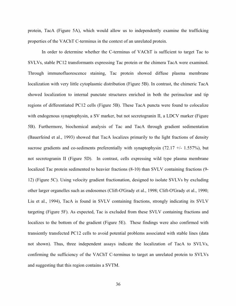
protein, TacA (Figure 5A), which would allow us to independently examine the trafficking
properties of the VAChT C-terminus in the context of an unrelated protein.
In order to determine whether the C-terminus of VAChT is sufficient to target Tac to
SVLVs, stable PC12 transformants expressing Tac protein or the chimera TacA were examined.
Through immunofluorescence staining, Tac protein showed diffuse plasma membrane
localization with very little cytoplasmic distribution (Figure 5B). In contrast, the chimeric TacA
showed localization to internal punctate structures enriched in both the perinuclear and tip
regions of differentiated PC12 cells (Figure 5B). These TacA puncta were found to colocalize
with endogenous synaptophysin, a SV marker, but not secretogranin II, a LDCV marker (Figure
5B). Furthermore, biochemical analysis of Tac and TacA through gradient sedimentation
(Bauerfeind et al., 1993) showed that TacA localizes primarily to the light fractions of density
sucrose gradients and co-sediments preferentially with synaptophysin (72.17 +/- 1.557%), but
not secretogranin II (Figure 5D). In contrast, cells expressing wild type plasma membrane
localized Tac protein sedimented to heavier fractions (8-10) than SVLV containing fractions (9-
12) (Figure 5C). Using velocity gradient fractionation, designed to isolate SVLVs by excluding
other larger organelles such as endosomes (Clift-O'Grady et al., 1998; Clift-O'Grady et al., 1990;
Liu et al., 1994), TacA is found in SVLV containing fractions, strongly indicating its SVLV
targeting (Figure 5F). As expected, Tac is excluded from these SVLV containing fractions and
localizes to the bottom of the gradient (Figure 5E). These findings were also confirmed with
transiently transfected PC12 cells to avoid potential problems associated with stable lines (data
not shown). Thus, three independent assays indicate the localization of TacA to SVLVs,
confirming the sufficiency of the VAChT C-terminus to target an unrelated protein to SVLVs
and suggesting that this region contains a SVTM.
36
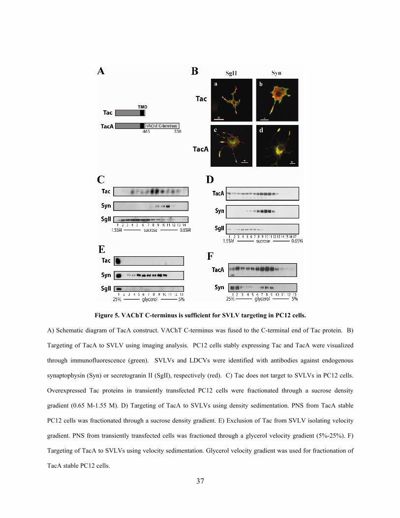
Figure 5. VAChT C-terminus is sufficient for SVLV targeting in PC12 cells.
A) Schematic diagram of TacA construct. VAChT C-terminus was fused to the C-terminal end of Tac protein. B)
Targeting of TacA to SVLV using imaging analysis. PC12 cells stably expressing Tac and TacA were visualized
through immunofluorescence (green). SVLVs and LDCVs were identified with antibodies against endogenous
synaptophysin (Syn) or secretogranin II (SgII), respectively (red). C) Tac does not target to SVLVs in PC12 cells.
Overexpressed Tac proteins in transiently transfected PC12 cells were fractionated through a sucrose density
gradient (0.65 M-1.55 M). D) Targeting of TacA to SVLVs using density sedimentation. PNS from TacA stable
PC12 cells was fractionated through a sucrose density gradient. E) Exclusion of Tac from SVLV isolating velocity
gradient. PNS from transiently transfected cells was fractioned through a glycerol velocity gradient (5%-25%). F)
Targeting of TacA to SVLVs using velocity sedimentation. Glycerol velocity gradient was used for fractionation of
TacA stable PC12 cells.
37
2.3.2 Dileucine containing motif is required for SVLV targeting
The cytosolic tail of VAChT consists of 60 amino acids which contain several previously defined
motifs including a classic dileucine motif [E(XXX)LL], a classical tyrosine motif (YXXØ), and
a nonclassical tyrosine motif. In order to investigate which of these motifs determines the SVLV
targeting of VAChT, a series of TacA chimera with deletions within the C-terminus of VAChT
were constructed (Figure 6A). These chimeras were then examined for their subcellular
localization in PC12 cells. Targeting to SVLVs was determined through two criteria: steady-state
colocalization with synaptophysin through immunofluorescent staining, and comigration with
SVLV containing fractions in velocity subcellular fractionation analysis. As shown in figure 6B,
constructs that met both of these criteria are indicated with a symbol [+] for their SVLV
targeting. In contrast, constructs unable to target to SVLVs are indicated with a symbol [--] for
SVLV targeting. Analysis of deletion mutants clearly indicated the requirement of a small
dileucine motif containing region (Figure 6A, indicated in grey) for SVLV targeting. The
deletion of this region disrupted SVLV targeting and led to the accumulation of the mutant on
the cell surface, presumably due to a loss of dileucine motif mediated internalization. This is
demonstrated through representative immunofluorescent images in Figure 6C. Deletion mutant
analysis suggested that the dileucine containing motif is the only region required for SVLV
targeting. Other regions, previously reported to play a role in VAChT trafficking, including
tyrosine motifs (Kim and Hersh, 2004), or the phosphorylation at serine 480 (Cho et al., 2000;
Krantz et al., 2000) were not found to be essential for SVLV targeting in our system. Thus the 10
a.a. dileucine motif containing sequence of the VAChT C-terminus is essential for the SVLV
targeting of VAChT.
38
Figure 6. Dileucine containing motif is necessary for SVLV targeting.
A) Amino acid sequence of VAChT C-terminus (residues 465 to 530). Dileucine containing motif is shaded in grey.
B) Targeting of deletion mutants of TacA to SVLVs in PC12 cells. A series of deletion mutations were made within
the C-terminus of VAChT using the TacA construct as template. SVLV targeting of the mutants were determined by
both immunofluorescent colocalization with synaptophysin, as well as presence in SVLV isolating glycerol velocity
gradients. Symbol (+) indicates the targeting of TacA mutant to SVLVs. Symbol (--) indicates non-targeting to
SVLVs. C) Immunofluorescent analysis of subcellular localization of key deletion mutants. PC12 cells transiently
transfected were stained for mutant TacA (red) and synaptophysin (green). Bar= 20µm.
39
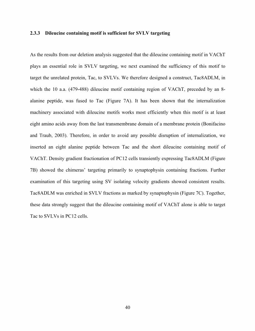
2.3.3 Dileucine containing motif is sufficient for SVLV targeting
As the results from our deletion analysis suggested that the dileucine containing motif in VAChT
plays an essential role in SVLV targeting, we next examined the sufficiency of this motif to
target the unrelated protein, Tac, to SVLVs. We therefore designed a construct, Tac8ADLM, in
which the 10 a.a. (479-488) dileucine motif containing region of VAChT, preceded by an 8-
alanine peptide, was fused to Tac (Figure 7A). It has been shown that the internalization
machinery associated with dileucine motifs works most efficiently when this motif is at least
eight amino acids away from the last transmembrane domain of a membrane protein (Bonifacino
and Traub, 2003). Therefore, in order to avoid any possible disruption of internalization, we
inserted an eight alanine peptide between Tac and the short dileucine containing motif of
VAChT. Density gradient fractionation of PC12 cells transiently expressing Tac8ADLM (Figure
7B) showed the chimeras’ targeting primarily to synaptophysin containing fractions. Further
examination of this targeting using SV isolating velocity gradients showed consistent results.
Tac8ADLM was enriched in SVLV fractions as marked by synaptophysin (Figure 7C). Together,
these data strongly suggest that the dileucine containing motif of VAChT alone is able to target
Tac to SVLVs in PC12 cells.
40
Figure 7. Dileucine containing motif is sufficient for SVLV targeting in PC12 cells.
A) Schematic diagram of Tac8ADLM. Eight alanines (8A), followed by the dileucine containing motif (DLM) of
VAChT, were fused to the C-terminal end of Tac protein. B) Tac8ADLM targets to SVLV in PC12 cells using
density gradient fractionation. PNS of PC12 cells transiently expressing Tac8ADLM were fractionated through
sucrose density gradients. Tac8ADLM comigrated with synaptophysin containing fractions. C) Tac8ADLM targets
to SVLV in PC12 cells using velocity sedimentation. Tac8ADLM comigrates primarily with synaptophysin
containing fractions.
2.3.4 The dileucine containing motif serves as an internalization motif as well as a SVTM
The above results demonstrate that the VAChT dileucine containing motif is able to target Tac to
SVLVs. The identified region contains a classical dileucine motif, E(XXX)LL, which has been
extensively characterized for its role in internalization (Bonifacino and Traub, 2003; Tan et al.,
41
1998). Therefore, it was unclear whether the trafficking signal that mediates internalization is
different than the one that mediates SVLV targeting. To address this, we generated a series of
pair-wise alanine mutations within this region (Figure 8A) and analyzed their effects on
internalization as well as SVLV targeting. To examine internalization, a semi-quantitative,
ELISA based biotin conjugated system was used. The assay detected a fraction of internalization
of TacA dialanine mutants over a 30 minute time period. Confirming the efficacy of our assay,
wild type Tac protein remained primarily on the cell surface with only 25% internalization,
whereas the majority (92%) of TacA was internalized. Truncation of TacA to TacA490 did not
significantly alter internalization. Using pair-wise alanine mutations of this dileucine containing
region, we showed that the mutation of LL-485/486-AA disrupted internalization dramatically
(31% internalization compared to control) (Figure 8B). This result is consistent with previous
studies that demonstrate the requirement for the leucine residues for efficient VAChT
internalization (Bonifacino and Traub, 2003; Santos et al., 2001; Tan et al., 1998). Previously,
the residues upstream of the leucines in many classical dileucine motifs have been indicated to
regulate the efficiency of internalization. Not surprisingly, mutation of upstream residues DV
483/484 to AA in the VAChT C-terminus led to a significant decrease in efficiency of
internalization over 30 minutes (Figure 8B). Mutation of residues 479/480 led to a small but
significant decrease in internalization. Neither downstream dileucine flanking residues 487/488
nor residues 481/482 significantly contributed to the internalization of TacA, as mutation of
these residues did not significantly alter the internalization of these mutant TacA proteins (Figure
8B).
42
Figure 8. Dileucine containing motif is essential for the internalization of TacA.
A) Schematic diagram of dialanine scanning mutants of TacA. Pairwise alanine mutations (grey box) were made
using TacA490 as the template. B) Mutation of leucines and neighboring residues reduces internalization of TacA.
PC12 cells transiently transfected with TacA dialanine mutants were analyzed semi-quantitatively for their
endocytosis for 30 minutes. Percent internalization of mutants was normalized to non-mutant TacA490 as control to
determine effect on internalization (* p<0.05, ***p<0.001). Error bars depict standard deviation.
43
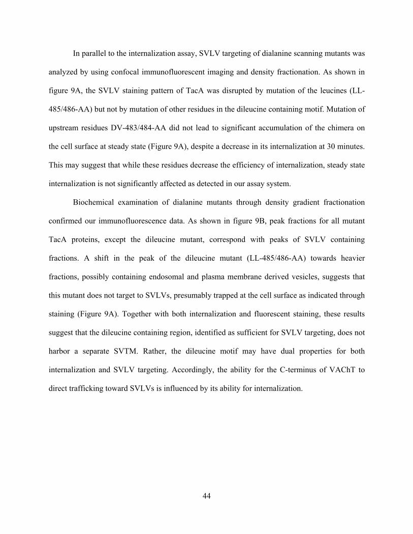
In parallel to the internalization assay, SVLV targeting of dialanine scanning mutants was
analyzed by using confocal immunofluorescent imaging and density fractionation. As shown in
figure 9A, the SVLV staining pattern of TacA was disrupted by mutation of the leucines (LL-
485/486-AA) but not by mutation of other residues in the dileucine containing motif. Mutation of
upstream residues DV-483/484-AA did not lead to significant accumulation of the chimera on
the cell surface at steady state (Figure 9A), despite a decrease in its internalization at 30 minutes.
This may suggest that while these residues decrease the efficiency of internalization, steady state
internalization is not significantly affected as detected in our assay system.
Biochemical examination of dialanine mutants through density gradient fractionation
confirmed our immunofluorescence data. As shown in figure 9B, peak fractions for all mutant
TacA proteins, except the dileucine mutant, correspond with peaks of SVLV containing
fractions. A shift in the peak of the dileucine mutant (LL-485/486-AA) towards heavier
fractions, possibly containing endosomal and plasma membrane derived vesicles, suggests that
this mutant does not target to SVLVs, presumably trapped at the cell surface as indicated through
staining (Figure 9A). Together with both internalization and fluorescent staining, these results
suggest that the dileucine containing region, identified as sufficient for SVLV targeting, does not
harbor a separate SVTM. Rather, the dileucine motif may have dual properties for both
internalization and SVLV targeting. Accordingly, the ability for the C-terminus of VAChT to
direct trafficking toward SVLVs is influenced by its ability for internalization.
44
Figure 9 Dileucine containing motif of VAChT C-terminus serves as a SVTM.
A) SVLV targeting analysis of dialanine mutants as detected through immunofluorescent staining. Overexpressed
TacA490 mutants in differentiated PC12 cells were visualized (red) and compared to staining patterns of
synaptophysin (green). Only TacA490 LL-485/486-AA showed disrupted SVLV targeting, localizing preferentially
to the cell surface. Arrows indicate the tips of the processes for colocalization of TacA variants and synaptophysin.
Bar= 20µm. B) SVLV targeting analysis of dialanine mutants through density gradient analysis. PNS of transiently
transfected PC12 cells were fractionated with sucrose gradients. Western blots for TacA490 mutant proteins were
semiquantified and calculated as a fraction of total signal. Grey shading marks SVLV corresponding fractions.
45
2.3.5 Specificity of the dileucine containing motif for SVLV targeting
Our dialanine scanning analysis demonstrated that the SVLV targeting of TacA is limited only
by mutation of the residues that interfered with internalization (leucine residues), indicating that
the motif’s role in SVLV targeting is not easily distinguishable from its previously defined role
in internalization. We therefore sought to address the specificity of the VAChT dileucine
containing motif as a SVTM. In order to address this, we examined additional Tac chimeras of
several membrane proteins that contain other cytosolic internalization motifs. First, a chimera
between Tac and the N-terminus of transferrin receptor (TacTfR), which contains a tyrosine
internalization motif, was analyzed for its subcellular distribution. TacTfR was internalized from
the plasma membrane but did not traffic to SVLVs as analyzed by density gradient (data not
shown), supporting that the specificity of the VAChT dileucine containing motif as a SVTM
extends beyond internalization. However, dialanine scanning mutagenesis of the VAChT
dileucine containing motif suggested that none of the specific residues within this motif, other
than the leucine residues, are required for the chimera’s SV targeting (Figure 9). Therefore, it
was unclear to what extent the exact motif in VAChT was specific as a SVTM and to what extent
the dileucine motif could be generalized as SVTM. Thus, we tested the potential SV targeting of
another classical dileucine motif containing protein, GLUT4. In PC12 cells GLUT4, regulated in
part through a C-terminal dileucine internalization motif, R(XXX)LL, traffics to a unique vesicle
population (Cope et al., 2000; Garippa et al., 1996; Herman et al., 1994; Thoidis and Kandror,
2001; Verhey et al., 1993). Examination of the steady state localization of TacGLUT revealed
that the chimera, similarly to the full length protein, segregated to fractions slightly heavier than
synaptophysin containing fractions in density gradients (Figure 10B). In order to ensure that
TacGLUT did not comigrate with SVLVs, velocity gradient fractionation was performed to
46
separate SVLV light fractions with neighboring fractions. Exclusion from SVLVs was
confirmed as TacGLUT was found only in the bottom fraction of the gradient (Figure 10C).
Although both GLUT4 and VAChT chimera contain similar dileucine motifs, [R(TPS)LL vs.
E(RDV)LL], their differential targeting suggests that the environment or nature of the residues of
the dileucine motif may influence the specificity of this motif for SVLV targeting. Finally, we
examined a closely related dileucine motif, E(XXX)[I/L]L, found in the similar environment of
the C-terminus of VMAT2. VMAT2 was previously shown to have preferential targeting to
LDCVs in PC12 cells, which is thought to be regulated by an acidic patch at the distal end of the
C-terminus (Waites et al., 2001). The presence of this additional signaling motif also allowed us
to examine how neighboring domain(s) influence the dileucine mediated targeting event. We
therefore generated a chimera, TacM, in which the C-terminus of VMAT2 was fused to Tac
protein (Figure 10A). In both transiently transfected and stable cell lines, as determined by
sucrose and velocity gradients, TacM was found to traffic preferentially to LDCV fractions, but
also to SVLV fractions (Figure 10D; velocity gradient not shown), suggesting the C-terminus of
VMAT2 is not sufficient for its preferential LDCV targeting. To determine if the distal sequence
of the acidic patch influences the function of C-terminus in targeting as indicated previously, we
next constructed TacMs in which the acidic patch was deleted (Figure 10A). In contrast to the
two peak distribution seen in the density gradient for TacM, the steady-state distribution of
TacMs showed a single peak that co-migrated with synaptophysin containing fractions (Figure
10E). The trafficking of TacMs to SVLVs strongly suggests that the VMAT2 dileucine motif,
when not influenced by neighboring signals, is also able to direct SVLV targeting, indicating that
this motif might be generalized as a SVTM for several SV proteins.
47
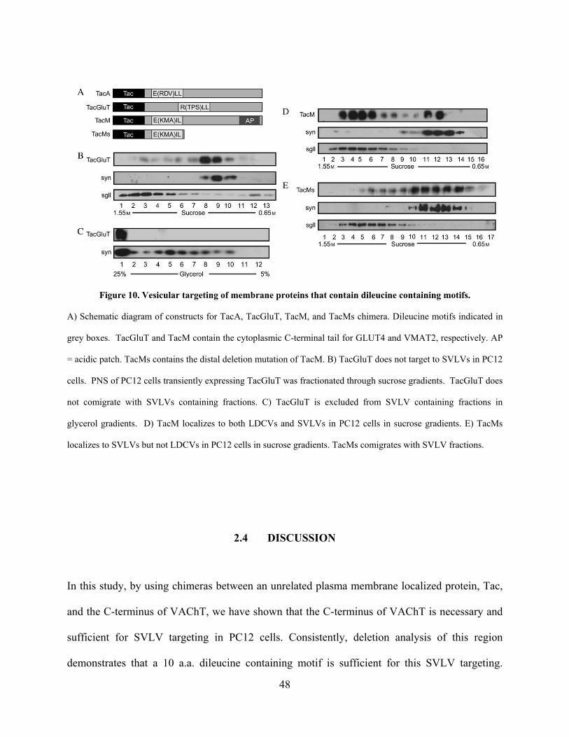
Figure 10. Vesicular targeting of membrane proteins that contain dileucine containing motifs.
A) Schematic diagram of constructs for TacA, TacGluT, TacM, and TacMs chimera. Dileucine motifs indicated in
grey boxes. TacGluT and TacM contain the cytoplasmic C-terminal tail for GLUT4 and VMAT2, respectively. AP
= acidic patch. TacMs contains the distal deletion mutation of TacM. B) TacGluT does not target to SVLVs in PC12
cells. PNS of PC12 cells transiently expressing TacGluT was fractionated through sucrose gradients. TacGluT does
not comigrate with SVLVs containing fractions. C) TacGluT is excluded from SVLV containing fractions in
glycerol gradients. D) TacM localizes to both LDCVs and SVLVs in PC12 cells in sucrose gradients. E) TacMs
localizes to SVLVs but not LDCVs in PC12 cells in sucrose gradients. TacMs comigrates with SVLV fractions.
2.4 DISCUSSION
In this study, by using chimeras between an unrelated plasma membrane localized protein, Tac,
and the C-terminus of VAChT, we have shown that the C-terminus of VAChT is necessary and
sufficient for SVLV targeting in PC12 cells. Consistently, deletion analysis of this region
demonstrates that a 10 a.a. dileucine containing motif is sufficient for this SVLV targeting.
48
Further examination of this motif has suggested that it contains dual signals indistinguishable for
internalization and synaptic vesicle targeting. Through the use of additional control chimeras,
we have shown that this signal is specific as a synaptic vesicle targeting motif (SVTM) and that
it may be generalized for multiple SV proteins.
Our approach to investigate the SVLV trafficking signals of VAChT through the use of
Tac chimeric proteins allowed us to determine the sufficiency of potential targeting signals
within VAChT. This system permitted independent analysis of the ability of VAChT C-terminal
cytosolic regions to traffic a non-related protein, Tac, to SVLVs. Previous studies of VAChT
trafficking have used mutagenesis of full length VAChT or chimera between VAChT and
VMAT2. Due to the high sequence similarity between these two transporters [~65% among
transmembrane regions (Roghani et al., 1994)], studies of this nature cannot exclude the potential
role of the transmembrane domains or the luminal loop of the transporters in vesicular targeting.
In addition to determining sufficiency, the Tac chimeric approach also allowed us to focus on
internalization and sorting steps from the plasma membrane, which mimic SV recycling and
biogenesis at neuronal terminals. Consistent with VNT chimeric studies (Tan et al., 1998), our
results using Tac chimera in PC12 cells imply that neither the N-terminal domain nor the
transmembrane domain regions of VAChT are required for SVLV trafficking at the terminal.
However, our data does not address whether these regions are important in axonal targeting, an
important additional sorting step for SV proteins in neurons (Prado and Prado, 2002). Our
preliminary studies using TacA chimera in primary neurons show that the steady state
distribution of the protein is not restricted to axons but extends to all neuronal processes (data not
shown). This may suggest that in neurons the N-terminus or transmembrane regions of VAChT
49
may be required for the proper axonal targeting of the protein while the C-terminus may be
sufficient to direct SV recycling at the terminal.
Our Tac chimeric analysis also first shows that the dileucine containing sequence
(residues 479-488) within the VAChT C-terminus is sufficient as a SVTM in PC12 cells.
Consistent with this, a similar region (residues 481 – 490) was previously indicated to be
required in the targeting of full length VAChT in SN56 cells (Ferreira et al., 2005). On the other
hand, our chimeric study was unable to confirm another report that a non-classical tyrosine motif
(Y524-Y527) at the distal end of the C-terminus of VAChT serves as an alternative
internalization motif or a SVLV trafficking role of a classical tyrosine motif (Kim and Hersh,
2004). Our systematic analysis of deletion mutants of these two tyrosine motifs failed to show
any significant decrease in their internalization at 30 minutes or any alteration of their steady-
state SVLV trafficking (Figures 6, 8). It is plausible to suggest that these previously identified
motifs, while not required for SVLV targeting in our system, may play a role in regulating the
efficiency of pathways of VAChT targeting. More sensitive approaches may help to address how
they influence the dominant dileucine containing motif.
Further examination of the signaling properties of the dileucine motif demonstrated that
this signal is specific as a SVTM. In agreement with our dialanine scanning results and analysis
of additional Tac chimera, the specific residues within the VAChT dileucine containing motif are
not essential for its SVLV targeting. However, TacGLUT containing a similar dileucine motif
did not traffic to SVLVs suggesting that the overall environment of the motif, as well as factors
such as proximity of the motif to the transmembrane domain, or accessibility of cytosolic
machinery to the motif may determine the specificity of the dileucine containing motif as a
SVTM. Furthermore, our data suggests that the dileucine containing motif can be influenced by
50
adjacent regions of the protein, such as the acidic patch (AP) in VMAT2. One possible
explanation is that this additional signaling motif may mask the SVTM and act as a dominant
earlier sorting signal at the TGN. The other possibility is that this motif may promote the Golgi-
endosomal trafficking during retrieval of VMAT2 and thus enhance its overall targeting to
secretory granules at TGN. When the acidic patch is deleted, the closely related dileucine motif
of VMAT2 was able to direct targeting to SVLVs, suggesting that this signal may be generalized
to several SV proteins. Interestingly, the notion that the VMAT2 C-terminus may contain signals
for targeting to both LDCVs and SVLVs suggests some regulation of secretory vesicle protein
trafficking between the two types of vesicles in neurons.
The potential for this type of dileucine motif to be a more general SVLV signal is further
supported by several previous findings. In synaptotagmin 1, the cytoplasmic motif, E(VDA)ML,
accounts for at least a portion of its SVLV trafficking in PC12 cells (Blagoveshchenskaya et al.,
1999). Furthermore, the dileucine motif of Tyrosinase, E(KQP)LL is responsible for its targeting
to SVLVs when exogenously expressed in PC12 cells (Blagoveshchenskaya et al., 1999).
Interestingly, dileucine-like motifs have been identified in several other vesicular transport
proteins such as VGLUT1 (EKCGFV) (Voglmaier et al., 2006). This recent report has suggested
that this dileucine-like motif is critical for the internalization and trafficking of the transporter.
This sequence is upstream of a polyproline motif that is responsible for interaction of VGLUT1
with endophilin 1, which may regulate this dileucine-like dependent trafficking (De Gois et al.,
2006; Voglmaier et al., 2006).
The mechanisms of the dileucine mediated trafficking of synaptotagmin and tyrosinase
are mediated by a fairly well characterized mechanism for SV biogenesis in PC12 cells (Faundez
et al., 1998), Arf GTPase-AP3 directed endosomal budding (Blagoveshchenskaya et al., 1999).
51
However, AP-3 binding has not been able to be detected for VAChT (Kim and Hersh, 2004).
Thus, VAChT targeting may be regulated through alternative cytosolic machinery. Accordingly,
we have identified a novel functional interaction between the C-terminus of VAChT and SNX5,
a lipid binding protein involved in the endosomal trafficking of membrane proteins (Colgan, et
al., and manuscript in preparation. See Chapter 3). This suggests that VAChT may also traffic
through an endosomal intermediate. The dual properties of the dileucine containing motif may
regulate trafficking through an endosomal intermediate by serving first as an internalization
signal at the plasma membrane and subsequently acting as a SVTM at an endosomal
compartment. This sequential targeting model may rely on the chronological function of multiple
regulatory sorting machineries or may take advantage of similar, but subcellularly
compartmentalized machinery that govern different targeting events. Similar mechanisms are
thought to occur in non-neuronal cell lines, where this type of dileucine motif can be recognized
both at the plasma membrane as well as endosomal and lysosomal compartments (Bonifacino
and Traub, 2003). Alternatively, the dileucine motif signal may mediate direct and selective
internalization from the plasma membrane which has been suggested as a major pathway for
recycling SVs in the nerve terminal (De Camilli and Takei, 1996). Nonetheless, the dual role of
the dileucine-containing motif in internalization and SV targeting provides a molecular
mechanism underlying the efficient recruitment of essential SV components during its biogenesis
and recycling.
52
2.5 MATERIALS AND METHODS
2.5.1 Chemicals and antibodies
General chemicals used in this report were purchased from Sigma (St. Louis, MO) unless
otherwise noted. The following antibodies were used in immunofluorescent staining: biotin
conjugated mouse anti-CD25 (Affinity BioReagents, Golden, CO), polyclonal synaptophysin and
secretogranin II (SYSY, Goettingen, Germany), secondary Cy3 conjugated goat anti-mouse and
Alexa 488 conjugated goat anti-rabbit (Jackson Immunoresearch Lab, West Grove, PA). The
following antibodies were used for Western blot: polyclonal IL-2R alpha (Santa Cruz Biotech.,
Santa Cruz, CA), monoclonal anti-IL2 Receptor (Covance, Princeton, NJ), polyclonal anti-
synaptophysin and anti-secretogranin II (SYSY, Goettingen, Germany), secondary HRP
conjugated goat anti-mouse and goat anti-rabbit (Pierce, Rockford, IL). Antibodies used for
ELISA internalization assay are as follows: biotin conjugated mouse anti-CD25 (Affinity
BioReagents, Golden, CO), polyclonal goat anti-mouse IgG (BD Pharmingen, San Diego, CA).
2.5.2 Plasmid construction and mutagenesis
Chimeric protein TacA was generated by PCR amplification of the C-terminus of VAChT (a.a.
465-530) with the introduction of Xba1 and Xho1 restriction sites flanking the region. PCR
product was digested and subcloned into Tac/pcDNA 3.1 as described (Tan et al., 1998). C-
terminus deletion mutant chimeras were constructed similarly by PCR amplification introducing
an Xba1 site at the 5’ region and a stop codon and Xho1 site at the 3’ region. Digestion was
followed by subcloning into Tac/pcDNA3.1 using Xba1 and Xho1. 8ATacDLM was constructed
53
using an extended 3’ primer encoding and Xba1 site followed by 8 alanines and consensus motif
for a.a. 479-483. PCR amplification introducing a stop codon and a Not1 site downstream of the
dileucine region was followed by digestion and subcloning into Tac/pcDNA3.1 using Xba1 and
Not1 restriction enzymes. Dialanine scanning point mutations were generated by using a
QuickChange site-directed mutagenesis kit (Stratagene, La Jolla, CA) as directed using
TacA490/pcDNA3.1 as a backbone. TacGLUT was generated by PCR with oligos that isolated
the C-terminus of human GLUT4 and introduced flanking Xba1 and Not1 restriction sites. PCR
product was digested and subcloned into Tac/pcDNA 3.1 using Xba1 and Not1. TacM constructs
were generated similarly to TacA with the exception that the C-terminus of VMAT2 was
amplified by PCR and used for subcloning. TacMs was generated by introducing a stop codon
after the dileucine containing region by QuickChange site directed mutagenesis using
TacM/pcDNA 3.1 as a backbone. All constructs were confirmed by sequencing.
2.5.3 Cell culture and transfection
All cells were maintained in 5% CO2 at 37ºC in medium containing penicillin and streptomycin
unless otherwise noted. PC12 cells were maintained in DMEM (Invitrogen) with 10% Equine
serum (Hyclone, Logan, UT), 5% Cosmic Calf serum (Hyclone, Logan, UT), and 2 mM L-
Glutamine (Invitrogen). For immunofluorescent analysis cells were seeded on poly-D-lysine and
Matrigel coated glass coverslips 24 hours before transfection. Cells on coverslips were then
transfected using either LipofectAMINE 2000 (Invitrogen, San Diego, CA), or Superfect
(Qaigen, Hilden, Germany) reagent according to the manufacturer’s instructions. Twelve hours
later cells were differentiated using 100 nM NGF for 24 hours before fixation with 4%
54
Paraformaldehyde. For biochemical assays, transfections were done using LipofectAMINE 2000.
Transfected cells were incubated at 37ºC for 36 - 48 hours before harvest.
Stable lines of wild type Tac, TacA, TacA490 dialanine scanning mutants, and TacM
were generated by further selection with G418 (500mg/ml). Positive transformants were
screened by immunofluorescence staining and western blot analysis. At least three stable lines
were tested for consistent subcellular localization of the heterogeneously expressed membrane
proteins. All stable cell line derived results were also confirmed in transiently expressing
independent PC12 cell lines.
2.5.4 Immunofluorescence and confocal microscopy
Immunofluorescent staining was performed as previously described (Liu et al., 2006). In brief,
cells seeded on glass coverslips were fixed with 4% paraformaldehyde in PBS, pH 7.4. After
fixation, cells were permeabilized and blocked for 30 min. in blocking buffer (BB, 2% BSA, 1%
fish skin gelatin, and 0.02% saponin in PBS). Cells were then incubated with primary antibody in
BB for 1 hour at room temperature. Coverslips were washed and incubated with the appropriate
Alexa- or Cy3- conjugated secondary antibody for 1 hour at room temperature. Confocal images
were acquired with a Fluoview 500 laser scanning confocal imaging system (Olympus, Tokyo,
Japan) configured with a fluorescence microscope fitted with Pan Apo 60 and 100 oil
objectives (Olympus). Confocal images were collected sequentially at 10241024 resolution to
minimize bleed through of fluorescence between channels.
55
2.5.5 Fractionation analysis
PC12 cells stably expressing, or transiently transfected with plasmid constructs, were harvested
in Buffer A (150 mM NaCl, 10 mM Hepes pH 7.4, 1 mM EGTA, 0.1mM MgCl2) with protease
inhibitors. Cells were cracked by eight passes through a cell cracker (Clearance-0.02um). Post-
nuclear supernatants were then loaded onto prepared density or velocity gradients and spun in a
Beckman SW41 rotor. For sucrose density fractionation, sucrose gradients were prepared using a
gradient mixer to form continuous gradients with sucrose concentrations from 0.65 M to 1.55 M
and spun in SW41 rotor at 30,000 rpm for 8 hours at 4C. (Beckman Instruments, Palo Alto,
CA). Glycerol velocity fractionation was done through a glycerol gradient of 5% to 25% glycerol
(V/V) and spun in SW41 rotor at 37,000 rpm for 1 hour at 4C. Fractions from density gradients
were collected from the bottom and velocity gradients collected from the top to minimize
contamination. All gradients were numbered from heavy to light.
2.5.6 Western blot analysis
Proteins were detected in gradient fractions by immunoblotting as previously described (Chen et
al., 2005). Equal amounts of gradient from each fraction were denatured in 3x SDS sample
buffer (New England Biolab, Beverly, MA), and separated by electrophoresis through 10% SDS-
PAGE. After electrophoresis, proteins were transferred to nitrocellulose (BA-85, Schleicher-
Schuell Bioscience, Keene, NH) and TacA chimeric proteins, synaptophysin, secretogranin II, or
other proteins were visualized by immunoblotting with appropriate antibodies in combination
with enhanced chemiluminescence (Super-Signal West Pico, Pierce, Rockford, IL). Protein
56
immunoreactive signals were scanned and the intensity of bands was semi-quantified using the
NIH Imaging program.
2.5.7 Internalization assay
Internalization assay was done as previously described (Tan et al., 1998). Briefly, PC12 cells
transfected by electroporation with cDNA plasmids were harvested 36 hours later and
resuspended with Serum Free Medium (DMEM, 20mM HEPES-KOH (pH7.2), 0.2% BSA). The
cell suspension was incubated with biotinylated anti-CD-25 (Tac) monoclonal antibody at 4˚C
for 2 hr. to label Tac chimeric proteins localized to the cell surface. After washing of unbound
antibody, internalization was allowed for 30min by 37C incubation. Internalization was stopped
and the sample equally divided. In one fraction, cell surface labeled proteins were quenched
through avidin incubation. Cells in both fractions were then solubilized and loaded in triplicate
aliquots onto 96 well ELISA plate pre-coated with goat anti-mouse IgG and incubated overnight
at 4˚C. Plates were washed of unbound biotin signal and developed using 3, 3’, 5, 5’
Tetramethylbenzidine (TMB) liquid substrate system for 30 minutes. Endpoint absorbance
measurements were taken at 450nm using a BioRAD plate reader. Percent internalized was
calculated as follows: (Ca-Va)/ (Ct-Vt) where Ca = internalized (avidin quenched) transfected
cells; Va = internalized (avidin quenched) vector transfected cells; Ct = total (unquenched)
transfected cells; Vt = total (unquenched) vector transfected cells. Standard curves were run with
each experiment to ensure inclusion within the linear range of detection. Mutants were
normalized to control TacA490 constructs to determined percent change in internalization and to
normalize across experiments.
57
3.0 SORTING NEXIN 5 REGULATES THE SYNAPTIC VESICLE SPECIFIC
TARGETING OF VESICULAR ACETYLCHOLINE TRANSPORTER
3.1 ABSRACT
Packaging of neurotransmitter into small synaptic vesicles or large dense core vesicles is
determined by vesicle-specific targeting of vesicular neurotransmitter transporters (VNTs).
Their regulated targeting is mediated through interaction of sorting motifs with cytosolic
machinery, however, machinery that regulates the vesicle specific trafficking of VNTs remains
to be identified. Here we identify that sorting nexin 5 regulates the specific trafficking of
vesicular acetylcholine transporter, responsible for packaging acetylcholine into synaptic
vesicles. Disruption of sorting nexin 5 function leads to mislocalization of vesicular
acetylcholine transporter to large dense core vesicles, suggesting a potential regulatory
mechanism for membrane trafficking between secretory vesicles.
58
3.2 RESULTS
Information transfer in the nervous system is mediated through the regulated release of
neurotransmitter (NT) from vesicular stores including clear synaptic vesicles (SVs) and large
dense-core vesicles (LDCVs). The distinct release properties of these secretory vesicles,
including calcium sensitivity, speed, and synchrony, determine characteristics of transmission
(Edwards, 1998). Specific packaging of NT into either SVs or LDCVs is determined by the
presence of vesicular neurotransmitter transporters (VNTs), whose localization to these secretory
vesicles is highly regulated. Therefore, in order to understand the determinants of vesicle-
specific NT packaging, we sought to identify machinery that regulates the trafficking of VNTs.
Vesicular Acetylcholine Transporter (VAChT) has been shown to localize preferentially
to SVs, which determines the fast, synchronous release of Acetylcholine (Ach) in both neurons
and neuroendocrine cell lines. We have previously shown that a synaptic vesicle targeting motif
in the C-terminal tail of VAChT is sufficient for this SV-specific targeting in PC12 cells, a
neuroendocrine cell line (Colgan et al., 2007). Therefore, in order to identify cytosolic
machinery that regulates this trafficking, we isolated the VAChT C-terminus in a fusion protein
with an unrelated plasma membrane protein, Tac. The chimera, TacA, efficiently traffics to SVs
(Colgan et al., 2007). Therefore, we used the SV-specific trafficking of TacA as a tool to
characterize interacting proteins that regulate SV trafficking.
3.2.1 Sorting Nexin 5 associates with VAChT
Using the C-terminus of VAChT as bait in a yeast-two-hybrid screen, we identified sorting nexin
5 (SNX5) as a potential interacting protein that could regulate trafficking to synaptic vesicles.
59
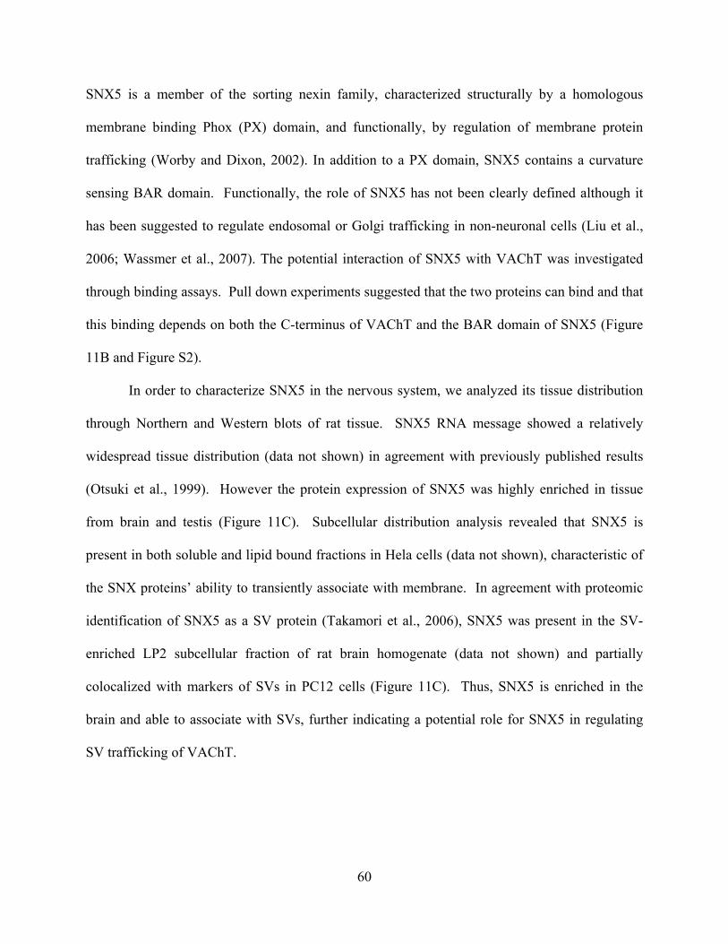
SNX5 is a member of the sorting nexin family, characterized structurally by a homologous
membrane binding Phox (PX) domain, and functionally, by regulation of membrane protein
trafficking (Worby and Dixon, 2002). In addition to a PX domain, SNX5 contains a curvature
sensing BAR domain. Functionally, the role of SNX5 has not been clearly defined although it
has been suggested to regulate endosomal or Golgi trafficking in non-neuronal cells (Liu et al.,
2006; Wassmer et al., 2007). The potential interaction of SNX5 with VAChT was investigated
through binding assays. Pull down experiments suggested that the two proteins can bind and that
this binding depends on both the C-terminus of VAChT and the BAR domain of SNX5 (Figure
11B and Figure S2).
In order to characterize SNX5 in the nervous system, we analyzed its tissue distribution
through Northern and Western blots of rat tissue. SNX5 RNA message showed a relatively
widespread tissue distribution (data not shown) in agreement with previously published results
(Otsuki et al., 1999). However the protein expression of SNX5 was highly enriched in tissue
from brain and testis (Figure 11C). Subcellular distribution analysis revealed that SNX5 is
present in both soluble and lipid bound fractions in Hela cells (data not shown), characteristic of
the SNX proteins’ ability to transiently associate with membrane. In agreement with proteomic
identification of SNX5 as a SV protein (Takamori et al., 2006), SNX5 was present in the SV-
enriched LP2 subcellular fraction of rat brain homogenate (data not shown) and partially
colocalized with markers of SVs in PC12 cells (Figure 11C). Thus, SNX5 is enriched in the
brain and able to associate with SVs, further indicating a potential role for SNX5 in regulating
SV trafficking of VAChT.
60
Figure 11. SNX5 associates with VAChT
A) C-Terminus of VAChT (black) was isolated and fused to Tac protein (TacA). SNX5 Phox domain (PX) and
SNX5 BAR domain (CC/CC) are indicated. B) HEK293 cell lysates, transiently transfected with HA-VAChT, HA-
SNX5, HA-SNX5 PX, or HA-SNX5BAR were incubated with glutathione-sepharose immobilized GST-SNX5 or
GST-VAChT C-terminus as indicated. Bound protein was eluted and detected by Western blot. C) Tissue
distribution of SNX5 protein in rat homogenate. SNX5 was detected through Western blot by specific antibody. D)
Immunofluorescence imaging of endogenous SNX5 (red) in differentiated PC12 cells. Markers of synaptic vesicles
including synaptophysin, TacA, and VAChT (green) were also detected.
61
3.2.2 SNX5 regulates SV Trafficking
Analysis of the potential function of SNX5 in regulating the SV specific trafficking of VAChT
was studied by altering SNX5 levels in PC12 cells stably expressing the chimeric protein TacA.
The expression level and localization of TacA were monitored to detect resulting changes in SV
protein trafficking. SNX5 levels were altered through three methods, overexpression of SNX5,
overexpression of dominant-negative truncated SNX5 constructs (SNX5BAR and SNX5PX),
and siRNA mediated knockdown of endogenous SNX5. Overexpression of SNX5 did not
significantly alter the expression level or localization of TacA. This may reflect the efficiency of
the interaction between SNX5 and the VAChT C-terminus. Furthermore, it suggests that
overexpression of SNX5 does not grossly disrupt cellular trafficking pathways. Overexpression
of SNX5 mutants or knockdown of endogenous SNX5, however, led to a significant and
dramatic (~40%) loss in SV targeting of TacA (Figure 12A, 12B) without an alteration of protein
level. This effect, underrepresented in the data due to the ~ 60% maximum transfection
efficiency in our system, suggests that SNX5 regulates trafficking of VAChT to SVs. The
regulation of this SV trafficking appears to have some specificity, as the trafficking of another
SV localized protein, synaptophysin (p38), was not altered (Figure 12A, B).
62
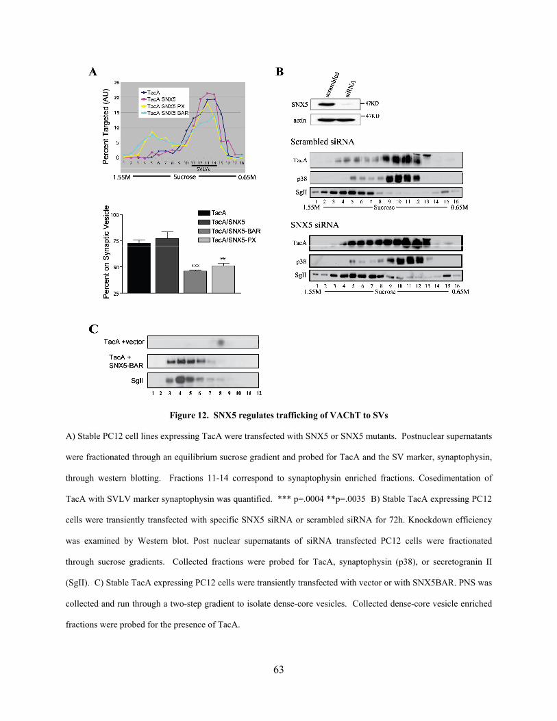
Figure 12. SNX5 regulates trafficking of VAChT to SVs
A) Stable PC12 cell lines expressing TacA were transfected with SNX5 or SNX5 mutants. Postnuclear supernatants
were fractionated through an equilibrium sucrose gradient and probed for TacA and the SV marker, synaptophysin,
through western blotting. Fractions 11-14 correspond to synaptophysin enriched fractions. Cosedimentation of
TacA with SVLV marker synaptophysin was quantified. *** p=.0004 **p=.0035 B) Stable TacA expressing PC12
cells were transiently transfected with specific SNX5 siRNA or scrambled siRNA for 72h. Knockdown efficiency
was examined by Western blot. Post nuclear supernatants of siRNA transfected PC12 cells were fractionated
through sucrose gradients. Collected fractions were probed for TacA, synaptophysin (p38), or secretogranin II
(SgII). C) Stable TacA expressing PC12 cells were transiently transfected with vector or with SNX5BAR. PNS was
collected and run through a two-step gradient to isolate dense-core vesicles. Collected dense-core vesicle enriched
fractions were probed for the presence of TacA.
63
As disruption of SNX5 decreased the SV localization of TacA without altering its
expression level, we next determined the localization of the mistargeted protein. Using density
gradient subcellular fractionation, we showed that TacA was shifted to fractions containing
heavier organelles including LDCVs, endosomes, plasma membrane, or Golgi (Figure 12A, B).
Using markers for these organelles, SGII, TfR, Na/K ATPase and TGN38 respectively, our data
suggested that disruption of SNX5 lead to an accumulation of TacA on LDCVs (Figure 12B,
Figure S2). However, density gradient fractionation alone is not sufficient to separate these
heavier organelles from one another. In order to determine if SNX5 disruption led to an
accumulation on LDCVs, we isolated LDCVs using a two-step gradient and examined the
presence of TacA before and after disruption of SNX5 function (Tooze and Stinchcombe, 1992).
Overexpression of SNX5 mutants led to a dramatic increase of TacA on LDCVs (Figure 12C)
without altering its overall expression level. Thus, disruption of SNX5 function altered targeting
of TacA from SVs to LDCVs.
The results of this work identify SNX5 as a novel regulator of the SV specific trafficking
of VAChT. Further study is required to determine the location and mechanism of SNX5
regulation of VAChT trafficking, as well as the physiological relevance of this regulation in the
nervous system. However, the identification of machinery for the vesicle specific trafficking of
VAChT suggests that regulation of VNT trafficking may serve as a potential mechanism in
defining the packaging of NT and thus, the properties of synaptic transmission.
64
4.0 COUPLING OF VESICULAR TRANSPORT AND SOMATIC RELEASE IN
SEROTONIN NEURONS
4.1 ABSTRACT
Although neurotransmission relies on the packaging of transmitters into vesicles, it has not been
possible to directly monitor the function of vesicular transporters as they support release in living
neurons. Here, a pH-sensitive, fluorescent serotonin analog is visualized with two-photon
microscopy to study vesicular monoamine transporter (VMAT) activity during somatic release in
dorsal raphe nucleus serotonin neurons. Following uptake by the serotonin transporter and
packaging by VMAT, glutamate receptor activation evoked somatic vesicular release of the
fluorescent monoamine. Release was accompanied by VMAT activity, which redistributed
monoamine from the nucleus, a compartment not previously implicated in neurotransmission.
Measurements of vesicular transport and release allowed for the contribution of activity
dependent packaging to somatic release to be assessed. While some monoamine packaged at rest
was held in reserve, monoamine packaged during stimulation was released efficiently,
suggesting a coupling between activity-dependent vesicular transport and somatic release.
65
4.2 INTRODUCTION
Neurotransmission requires vesicular transporters (VNTs), which package neurotransmitters into
vesicles, and the exocytosis/endocytosis vesicle cycle, which supports release of packaged
content. Although the vesicle cycle can be assayed in single neurons, it has not been possible to
dynamically monitor packaging. For example, pH-sensitive, fluorescent secretory vesicle
proteins can be used to track synaptic vesicle exocytosis and endocytosis based on the luminal
acidic pH of synaptic vesicles (Burrone et al., 2006). However, this approach provides insight
into vesicle cycling without detecting vesicle content (i.e., signals are identical for filled and
empty vesicles). This limitation precludes detection of vesicular transporter activity, and likely
accounts for reported mismatches between measurements of vesicle cycling and transmitter
release (Ertunc et al., 2007; Tabares et al., 2007). Potentially, vesicular transport could be
assayed by imaging transmitter dynamics directly. In fact, serotonin (5-hydroxytryptamine, 5-
HT) is detected with three-photon microscopy, but vesicular content has only been detected in
large granules and vesicle clusters. Thus far, transmitter in disperse, small vesicles and transport
of 5-HT from the cytoplasm into vesicles has not been observed with this approach (Kaushalya
et al., 2008a; Kaushalya et al., 2008b; Maiti et al., 1997). Thus, current methods cannot directly
monitor vesicular transporter activity during stimulated release.
The complementary advantages of imaging pH-sensitive vesicle markers and vesicle
content suggest a new possibility: imaging a fluorescent, pH-sensitive neurotransmitter analog.
Such an analog would have to be recognized by native transporters and released in response to
activity. Furthermore, in contrast to serotonin4, the signal from the analog would change when
transported from the neutral cytoplasm into acidic vesicles and allow for quantification of
vesicular content even when individual vesicles cannot be resolved. A possible candidate for
66
this novel approach is the serotonin analog 5,7-dihydroxytryptamine (dHT). This monoamine
absorbs near-UV light in a pH-dependent manner and emits visible light (Schlossberger, 1978;
Vaney, 1986). It is a substrate of the serotonin transporter (SERT) and thus accumulates in
serotonin neurons (Bjorklund et al., 1974). Furthermore, within hours of injection into animals,
dHT is detected in serotonin neuron vesicles, raising the possibility that it is a substrate of the
vesicular monoamine transporter (VMAT) (Gershon and Sherman, 1982). Prolonged exposure
to dHT is toxic, but this effect is inhibited by preventing its chemical and enzymatic oxidation
(Bjorklund et al., 1975; Silva et al., 1988). Indeed, acute dHT uptake identifies viable
monoaminergic neurons without altering neuronal morphology or electrical properties (Hahn et
al., 2006; Hahn et al., 2003; Silva et al., 1988). Therefore, imaging dHT might provide an
approach to study vesicular transport during release in serotonin neurons.
Serotonin neurons located in the dorsal raphe nucleus (DR) project widely throughout the
brain to control mood and behavior. Reduction of serotonin release, which can occur as a result
of decreased VMAT activity, is linked to psychiatric disorders, while increasing serotonin levels
with SSRIs (selective serotonin reuptake inhibitors) relieves depression (Fukui et al., 2007). In
addition to release in projection areas, vesicular serotonin release occurs in the DR from the cell
body and dendrites (de Kock et al., 2006) and can regulate the activity of serotonin release from
terminals. This regulation is mediated by activation of inhibitory 5HT1A autoreceptors that
decrease neuronal firing rate, and is relevant to the delay in therapeutic efficacy of SSRI
antidepressant drugs (Blier et al., 1998). Despite the importance of local serotonin release in the
DR, little is known about the mechanisms and regulation of this somatic vesicular release.
Here, a new optical approach, two-photon imaging of a pH-sensitive, serotonin analog is
developed to study vesicular transport and release in serotonin neurons of the DR. First, the
67
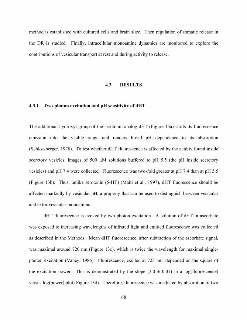
method is established with cultured cells and brain slice. Then regulation of somatic release in
the DR is studied. Finally, intracellular monoamine dynamics are monitored to explore the
contributions of vesicular transport at rest and during activity to release.
4.3 RESULTS
4.3.1 Two-photon excitation and pH sensitivity of dHT
The additional hydroxyl group of the serotonin analog dHT (Figure 13a) shifts its fluorescence
emission into the visible range and renders broad pH dependence to its absorption
(Schlossberger, 1978). To test whether dHT fluorescence is affected by the acidity found inside
secretory vesicles, images of 500 µM solutions buffered to pH 5.5 (the pH inside secretory
vesicles) and pH 7.4 were collected. Fluorescence was two-fold greater at pH 7.4 than at pH 5.5
(Figure 13b). Thus, unlike serotonin (5-HT) (Maiti et al., 1997), dHT fluorescence should be
affected markedly by vesicular pH, a property that can be used to distinguish between vesicular
and extra-vesicular monoamine.
dHT fluorescence is evoked by two-photon excitation. A solution of dHT in ascorbate
was exposed to increasing wavelengths of infrared light and emitted fluorescence was collected
as described in the Methods. Mean dHT fluorescence, after subtraction of the ascorbate signal,
was maximal around 720 nm (Figure 13c), which is twice the wavelength for maximal single-
photon excitation (Vaney, 1986). Fluorescence, excited at 725 nm, depended on the square of
the excitation power. This is demonstrated by the slope (2.0 ± 0.01) in a log(fluorescence)
versus log(power) plot (Figure 13d). Therefore, fluorescence was mediated by absorption of two
68
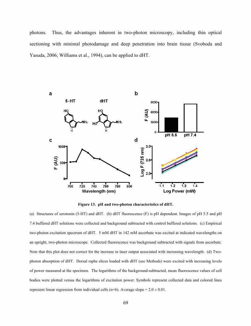
photons. Thus, the advantages inherent in two-photon microscopy, including thin optical
sectioning with minimal photodamage and deep penetration into brain tissue (Svoboda and
Yasuda, 2006; Williams et al., 1994), can be applied to dHT.
Figure 13. pH and two-photon characteristics of dHT.
(a) Structures of serotonin (5-HT) and dHT. (b) dHT fluorescence (F) is pH dependent. Images of pH 5.5 and pH
7.4 buffered dHT solutions were collected and background subtracted with control buffered solutions. (c) Empirical
two-photon excitation spectrum of dHT. 5 mM dHT in 142 mM ascorbate was excited at indicated wavelengths on
an upright, two-photon microscope. Collected fluorescence was background subtracted with signals from ascorbate.
Note that this plot does not correct for the increase in laser output associated with increasing wavelength. (d) Two-
photon absorption of dHT. Dorsal raphe slices loaded with dHT (see Methods) were excited with increasing levels
of power measured at the specimen. The logarithms of the background-subtracted, mean fluorescence values of cell
bodies were plotted versus the logarithms of excitation power. Symbols represent collected data and colored lines
represent linear regression from individual cells (n=6). Average slope = 2.0 ± 0.01.
69
4.3.2 Cellular uptake and vesicular packaging of dHT by SERT and VMAT
To demonstrate specific uptake of dHT, PC12 cells transfected with SERT and GFP were
incubated with 20 µM dHT. The antioxidant ascorbate and the monoamine oxidase inhibitor
pargyline were also present to inhibit oxidation of dHT. Cells expressing SERT, but not a
control vector, accumulated dHT within an hour (Figure 14). This accumulation was inhibited
by preincubation of cells with the specific SERT inhibitor fluoxetine (Figure 14). Thus, SERT-
mediated dHT uptake into cells was detected.
Figure 14. SERT-mediated dHT loading detected by two-photon microscopy.
Upper row: Two-photon dHT fluorescence images of representative undifferentiated PC12 cells transfected with
control (Con) or SERT DNA and incubated for 1 h with dHT or a combination of dHT and fluoxetine. Lower row:
Confocal single photon fluorescence of GFP was used to identify transfected PC12 cells. Scale bar = 10 µm.
70
Several experiments demonstrated that dHT is packaged by VMAT into secretory
vesicles. First, SERT-transfected, NGF-differentiated PC12 cells were incubated overnight in
dHT, washed for 1 hour and then imaged. In contrast to the distributed fluorescence pattern
produced with 1 hour incubation, this longer protocol resulted in fluorescence localized to
growth cones, the location of secretory vesicles in PC12 cells (Figure 15a). Second, application
of 1 µM monensin, a cation ionophore that neutralizes the acidic pH in PC12 cell secretory
vesicles (Han et al., 1999a), led to a two-fold increase in fluorescence in the growth cone (Figure
15b). The sensitivity to collapse of the vesicular pH gradient, along with the pH-sensitive
fluorescence of dHT (Figure 13b), is consistent with efficient packaging of dHT into secretory
vesicles. Third, incubation of SERT-transfected cells with 100 nM reserpine, a specific VMAT
inhibitor, before and during the dHT incubation prevented the preferential accumulation of
fluorescence in growth cones (Figure 15c, top). This was evident by the change in the ratio of
fluorescence in growth cones to cell bodies (FGC: FCB) induced by reserpine (Figure 15c,
bottom). Finally, induction of exocytosis by K+-induced depolarization for 10 minutes evoked a
50 ± 3% loss of dHT fluorescence. In PC12 cells, VMAT-containing secretory vesicles also
contain neuropeptides. Therefore, to confirm that this response is consistent with vesicular
release, PC12 cells that express a GFP-tagged neuropeptide (ANF-GFP) (Burke et al., 1997; Han
et al., 1999b) were transfected with SERT and loaded with dHT overnight. The fluorescence
signals from dHT and the secretory vesicle marker colocalized in growth cones (Figure 15d, top).
Furthermore, depolarization induced decreases in dHT and ANF-GFP fluorescence signals that
were similar in magnitude and kinetics (Figure 15d, bottom). Thus, dHT fluorescence loss
paralleled release of secretory vesicle content. Together, four independent criteria show that
VMAT packages dHT into acidic, release competent secretory vesicles.
71
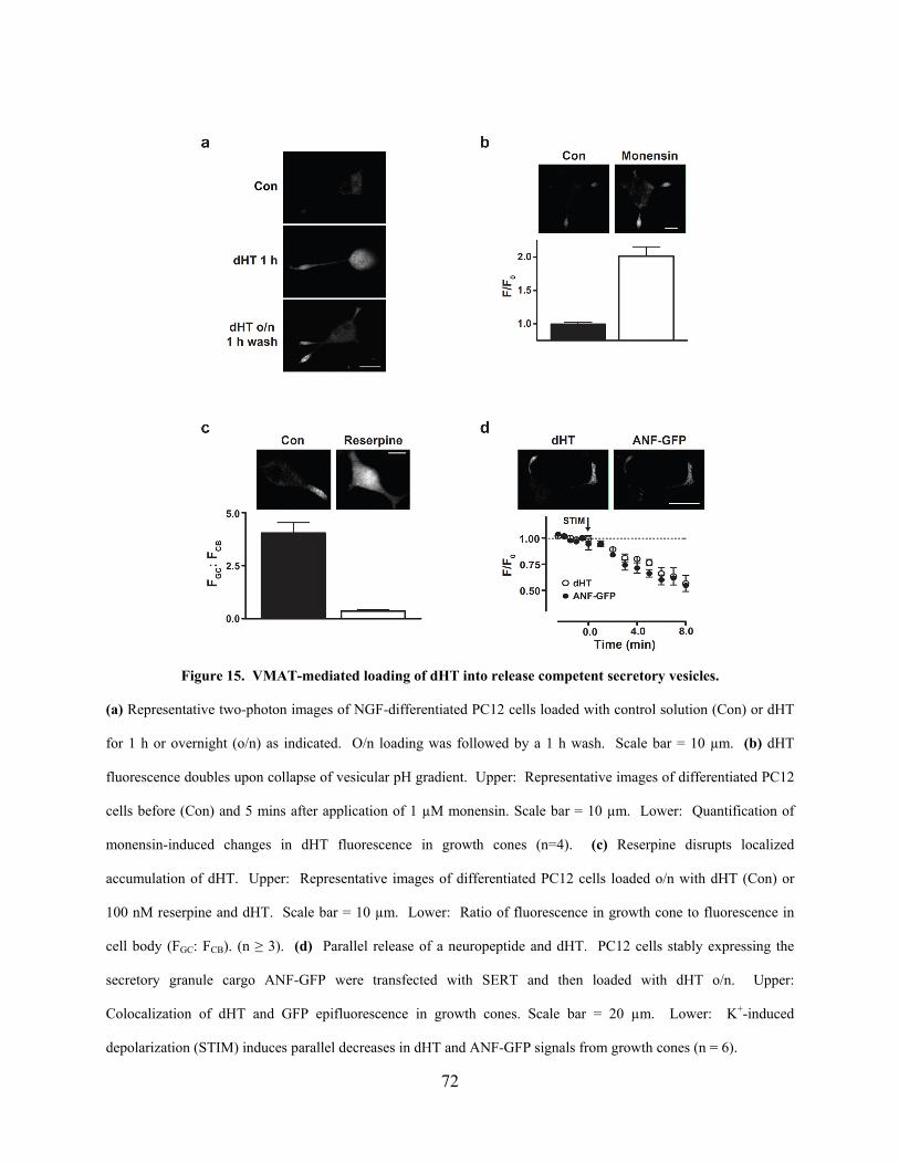
Figure 15. VMAT-mediated loading of dHT into release competent secretory vesicles.
(a) Representative two-photon images of NGF-differentiated PC12 cells loaded with control solution (Con) or dHT
for 1 h or overnight (o/n) as indicated. O/n loading was followed by a 1 h wash. Scale bar = 10 µm. (b) dHT
fluorescence doubles upon collapse of vesicular pH gradient. Upper: Representative images of differentiated PC12
cells before (Con) and 5 mins after application of 1 µM monensin. Scale bar = 10 µm. Lower: Quantification of
monensin-induced changes in dHT fluorescence in growth cones (n=4). (c) Reserpine disrupts localized
accumulation of dHT. Upper: Representative images of differentiated PC12 cells loaded o/n with dHT (Con) or
100 nM reserpine and dHT. Scale bar = 10 µm. Lower: Ratio of fluorescence in growth cone to fluorescence in
cell body (FGC: FCB). (n ≥ 3). (d) Parallel release of a neuropeptide and dHT. PC12 cells stably expressing the
secretory granule cargo ANF-GFP were transfected with SERT and then loaded with dHT o/n. Upper:
Colocalization of dHT and GFP epifluorescence in growth cones. Scale bar = 20 µm. Lower: K+-induced
depolarization (STIM) induces parallel decreases in dHT and ANF-GFP signals from growth cones (n = 6).
72
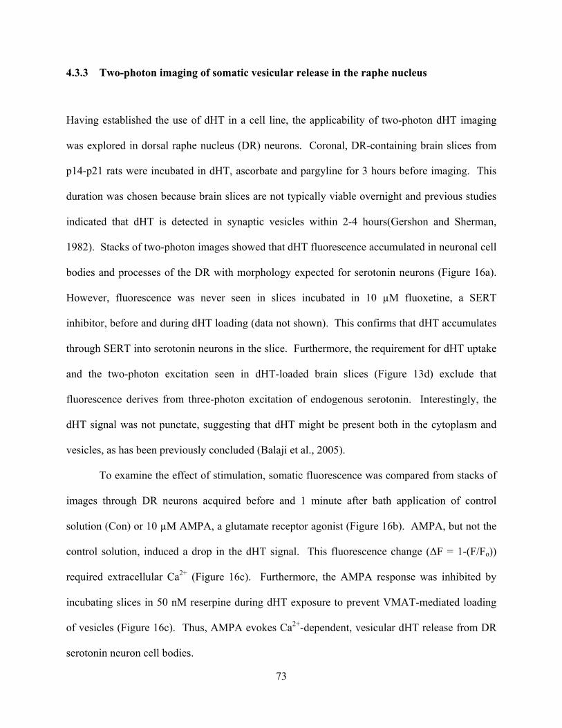
4.3.3 Two-photon imaging of somatic vesicular release in the raphe nucleus
Having established the use of dHT in a cell line, the applicability of two-photon dHT imaging
was explored in dorsal raphe nucleus (DR) neurons. Coronal, DR-containing brain slices from
p14-p21 rats were incubated in dHT, ascorbate and pargyline for 3 hours before imaging. This
duration was chosen because brain slices are not typically viable overnight and previous studies
indicated that dHT is detected in synaptic vesicles within 2-4 hours(Gershon and Sherman,
1982). Stacks of two-photon images showed that dHT fluorescence accumulated in neuronal cell
bodies and processes of the DR with morphology expected for serotonin neurons (Figure 16a).
However, fluorescence was never seen in slices incubated in 10 µM fluoxetine, a SERT
inhibitor, before and during dHT loading (data not shown). This confirms that dHT accumulates
through SERT into serotonin neurons in the slice. Furthermore, the requirement for dHT uptake
and the two-photon excitation seen in dHT-loaded brain slices (Figure 13d) exclude that
fluorescence derives from three-photon excitation of endogenous serotonin. Interestingly, the
dHT signal was not punctate, suggesting that dHT might be present both in the cytoplasm and
vesicles, as has been previously concluded (Balaji et al., 2005).
To examine the effect of stimulation, somatic fluorescence was compared from stacks of
images through DR neurons acquired before and 1 minute after bath application of control
solution (Con) or 10 µM AMPA, a glutamate receptor agonist (Figure 16b). AMPA, but not the
control solution, induced a drop in the dHT signal. This fluorescence change (ΔF = 1-(F/Fo))
required extracellular Ca2+ (Figure 16c). Furthermore, the AMPA response was inhibited by
incubating slices in 50 nM reserpine during dHT exposure to prevent VMAT-mediated loading
of vesicles (Figure 16c). Thus, AMPA evokes Ca2+-dependent, vesicular dHT release from DR
serotonin neuron cell bodies.
73
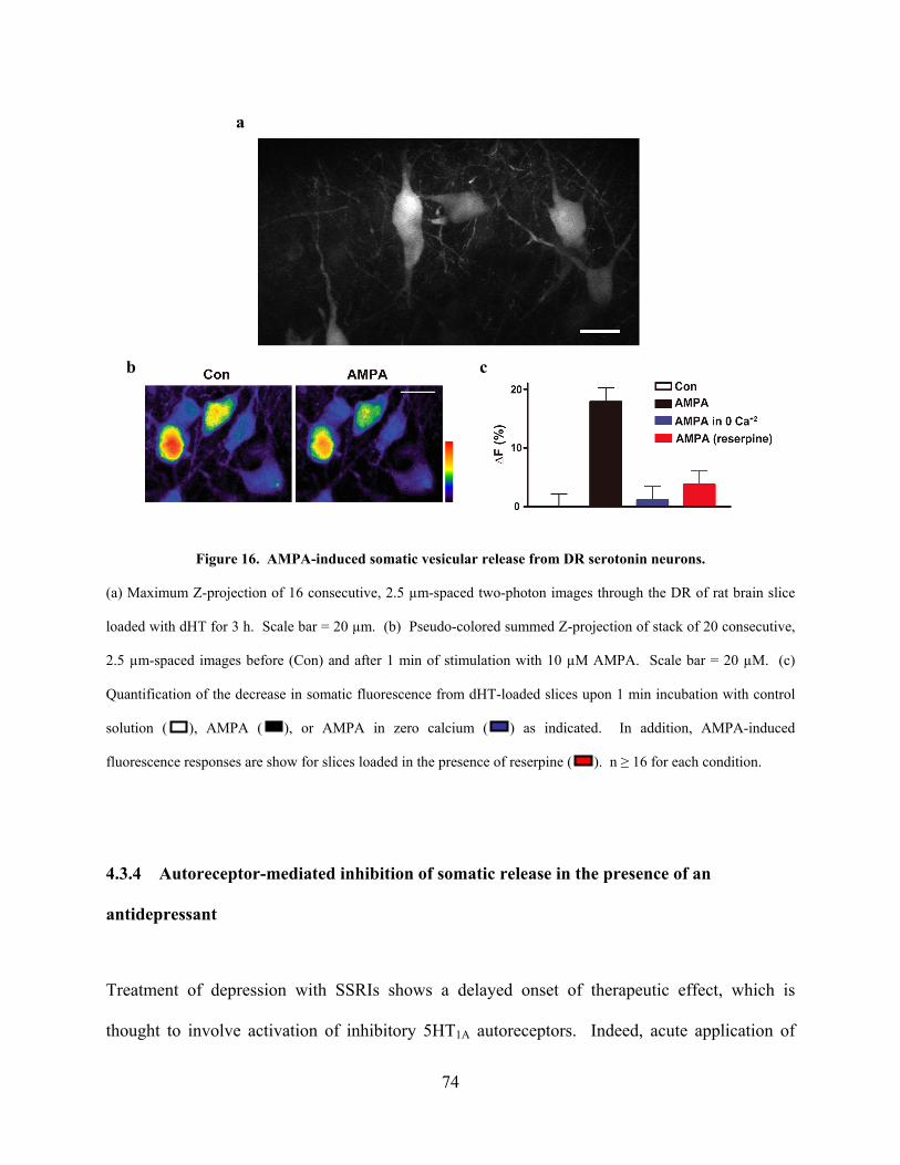
a
b c
Figure 16. AMPA-induced somatic vesicular release from DR serotonin neurons.
(a) Maximum Z-projection of 16 consecutive, 2.5 µm-spaced two-photon images through the DR of rat brain slice
loaded with dHT for 3 h. Scale bar = 20 µm. (b) Pseudo-colored summed Z-projection of stack of 20 consecutive,
2.5 µm-spaced images before (Con) and after 1 min of stimulation with 10 µM AMPA. Scale bar = 20 µM. (c)
Quantification of the decrease in somatic fluorescence from dHT-loaded slices upon 1 min incubation with control
solution ( ), AMPA ( ), or AMPA in zero calcium ( ) as indicated. In addition, AMPA-induced
fluorescence responses are show for slices loaded in the presence of reserpine ( ). n ≥ 16 for each condition.
4.3.4 Autoreceptor-mediated inhibition of somatic release in the presence of an
antidepressant
Treatment of depression with SSRIs shows a delayed onset of therapeutic effect, which is
thought to involve activation of inhibitory 5HT1A autoreceptors. Indeed, acute application of
74
SSRIs in the DR leads to an autoreceptor-mediated decrease in serotonin release (Adell and
Artigas, 1991). However, the involvement of somatic release in this response is not known. To
test whether this inhibition of release occurs at the serotonin neuron cell body, the SSRI
fluoxetine was applied for 5 minutes before and during stimulation with AMPA. This treatment
attenuated the AMPA-evoked somatic decrease in dHT fluorescence (Figure 17a). To determine
whether the inhibitory effect of fluoxetine was mediated by 5HT1A autoreceptors, the experiment
was repeated in the presence of the selective 5HT1A receptor antagonist WAY 100635. Bath
application of 2 µM WAY 100635 did not alter resting fluorescence or AMPA-induced release.
However, the autoreceptor antagonist blocked the fluoxetine-induced attenuation of release
(Figure 17b). Thus, inhibition of somatic release by fluoxetine is mediated by activation of 5-
HT1A autoreceptors.
Several conclusions can be drawn from these data. First, because application of the 5-
HT1A receptor antagonist did not increase AMPA-evoked release, inhibitory autoreceptors do not
affect responses to AMPA in the absence of fluoxetine. Second, fluoxetine had no effect on
AMPA-induced release independent of 5-HT1A receptors. Therefore, SERT does not mediate
somatic release induced by AMPA stimulation. This confirms the finding that all detected
release is vesicular (Figure 16c). Third, the inhibition of release in response to fluoxetine,
through activation of the G-protein coupled autoreceptor, demonstrates the viability of the
preparation and the fidelity of two-photon dHT imaging. Finally, autoreceptor-mediated, SSRI-
induced inhibition of release occurs at the serotonin neuron cell body in the DR.
75
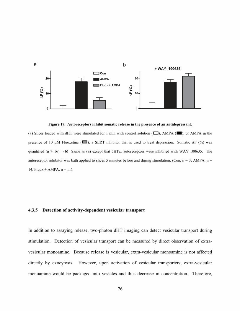
Figure 17. Autoreceptors inhibit somatic release in the presence of an antidepressant.
(a) Slices loaded with dHT were stimulated for 1 min with control solution ( ), AMPA ( ), or AMPA in the
presence of 10 µM Fluoxetine ( ), a SERT inhibitor that is used to treat depression. Somatic ΔF (%) was
quantified (n ≥ 16). (b) Same as (a) except that 5HT1A autoreceptors were inhibited with WAY 100635. The
autoreceptor inhibitor was bath applied to slices 5 minutes before and during stimulation. (Con, n = 3; AMPA, n =
14; Fluox + AMPA, n = 11).
4.3.5 Detection of activity-dependent vesicular transport
In addition to assaying release, two-photon dHT imaging can detect vesicular transport during
stimulation. Detection of vesicular transport can be measured by direct observation of extra-
vesicular monoamine. Because release is vesicular, extra-vesicular monoamine is not affected
directly by exocytosis. However, upon activation of vesicular transporters, extra-vesicular
monoamine would be packaged into vesicles and thus decrease in concentration. Therefore,
76
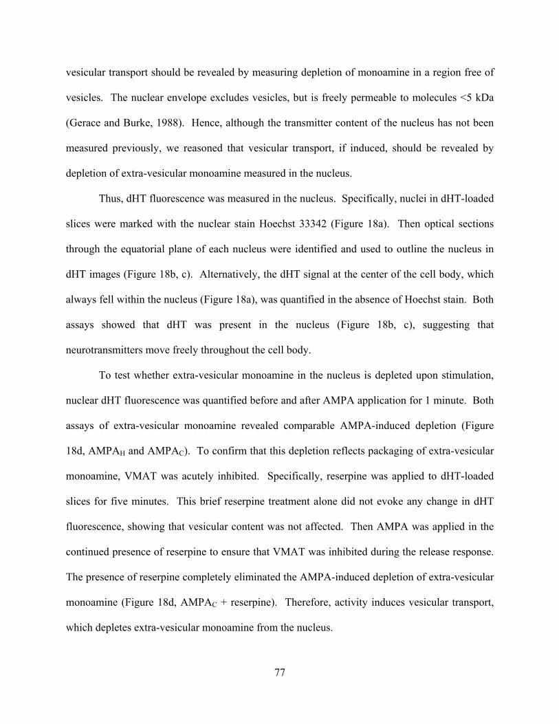
vesicular transport should be revealed by measuring depletion of monoamine in a region free of
vesicles. The nuclear envelope excludes vesicles, but is freely permeable to molecules <5 kDa
(Gerace and Burke, 1988). Hence, although the transmitter content of the nucleus has not been
measured previously, we reasoned that vesicular transport, if induced, should be revealed by
depletion of extra-vesicular monoamine measured in the nucleus.
Thus, dHT fluorescence was measured in the nucleus. Specifically, nuclei in dHT-loaded
slices were marked with the nuclear stain Hoechst 33342 (Figure 18a). Then optical sections
through the equatorial plane of each nucleus were identified and used to outline the nucleus in
dHT images (Figure 18b, c). Alternatively, the dHT signal at the center of the cell body, which
always fell within the nucleus (Figure 18a), was quantified in the absence of Hoechst stain. Both
assays showed that dHT was present in the nucleus (Figure 18b, c), suggesting that
neurotransmitters move freely throughout the cell body.
To test whether extra-vesicular monoamine in the nucleus is depleted upon stimulation,
nuclear dHT fluorescence was quantified before and after AMPA application for 1 minute. Both
assays of extra-vesicular monoamine revealed comparable AMPA-induced depletion (Figure
18d, AMPAH and AMPAC). To confirm that this depletion reflects packaging of extra-vesicular
monoamine, VMAT was acutely inhibited. Specifically, reserpine was applied to dHT-loaded
slices for five minutes. This brief reserpine treatment alone did not evoke any change in dHT
fluorescence, showing that vesicular content was not affected. Then AMPA was applied in the
continued presence of reserpine to ensure that VMAT was inhibited during the release response.
The presence of reserpine completely eliminated the AMPA-induced depletion of extra-vesicular
monoamine (Figure 18d, AMPAC + reserpine). Therefore, activity induces vesicular transport,
which depletes extra-vesicular monoamine from the nucleus.
77
Figure 18. VMAT-mediated depletion of extra-vesicular dHT from the nucleus.
(a) 3D projection of a stack of optical sections through a dHT-loaded neuron stained with Hoechst 33342 to mark the
nucleus (dHT, green; nucleus, blue). (b) Optical section through the center of the nucleus shows dHT (green)
throughout the Hoechst-labeled nucleus (blue). (c) Pseudo-colored dHT equatorial section before (Con) and after
AMPA stimulation. The nucleus as marked by Hoechst stain is outlined. (d) Quantification of the AMPA-induced
decrease in nuclear fluorescence measured within the Hoechst defined nuclear border (AMPAH; n=6) or the center of
the neuron (AMPAC; n=7). Acute application of reserpine just before and during stimulation blocked the AMPA-
induced dHT depletion in the nucleus (AMPAC + reserpine; n=7). Scale bars = 10µm.
78
4.3.6 Quantification of VMAT-mediated packaging and release during stimulation
In order to determine the contributions of activity dependent vesicular transport and release to
the somatic AMPA response, the vesicular dHT pool was quantified before and after stimulation.
Quantification took advantage of the broad pH sensitivity of dHT, the ability to collapse the
vesicular pH gradient with ammonium chloride and reserpine inhibition of VMAT. Application
of 50 mM ammonium chloride (pH 7.4) for 30 s maximally increased somatic dHT fluorescence
of resting neurons (Figure 19a). However, because ammonium shifted the pH of both vesicles
(pH 5.5 pH 7.4) and cytoplasm (pH 7.1 pH 7.4), it was necessary to determine the portion
of the fluorescence increase derived from the vesicular pool. Therefore, slices were incubated
with reserpine during the initial dHT loading to prevent vesicular packaging of dHT.
Application of ammonium to these slices led to a smaller fractional increase in fluorescence that
represents the pH effect on extra-vesicular dHT. By subtracting this extra-vesicular fractional
change from the total fractional change seen without reserpine, the effect of collapsing the pH
gradient in VMAT-containing vesicles was measured (Figure 19b). The observed 13% increase
in vesicular fluorescence coupled with the pH sensitivity of dHT (Figure 13b and 15b) indicates
that 26% of total somatic content was packaged into secretory vesicles at rest. Ammonium
experiments were then repeated on slices after AMPA treatment to determine vesicular content
after stimulation. The 6% change in vesicular fluorescence observed with collapse of the pH
gradient (Figure 18c) indicates that 12% of somatic content remaining after stimulation was
packaged into vesicles.
79
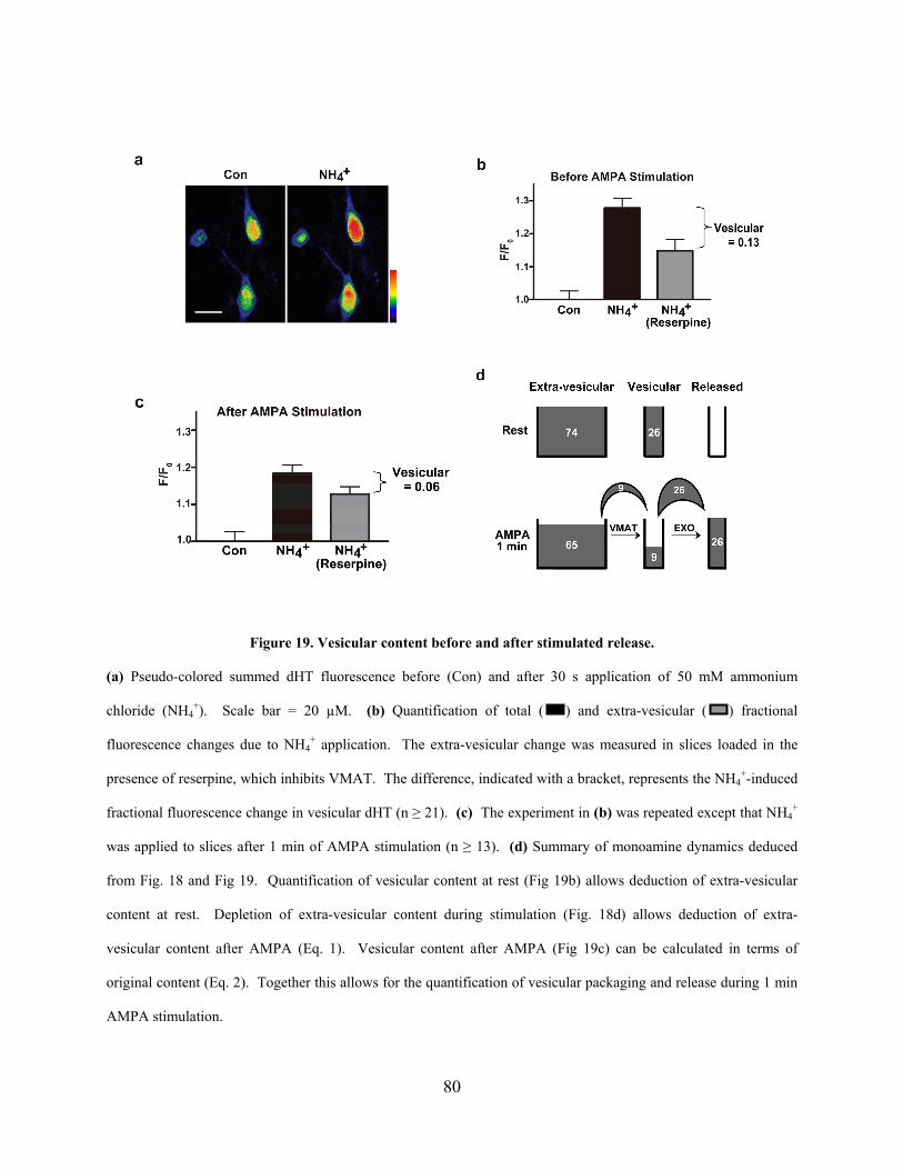
Figure 19. Vesicular content before and after stimulated release.
(a) Pseudo-colored summed dHT fluorescence before (Con) and after 30 s application of 50 mM ammonium
chloride (NH4+). Scale bar = 20 µM. (b) Quantification of total ( ) and extra-vesicular ( ) fractional
fluorescence changes due to NH4+ application. The extra-vesicular change was measured in slices loaded in the
presence of reserpine, which inhibits VMAT. The difference, indicated with a bracket, represents the NH4+-induced
fractional fluorescence change in vesicular dHT (n ≥ 21). (c) The experiment in (b) was repeated except that NH4+
was applied to slices after 1 min of AMPA stimulation (n ≥ 13). (d) Summary of monoamine dynamics deduced
from Fig. 18 and Fig 19. Quantification of vesicular content at rest (Fig 19b) allows deduction of extra-vesicular
content at rest. Depletion of extra-vesicular content during stimulation (Fig. 18d) allows deduction of extra-
vesicular content after AMPA (Eq. 1). Vesicular content after AMPA (Fig 19c) can be calculated in terms of
original content (Eq. 2). Together this allows for the quantification of vesicular packaging and release during 1 min
AMPA stimulation.
80
The above quantification of the vesicular pool at rest and after stimulation, together with
the independently measured AMPA-induced change in the extra-vesicular pool (Figure 18d) can
be used to calculate vesicular packaging and release during stimulation which is summarized in
Figure 19d. Prior to stimulation, 26% of somatic dHT is packaged into vesicles (Figure 19b),
which implies that the extra-vesicular pool contains 74% of somatic content. Stimulation
depleted the extra-vesicular pool by 12% (Figure 18d), which corresponds to packaging of 9% of
total somatic content (i.e., 0.09 = (0.74)*(0.12)). That leaves 65% of the initial total content in
the extra-vesicular pool after stimulation (i.e., 74 - 9, also see Eq. 1). This last parameter
coupled with the ammonia response after stimulation (Figure 19c) can be used to calculate that
9% of the total initial content remains in vesicles after stimulation (Eq. 2). Strikingly, the
content that remains in vesicles after stimulation equals the amount that was packaged during
stimulation (Fig. 19d, 9%).
These results lead to the conclusion that 26% of total content was released (Figure 19d).
First, this amount equals the AMPA-induced decreases in the extra-vesicular and vesicular pools
([74-65] + [26-9]). Second, because the monoamine transported into vesicles during stimulation
equals vesicular content after stimulation, an amount equivalent to the initial vesicular pool
(26%) must have been released. Finally and most importantly, a prediction of the AMPA-
induced change in fluorescence based on measurements of extra-vesicular content (Figure 18-
nuclear depletion experiments) and vesicular content (Figure 19- pH collapse experiments)
agrees with the independently measured somatic, AMPA-evoked fluorescence response (Figure
16). Specifically, taking into consideration the pH-dependent halving of dHT fluorescence in
vesicles (Figure 13b, Figure 15b), the AMPA-induced dynamics summarized in Figure 19d
predict a total ΔF of 20% (Eq. 3). This prediction is within the error of the measured AMPA-
81
induced somatic fluorescence response (18 ± 2.4%, Figure 16c), showing that results from
diverse experimental designs all confirm the same conclusion (Figure 19d). Thus, the
contributions of packaging and release to the somatic AMPA response have been quantified.
4.3.7 Efficient release of monoamine packaged during stimulation
The above results are compatible with a simple model in which all somatic vesicles undergo
exocytosis once to completely release their preloaded content
and then are refilled by VMAT (Figure 20ai). According to this hypothesis, VMAT activity
during the 1 minute stimulation partially replenishes emptied vesicular stores, but does not
contribute to release. However, an alternative hypothesis is that AMPA evokes release of only a
portion of preloaded vesicular content. In this case, the rest of the response derives from
efficient release of monoamine packaged by VMAT during stimulation (Figure 20aii).
Preloaded vesicular content held in reserve would then account for the vesicular content detected
after AMPA.
The above alternatives diverge with respect to the effect of acute inhibition of VMAT on
release evoked by AMPA. According to the first hypothesis, blocking VMAT acutely during
stimulation would not affect release because all release derives from monoamine that was
packaged prior to AMPA application (Figure 20ai). In contrast, the second hypothesis predicts
that acute inhibition of VMAT during stimulation would reduce release because monoamine
packaged in the presence of AMPA contributes to release (Figure 20aii). In fact, AMPA-induced
fluorescence changes during acute inhibition of VMAT can be predicted for each hypothesis
based on the results presented thus far (Eq. 4). Specifically, if release is supported exclusively
by preloaded vesicles (Figure 20ai), then AMPA stimulation after acute inhibition of VMAT
82
should evoke a 15% decrease in fluorescence (Figure 20b, upper dashed line i). On the other
hand, if all transmitter packaged during stimulation is released (Figure 20aii), then acute
inhibition of VMAT would reduce release to the fraction supported by preloaded vesicles. This
would correspond to a 9.8% decrease in fluorescence (Figure 20b, lower dashed line ii). Finally,
an intermediate balance between these two hypotheses would yield an intermediate result.
To inhibit VMAT during stimulation, reserpine was applied acutely (i.e., just before and
during AMPA application) as described in previous experiments (Figure 18). AMPA stimulation
after acute reserpine application evoked a 9.0 ± 2.0 % decrease in somatic fluorescence (Figure
20b). This result agrees with the second hypothesis (Figure 20aii): while some monoamine
transported into somatic vesicles at rest is held in reserve, virtually all monoamine transported by
VMAT during AMPA stimulation is released. Hence, activity-dependent vesicular transport is
efficiently coupled to somatic release.
83
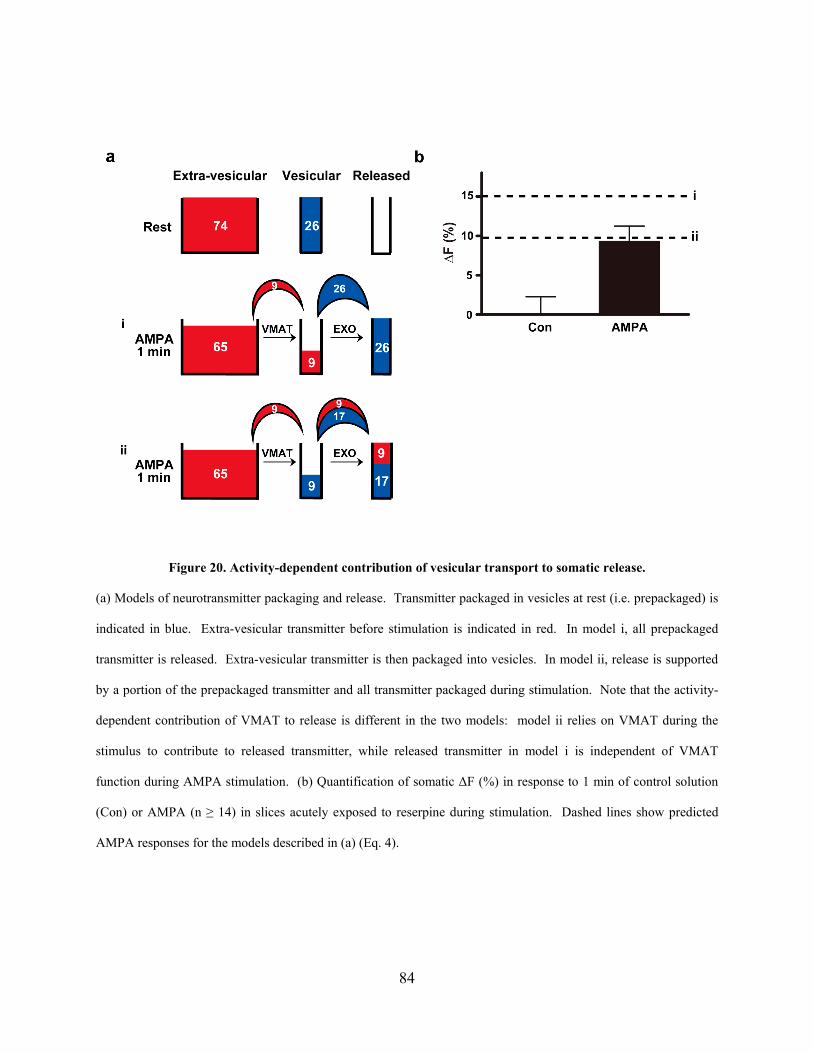
Figure 20. Activity-dependent contribution of vesicular transport to somatic release.
(a) Models of neurotransmitter packaging and release. Transmitter packaged in vesicles at rest (i.e. prepackaged) is
indicated in blue. Extra-vesicular transmitter before stimulation is indicated in red. In model i, all prepackaged
transmitter is released. Extra-vesicular transmitter is then packaged into vesicles. In model ii, release is supported
by a portion of the prepackaged transmitter and all transmitter packaged during stimulation. Note that the activity-
dependent contribution of VMAT to release is different in the two models: model ii relies on VMAT during the
stimulus to contribute to released transmitter, while released transmitter in model i is independent of VMAT
function during AMPA stimulation. (b) Quantification of somatic ΔF (%) in response to 1 min of control solution
(Con) or AMPA (n ≥ 14) in slices acutely exposed to reserpine during stimulation. Dashed lines show predicted
AMPA responses for the models described in (a) (Eq. 4).
84

4.4 DISCUSSION
Here an approach for measuring vesicular transport and release in living DR serotonin neurons
was developed to make fundamental insights into the activity-dependent function of vesicular
neurotransmitter transporters. Although the use of a transmitter analog has limitations in
assessing quantitative measurements, the ability to monitor VMAT activity during stimulation
for the first time has allowed for several qualitative conclusions. First, while only a fraction of
monoamine packaged at rest is released, release of monoamine packaged during activity is
complete. This implies that basal VMAT activity does not discriminate between releasable and
reserve pools. In contrast, during stimulation VMAT is active specifically in vesicles that
rapidly undergo exocytosis. The coupling of activity-dependent vesicular transport and
exocytosis is optimal for supporting sustained somatic release. Furthermore, this coupling
provides an explanation for the classic observation that newly synthesized (and hence newly
packaged) transmitter is released preferentially (Collier, 1969). Finally, these results reveal that
both basal and activity-dependent vesicular transport contribute to somatic release by serotonin
neurons.
Second, the nuclear compartment is relevant for vesicular transport in the serotonin
neuron cell body. Previously, the role of the nucleus had not been considered because nuclear
transmitter content had never been assayed. The permeability of the nuclear envelope, however,
implies that endogenous transmitters in intact cells equilibrate between the cytoplasm and the
nucleus. The nucleus occupies a large volume in the DR serotonin neuron cell body (Figure 18a)
and does not contain monoamine oxidase, the enzyme responsible for intracellular catabolism of
monoamine transmitters. Thus, the nucleus can serve as a large depot of diffusible transmitter
85
for somatic vesicle loading. The participation of the nucleus distinguishes monoamine dynamics
at the serotonin neuron cell body from terminals.
The development of an assay that can measure vesicular transport in the context of
neurotransmission will allow for future studies examining the regulation of quantal size.
Emerging evidence suggests that regulation of vesicular packaging may define properties of
neurotransmission during activity-dependent plasticity, exposure to drugs and during
neuropathology (Edwards, 2007). However, the physiologic relevance and mechanisms of these
changes had been difficult to assess due to the lack of a suitable live cell system. The regulation
of VMAT can now be directly assayed in brain slice. Moreover, the relevance of VMAT
regulation to neurotransmitter release can be studied.
Other aspects of serotonin neuron function are also amenable to study with two-photon
dHT imaging. For example, antidepressant drug-dependent autoreceptor-mediated inhibition,
which is thought to contribute to the delay of therapeutic efficacy of SSRIs (Blier et al., 1998),
controls release from the cell body (Figure 17). Likewise, real-time SERT function in the brain
slice can be assayed. Finally, two-photon dHT imaging is feasible with serotonin neuron
dendrites and presynaptic terminals in brain slices (unpublished results). The optical detection of
intracellular monoamine dynamics will complement other approaches to comprehensively
examine neurotransmission by central serotonin neurons.
86
4.5 METHODS
4.5.1 PC12 cell experiments
PC12 cells were cultured in DMEM supplemented with 10% fetal bovine serum at 37° with 5%
CO2. For imaging, cells were seeded on poly-D-lysine coated coverslips (#1.5, VWR) and
transfected via Tfx50 (Promega) with HA-SERT and/ or eGFP. When indicated, PC12 cells
were differentiated by application of 50 ng/ ml 2.5 S NGF for 36 hours. Five days after
transfection, to allow for expression and trafficking of SERT, cells were incubated with 20 M
dHT (5,7-dihydroxytryptamine creatine sulfate, Regis Technologies), 568 µM ascorbate and 100
μM pargyline in culture medium for 1 hour or overnight. Overnight loading was followed by a 1
hour wash in culture medium at 37°C to allow cytoplasmic dHT to clear. Cells stably expressing
ANF-emerald GFP(Han et al., 1999b) were transfected with SERT, differentiated, and loaded
with dHT as described above. When cells were loaded in the presence of fluoxetine (10 μM) or
reserpine (100 nM), drug was added along with dHT. For imaging, cells were bathed in normal
saline (in mM, 5.4 KCl, 140 NaCl, 2 CaCl2, 0.8 MgCl2, 10mM Na-HEPES, 10 mM glucose; pH
7.4) and excited by epifluorescence or two-photon microscopy as described. For pH collapse
experiments, monensin (1 µM) was bath applied during imaging. PC12 cells were stimulated by
exchange of normal saline for high K+ saline (in mM: 100 KCl, 45 NaCl, 5 BaCl2, 0.8 MgCl2, 10
Na-HEPES, 10 glucose; pH 7.4).
87
4.5.2 Slice Experiments
All experiments were conducted in accordance with protocols approved by the University of
Pittsburgh Institutional Animal Care and Use Committee. Male Sprague Dawley rats p14-p21
(Hilltop Labs) were anesthetized with isoflurane and decapitated. Brains were removed and
bathed in 95% O2 and 5% CO2-saturated, ice-cold, sucrose-based artificial cerebral spinal fluid
(s-aCSF; in mM: 87 NaCl, 75 sucrose, 2.5 KCl, 25 NaHCO3, 1.25 NaH2PO4, 0.5 CaCl2, 7.0
MgSO4, 25 glucose, 0.15 ascorbic acid, 1 kynurenic acid, pH 7.4). 250 µm coronal brain slices
containing the DR were cut with a vibratome (The Vibratome Company) and incubated in 20 µM
dHT, 568 µM ascorbate, and 100 μM pargyline in s-aCSF for 3 hours at 37°C. Slices were
washed once in normal aCSF (in mM, 124 NaCl, 4 KCl, 25.7 NaHCO3, 1.25 NaH2PO4, 2.45
CaCl2, 1.2 MgSO4, 11 glucose, 0.15 ascorbic acid, pH 7.4 ) before imaging. For slices loaded in
the presence of fluoxetine (10 µM) or reserpine (50 nM), drugs were applied 10 minutes before
addition of dHT. Stimulation of slices was induced by a bath exchange with aCSF supplemented
with 10 µM AMPA. For stimulation with AMPA in zero calcium, CaCl2 in normal aCSF was
replaced with MgSO4 and 1 mM EGTA was added. For autoreceptor studies, fluoxetine (10
µM) was added 5 minutes before stimulation with AMPA. The autoreceptor inhibitor WAY
100635 (2 µM) was added 5 minutes before fluoxetine application. When indicated, nuclei were
stained by a 30-45 minute incubation of loaded slices with 1.6 mM Hoechst 33342 at the end of
the experiment (i.e. after AMPA stimulation). For experiments requiring acute inhibition of
VMAT, reserpine (50 nM) was added to slices 5 minutes before stimulation with AMPA.
Finally, for pH collapse experiments, 50 mM NH4Cl replaced 50 mM of NaCl in normal aCSF.
NH4+ aCSF was applied to slices at rest or after AMPA stimulation as indicated.
88
4.5.3 Optical Setups
Widefield epifluorescence experiments were done on an Olympus IX71 inverted microscope
equipped with a 340UV 40x 1.35 numerical aperture (NA) oil-immersion objective and a xenon
arc lamp. DHT epifluorescence used a 360/40 excitation filter and 420 nm long-pass emission
filter, which was collected by a cooled CCD camera (Hamamatsu Orca ER). Two-photon
imaging experiments were done on an Olympus Fluoview FV1000 upright confocal scanning
microscope. 725 nm excitation illumination from a Coherent chameleon ultra titanium sapphire
laser was attenuated with an acoustical optical modulator and expanded with a motorized
telescope (LSMtech) before being focused by a 60x, 1.1 NA water-immersion objective.
Emission (400 - 480 nm) was quantified with a non-descanned detector (LSMtech), which
contains a cooled Hamamatsu photomultiplier tube. Stacks of consecutive, 2.5 µm spaced images
were taken through loaded neurons of the DR. All optical measurements were performed at
room temperature.
4.5.4 Image Analysis
Analysis was done through Image J (NIH). Images were contrast enhanced or pseudo-colored to
aid figure presentation without altering the primary data. When necessary, series of images were
aligned with image j plug-in Stack Reg or stacks aligned with image j plugin Align 3TP. Mean
fluorescence intensity was measured in regions of interest (ROIs) in single images or summed z-
projections of image stacks and subtracted by background values (i.e. unloaded cells or tissue
background fluorescence). Percent change in fluorescence (ΔF (%) = (1 – (F/F0)) * 100) was
normalized to control experiments to allow for comparisons across experimental manipulations.
89
‘n’ represents the number of cells from which data was collected. All data was compiled from at
least three independent experiments. Error bars represent standard error of the mean.
4.5.5 Arithmetic Analysis
‘v’ is defined as the fraction of dHT in vesicles and ‘c’ as the fraction of extra-vesicular dHT.
The subscript ‘0’ defines the variable in resting cells (i.e. before experimental manipulation). In
all cases ‘c’ and ‘v’ are expressed in terms of fraction of total content at rest. Hence, v0 + c0= 1.
The fractional fluorescence of extra-vesicular dHT is denoted as ‘Fc’ and equals the fraction in
extra-vesicular content ‘c’. The fractional fluorescence of vesicular dHT is denoted ‘Fv’ and
equals ‘½v’ due to the pH-dependent halving of dHT fluorescence in acidic vesicles (Figure 13b,
Figure 15c). Hence, the somatic fluorescence can be written in terms of the fractional dHT
content in the vesicular and extra-vesicular pools (F0 = Fc0 + Fv0 = c0 +(1/2)v0).
Equation 1: Figure 18 demonstrates a 12% change in the extra-vesicular fraction (ΔFc
(%)) during AMPA stimulation. In order to calculate the extra-vesicular fraction after
stimulation (cAMPA) the following calculation was performed.
ΔFc (%) = (1 – (FcAMPA/Fc0)) * 100; c0 = 0.74 (Figure 7b)
12% = (1 - (cAMPA / 0.74)) * 100
cAMPA = 0.65
Equation 2: Figure 19c demonstrates that 12% (Eq.1) of the somatic content after
stimulation is in vesicles. In order to express the vesicular content after stimulation in terms of
fraction of total content at rest, the following calculation is performed.
vAMPA = 0.12 (cAMPA + vAMPA) ; cAMPA = 0.65 (Eq. 1, Figure 6)
vAMPA = 0.12 (0.65 + vAMPA)
90
vAMPA = 0.09
Equation 3: A prediction of the AMPA-induced change in fluorescence can be made
from measurements of vesicular and extra-vesicular content before and after stimulation (derived
from Figures 18 and 19). This prediction can be compared to actual AMPA- induced
fluorescence changes measured in Figure 16.
ΔFpred (%) = (1 - (FAMPA/ F0)) *100
= (1 - (FcAMPA + FvAMPA)/ (Fc0 + Fv0)) *100
= (1 - (cAMPA + ½vAMPA)/ (c0 + ½v0)) *100
= (1 - (0.65 + ½(0.09))/ (0.74 + ½(0.26))) *100
= 20 %
Equation 4: Predictions of the change in fluorescence while acutely inhibiting VMAT
during stimulation can be made for each model. Without AMPA-induced packaging of extra-
vesicular dHT, the extra-vesicular pool is not depleted. Hence, cAMPA = c0 = 0.74.
ΔFpred. (%) = (1 - (FAMPA/F0)) *100
= (1 - (FcAMPA + FvAMPA)/ (Fc0 + Fv0)) *100
= (1 - (c0 + ½vAMPA)/ (c0 + ½v0))*100
= (1 - (0.74 + ½vAMPA)/ (0.74 + ½v0))*100
In model i all preloaded vesicular transmitter is released and then vesicles are refilled by
VMAT. With acute VMAT inhibition, vesicles cannot be refilled. Therefore, no transmitter
remains in vesicles after stimulation (vAMPA= 0.0).
ΔFpred.i (%) = (1 - (0.74 + ½vAMPA)/ (0.74 + ½v0))*100
= (1 - (0.74 + ½(0.0)/ (0.74 + ½(0.26))) *100
= 15 %
91
In model ii only a portion of preloaded transmitter is released, while the rest is held in
reserve and accounts for the vesicular content after AMPA stimulation. Because VMAT activity
during the stimulus does not contribute to the amount remaining in vesicles after stimulation in
this model, acute reserpine does not alter vAMPA. Hence, vAMPA = 0.09.
ΔFpred.ii (%) = (1 - (0.74 + ½vAMPA)/ (0.74 + ½v0))*100
= (1 - (0.74 + ½(0.09)/ (0.74 + ½(0.26))) *100
= 9.8 %
92
5.0 DISCUSSION
5.1 SUMMARY AND SIGNIFICANCE OF FINDINGS
Neurotransmission must be highly regulated while maintaining efficiency in order to mediate the
complex and varied tasks demanded of it. Homeostatic and plastic changes must continually
refine synaptic efficacy to respond to changing conditions. However, mechanisms that mediate
this ‘fine tuning’ of transmission are not well understood. Classic interpretations of
neurotransmission viewed neurotransmitter release as a fixed, stereotypic response to input, and
thus limited mechanisms of plasticity primarily to changes in receptor responses. However
modern understanding has now clearly defined an important role for the regulation of
neurotransmitter release as well.
Release of transmitter is mediated by a series of highly regulated events including the
packaging of secretory vesicles and their exocytosis. The packaging of transmitter is determined
by the vesicular neurotransmitter transporter (VNT) family, whose presence and function are
essential for neurotransmission. During both the biogenesis and the activity-dependent recycling
of secretory vesicles, VNTs undergo trafficking that can determine the quality, quantity, and
location of packaged neurotransmitter. Thus, understanding the signals and mechanisms of VNT
trafficking are essential to understanding the regulation of neurotransmission.
93
In chapter 2 and chapter 3 of this thesis, the synaptic vesicle specific targeting of VNTs
are investigated. Using the SV specific trafficking of VAChT in the PC12 neuroendocrine cell
line as a model system, a specific synaptic vesicle targeting motif was identified. A chimera
between an unrelated, plasma membrane localized protein and the C-terminus of VAChT
trafficked to SVLVs, indicating the sufficiency of the C-terminus for SV targeting from the
plasma membrane. Next, deletion analysis revealed the requirement and sufficiency of a 10
amino acid region containing a classic dileucine motif for both internalization and SVLV
targeting. The duality of the motif as both an internalization and SV trafficking signal suggested
efficiency in SV trafficking, consistent with the rapid kinetics of SV recycling at the terminal
(Cousin and Robinson, 1999). Further examination of this motif revealed specificity for SVLV
targeting over other internalization motifs and suggested that specificity lies in the type and
environment of the motif. Specifically, while some dileucine motifs were not able to direct
targeting to SVLVs, the closely related motif in VMAT2 suggested that this signal may be
generalized as a SVTM across several synaptic vesicle proteins.
In Chapter 3, sorting nexin 5 was identified as a novel regulator of the SVLV specific
trafficking of VAChT. The characterization of SNX5 as a brain enriched protein that associates
with SVs and the ability of SNX5 to bind to the C-terminus of VAChT suggested a functional
interaction. Disruption of SNX5 by overexpression of mutant protein or siRNA mediated
knockdown led to the mistargeting of the SVLV targeted protein to LDCVs. This
mislocalization suggested a potential regulatory mechanism for membrane trafficking between
different types of secretory vesicles. Thus, SNX5 was identified as a novel regulatory protein of
SV specific trafficking, and suggested a potential mechanism of regulation that may be relevant
for the modulation of neurotransmission.
94
Regulation of VNT trafficking or function has been reported to be involved in activity
dependent plasticity, mechanisms of drug action, and disorders of the nervous system. This
relevance suggests that VNTs may be an important pharmacological target (Iversen, 2000).
However, in order to understand the regulation of VNTs, and the subsequent consequences for
neurotransmission in-vivo, an assay that can measure vesicular transport and release in live
neurons was necessary. Therefore, in Chapter 4, a novel method was established in order to
measure these parameters. The use of a pH-sensitive, fluorescent serotonin analog, 5,7-
dihydroxytryptamine, visualized with two-photon microscopy, allowed measurements of both
vesicular transport and release in dorsal raphe nucleus serotonin neurons in the brain slice.
Somatic release was accompanied by VMAT activity, which redistributed monoamine from the
extra-vesicular compartment, including the nucleus. A portion of monoamine packaged at rest
was held in reserve, however, all monoamine packaged during stimulation was released
efficiently. This novel assay measured resting and activity dependent contributions of vesicular
transport to release in living neurons for the first time and established a means of studying VNT
regulation and the resulting modulation of neurotransmitter release.
The work presented in this thesis provides important information on the mechanisms and
regulation of the synaptic vesicle trafficking of vesicular transporters. Moreover, it establishes a
novel assay of vesicular transport in living neurons that allows study of activity dependent VNT
function. The recent shift in the understanding of VNTs as passive ‘gatekeepers’ for secretory
vesicles to targets of active regulation that define properties of neurotransmission has come as
numerous studies conclude that VNT regulation is behaviorally relevant. These studies have
implicated VNTs in the psychotropic action as well as the toxicity of drugs of abuse, the
pathology of neurologic disease as well as the efficacy of pharmacologic treatments, and finally,
95

behaviorally relevant, activity-dependent plasticity. The centrality of VNT regulation to brain
function should not be surprising due to the essential role of VNTs in neurotransmission.
Despite this, the development of pharmacological agents to specifically target VNTs has
received only limited attention. This thesis, in addition to providing novel information on the
trafficking and activity-dependent function of vesicular transporters, lays a foundation for
comprehensive studies on the active role of VNTs in shaping the properties of
neurotransmission. Greater understanding in this area will undoubtedly yield advances in the
development of novel therapies targeting VNTs.
5.2 REGULATION OF VACHT TRAFFICKING
5.2.1 Multiplicity of Pathways
The coexistence and relative prevalence of various recycling pathways including ‘kiss and run’
and clathrin mediated endocytosis has been a controversial topic (Rizzoli and Jahn, 2007).
However, it is this coexistence of multiple pathways that would allow for the highly
advantageous balance between efficient and regulated SV recycling that is likely required for
neurotransmission. Activity- dependent recycling through a ‘kiss and run’ mechanism allows for
faithful recycling of SVs in their current composition. In this case, vesicle proteins do not
require sorting, as vesicle identity is maintained, and recycling is mediated by the closing of the
fusion pore. Thus, this pathway is highly efficient, allowing rapid and faithful regeneration of
SVs for further release. However, this mechanism does not allow for regulation of protein levels
on the secretory vesicle, a potential means of plasticity.
96
On the other hand, clathrin mediated internalization, while less efficient allows for
regulation of the SV proteome. A direct uncoating of internalized clathrin-coated pits to form
SVs still maintains fast kinetics of recycling while allowing for modulation of the stoichiometry
of SV proteins. Vesicle recycling through an endosomal intermediate may have slower kinetics
and require multiple trafficking steps, however it allows for the greatest level of regulation. In
addition to regulation of protein stoichiometry, recycling through an endosomal intermediate
may regulate the balance of proteins between secretory vesicle types. LDCV proteins and SV
proteins are seen together in endosomal intermediates, suggesting a potential site of component
intermixing (Partoens et al., 1998). Moreover, trafficking of SV proteins through an endosomal
intermediate may allow for independent regulation of vesicle subpopulations. Optical evidence
supports the idea that pools of vesicles (i.e. readily releasable and reserve) use unique recycling
pathways and that the properties of secretory vesicles in different pools are unique. Vesicles that
are recycled through an endosomal intermediate are thought to populate the reserve pool of
vesicles (Richards et al., 2000). Thus an endosomal pathway of recycling may provide ‘storage’
of a pool of vesicles (i.e. the reserve pool) with unique characteristics. This pathway may
therefore provide a means for ‘bulk’ regulation, in which larger amounts of proteins and lipid are
internalized together before precise sorting. Recruitment of the reserve pool for release, through
changes in phosphorylation, could then serve as an efficient mechanism not only of recruiting
vesicles for release, but also altering properties of neurotransmission.
Thus, the presence of multiple recycling pathways including ‘kiss and run’ and clathrin
mediated internalization has been controversial for synaptic vesicles in the CNS. However it is
precisely the coexistance of these multiple pathways that would allow for control between rapid
97
regeneration of SVs in their current composition and regulation of the properties of SVs,
providing a balance between efficiency and plasticity that neurotransmission demands.
The question is then raised as to how the trafficking of specific proteins, such as VAChT,
are regulated through these multiple pathways. First, the signals that mediate the trafficking
through these pathways must be identified. A single dileucine-containing motif in the C-
terminus of VAChT was identified as sufficient for both its internalization and synaptic vesicle
targeting. Although this motif was identified under resting conditions, it is likely that this motif
also directs VAChT targeting during activity-dependent vesicle recycling. Trafficking through a
‘kiss and run’ mechanism likely does not require targeting sequences. As vesicle identity is
maintained during ‘kiss and run’, proteins do not need to be sorted. Thus, the identified
dileucine motif is likely involved in clathrin-mediated pathways of recycling. The motif is
consistent with a classic E(XXX)LL dileucine motif identified in non-neuronal, clathrin
dependent internalization (Bonifacino and Traub, 2003). Moreover, the reported adaptor protein
AP-2 binding to this dileucine motif in VAChT is consistent with trafficking through a clathrin-
mediated recycling pathway (Barbosa et al., 2002). The presence of a single motif that has dual
properties for both internalization and SV targeting suggests efficiency of trafficking and the
ability of this motif to mediate direct SV formation from the plasma membrane. However, this
does not exclude a role for this motif in mediating trafficking through an endosomal
intermediate. The targeting of VAChT through an endosomal intermediate has not been
explored directly. However, it is likely that endosomal pathways may be particularly relevant for
VNTs, which can be found on multiple vesicle types and in multiple vesicle pools. The
regulation of VNTs between vesicle types can define the relative content of vesicle types. In fact
our studies from Chapter 3 suggest that VAChT trafficking has the potential to be regulated
98
between vesicle-types. It is likely that VAChT trafficking through an endosomal intermediate
may also be mediated through the identified SVTM. However, the inability to distinguish
between internalization signals and SV targeting signals within this regions suggests that the
identified dileucine motif must act both at the plasma membrane for internalization and at the
endosome for budding of the SV. Consistent with this, endosomal budding of SVs is thought to
be mediated by an AP-3 dependent mechanism which has been shown to interact with dileucine
motifs of the E(XXX)LL form (Bonifacino and Traub, 2003). However, interaction between
VAChT and AP-3 has not been identified in binding studies (Kim and Hersh, 2004). This
interaction may be transient in nature or occur only under certain conditions (i.e. under high
levels of stimulation), thus it is difficult to interpret this negative result. However, it is also
possible that VAChT may interact with unique cellular machinery.
The trafficking of VAChT through multiple trafficking pathways provides a means of
increasing its regulation. However the mechanisms that determine the balance of VAChT
trafficking between these recycling pathways needs further investigation. The prevalence of
‘kiss and run’ or full fusion mechanisms of endocytosis are thought to be regulated at least in
part by activity (de Lange et al., 2003). In many cases, low frequency of stimulation leads to
maintenance of synaptic efficacy through ‘kiss and run’ endocytosis. Under more intense
stimulation, clathrin-mediated internalization through direct or endosomal trafficking may be
most prevalent. One could hypothesize that it is precisely under these situations of high
frequency stimulation that greater regulation of synaptic transmission is needed and that a
clathrin mediated recycling pathway would allow for that regulation. Various readouts of
synaptic activity, such as calcium levels, could regulate recycling machinery to induce a shift in
recycling pathways. In fact, calcium mediated regulation of fusion pore collapse has been
99
reported (Fulop and Smith, 2006; He et al., 2006). Activity dependence may also regulate
trafficking between a direct and endosomal mediated clathrin pathway. Shifts between these
pathways could potentially be mediated by saturation of recycling machinery or machinery
compartmentalization. Recently, the trafficking of VGluT was studied through the use of a
chimeric protein between VGluT-1 and a pH-sensitive GFP variant in primary neurons. The
trafficking of VGluT was regulated between an AP-2 dependent and an AP-3 dependent
mechanism based on the intensity of stimulation. Regulation between these two pathways was
hypothesized to be mediated by the competitive recruitment of VGluT-1 into the AP-2 dependent
pathway by endophilin, an adaptor protein involved in clathrin mediated endocytosis (Voglmaier
et al., 2006). Trafficking of VGluT through the two pathways relied on a common dileucine-like
motif. This suggests that VAChT may be regulated similarly through multiple pathways relying
on a single primary dileucine motif. The prevalence of recycling pathways may be regulated by
interaction with regulatory machinery. Identification of trafficking proteins, like SNX5, that
may regulate the prevalence of VAChT trafficking through different recycling pathways is
needed.
5.2.2 Multiplicity of Signals
Analysis of the specificity and generality of the dileucine motif in VAChT suggested that a
similar motif in VMAT2 was also able to direct trafficking to SVLVs in the absence of a LDCV
targeting sequence. Furthermore, dileucine-like motifs have been identified in several other SV
proteins such as synaptotagmin and synaptobrevin 2 (Blagoveshchenskaya et al., 1999; Grote et
al., 1995). However there is significant variability in the amino acid environments of these
dileucine-like motifs. Furthermore, unrelated SV targeting motifs have been identified in other
100
proteins. In certain SV proteins, synaptic vesicle trafficking signals do not appear to exist and
their SV-specific location relies on their association with other SV proteins (Pennuto et al.,
2003). This multiplicity of signals has made understanding the regulation of SV targeting
difficult. Furthermore, the reconciliation of a large number of trafficking motifs with a limited
number of common trafficking pathways has been difficult.
One possible explanation of the multiplicity of signals identified in the SV targeting of
proteins is to allow for individual recruitment and regulation through common pathways. Two
proteins may have signals that direct them through clathrin and AP-2 dependent internalization
and SV targeting from the membrane, however, they may rely on unique protein interactions that
distinguish the efficiency of their recruitment. The signals that mediate this trafficking may have
some similarities such as a dileucine-like motif, however the environment of the motif or
additional motifs may regulate the efficiency or recruitment into clathrin pits or association with
other SV proteins that are co-regulated.
The presence of a unique and sufficient dileucine motif in VAChT allows it to be
individually regulated during SV recycling. Modulation of VAChT recycling efficiency to alter
VAChT protein levels on vesicles is likely regulated by additional factors that remain to be
identified. One such factor could be the phosphorylation state of the dileucine motif of VAChT
by PKC, which has been shown to alter the SV trafficking of VAChT (Cho et al., 2000). This
may reduce the efficiency of internalization or alter interactions with adaptor proteins.
Additionally, the presence of two tyrosine based motifs in the C-terminus of VAChT have been
suggested to regulate the efficiency of VAChT internalization or SV targeting, although the
mechanisms of this have not yet been determined (Kim and Hersh, 2004).
101
5.2.3 Potential mechanisms of SNX5 regulation
The identification of sorting nexin 5 as a regulator of the synaptic vesicle trafficking of VAChT
confirmed the involvement of specific protein interactions in the regulation of VAChT
trafficking. The mislocalization of the VAChT chimera to LDCVs upon the disruption of SNX5
function suggested that SNX5 regulates the trafficking of VAChT between secretory vesicle
types. Although VAChT has been characterized as highly enriched in SVs, it has been identified
to reside to a lesser extent on LDCVs (Agoston and Whittaker, 1989; Lundberg et al., 1981).
Moreover the finding that disruption of the interaction between SNX5 and VAChT can shift
trafficking between secretory granules defines an important potential mechanism of regulation
that may be common to other VNTs such as VMAT, which is often found in multiple vesicle
types in a single neuron. In fact, preliminary evidence suggests that SNX5 may bind VMAT2
(unpublished data). The mechanism of SNX5 regulation remains to be identified, however
potential models are explored below.
Regulation between secretory vesicle types may suggest a common pathway of SV and
LDCV biogenesis. Classically, proteins destined for different vesicle types were thought to
segregate at the level of the TGN into the regulated and constitutive secretory pathways.
However, certain SV bound proteins may take an alternate route. These proteins may traffic into
the regulated secretion pathway. This could be due to the presence of targeting motifs similar to
LDCV bound proteins, association with other proteins bound for this pathway, or inefficient
TGN sorting. During the maturation of LDCVs, proteins destined for other locations in the cell
are removed in a more stringent sorting process by the budding off of transport vesicles. This
sorting process is an AP-1, ARF1, and GGA dependent process (Dittie et al., 1996; Kakhlon et
al., 2006).
102
Interestingly, VAChT has been shown to bind to AP-1 through its C terminus dileucine
motif and thus has been suggested to traffic in this manner to SVs (Kim and Hersh, 2004). This
alternative biogenesis pathway suggests a common initial pathway of VNTs destined for both
types of secretory vesicles, which would allow direct regulation of trafficking between them.
The data presented in Chapter 3 suggests that SNX5 may regulate this trafficking. In the model
of biogenesis proposed above, SNX5 would promote the budding of VAChT from immature
secretory granules, removing it from the regulated secretory pathway. Disruption of SNX5
function, or the interaction between SNX5 and VAChT, would therefore prevent the removal of
VAChT from immature granules and lead to its accumulation in LDCVs.
This potential model of regulation is consistent with the known properties of SNX5. The
role of the sorting nexin family in regulating the trafficking of membrane proteins has been well
defined, although the mechanisms of this regulation are not clear (Carlton and Cullen, 2005).
The presence of a curvature sensing/forming BAR domain in SNX5 suggests that is likely
involved in trafficking of proteins via a budding mechanism. The PX domain of the sorting
nexin family was originally characterized for its affinity for the phosphoinositide PI3P lipids,
localizing the sorting nexin proteins to membranes enriched in these lipids. However, recent
evidence has suggested that unique amino acid sequences in various PX domains of the members
of the sorting nexin proteins may mediate different phospohinositide specificity. Recently,
SNX5 has been characterized in non-neuronal cells. Its PX domain has shown affinity for PI4P,
which is enriched in TGN derived membrane (Liu et al., 2006). Moreover, PI4P recruits GGAs
and AP-1 and thus SNX5 is well suited to be involved in membrane trafficking involving these
characteristics. Thus, properties of SNX5 in non-neuronal cells suggests characteristics that are
consistent with regulating trafficking from immature secretory granules in neurons.
103
SNX5 has also been recently characterized through genetic studies as a potential
component of the mammalian retromer complex (Wassmer et al., 2007). Classically, the
retromer complex is thought to mediate, endosomal/lysosomal to TGN trafficking, best described
in the recycling of the cation independent mannose-6-phosphate receptor (CI-MPR) after
delivery of its cargo to the lysosome (Bonifacino and Hurley, 2008). Consistent with this role,
our lab has previously demonstrated that SNX5 inhibits the degradation of EGFR from late
endosomes (Liu et al., 2006). A role for the retromer in regulating targeting between secretory
vesicle types is not apparent, however the CI-MPR is known to traffic through immature
secretory granules. This receptor is removed during LDCV maturation in an AP-1 dependent
process as described above (Klumperman et al., 1998). Thus, SNX5 which may play a role in
CI-MPR trafficking as a component of the retromer, may also play a role individually or as a
larger protein complex in the removal of this receptor and other proteins from the regulated
secretion pathway, including VAChT. The trafficking pathway of VAChT once removed from
ISGs remains to be determined. Vesicles budding off of ISGs have been reported to traffic to
various locations including the plasma membrane in a constitutive-like pathway, the TGN, or
endosomes, all of which could be intermediates in the formation of SVs (Tooze and
Stinchcombe, 1992).
This biosynthetic pathway is also consistent with the known regulation of VMAT
trafficking between secretory vesicles. The C-terminus acidic patch in VMAT may disrupt
interaction with regulatory machinery, such as SNX5, that mediates sorting away from the
regulated secretory pathway. In this way, this motif would promote retention to the regulated
secretion pathway and increase VMAT targeting to LDCVs. Interestingly, phosphorylation of
residues in the acidic patch promotes a shift in VMAT2 targeting to SVs (Waites et al., 2001).
104
Thus, the inclusion of VMAT2 in vesicles budding off of ISGs may be regulated by
phosphorylation.
Interestingly the finding that SNX5 associates with synaptic vesicles (Figure 11;
(Takamori et al., 2006) suggests that SNX5 may have a more permanent interaction with VAChT
than described above. Thus, SNX5 may regulate VAChT targeting at multiple steps in the SV
trafficking pathway. As mentioned previously, the endosome is another potential site of
regulation between secretory vesicles. Proteins from both LDCVs and SVs intermix in
endosomal compartments after stimulation and must be sorted (Partoens et al., 1998). Interaction
of VAChT with SNX5 may therefore regulate SV sorting in the endosome. Proteins that are not
selectively sorted for SV retention at this stage may be trafficked to the lysosome for degradation
or the TGN for further sorting into secretory pathways as described above. Interestingly, several
studies have indicated an endosomal localization of SNX5 in non-neuronal cells, suggesting that
it may play a role in trafficking steps at the early endosome (Merino-Trigo et al., 2004; Yoo et
al., 2006). The identification of a regulatory protein that may act to direct SV specific trafficking
of VAChT at several steps of a trafficking pathway is appealing as it is both efficient and
provides for redundant levels of regulation. This may be particularly relevant for the VNT
family of proteins whose location to secretory granules is essential for neurotransmission and
whose trafficking properties define the vesicle type and amount of packaged neurotransmitter. It
is interesting to note that interaction of SNX5 showed at least some specificity of regulating
VNT traffic as trafficking of another SV protein, synaptophysin, was not regulated by SNX5
(Figure 12).
Further studies that examine the interaction of SNX5 with other SV proteins would be of
great interest. In particular, confirmation of the functional interaction of VMAT2 and SNX5
105
would help to define the mechanisms of SNX5 function. As mechanisms that mediate regulation
of VMAT2 targeting between vesicle types have been identified, such as changes in
phosphorylation in the acidic patch, further study on the regulation of the interaction between
SNX5 and VMAT2 may provide more insight into physiologic regulation of VNT trafficking.
Confirmation of VAChT trafficking through the ISGs as well as further study on the endosomal
trafficking of VAChT will allow for greater understanding of the sites and mechanism of SNX5
regulation.
5.2.4 Future Studies of VNT trafficking
Further elucidation of mechanisms that regulate the trafficking of VAChT and other VNTs is
necessary. Future studies will have to examine trafficking motifs and machinery not only under
resting, steady-state conditions, but also during stimulation. Multiple stimulation conditions to
examine the prevalence of one recycling pathway over another will be helpful to identify
cytosolic machinery responsible for trafficking regulation. Moreover, it will be important to
incorporate multiple identified motifs and regulation of VNT trafficking into a cohesive picture
of SV recycling. Finally, although the primary focus has been on SV trafficking at the synapse,
understanding the polarized targeting of VNTs, which may regulate the axonal/somatodendritic
localization of the proteins will be important to examine. It is important to note, that
experiments that study trafficking will need to examine the efficiency of trafficking rather than
an end-point read out to understand trafficking regulation. Moreover, studies that are able to
examine trafficking of multiple synaptic vesicle proteins, or the trafficking of SV proteins
relative to cycling lipid, will increase our understanding of the relative regulation of SV proteins
that define vesicle protein stoichiometry. This will require a combination of sensitive and
106
dynamic techniques including optical, biochemical and genetic methodology. A greater
understanding of mechanisms that mediate the selective regulation of VNT trafficking will allow
us to understand complex mechanisms of regulation used to define synaptic transmission such as
the recent identification of diurnal trafficking regulation of VGluT (Darna et al., 2008) as well as
other regulation that may act to acutely regulate properties of transmission.
5.3 PHYSIOLOGIC RELEVANCE OF VNT REGULATION
The measurement of VNT function in the context of neurotransmission in living neurons holds
great significance for the field of VNTs. For the first time potential mechanisms of VNT
regulation can be studied in intact neurons and the physiologic relevance of this regulation can
be simultaneously measured. Moreover, the establishment of this assay allowed for direct
measurements of the relative contribution of activity-dependent vesicular transport to release.
The use of a false transmitter in this assay suggests the quantitative measurement of this
contribution may not be indicative of native transmitter. As the handling properties (i.e. km and
Vmax for VMAT or SERT) and/or the cytosolic concentration of the false transmitter are likely
not precisely the same as that of the native transmitter, the quantitative measurements of
vesicular transport and release may not be reflective of native responses (although its not obvious
in what direction the use of DHT rather than native transmitter might affect these values).
However, while the quantitative measurements of vesicular transport may not be precise, the
qualitative conclusion, that transmitter packaged during stimulation is released efficiently holds
true.
107
This conclusion underscores the importance of the VNT family of proteins as an efficient
means of regulating properties of transmission. There are at least two possible interpretations of
the transmitter dynamics that mediate this result. One is that upon stimulation VMAT2 activity is
upregulated specifically in a pool of vesicles that will undergo release, loading additional
transmitter in this subset of vesicles before release. This subset of vesicles would likely
comprise a readily releasable or ‘primed’ pool of vesicles. The association of this pool of
vesicles with certain machinery or the conformation of certain proteins within these vesicles may
serve to distinguish this subpopulation and allow for the unique regulation of VMAT within this
group of vesicles. Interestingly, a recent study suggested that the vesicle pre-fusion protein Ca+2
dependent activator of protein secretion (CAPS) has been shown to increase vesicular
monoamine transporter function, suggesting a potential mechanism of the unique regulation of
vesicular transport in vesicles destined for release (Brunk et al., 2009). This potential regulation
would be a very efficient mechanism of altering quantal size and is deserving of further study.
An alternative interpretation of the results presented is that upon stimulation a pool of
vesicles undergoes exocytosis, refilling and subsequent exocytosis. Vesicles that are refilled
undergo exocytosis such that virtually everything that is packaged during stimulation is released.
This may suggest that vesicles are rapidly cycling compared to the one-minute time point of
analysis. Estimates of vesicle reacidification, through studies using pH sensitive fluorescently
tagged SV proteins demonstrated kinetics on the order of a few seconds, consistent with the
potential for multiple cycles of exocytosis within one minute (Atluri and Ryan, 2006).
Furthermore, vesicle cycling studies have suggested that recently recycled vesicles are released
preferentially, thus defining a subset of cycling vesicles (Pyle et al., 2000). These observations
are consistent with the observations that vesicles pools may recycle within themselves (Richards
108
et al., 2000). Thus, vesicles that are released from the readily releasable pool recycling back into
a readily releasable pool. This mechanism, which implies trafficking of VNTs through the
recycling of a subset of SVs, suggests that regulation of VNT trafficking has the potential to
influence release acutely. Interestingly, recent work at CA-1 pyramidal terminals showed an
increase in synaptic depression upon the blockade of vesicle acidification within milliseconds
(Ertunc et al., 2007). The speed of synaptic depression induced depended on stimulation
frequency, implying an activity-dependent regulation of vesicle trafficking through recycling
pathways of different kinetics. This is consistent with the potential for regulation of VNT
trafficking to acutely affect release. The dependence of activity dependent vesicular transport on
vesicle cycling can be tested through inhibition of vesicle endocytosis to clarify our results.
An important characteristic of the studies done in Chapter 4 that has not been expressly
discussed is that release and vesicular transport were measured at the somatodendritic
compartment of Raphe neurons and not at terminals. The classic observation that neurons are
unidirectional in nature has clearly been redefined as somatodendritic release of transmitter has
been described for many classes of neurons. Mechanisms that mediate somatodendritic release
are not as well understood and appear to vary by cell type. However, somatodendritic release in
the Raphe nucleus has been shown to be vesicular and calcium dependent and thus may share
many of the same mechanisms as release from terminals (de Kock et al., 2006). However, the
role of vesicle cycling or vesicular transport at the somatodendritic compartment has not been
explored. One major difference in vesicular release from terminals and somatodendritic
compartments in the serotonin neurons of the Raphe is the absence of defined synaptic
specializations in the latter. While clusters of small vesicles are often seen in somatodendritic
compartments, they do not appear to be associated with presynaptic specialties. Thus, any
109
cycling of vesicles in the soma is occurring in the absence of the characterized active zones
present at axon terminals. It is unclear whether the same complement of trafficking machinery
proteins are present at release sites in somatic compartments. Further study examining the
protein composition and morphology of release sites in the soma as well as optical studies of
vesicle cycling in this compartment are needed. The efficient release of transmitter packaged
during stimulation therefore suggests remarkable and perhaps unexpected efficiency of vesicular
transport and release at the somatic compartment.
Whether mediated through selective activation of VMAT2 in a subset of vesicles or the
rapid cycling of vesicles through rounds of release, the results presented demonstrate that
activity-dependent vesicle filling contributes to release on an acute time scale. This suggests that
regulation of VMAT function, through trafficking or other means, would have an immediate
impact on release. Thus, regulation of VNT function can induce acute changes in
neurotransmission.
5.4 CONCLUDING REMARKS
While recent work has identified the potential of VNT regulation as a means of synaptic
plasticity, the mechanisms of this regulation as well as the physiologic significance is not well
understood. In this thesis, I have identified important properties of VNT trafficking and
function. Specifically, I have identified a SVTM necessary and sufficient to mediate SVLV
targeting of VAChT in the neuroendocrine PC12 cell line. Moreover, I have identified SNX5 as
a novel regulator of this trafficking. Disruption of SNX5 function revealed a potential
mechanism of regulation between secretory vesicle specific trafficking. Finally, an assay was
110
established to measure vesicular transport in live neurons. Measurements of vesicular transport
and release revealed the efficient release of transmitter packaged during stimulation. This
collection of work has revealed important properties of VNT trafficking and activity-dependent
function and has laid the foundation for studying the regulation of VNT function as a means of
modulating neurotransmitter release.
The future of the VNT field lies in understanding the mechanisms that regulate VNT
targeting during stimulation, drug application or disease pathology. Studies that move beyond
identification of steady-state trafficking properties and examine the efficiency of trafficking over
time or during changing conditions, rather than end point readouts, will be most informative.
The identification of mechanisms that regulate the efficiency of VNT trafficking allow for the
potential of manipulating synaptic efficacy. The development of an assay for vesicular transport
in live cells will be invaluable in future studies of VNT regulation. Not only will this assay
allow for the exploration of mechanisms that regulate vesicular transport, but will also allow for
the analysis of the physiologic relevance of this regulation in terms of neurotransmission.
Additional assays that will allow for measurements of vesicular transport in other
neurotransmitter systems and that provide spatial discrimination of vesicles will further enhance
the progress of the field. The appreciation of VNT regulation as a means to modulating synaptic
efficacy will hopefully lead to a greater focus of the VNT family of proteins as potential
therapeutic targets.
111
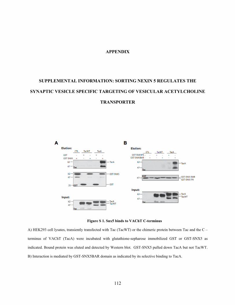
APPENDIX
SUPPLEMENTAL INFORMATION: SORTING NEXIN 5 REGULATES THE
SYNAPTIC VESICLE SPECIFIC TARGETING OF VESICULAR ACETYLCHOLINE
TRANSPORTER
Figure S 1. Snx5 binds to VAChT C-terminus
A) HEK293 cell lysates, transiently transfected with Tac (TacWT) or the chimeric protein between Tac and the C –
terminus of VAChT (TacA) were incubated with glutathione-sepharose immobilized GST or GST-SNX5 as
indicated. Bound protein was eluted and detected by Western blot. GST-SNX5 pulled down TacA but not TacWT.
B) Interaction is mediated by GST-SNX5BAR domain as indicated by its selective binding to TacA.
112
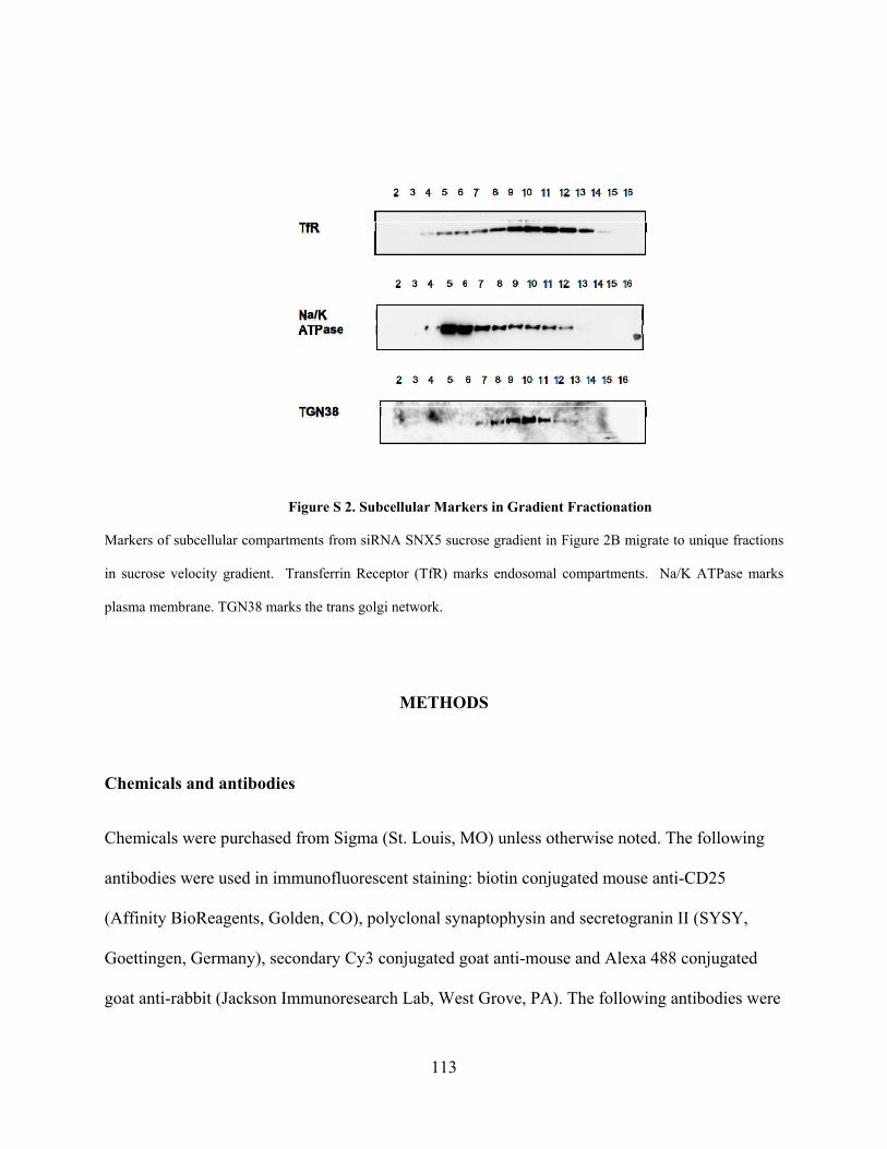
Figure S 2. Subcellular Markers in Gradient Fractionation
Markers of subcellular compartments from siRNA SNX5 sucrose gradient in Figure 2B migrate to unique fractions
in sucrose velocity gradient. Transferrin Receptor (TfR) marks endosomal compartments. Na/K ATPase marks
plasma membrane. TGN38 marks the trans golgi network.
METHODS
Chemicals and antibodies
Chemicals were purchased from Sigma (St. Louis, MO) unless otherwise noted. The following
antibodies were used in immunofluorescent staining: biotin conjugated mouse anti-CD25
(Affinity BioReagents, Golden, CO), polyclonal synaptophysin and secretogranin II (SYSY,
Goettingen, Germany), secondary Cy3 conjugated goat anti-mouse and Alexa 488 conjugated
goat anti-rabbit (Jackson Immunoresearch Lab, West Grove, PA). The following antibodies were
113
used for Western blot: polyclonal IL-2R alpha (Santa Cruz Biotech., Santa Cruz, CA),
monoclonal anti-IL2 Receptor (Covance, Princeton, NJ), polyclonal anti-synaptophysin and anti-
secretogranin II (SYSY, Goettingen, Germany), secondary HRP conjugated goat anti-mouse and
goat anti-rabbit (Pierce, Rockford, IL).
Plasmid construction and mutagenesis
Chimeric protein TacA was generated as previously described (citation) . Briefly, the C-terminus
of VAChT (a.a. 465-530) was amplified by PCR with the introduction of Xba1 and Xho1
restriction sites flanking the region. PCR product was digested and subcloned into Tac/pcDNA
3.1 as described (Tan et al., 1998). Glutathione S-transferase (GST)-tagged SNX5 and SNX5PX,
SNX5BAR plasmids were generated by cloning full length SNX5 (1-404), SNX5PX (1-179), or
SNX5BAR (180-404) cDNA into pGEX-6p-1 vector (Amersham) in frame with the GST coding
sequence. Similarly, HA-tagged SNX5 and its truncated mutants were made by inserting cDNAs
into a modified pcDNA3.1 vector in frame with the HA coding sequence. All constructs were
confirmed by sequencing.
Cell culture and transfection
All cells were maintained in 5% CO2 at 37ºC in medium containing penicillin and streptomycin
unless otherwise noted. PC12 cells were maintained in DMEM (Invitrogen) with 10% Equine
serum (Hyclone, Logan, UT), 5% Cosmic Calf serum (Hyclone, Logan, UT), and 2 mM L-
Glutamine (Invitrogen). Stable lines of wild type Tac, TacA, were generated as previously
described (Colgan et al., 2007). Hek293 cells were grown in DMEM with 10% fetal bovine
serum (Hyclone, Logan, UT). All cells were transfected using LipofectAMINE 2000
114
(Invitrogen, San Diego, CA) reagent or electroporation according to the manufacturer’s
instructions. PC12 cells were differentiated using 100 nM NGF for 24 hours. Transfected cells
were incubated at 37ºC for 36 - 48 hours before harvest.
Immunofluorescence and confocal microscopy
Immunofluorescent staining was performed as previously described (Liu et al., 2006). In brief,
cells seeded on glass coverslips were fixed with 4% paraformaldehyde in PBS, pH 7.4. After
fixation, cells were permeabilized and blocked for 30 min. in blocking buffer (BB, 2% BSA, 1%
fish skin gelatin, and 0.02% saponin in PBS). Cells were then incubated with primary antibody in
BB for 1 hour at room temperature. Coverslips were washed and incubated with the appropriate
Alexa- or Cy3- conjugated secondary antibody for 1 hour at room temperature. Confocal images
were acquired with a Fluoview 500 laser scanning confocal imaging system (Olympus, Tokyo,
Japan) configured with a fluorescence microscope fitted with Pan Apo 60 and 100 oil
objectives (Olympus). Confocal images were collected sequentially at 10241024 resolution to
minimize bleed through of fluorescence between channels.
Yeast-two-Hybrid
The MATCHMAKER LexA yeast two-hybrid system (BD Clontech) was used to identify the
interacting proteins for VAChT. The C-terminus of VAChT was used as bait to screen a PC12
cDNA library as described in the user manual (BD Clontech). Plasmids for both cDNA library
and bait were transformed sequentially into yeast strain EGY48 using the lithium acetate method
and cultured on SD selection medium according to the manufacturer’s manual (PT3040, BD
Clontech). The yeast transformants were restreaked on SD/His-/Trp-/Leu-/Ura- induction medium
115
plates containing Galactose, Raffinose and X-gal. Only transformants displaying blue color
within 3 days were considered as positive result of protein-protein interaction. Positive colonies
were restreaked for single colonies and then the cDNA plasmids were retrieved and the
contained cDNA inserts were PCR amplified for sequencing.
Immunoprecipitation
Expression and purification of GST fusion proteins were done as described previously (Liu et al,
2006). 40ug of GST-fusion proteins or control GST protein coupled to glutathione sepharose
beads were incubated with transfected HEK293 cell lysate at 4°C overnight. Beads were then
carefully washed for four times with washing buffer (50 mM Tris-HCl, pH 7.4; 150 mM NaCl,
0.1% NP-40 and protease inhibitors). The bound proteins were eluted using SDS–PAGE sample
buffer and were subjected to SDS–PAGE and immunoblotting with specific antibodies. Protein
bands were visualized by the ECL detection system (PIERCE).
Sucrose Gradient Fractionation
PC12 cells stably expressing, or transiently transfected with plasmid constructs, were harvested
in Buffer A (150 mM NaCl, 10 mM Hepes pH 7.4, 1 mM EGTA, 0.1mM MgCl2) with protease
inhibitors. Cells were cracked by eight passes through a cell cracker (Clearance-0.02um). Post-
nuclear supernatants (PNS) were then loaded onto prepared density gradients and spun in a
Beckman SW41 rotor. For sucrose density fractionation, sucrose gradients were prepared using a
gradient mixer to form continuous gradients with sucrose concentrations from 0.65 M to 1.55 M
and spun in SW41 rotor at 30,000 rpm for 8 hours at 4C. (Beckman Instruments, Palo Alto,
CA). Aliquots were taken from each gradient for western analysis as described below.
116
Two-step gradient for LDCV purification
LDCVs purification is performed by sequential fractionation through two sucrose gradients
(Stinchombe and Huttner, 1994). PC12 cells were loaded overnight with 2.5 Ci/ml [3H]
norepinephrine to label LDCVs. PNS was prepared as described above in the presence of
protease inhibitors. PNS was layered onto a 0.3-1.2 M sucrose gradient and sedimented at
25,400 rpm in an SW41 rotor for 30 min at 4C. Fractions were collected and radioactivity
measured in an aliquot of each fraction by scintillation counting. Pooled fractions containing the
bulk of the radioactivity were then loaded onto a 0.65-1.55 M sucrose gradient and sedimented to
equilibrium at 30,000 rpm in an SW41 rotor for 6-12 hours at 4C. Collected fractions were
loaded onto gels for western blotting detection as described.
Western blot analysis
Proteins were detected in gradient fractions by immunoblotting as previously described (Chen et
al., 2005). Equal amounts of gradient from each fraction were denatured in 3x SDS sample
buffer (New England Biolab, Beverly, MA), and separated by electrophoresis through 10% SDS-
PAGE. After electrophoresis, proteins were transferred to nitrocellulose (BA-85, Schleicher-
Schuell Bioscience, Keene, NH) and TacA chimeric proteins, synaptophysin, secretogranin II, or
other marker proteins were visualized by immunoblotting with appropriate antibodies in
combination with enhanced chemiluminescence (Super-Signal West Pico, Pierce, Rockford, IL).
Protein immunoreactive signals were scanned and the intensity of bands was semi-quantified
using the NIH Imaging program.
117
BIBLIOGRAPHY
Adell, A., and Artigas, F. (1991). Differential effects of clomipramine given locally or systemically on extracellular 5-hydroxytryptamine in raphe nuclei and frontal cortex. An in vivo brain microdialysis study. Naunyn Schmiedebergs Arch Pharmacol 343, 237-244.
Agoston, D.V., and Whittaker, V.P. (1989). Characterization, by size, density, osmotic fragility, and immunoaffinity, of acetylcholine- and vasoactive intestinal polypeptide-containing storage particles from myenteric neurones of the guinea-pig. J Neurochem 52, 1474-1480.
Ahnert-Hilger, G., Nurnberg, B., Exner, T., Schafer, T., and Jahn, R. (1998). The heterotrimeric G protein Go2 regulates catecholamine uptake by secretory vesicles. EMBO J 17, 406-413.
Alfonso, A., Grundahl, K., Duerr, J.S., Han, H.P., and Rand, J.B. (1993). The Caenorhabditis elegans unc-17 gene: a putative vesicular acetylcholine transporter. Science 261, 617-619.
Anderson, D.C., King, S.C., and Parsons, S.M. (1982). Proton gradient linkage to active uptake of [3H]acetylcholine by Torpedo electric organ synaptic vesicles. Biochemistry 21, 3037-3043.
Atluri, P.P., and Ryan, T.A. (2006). The kinetics of synaptic vesicle reacidification at hippocampal nerve terminals. J Neurosci 26, 2313-2320.
Augustine, G.J., and Kasai, H. (2007). Bernard Katz, quantal transmitter release and the foundations of presynaptic physiology. J Physiol 578, 623-625.
Balaji, J., Desai, R., Kaushalya, S.K., Eaton, M.J., and Maiti, S. (2005). Quantitative measurement of serotonin synthesis and sequestration in individual live neuronal cells. J Neurochem 95, 1217-1226.
Barbosa, J., Jr., Ferreira, L.T., Martins-Silva, C., Santos, M.S., Torres, G.E., Caron, M.G., Gomez, M.V., Ferguson, S.S., Prado, M.A., and Prado, V.F. (2002). Trafficking of the vesicular
118
acetylcholine transporter in SN56 cells: a dynamin-sensitive step and interaction with the AP-2 adaptor complex. J Neurochem 82, 1221-1228.
Bauerfeind, R., Regnier-Vigouroux, A., Flatmark, T., and Huttner, W.B. (1993). Selective storage of acetylcholine, but not catecholamines, in neuroendocrine synaptic-like microvesicles of early endosomal origin. Neuron 11, 105-121.
Bellocchio, E.E., Reimer, R.J., Fremeau, R.T., Jr., and Edwards, R.H. (2000). Uptake of glutamate into synaptic vesicles by an inorganic phosphate transporter. Science 289, 957-960.
Bjorklund, A., Baumgarten, H.G., and Nobin, A. (1974). Chemical lesioning of central monoamine axons by means of 5,6-dihydroxytryptamine and 5,7-dihydroxytryptamine. Adv Biochem Psychopharmacol 10, 13-33.
Bjorklund, A., Horn, A.S., Baumgarten, H.G., Nobin, A., and Schlossberger, H.G. (1975). Neurotoxicity of hydroxylated tryptamines: structure-activity relationships. 2. In vitro studies on monoamine uptake inhibition and uptake impairment. Acta Physiol Scand Suppl 429, 29-60.
Blagoveshchenskaya, A.D., Hewitt, E.W., and Cutler, D.F. (1999). Di-Leucine Signals Mediate Targeting of Tyrosinase and Synaptotagmin to Synaptic-like Microvesicles within PC12 Cells. Mol. Biol. Cell 10, 3979-3990.
Blier, P., Pineyro, G., el Mansari, M., Bergeron, R., and de Montigny, C. (1998). Role of somatodendritic 5-HT autoreceptors in modulating 5-HT neurotransmission. Ann N Y Acad Sci 861, 204-216.
Bonifacino, J.S., and Hurley, J.H. (2008). Retromer. Curr Opin Cell Biol 20, 427-436.
Bonifacino, J.S., and Traub, L.M. (2003). Signals for sorting of transmembrane proteins to endosomes and lysosomes. Annu Rev Biochem 72, 395-447.
Brown, J.M., Hanson, G.R., and Fleckenstein, A.E. (2001). Cocaine-induced increases in vesicular dopamine uptake: role of dopamine receptors. J Pharmacol Exp Ther 298, 1150-1153.
Brunk, I., Blex, C., Rachakonda, S., Holtje, M., Winter, S., Pahner, I., Walther, D.J., and Ahnert-Hilger, G. (2006). The first luminal domain of vesicular monoamine transporters mediates G-protein-dependent regulation of transmitter uptake. J Biol Chem 281, 33373-33385.
119
Burke, N.V., Han, W., Li, D., Takimoto, K., Watkins, S.C., and Levitan, E.S. (1997). Neuronal peptide release is limited by secretory granule mobility. Neuron 19, 1095-1102.
Burrone, J., Li, Z., and Murthy, V.N. (2006). Studying vesicle cycling in presynaptic terminals using the genetically encoded probe synaptopHluorin. Nat Protoc 1, 2970-2978.
Carlton, J.G., and Cullen, P.J. (2005). Sorting nexins. Curr Biol 15, R819-820.
Ceccarelli, B., Hurlbut, W.P., and Mauro, A. (1973). Turnover of transmitter and synaptic vesicles at the frog neuromuscular junction. J Cell Biol 57, 499-524.
Chaudhry, F.A., Edwards, R.H., and Fonnum, F. (2008). Vesicular neurotransmitter transporters as targets for endogenous and exogenous toxic substances. Annu Rev Pharmacol Toxicol 48, 277-301.
Chen, C.X., Huang, S.Y., Zhang, L., and Liu, Y.J. (2005). Synaptophysin enhances the neuroprotection of VMAT2 in MPP+-induced toxicity in MN9D cells. Neurobiol Dis 19, 419-426.
Cho, G.W., Kim, M.H., Chai, Y.G., Gilmor, M.L., Levey, A.I., and Hersh, L.B. (2000). Phosphorylation of the rat vesicular acetylcholine transporter. J Biol Chem 275, 19942-19948.
Cidon, S., and Sihra, T.S. (1989). Characterization of a H+-ATPase in rat brain synaptic vesicles. Coupling to L-glutamate transport. J Biol Chem 264, 8281-8288.
Clift-O'Grady, L., Desnos, C., Lichtenstein, Y., Faundez, V., Horng, J.T., and Kelly, R.B. (1998). Reconstitution of synaptic vesicle biogenesis from PC12 cell membranes. Methods 16, 150-159.
Clift-O'Grady, L., Linstedt, A.D., Lowe, A.W., Grote, E., and Kelly, R.B. (1990). Biogenesis of synaptic vesicle-like structures in a pheochromocytoma cell line PC-12. J Cell Biol 110, 1693-1703.
Colebrooke, R.E., Humby, T., Lynch, P.J., McGowan, D.P., Xia, J., and Emson, P.C. (2006). Age-related decline in striatal dopamine content and motor performance occurs in the absence of nigral cell loss in a genetic mouse model of Parkinson's disease. Eur J Neurosci 24, 2622-2630.
120
Colgan, L., Liu, H., Huang, S.Y., and Liu, Y.J. (2007). Dileucine motif is sufficient for internalization and synaptic vesicle targeting of vesicular acetylcholine transporter. Traffic 8, 512-522.
Collier, B. (1969). The preferential release of newly synthesized transmitter by a sympathetic ganglion. J Physiol 205, 341-352.
Cope, D.L., Lee, S., Melvin, D.R., and Gould, G.W. (2000). Identification of further important residues within the Glut4 carboxy-terminal tail which regulate subcellular trafficking. FEBS Lett 481, 261-265.
Cousin, M.A., and Robinson, P.J. (1999). Mechanisms of synaptic vesicle recycling illuminated by fluorescent dyes. J Neurochem 73, 2227-2239.
Croft, B.G., Fortin, G.D., Corera, A.T., Edwards, R.H., Beaudet, A., Trudeau, L.E., and Fon, E.A. (2005). Normal biogenesis and cycling of empty synaptic vesicles in dopamine neurons of vesicular monoamine transporter 2 knockout mice. Mol Biol Cell 16, 306-315.
Daly, C., Sugimori, M., Moreira, J.E., Ziff, E.B., and Llinas, R. (2000). Synaptophysin regulates clathrin-independent endocytosis of synaptic vesicles. Proc Natl Acad Sci U S A 97, 6120-6125.
Daly, C., and Ziff, E.B. (2002). Ca2+-dependent formation of a dynamin-synaptophysin complex: potential role in synaptic vesicle endocytosis. J Biol Chem 277, 9010-9015.
Darna, M., Schmutz, I., Richter, K., Yelamanchili, S.V., Pendyala, G., Holtje, M., Albrecht, U., and Ahnert-Hilger, G. (2008). Time-of-day dependent sorting of vesicular glutamate transporter to the plasma membrane. J Biol Chem.
De Camilli, P., and Takei, K. (1996). Molecular mechanisms in synaptic vesicle endocytosis and recycling. Neuron 16, 481-486.
de Castro, B.M., Pereira, G.S., Magalhaes, V., Rossato, J.I., De Jaeger, X., Martins-Silva, C., Leles, B., Lima, P., Gomez, M.V., Gainetdinov, R.R., et al. (2008). Reduced expression of the vesicular acetylcholine transporter causes learning deficits in mice. Genes Brain Behav.
De Gois, S., Jeanclos, E., Morris, M., Grewal, S., Varoqui, H., and Erickson, J.D. (2006). Identification of Endophilins 1 and 3 as Selective Binding Partners for VGLUT1 and Their Co-Localization in Neocortical Glutamatergic Synapses: Implications for Vesicular Glutamate Transporter Trafficking and Excitatory Vesicle Formation. Cell Mol Neurobiol.
121
de Kock, C.P., Cornelisse, L.N., Burnashev, N., Lodder, J.C., Timmerman, A.J., Couey, J.J., Mansvelder, H.D., and Brussaard, A.B. (2006). NMDA receptors trigger neurosecretion of 5-HT within dorsal raphe nucleus of the rat in the absence of action potential firing. J Physiol 577, 891-905.
de Lange, R.P., de Roos, A.D., and Borst, J.G. (2003). Two modes of vesicle recycling in the rat calyx of Held. J Neurosci 23, 10164-10173.
Del Castillo, J., and Katz, B. (1954). Quantal components of the end-plate potential. J Physiol 124, 560-573.
Del Castillo, J., and Katz, B. (1956). Biophysical aspects of neuro-muscular transmission. Prog Biophys Biophys Chem 6, 121-170.
Dittie, A.S., Hajibagheri, N., and Tooze, S.A. (1996). The AP-1 adaptor complex binds to immature secretory granules from PC12 cells, and is regulated by ADP-ribosylation factor. J Cell Biol 132, 523-536.
Doherty, P., Hawgood, B.J., and Smith, I.C. (1984). Changes in miniature end-plate potentials after brief nervous stimulation at the frog neuromuscular junction. J Physiol 356, 349-358.
Edwards, R.H. (1998). Neurotransmitter release: variations on a theme. Curr Biol 8, R883-885.
Edwards, R.H. (2007). The neurotransmitter cycle and quantal size. Neuron 55, 835-858.
Eiden, L.E. (2000). The vesicular neurotransmitter transporters: current perspectives and future prospects. FASEB J 14, 2396-2400.
Emmanuel Pothos, M.D. (1996). l-3,4-Dihydroxyphenylalanine Increases the Quantal Size of Exocytotic Dopamine Release In Vitro. Journal of Neurochemistry 66, 629-636.
Erickson, J.D., Eiden, L.E., and Hoffman, B.J. (1992). Expression cloning of a reserpine-sensitive vesicular monoamine transporter. Proc Natl Acad Sci U S A 89, 10993-10997.
Erickson, J.D., Varoqui, H., Schafer, M.K., Modi, W., Diebler, M.F., Weihe, E., Rand, J., Eiden, L.E., Bonner, T.I., and Usdin, T.B. (1994). Functional identification of a vesicular acetylcholine transporter and its expression from a "cholinergic" gene locus. J Biol Chem 269, 21929-21932.
122
Ertunc, M., Sara, Y., Chung, C., Atasoy, D., Virmani, T., and Kavalali, E.T. (2007). Fast synaptic vesicle reuse slows the rate of synaptic depression in the CA1 region of hippocampus. J Neurosci 27, 341-354.
Faundez, V., Horng, J.T., and Kelly, R.B. (1998). A function for the AP3 coat complex in synaptic vesicle formation from endosomes. Cell 93, 423-432.
Fei, H., Grygoruk, A., Brooks, E.S., Chen, A., and Krantz, D.E. (2008). Trafficking of vesicular neurotransmitter transporters. Traffic 9, 1425-1436.
Ferreira, L.T., Santos, M.S., Kolmakova, N.G., Koenen, J., Barbosa, J., Jr., Gomez, M.V., Guatimosim, C., Zhang, X., Parsons, S.M., Prado, V.F., and Prado, M.A. (2005). Structural requirements for steady-state localization of the vesicular acetylcholine transporter. J Neurochem 94, 957-969.
Fleckenstein, A.E., and Hanson, G.R. (2003). Impact of psychostimulants on vesicular monoamine transporter function. Eur J Pharmacol 479, 283-289.
Fon, E.A., Pothos, E.N., Sun, B.C., Killeen, N., Sulzer, D., and Edwards, R.H. (1997). Vesicular transport regulates monoamine storage and release but is not essential for amphetamine action. Neuron 19, 1271-1283.
Freis, E.D. (1954). Mental depression in hypertensive patients treated for long periods with large doses of reserpine. N Engl J Med 251, 1006-1008.
Frerking, M., and Wilson, M. (1996). Saturation of postsynaptic receptors at central synapses? Curr Opin Neurobiol 6, 395-403.
Fukui, M., Rodriguiz, R.M., Zhou, J., Jiang, S.X., Phillips, L.E., Caron, M.G., and Wetsel, W.C. (2007). Vmat2 heterozygous mutant mice display a depressive-like phenotype. J Neurosci 27, 10520-10529.
Fulop, T., and Smith, C. (2006). Physiological stimulation regulates the exocytic mode through calcium activation of protein kinase C in mouse chromaffin cells. Biochem J 399, 111-119.
Gainetdinov, R.R., Jones, S.R., Fumagalli, F., Wightman, R.M., and Caron, M.G. (1998). Re-evaluation of the role of the dopamine transporter in dopamine system homeostasis. Brain Res Brain Res Rev 26, 148-153.
123
Garippa, R.J., Johnson, A., Park, J., Petrush, R.L., and McGraw, T.E. (1996). The carboxyl terminus of GLUT4 contains a serine-leucine-leucine sequence that functions as a potent internalization motif in Chinese hamster ovary cells. J Biol Chem 271, 20660-20668.
Gerace, L., and Burke, B. (1988). Functional organization of the nuclear envelope. Annu Rev Cell Biol 4, 335-374.
Gershon, M.D., and Sherman, D. (1982). Selective demonstration of serotonergic neurons and terminals in electron micrographs: loading with 5,7-dihydroxytryptamine and fixation with NaMnO4. J Histochem Cytochem 30, 769-773.
Gilmor, M.L., Nash, N.R., Roghani, A., Edwards, R.H., Yi, H., Hersch, S.M., and Levey, A.I. (1996). Expression of the putative vesicular acetylcholine transporter in rat brain and localization in cholinergic synaptic vesicles. J Neurosci 16, 2179-2190.
Grote, E., Hao, J.C., Bennett, M.K., and Kelly, R.B. (1995). A targeting signal in VAMP regulating transport to synaptic vesicles. Cell 81, 581-589.
Guo, J.T., Chen, A.Q., Kong, Q., Zhu, H., Ma, C.M., and Qin, C. (2008). Inhibition of vesicular monoamine transporter-2 activity in alpha-synuclein stably transfected SH-SY5Y cells. Cell Mol Neurobiol 28, 35-47.
Hahn, J., Kullmann, P.H., Horn, J.P., and Levitan, E.S. (2006). D2 autoreceptors chronically enhance dopamine neuron pacemaker activity. J Neurosci 26, 5240-5247.
Hahn, J., Tse, T.E., and Levitan, E.S. (2003). Long-term K+ channel-mediated dampening of dopamine neuron excitability by the antipsychotic drug haloperidol. J Neurosci 23, 10859-10866.
Han, W., Li, D., Stout, A.K., Takimoto, K., and Levitan, E.S. (1999a). Ca2+-induced deprotonation of peptide hormones inside secretory vesicles in preparation for release. J Neurosci 19, 900-905.
Han, W., Ng, Y.K., Axelrod, D., and Levitan, E.S. (1999b). Neuropeptide release by efficient recruitment of diffusing cytoplasmic secretory vesicles. Proc Natl Acad Sci U S A 96, 14577-14582.
Han, W., Rhee, J.S., Maximov, A., Lao, Y., Mashimo, T., Rosenmund, C., and Sudhof, T.C. (2004). N-glycosylation is essential for vesicular targeting of synaptotagmin 1. Neuron 41, 85-99.
124
Hannah, M.J., Schmidt, A.A., and Huttner, W.B. (1999). Synaptic vesicle biogenesis. Annu Rev Cell Dev Biol 15, 733-798.
Hastings, T.G., and Zigmond, M.J. (1997). Loss of dopaminergic neurons in parkinsonism: possible role of reactive dopamine metabolites. J Neural Transm Suppl 49, 103-110.
He, L., Wu, X.S., Mohan, R., and Wu, L.G. (2006). Two modes of fusion pore opening revealed by cell-attached recordings at a synapse. Nature 444, 102-105.
Herman, G.A., Bonzelius, F., Cieutat, A.M., and Kelly, R.B. (1994). A distinct class of intracellular storage vesicles, identified by expression of the glucose transporter GLUT4. Proc Natl Acad Sci U S A 91, 12750-12754.
Heuser, J.E. (1989). Review of electron microscopic evidence favouring vesicle exocytosis as the structural basis for quantal release during synaptic transmission. Q J Exp Physiol 74, 1051-1069.
Heuser, J.E., and Reese, T.S. (1973). Evidence for recycling of synaptic vesicle membrane during transmitter release at the frog neuromuscular junction. J Cell Biol 57, 315-344.
Hillarp, N.A. (1958a). Isolation and some biochemical properties of the catechol amine granules in the cow adrenal medulla. Acta Physiol Scand 43, 82-96.
Hillarp, N.A. (1958b). The release of catechol amines from the amine containing granules of the adrenal medulla. Acta Physiol Scand 43, 292-302.
Hillarp, N.A. (1960). Effect of reserpine on the nucleotide and catecholamine content of the denervated adrenal medulla of the rat. Nature 187, 1032.
Hogan, K.A., Staal, R.G., and Sonsalla, P.K. (2000). Analysis of VMAT2 binding after methamphetamine or MPTP treatment: disparity between homogenates and vesicle preparations. J Neurochem 74, 2217-2220.
Holtje, M., von Jagow, B., Pahner, I., Lautenschlager, M., Hortnagl, H., Nurnberg, B., Jahn, R., and Ahnert-Hilger, G. (2000). The neuronal monoamine transporter VMAT2 is regulated by the trimeric GTPase Go(2). J Neurosci 20, 2131-2141.
Holtje, M., Winter, S., Walther, D., Pahner, I., Hortnagl, H., Ottersen, O.P., Bader, M., and Ahnert-Hilger, G. (2003). The vesicular monoamine content regulates VMAT2 activity through
125
Galphaq in mouse platelets. Evidence for autoregulation of vesicular transmitter uptake. J Biol Chem 278, 15850-15858.
Iversen, L. (2000). Neurotransmitter transporters: fruitful targets for CNS drug discovery. Mol Psychiatry 5, 357-362.
Jackson, M.B., and Chapman, E.R. (2008). The fusion pores of Ca(2+)-triggered exocytosis. Nat Struct Mol Biol 15, 684-689.
Jonsson, G., and Sachs, C. (1969). Subcellular distribution of 3H-noradrenaline in adrenergic nerves of mouse atrium--effect of reserpine, monoamine oxidase and tyrosine hydroxylase inhibition. Acta Physiol Scand 77, 344-357.
Kakhlon, O., Sakya, P., Larijani, B., Watson, R., and Tooze, S.A. (2006). GGA function is required for maturation of neuroendocrine secretory granules. EMBO J 25, 1590-1602.
Kantheti, P., Qiao, X., Diaz, M.E., Peden, A.A., Meyer, G.E., Carskadon, S.L., Kapfhamer, D., Sufalko, D., Robinson, M.S., Noebels, J.L., and Burmeister, M. (1998). Mutation in AP-3 delta in the mocha mouse links endosomal transport to storage deficiency in platelets, melanosomes, and synaptic vesicles. Neuron 21, 111-122.
Kaushalya, S.K., Desai, R., Arumugam, S., Ghosh, H., Balaji, J., and Maiti, S. (2008a). Three-photon microscopy shows that somatic release can be a quantitatively significant component of serotonergic neurotransmission in the mammalian brain. J Neurosci Res 86, 3469-3480.
Kaushalya, S.K., Nag, S., Ghosh, H., Arumugam, S., and Maiti, S. (2008b). A high-resolution large area serotonin map of a live rat brain section. Neuroreport 19, 717-721.
Kim, M.H., and Hersh, L.B. (2004). The vesicular acetylcholine transporter interacts with clathrin-associated adaptor complexes AP-1 and AP-2. J Biol Chem 279, 12580-12587.
Kitamoto, T., Xie, X., Wu, C.F., and Salvaterra, P.M. (2000). Isolation and characterization of mutants for the vesicular acetylcholine transporter gene in Drosophila melanogaster. J Neurobiol 42, 161-171.
Klumperman, J., Kuliawat, R., Griffith, J.M., Geuze, H.J., and Arvan, P. (1998). Mannose 6-phosphate receptors are sorted from immature secretory granules via adaptor protein AP-1, clathrin, and syntaxin 6-positive vesicles. J Cell Biol 141, 359-371.
126
Klyachko, V.A., and Jackson, M.B. (2002). Capacitance steps and fusion pores of small and large-dense-core vesicles in nerve terminals. Nature 418, 89-92.
Knoth, J., Zallakian, M., and Njus, D. (1981). Stoichiometry of H+-linked dopamine transport in chromaffin granule ghosts. Biochemistry 20, 6625-6629.
Koenig, J.H., and Ikeda, K. (1996). Synaptic vesicles have two distinct recycling pathways. J Cell Biol 135, 797-808.
Krantz, D.E., Waites, C., Oorschot, V., Liu, Y., Wilson, R.I., Tan, P.K., Klumperman, J., and Edwards, R.H. (2000). A phosphorylation site regulates sorting of the vesicular acetylcholine transporter to dense core vesicles. J Cell Biol 149, 379-396.
Letourneur, F., and Klausner, R.D. (1991). T-cell and basophil activation through the cytoplasmic tail of T-cell-receptor zeta family proteins. Proc Natl Acad Sci U S A 88, 8905-8909.
Li, H., Waites, C.L., Staal, R.G., Dobryy, Y., Park, J., Sulzer, D.L., and Edwards, R.H. (2005). Sorting of vesicular monoamine transporter 2 to the regulated secretory pathway confers the somatodendritic exocytosis of monoamines. Neuron 48, 619-633.
Li, Z., and Murthy, V.N. (2001). Visualizing postendocytic traffic of synaptic vesicles at hippocampal synapses. Neuron 31, 593-605.
Liu, G., Choi, S., and Tsien, R.W. (1999). Variability of neurotransmitter concentration and nonsaturation of postsynaptic AMPA receptors at synapses in hippocampal cultures and slices. Neuron 22, 395-409.
Liu, H., Liu, Z.Q., Chen, C.X., Magill, S., Jiang, Y., and Liu, Y.J. (2006). Inhibitory regulation of EGF receptor degradation by sorting nexin 5. Biochem Biophys Res Commun 342, 537-546.
Liu, Y., and Edwards, R.H. (1997). The role of vesicular transport proteins in synaptic transmission and neural degeneration. Annu Rev Neurosci 20, 125-156.
Liu, Y., Peter, D., Merickel, A., Krantz, D., Finn, J.P., and Edwards, R.H. (1996). A molecular analysis of vesicular amine transport. Behav Brain Res 73, 51-58.
127
Liu, Y., Peter, D., Roghani, A., Schuldiner, S., Prive, G.G., Eisenberg, D., Brecha, N., and Edwards, R.H. (1992a). A cDNA that suppresses MPP+ toxicity encodes a vesicular amine transporter. Cell 70, 539-551.
Liu, Y., Roghani, A., and Edwards, R.H. (1992b). Gene transfer of a reserpine-sensitive mechanism of resistance to N-methyl-4-phenylpyridinium. Proc Natl Acad Sci U S A 89, 9074-9078.
Liu, Y., Schweitzer, E.S., Nirenberg, M.J., Pickel, V.M., Evans, C.J., and Edwards, R.H. (1994). Preferential localization of a vesicular monoamine transporter to dense core vesicles in PC12 cells. J Cell Biol 127, 1419-1433.
Lundberg, J.M., Fried, G., Fahrenkrug, J., Holmstedt, B., Hokfelt, T., Lagercrantz, H., Lundgren, G., and Anggard, A. (1981). Subcellular fractionation of cat submandibular gland: comparative studies on the distribution of acetylcholine and vasoactive intestinal polypeptide (VIP). Neuroscience 6, 1001-1010.
Mainen, Z.F., Malinow, R., and Svoboda, K. (1999). Synaptic calcium transients in single spines indicate that NMDA receptors are not saturated. Nature 399, 151-155.
Maiti, S., Shear, J.B., Williams, R.M., Zipfel, W.R., and Webb, W.W. (1997). Measuring serotonin distribution in live cells with three-photon excitation. Science 275, 530-532.
Malenka, R.C. (2003). Synaptic plasticity and AMPA receptor trafficking. Ann N Y Acad Sci 1003, 1-11.
Marks, M.S., Roche, P.A., van Donselaar, E., Woodruff, L., Peters, P.J., and Bonifacino, J.S. (1995). A lysosomal targeting signal in the cytoplasmic tail of the beta chain directs HLA-DM to MHC class II compartments. J Cell Biol 131, 351-369.
Maron, R., Fishkes, H., Kanner, B.I., and Schuldiner, S. (1979). Solubilization and reconstitution of the catecholamine transporter from bovine chromaffin granules. Biochemistry 18, 4781-4785.
Marshall, I.G. (1970). Studies on the blocking action of 2-(4-phenyl piperidino) cyclohexanol (AH5183). Br J Pharmacol 38, 503-516.
Martin, T.F. (2003). Tuning exocytosis for speed: fast and slow modes. Biochim Biophys Acta 1641, 157-165.
128
McAllister, A.K., and Stevens, C.F. (2000). Nonsaturation of AMPA and NMDA receptors at hippocampal synapses. Proc Natl Acad Sci U S A 97, 6173-6178.
McIntire, S.L., Reimer, R.J., Schuske, K., Edwards, R.H., and Jorgensen, E.M. (1997). Identification and characterization of the vesicular GABA transporter. Nature 389, 870-876.
Merino-Trigo, A., Kerr, M.C., Houghton, F., Lindberg, A., Mitchell, C., Teasdale, R.D., and Gleeson, P.A. (2004). Sorting nexin 5 is localized to a subdomain of the early endosomes and is recruited to the plasma membrane following EGF stimulation. J Cell Sci 117, 6413-6424.
Mooslehner, K.A., Chan, P.M., Xu, W., Liu, L., Smadja, C., Humby, T., Allen, N.D., Wilkinson, L.S., and Emson, P.C. (2001). Mice with very low expression of the vesicular monoamine transporter 2 gene survive into adulthood: potential mouse model for parkinsonism. Mol Cell Biol 21, 5321-5331.
Moriyama, Y., and Yamamoto, A. (1995). Vesicular L-glutamate transporter in microvesicles from bovine pineal glands. Driving force, mechanism of chloride anion activation, and substrate specificity. J Biol Chem 270, 22314-22320.
Mosharov, E.V., Staal, R.G., Bove, J., Prou, D., Hananiya, A., Markov, D., Poulsen, N., Larsen, K.E., Moore, C.M., Troyer, M.D., et al. (2006). Alpha-synuclein overexpression increases cytosolic catecholamine concentration. J Neurosci 26, 9304-9311.
Nakatsu, F., Okada, M., Mori, F., Kumazawa, N., Iwasa, H., Zhu, G., Kasagi, Y., Kamiya, H., Harada, A., Nishimura, K., et al. (2004). Defective function of GABA-containing synaptic vesicles in mice lacking the AP-3B clathrin adaptor. J Cell Biol 167, 293-302.
Nirenberg, M.J., Chan, J., Liu, Y., Edwards, R.H., and Pickel, V.M. (1996). Ultrastructural localization of the vesicular monoamine transporter-2 in midbrain dopaminergic neurons: potential sites for somatodendritic storage and release of dopamine. J Neurosci 16, 4135-4145.
Okada, Y., Yamazaki, H., Sekine-Aizawa, Y., and Hirokawa, N. (1995). The neuron-specific kinesin superfamily protein KIF1A is a unique monomeric motor for anterograde axonal transport of synaptic vesicle precursors. Cell 81, 769-780.
Otsuki, T., Kajigaya, S., Ozawa, K., and Liu, J.M. (1999). SNX5, a new member of the sorting nexin family, binds to the Fanconi anemia complementation group A protein. Biochem Biophys Res Commun 265, 630-635.
129
Parsons, R.L., Calupca, M.A., Merriam, L.A., and Prior, C. (1999). Empty synaptic vesicles recycle and undergo exocytosis at vesamicol-treated motor nerve terminals. J Neurophysiol 81, 2696-2700.
Partilla, J.S., Dempsey, A.G., Nagpal, A.S., Blough, B.E., Baumann, M.H., and Rothman, R.B. (2006). Interaction of amphetamines and related compounds at the vesicular monoamine transporter. J Pharmacol Exp Ther 319, 237-246.
Partoens, P., Slembrouck, D., Quatacker, J., Baudhuin, P., Courtoy, P.J., and De Potter, W.P. (1998). Retrieved constituents of large dense-cored vesicles and synaptic vesicles intermix in stimulation-induced early endosomes of noradrenergic neurons. J Cell Sci 111 ( Pt 6), 681-689.
Pennuto, M., Bonanomi, D., Benfenati, F., and Valtorta, F. (2003). Synaptophysin I Controls the Targeting of VAMP2/Synaptobrevin II to Synaptic Vesicles. Mol. Biol. Cell 14, 4909-4919.
Peter, D., Liu, Y., Sternini, C., de Giorgio, R., Brecha, N., and Edwards, R.H. (1995). Differential expression of two vesicular monoamine transporters. J Neurosci 15, 6179-6188.
Poodry, C.A., and Edgar, L. (1979). Reversible alteration in the neuromuscular junctions of Drosophila melanogaster bearing a temperature-sensitive mutation, shibire. J Cell Biol 81, 520-527.
Pothos, E.N., Larsen, K.E., Krantz, D.E., Liu, Y., Haycock, J.W., Setlik, W., Gershon, M.D., Edwards, R.H., and Sulzer, D. (2000). Synaptic vesicle transporter expression regulates vesicle phenotype and quantal size. J Neurosci 20, 7297-7306.
Prado, V.F., Martins-Silva, C., de Castro, B.M., Lima, R.F., Barros, D.M., Amaral, E., Ramsey, A.J., Sotnikova, T.D., Ramirez, M.R., Kim, H.G., et al. (2006). Mice deficient for the vesicular acetylcholine transporter are myasthenic and have deficits in object and social recognition. Neuron 51, 601-612.
Prado, V.F., and Prado, M.A. (2002). Signals involved in targeting membrane proteins to synaptic vesicles. Cell Mol Neurobiol 22, 565-577.
Pyle, J.L., Kavalali, E.T., Piedras-Renteria, E.S., and Tsien, R.W. (2000). Rapid reuse of readily releasable pool vesicles at hippocampal synapses. Neuron 28, 221-231.
Rand, J.B. (1989). Genetic analysis of the cha-1-unc-17 gene complex in Caenorhabditis. Genetics 122, 73-80.
130
Richards, D.A., Guatimosim, C., and Betz, W.J. (2000). Two endocytic recycling routes selectively fill two vesicle pools in frog motor nerve terminals. Neuron 27, 551-559.
Riddle, E.L., Topham, M.K., Haycock, J.W., Hanson, G.R., and Fleckenstein, A.E. (2002). Differential trafficking of the vesicular monoamine transporter-2 by methamphetamine and cocaine. Eur J Pharmacol 449, 71-74.
Rizzoli, S.O., and Jahn, R. (2007). Kiss-and-run, collapse and 'readily retrievable' vesicles. Traffic 8, 1137-1144.
Robertson, J.D. (1956). The ultrastructure of a reptilian myoneural junction. J Biophys Biochem Cytol 2, 381-394.
Roghani, A., Feldman, J., Kohan, S.A., Shirzadi, A., Gundersen, C.B., Brecha, N., and Edwards, R.H. (1994). Molecular cloning of a putative vesicular transporter for acetylcholine. Proc Natl Acad Sci U S A 91, 10620-10624.
Santos, M.S., Barbosa, J., Jr., Veloso, G.S., Ribeiro, F., Kushmerick, C., Gomez, M.V., Ferguson, S.S., Prado, V.F., and Prado, M.A. (2001). Trafficking of green fluorescent protein tagged-vesicular acetylcholine transporter to varicosities in a cholinergic cell line. J Neurochem 78, 1104-1113.
Sawada, K., Echigo, N., Juge, N., Miyaji, T., Otsuka, M., Omote, H., Yamamoto, A., and Moriyama, Y. (2008). Identification of a vesicular nucleotide transporter. Proc Natl Acad Sci U S A 105, 5683-5686.
Schlossberger, H.G. (1978). Synthesis and chemical properties of some indole derivatives. Annals of the New York Academy of Sciences 305, 25-35.
Silva, N.L., Mariani, A.P., Harrison, N.L., and Barker, J.L. (1988). 5,7-Dihydroxytryptamine identifies living dopaminergic neurons in mesencephalic cultures. Proc Natl Acad Sci U S A 85, 7346-7350.
Smith, R., Brundin, P., and Li, J.Y. (2005). Synaptic dysfunction in Huntington's disease: a new perspective. Cell Mol Life Sci 62, 1901-1912.
Smith, R., Chung, H., Rundquist, S., Maat-Schieman, M.L., Colgan, L., Englund, E., Liu, Y.J., Roos, R.A., Faull, R.L., Brundin, P., and Li, J.Y. (2006). Cholinergic neuronal defect without cell loss in Huntington's disease. Hum Mol Genet 15, 3119-3131.
131
Song, H., Ming, G., Fon, E., Bellocchio, E., Edwards, R.H., and Poo, M. (1997). Expression of a putative vesicular acetylcholine transporter facilitates quantal transmitter packaging. Neuron 18, 815-826.
Staal, R.G., Hananiya, A., and Sulzer, D. (2008). PKC theta activity maintains normal quantal size in chromaffin cells. J Neurochem 105, 1635-1641.
Steinert, J.R., Kuromi, H., Hellwig, A., Knirr, M., Wyatt, A.W., Kidokoro, Y., and Schuster, C.M. (2006). Experience-dependent formation and recruitment of large vesicles from reserve pool. Neuron 50, 723-733.
Sudhof, T.C. (2004). The synaptic vesicle cycle. Annu Rev Neurosci 27, 509-547.
Sulzer, D., Pothos, E., Sung, H.M., Maidment, N.T., Hoebel, B.G., and Rayport, S. (1992). Weak base model of amphetamine action. Ann N Y Acad Sci 654, 525-528.
Svoboda, K., and Yasuda, R. (2006). Principles of Two-Photon Excitation Microscopy and Its Applications to Neuroscience. Neuron 50, 823-839.
Tabares, L., Ruiz, R., Linares-Clemente, P., Gaffield, M.A., Alvarez de Toledo, G., Fernandez-Chacon, R., and Betz, W.J. (2007). Monitoring synaptic function at the neuromuscular junction of a mouse expressing synaptopHluorin. J Neurosci 27, 5422-5430.
Takahashi, N., Miner, L.L., Sora, I., Ujike, H., Revay, R.S., Kostic, V., Jackson-Lewis, V., Przedborski, S., and Uhl, G.R. (1997). VMAT2 knockout mice: heterozygotes display reduced amphetamine-conditioned reward, enhanced amphetamine locomotion, and enhanced MPTP toxicity. Proc Natl Acad Sci U S A 94, 9938-9943.
Takamori, S., Holt, M., Stenius, K., Lemke, E.A., Gronborg, M., Riedel, D., Urlaub, H., Schenck, S., Brugger, B., Ringler, P., et al. (2006). Molecular anatomy of a trafficking organelle. Cell 127, 831-846.
Takamori, S., Rhee, J.S., Rosenmund, C., and Jahn, R. (2000). Identification of a vesicular glutamate transporter that defines a glutamatergic phenotype in neurons. Nature 407, 189-194.
Tan, P.K., Waites, C., Liu, Y., Krantz, D.E., and Edwards, R.H. (1998). A leucine-based motif mediates the endocytosis of vesicular monoamine and acetylcholine transporters. J Biol Chem 273, 17351-17360.
132
Thoidis, G., and Kandror, K.V. (2001). A Glut4-vesicle marker protein, insulin-responsive aminopeptidase, is localized in a novel vesicular compartment in PC12 cells. Traffic 2, 577-587.
Toll, L., and Howard, B.D. (1980). Evidence that an ATPase and a protonmotive force function in the transport of acetylcholine into storage vesicles. J Biol Chem 255, 1787-1789.
Tooze, S.A., and Stinchcombe, J.C. (1992). Biogenesis of secretory granules. Semin Cell Biol 3, 357-366.
Van der Kloot, W., and Branisteanu, D.D. (1992). Effects of activators and inhibitors of protein kinase A on increases in quantal size at the frog neuromuscular junction. Pflugers Arch 420, 336-341.
Van der Kloot, W., and Molgo, J. (1994). Quantal acetylcholine release at the vertebrate neuromuscular junction. Physiol. Rev. 74, 899-991.
Vaney, D.I. (1986). Morphological identification of serotonin-accumulating neurons in the living retina. Science 233, 444-446.
Varoqui, H., Diebler, M.F., Meunier, F.M., Rand, J.B., Usdin, T.B., Bonner, T.I., Eiden, L.E., and Erickson, J.D. (1994). Cloning and expression of the vesamicol binding protein from the marine ray Torpedo. Homology with the putative vesicular acetylcholine transporter UNC-17 from Caenorhabditis elegans. FEBS Lett 342, 97-102.
Varoqui, H., and Erickson, J.D. (1998). The cytoplasmic tail of the vesicular acetylcholine transporter contains a synaptic vesicle targeting signal. J Biol Chem 273, 9094-9098.
Verhey, K.J., Hausdorff, S.F., and Birnbaum, M.J. (1993). Identification of the carboxy terminus as important for the isoform-specific subcellular targeting of glucose transporter proteins. J Cell Biol 123, 137-147.
Voglmaier, S.M., Kam, K., Yang, H., Fortin, D.L., Hua, Z., Nicoll, R.A., and Edwards, R.H. (2006). Distinct endocytic pathways control the rate and extent of synaptic vesicle protein recycling. Neuron 51, 71-84.
Waites, C.L., Mehta, A., Tan, P.K., Thomas, G., Edwards, R.H., and Krantz, D.E. (2001). An acidic motif retains vesicular monoamine transporter 2 on large dense core vesicles. J Cell Biol 152, 1159-1168.
133
134
Wang, X., Li, Y., Engisch, K.L., Nakanishi, S.T., Dodson, S.E., Miller, G.W., Cope, T.C., Pinter, M.J., and Rich, M.M. (2005). Activity-dependent presynaptic regulation of quantal size at the mammalian neuromuscular junction in vivo. J Neurosci 25, 343-351.
Wang, Y.M., Gainetdinov, R.R., Fumagalli, F., Xu, F., Jones, S.R., Bock, C.B., Miller, G.W., Wightman, R.M., and Caron, M.G. (1997). Knockout of the vesicular monoamine transporter 2 gene results in neonatal death and supersensitivity to cocaine and amphetamine. Neuron 19, 1285-1296.
Wassmer, T., Attar, N., Bujny, M.V., Oakley, J., Traer, C.J., and Cullen, P.J. (2007). A loss-of-function screen reveals SNX5 and SNX6 as potential components of the mammalian retromer. J Cell Sci 120, 45-54.
Weihe, E., Tao-Cheng, J.H., Schafer, M.K., Erickson, J.D., and Eiden, L.E. (1996). Visualization of the vesicular acetylcholine transporter in cholinergic nerve terminals and its targeting to a specific population of small synaptic vesicles. Proc Natl Acad Sci U S A 93, 3547-3552.
Williams, R.M., Piston, D.W., and Webb, W.W. (1994). Two-photon molecular excitation provides intrinsic 3-dimensional resolution for laser-based microscopy and microphotochemistry. FASEB J 8, 804-813.
Worby, C.A., and Dixon, J.E. (2002). Sorting out the cellular functions of sorting nexins. Nat Rev Mol Cell Biol 3, 919-931.
Yelamanchili, S.V., Pendyala, G., Brunk, I., Darna, M., Albrecht, U., and Ahnert-Hilger, G. (2006). Differential sorting of the vesicular glutamate transporter 1 into a defined vesicular pool is regulated by light signaling involving the clock gene Period2. J Biol Chem 281, 15671-15679.
Yoo, K.W., Kim, E.H., Jung, S.H., Rhee, M., Koo, B.K., Yoon, K.J., Kong, Y.Y., and Kim, C.H. (2006). Snx5, as a Mind bomb-binding protein, is expressed in hematopoietic and endothelial precursor cells in zebrafish. FEBS Lett 580, 4409-4416.
Related Documents


![Cell Cycle–Dependent Regulation and Function of ARGONAUTE1 … · LARGE-SCALE BIOLOGY ARTICLE Cell Cycle–Dependent Regulation and Function of ARGONAUTE1 in Plants[OPEN] AdrienTrolet,a,1](https://static.cupdf.com/doc/110x72/5f363a1287ada873697d174b/cell-cycleadependent-regulation-and-function-of-argonaute1-large-scale-biology.jpg)

