TRAF6 promotes atypical ubiquitination of mutant DJ-1 and alpha-synuclein and is localized to Lewy bodies in sporadic Parkinson’s disease brains Silvia Zucchelli 1,2,3 , Marta Codrich 1 , Federica Marcuzzi 1 , Milena Pinto 1 , Sandra Vilotti 1 , Marta Biagioli 1,3 , Isidro Ferrer 4 and Stefano Gustincich 1,2,3, ∗ 1 Sector of Neurobiology, International School for Advanced Studies (SISSA), AREA Science Park, s.s. 14, Km 163.5, Basovizza, 34012 Trieste, Italy, 2 SISSA Unit, Italian Institute of Technology (IIT), AREA Science Park, s.s. 14, Km 163.5, Basovizza, 34012 Trieste, Italy, 3 The Giovanni Armenise-Harvard Foundation Laboratory, International School for Advanced Studies (SISSA), AREA Science Park, s.s. 14, Km 163.5, Basovizza, 34012 Trieste, Italy and 4 Institute of Neuropathology, Institut d’Investigacio Biomedica de Bellvitge – University Hospital Bellvitge, University of Barcelona, 08907 Hospitalet de LLobregat, Spain Received April 9, 2010; Revised and Accepted July 7, 2010 Parkinson’s disease (PD) is a neurodegenerative disorder characterized by loss of dopaminergic neurons in the Substantia Nigra and the formation of ubiquitin- and alpha-synuclein (aSYN)-positive cytoplasmic inclusions called Lewy bodies (LBs). Although most PD cases are sporadic, families with genetic mutations have been found. Mutations in PARK7/DJ-1 have been associated with autosomal recessive early-onset PD, while missense mutations or duplications of aSYN (PARK1, PARK4) have been linked to dominant forms of the disease. In this study, we identify the E3 ubiquitin ligase tumor necrosis factor-receptor associated factor 6 (TRAF6) as a common player in genetic and sporadic cases. TRAF6 binds misfolded mutant DJ-1 and aSYN. Both proteins are substrates of TRAF6 ligase activity in vivo. Interestingly, rather than conventional K63 assembly, TRAF6 promotes atypical ubiquitin linkage formation to both PD targets that share K6-, K27- and K29- mediated ubiquitination. Importantly, TRAF6 stimulates the accumulation of insoluble and polyubi- quitinated mutant DJ-1 into cytoplasmic aggregates. In human post-mortem brains of PD patients, TRAF6 protein colocalizes with aSYN in LBs. These results reveal a novel role for TRAF6 and for atypical ubiquitina- tion in PD pathogenesis. INTRODUCTION Parkinson’s disease (PD) is one of the most common neurode- generative disorders. It is characterized by loss of dopamin- ergic (DA) neurons in the Substantia Nigra (SN) and the presence of cytoplasmic inclusions called Lewy bodies (LBs) (1). Most PD cases are sporadic, but a minority of forms is associated to familial transmission. Among them, PARK7/DJ-1 has been linked to autosomal recessive PD, whereas PARK1/PARK4/alpha-synuclein (aSYN) is found in dominant forms. aSYN is a major component of LBs providing a molecular link between sporadic and genetic cases. The accumulation of misfolded proteins into cellular aggregates is a prominent feature common to most neurodegenerative diseases. These insoluble proteinaceous deposits contain ubiqui- tin and components of the ubiquitin – proteasome system, includ- ing those encoded by genes mutated in familiar cases, like the ubiquitin ligase parkin/PARK2 and ubiquitin C-terminal hydrolase-L1 (UCH-L1/PARK5) (2 – 5). These data suggest that cellular handling of misfolded and aggregation-prone proteins play a central role in PD pathogenesis. Substrate ubiquitination is a signal for diverse cellular func- tions. Polyubiquitin chains can be formed through covalent conjugation using any of the seven lysines present in the ubi- quitin moiety. Ubiquitination through K48 generally targets proteins for degradation, whereas K63 linkage plays a role in signaling and protein trafficking (6). Recent data indicate ∗ To whom correspondence should be addressed. Tel: +39 0403756505; Fax: +39 0403756502; Email: [email protected] # The Author 2010. Published by Oxford University Press. All rights reserved. For Permissions, please email: [email protected] Human Molecular Genetics, 2010, Vol. 19, No. 19 3759–3770 doi:10.1093/hmg/ddq290 Advance Access published on July 14, 2010 Downloaded from https://academic.oup.com/hmg/article/19/19/3759/635943 by guest on 22 December 2021

Welcome message from author
This document is posted to help you gain knowledge. Please leave a comment to let me know what you think about it! Share it to your friends and learn new things together.
Transcript

TRAF6 promotes atypical ubiquitination of mutantDJ-1 and alpha-synuclein and is localized to Lewybodies in sporadic Parkinson’s disease brains
Silvia Zucchelli1,2,3, Marta Codrich1, Federica Marcuzzi1, Milena Pinto1, Sandra Vilotti1,
Marta Biagioli1,3, Isidro Ferrer4 and Stefano Gustincich1,2,3,∗
1Sector of Neurobiology, International School for Advanced Studies (SISSA), AREA Science Park, s.s. 14, Km 163.5,
Basovizza, 34012 Trieste, Italy, 2SISSA Unit, Italian Institute of Technology (IIT), AREA Science Park, s.s. 14, Km
163.5, Basovizza, 34012 Trieste, Italy, 3The Giovanni Armenise-Harvard Foundation Laboratory, International School
for Advanced Studies (SISSA), AREA Science Park, s.s. 14, Km 163.5, Basovizza, 34012 Trieste, Italy and 4Institute
of Neuropathology, Institut d’Investigacio Biomedica de Bellvitge – University Hospital Bellvitge, University of
Barcelona, 08907 Hospitalet de LLobregat, Spain
Received April 9, 2010; Revised and Accepted July 7, 2010
Parkinson’s disease (PD) is a neurodegenerative disorder characterized by loss of dopaminergic neurons inthe Substantia Nigra and the formation of ubiquitin- and alpha-synuclein (aSYN)-positive cytoplasmicinclusions called Lewy bodies (LBs). Although most PD cases are sporadic, families with genetic mutationshave been found. Mutations in PARK7/DJ-1 have been associated with autosomal recessive early-onset PD,while missense mutations or duplications of aSYN (PARK1, PARK4) have been linked to dominant forms ofthe disease. In this study, we identify the E3 ubiquitin ligase tumor necrosis factor-receptor associated factor6 (TRAF6) as a common player in genetic and sporadic cases. TRAF6 binds misfolded mutant DJ-1 and aSYN.Both proteins are substrates of TRAF6 ligase activity in vivo. Interestingly, rather than conventional K63assembly, TRAF6 promotes atypical ubiquitin linkage formation to both PD targets that share K6-, K27-and K29- mediated ubiquitination. Importantly, TRAF6 stimulates the accumulation of insoluble and polyubi-quitinated mutant DJ-1 into cytoplasmic aggregates. In human post-mortem brains of PD patients, TRAF6protein colocalizes with aSYN in LBs. These results reveal a novel role for TRAF6 and for atypical ubiquitina-tion in PD pathogenesis.
INTRODUCTION
Parkinson’s disease (PD) is one of the most common neurode-generative disorders. It is characterized by loss of dopamin-ergic (DA) neurons in the Substantia Nigra (SN) and thepresence of cytoplasmic inclusions called Lewy bodies(LBs) (1). Most PD cases are sporadic, but a minority offorms is associated to familial transmission. Among them,PARK7/DJ-1 has been linked to autosomal recessive PD,whereas PARK1/PARK4/alpha-synuclein (aSYN) is found indominant forms. aSYN is a major component of LBs providinga molecular link between sporadic and genetic cases. Theaccumulation of misfolded proteins into cellular aggregates isa prominent feature common to most neurodegenerative
diseases. These insoluble proteinaceous deposits contain ubiqui-tin and components of the ubiquitin–proteasome system, includ-ing those encoded by genes mutated in familiar cases, like theubiquitin ligase parkin/PARK2 and ubiquitin C-terminalhydrolase-L1 (UCH-L1/PARK5) (2–5). These data suggestthat cellular handling of misfolded and aggregation-proneproteins play a central role in PD pathogenesis.
Substrate ubiquitination is a signal for diverse cellular func-tions. Polyubiquitin chains can be formed through covalentconjugation using any of the seven lysines present in the ubi-quitin moiety. Ubiquitination through K48 generally targetsproteins for degradation, whereas K63 linkage plays a rolein signaling and protein trafficking (6). Recent data indicate
∗To whom correspondence should be addressed. Tel: +39 0403756505; Fax: +39 0403756502; Email: [email protected]
# The Author 2010. Published by Oxford University Press. All rights reserved.For Permissions, please email: [email protected]
Human Molecular Genetics, 2010, Vol. 19, No. 19 3759–3770doi:10.1093/hmg/ddq290Advance Access published on July 14, 2010
Dow
nloaded from https://academ
ic.oup.com/hm
g/article/19/19/3759/635943 by guest on 22 Decem
ber 2021

that degradative and nondegradative functions of protein ubi-quitination can be also associated with atypical chain for-mation. All lysine residues, with the exception of K63,accumulate in response to proteasome inhibition (7). K11has been implicated in endoplasmic reticulum-associateddegradation (7), while K27 and K29 appear to be connectedto lysosomal localization and degradation (8,9). K6, K27,K29 and K33 have also been proven to have nondegradativefunctions on selected substrates (10–12).
We previously showed that tumor necrosis factor-receptorassociated factor 6 (TRAF6)-binding protein TRAF- andTNF-receptor associated protein (TTRAP) (13) is a novel inter-actor of DJ-1 able to bind the PD-associated mutant L166Pmore strongly than wild type (wt) (14). TRAF6 is an E3 ubiqui-tin ligase that promotes K63-specific chain assembly in thesignal transduction pathway that ultimately leads to nuclearfactor-kB (NFkB) activation (15). Although in the immunesystem it has an essential role in tumor necrosis factor andinterleukin-1/Toll-like receptors signaling, in the brain,TRAF6 activity has been associated with the transductioncascade of the neurotrophin receptors p75 and TrkA (16–18).So far, the only evidence that supports a role for TRAF6 in neu-rodegeneration has been limited to its colocalization with tau inbrains of Alzheimer’s disease (AD) patients (19).
In the current study, we provide evidence that TRAF6 inter-acts with and ubiquitinates mutant DJ-1 and aSYN. Unexpect-edly, TRAF6 promotes an atypical mode of polyubiquitinchain formation onto its PD-associated target proteins thatshare K6, K27 and K29 ubiquitination. We also show thatTRAF6-mediated ubiquitination stimulates the accumulationof mutant DJ-1 into insoluble aggregates. In post-mortembrains of sporadic PD patients, TRAF6 is present in LBs.
Altogether, our data imply a novel role for TRAF6 in med-iating atypical ubiquitination of proteins relevant for sporadicand familial PD.
RESULTS
TRAF6 binds to and ubiquitinates misfolded mutant DJ-1
We have previously shown that TTRAP is a potent interactorof PD-associated mutant DJ-1, with misfolded L166P beingthe stronger partner (14). Since TTRAP was originally ident-ified as a TRAF-interacting protein with highest selectivityfor TRAF6 (13), we analyzed whether TRAF6 itself couldassociate with DJ-1.
We performed coimmunoprecipitation experiments usingHEK cells transfected with HA–TRAF6 and FLAG–DJ-1wt or PD-linked L166P mutant. Although protein levelswere significantly lower than those of wt protein, misfoldedDJ-1 mutant could specifically associate with TRAF6 inuntreated conditions (Fig. 1A). Since proteasome inhibitionstabilizes L166P levels, we then analyzed the binding capabili-ties of wt and mutant proteins in conditions of proteasomeblock. Coimmunoprecipitation in cells treated with MG132revealed an enhanced binding of TRAF6 to L166P and con-firmed not detectable binding with wt DJ-1, proving thatTRAF6 is able to discriminate misfolded mutant DJ-1. Inter-action data were confirmed by reverse immunoprecipitationwith HA–TRAF6 and FLAG–DJ-1 (Fig. 1B) as well as by
the use of different protein tags (Fig. 1C). To identify whichportion of TRAF6 binds to misfolded DJ-1, we expressed wtand mutant form of TRAF6 lacking the N-terminal RINGdomain in HEK cells and verified binding to L166P. Similarto other TRAF6-binding proteins (16), the C-terminal TRAFdomain was sufficient to mediate its association with mutantDJ-1 (Fig. 1D). Together, our data indicate that TRAF6binds misfolded mutant DJ-1, but not wt protein.
Since TRAF6 is an E3 ubiquitin ligase, we investigatedwhether mutant DJ-1 might be substrate of TRAF6 activity.To test this hypothesis, we performed in vivo ubiquitinationassays. HEK cells were transfected with MYC–DJ-1 wt orL166P with HA–ubiquitin in the presence or absence ofFLAG–TRAF6. Controls were included with HA–ubiquitinalone or with FLAG–TRAF6 to avoid artifacts due to ageneral increase in polyubiquitination. We found that, in theabsence of exogenous TRAF6, both wt and mutant DJ-1could be ubiquitinated, even though at very low levels(Fig. 2A). Overexpression of TRAF6 had no effect on the ubi-quitination of wt DJ-1, but significantly enhanced ubiquitina-tion of L166P, as expected from their interaction pattern.Mutant DJ-1 was mainly observed in the polyubiquitinatedform. To provide further support on the role of TRAF6 inmediating misfolded DJ-1 ubiquitination, we performedin vivo ubiquitination assays with a form of TRAF6 lackingthe N-terminal ubiquitin ligase RING domain (DN) (Sup-plementary Material, Fig. S1A). The effects on wt DJ-1were used as internal control. We found that overexpressionof TRAF6–DN abolished L166P ubiquitination (Fig. 2B).Therefore, E3 ligase activity of TRAF6 is required formutant DJ-1 polyubiquitination.
To examine the specificity of TRAF6-mediated effects onL166P, we compared TRAF6 activity with that of TRAF2,another E3 ligase that belongs to the TRAF family. MYC–L166P was transfected with HA–ubiquitin and FLAG–TRAF2 wt or DN mutant. FLAG–TRAF6 wt and DN wereincluded in the same experiment as internal reference. Theamount of polyubiquitinated L166P by TRAF2 was muchless than by TRAF6 (Fig. 2C). These results indicate thatthe effects of TRAF6 are specific, but not exclusive.
TRAF6-mediated ubiquitination of L166P involvesatypical chain linkage
E3 ligases can promote the formation of polyubiquitin chainson target substrates through any of the lysines present on ubi-quitin molecules. TRAF6 activity has been widely associatedwith chain formation mainly via K63-specific linkage. There-fore, we investigated whether K63 or canonical K48 linkageswere used for mutant DJ-1 ubiquitination by TRAF6. We usedubiquitin mutants in which either of these residues was substi-tuted with an arginine (K48R and K63R). An ubiquitin mutantwith all lysines substituted with arginine was also included asnegative control (K0). HEK cells were thus transfected withMYC–L166P with HA–wt or mutant ubiquitin in the pres-ence of FLAG–TRAF6. As expected, a robust polyubiquitina-tion of L166P with wt ubiquitin was promoted by TRAF6overexpression, while it was abolished in the presence of K0mutant. Strikingly, we found that TRAF6-mediated ubiquiti-nation of L166P was maintained with K48R and K63R
3760 Human Molecular Genetics, 2010, Vol. 19, No. 19
Dow
nloaded from https://academ
ic.oup.com/hm
g/article/19/19/3759/635943 by guest on 22 Decem
ber 2021

mutants (Fig. 3A). We then generated ubiquitin mutants withonly one lysine available for polymerization. Consistent withthe data obtained with K48R and K63R, neither K63 norK48 were used by TRAF6 for mutant DJ-1 ubiquitination.Instead, TRAF6 promoted a robust polyubiquitination ofL166P in the presence of K27 and K29 mutants. Weaker butreproducible ubiquitination was also induced with K6 andK33 ubiquitins. Background signals were observed with K0(Fig. 3B).
Altogether, our data indicate that TRAF6 promotes atypicalpolyubiquitination of misfolded mutant DJ-1 by using K6,K27, K29 and K33 as isotype linkages.
TRAF6 ubiquitination promotes the accumulationof mutant DJ-1 into insoluble aggregates
One fundamental question was to determine whether TRAF6-mediated atypical ubiquitination of L166P might trigger itsdegradation. Human neuroblastoma SH–SY5Y cells stably
expressing FLAG–L166P were transfected with FLAG–TRAF6 wt or DN, and the steady-state levels of misfoldedDJ-1 were followed by western blot. TRAF6 had no effecton L166P protein levels, indicating that its activity has a non-degradative role (Supplementary Material, Fig. S2A).Similarly, pulse chase experiments with cyclohexamideshowed no evident effects of TRAF6 ubiquitination onL166P stability (Supplementary Material, Fig. S2B). Sincenondegradative ubiquitination has been previously associatedwith altered solubility of PD-associated aSYN and synphilin-1(20,21), we next investigated whether TRAF6-mediatedubiquitination of L166P might have an impact on its bio-chemical status. MYC–L166P and HA–ubiquitin wereco-expressed in HEK cells in the presence or absence ofFLAG–TRAF6 wt or DN. Lysates were separated intoTriton X-100 soluble and insoluble fractions. We found thatnonubiquitinated forms of L166P were distributed in bothfractions and TRAF6 had no effect on total levels of solublemutant DJ-1. In contrast, polyubiquitinated misfolded DJ-1
Figure 1. TRAF6 interacts with mutant DJ-1. (A) HEK cells were transfected with HA-TRAF6 alone or with FLAG-DJ-1 wt (W) or L166P (L) and left untreatedor incubated with 5 mM MG132 for 16 h. Lysates were immunoprecipitated (IP) with anti-FLAG agarose beads and bound proteins were revealed by immunoblot(IB) with anti-HA and anti-FLAG antibodies. Lysates were tested for the expression of TRAF6 and DJ-1 proteins. b-actin was used as loading control. (B) Cellswere transfected with FLAG–DJ-1 wt (W) or L166P (L) and HA–TRAF6 and treated with MG132. Lysates were subjected to coimmunoprecipitation withanti-HA antibody. Immunoprecipitates and lysates were analyzed with anti-FLAG, anti-HA and beta-actin antibodies. (C) Coimmunoprecipitationwith FLAG–TRAF6 and MYC–DJ-1 constructs. Experiment was performed as in (A) with HEK cells treated with MG132. (D) Cells were transfected withMYC–L166P alone or with full length (WT) or N-terminally deleted (DN) FLAG–TRAF6. Lysates were immunoprecipitated with anti-FLAG agarosebeads. Bound proteins and lysates were analyzed with anti-MYC and anti-FLAG antibodies.
Human Molecular Genetics, 2010, Vol. 19, No. 19 3761
Dow
nloaded from https://academ
ic.oup.com/hm
g/article/19/19/3759/635943 by guest on 22 Decem
ber 2021

was present only in Triton-insoluble fraction (Fig. 4A). Con-sistent with our in vivo ubiquitination data, we observed apotent increase in the amount of ubiquitinated L166P onlywhen TRAF6 wt, but not DN, was overexpressed.
To assess whether the increased insolubility of L166Pinduced by TRAF6 is associated with a propensity to formprotein aggregates, we monitored the effects of TRAF6overexpression on L166P inclusion formation. We createdwt–TRAF6 and DN–TRAF6 fused to GFP (SupplementaryMaterial, Fig. S1B) and used these constructs with
FLAG–L166P and HA–ubiquitin. The formation of ubiquitin-and L166P-positive aggregates was followed by double immu-nofluorescence coupled with GFP autofluorescence. To detectonly insoluble inclusions, a permeabilization protocol wasperformed (14). L166P showed a diffused pattern whenexpressed alone or with ubiquitin (Fig. 4B). Addition ofligase-competent TRAF6 generated larger insoluble L166P-containing aggregates in .90% of the cells. These were posi-tively stained for TRAF6 and ubiquitin. No aggregates wereobserved when TRAF6 DN was used, proving that inclusion
Figure 2. Mutant DJ-1 is substrate of TRAF6 ubiquitin ligase activity. (A) For in vivo ubiquitination assay, HEK cells were transfected with HA–ubiquitin,FLAG–TRAF6 and MYC–DJ-1 wt (W) or L166P (L) and treated with 10 mM MG132 for 3 h. Ubiquitinated DJ-1 was visualized with anti-HA antibodyafter immunoprecipitation with anti-MYC. Immunoprecipitated DJ-1 was verified by anti-MYC immunoblot. Inputs were checked with anti-FLAG,ant-MYC and anti-HA antibodies. (B) Cells were transfected with HA–ubiquitin and MYC–DJ-1 wt or L166P with WT or DN FLAG–TRAF6. Ubiquitinationassay was performed as in (A). (C) Ubiquitination experiment was done with HA–ubiquitin, MYC–L166P and FLAG–TRAF6 (wt and DN) or FLAG–TRAF2(wt and DN). Immunoprecipitates and lysates were analyzed with anti-HA, anti-MYC and anti-FLAG antibodies.
3762 Human Molecular Genetics, 2010, Vol. 19, No. 19
Dow
nloaded from https://academ
ic.oup.com/hm
g/article/19/19/3759/635943 by guest on 22 Decem
ber 2021

formation is a consequence of TRAF6 activity on mutantDJ-1. As expected, wt DJ-1 staining was not altered by ubiqui-tin or TRAF6 expression (Fig. 4B). To prove that TRAF6action on L166P involved atypical ubiquitination, we carriedout analogous experiments with ubiquitin mutants that wereshown to be involved in L166P ubiquitination (K27 andK29). As controls, K0 was used as well as K11 and K48
ubiquitins. These are mutants that mediate poly-chain for-mation, but are not TRAF6 substrates. K27 and K29, but notK0, K11 and K48, were able to recapitulate the phenotypeobserved with wt ubiquitin, proving the specificity of TRAF6activity (Fig. 4C). Experiments were also carried out with dif-ferentially tagged mutant DJ-1 and all the remaining ubiquitinmutants (Supplementary Material, Fig. S3). Together, these
Figure 3. TRAF6 mediates atypical ubiquitin chain formation on mutant DJ-1. (A) HA–ubiquitin constructs with either wt or mutated K48 (48R) and K63 (63R)lysine residues were used for ubiquitination assay with MYC–L166P and FLAG–TRAF6. A polyubiquitination incompetent mutant (K0) was used as control.Immunoprecipitates and lysates were analyzed with anti-FLAG, anti-MYC and anti-HA antibodies. (B) HA–ubiquitin mutants in which only the indicated lysineresidue is available for chain formation were used. Wt and K0 ubiquitin constructs were used as positive and negative controls. Ubiquitination assay was per-formed as in (A).
Human Molecular Genetics, 2010, Vol. 19, No. 19 3763
Dow
nloaded from https://academ
ic.oup.com/hm
g/article/19/19/3759/635943 by guest on 22 Decem
ber 2021

Figure 4. TRAF6 promotes the accumulation of ubiquitinated mutant DJ-1 in insoluble aggregates. (A) HEK cells were transfected with HA–ubiquitin, MYC–L166P and FLAG–TRAF6 (wt or DN). After treatment with 10 mM MG132 for 3 h, Triton X-100 soluble and insoluble fractions were separated. Lysates wereanalyzed with anti-MYC, anti-HA, anti-FLAG and anti-actin antibodies. (B) Cells were transfected with HA–ubiquitin wt, FLAG–L166P and GFP–TRAF6 (wtor DN) and treated as in (A). Before fixation, cells were permeabilized with Triton X-100. Insoluble aggregates were analyzed by double immunofluorescencecoupled with GFP autofluorescence. (C) HA–ubiquitin wt or K0, K11 K27, K29 and K48 mutants were used with FLAG–L166P and GFP–TRAF6 wt to followinsoluble aggregate formation. Double immunofluorescence coupled with GFP autofluorescence was performed as in (B) on permeabilized cells.
3764 Human Molecular Genetics, 2010, Vol. 19, No. 19
Dow
nloaded from https://academ
ic.oup.com/hm
g/article/19/19/3759/635943 by guest on 22 Decem
ber 2021

data provide evidence that TRAF6-mediated atypical ubiquiti-nation of DJ-1 L166P increases its propensity to accumulateinto insoluble aggregates.
TRAF6 binds to and ubiquitinates aSYN with atypicallinkage formation
It has been shown that the ubiquitin ligase seven in absentiahomolog (SIAH)-1 binds to and ubiquitinates aSYN andaSYN-binding protein synphilin-1 (4). Structural studiesproved that members of the SIAH family possess a domainhighly similar to the TRAF-C region of TRAF proteins (22).To study a potential role of TRAF6 in PD sporadic cases,we analyzed the interplay between TRAF6 and aSYN. Coim-munoprecipitation experiments were carried out using MYC–aSYN and FLAG–TRAF6. Both wt and PD-associated A53Tmutant aSYN constructs were tested. aSYN was found tospecifically interact with TRAF6 (Fig. 5A). No preferentialbinding toward the wt or mutant form of aSYN could beobserved, suggesting a potential role of TRAF6 in both spora-dic and aSYN-linked genetic cases.
The interaction between TRAF6 and aSYN raised the possi-bility that aSYN might be an additional PD-associated targetof TRAF6 ligase activity. We thus performed in vivo ubiquiti-nation assay with HA–ubiquitin and MYC–aSYN wt or A53Twith or without FLAG–TRAF6 wt and DN. In this exper-iment, the level of aSYN polyubiquitinated species was sig-nificantly enhanced by TRAF6 overexpression (Fig. 5B). Noaccumulation of ubiquitinated aSYN was observed whenTRAF6 DN was used. Both wt and A53T mutant weretargets of TRAF6.
We then evaluated whether TRAF6 activity on aSYN mightinvolve atypical ubiquitin chain formation (Fig. 5C). We thusperformed ubiquitination assay with all ubiquitin mutants.TRAF6 supported aSYN ubiquitination mainly via K6, K27and K29 chains. Importantly, these are shared motifs with mis-folded mutant DJ-1.
TRAF6 is present in LBs and accumulates in PD brains
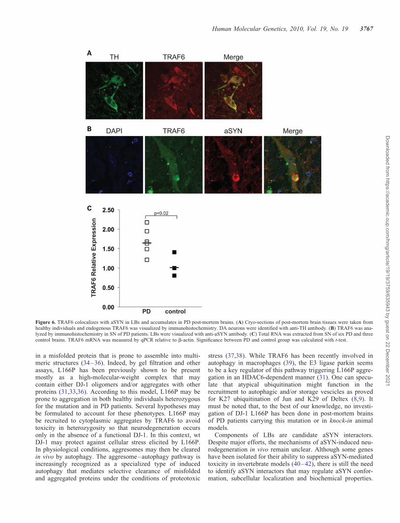
To gain further insights into the relevance of TRAF6 for PDpathogenesis, we analyzed the presence of endogenousTRAF6 in SN DA neurons in human post-mortem brains. Asshown in Fig. 6A, TRAF6 is expressed in the cytoplasm ofDA neurons, as identified by tyrosine hydroxylase (TH) stain-ing. Some TRAF6 immunoreactivity was also found innon-DA neurons, proving a quite broad distribution in mid-brain cell populations.
Several E3 ligases, including Parkin, carbxyl terminus ofHsc70-interaction protein (CHIP) and SIAH, have beenshown to be part of LBs in surviving DA neurons in PDbrains. Therefore, we examined human post-mortem brainsof pathologically confirmed cases of sporadic PD. Exper-iments were performed on brain samples from two differentpatients. LBs were identified using an aSYN antibody. Inter-estingly, virtually all nigral LBs were immunoreactive for ananti-TRAF6 antibody in both individuals (Fig. 6B and Sup-plementary Material, Fig. S4). TRAF6 was expressed through-out the cytoplasm, but the protein was clearly accumulatedat the border of LBs, a site where aSYN staining was more
prominent. The specificity of TRAF6 accumulation in LBswas verified using three antibodies directed against N- or C-terminal epitopes of the protein (Supplementary Material,Fig. S4). No immunoreactivity was detected with unrelatedIgG (data not shown).
In response to an excess burden of misfolded proteins,neurons tend to increase the expression of E3 ligases anddownregulate the level of deubiquitinating enzymes as pre-viously shown in PD post-mortem brains for Parkin andUCH-L1 (23–26). Therefore, we tested TRAF6 mRNAlevels in sporadic PD (n ¼ 6) and healthy control (n ¼ 3)brains (27). RNA was extracted selectively from SN, the siteof PD pathogenesis. Quantitative real-time polymerase chainreaction (qPCR) amplification showed that the amount ofTRAF6 mRNA was statistically increased in sporadic PDsamples when compared with normal controls (P , 0.02),with an average 1.7-fold upregulation (Fig. 6C).
To strengthen the significance of the increased amount ofTRAF6 expression in PD, we also checked for the status ofregulators of TRAF6 activity. By qPCR analysis, we foundno significant differences in the expression of p62, CYLDand A20 between PD and control samples (SupplementaryMaterial, Fig. S5). Overall, our results show that the ubiquitinligase TRAF6 is present in DA neurons in the midbrain of bothnormal and PD brains. In sporadic patients, TRAF6 coloca-lizes with aSYN within LBs and the amount of its mRNAseems increased in SN.
DISCUSSION
The description of the molecular components of LBs and thediscovery of new players in the recruitment of these moleculesto aggregates are important for the understanding of the mol-ecular mechanisms of PD and for the identification of newdrug targets. More than 76 proteins have been described sofar as localized in LBs (28–30) and the significance of theirrelocalization may depend on protein identity.
Ubiquitin ligases are responsible for the formation of poly-ubiquitin chains in substrate proteins and are frequentlyassociated with neurodegenerative diseases for their presencein intracellular aggregates and their role in aggregate for-mation. The E3 ligases Parkin, CHIP and SIAH have beenfound to accumulate in LBs of PD patients (2,4,5). Whiletheir enzymatic activities may be important for protein recruit-ment into the aggregates, their sequestration may decrease thequantity of soluble, active enzymes inducing a ‘loss of func-tion’ phenotype for the lack of appropriate physiological ubi-quitination of targets. Interestingly, they ubiquitinatemisfolded L166P DJ-1 and aSYN with degradative or nonde-gradative effects on either of PD substrates (4,5,21,31).
Structural studies proved that members of the SIAH familypossess a domain highly similar to the TRAF-C region ofTRAF proteins (22). In this study, we demonstrate that the ubi-quitin E3 ligase TRAF6 is a component of LBs. Virtually allLBs in the post-mortem brains of two individuals were posi-tive for TRAF6. Three different antibodies against both theN- and C-terminals of the protein have been used.
TRAF6 ligase activity has been extensively characterized inthe context of NFkB activation (15). In the brain, TRAF6 has
Human Molecular Genetics, 2010, Vol. 19, No. 19 3765
Dow
nloaded from https://academ
ic.oup.com/hm
g/article/19/19/3759/635943 by guest on 22 Decem
ber 2021

been previously linked to K63-specific polyubiquitination ofAD-associated protein tau (19) and in nerve growth factor-receptor signaling (18). Most importantly, TRAF6 localizesin tau-containing, intracellular aggregates in AD post-mortembrains, suggesting that TRAF6 sequestration may be acommon mechanism in neurodegeneration (19).
In post-mortem brains of non-PD individuals, we show thatTRAF6 protein is expressed in DA cells of SN. In the mousebrain, TRAF6 mRNA is present at comparable levels in DAcells from SN and Ventral Tegmental Area, a site that isspared in PD (32). This is consistent with the hypothesis thatTRAF6 expression per se is not related to cellular vulner-ability.
TRAF6 interacts with mutant L166P DJ-1 and aSYN topromote their ubiquitination via atypical ubiquitin chain for-mation involving residues K6, K27, K29 and K33. Surpris-ingly, none of the PD targets that we tested were modified
via TRAF6 canonical activity with K63 specificity. Noncon-ventional ubiquitination of L166P is sufficient for its accumu-lation into insoluble inclusions.
In a quantitative proteomic analysis of atypical ubiquitinchains in yeast, substrates for K6, K27, K29 and K33 havebeen shown to accumulate upon proteasome block, althoughto a lower extent than for K48, suggesting their potentialrole in protein degradation (7). Interestingly, these four ubiqui-tin moieties showed some functional redundancy. Unfortu-nately, the low cellular concentrations of their substrateshave limited so far their identification in yeast or in ex vivomammalian samples (7).
TRAF6 substrate L166P DJ-1 is very unstable and itsexpression level, both in transfection studies and in patientlymphoblasts, is lower than wt (33). This suggests thatL166P mutation may induce a loss of DJ-1 function.However, L166P disrupts DJ-1 protein conformation resulting
Figure 5. TRAF6 binds aSYN and enhances its ubiquitination with atypical chains. (A) HEK cells were transfected with FLAG–TRAF6 and MYC–aSYN wt orA53T (mut). Lysates were immunoprecipitated with anti-FLAG agarose beads and bound proteins were revealed with anti-MYC and anti-FLAG antibodies.Inputs were tested for the expression of TRAF6 and aSYN proteins. (B) Cells were transfected with HA–ubiquitin, MYC–aSYN (wt or A53T) and FLAG–TRAF6 (wt or DN). For in vivo ubiquitination assay, lysates were immunoprecipitated with anti-MYC antibody. Immunoprecipitates and lysates were analyzedwith anti-MYC, anti-FLAG and anti-HA antibodies. (C) HA–ubiquitin mutants in which only the indicated lysine residue is available for chain formation wereused. Wt and K0 ubiquitin were included as controls. In vivo ubiquitination of aSYN wt was performed as in (B).
3766 Human Molecular Genetics, 2010, Vol. 19, No. 19
Dow
nloaded from https://academ
ic.oup.com/hm
g/article/19/19/3759/635943 by guest on 22 Decem
ber 2021
in a misfolded protein that is prone to assemble into multi-meric structures (34–36). Indeed, by gel filtration and otherassays, L166P has been previously shown to be presentmostly as a high-molecular-weight complex that maycontain either DJ-1 oligomers and/or aggregates with otherproteins (31,33,36). According to this model, L166P may beprone to aggregation in both healthy individuals heterozygousfor the mutation and in PD patients. Several hypotheses maybe formulated to account for these phenotypes. L166P maybe recruited to cytoplasmic aggregates by TRAF6 to avoidtoxicity in heterozygosity so that neurodegeneration occursonly in the absence of a functional DJ-1. In this context, wtDJ-1 may protect against cellular stress elicited by L166P.In physiological conditions, aggresomes may then be clearedin vivo by autophagy. The aggresome–autophagy pathway isincreasingly recognized as a specialized type of inducedautophagy that mediates selective clearance of misfoldedand aggregated proteins under the conditions of proteotoxic
stress (37,38). While TRAF6 has been recently involved inautophagy in macrophages (39), the E3 ligase parkin seemsto be a key regulator of this pathway triggering L166P aggre-gation in an HDAC6-dependent manner (31). One can specu-late that atypical ubiquitination might function in therecruitment to autophagic and/or storage vescicles as provedfor K27 ubiquitination of Jun and K29 of Deltex (8,9). Itmust be noted that, to the best of our knowledge, no investi-gation of DJ-1 L166P has been done in post-mortem brainsof PD patients carrying this mutation or in knock-in animalmodels.
Components of LBs are candidate aSYN interactors.Despite major efforts, the mechanisms of aSYN-induced neu-rodegeneration in vivo remain unclear. Although some geneshave been isolated for their ability to suppress aSYN-mediatedtoxicity in invertebrate models (40–42), there is still the needto identify aSYN interactors that may regulate aSYN confor-mation, subcellular localization and biochemical properties.
Figure 6. TRAF6 colocalizes with aSYN in LBs and accumulates in PD post-mortem brains. (A) Cryo-sections of post-mortem brain tissues were taken fromhealthy individuals and endogenous TRAF6 was visualized by immunohistochemistry. DA neurons were identified with anti-TH antibody. (B) TRAF6 was ana-lyzed by immunohistochemistry in SN of PD patients. LBs were visualized with anti-aSYN antibody. (C) Total RNA was extracted from SN of six PD and threecontrol brains. TRAF6 mRNA was measured by qPCR relative to b-actin. Significance between PD and control group was calculated with t-test.
Human Molecular Genetics, 2010, Vol. 19, No. 19 3767
Dow
nloaded from https://academ
ic.oup.com/hm
g/article/19/19/3759/635943 by guest on 22 Decem
ber 2021
Both SIAH (4) and CHIP (5) bind to aSYN and regulate itslevels and aggregation properties (21,43,44). Here we identifyTRAF6 as a new aSYN modifier through unconventional ubi-quitination.
LBs contain phosphorylated, ubiquitinated and nitratedaSYN (45–47). The type of ubiquitin moieties covalentlyattached to aSYN in PD and control brains remains unclear.Interestingly, only 10% of aSYN is ubiquitinated with evi-dences for conjugation of one, two or three ubiquitins (44).Monoubiquitination by SIAH has been proposed to act as aseed for further protein aggregation (48).
In our experimental settings, we failed to trigger aSYNaggregates by TRAF6 overexpression. Its induction in cell cul-tures depends on many factors including a permissive cellularenvironment, expression of synphilin-1 (49) and a proteolyticcleavage that releases an inhibitory domain at the C-terminal(50). It will be interesting to assess whether TRAF6 maytrigger aggregation of aSYN in these conditions. It alsoremains unclear the role of TRAF6-mediated atypical ubiqui-tination in the degradation of aSYN via the lysosomal/autop-hagy pathways (51).
Interestingly, TRAF6 expression seems increased in SN ofsporadic PD brains. These data need to be validated in alarger sample size since the reproducibility of gene expressionanalysis of human post-mortem brains may suffer fromsampling a heterogeneous tissue. If confirmed, this expressionmay be part of a cellular response to an excess burden ofunfolded proteins, as shown for other E3 ubiquitin ligases.
However, TRAF6 may also ubiquitinate soluble aSYN inthe brain and thus be important for aSYN physiological func-tion and not necessarily for its recruitment to LBs. In thismodel, the increased expression of TRAF6 may be a responseto its sequestration to LBs to maintain the appropriate post-translational modifications of its targets including aSYN.Further experiments are needed to verify this hypothesis.
In conclusion, our data indicate a role for TRAF6 in PD forits localization in LBs and handling of aggregation-prone mis-folded proteins as atypical ubiquitin E3 ligase. These resultspose new exciting questions about the role of atypical ubiqui-tination in cellular physiology and dysfunction.
MATERIALS AND METHODS
Further experimental details are provided as SupplementaryMaterial.
Immunoprecipitation and western blot
For coimmunoprecipitation experiments, cells were lysed inTRAF6 (200 mM NaCl, 50 mM Tris, pH 7.5, 0.5% NP40,10% glycerol) or aSYN (150 mM NaCl, 50 mM Tris, pH 7.5,0.5% CHAPS) buffers supplemented with anti-protease cock-tail (Roche) and 5 mM N-ethyl-maleimide (NEM). Cell lysateswere incubated with anti-FLAG agarose beads (Sigma) or withthe appropriate antibody. After washing, immunoprecipitatedproteins were eluted with 2× sodium dodecyl sulphate(SDS) sample buffer, boiled and analyzed by western blot.The following antibodies were used: anti-FLAG 1:2000(Sigma), anti-MYC 1:4000 (Cell Signaling), anti-HA 1:1000
(from ibridoma supernatant, kindly provided by Dr LicioCollavin), anti-b-actin 1:5000 (Sigma). For detection, anti-mouse-horseradish peroxidase (HRP) and anti-rabbit-HRP(Dako) or protein A-HRP (Upstate) in combination withenhanced chemiluminescence (ECL) (GE Healthcare) wereused.
In vivo ubiquitination assay
For in vivo ubiquitination assays, HEK cells were transfectedwith HA–ubiquitin and the indicated DJ-1, aSYN, TRAF6 orTRAF2 constructs. After transfection, cells were either leftuntreated or incubated with 10 mM MG132 for 3 h. Cellswere then lysed with radioimmunoprecipitation assay (RIPA)buffer (150 mM NaCl, 50 mM Tris, pH 7.5, 1% Triton X-100,1% deoxycholic acid and 0.1% SDS) and samples werebriefly sonicated. After centrifugation, clear lysates wereimmunoprecipitated with anti-MYC, anti-FLAG or anti-GFP(Invitrogen) antibodies. Immunocomplexes were analyzed bywestern blot using anti-HA antibody to detect ubiquitin conju-gates. For development, protein A-HRP was used.
Cell fractionation
Transfected HEK cells were lysed in a buffer containing150 mM NaCl, 50 mM Tris, pH 7.5, and 0.2% Triton X-100,supplemented with protease inhibitor cocktail and 5 mM
NEM (Sigma). Lysates were centrifuged at 20 000 g for30 min at 48C and separated into Triton X-100 soluble (super-natant) and insoluble (pellet) fractions. Insoluble pellets wereresuspended in boiling sample buffer, sonicated and used forwestern blot analysis.
Immunocytochemistry and immunohistofluorescence
To detect insoluble aggregates of mutant DJ-1, immunocyto-chemistry was performed as previously described (14). Afterpermeabilization, cells were fixed in 4% paraformaldehydeand indirect immunofluorescence was carried out followingstandard methods. Anti-FLAG (1:1000) and anti-ubiquitin(Dako) (1:50) antibodies were used. For detection, AlexaFluor-405-labeled (blu) or -594-labeled (red) (Invitrogen) anti-mouse or anti-rabbit antibodies were used while GFP fluor-escence was evidenced at 488 laser. Immunohistofluorescenceon human post-mortem brain sections was performed asreported (52). Anti-TRAF6 antibodies were from Abnova(PAB0279) and Santa Cruz (sc-8409 and sc-7221). Mousemonoclonal (#2647) and rabbit polyclonal (#2628) anti-aSYNantibodies were from Cell Signaling. All images were col-lected using a confocal microscope (LEICA TCS SP2).
Post-mortem human brain samples
Brain samples are from the brain bank at the Institute of Neu-ropathology, Bellvitge Hospital (University of Barcelona,Spain). Samples were dissected at autopsy with the informedconsent of patients or their relatives and the institutionalapproval of the Ethics Committee of the University of Barce-lona. Brains were obtained from Caucasian, pathologicallyconfirmed PD cases and age-matched controls (27). Briefly,
3768 Human Molecular Genetics, 2010, Vol. 19, No. 19
Dow
nloaded from https://academ
ic.oup.com/hm
g/article/19/19/3759/635943 by guest on 22 Decem
ber 2021

all cases of PD had suffered from classical PD, none of themhad cognitive impairment and their neuropathological charac-terization was made according to the established criteria.Control healthy subjects showed the absence of neurologicalsymptoms and of metabolic and vascular diseases, and theneuropathological study disclosed no abnormalities, includinglack of AD and related pathology. The time between death andtissue preparation was in the range of 3–5 h. The ventral mid-brain region was sectioned horizontally. The dark-pigmentedzones of the SN were readily apparent from all surroundingstructures and were then isolated from the ventral midbrain.SN autopsy tissues were immediately frozen for RNA extrac-tion. For histological analysis, samples were cryoprotectedwith 30% sucrose in 4% formaldehyde, frozen in dry iceand stored at 2808C until use.
Quantitative real-time polymerase chain reaction
Total RNA from SN of six pathologically confirmed and threecontrol subjects (27) was isolated using the TRIZOL reagent(Invitrogen). cDNA was obtained from 1 mg of purifiedRNA using the iSCRIPTTM cDNA Synhesis Kit (Bio-Rad).qPCR was performed using SYBR-Green PCR Master Mix(Applied Biosystem). Expression of TRAF6, CYLD, A20and p62/SQSTM1 was analyzed using specific oligonucleo-tides.
SUPPLEMENTARY MATERIAL
Supplementary Material is available at HMG online.
ACKNOWLEDGEMENTS
We are indebted to all the members of the laboratory ofStefano Gustincich for discussions. We thank Prof. PeterT. Lansbury (Harvard Medical School, Cambridge, MA,USA) for aSYN cDNA, Dr Sandro Goruppi (Tufts University,Boston, MA, USA) for TRAF2 constructs and Dr LicioCollavin (University of Trieste, Italy) for anti-HA antibody.We thank Prof. Francesca Persichetti (University of PiemonteOrientale, Italy) for carefully reading the manuscript.
Conflict of Interest statement. None declared.
FUNDING
This work was supported by Telethon Grant GGP06268, TheGiovanni Armenise-Harvard Foundation and the Italian Insti-tute of Technology.
REFERENCES
1. Lesage, S. and Brice, A. (2009) Parkinson’s disease: from monogenicforms to genetic susceptibility factors. Hum. Mol. Genet., 18, R48–R59.
2. Schlossmacher, M.G., Frosch, M.P., Gai, W.P., Medina, M., Sharma, N.,Forno, L., Ochiishi, T., Shimura, H., Sharon, R., Hattori, N. et al. (2002)Parkin localizes to the Lewy bodies of Parkinson disease and dementiawith Lewy bodies. Am. J. Pathol., 160, 1655–1667.
3. Ito, T. (2003) Dorfin localizes to Lewy bodies and ubiquitylatesSynphilin-1. J. Biol. Chem., 278, 29106–29114.
4. Liani, E., Eyal, A., Avraham, E., Shemer, R., Szargel, R., Berg, D.,Bornemann, A., Riess, O., Ross, C.A., Rott, R. et al. (2004)Ubiquitylation of synphilin-1 and alpha-synuclein by SIAH and itspresence in cellular inclusions and Lewy bodies imply a role inParkinson’s disease. Proc. Natl Acad. Sci. USA, 101, 5500–5505.
5. Shin, Y. (2005) The co-chaperone carboxyl terminus of Hsp70-interactingprotein (CHIP) mediates alpha-synuclein degradation decisions betweenproteasomal and lysosomal pathways. J. Biol. Chem., 280, 23727–23734.
6. Chen, Z. and Sun, L. (2009) Nonproteolytic functions of ubiquitin in cellsignaling. Mol. Cell, 33, 275–286.
7. Xu, P., Duong, D.M., Seyfried, N.T., Cheng, D., Xie, Y., Robert, J., Rush,J., Hochstrasser, M., Finley, D. and Peng, J. (2009) Quantitativeproteomics reveals the function of unconventional ubiquitin chains inproteasomal degradation. Cell, 137, 133–145.
8. Ikeda, H. and Kerppola, T.K. (2008) Lysosomal localization ofubiquitinated Jun requires multiple determinants in a lysine-27-linkedpolyubiquitin conjugate. Mol. Biol. Cell, 19, 4588–4601.
9. Chastagner, P., Israel, A. and Brou, C. (2006) Itch/AIP4 mediates Deltexdegradation through the formation of K29-linked polyubiquitin chains.EMBO Rep., 7, 1147–1153.
10. Jiang, J., Ballinger, C.A., Wu, Y., Dai, Q., Cyr, D.M., Hohfeld, J. andPatterson, C. (2001) CHIP is a U-box-dependent E3 ubiquitin ligase:identification of Hsc70 as a target for ubiquitylation. J. Biol. Chem., 276,42938–42944.
11. Al-Hakim, A.K., Zagorska, A., Chapman, L., Deak, M., Peggie, M. andAlessi, D.R. (2008) Control of AMPK-related kinases by USP9X andatypical Lys29/Lys33-linked polyubiquitin chains. Biochem. J., 411, 249.
12. Xu, K., Shimelis, H., Linn, D.E., Jiang, R., Yang, X., Sun, F., Guo, Z.,Chen, H., Li, W., Chen, H. et al. (2009) Regulation of androgen receptortranscriptional activity and specificity by RNF6-induced ubiquitination.Cancer Cell, 15, 270–282.
13. Pype, S., Declercq, W., Ibrahimi, A., Michiels, C., Van Rietschoten, J.G.,Dewulf, N., De Boer, M., Vandenabeele, P., Huylebroeck, D. andRemacle, J.E. (2000) TTRAP, a novel protein that associates with CD40,tumor necrosis factor (TNF) receptor-75 and TNF receptor-associatedfactors (TRAFs), and that inhibits nuclear factor-kappa B activation.J. Biol. Chem., 275, 18586–18593.
14. Zucchelli, S., Vilotti, S., Calligaris, R., Lavina, Z.S., Biagioli, M., Foti, R.,De Maso, L., Pinto, M., Gorza, M., Speretta, E. et al. (2009)Aggresome-forming TTRAP mediates pro-apoptotic properties ofParkinson’s disease-associated DJ-1 missense mutations. Cell Death
Differ., 16, 428–438.15. Chen, Z. (2005) Ubiquitin signalling in the NF-kappaB pathway. Nat. Cell
Biol., 7, 758–765.16. Wu, H. and Arron, J. (2003) TRAF6, a molecular bridge spanning
adaptive immunity, innate immunity and osteoimmunology. Bioessays,25, 1096–1105.
17. Khursigara, G., Orlinick, J.R. and Chao, M.V. (1999) Association of thep75 neurotrophin receptor with TRAF6. J. Biol. Chem., 274, 2597–2600.
18. Geetha, T., Jiang, J. and Wooten, M. (2005) Lysine 63 Polyubiquitinationof the nerve growth factor receptor TrkA directs internalization andsignaling. Mol. Cell, 20, 301–312.
19. Babu, J., Geetha, T. and Wooten, M. (2005) Sequestosome 1/p62 shuttlespolyubiquitinated tau for proteasomal degradation. J. Neurochem., 94,192–203.
20. Lim, K.L., Chew, K.C., Tan, J.M., Wang, C., Chung, K.K., Zhang, Y.,Tanaka, Y., Smith, W., Engelender, S., Ross, C.A. et al. (2005) Parkinmediates nonclassical, proteasomal-independent ubiquitination ofSynphilin-1: implications for Lewy body formation. J. Neurosci., 25,2002–2009.
21. Lee, J., Wheeler, T., Li, L. and Chin, L. (2007) Ubiquitination ofalpha-synuclein by Siah-1 promotes alpha-synuclein aggregation andapoptotic cell death. Hum. Mol. Genet., 17, 906–917.
22. Polekhina, G., House, C.M., Traficante, N., Mackay, J.P., Relaix, F.,Sassoon, D.A., Parker, M.W. and Bowtell, D.D. (2002) Siah ubiquitinligase is structurally related to TRAF and modulates TNF-a signaling.Nat. Struct. Biol., 9, 68–75.
23. Humbert, J., Beyer, K., Carrato, C., Mate, J.L., Ferrer, I. and Ariza, A.(2007) Parkin and synphilin-1 isoform expression changes in Lewy bodydiseases. Neurobiol. Dis., 26, 681–687.
24. Choi, J., Levey, A.I., Weintraub, S.T., Rees, H.D., Gearing, M., Chin, L.S.and Li, L. (2004) Oxidative modifications and down-regulation of
Human Molecular Genetics, 2010, Vol. 19, No. 19 3769
Dow
nloaded from https://academ
ic.oup.com/hm
g/article/19/19/3759/635943 by guest on 22 Decem
ber 2021

ubiquitin carboxyl-terminal hydrolase L1 associated with idiopathicParkinson’s and Alzheimer’s diseases. J. Biol. Chem., 279, 13256–13264.
25. Culpan, D., Cram, D., Chalmers, K., Cornish, A., Palmer, L., Palmer, J.,Hughes, A., Passmore, P., Craigs, D., Wilcock, G.K. et al. (2009)TNFR-associated factor-2 (TRAF2) in Alzheimer’s disease. Neurobiol.Aging, 30, 1052–1060.
26. Sahara, N., Murayama, M., Mizoroki, T., Urushitani, M., Imai, Y.,Takahashi, R., Murata, S., Tanaka, K. and Takashima, A. (2005) In vivoevidence of CHIP up-regulation attenuating tau aggregation.J. Neurochem., 94, 1254–1263.
27. Navarro, A., Boveris, A., Bandez, M.J., Sanchez-Pinto, M.J., Gomez, C.,Muntane, G. and Ferrer, I. (2009) Human brain cortex: mitochondrialoxidative damage and adaptative response in Parkinson disease anddementia with Lewy bodies. Free Radic. Biol. Med., 46, 1574–1580.
28. Wakabayashi, K., Tanji, K., Mori, F. and Takahashi, H. (2007) The Lewybody in Parkinson’s disease: molecules implicated in the formation anddegradation of alpha-synuclein aggregates. Neuropathology, 27, 494–506.
29. Beyer, K., Domingo-Sabat, M. and Ariza, A. (2009) Molecular pathologyof lewy body diseases. Int. J. Mol. Sci., 10, 724–745.
30. Spillantini, M.G., Schmidt, M.L., Lee, V.M., Trojanowski, J.Q., Jakes, R.and Goedert, M. (1997) Alpha-synuclein in Lewy bodies. Nature, 388,839–840.
31. Olzmann, J.A., Li, L., Chudaev, M.V., Chen, J., Perez, F.A., Palmiter,R.D. and Chin, L.S. (2007) Parkin-mediated K63-linkedpolyubiquitination targets misfolded DJ-1 to aggresomes via binding toHDAC6. J. Cell Biol., 178, 1025–1038.
32. Biagioli, M., Pinto, M., Cesselli, D., Zaninello, M., Lazarevic, D.,Roncaglia, P., Simone, R., Vlachouli, C., Plessy, C., Bertin, N. et al.(2009) Unexpected expression of alpha- and beta-globin in mesencephalicdopaminergic neurons and glial cells. Proc. Natl Acad. Sci. USA, 106,15454–15459.
33. Macedo, M.G., Anar, B., Bronner, I.F., Cannella, M., Squitieri, F.,Bonifati, V., Hoogeveen, A., Heutink, P. and Rizzu, P. (2003) TheDJ-1L166P mutant protein associated with early onset Parkinson’s diseaseis unstable and forms higher-order protein complexes. Hum. Mol. Genet.,12, 2807–2816.
34. Olzmann, J.A., Brown, K., Wilkinson, K.D., Rees, H.D., Huai, Q., Ke, H.,Levey, A.I., Li, L. and Chin, L.S. (2004) Familial Parkinson’sdisease-associated L166P mutation disrupts DJ-1 protein folding andfunction. J. Biol. Chem., 279, 8506–8515.
35. Shendelman, S., Jonason, A., Martinat, C., Leete, T. and Abeliovich, A.(2004) DJ-1 is a redox-dependent molecular chaperone that inhibitsalpha-synuclein aggregate formation. PLoS Biol., 2, e362.
36. Herrera, F.E., Zucchelli, S., Jezierska, A., Lavina, Z.S., Gustincich, S. andCarloni, P. (2007) On the oligomeric state of DJ-1 protein and its mutantsassociated with Parkinson Disease. A combined computational andin vitro study. J. Biol. Chem., 282, 24905–24914.
37. Chin, L.S., Olzmann, J.A. and Li, L. (2010) Parkin-mediated ubiquitinsignalling in aggresome formation and autophagy. Biochem. Soc. Trans.,38, 144–149.
38. Olzmann, J.A. and Chin, L.S. (2008) Parkin-mediated K63-linkedpolyubiquitination: a signal for targeting misfolded proteins to theaggresome–autophagy pathway. Autophagy, 4, 85–87.
39. Subauste, C.S., Andrade, R.M. and Wessendarp, M. (2007) CD40-TRAF6and autophagy-dependent anti-microbial activity in macrophages.Autophagy, 3, 245–248.
40. Auluck, P.K., Chan, H.Y., Trojanowski, J.Q., Lee, V.M. and Bonini, N.M.(2002) Chaperone suppression of alpha-synuclein toxicity in a Drosophilamodel for Parkinson’s disease. Science, 295, 865–868.
41. Qiao, L., Hamamichi, S., Caldwell, K.A., Caldwell, G.A., Yacoubian,T.A., Wilson, S., Xie, Z.L., Speake, L.D., Parks, R., Crabtree, D. et al.(2008) Lysosomal enzyme cathepsin D protects against alpha-synucleinaggregation and toxicity. Mol. Brain, 1, 17.
42. Kuwahara, T., Koyama, A., Koyama, S., Yoshina, S., Ren, C.H., Kato, T.,Mitani, S. and Iwatsubo, T. (2008) A systematic RNAi screen revealsinvolvement of endocytic pathway in neuronal dysfunction in alpha-synuclein transgenic C. elegans. Hum. Mol. Genet., 17, 2997–3009.
43. Nagano, Y., Yamashita, H., Takahashi, T., Kishida, S., Nakamura, T.,Iseki, E., Hattori, N., Mizuno, Y., Kikuchi, A. and Matsumoto, M. (2003)Siah-1 facilitates ubiquitination and degradation of Synphilin-1. J. Biol.Chem., 278, 51504–51514.
44. Engelender, S. (2008) Ubiquitination of alpha-synuclein and autophagy inParkinson’s disease. Autophagy, 4, 372–374.
45. Hasegawa, M., Fujiwara, H., Nonaka, T., Wakabayashi, K., Takahashi, H.,Lee, V.M., Trojanowski, J.Q., Mann, D. and Iwatsubo, T. (2002)Phosphorylated alpha-synuclein is ubiquitinated in alpha-synucleinopathylesions. J. Biol. Chem., 277, 49071–49076.
46. Tofaris, G.K., Razzaq, A., Ghetti, B., Lilley, K.S. and Spillantini, M.G.(2003) Ubiquitination of alpha-synuclein in Lewy bodies is a pathologicalevent not associated with impairment of proteasome function. J. Biol.Chem., 278, 44405–44411.
47. Anderson, J.P., Walker, D.E., Goldstein, J.M., de Laat, R., Banducci, K.,Caccavello, R.J., Barbour, R., Huang, J., Kling, K., Lee, M. et al. (2006)Phosphorylation of Ser-129 is the dominant pathological modification ofalpha-synuclein in familial and sporadic Lewy body disease. J. Biol.Chem., 281, 29739–29752.
48. Rott, R., Szargel, R., Haskin, J., Shani, V., Shainskaya, A., Manov, I.,Liani, E., Avraham, E. and Engelender, S. (2008) Monoubiquitylationof alpha-synuclein by seven in absentia homolog (SIAH)promotes its aggregation in dopaminergic cells. J. Biol. Chem., 283,3316–3328.
49. Engelender, S., Kaminsky, Z., Guo, X., Sharp, A.H., Amaravi, R.K.,Kleiderlein, J.J., Margolis, R.L., Troncoso, J.C., Lanahan, A.A., Worley,P.F. et al. (1999) Synphilin-1 associates with alpha-synuclein andpromotes the formation of cytosolic inclusions. Nat. Genet., 22, 110–114.
50. Li, W., West, N., Colla, E., Pletnikova, O., Troncoso, J.C., Marsh, L.,Dawson, T.M., Jakala, P., Hartmann, T., Price, D.L. et al. (2005)Aggregation promoting C-terminal truncation of alpha-synuclein is anormal cellular process and is enhanced by the familial Parkinson’sdisease-linked mutations. Proc. Natl Acad. Sci. USA, 102, 2162–2167.
51. Cuervo, A.M., Stefanis, L., Fredenburg, R., Lansbury, P.T. and Sulzer, D.(2004) Impaired degradation of mutant alpha-synuclein bychaperone-mediated autophagy. Science, 305, 1292–1295.
52. Waldvogel, H.J., Curtis, M.A., Baer, K., Rees, M.I. and Faull, R.L. (2006)Immunohistochemical staining of post-mortem adult human brainsections. Nat. Protoc., 1, 2719–2732.
3770 Human Molecular Genetics, 2010, Vol. 19, No. 19
Dow
nloaded from https://academ
ic.oup.com/hm
g/article/19/19/3759/635943 by guest on 22 Decem
ber 2021
Related Documents

![Preclinical development of a vaccine against oligomeric alpha-synuclein … · 2017. 11. 15. · gated alpha-synuclein [6–9]. Alpha-synuclein (a-syn) is an abundant protein in the](https://static.cupdf.com/doc/110x72/5fc07f533588d914ed7a20f9/preclinical-development-of-a-vaccine-against-oligomeric-alpha-synuclein-2017-11.jpg)