24 1 Department of Neurology, Slagelse Hospital, 4200 Slagelse-Denmark 2 Department of Surgical Sciences, Uppsala University, Uppsala, Sweden And, Medical Physics, Uppsala University Hospital, Uppsala Sweden *Corresponding author: Homayoun Roshanisefat, Department of Neurology, Slagelse Hospital, 4200 Slagelse-DK, Email: [email protected] Received: October 21, 2020 Published: November 12, 2020 Introduction Multiple Sclerosis (MS) is a chronic inflammatory disease of myelin, mostly affecting patients between 20 and 50 years of age. MS is reported to occur at least twice as often in females as in males[1] and accompanies a variety of signs and symptoms.[2] Its etiology is unknown and the pathology is characterized by a multifactorial, immune-mediated disease caused by complex gene–environmental interactions and plaque burden of the central nerve system (CNS),[3] including the trigeminal nerve (TGN).[4] Patients with MS-trigeminal neuralgia (MS-TN) frequently suffer from bilateral plaques, but the pain is overwhelmingly unilateral.[5] Therefore, the contribution of plaques to the clinical presentation of pain is not fully understood and is in urgent need of being explored further. In several MRI studies, trigeminal root entry zone (REZ) abnormality has been reported in patients with MS (PwMS),[6- 8] particularly in those with signs and symptoms related to the TGN. Findings suggest that using a 3T MRI scanner might show TGN (Fig. 1), selectively impairs with widespread white matter (WM) lesion, especially in the contralateral hemisphere, which, in turn, may be the hallmark of disease severity in patients with MS and TN (PwMS-TS).[9] The mi- crostructure of the tissue damage may affect the TGN in patients with TN (PwTN), and as a possible consequence, WM microstructural alterations in the CNS.[10] Some evidence also indicates that injury to TGN may include trauma, neu- roinflammation, and edema.[11] Affection on the TGN can also be visualized by an immunohistochemical method which shows focal CNS demyelination.[12] The REZ is regarded as a highly important area of pathophysiological exchanges between the peripheral part (PNS) of the TGN and the CNS.[7, 13] Especially, TGN demyelination[14] and WM lesions may be explained by performing a bilateral representation of the transcallosal pathways or the trigeminothalamic sys- tem.[15] Further, specific lesions in MS, such as Dowson’s fingers might probably manifest.[16] SVOA Neurology Research Article ISSN: 2753-9180 Tractography innovative knowledge of multiple sclerosis and trigeminal neuralgia Homayoun Roshanisefat* 1 and Johanna Mårtensson 2 Abstract In Multiple Sclerosis (MS) pathophysiology, a detailed history is known to be the most innovative tool for clarifying the trigeminal nerve (TGN) and predicting its possible interaction. TGN is one of the largest and most engaged cranial nerves (CN) in MS pathology, which probably has received limited attention so far. In addition, it has a very active peripheral ascending and descending neural transport. Patients with classic trigeminal neuralgia (TN) will be used as a proxy to obtain additional information on MS pathology when symptoms are unilateral, with magnetic resonance imaging (MRI) showing bilateral pathology, and neurophysiological result supporting the MRI findings. Using MRI was found to raise the level of information on the microstructure and neural interconnection of TGN, for example, using the T2 and diffu- sion tensor imaging (DTI) with tractography can improve our understanding in this regard. Microvascular information with retrograde reflux of TGN venous contact can also be followed to the central venous branches in the corpus callosum and read out from the neurosurgical report. In this study, a sign of common demographical factors, such as predominant- ly female, younger ages, and side specific, white matter (WM) lesions when reviewing diffusion MRI data of naïve TN, and MS-TN was found. Other findings included anatomical differences, e.g., smaller diameter, volume, and greater atro- phy, when looking through findings on female associated diffusion MRI. A tractographical comparison between TN pa- tients without MS and TN patients with MS has facilitated a better understanding about the possible role of TGN in MS pathology. Keywords: Multiple Sclerosis , MRI, trigeminal neuralgia, TGN SVOA Neurology

Welcome message from author
This document is posted to help you gain knowledge. Please leave a comment to let me know what you think about it! Share it to your friends and learn new things together.
Transcript

24
1 Department of Neurology, Slagelse Hospital, 4200 Slagelse-Denmark
2 Department of Surgical Sciences, Uppsala University, Uppsala, Sweden And, Medical Physics, Uppsala University Hospital,
Uppsala Sweden
*Corresponding author: Homayoun Roshanisefat, Department of Neurology, Slagelse Hospital, 4200 Slagelse-DK, Email:
Received: October 21, 2020 Published: November 12, 2020
Introduction
Multiple Sclerosis (MS) is a chronic inflammatory disease of myelin, mostly affecting patients between 20 and 50 years
of age. MS is reported to occur at least twice as often in females as in males[1] and accompanies a variety of signs and
symptoms.[2] Its etiology is unknown and the pathology is characterized by a multifactorial, immune-mediated disease
caused by complex gene–environmental interactions and plaque burden of the central nerve system (CNS),[3] including
the trigeminal nerve (TGN).[4] Patients with MS-trigeminal neuralgia (MS-TN) frequently suffer from bilateral plaques,
but the pain is overwhelmingly unilateral.[5] Therefore, the contribution of plaques to the clinical presentation of pain
is not fully understood and is in urgent need of being explored further.
In several MRI studies, trigeminal root entry zone (REZ) abnormality has been reported in patients with MS (PwMS),[6-
8] particularly in those with signs and symptoms related to the TGN. Findings suggest that using a 3T MRI scanner
might show TGN (Fig. 1), selectively impairs with widespread white matter (WM) lesion, especially in the contralateral
hemisphere, which, in turn, may be the hallmark of disease severity in patients with MS and TN (PwMS-TS).[9] The mi-
crostructure of the tissue damage may affect the TGN in patients with TN (PwTN), and as a possible consequence, WM
microstructural alterations in the CNS.[10] Some evidence also indicates that injury to TGN may include trauma, neu-
roinflammation, and edema.[11] Affection on the TGN can also be visualized by an immunohistochemical method which
shows focal CNS demyelination.[12] The REZ is regarded as a highly important area of pathophysiological exchanges
between the peripheral part (PNS) of the TGN and the CNS.[7, 13] Especially, TGN demyelination[14] and WM lesions
may be explained by performing a bilateral representation of the transcallosal pathways or the trigeminothalamic sys-
tem.[15] Further, specific lesions in MS, such as Dowson’s fingers might probably manifest.[16]
SVOA Neurology
Research Article
ISSN: 2753-9180
Tractography innovative knowledge of multiple sclerosis and
trigeminal neuralgia Homayoun Roshanisefat*1 and Johanna Mårtensson2
Abstract
In Multiple Sclerosis (MS) pathophysiology, a detailed history is known to be the most innovative tool for clarifying the
trigeminal nerve (TGN) and predicting its possible interaction. TGN is one of the largest and most engaged cranial nerves
(CN) in MS pathology, which probably has received limited attention so far. In addition, it has a very active peripheral
ascending and descending neural transport. Patients with classic trigeminal neuralgia (TN) will be used as a proxy to
obtain additional information on MS pathology when symptoms are unilateral, with magnetic resonance imaging (MRI)
showing bilateral pathology, and neurophysiological result supporting the MRI findings. Using MRI was found to raise
the level of information on the microstructure and neural interconnection of TGN, for example, using the T2 and diffu-
sion tensor imaging (DTI) with tractography can improve our understanding in this regard. Microvascular information
with retrograde reflux of TGN venous contact can also be followed to the central venous branches in the corpus callosum
and read out from the neurosurgical report. In this study, a sign of common demographical factors, such as predominant-
ly female, younger ages, and side specific, white matter (WM) lesions when reviewing diffusion MRI data of naï ve TN,
and MS-TN was found. Other findings included anatomical differences, e.g., smaller diameter, volume, and greater atro-
phy, when looking through findings on female associated diffusion MRI. A tractographical comparison between TN pa-
tients without MS and TN patients with MS has facilitated a better understanding about the possible role of TGN in MS
pathology.
Keywords: Multiple Sclerosis , MRI, trigeminal neuralgia, TGN
SVOA Neurology

25
Recent theories concerning the nature and role of TGN in CNS pathogenesis have been studied to use the TGN as a
proxy or a measurable mechanism to identify pathways for spreading pathogens from peripheral fibers of the TGN to
the central sensory radiators.[5, 17, 18] In addition to the fact that the focus is on pain per se, the vascular consequenc-
es are also of interest, given that it acts like a venous angioma at the REZ of TGN which retrogradely gives raised pres-
sure to the CNS venous system inclusive of the central vein in Corpus Callosum (CC), which has been shown having a
pathognomonic role in MS radially.[19] Some evidence attributed to reversible splenial lesions of the CC in various clin-
ical conditions has also been reported[20] and it could lead to widespread abnormalities in the microstructure of WM
tracts related to sensory, motor, cognitive, and pain functions, including a focal area of the CC[21]. Comparing findings
by performing tractography can help to find some common parameters to emphasize the common affection in PwTN
and PwMS-TN with a view to better understanding the peripheral etiology in MS.
Herein, we describe the tractographical patient studies, which compared the association of TGNs role with non-MS con-
trols with and without TN. Also, more detailed studies of TN to enhance the understanding of TGN in general would
help provide more information about TGN in MS.
Materials and Methods
A “targeted” review of the scientific literature within the PubMed, EMBASE, and the Cochrane Database of Systematic
Reviews and Google Scholar was carried out. Publications up to December 1, 2019, were included. We extracted all pa-
rameters using the following keywords: multiple sclerosis and tractography combined with/or trigeminal neuralgia,
facial pain or numbness, microstructure, WM, diffusion-MRI (dMRI), MRI, Tesla, Gadolinium, slice thickness, neurophys-
iology, and pathology. Using the bibliographies of the publications retrieved, the primary search was supplemented by
secondary tracking details. Only full- length, original communications were valid, and the search was primarily confined
to English language publications or, in some cases, other languages (Danish, Swedish, Norwegian, and German) if indi-
cated. The animal model studies were only used to support clinical context if no human study was available.
Results
This search yielded a total of approximately 586 hits, which were reviewed by title and abstract for potential relevance
to this topic; the body-text itself was reviewed in case the abstract was found lacking relevance search information. Af-
ter excluding duplicates, malignancy, tumor, bleeding, and hereditary cause, reviews, animal, migraine, and headache
studies (flow diagram), 60 studies were included in the final review (PRISMA Flow Diagram).
Records identified through
Medline searching
(n = 60)
Additional records identified
through other sources
(n = 585 )
Records after duplicates removed
(n = 260 )
Records screened
(n = 260 )
Records excluded
(n = 210 )
Full-text articles assessed for eligibility
(n = 260 )
Full-text articles excluded, with reasons
(n = 140 )
Studies included in qualitative synthesis
(n = 120 )
Studies included in quantitative synthesis
(n = 60 )
Iden
tifi
cati
on
Sc
reen
ing
Elig
ibili
ty
Incl
ud
ed
SVOA Neurology
Tractography innovative knowledge of multiple sclerosis and trigeminal neuralgia

26
The background of TGN
TGN is a mixed nerve that primarily encompasses sensory neurons, albeit with embryological origin from the lymph node.[22] Three divisions of neurons converge the trigeminal ganglion (TG): the ophthalmic nerve (V1), the maxillary nerve (V2), and the mandibular nerve (V3), which also carries components of descending motoric fibers that leave the CNS to innervate different facial muscles.[23] The sensory neurons build up the Gasser ganglion, i.e. a diverse popula-tion of cells, that can be classified according to cellular morphology, neurochemistry, and functional characteristics.[22, 24, 25] In this regard, it is pertinent to note that the peripheral and CNS connection of TGN has been studied tracto-graphically.[26] In analyzes of myelinated axons[27] and nerve volumes,[28] gender differences have been found, e.g. lower values in females.[29] Further, comparisons of the diameter of the TGN’s cisternal portion using a 3.0 T MRI, re-veals significantly smaller in left TGN volumes in females as compared to corresponding findings in males (p<0.0001).[30] Previous studies using 1.5T MRI have pointed out that even in general, the diameter of the left-sided mean volume of TGN is smaller, with the dimensions of right-sided nerves being 0.093 cm3 (0.055–0.147 cm3) and that of left-sided nerves being 0.091 cm3 (0.057–0.142 cm3).[31] Further, heart rate alterations due to stimulations of the TGN have been elucidated with significant gender differences.[32]
Trigeminal free nerve endings, which are sensitive to mechanical and thermal stimuli, appear to primarily respond to
noxious and potentially harmful levels of chemical stimulation, CO2 included.[33] Peripheral temperature sensing cell
bodies located in TGN mediating primarily two classes of neurons activated by innocuous warmth: (34 - 42°C) or cold
(14 -30°C).[34] Further, it has been shown that TGN activation depends on the sex hormone.[35] The role of TGN in
cardiovascular alterations, diving, and temperature reflex is specialized for each branch of TGN and sex-dependent.[32,
36, 37] The thermoregulatory system shifted to hypometabolism could cause obesity, which again is more frequent
among females.[38]
The imaging technology has grown successfully over the last few decades.[39] With advancements in MRI technology, it
has become possible to visualize 1 (51.2%), 2 (37.5%), or even 3 (11.2%) small motor roots typically emerging from
the pons anterosuperomedially to the entry point of the large sensory root.[40, 41] However, detailed pathways for
sensory, motoric, and autonomic is more complex[42] and may need high field MRI as 11.75 T for the purpose of visual-
ization. Further, it is also essential to verify that detected damages of the optic and TGN appeared before lesions in CC
occurred as a result of CNS malaria that had been triggered in mice.[43]
Definitions and epidemiology
With an annual incidence of ~4.5 per 100 000, TN is characterized by a recurrent, unilateral, brief, electric shock-like
pain, abrupt in onset and termination, as well as with lancinating jerk localized to small areas of the face. Pain is limited
to the distribution of one or more divisions (V1, V2, or V3) of the TGN and intensely triggered by innocuous stimuli. In
addition, there may be concomitant continuous pain of moderate-intensity within the distribution(s) of the affected
nerve branches; either idiopathic or in conflict with arteries or veins.[44] Secondary TN occurs in up to 37%[45] of
PwTN and the diagnosis is made in the presence of a structural abnormality, other than vascular compression, albeit
including MS plaques, tumors, and abnormalities of the skull base, that affects the TGN. Previously lesions could be de-
tected using low field MRI, but only in 15%[46] MS plaques are the most commonly identified secondary abnormalities
in TN.[47] Patients with MS have a 20-fold increased risk of developing TN[47] and according to current information,
1.9 - 4.9% of these patients have an increased risk of suffering from TN without differences between MS-courses, re-
lapsing-remitting (RRMS), secondary (SPMS) and primary progressive (PPMS). Conversely, MS is detected in 2%-14%
of PwTN, and 10% of patients with PwMS have a higher risk of bilateral TN.[48] Also, the reasons why both facial pains
including TN and non-painful facial sensory disturbances are a common phenomenon in PwMS are not fully known.
Painful facial attacks determined in patients under the age of 50 are most likely to have MS etiology. In patients, with
late-onset of MS at even higher ages, these attacks may occur with a more aggressive disability impact.[49] Agreeing
with these similarities in PwTN, the histopathologic findings show large demyelinated axons, loss of axon quantity, and
abnormal remyelination in the nerve specimens.[14, 50]
dMRI, DTI and Tractography
The dMRI is one of the basic approaches of investigating the WM of the human brain. Tractography is a postprocessing
method that can help derive further information of the WM microstructure by visualizing WM pathways, for example,
the TGN, as 3D reconstructed tracts.[51-53]. In deterministic tractography, the algorithm uses the diffusion tensor (DT)
to point out the diffusion direction from seeding points, proceeding stepwise from voxel to voxel along with the voxel-
wise calculated DT. In probabilistic tractography (Fig 1), the algorithm performs a probability distribution of the diffu-
sion directions.[51, 54] It is notable that other imaging-based approaches used to identify the TGN traditionally rely on
T2-weighted MR images, which provide localization of the cisternal portion of the TGN, where the contrast between
nerve and cerebrospinal fluid (CSF) is high enough to allow differentiation.[55]
SVOA Neurology
Tractography innovative knowledge of multiple sclerosis and trigeminal neuralgia

27
The use of DTI-MRI can add information about WM microstructure and evaluation of the diffusional measures, together
with visual reviews and other measures. This increases the possibilities to perform clinical diagnostics with higher ac-
curacy.[56]
Fig 1. Probabilistic tractography of the TGN bilaterally in a healthy control. Image acquisition was performed at Philips
Achieva 3.0T MR scanner (Philips Achieva, Best, the Netherlands) using a DTI sequence, measured in 48 directions with b
values 0 and 1000.
DTI in TN
A reconstruction of four WM pathways within the TGN region can be enabled with the utilization of DTI: the REZ, the
spinal trigeminal tract (TGT), as well as the ipsilateral and contralateral ventral trigeminothalamic tracts, which can be
illustrated in figure 2[57]. In a previous MR study performed at 3.0T, variances of fractional anisotropy (FA) in affected
regions of TGN in PwTN were observed to be significantly higher in comparison to the corresponding values of TGN in
healthy controls (HC). In addition, a significant positive correlation between FA and contralateral values of TGN was
obtained.[58] In an investigation of TGN in PwTN without MS performed at a 7T MR scanner, affected parts of TGN had
significantly lower FA values (0.31 ± 0.09) than unaffected parts of TGN (0.43 ± 0.10).[15]
Fig 2. The above pictures were borrowed from Guoqiang et al. This illustrates the normal anatomical assessment criteria
of the TGN for expert evaluation. Sagittal, coronal, and axial views of an example TGN (yellow) are overlaid on T2-
weighted MRI. Criteria include: (a) presence (or absence) of branch-like structure, (b) quality of cisternal portion and T2
overlap, (c) presence (or absence) of the mesencephalic trigeminal tract, (d) presence (or absence) of spinal cord tract of
the TGN, (e) avoiding entering (or entering) into the temporal lobe, (f) avoiding (or entering) inferior cerebellar peduncle,
and (g) avoiding (or entering) middle cerebellar peduncle.[26]
SVOA Neurology
Tractography innovative knowledge of multiple sclerosis and trigeminal neuralgia

28
Volume
Investigation of volume may help to identify some common parameters to emphasize the associated affection in PwTN
and PwMS-TN. In this regard, Leal et al determined that the mean volume (V) of the TGN ipsilateral to pain (64.75 ±
14.12 mm3; Confidence Interval (CI), 53.22 to 76.28) and the volume was significantly smaller than contralateral (p <
0.05).[29] Decreased TGN volume has been detected in primary TN in comparison to controls. Additionally, volume
reduction has been detected for grey matter (GM) in several regions associated with pain in primary TGN subjects, in-
cluding the insula, secondary somatosensory cortex, hippocampus, dorsal anterior cingulate cortex, precuneus, and
several areas of the temporal lobe.[15, 59] Across all patients, thalamus volume was reduced ipsilateral compared to
contralateral to the side of pain. Between responders and non-responders, the latter exhibited larger contralateral TGN
volume, and larger ipsilateral and contralateral hippocampus volume.[60]
MRI at higher field strengths allow for improved visualization of the brain and increases the possibility of detecting mi-
crostructural changes of the WM in PwTN.[15, 61] Significant differences in fractional anisotropy of TN can be meas-
ured in the rostral and caudal pathway of TGN in the first part[15] and then posteromedially into the pons before turn-
ing and descending caudally into the spinal TGT.[61] Compared with control subjects, TN subjects exhibit significant
ipsilateral reductions in grey matter volume (GMV).[62] Further, higher field of imaging was performed at MRI 7.0T,
and the volumes were significantly smaller in the affected TGN (33.83 ± 23.12 mm3) than in the unaffected (47.76 ±
32.48 mm3; p = 0.008).[63]
The trigeminovagal complex in humans was investigated post-mortem by performing imaging at 11.7 T MRI, and the
results gave a very new insight to the spinal cord nerves and nuclei, thus contributing to the investigation of pain and
other CNS diseases.[64] Also, the existence of a bifurcation of the TGT into a ventral trigeminothalamic tract and dorsal
trigeminothalamic tract could be described when performing 11.7 T MRI scans of the human brainstem.[65]
Microstructural Changes
The orientation of WM pathways can be reconstructed successfully to characterize the microstructure changes of the
TGN along with its divisions at the skull base by using deterministic and probabilistic tractography algorithms at 3.0
and 7.0 T MRI. The microvascular changes are confined with or without vascular conflict and TN respectively.[15, 50]
Tractography was applied to the thalamic-somatosensory tract (TST) ipsilateral to the site of neurovascular compres-
sion and the FA values were reduced in patients with TN in comparison to side-matched healthy controls, (mean 0.43)
versus (mean 0.47, p = 0.01) (ref). The finding of these WM pathways’ reduced FA may provide credible evidence of
microstructural alteration at the level of the thalamus and S1, thus deepening the understanding of TN neurobiology.
[66]
Correspondingly, images with 7.0T MRI study revealed ipsilateral reduced microstructural integrity of TST which, when
found at the site of neurovascular compression in patients with TN and the altered structure, is followed to the cortex.
[67] After removal of the compression on classic TGN, the loss of FA remained, probably indicating altered microstruc-
tural arrangement. However, after decompression surgery was performed, some part of damages improved in conduc-
tion sensitivity and reduction of edema in the REZ.[68]
Further, when using multiple DTI metrics, microstructural abnormalities of CN V in patients with TN due to NVC was
found. Compared with the unaffected side, the affected side exhibited significantly decreased FA, increased ADC and RD,
and no significant change of AD. [69]
Atrophic changes
Examinations of PwTN with ultra-high field MRI at 7.0 T have shown volumes and structure looseness [15, 61], an affec-
tion expressed with nerve atrophy.[68] Scans at 7.0 T MRI allow identifications of the grade of atrophy and diffusion
abnormalities of the trigeminal nerves in PwTN, f.e. FA, as well as quality assurance (QA) values of the cisternal seg-
ment, which were found to be significantly lower in affected TGN than in unaffected ones.[63] In addition, a sign of atro-
phy detected at an earlier timepoint during the study, even with low field MRI technique, without other relevant find-
ings, correlated to the side of symptoms.[70] The mean diameter of the TGN on the symptomatic side was found to be
significantly smaller than the mean diameter on the asymptomatic side in 30 of 31 patients (2.11 mm ± 0.40 [standard
deviation] and 2.62 mm ± 0.56, P < .001, 95% CI: −0.35 to −0.67 mm). Similarly, the cross-sectional area on the sympto-
matic side was found to be significantly smaller than the area on the asymptomatic side in 27 of 31 patients (4.50 mm2
± 1.75 and 6.28 mm2 ± 2.19, P < .001, 95% CI: −2.41 to −1.16 mm2).
SVOA Neurology
Tractography innovative knowledge of multiple sclerosis and trigeminal neuralgia
29
The results indicate that TGN atrophy can be depicted noninvasively in patients with TN.[71] Right-sided TN patients
are described with significant volumetric reductions in ipsilateral cornu ammo- 1 (CA1), CA4, dentate gyrus, molecular
layer, and hippocampus-amygdala transition area, resulting in decreased whole ipsilateral hippocampal volume, in
comparison to healthy controls.[72] In addition, another study revealed GMV atrophy in the multiple temporal subre-
gions in primary TN patients, including the bilateral temporal pole, right inferior temporal gyrus, and left middle tem-
poral gyrus.[73]
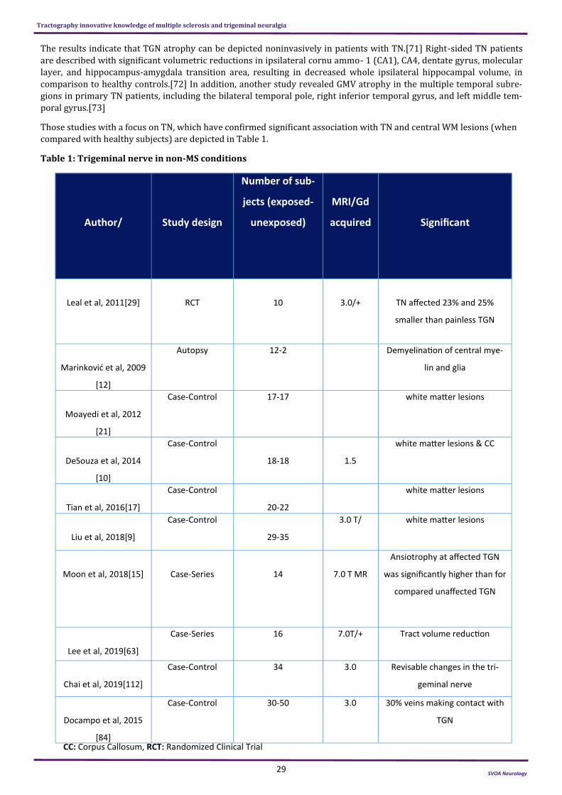
Those studies with a focus on TN, which have confirmed significant association with TN and central WM lesions (when
compared with healthy subjects) are depicted in Table 1.
Table 1: Trigeminal nerve in non-MS conditions
Author/
Study design
Number of sub-
jects (exposed-
unexposed)
MRI/Gd
acquired
Significant
Leal et al, 2011[29]
RCT
10
3.0/+
TN affected 23% and 25%
smaller than painless TGN
Marinković et al, 2009
[12]
Autopsy 12-2 Demyelination of central mye-
lin and glia
Moayedi et al, 2012
[21]
Case-Control 17-17 white matter lesions
DeSouza et al, 2014
[10]
Case-Control
18-18
1.5
white matter lesions & CC
Tian et al, 2016[17]
Case-Control
20-22
white matter lesions
Liu et al, 2018[9]
Case-Control
29-35
3.0 T/ white matter lesions
Moon et al, 2018[15]
Case-Series
14
7.0 T MR
Ansiotrophy at affected TGN
was significantly higher than for
compared unaffected TGN
Lee et al, 2019[63]
Case-Series 16 7.0T/+ Tract volume reduction
Chai et al, 2019[112]
Case-Control 34 3.0 Revisable changes in the tri-
geminal nerve
Docampo et al, 2015
[84]
Case-Control 30-50 3.0 30% veins making contact with
TGN
CC: Corpus Callosum, RCT: Randomized Clinical Trial
SVOA Neurology
Tractography innovative knowledge of multiple sclerosis and trigeminal neuralgia

30
MRI in MS
The number of visual lesions in or related to different segments of TGN has changed during the increased access to ad-
vanced analysis methods as well as the use of higher field strengths at MRI. Previous studies using MRI at lower field
strengths and with large slice thickness suggested that the prevalence of this lesion was between 3% and 7%,[7, 74]
and with bilateral enhancement found in 55-75% MS subjects with TGN lesion.[75] However, the use of 3D MRI at a
higher resolution (3.0 T) with thinner slice thickness (1 mm), showed that the prevalence increased up to 23%.[75]
Studies have shown that TGN lesions can be already present in MS patient's initial attack[75] and that demyelination
may occur in younger ages at MS onset.[76] These radiological findings however, were not associated with TN, despite a
subgroup (37.5%) with painless paraesthesia of the V3 innervation territory of TGN. When studies explore the neuro-
anatomical and diagnostic benefits of MRI at 7.0 T, information that could not be obtained from MRI at lower field
strengths may now compensate with better accuracies.[77] Clinically, TN is shown headed to the diagnosis of MS in
15% and more likely to be female[78] while a recent revision to MS criteria complete palsy of the right III nerve have
been included in MS attack.[79]
In addition to the intrapontine tract, the TGN, the pontine ERZ, the cisternal segment, and the trigeminal nuclei are
characteristic sites of MS lesions. TGN lesions can be shown with a frequency of 12%-38% at 3.0 T MRI.[75] Thickened
TGN have been elucidated in MS patients and should be recognized as a rare feature of this disease and, in cases, more
frequent in right-sided CNS lesions[80] and even more aggressive right-sided TGN lesion.[81] This was in correspond-
ence to MRI tractography findings at 3.0 T that was more frequent left hemisphere lesions in the clinically isolated
course of MS.[82] In agreement with this finding, another study with MRI at 3.0T showed a demyelinating plaque in the
pontine and trigeminal REZ on the affected side. The frequency of the neurovascular compression and its association
with the pontine demyelinating plaque were higher on the affected side than on the unaffected side (54% vs 0%; p =
0.0001).[4] The MS-TN plaque distribution was ruled out with the coalescence of plaques bilaterally along the intrapon-
tine TGN pathway at the proximal pontine segment (PPS). Brainstem plaques at the PPS appear to affect the laterality of
MS-TN pain. The distribution of plaques indicates that 11 patients (61%) had bilateral plaques and 4 patients (22%)
had unilateral plaques near the PPS fibers; however, the remaining 3 did not reveal any radiologic mark of plaques
along the intrapontine TGN.[83]
Volume changes in PwMS-TN
In PwTN without MS, the diagnosis is shown as in MS with predominantly female manifestation. This gender difference
can be followed by the volume of the normal cisternal TGN measured on 3.0 T MRI in vivo, which shows 77.4–78 mm3
in male and 66.1–66.4 mm3 in female,[30, 84] implying a difference between 11.3 mm3 to 11.6 mm3.
In non-MS TN, the posterior fossa volume in males was found to be larger than the posterior fossa volume in females
[85]. In the TN group, females were found to be younger than males and less likely to have neurovascular compression.
[86] In a case-report of females with the clinical sign as MS investigated with MRI, revisable contrast was found with
enhancement in REZ.[87] When compared to the general population, patients with MS have a 20-fold higher risk of TN,
regardless of the type of MS.[47] The risk of getting diagnosed with MS-TN is most frequent in females.[78] MS-TN be-
haves like classical and idiopathic TN and is known to be more common in females than in males, affecting the right side
more frequently than the left side[47] and showing significantly increase d serum levels of IL-1β, IL-6, IL-8 and TNF-α
in comparison to healthy volunteers.[88]
In agreement with these findings regarding the TN, TN-MS studies of T2-hyperintense lesions within the pontine tri-
geminal pathway found evidence that FA was significantly lower and ADC higher, within the affected TGN and the non-
affected TGN in patients with MS, when compared to patients with idiopathic TN or healthy controls (p < 0.001) .[89]
Patients with a higher field of MRI examination could show significantly lowered FA and QA values in affected-side cis-
ternal segments of TGN than in unaffected-side cisternal segments of TGN. However, this affection in other studies is
mostly seen as volume reduction.[63]
Most importantly, 89% of patients in TN-group were found to have abnormal trigeminal reflexes, when the neurophysi-
ological test was used and showed that stimulation were strongly delayed on the affected side.[90] Also, tongue soma-
tosensory-evoked potentials (tSSEPs) performed on MS patients has indicated 90% prolonged; even the MRI showed
brainstem lesions in only 50% of 10 cases.[91] Furthermore, the significantly altered tSSEP score was a predictor for
the presence of midbrain lesions in CIS patients.[92] Further trigeminal SSEPs, as well as facial nerve conduction stud-
ies performed in MS patients with RRMS course, showed abnormalities in all included patients in this study.[93]
Microstructural changes:
The findings of FA and ADC values enables assessment of microstructural changes within the TGN in patients with de-
myelination.
SVOA Neurology
Tractography innovative knowledge of multiple sclerosis and trigeminal neuralgia

31
The existence of microstructural changes have been obtained from DTI within the TGN as well as in-NVC-related PwTN.
[94] The MS in comparison to a group with TN only, TNG group showed lower FA in the ipsilateral peri-lesional seg-
ments, thus suggesting differential microstructural changes along with the CN V in MS patients.[95] In another study,
17 of 18 patients were non- responders to surgical treatment. The lesions were uniformly located along the affected
trigeminal pontine pathway, where the site of maximum overlap across patients was in the area of the trigeminal nucle-
us (TGNC). These microstructural abnormalities in WM were characterized by lower FA values and higher mean diffu-
sivities when compared to the unaffected side. The microstructure of the brainstem TGN within a lesion highlighted the
difference between solitary pontine lesion (SPL) of TN and MS plaques. In conclusion, although SPL-TN patients have
identical clinical features to TN, their single pontine lesion does not even fulfill criteria of MS, and are refractory to sur-
gical management.[83]
MS-TN with atrophy
Previous studies showed that neurovascular compression, i.e. with morphological changes of the TGN such as atrophy,
dislocation, indentation, or flattening, was strongly associated with the symptomatic side in PwMS-TN.[4, 47] With or
without neurovascular conflict, constructive interference demonstrates TGN atrophy[4]; with lesions in the typical,
periventricular, and brain stem distribution, including the pontine trigeminal pathway on the affected side.[89]
The comparison of PwMS-TN and the none-MS subject has been outlined as in Table 2.
Table 2: Trigeminal nerve in MS
TRE=Trigeminal Root Entry, Gd=Gadulinium, TGN=Trigeminal Nerve
Study
Study design
Number of sub-
jects (MS-Non-
MS)
MRI/Gd
acquired
Significant
Meaney et al, 1995[113] Case-Series 7 1.5 T/+ One case demyelination of TGN
van der Meijs et al, 2002[114]
Case-Series 851 0.5-1.5 T/ Enhancement of TGN (3%) clini-
cally silent, the incidence of tri-
geminal nerve demyelination
Silva et al, 2005[74]
Case-Series 275 1.0 T/+ TRE
Cruccu et al, 2008[90] Case-Control 80-50 Vaxalbased TGN lesion
Shor et al, 2016[79]
Case report 1 1.5 T/+ showed enhancement and thick-
ening of the cisternal right III
nerve
Zhu et al, 2012[115] Case-report 1 1.5T/- demyelination in the
right pons at the TRE and Daw-
sons Fingers
Mills et al, 2010[75] Case-Control 47-2 3.0/- TGN lesion
Sugiyama et al, 2015 Case-Control 128-46 1.5 T/- TRE lesion
Fallata, et al, 2017[78] survey 8590 NA 15% TN before MS diagnosis
Danyluk et al, 2019[60] Case-Series 34 1.5T TGN volumetry
Truini et al, 2016[4]
Case-Series 28 3.0 T demyelinating plaque in the
pontine trigeminal root entry
zone
SVOA Neurology
Tractography innovative knowledge of multiple sclerosis and trigeminal neuralgia

32
Discussion There is a clinical need to improve the visualization of CNs within the nasal cavity, particularly in the context of pathol-ogy, and to enable detailed analysis of the affected nerves' microstructure when studying MS pathophysiology. The pro-spect of obtaining 7.0 T MR imaging in the routine clinical setting should improve the ability to reliably visualize the larger CNs such as the TGN. In this regard, knowledge of the exact neuroanatomy of the TGTs will contribute to a deep-er understanding of its role in MS pathology. As shown in this review, the management of TN has revealed a new path-way in order to better understand the role of TGN in MS; this nerve has the capability to transmit or mediate the pe-ripheral injuries to CNS and improve the knowledge of pathophysiology in MS with or without MRI pathognomonic le-sions.
In this study, we have found a sign of common demographical factors such as predominantly female, younger ages, and
side specific WM lesions when reviewing diffusion MRI data of TN and MS-TN. We have also found anatomical differ-
ences, e.g., smaller diameter, volume, and more atrophy, when looking through findings on female associated diffusion
MRI. Further, the similarity in microstructural changes in both TN and MS-TN has also been identified, when studying
DTI parameters.
However, the pathogenesis of many chronic TGN pain conditions, such as TN, migraine, and temporo-mandibular disor-
ders, is still not clear. When fibers display a wide innervation in CNS, one of the proposed biochemical mechanisms in-
volves calcitonin gene-related peptide (CGRP), which is considered as the most important neuropeptide in the trigemi-
nal system (TS).[96] The higher concentration of intraganglionic of CGRP can modulate the neuronal transmission of
pain signals. Expression of CGRP in the cerebral cortex and TG one week post‐injury is altered when compared to unin-
jured control animals.[97] Further, there is evidence that peripheral inflammatory reaction in the area of trigeminal
nociceptors cascades increases expression of CGRP and brain-derived neurotrophic factor gene in TG.[98] In various
animal models of TGN-associated disorders, the concentration of CGRP was observed to increase in TG. In most of these
models, pathological changes in the TGN are accompanied by inflammation within peripheral neuronal endings of TGN,
and as shown in other studies, with a significant sex depended difference in CGRP expression in female mice.[99, 100]
This finding could probably be suggesting some association with the anatomically smaller volume of TGN in females.
[101]
The studies with proton density-weighted image show a classic MS "ovoid lesion" with the long axis perpendicular to
the lateral ventricle[102] MS is known to commonly surround the subependymal veins that drain perpendicular to the
ventricles, named "Dawson’s fingers" signs at MRI and pathological preparation.[103-105] According to studies, the
proton density can detect the cortical changes in only 21 years females with suddenly aggressive dementia as the first
symptom in MS cases.[105] The sagittal proton density-weighted image turned out with multiple WM lesions with
Dawson’s finger signs.[105] This method, in conjunction with diffusion MRI and tractography, would help to better un-
derstand GM connectivity in the brain.
An animal study could by studying cortical spreading depolarization (CSD) carry out pathological phenomenon with
proven relevance to functional outcome from traumatic brain injuries (TBI). The effects of TGN stimulation in targeting
CSDs to completely terminate CSDs in the injured rat’s brain exhibited significantly decreased numbers of the CSD from
60% to 49%. The finding supports the intimate connection between TGN and cerebral and meningeal blood vessels,
referred to as the trigemino‐cerebrovascular system (TCVS).[106] The TCVS are also capable of activating the so called
‘diving reflex’, where the primary role is to conserve oxygen for the sensitive brain and heart tissue.[106] This reflex
has different outcomes in females than in males.[32] This connection can probably explain the increased stroke risk in
MS and regardless to course of MS,[107] and beyond the effect of surveillance bias.
The ophthalmic branch of the trigeminal nerve seems to be commonly affected in MS.[108] This division of TGN has the
highest amount of myelinated and unmyelinated nerve branches on the cornea, sinus cavities, as well as internal and
external nasal environment. All these surfaces can make a specifically route via epithelium transportation, as nano-
peptide research has shown a direct connection between the nerve ending and CNS.[109, 110] Further, TGN exhibited
higher concentrations of tested nanocarrier than any other sampled CNS tissues.[111] This finding can contribute to
developing a better understanding of the possibility of peripheral pathogens into CNS.
Limitations
Despite the clear advantages of using comorbidity like TN and strengthening pathophysiological understanding in MS,
there are potential limitations of each procedure. There is a possibility of selection bias in each of those studies that
sought to use patient with pain caused by TN. Another limitation that could have been hidden in those included studies
was the pharmacological treatment effect and hormonal impact which have not been exclusively described in any of the
included studies. Therefore, factors relating to the female sex, such as differences in sex hormone levels, may be risk
factors in TN and MS and should be included in future studies, even when ruling out the confounder effect of MS thera-
pies.
SVOA Neurology
Tractography innovative knowledge of multiple sclerosis and trigeminal neuralgia

33
Conclusion
The authors reiterate the importance of evaluating TGN impact in MS through the inclusion of DT-MRI and tractog-
raphy and an emphasis on the fact that the knowledge in classical TN and MS-TN may be pivotal in pointing out TGN as
the suggested main port of MS pathology. A series of studies can be carried out to further discuss the TGNs in different
genetic and environmental risk associations.
References
1. Filippi, M., et al., Multiple sclerosis. Nat Rev Dis Primers, 2018. 4(1): p. 43.
2. Miller, D.H., et al., Differential diagnosis of suspected multiple sclerosis: a consensus approach. Multiple Sclerosis Jour-
nal, 2008. 14(9): p. 1157-1174.
3. Christogianni, A., et al., Temperature sensitivity in multiple sclerosis: An overview of its impact on sensory and cogni-
tive symptoms. Temperature, 2018. 5(3): p. 208-223.
4. Truini, A., et al., A dual concurrent mechanism explains trigeminal neuralgia in patients with multiple sclerosis. Neu-
rology, 2016. 86(22): p. 2094-2099.
5. Pichiecchio, A., et al., Bilateral trigeminal enhancement on magnetic resonance imaging in a patient with multiple
sclerosis and trigeminal neuralgia. Multiple Sclerosis Journal, 2007. 13(6): p. 814-816.
6. Mills, R.J., C.A. Young, and E.T. Smith, Central trigeminal involvement in multiple sclerosis using high-resolution MRI at
3 T. The British Journal of Radiology, 2010. 83(990): p. 493-498.
7. Nakashima, I., et al., Linear pontine trigeminal root lesions in multiple sclerosis: clinical and magnetic resonance imag-
ing studies in 5 cases. Archives of neurology, 2001. 58(1): p. 101-104.
8. Haider, L., et al., Cranial Nerve Enhancement in Multiple Sclerosis Is Associated With Younger Age at Onset and More
Severe Disease. Frontiers in Neurology, 2019. 10(1085).
9. Liu, J., et al., Abnormal brain white matter in patients with right trigeminal neuralgia: a diffusion tensor imaging
study. The journal of headache and pain, 2018. 19(1): p. 46-46.
10. DeSouza, D.D., M. Hodaie, and K.D. Davis, Abnormal trigeminal nerve microstructure and brain white matter in idio-
pathic trigeminal neuralgia. PAIN®, 2014. 155(1): p. 37-44.
11. Renton, T., Trigeminal nerve injuries. Australian Endodontic Journal, 2018. 44(2): p. 159-169.
12. Marinkovic , S., et al., Ultrastructure and immunohistochemistry of the trigeminal peripheral myelinated axons in pa-
tients with neuralgia. Clinical neurology and neurosurgery, 2009. 111(10): p. 795-800.
13. DeSouza, D.D., M. Hodaie, and K.D. Davis, Structural magnetic resonance imaging can identify trigeminal system ab-
normalities in classical trigeminal neuralgia. Frontiers in neuroanatomy, 2016. 10: p. 95.
14. Hilton, D.A., et al., Pathological findings associated with trigeminal neuralgia caused by vascular compression. Neuro-
surgery, 1994. 35(2): p. 299-303.
15. Moon, H.C., et al., 7.0 Tesla MRI tractography in patients with trigeminal neuralgia. Magnetic resonance imaging,
2018. 54: p. 265-270.
16. Rocha, A.J.d., et al., Central nervous system infectious diseases mimicking multiple sclerosis: recognizing distinguisha-
ble features using MRI. Arquivos de neuro-psiquiatria, 2013. 71(9B): p. 738-746.
17. Tian, T., et al., Brain white matter plasticity and functional reorganization underlying the central pathogenesis of tri-
geminal neuralgia. Scientific reports, 2016. 6: p. 36030.
18. Costa, G.M.F. and C.M.d.A. Leite, Trigeminal neuralgia: peripheral and central mechanisms. Revista Dor, 2015. 16(4):
p. 297-301.
SVOA Neurology
Tractography innovative knowledge of multiple sclerosis and trigeminal neuralgia

34
19. Samadian, M., et al., Trigeminal neuralgia caused by venous angioma: a case report and review of the literature.
World neurosurgery, 2015. 84(3): p. 860-864.
20. Mori, H., et al., Reversible splenial lesion in the corpus callosum following rapid withdrawal of carbamazepine after
neurosurgical decompression for trigeminal neuralgia. Journal of Clinical Neuroscience, 2012. 19(8): p. 1182-1184.
21. Moayedi, M., et al., White matter brain and trigeminal nerve abnormalities in temporomandibular disorder. Pain,
2012. 153(7): p. 1467-1477.
22. Nguyen, J. and H. Duong, Anatomy, Head and Neck, Mandibular Foramen, in StatPearls [Internet]. 2019, StatPearls
Publishing.
23. Kruger, L. and R. Young, Specialized features of the trigeminal nerve and its central connections, in The cranial
nerves. 1981, Springer. p. 273-301.
24. Leston, J.M., Anatomie fonctionnelle du nerf trijumeau. Neurochirurgie, 2009. 55(2): p. 99-112.
25. Netter, F.H., Atlas of Human Anatomy, Saunders Elsevier, 2014: Atlas of Human Anatomy. Vol. 1. 2014: Bukupedia.
26. Xie, G., et al., Anatomical assessment of trigeminal nerve tractography using diffusion MRI: A comparison of acquisi-
tion b-values and single- and multi-fiber tracking strategies. NeuroImage: Clinical, 2020. 25: p. 102160.
27. Moriyama, H., et al., Sex differences in morphometric aspects of the peripheral nerves and related diseases. Neu-
roRehabilitation, 2016. 39(3): p. 413-422.
28. Kress, B., M. Schindler, and D. Rasche, Why Do Advanced Age and Female Gender Predispose to Trigeminal Neural-
gia? Clinical Neuroradiology, 2008. 18(1): p. 60-64.
29. Leal, P.R.L., et al., Structural abnormalities of the trigeminal root revealed by diffusion tensor imaging in patients with
trigeminal neuralgia caused by neurovascular compression: a prospective, double-blind, controlled study. PAIN®,
2011. 152(10): p. 2357-2364.
30. Yildiz, E., et al., Volume of the Cisternal Portion of the Trigeminal Nerve: A Study With 3.0-Tesla Constructive-
Interference-in-Steady-State Imaging of Healthy Subjects. Journal of Neurological Sciences, 2015. 32(1).
31. Kress, B., et al., MRI volumetry for the preoperative diagnosis of trigeminal neuralgia. Eur Radiol, 2005. 15(7): p.
1344-8.
32. Prodel, E., et al., Cardiovascular response to trigeminal nerve stimulation at rest and during exercise in humans: does
sex matter? American Journal of Physiology-Regulatory, Integrative and Comparative Physiology, 2018. 315(1): p.
R68-R75.
33. Armstrong, S.A. and M.J. Herr, Physiology, Nociception, in StatPearls [Internet]. 2019, StatPearls Publishing.
34. Tan, C.L. and Z.A. Knight, Regulation of body temperature by the nervous system. Neuron, 2018. 98(1): p. 31-48.
35. Fejes-Szabo , A., et al., Chronic 17β-estradiol pretreatment has pronociceptive effect on behavioral and morphological
changes induced by orofacial formalin in ovariectomized rats. Journal of pain research, 2018. 11: p. 2011.
36. Andersen, H.T., The Reflex Nature of the Physiological Adjustments to Diving and Their Afferent Pathway. Acta Physi-
ologica Scandinavica, 1963. 58(2‐3): p. 263-273.
37. Nakamura, K., Central circuitries for body temperature regulation and fever. American journal of Physiology-
Regulatory, integrative and comparative Physiology, 2011. 301(5): p. R1207-R1228.
38. Garawi, F., et al., Global differences between women and men in the prevalence of obesity: is there an association with
gender inequality? European Journal of Clinical Nutrition, 2014. 68(10): p. 1101-1106.
39. Cowan, J., I. Ormerod, and P. Rudge, Hemiparetic multiple sclerosis. Journal of Neurology, Neurosurgery & Psychia-
try, 1990. 53(8): p. 675-680.
40. Haller, S. and L. Etienne, Kö vari E, Varoquaux AD, Urbach H, Becker M. Imaging of Neurovascular Compression Syn-
dromes: Trigeminal Neuralgia, Hemifacial Spasm, Vestibular Paroxysmia, and Glossopharyngeal Neuralgia. AJNR Am
J Neuroradiol, 2016. 37: p. 1384-1392.
SVOA Neurology
Tractography innovative knowledge of multiple sclerosis and trigeminal neuralgia

35
41. Yousry, I., et al., Detailed anatomy of the motor and sensory roots of the trigeminal nerve and their neurovascular rela-
tionships: a magnetic resonance imaging study. Journal of neurosurgery, 2004. 101(3): p. 427-434.
42. Moayedi, M. and M. Hodaie, Trigeminal nerve and white matter brain abnormalities in chronic orofacial pain disor-
ders. Pain Reports, 2019. 4(4).
43. Saggu, R., et al., In the eye of experimental cerebral malaria. The American journal of pathology, 2011. 179(3): p.
1104-1109.
44. Shi, L., et al., After microvascular decompression to treat trigeminal neuralgia, both immediate pain relief and recur-
rence rates are higher in patients with arterial compression than with venous compression. Oncotarget, 2017. 8(27):
p. 44819.
45. Childs, A., et al., Neurovascular compression of the trigeminal and glossopharyngeal nerve: three case reports. Ar-
chives of disease in childhood, 2000. 82(4): p. 311-315.
46. Gronseth, G., et al., Practice parameter: the diagnostic evaluation and treatment of trigeminal neuralgia (an evidence-
based review): report of the Quality Standards Subcommittee of the American Academy of Neurology and the Europe-
an Federation of Neurological Societies. Neurology, 2008. 71(15): p. 1183-90.
47. Di Stefano, G., S. Maarbjerg, and A. Truini, Trigeminal neuralgia secondary to multiple sclerosis: from the clinical pic-
ture to the treatment options. The journal of headache and pain, 2019. 20(1): p. 20.
48. Cruccu, G., et al., Trigeminal neuralgia. New classification and diagnostic grading for practice and research, 2016. 87
(2): p. 220-228.
49. Greca, L., Late Onset Multiple Sclerosis: An Underlying Disease for Trigeminal Neuralgia.(2019) Intl J Cancer Oncol 6
(1): 14-16. Copy Rights, 2019: p. 2377-1348.19.
50. Lutz, J., et al., Trigeminal Neuralgia due to Neurovascular Compression: High-Spatial-Resolution Diffusion-Tensor Im-
aging Reveals Microstructural Neural Changes. Radiology, 2011. 258(2): p. 524-530.
51. Parker, G.J.M. and D.C. Alexander, Probabilistic anatomical connectivity derived from the microscopic persistent angu-
lar structure of cerebral tissue. Philosophical Transactions of the Royal Society B: Biological Sciences, 2005. 360
(1457): p. 893-902.
52. Ciccarelli, O., et al., Diffusion-based tractography in neurological disorders: concepts, applications, and future develop-
ments. Lancet Neurol, 2008. 7(8): p. 715-27.
53. Ma rtensson, J., et al., Spatial analysis of diffusion tensor tractography statistics along the inferior fronto-occipital fas-
ciculus with application in progressive supranuclear palsy. Magnetic Resonance Materials in Physics, Biology and
Medicine, 2013. 26(6): p. 527-537.
54. Petersen, M.V., et al., Probabilistic versus deterministic tractography for delineation of the cortico-subthalamic hy-
perdirect pathway in patients with Parkinson disease selected for deep brain stimulation. 2017. 126(5): p. 1657.
55. Shapey, J., et al., Clinical Applications for Diffusion MRI and Tractography of Cranial Nerves Within the Posterior Fos-
sa: A Systematic Review. Frontiers in Neuroscience, 2019. 13(23).
56. Ma rtensson, J., Diffusion tensor magnetic resonance imaging of the brain: Tractography analysis with application in
healthy individuals and patients. 2017, Acta Universitatis Upsaliensis.
57. Henssen, D.J.H.A., et al., New Insights in Trigeminal Anatomy: A Double Orofacial Tract for Nociceptive Input. Fron-
tiers in Neuroanatomy, 2016. 10(53).
58. Fujiwara, S., et al., High‐resolution Diffusion Tensor Imaging for the Detection of Diffusion Abnormalities in the Tri-
geminal Nerves of Patients with Trigeminal Neuralgia Caused by Neurovascular Compression. Journal of Neuroimag-
ing, 2011. 21(2): p. e102-e108.
59. Wang, Y., et al., Correlation between nerve atrophy, brain grey matter volume and pain severity in patients with pri-
mary trigeminal neuralgia. Cephalalgia, 2019. 39(4): p. 515-525.
60. Danyluk, H., et al., Hippocampal and trigeminal nerve volume predict outcome of surgical treatment for trigeminal
neuralgia. Cephalalgia, 2019: p. 0333102419877659.
SVOA Neurology
Tractography innovative knowledge of multiple sclerosis and trigeminal neuralgia

36
61. Burkett, D.J., et al., Deterministic tractography of the descending tract of the spinal trigeminal nerve using diffusion
tensor imaging. Journal of Neuroimaging, 2017. 27(5): p. 539-544.
62. Wilcox, S.L., et al., Anatomical changes at the level of the primary synapse in neuropathic pain: evidence from the spi-
nal trigeminal nucleus. Journal of Neuroscience, 2015. 35(6): p. 2508-2515.
63. Lee, Y.J., et al., Atrophic Changes and Diffusion Abnormalities of Affected Trigeminal Nerves in Trigeminal Neuralgia
Using 7-T MRI. Stereotactic and functional neurosurgery, 2019. 97(3): p. 169-175.
64. Henssen, D., et al., Visualizing the trigeminovagal complex in the human medulla by combining ex-vivo ultra-high
resolution structural MRI and polarized light imaging microscopy. Scientific reports, 2019. 9(1): p. 1-11.
65. Henssen, D.J., et al., Ex vivo visualization of the trigeminal pathways in the human brainstem using 11.7 T diffusion
MRI combined with microscopy polarized light imaging. Brain Structure and Function, 2019. 224(1): p. 159-170.
66. John, W.R., et al., First application of 7-T ultra–high field diffusion tensor imaging to detect altered microstructure of
thalamic-somatosensory anatomy in trigeminal neuralgia. Journal of Neurosurgery JNS, 2019: p. 1-9.
67. Rutland, J.W., et al., First application of 7-T ultra–high field diffusion tensor imaging to detect altered microstructure
of thalamic-somatosensory anatomy in trigeminal neuralgia. Journal of neurosurgery, 2019. 1(aop): p. 1-9.
68. Leal, P.R., et al., Atrophic changes in the trigeminal nerves of patients with trigeminal neuralgia due to neurovascular
compression and their association with the severity of compression and clinical outcomes. J Neurosurg, 2014. 120(6):
p. 1484-95.
69. Chen, S.-T., et al., Using Diffusion Tensor Imaging to Evaluate Microstructural Changes and Outcomes after Radiofre-
quency Rhizotomy of Trigeminal Nerves in Patients with Trigeminal Neuralgia. PLOS ONE, 2016. 11(12): p.
e0167584.
70. Sindou, M., T. Howeidy, and G. Acevedo, Anatomical Observations During Microvascular Decompression for Idio-
pathic Trigeminal Neuralgia (with Correlations Between Topography of Pain and Site of the Neurovascular Con-
flict). Prospective Study in a Series of 579 Patients. Acta Neurochirurgica, 2002. 144(1): p. 1-13.
71. Erbay, S.H., et al., Nerve Atrophy in Severe Trigeminal Neuralgia: Noninvasive Confirmation at MR Imaging—Initial
Experience. Radiology, 2006. 238(2): p. 689-692.
72. Vaculik, M.F., et al., Selective hippocampal subfield volume reductions in classic trigeminal neuralgia. NeuroImage:
Clinical, 2019. 23: p. 101911.
73. Wang, Y., et al., Correlation between nerve atrophy, brain grey matter volume and pain severity in patients with pri-
mary trigeminal neuralgia. Cephalalgia, 2018. 39(4): p. 515-525.
74. Da Silva, C., da rocha AJ, Mendes MF, Maia AC Jr, Braga FT, Tilbery CP. Trigeminal involvement in multiple sclerosis:
magnetic resonance imaging findings with clinical correlation in a series of patients. Mult Scler, 2005. 11: p. 282-5.
75. Mills, R., C. Young, and E. Smith, Central trigeminal involvement in multiple sclerosis using high-resolution MRI at 3 T.
The British journal of radiology, 2010. 83(990): p. 493-498.
76. Haider, L., et al., Cranial Nerve Enhancement in Multiple Sclerosis Is Associated With Younger Age at Onset and More
Severe Disease. Frontiers in neurology, 2019. 10: p. 1085.
77. Arrighi-Allisan, A.E., et al., Neuroanatomical Determinants of Secondary Trigeminal Neuralgia: Application of 7T Ul-
tra-High Field Multimodal Magnetic Resonance Imaging. World Neurosurgery, 2019.
78. Fallata, A., et al., Trigeminal neuralgia commonly precedes the diagnosis of multiple sclerosis. International journal of
MS care, 2017. 19(5): p. 240-246.
79. Shor, N., et al., Involvement of peripheral III nerve in multiple sclerosis patient: report of a new case and discussion of
the underlying mechanism. Multiple Sclerosis Journal, 2017. 23(5): p. 748-750.
80. Vattakatuchery, J.J., H. Rickards, and A.E. Cavanna, Pathogenic mechanisms of depression in multiple sclerosis. The
Journal of neuropsychiatry and clinical neurosciences, 2011. 23(3): p. 261-276.
81. Elsone, L., et al., Tumefactive multiple sclerosis mimicking neoplasm. Acta Chirurgica Latviensis, 2010. 10(2): p. 91-
97.
82. Pawlitzki, M., et al., Loss of corticospinal tract integrity in early MS disease stages. Neurology-Neuroimmunology
Neuroinflammation, 2017. 4(6): p. e399.
SVOA Neurology
Tractography innovative knowledge of multiple sclerosis and trigeminal neuralgia
37
83. Li, C.M.F., et al., Trigeminal neuralgia associated with multiple sclerosis: A multimodal assessment of brainstem
plaques and response to Gamma Knife radiosurgery. Multiple Sclerosis Journal, 2019: p. 1352458519886070.
84. Docampo, J., et al., Neurovascular Study of the Trigeminal Nerve at 3T MRI. The Neuroradiology Journal, 2015. 28
(1): p. 27-35.
85. Hardaway, F.A., et al., Sex-dependent posterior fossa anatomical differences in trigeminal neuralgia patients with and
without neurovascular compression: a volumetric MRI age- and sex-matched case-control study. J Neurosurg, 2019.
132(2): p. 631-638.
86. Hardaway, F.A., et al., Sex-dependent posterior fossa anatomical differences in trigeminal neuralgia patients with and
without neurovascular compression: a volumetric MRI age-and sex-matched case-control study. Journal of neurosur-
gery, 2019. 1(aop): p. 1-8.
87. Rukmangadachar, L., M. Ansari, and M. Burnett, Trigeminal nerve root enhancement in multiple sclerosis (P5. 2-066).
2019, AAN Enterprises.
88. Liu, M.-X., et al., A correlative analysis between inflammatory cytokines and trigeminal neuralgia or hemifacial spasm.
Neurological research, 2019. 41(4): p. 335-340.
89. Lummel, N., et al., Diffusion tensor imaging of the trigeminal nerve in patients with trigeminal neuralgia due to multi-
ple sclerosis. Neuroradiology, 2015. 57(3): p. 259-267.
90. Cruccu, G., et al., Trigeminal neuralgia and pain related to multiple sclerosis. PAIN®, 2009. 143(3): p. 186-191.
91. Gabelic , T., et al., Tongue somatosensory-evoked potentials: evaluation of the afferent trigeminal pathway in patients
with early multiple sclerosis. Clinical EEG and neuroscience, 2013. 44(4): p. 286-290.
92. Krbot Skoric , M., et al., Tongue somatosensory evoked potentials reflect midbrain involvement in patients with clini-
cally isolated syndrome. Croatian medical journal, 2016. 57(6): p. 558-565.
93. Koutsis, G., et al., A neurophysiological study of facial numbness in multiple sclerosis: Integration with clinical data
and imaging findings. Multiple sclerosis and related disorders, 2016. 9: p. 140-146.
94. Neetu, S., et al., Microstructural abnormalities of the trigeminal nerve by diffusion-tensor imaging in trigeminal neu-
ralgia without neurovascular compression. The Neuroradiology Journal, 2015. 29(1): p. 13-18.
95. Chen, D.Q., et al., Diffusivity signatures characterize trigeminal neuralgia associated with multiple sclerosis. Multiple
Sclerosis Journal, 2016. 22(1): p. 51-63.
96. Russell, F.A., et al., Calcitonin Gene-Related Peptide: Physiology and Pathophysiology. Physiological Reviews, 2014.
94(4): p. 1099-1142.
97. Close, L., et al. DEVELOPMENT OF TACTILE ALLODYNIA AND ALTERED CGRP EXPRESSION FOLLOWING BLAST IN-
DUCED MILD TRAUMATIC BRAIN INJURY IN MICE. in JOURNAL OF NEUROTRAUMA. 2019. MARY ANN LIEBERT, INC
140 HUGUENOT STREET, 3RD FL, NEW ROCHELLE, NY 10801 USA.
98. Schou, W.S., et al., Calcitonin gene-related peptide and pain: a systematic review. The Journal of Headache and Pain,
2017. 18(1): p. 34.
99. Avona, A., et al., Dural Calcitonin Gene-Related Peptide Produces Female-Specific Responses in Rodent Migraine Mod-
els. The Journal of Neuroscience, 2019. 39(22): p. 4323-4331.
100. Kuzawinska, O., et al., Gender differences in the neurochemical response of trigeminal ganglion neurons to peripheral
inflammation in mice. Acta Neurobiol Exp (Wars), 2014. 74(2): p. 227-32.
101. Gupta, S., et al., Mechanisms of Pain Modulation by Sex Hormones in Migraine. Headache: The Journal of Head and
Face Pain, 2011. 51(6): p. 905-922.
102. Filippi, M., et al., Assessment of lesions on magnetic resonance imaging in multiple sclerosis: practical guidelines.
Brain, 2019. 142(7): p. 1858-1875.
103. Butteriss, D.J.A., et al., Use of serial proton magnetic resonance spectroscopy to differentiate low grade glioma from
tumefactive plaque in a patient with multiple sclerosis. The British Journal of Radiology, 2003. 76(909): p. 662-665.
107. Tillema, J.M. and I. Pirko, Neuroradiological evaluation of demyelinating disease. Ther Adv Neurol Disord, 2013. 6(4): p. 249-68.
SVOA Neurology
Tractography innovative knowledge of multiple sclerosis and trigeminal neuralgia

38
104. Tillema, J.M. and I. Pirko, Neuroradiological evaluation of demyelinating disease. Ther Adv Neurol Disord, 2013. 6
(4): p. 249-68.
105. Pang, H., H. Sun, and G. Fan, Correlations between the trigeminal nerve microstructural changes and the trigeminal-
pontine angle features. Acta neurochirurgica, 2019. 161(12): p. 2505-2511.
106. Shah, K., R. Narayan, and C. Li. Towards precision medicine for the injured brain: auto-tuning of cortical spreading
depolarization. In journal of neurotrauma. 2019. Mary ann liebert, inc 140 huguenot street, 3rd fl, new rochelle, ny
10801 usa.
107. Roshanisefat, H., et al., Multiple sclerosis clinical course and cardiovascular disease risk - Swedish cohort study. Eur J
Neurol, 2014. 21(11): p. 1353-e88.
108. Mazhari, A., Multiple sclerosis-related pain syndromes: an imaging update. Current pain and headache reports,
2016. 20(12): p. 63.
109. Sonvico, F., et al., Surface-modified nanocarriers for nose-to-brain delivery: from bioadhesion to targeting. Phar-
maceutics, 2018. 10(1): p. 34.
110. Samaridou, E. and M.J. Alonso, Nose-to-brain peptide delivery–The potential of nanotechnology. Bioorganic & me-
dicinal chemistry, 2018. 26(10): p. 2888-2905.
111. Thorne, R., et al., Delivery of insulin-like growth factor-I to the rat brain and spinal cord along olfactory and tri-
geminal pathways following intranasal administration. Neuroscience, 2004. 127(2): p. 481-496.
112. Chai, W., et al., Diffusion tensor imaging of microstructural alterations in the trigeminal nerve due to neurovascu-
lar contact/compression. Acta Neurochir (Wien), 2019. 161(7): p. 1407-1413.
113. Meaney, J.F., et al., Association between trigeminal neuralgia and multiple sclerosis: role of magnetic resonance
imaging. J Neurol Neurosurg Psychiatry, 1995. 59(3): p. 253-9.
114. van der Meijs, A.H., I.L. Tan, and F. Barkhof, Incidence of enhancement of the trigeminal nerve on MRI in patients
with multiple sclerosis. Mult Scler, 2002. 8(1): p. 64-7.
115. Zou, Z., et al., Cytotoxic T Lymphocyte Trafficking and Survival in an Augmented Fibrin Matrix Carrier. PLOS ONE,
2012. 7(4): p. e34652.
Citation: Homayoun Roshanisefat. “Tractography innovative knowledge of multiple sclerosis and trigeminal
neuralgia”. SVOA Neurology 1:1(2020) 24-38.
Copyright: © 2020 All rights reserved by Homayoun Roshanisefat., et al. This is an open access article distributed
under the Creative Commons Attribution License, which permits unrestricted use, distribution, and reproduction in
any medium, provided the original work is properly cited.
SVOA Neurology
Tractography innovative knowledge of multiple sclerosis and trigeminal neuralgia
Related Documents