Potential of extracellular microRNAs as biomarkers of acetaminophen toxicity in children Xi Yang a, ⁎, William F. Salminen a,1 , Qiang Shi a , James Greenhaw a , Pritmohinder S. Gill b,c , Sudeepa Bhattacharyya b,c , Richard D. Beger a , Donna L. Mendrick a , William B. Mattes a , Laura P. James b,c, ⁎⁎ a Division of Systems Biology, National Center for Toxicological Research, Food and Drug Administration, 3900 NCTR Road, Jefferson, AR, USA b Department of Pediatrics, University of Arkansas for Medical Sciences, Little Rock, AR, USA c Clinical Pharmacology and Toxicology Section, Arkansas Children's Hospital, Little Rock, AR, USA abstract article info Article history: Received 18 December 2014 Revised 11 February 2015 Accepted 13 February 2015 Available online 21 February 2015 Keywords: Acetaminophen Pediatric Urine MicroRNA DILI Developing biomarkers for detecting acetaminophen (APAP) toxicity has been widely investigated. Recent stud- ies of adults with APAP-induced liver injury have reported human serum microRNA-122 (miR-122) as a novel biomarker of APAP-induced liver injury. The goal of this study was to examine extracellular microRNAs (miRNAs) as potential biomarkers for APAP liver injury in children. Global levels of serum and urine miRNAs were exam- ined in three pediatric subgroups: 1) healthy children (n = 10), 2) hospitalized children receiving therapeutic doses of APAP (n = 10) and 3) children hospitalized for APAP overdose (n = 8). Out of 147 miRNAs detected in the APAP overdose group, eight showed significantly increased median levels in serum (miR-122, -375, -423-5p, -30d-5p, -125b-5p, -4732-5p, -204-5p, and -574-3p), compared to the other groups. Analysis of urine samples from the same patients had significantly increased median levels of four miRNAs (miR-375, -940, -9- 3p and -302a) compared to the other groups. Importantly, correlation of peak serum APAP protein adduct levels (an indicator of the oxidation of APAP to the reactive metabolite N-acetyl-para-quinone imine) with peak miRNA levels showed that the highest correlation was observed for serum miR-122 (R = 0.94; p b 0.01) followed by miR-375 (R = 0.70; p = 0.05). Conclusion: Our findings demonstrate that miRNAs are increased in children with APAP toxicity and correlate with APAP protein adducts, suggesting a potential role as biomarkers of APAP toxicity. Published by Elsevier Inc. Introduction Acetaminophen (APAP) is one of the most commonly used drugs for pain and fever in adults and children (James et al., 2008; Algren, 2008). The drug is generally considered to be safe when administered at doses recommended by the manufacturer. However, APAP overdose is a very common cause of acute liver failure (ALF) in adults, and accounts for ~14% of ALF in children (Squires et al., 2006). Since the etiology of ALF for up to 40% of pediatric cases is unknown, it is possible that undiag- nosed APAP overdose is responsible for some of these indeterminate ALF cases (James et al., 2008). While liver injury itself is detected by increases in serum transaminase and bilirubin, the current approach for diagnosing APAP poisoning relies on a history of APAP exposure and quantitation of APAP in peripheral blood within the first 24 h after the overdose (i.e., the Rumack nomogram). Limitations of this approach have been well-described and more sensitive, mechanism- based biomarkers are needed (McGill et al., 2012, 2014a, 2014b). Early biomarkers, which correctly identify individuals with toxicity, could be important to detect patients at risk for developing ALF. MicroRNAs (miRNAs) show promise as possible new biomarkers of disease and injury (Chen et al., 2008). miRNAs are typically 21–23 nu- cleotides long and regulate gene expression by binding to the 3′ untranslated regions (3′UTR) of their target mRNAs (Ambros, 2004; Krol et al., 2010). Since miRNAs can be detected in body fluids, they are appealing as noninvasive biomarker candidates (Etheridge et al., 2011). Our laboratory previously reported that urinary miRNA pro- files were altered in rats after administration of hepatotoxic doses of acetaminophen or carbon tetrachloride (Yang et al., 2012a). Studies in the mouse model of APAP toxicity found that miR-122 and miR-192, Toxicology and Applied Pharmacology 284 (2015) 180–187 Abbreviations: miRNA, microRNA; DILI, drug induced liver injury; APAP, acetamino- phen; ALT, alanine aminotransferase; ALF, acute liver failure. ⁎ Correspondence to: X. Yang, Division of Systems Biology, National Center for Toxicological Research, Food and Drug Administration, 3900 NCTR Road, Jefferson, AR 72079, USA. Fax: +1 870 543 7736. ⁎⁎ Correspondence to: L.P. James, Arkansas Children's Hospital, 1 Children's Way, Little Rock, AR 72202, USA. Fax: +1 501 364 3551. E-mail addresses: [email protected] (X. Yang), [email protected] (W.F. Salminen), [email protected] (Q. Shi), [email protected] (J. Greenhaw), [email protected] (P.S. Gill), [email protected] (S. Bhattacharyya), [email protected] (R.D. Beger), [email protected] (D.L. Mendrick), [email protected] (W.B. Mattes), [email protected] (L.P. James). 1 Current address: PAREXEL International, 7321 Hemlock Lane, Sarasota, FL, USA. http://dx.doi.org/10.1016/j.taap.2015.02.013 0041-008X/Published by Elsevier Inc. Contents lists available at ScienceDirect Toxicology and Applied Pharmacology journal homepage: www.elsevier.com/locate/ytaap

Welcome message from author
This document is posted to help you gain knowledge. Please leave a comment to let me know what you think about it! Share it to your friends and learn new things together.
Transcript
Toxicology and Applied Pharmacology 284 (2015) 180–187
Contents lists available at ScienceDirect
Toxicology and Applied Pharmacology
j ourna l homepage: www.e lsev ie r .com/ locate /ytaap
Potential of extracellular microRNAs as biomarkers of acetaminophentoxicity in children
Xi Yang a,⁎, William F. Salminen a,1, Qiang Shi a, James Greenhaw a, Pritmohinder S. Gill b,c,Sudeepa Bhattacharyya b,c, Richard D. Beger a, Donna L. Mendrick a, William B. Mattes a, Laura P. James b,c,⁎⁎a Division of Systems Biology, National Center for Toxicological Research, Food and Drug Administration, 3900 NCTR Road, Jefferson, AR, USAb Department of Pediatrics, University of Arkansas for Medical Sciences, Little Rock, AR, USAc Clinical Pharmacology and Toxicology Section, Arkansas Children's Hospital, Little Rock, AR, USA
Abbreviations:miRNA, microRNA; DILI, drug inducedphen; ALT, alanine aminotransferase; ALF, acute liver failur⁎ Correspondence to: X. Yang, Division of Systems
Toxicological Research, Food and Drug Administration,72079, USA. Fax: +1 870 543 7736.⁎⁎ Correspondence to: L.P. James, Arkansas Children's HRock, AR 72202, USA. Fax: +1 501 364 3551.
E-mail addresses: [email protected] (X. Yang), Will(W.F. Salminen), [email protected] (Q. Shi), James.G(J. Greenhaw), [email protected] (P.S. Gill), SBhattacharyy(S. Bhattacharyya), [email protected] (R.D. Beger(D.L. Mendrick), [email protected] (W.B. Matte(L.P. James).
1 Current address: PAREXEL International, 7321 Hemlo
http://dx.doi.org/10.1016/j.taap.2015.02.0130041-008X/Published by Elsevier Inc.
a b s t r a c t
a r t i c l e i n f oArticle history:Received 18 December 2014Revised 11 February 2015Accepted 13 February 2015Available online 21 February 2015
Keywords:AcetaminophenPediatricUrineMicroRNADILI
Developing biomarkers for detecting acetaminophen (APAP) toxicity has beenwidely investigated. Recent stud-ies of adults with APAP-induced liver injury have reported human serum microRNA-122 (miR-122) as a novelbiomarker of APAP-induced liver injury. The goal of this studywas to examine extracellularmicroRNAs (miRNAs)as potential biomarkers for APAP liver injury in children. Global levels of serum and urine miRNAs were exam-ined in three pediatric subgroups: 1) healthy children (n = 10), 2) hospitalized children receiving therapeuticdoses of APAP (n = 10) and 3) children hospitalized for APAP overdose (n = 8). Out of 147 miRNAs detectedin the APAP overdose group, eight showed significantly increased median levels in serum (miR-122, -375,-423-5p, -30d-5p, -125b-5p, -4732-5p, -204-5p, and -574-3p), compared to the other groups. Analysis of urinesamples from the same patients had significantly increased median levels of four miRNAs (miR-375, -940, -9-3p and -302a) compared to the other groups. Importantly, correlation of peak serum APAP protein adduct levels(an indicator of the oxidation of APAP to the reactivemetabolite N-acetyl-para-quinone imine)with peakmiRNAlevels showed that the highest correlation was observed for serum miR-122 (R = 0.94; p b 0.01) followed bymiR-375 (R = 0.70; p = 0.05). Conclusion: Our findings demonstrate that miRNAs are increased in childrenwith APAP toxicity and correlate with APAP protein adducts, suggesting a potential role as biomarkers of APAPtoxicity.
Published by Elsevier Inc.
Introduction
Acetaminophen (APAP) is one of themost commonly used drugs forpain and fever in adults and children (James et al., 2008; Algren, 2008).The drug is generally considered to be safe when administered at dosesrecommended by the manufacturer. However, APAP overdose is a verycommon cause of acute liver failure (ALF) in adults, and accounts for~14% of ALF in children (Squires et al., 2006). Since the etiology of ALF
liver injury; APAP, acetamino-e.Biology, National Center for
3900 NCTR Road, Jefferson, AR
ospital, 1 Children's Way, Little
[email protected]@[email protected]), [email protected]), [email protected]
ck Lane, Sarasota, FL, USA.
for up to 40% of pediatric cases is unknown, it is possible that undiag-nosed APAP overdose is responsible for some of these indeterminateALF cases (James et al., 2008). While liver injury itself is detected byincreases in serum transaminase and bilirubin, the current approachfor diagnosing APAP poisoning relies on a history of APAP exposureand quantitation of APAP in peripheral blood within the first 24 hafter the overdose (i.e., the Rumack nomogram). Limitations of thisapproach have been well-described and more sensitive, mechanism-based biomarkers are needed (McGill et al., 2012, 2014a, 2014b). Earlybiomarkers, which correctly identify individuals with toxicity, couldbe important to detect patients at risk for developing ALF.
MicroRNAs (miRNAs) show promise as possible new biomarkers ofdisease and injury (Chen et al., 2008). miRNAs are typically 21–23 nu-cleotides long and regulate gene expression by binding to the 3′untranslated regions (3′UTR) of their target mRNAs (Ambros, 2004;Krol et al., 2010). Since miRNAs can be detected in body fluids, theyare appealing as noninvasive biomarker candidates (Etheridge et al.,2011). Our laboratory previously reported that urinary miRNA pro-files were altered in rats after administration of hepatotoxic doses ofacetaminophen or carbon tetrachloride (Yang et al., 2012a). Studies inthe mouse model of APAP toxicity found that miR-122 and miR-192,
181X. Yang et al. / Toxicology and Applied Pharmacology 284 (2015) 180–187
both found at high levels in liver tissue, are potential liver injury bio-markers (Wang et al., 2009). Compared to alanine aminotransferase(ALT), miR-122 is liver specific and represents over 70% of the totallivermiRNAs (Chang et al., 2004). Recent clinical studies support the in-crease of serummiR-122 under conditions of hepatotoxicity in patientswith APAP overdose (Antoine et al., 2013; Starkey Lewis et al., 2011,2012; Thulin et al., 2013; Krauskopf et al., 2014). Elevations of miR-122 have also been reported in heparin-induced liver necrosis (Harrillet al., 2012), liver steatosis (Cermelli et al., 2011) and hepatitis B and Cinfections (Su et al., 2013; Arataki et al., 2013; Laterza et al., 2013). Itis important to note that these studies did not address the usefulnessof measuring serum miR-122 levels in children exposed to high dosesof APAP.
In this investigation, globalmiRNA levelswere examined using smallRNA sequencing of serum samples and human miRNA PCR array analy-sis on urine samples from three pediatric subgroups. The subgroupswere 1) children with no recent APAP exposure, 2) hospitalized chil-dren receiving APAP for treatment of pain and fever, and 3) childrenwith APAP overdose. We evaluated the hypothesis that miRNA expres-sion profilesmay have diagnostic potential for APAP toxicity in children.
Method
Study population and design. The study was approved by the institu-tional review board of the University of Arkansas for Medical Sciences.Following informed consent and assent when age appropriate, bloodand urine samples were collected from study subjects (n = 28). Therewere three subject groups: 1) control group, defined as healthy childrenwith no use of APAP in the preceding 14 days (N = 10); 2) APAP ther-apeutic group, defined as hospitalized children receiving APAP per stan-dard of care (N=10); and 3) APAP overdose group, defined as childrenrequiring hospitalization for treatment of APAP overdose (N= 8). Clin-ical information on study subjects included gender, weight, and dose ordoses of APAP received (Group 2) or ingested (Group 3), reported asmg/kg. A single sample was collected in Groups 1 and 2, while multiplesamples were collected from some patients in Group 3. Blood sampleswere centrifuged immediately after collection, and serum and urinesamples were stored at−80 °C until further analysis.
Serum ALT and APAP protein adducts quantification. Serum ALT levelswere quantified in the clinical laboratory of Arkansas Children's Hospi-tal. Serum APAP protein adduct levels were quantified through a high-performance liquid chromatography with electrochemical detectionassay as previously described (Muldrew et al., 2002; James et al., 2003,2009).
Serum and urine miRNA profiling. Total RNA was isolated from serum(50 μl) using the method described previously (Yang et al., 2012a). TheserummiRNA (minimum of 300 ng total RNA) profiling was conductedby Illumina high-throughput small RNA sequencing (HiSeq 2000,Illumina), according to the manufacturer's protocol. Four sampleswere not included in the analysis due to low RNA volume; therefore, atotal of 36 serum RNA samples were sequenced, including: 1) controlgroup, 9 samples from 9 subjects; 2) APAP therapeutic group, 8 samplesfrom 8 subjects; and 3) APAP overdose group, 19 samples from 8 sub-jects. The Upper Quartile normalization method was used for data anal-ysis (Bullard et al., 2010; Dillies et al., 2013), where the counts weredivided by the upper quartile of counts associated with their lane andmultiplied by the mean upper quartile of counts across all the samplesof the dataset. Absolute fold changes of N2-fold at p b 0.05 were consid-ered significant.
Total RNA was isolated from urine (300 μl) by the TRIzol method(Yang et al., 2012a), and 300 ng of RNA was used to generate cDNA.Whole genome profiling of urinary RNA samples was performed usinghuman miRNome miRNA PCR arrays (MAH-3200E, Qiagen, Frederick,MD) covering 752 human miRNAs. These PCR arrays were run per the
manufacturer's protocol (Qiagen) on a 7900 real-time PCR system(Applied Biosystems Inc., Foster, CA). Relative miRNA expression levelswere determined with the ΔCt method per the manufacturer's recom-mendations. Three miRNAs (miR-7, miR-671-3p, and miR-943), show-ing low standard deviation, were selected to normalize the 752miRNAs from PCR array. Fold changes of N2-fold at p b 0.05 were usedto select the significantly altered miRNAs. Hierarchical unsupervisedclustering analysis (Heatmap) was performed as described previously(Fang et al., 2009); normalized counts (serum) or ΔCt values (urine)were used for these analyses.
Quantitative PCR validation. To validate the miRNA profiling results,TaqMan miRNA qRT-PCR assay (Applied Biosystems) was performed onselected miRNAs (miR-122 and miR-375) for the serum and urine sam-ples and miR-940 levels were confirmed by SYBR Green Qiagen kit asthe TaqMan assay was not available. This analysis was limited tomiRNAswith a significant correlation (p b 0.05) with APAP protein adducts.
Non-humanmiRNA, ath-miR159a (Arabidopsis thaliana), was spikedinto RNA samples as a control for extraction and amplification steps.Let-7d was used for normalization of serum samples based on the pre-vious publication (Antoine et al., 2013) and miR-671-3p was used fornormalization of urine samples which was the most consistent urinarymiRNA.
Statistical analysis. A non-parametric method (Kruskal–Wallis one-way analysis of variance by ranks) was used to determine whetherthere was a significant difference among the three subgroups. Dunn'smethod was used for all pairwise multiple comparisons. For correlationanalysis between peakmiRNA and peak APAP protein adduct, Pearson'scorrelation test was performed in SigmaPlot (version 11.0, Systat Soft-ware Inc.), with p b 0.05 considered as statistically significant.
Results
Elevations of ALT and APAP protein adducts
Serum ALT and APAP protein adduct values are summarized inTable 1. Children in Groups 2 and 3 were older than those in Group 1.The median unit dose of APAP in the therapeutic group was12.6 mg/kg (range: 10–17.5 mg/kg) and the median daily dose was17 mg/kg (range: 10.2–28.5 mg/kg). The median reported total APAPexposure in overdose patients was 198.4 mg/kg (range: 58.6–559.4mg/kg).Median values of ALT and APAP adductswere significant-ly (p b 0.05) higher in Group 3 than in the other groups.
Global serum and urine miRNA analysis
Small RNA sequencing (HiSeq 2000, Illumina) detected and quanti-fied a total of 147 miRNAs in all serum samples (n = 36). Comparisonof the subgroups revealed that eight serum miRNAs (miR-122, -375,-423-5p, -30d-5p, -125b-5p, -4732-5p, -204-5p, and -574-3p) were in-creased more than 2-fold in samples from the APAP overdose group(Group 3) compared to the other subgroups (Table 2). Urinary miRNAprofiling using the whole genome PCR array found that miR-375, miR-940, miR-9-3p andmiR-302awere increased in Group 3 (Table 3) com-pared to the other two groups.miR-375was increased in both urine andserum samples inGroup 3patients (Tables 2 and 3).WhilemiR-122wasdetected in urine, it was not statistically different among the groups(data not shown). To explore relationships between groups, hierarchi-cal cluster analysis (HCA) was performed on the miRNA levels in indi-vidual samples. Since repeated measures were available for the Group3 subjects (at multiple time points during the hospitalization), onlythe peak values were selected for HCA analysis. As shown in Fig. 1A,levels of serum miRNAs could separate Group 3 (elevated ALT) fromGroups 1 and 2 (normal to low ALT). This panel included eight up-regulated miRNAs (Table 2) and three down-regulated miRNAs (miR-
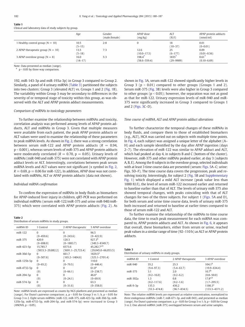
Table 1Clinical and laboratory data of study subjects by group.
Age Gender(male:female)
APAP dose(mg/kg)
ALT(IU/l)
APAP protein adducts(nmol/ml)
1 Healthy control group (N = 10) 10.5 2:8 0 18 0.01(5–15) (10–37) (0–0.01)
2 APAP therapeutic group (N = 10) 13.5 7:3 12.6 25 0.09(5–18) (10.0–17.5) (6–177) (0.01–0.56)
3 APAP overdose group (N = 8) 16.0 5:3 198.4⁎ 1835⁎ 0.61⁎
(14–17) (58.6–559.4) (29–9909) (0.10–6.69)
Note: Data presented as median (range).⁎ p b 0.05 by three way comparison.
182 X. Yang et al. / Toxicology and Applied Pharmacology 284 (2015) 180–187
192, miR-143-3p and miR-193a-3p) in Group 3 compared to Group 2.Similarly, a panel of 4 urinary miRNA (Table 3) partitioned the subjectsinto two clusters: Group 3 (elevated ALT) vs. Groups 1 and 2 (Fig. 1B).The variability within Group 3 may be secondary to differences in theseverity of or temporal stage of toxicity within this group, as was ob-served with the ALT and APAP protein adduct measurements.
Comparison of miRNAs to toxicology parameters
To further examine the relationship between miRNAs and toxicity,correlation analysis was performed among levels of APAP protein ad-ducts, ALT and miRNAs in Group 3. Given that multiple measureswere available from each patient, the peak APAP protein adducts orALT values were used to examine the relationship of these parametersto peakmiRNA levels. As shown in Fig. 2, there was a strong correlationbetween serum miR-122 and APAP protein adducts (R = 0.94,p b 0.001), whereas serum levels of miR-375 and APAP protein adductswere moderately correlated (R = 0.70, p = 0.05). Urinary levels ofmiRNAs (miR-940 andmiR-375)were not correlatedwith APAP proteinadduct levels or ALT. Interestingly, correlations between peak serummiRNA levels and ALT values were not significant (x symbol in Fig. 2,R=0.69, p = 0.06 for miR-122), In addition, APAP dose was not corre-lated with miRNAs, ALT or APAP protein adducts (data not shown).
Individual miRNA confirmation
To confirm the expression of miRNAs in body fluids as biomarkersfor APAP-induced liver injury in children, qRT-PCR was performed onindividual miRNAs (serum miR-122/miR-375 and urine miR-940/miR-375) which were correlated with APAP protein adducts (Fig. 2). As
Table 2Distribution of serummiRNAs in study groups.
miRNA ID 1 Control 2 APAP therapeutic 3 APAP overdose
miR-122 0 0 96.5(0–459.6) (0–243.6) (0–422.9)
miR-375 420.9 120.3 927.3b
(0–698.0) (0–1883.7) (349.3–8349.7)miR-423-5p 15,785.7 6573.6 45,282.7a,b
(5833.3–29,882.2) (5691.1–25,723.4) (29,045.0–66,053.5)miR-30d-5p 0 661.7 1829.5a
(0–507.6) (165.2–1404.6) (525.1–2701.4)miR-125b-5p 0 0 110.0
(0–65.2) (0–79.5) (0–446.4)miR-4732-5p 0 0 61.0a
(0) (0–66.1) (0–238.7)miR-204-5p 0 0 46.6a
(0) (0–24.1) (0–69.9)miR-574-3p 0 0 63.3
(0) (0–31.6) (0–358.0)
Note: miRNA levels are expressed as counts by NGS platform and presented as median(range). For Dunn's pairwise comparison: a, p b 0.05 for Group 3 vs 1; b, p b 0.05 forGroup 3 vs 2. Eight serum miRNAs (miR-122, miR-375, miR-423-5p, miR-30d-5p, miR-125b-5p, miR-4732-5p, miR-204-5p, and miR-574-3p) were increased in Group 3(ANOVA, p b 0.05).
shown in Fig. 3A, serum miR-122 showed significantly higher levels inGroup 3 (p b 0.01) compared to other groups (Groups 1 and 2).Serum miR-375 (Fig. 3B) levels were also higher in Group 3 comparedto other groups (p b 0.05); however, the separation was not as goodas that for miR-122. Urinary expression levels of miR-940 and miR-375 were significantly increased in Group 3 compared to Groups 1and 2 (Figs. 3C–D).
Time course of miRNA, ALT and APAP protein adduct alterations
To further characterize the temporal changes of these miRNAs inbody fluids, and compare them to those of established biomarkers(e.g., ALT), HCA was carried out on subjects with multiple time points.In Fig. 4, each subject was assigned a unique letter of the alphabet (A–H) and each sample identified by the day after APAP ingestion (days2–7). The elevation of miR-122 was similar to APAP adduct and ALT,which had peaked at day 4, in subjects B and C (bottom of the cluster).However, miR-375 and other miRNAs peaked earlier, at day 3 (subjectsA, B, E). Among the8 subjects in the overdose group, selected individualswith at least 3 time course data are presented (serum: Figs. 5A–C; urine:Figs. 5D–F). The time course data covers the progression, peak and re-solving toxicity. Interestingly, for subject 2 (Fig. 5B and SupplementaryFig. 1) which displayed a mild ALT increase (peak value less than1000 IU/l), the level of serum miR-122 increased earlier and returnedto baseline earlier than that of ALT. The levels of urinary miR-375 alsoshowed temporal changes, with peaks coinciding with that of ALTchanges for two of the three subjects. For subject 1 (Figs. 5A and D),for both serum and urine time course data, levels of urinary miR-375both increased and returned to baseline at earlier times compared tothose of serum miR-122 and ALT.
To further examine the relationship of the miRNAs to time coursedata, the time to reach peak measurement for each miRNA was com-pared to, APAP protein adducts and ALT. As shown in Fig. 6, it appearsthat overall, these biomarkers, either from serum or urine, reachedpeak values in a similar range of time (92–110h) as ALT or APAP proteinadducts.
Table 3Distribution of urinary miRNAs in study groups.
miRNA ID 1 Control 2 APAP therapeutic 3 APAP overdose
miR-940 35.2 25.3 104.1b
(9.4–97.3) (5.4–63.7) (19.9–634.4)miR-375 3.1 2.0 10.7b
(0.2–16.0) (0.2–6.2) (0.8–50.0)miR-302a 1.3 0.8 17.8a,b
(0.2–117.6) (0.2–1.6) (1.7–295.5)miR-9-3p 135.3 436.2 1186.3a
(55.3–474.8) (96.7–834.5) (133.3–4371.2)
Note: The relative miRNA levels are expressed as relative concentration, normalized tothree endogenous miRNAs (miR-7, miR-671-3p, and miR-943), and presented as median(range). For Dunn's pairwise comparison: a, p b 0.05 for Group 3 vs 1; b, p b 0.05 for Group3 vs 2. One altered miRNA (miR-375) overlapped between serum and urine samples.
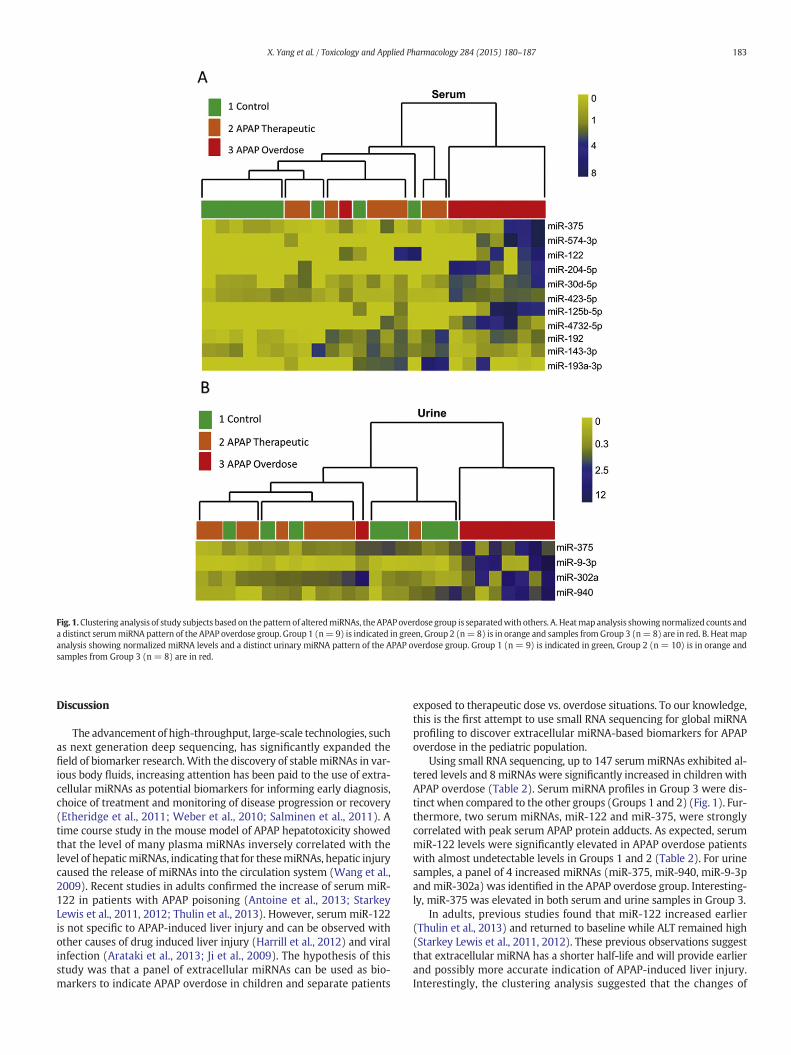
Fig. 1.Clustering analysis of study subjects based on thepattern of alteredmiRNAs, theAPAPoverdose group is separatedwith others. A.Heatmap analysis showing normalized counts anda distinct serummiRNA pattern of the APAP overdose group. Group 1 (n=9) is indicated in green, Group 2 (n=8) is in orange and samples fromGroup 3 (n=8) are in red. B. Heat mapanalysis showing normalized miRNA levels and a distinct urinary miRNA pattern of the APAP overdose group. Group 1 (n = 9) is indicated in green, Group 2 (n = 10) is in orange andsamples from Group 3 (n = 8) are in red.
183X. Yang et al. / Toxicology and Applied Pharmacology 284 (2015) 180–187
Discussion
The advancement of high-throughput, large-scale technologies, suchas next generation deep sequencing, has significantly expanded thefield of biomarker research.With the discovery of stablemiRNAs in var-ious body fluids, increasing attention has been paid to the use of extra-cellular miRNAs as potential biomarkers for informing early diagnosis,choice of treatment and monitoring of disease progression or recovery(Etheridge et al., 2011; Weber et al., 2010; Salminen et al., 2011). Atime course study in the mouse model of APAP hepatotoxicity showedthat the level of many plasma miRNAs inversely correlated with thelevel of hepaticmiRNAs, indicating that for thesemiRNAs, hepatic injurycaused the release of miRNAs into the circulation system (Wang et al.,2009). Recent studies in adults confirmed the increase of serum miR-122 in patients with APAP poisoning (Antoine et al., 2013; StarkeyLewis et al., 2011, 2012; Thulin et al., 2013). However, serum miR-122is not specific to APAP-induced liver injury and can be observed withother causes of drug induced liver injury (Harrill et al., 2012) and viralinfection (Arataki et al., 2013; Ji et al., 2009). The hypothesis of thisstudy was that a panel of extracellular miRNAs can be used as bio-markers to indicate APAP overdose in children and separate patients
exposed to therapeutic dose vs. overdose situations. To our knowledge,this is the first attempt to use small RNA sequencing for global miRNAprofiling to discover extracellular miRNA-based biomarkers for APAPoverdose in the pediatric population.
Using small RNA sequencing, up to 147 serummiRNAs exhibited al-tered levels and 8 miRNAs were significantly increased in children withAPAP overdose (Table 2). Serum miRNA profiles in Group 3 were dis-tinct when compared to the other groups (Groups 1 and 2) (Fig. 1). Fur-thermore, two serum miRNAs, miR-122 and miR-375, were stronglycorrelated with peak serum APAP protein adducts. As expected, serummiR-122 levels were significantly elevated in APAP overdose patientswith almost undetectable levels in Groups 1 and 2 (Table 2). For urinesamples, a panel of 4 increased miRNAs (miR-375, miR-940, miR-9-3pand miR-302a) was identified in the APAP overdose group. Interesting-ly, miR-375 was elevated in both serum and urine samples in Group 3.
In adults, previous studies found that miR-122 increased earlier(Thulin et al., 2013) and returned to baseline while ALT remained high(Starkey Lewis et al., 2011, 2012). These previous observations suggestthat extracellular miRNA has a shorter half-life and will provide earlierand possibly more accurate indication of APAP-induced liver injury.Interestingly, the clustering analysis suggested that the changes of
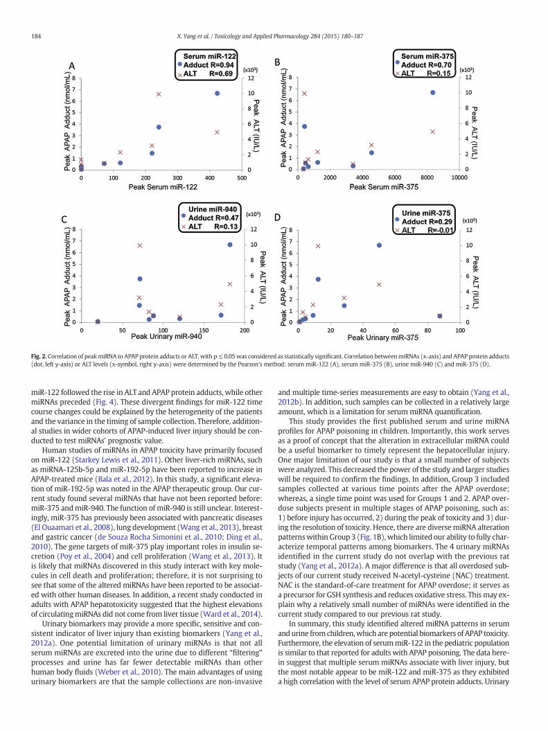
Fig. 2. Correlation of peakmiRNA to APAP protein adducts or ALT, with p≤ 0.05was considered as statistically significant. Correlation betweenmiRNAs (x-axis) and APAP protein adducts(dot, left y-axis) or ALT levels (x-symbol, right y-axis) were determined by the Pearson's method: serum miR-122 (A), serum miR-375 (B), urine miR-940 (C) and miR-375 (D).
184 X. Yang et al. / Toxicology and Applied Pharmacology 284 (2015) 180–187
miR-122 followed the rise in ALT and APAP protein adducts, while othermiRNAs preceded (Fig. 4). These divergent findings for miR-122 timecourse changes could be explained by the heterogeneity of the patientsand the variance in the timing of sample collection. Therefore, addition-al studies in wider cohorts of APAP-induced liver injury should be con-ducted to test miRNAs' prognostic value.
Human studies of miRNAs in APAP toxicity have primarily focusedon miR-122 (Starkey Lewis et al., 2011). Other liver-rich miRNAs, suchas miRNA-125b-5p and miR-192-5p have been reported to increase inAPAP-treated mice (Bala et al., 2012). In this study, a significant eleva-tion of miR-192-5p was noted in the APAP therapeutic group. Our cur-rent study found several miRNAs that have not been reported before:miR-375 andmiR-940. The function of miR-940 is still unclear. Interest-ingly, miR-375 has previously been associated with pancreatic diseases(El Ouaamari et al., 2008), lung development (Wang et al., 2013), breastand gastric cancer (de Souza Rocha Simonini et al., 2010; Ding et al.,2010). The gene targets of miR-375 play important roles in insulin se-cretion (Poy et al., 2004) and cell proliferation (Wang et al., 2013). Itis likely that miRNAs discovered in this study interact with key mole-cules in cell death and proliferation; therefore, it is not surprising tosee that some of the altered miRNAs have been reported to be associat-ed with other human diseases. In addition, a recent study conducted inadults with APAP hepatotoxicity suggested that the highest elevationsof circulatingmiRNAs did not come from liver tissue (Ward et al., 2014).
Urinary biomarkers may provide a more specific, sensitive and con-sistent indicator of liver injury than existing biomarkers (Yang et al.,2012a). One potential limitation of urinary miRNAs is that not allserum miRNAs are excreted into the urine due to different “filtering”processes and urine has far fewer detectable miRNAs than otherhuman body fluids (Weber et al., 2010). The main advantages of usingurinary biomarkers are that the sample collections are non-invasive
and multiple time-series measurements are easy to obtain (Yang et al.,2012b). In addition, such samples can be collected in a relatively largeamount, which is a limitation for serum miRNA quantification.
This study provides the first published serum and urine miRNAprofiles for APAP poisoning in children. Importantly, this work servesas a proof of concept that the alteration in extracellular miRNA couldbe a useful biomarker to timely represent the hepatocellular injury.One major limitation of our study is that a small number of subjectswere analyzed. This decreased the power of the study and larger studieswill be required to confirm the findings. In addition, Group 3 includedsamples collected at various time points after the APAP overdose;whereas, a single time point was used for Groups 1 and 2. APAP over-dose subjects present in multiple stages of APAP poisoning, such as:1) before injury has occurred, 2) during the peak of toxicity and 3) dur-ing the resolution of toxicity. Hence, there are diversemiRNA alterationpatternswithin Group 3 (Fig. 1B),which limited our ability to fully char-acterize temporal patterns among biomarkers. The 4 urinary miRNAsidentified in the current study do not overlap with the previous ratstudy (Yang et al., 2012a). A major difference is that all overdosed sub-jects of our current study received N-acetyl-cysteine (NAC) treatment.NAC is the standard-of-care treatment for APAP overdose; it serves asa precursor for GSH synthesis and reduces oxidative stress. Thismay ex-plain why a relatively small number of miRNAs were identified in thecurrent study compared to our previous rat study.
In summary, this study identified altered miRNA patterns in serumandurine fromchildren,which are potential biomarkers of APAP toxicity.Furthermore, the elevation of serummiR-122 in the pediatric populationis similar to that reported for adults with APAP poisoning. The data here-in suggest that multiple serum miRNAs associate with liver injury, butthe most notable appear to be miR-122 and miR-375 as they exhibiteda high correlation with the level of serum APAP protein adducts. Urinary
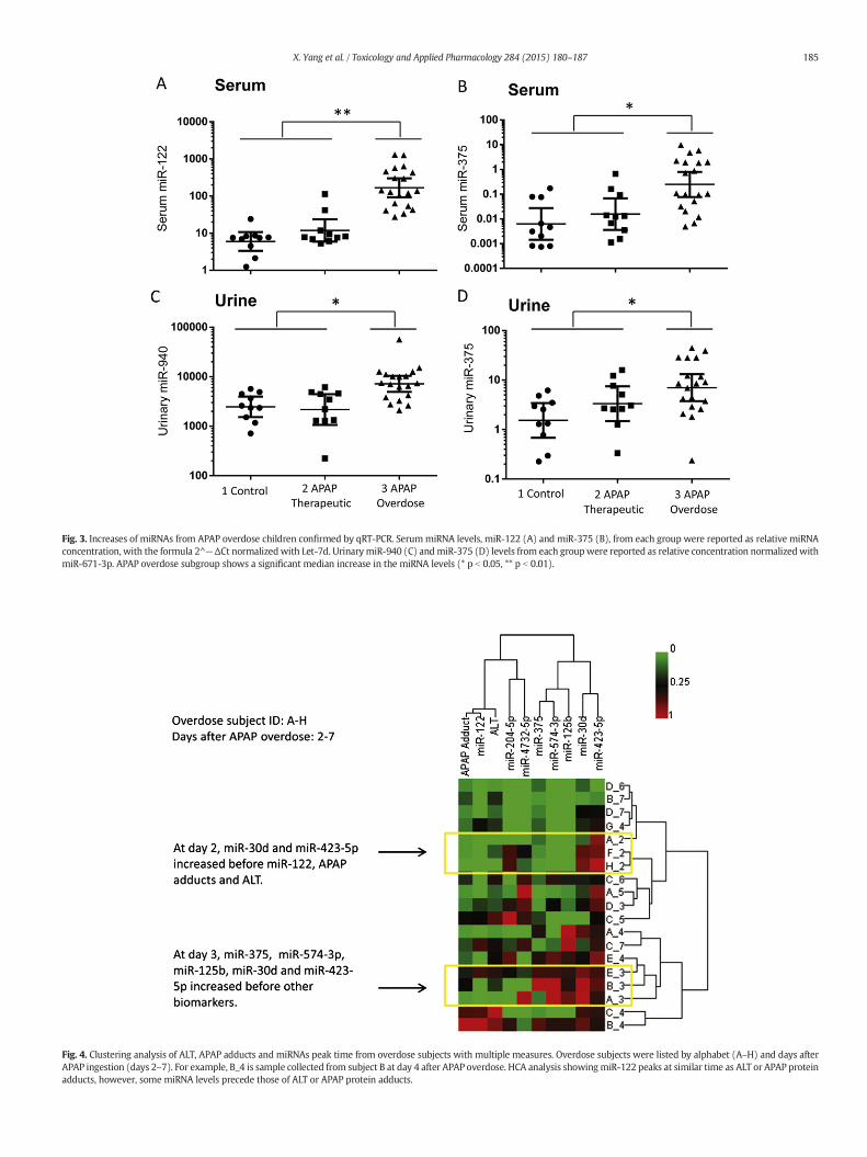
Fig. 3. Increases of miRNAs from APAP overdose children confirmed by qRT-PCR. SerummiRNA levels, miR-122 (A) and miR-375 (B), from each group were reported as relative miRNAconcentration, with the formula 2^−ΔCt normalizedwith Let-7d. Urinary miR-940 (C) and miR-375 (D) levels from each groupwere reported as relative concentration normalizedwithmiR-671-3p. APAP overdose subgroup shows a significant median increase in the miRNA levels (* p b 0.05, ** p b 0.01).
Fig. 4. Clustering analysis of ALT, APAP adducts and miRNAs peak time from overdose subjects with multiple measures. Overdose subjects were listed by alphabet (A–H) and days afterAPAP ingestion (days 2–7). For example, B_4 is sample collected from subject B at day 4 after APAP overdose. HCA analysis showingmiR-122 peaks at similar time as ALT or APAP proteinadducts, however, some miRNA levels precede those of ALT or APAP protein adducts.
185X. Yang et al. / Toxicology and Applied Pharmacology 284 (2015) 180–187
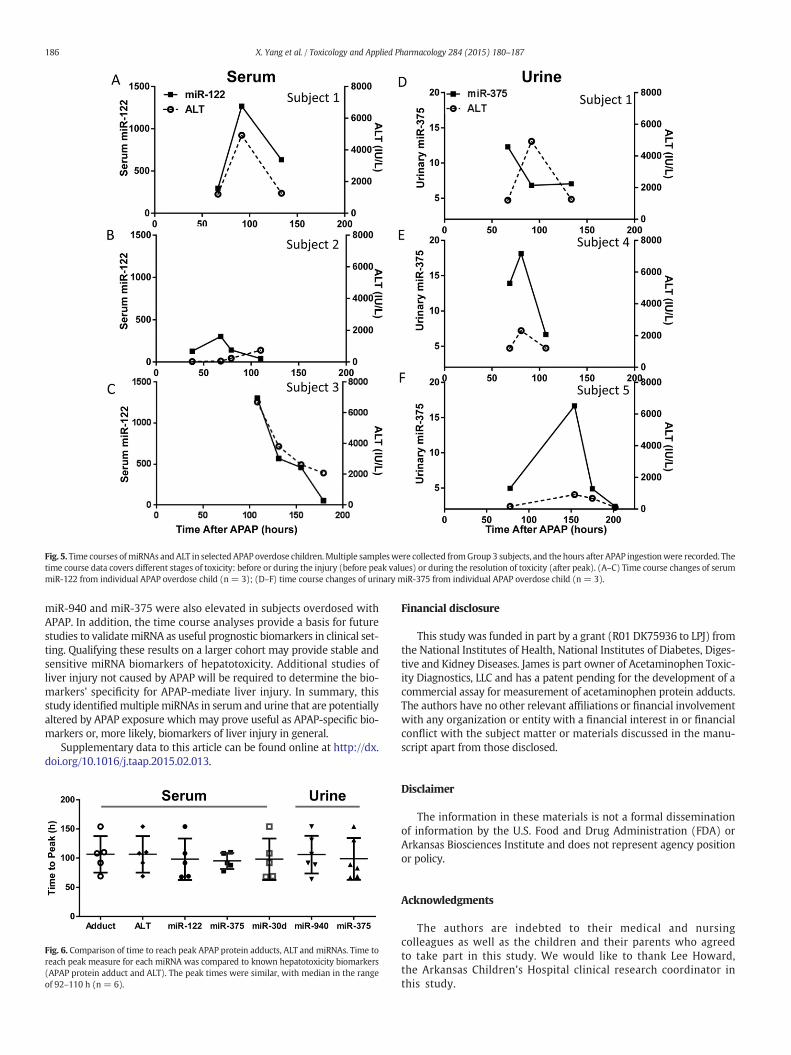
Fig. 5. Time courses ofmiRNAs and ALT in selected APAP overdose children.Multiple sampleswere collected fromGroup 3 subjects, and the hours after APAP ingestionwere recorded. Thetime course data covers different stages of toxicity: before or during the injury (before peak values) or during the resolution of toxicity (after peak). (A–C) Time course changes of serummiR-122 from individual APAP overdose child (n = 3); (D–F) time course changes of urinary miR-375 from individual APAP overdose child (n = 3).
186 X. Yang et al. / Toxicology and Applied Pharmacology 284 (2015) 180–187
miR-940 and miR-375 were also elevated in subjects overdosed withAPAP. In addition, the time course analyses provide a basis for futurestudies to validate miRNA as useful prognostic biomarkers in clinical set-ting. Qualifying these results on a larger cohort may provide stable andsensitive miRNA biomarkers of hepatotoxicity. Additional studies ofliver injury not caused by APAP will be required to determine the bio-markers' specificity for APAP-mediate liver injury. In summary, thisstudy identifiedmultiplemiRNAs in serum and urine that are potentiallyaltered by APAP exposure which may prove useful as APAP-specific bio-markers or, more likely, biomarkers of liver injury in general.
Supplementary data to this article can be found online at http://dx.doi.org/10.1016/j.taap.2015.02.013.
Fig. 6. Comparison of time to reach peak APAP protein adducts, ALT and miRNAs. Time toreach peak measure for each miRNA was compared to known hepatotoxicity biomarkers(APAP protein adduct and ALT). The peak times were similar, with median in the rangeof 92–110 h (n = 6).
Financial disclosure
This study was funded in part by a grant (R01 DK75936 to LPJ) fromthe National Institutes of Health, National Institutes of Diabetes, Diges-tive and Kidney Diseases. James is part owner of Acetaminophen Toxic-ity Diagnostics, LLC and has a patent pending for the development of acommercial assay for measurement of acetaminophen protein adducts.The authors have no other relevant affiliations or financial involvementwith any organization or entity with a financial interest in or financialconflict with the subject matter or materials discussed in the manu-script apart from those disclosed.
Disclaimer
The information in these materials is not a formal disseminationof information by the U.S. Food and Drug Administration (FDA) orArkansas Biosciences Institute and does not represent agency positionor policy.
Acknowledgments
The authors are indebted to their medical and nursingcolleagues as well as the children and their parents who agreedto take part in this study. We would like to thank Lee Howard,the Arkansas Children's Hospital clinical research coordinator inthis study.
187X. Yang et al. / Toxicology and Applied Pharmacology 284 (2015) 180–187
References
Algren, D.A., 2008. Review of N-acetylcysteine for the treatment of acetaminophen(paracetamol) toxicity in pediatrics. Geneva http://www.who.int/selection_medicines/committees/subcommittee/2/acetylcysteine_rev.pdf.
Ambros, V., 2004. The functions of animal microRNAs. Nature 431, 350–355.Antoine, D.J., Dear, J.W., Lewis, P.S., Platt, V., Coyle, J.,Masson,M., Thanacoody, R.H., et al., 2013.
Mechanistic biomarkers provide early and sensitive detection of acetaminophen-induced acute liver injury at first presentation to hospital. Hepatology 58, 777–787.
Arataki, K., Hayes, C.N., Akamatsu, S., Akiyama, R., Abe, H., Tsuge, M., Miki, D., et al., 2013.Circulating microRNA-22 correlates with microRNA-122 and represents viral replica-tion and liver injury in patients with chronic hepatitis B. J. Med. Virol. 85, 789–798.
Bala, S., Petrasek, J., Mundkur, S., Catalano, D., Levin, I., Ward, J., Alao, H., et al., 2012. Cir-culating microRNAs in exosomes indicate hepatocyte injury and inflammation in al-coholic, drug-induced, and inflammatory liver diseases. Hepatology 56, 1946–1957.
Bullard, J., Purdom, E., Hansen, K., Dudoit, S., 2010. Evaluation of statisticalmethods for nor-malization anddifferential expression inmRNA-Seq experiments. BMCBioinforma. 11,94.
Cermelli, S., Ruggieri, A., Marrero, J.A., Ioannou, G.N., Beretta, L., 2011. CirculatingmicroRNAs in patients with chronic hepatitis C and non-alcoholic fatty liver disease.PLoS One 6, e23937.
Chang, J., Nicolas, E., Marks, D., Sander, C., Lerro, A., Buendia, M.A., Xu, C., et al., 2004. miR-122, a mammalian liver-specific microRNA, is processed from hcr mRNA and maydownregulate the high affinity cationic amino acid transporter CAT-1. RNA Biol. 1,106–113.
Chen, X., Ba, Y., Ma, L., Cai, X., Yin, Y., Wang, K., Guo, J., et al., 2008. Characterization ofmicroRNAs in serum: a novel class of biomarkers for diagnosis of cancer and otherdiseases. Cell Res. 18, 997–1006.
de Souza Rocha Simonini, P., Breiling, A., Gupta, N., Malekpour, M., Youns, M.,Omranipour, R., Malekpour, F., et al., 2010. Epigenetically deregulated microRNA-375 is involved in a positive feedback loop with estrogen receptor α in breast cancercells. Cancer Res. 70, 9175–9184.
Dillies, M.-A., Rau, A., Aubert, J., Hennequet-Antier, C., Jeanmougin, M., Servant, N., Keime,C., et al., 2013. A comprehensive evaluation of normalization methods for Illuminahigh-throughput RNA sequencing data analysis. Brief. Bioinform. 14, 671–683.
Ding, L., Xu, Y., Zhang, W., Deng, Y., Si, M., Du, Y., Yao, H., et al., 2010. MiR-375 frequentlydownregulated in gastric cancer inhibits cell proliferation by targeting JAK2. Cell Res.20, 784–793.
El Ouaamari, A., Baroukh, N., Martens, G.A., Lebrun, P., Pipeleers, D., van Obberghen, E.,2008. miR-375 targets 3′-phosphoinositide-dependent protein kinase-1 and regu-lates glucose-induced biological responses in pancreatic β-cells. Diabetes 57,2708–2717.
Etheridge, A., Lee, I., Hood, L., Galas, D., Wang, K., 2011. Extracellular microRNA: a newsource of biomarkers. Mutat. Res. 717, 85–90.
Fang, H., Harris, S.C., Su, Z., Chen, M., Qian, F., Shi, L., Perkins, R., et al., 2009. ArrayTrack: anFDA and public genomic tool. Methods Mol. Biol. 563, 379–398.
Harrill, A.H., Roach, J., Fier, I., Eaddy, J.S., Kurtz, C.L., Antoine, D.J., Spencer, D.M., et al., 2012.The effects of heparins on the liver: application of mechanistic serum biomarkers in arandomized study in healthy volunteers. Clin. Pharmacol. Ther. 92, 214–220.
James, L.P., Mayeux, P.R., Hinson, J.A., 2003. Acetaminophen-induced hepatotoxicity. DrugMetab. Dispos. 31, 1499–1506.
James, L.P., Capparelli, E.V., Simpson, P.M., Letzig, L., Roberts, D., Hinson, J.A., Kearns, G.L.,et al., 2008. Acetaminophen-associated hepatic injury: evaluation of acetaminophenprotein adducts in children and adolescents with acetaminophen overdose. Clin.Pharmacol. Ther. 84, 684–690.
James, L.P., Letzig, L., Simpson, P.M., Capparelli, E., Roberts, D.W., Hinson, J.A., Davern, T.J.,et al., 2009. Pharmacokinetics of acetaminophen-protein adducts in adults with acet-aminophen overdose and acute liver failure. Drug Metab. Dispos. 37, 1779–1784.
Ji, J., Shi, J., Budhu, A., Yu, Z., Forgues, M., Roessler, S., Ambs, S., et al., 2009. MicroRNA ex-pression, survival, and response to interferon in liver cancer. N. Engl. J. Med. 361,1437–1447.
Krauskopf, J., Caiment, F., Claessen, S.M., Johnson, K.J., Warner, R.L., Schomaker, S.J., Burt,D.A., et al., 2015. Application of high-throughput sequencing to circulating
microRNAs reveals novel biomarkers for drug-induced liver injury. Toxicol. Sci. 143(2), 268–276.
Krol, J., Loedige, I., Filipowicz, W., 2010. The widespread regulation of microRNA biogen-esis, function and decay. Nat. Rev. Genet. 11, 597–610.
Laterza, O.F., Scott, M.G., Garrett-Engele, P.W., Korenblat, K.M., Lockwood, C.M., 2013. Cir-culating miR-122 as a potential biomarker of liver disease. Biomark. Med 7, 205–210.
McGill, M.R., Sharpe, M.R., Williams, C.D., Taha, M., Curry, S.C., Jaeschke, H., 2012. Themechanism underlying acetaminophen-induced hepatotoxicity in humans andmice involves mitochondrial damage and nuclear DNA fragmentation. J. Clin. Invest.122, 1574–1583.
McGill, M.R., Cao, M., Svetlov, A., Sharpe, M.R., Williams, C.D., Curry, S.C., Farhood, A., et al.,2014a. Argininosuccinate synthetase as a plasma biomarker of liver injury after acet-aminophen overdose in rodents and humans. Biomarkers 19, 222–230.
McGill, M.R., Li, F., Sharpe, M.R., Williams, C.D., Curry, S.C., Ma, X., Jaeschke, H., 2014b. Cir-culating acylcarnitines as biomarkers of mitochondrial dysfunction after acetamino-phen overdose in mice and humans. Arch. Toxicol. 88, 391–401.
Muldrew, K.L., James, L.P., Coop, L., McCullough, S.S., Hendrickson, H.P., Hinson, J.A.,Mayeux, P.R., 2002. Determination of acetaminophen-protein adducts in mouseliver and serum and human serum after hepatotoxic doses of acetaminophen usinghigh-performance liquid chromatography with electrochemical detection. DrugMetab. Dispos. 30, 446–451.
Poy, M.N., Eliasson, L., Krutzfeldt, J., Kuwajima, S., Ma, X., MacDonald, P.E., Pfeffer, S., et al.,2004. A pancreatic islet-specific microRNA regulates insulin secretion. Nature 432,226–230.
Salminen, W.F., Yang, X., Shi, Q., Mendrick, D.L., 2011. Using microRNA as biomarkers ofdrug-induced liver injury. J. Mol. Biomark. Diagn. 2, 119.
Squires Jr., R.H., Shneider, B.L., Bucuvalas, J., Alonso, E., Sokol, R.J., Narkewicz, M.R.,Dhawan, A., et al., 2006. Acute liver failure in children: the first 348 patients in the pe-diatric acute liver failure study group. J. Pediatr. 148, 652–658 (e652).
Starkey Lewis, P.J., Dear, J., Platt, V., Simpson, K.J., Craig, D.G., Antoine, D.J., French, N.S., etal., 2011. Circulating microRNAs as potential markers of human drug-induced liverinjury. Hepatology 54, 1767–1776.
Starkey Lewis, P.J., Merz, M., Couttet, P., Grenet, O., Dear, J., Antoine, D.J., Goldring, C., et al.,2012. Serum microRNA biomarkers for drug-induced liver injury. Clin. Pharmacol.Ther. 92, 291–293.
Su, T.H., Liu, C.H., Liu, C.J., Chen, C.L., Ting, T.T., Tseng, T.C., Chen, P.J., et al., 2013. SerummicroRNA-122 level correlates with virologic responses to pegylated interferon ther-apy in chronic hepatitis C. Proc. Natl. Acad. Sci. U. S. A. 110, 7844–7849.
Thulin, P., Nordahl, G., Gry, M., Yimer, G., Aklillu, E., Makonnen, E., Aderaye, G., et al., 2014.Keratin-18 and microRNA-122 complement alanine aminotransferase as novel safetybiomarkers for drug-induced liver injury in two human cohorts. Liver Int. 34,367–378.
Wang, K., Zhang, S., Marzolf, B., Troisch, P., Brightman, A., Hu, Z., Hood, L.E., et al., 2009.Circulating microRNAs, potential biomarkers for drug-induced liver injury. Proc.Natl. Acad. Sci. U. S. A. 106, 4402–4407.
Wang, Y., Huang, C., Reddy Chintagari, N., Bhaskaran, M., Weng, T., Guo, Y., Xiao, X., et al.,2013. miR-375 regulates rat alveolar epithelial cell trans-differentiation by inhibitingWnt/β-catenin pathway. Nucleic Acids Res. 41, 3833–3844.
Ward, J., Kanchagar, C., Veksler-Lublinsky, I., Lee, R.C., McGill, M.R., Jaeschke, H., Curry, S.C.,et al., 2014. Circulating microRNA profiles in human patients with acetaminophenhepatotoxicity or ischemic hepatitis. Proc. Natl. Acad. Sci. U. S. A. 111 (33),12169–12174.
Weber, J.A., Baxter, D.H., Zhang, S., Huang, D.Y., Huang, K.H., Lee, M.J., Galas, D.J., et al.,2010. The microRNA spectrum in 12 body fluids. Clin. Chem. 56, 1733–1741.
Yang, X., Greenhaw, J., Shi, Q., Su, Z., Qian, F., Davis, K., Mendrick, D.L., et al., 2012a. Iden-tification of urinary microRNA profiles in rats that may diagnose hepatotoxicity.Toxicol. Sci. 125, 335–344.
Yang, X., Salminen, W., Schnackenberg, L., 2012b. Current and emerging biomarkers ofhepatotoxicity. Curr. Biomark. Find. 2, 43–55.
Related Documents