Toxic Alexandrium minutum (Dinophyceae) from Vietnam with new gonyautoxin analogue Po-Teen Lim a,b, * , Shigeru Sato a , Chu Van Thuoc c , Pham The Tu c , Nguyen Thi Minh Huyen c , Yoshinobu Takata a , Makoto Yoshida d , Atsushi Kobiyama a , Kazuhiko Koike a , Takehiko Ogata a a School of Fisheries Science, Kitasato University, Sanriku, Ofunato, Iwate 022-0101, Japan b Faculty of Resource Science and Technology, Universiti Malaysia Sarawak, Kuching, Sarawak, Malaysia 94300 c Department of Biological Resource and Ecology, Institute of Marine Environment and Resources, 246 Da Nang, Hai Phong, Vietnam d Faculty of Environmental and Symbiotic Sciences, Prefecture University of Kumamoto, 3-1-100 Tsukide, Kumamoto 862-8502, Japan Received 5 December 2005; received in revised form 24 March 2006; accepted 14 April 2006 Abstract Clonal cultures of Alexandrium species collected from a shrimp pond on the northern coast of Vietnam were established and morphologically identified as Alexandrium minutum. Nucleotide sequences of domains 1 and 2 of the large subunit ribosomal (LSU) rRNA gene showed high sequence similarity to A. minutum isolates from Malaysia. Paralytic shellfish toxin profile of the clones was characterized by the dominance of GTX4, GTX1, and NEO. GTX3, GTX2, and dcSTX were also present in trace amount. Toxin content varied among the strains and growth stages, ranged from 3.0 to 12.5 fmol cell 1 . In addition to these known toxin components, a new gonyautoxin derivative was detected by HPLC, eluting between GTX4 and GTX1. The peak of this compound disappeared under non-oxidizing HPLC condition but unchanged either after treated with 0.05 M ammonium phosphate/10% mercaptoethanol or 0.1N HCl hydrolysis. LCMS ion scanning showed a parental ion of [M + H] + at m/z 396, [M SO 3 ] + at m/z 316, and [M SO 4 ] + at m/z 298. Based on these results, the derivative was identified as deoxy-GTX4-12ol, and this represents the first report of this toxin analogue. # 2006 Elsevier B.V. All rights reserved. Keywords: Alexandrium minutum; Large subunit ribosomal RNA; Morphology paralytic shellfish toxins; Vietnam 1. Introduction In the Asia Pacific region, the toxic Alexandrium minutum Halim was first reported in Taiwan (Su and Chiang, 1991; Hwang and Lu, 2000), followed by Australia (Cannon, 1990), Japan (Yuki, 1994), Thailand (Matsuoka et al., 1997), New Zealand (Chang et al., 1995; Mackenzie and Berkett, 1997), Vietnam (Yoshida et al., 2000), Malaysia (Usup et al., 2002), and Philippine (Bajarias et al., 2003). A. minutum was commonly occurred in semi-enclosed water such as harbor and coastal lagoon (Delgado et al., 1990; Giacobbe et al., 1996), estuary (Lim et al., 2004), and shrimp ponds (Matsuoka et al., 1997; Yoshida et al., 2000). Strong freshwater influence coupled with a stratified water column has been suggested as an essential prerequisite for blooms of this species (Delgado et al., 1990; Giacobbe et al., 1996; Lim and Ogata, 2005). Most A. minutum isolates studied to date produced paralytic shellfish toxins (PSTs) which were dominated www.elsevier.com/locate/hal Harmful Algae 6 (2007) 321–331 * Corresponding author. Tel.: +81 192 44 2121; fax: +81 192 44 2125. E-mail addresses: [email protected], [email protected] (P.-T. Lim). 1568-9883/$ – see front matter # 2006 Elsevier B.V. All rights reserved. doi:10.1016/j.hal.2006.04.004

Welcome message from author
This document is posted to help you gain knowledge. Please leave a comment to let me know what you think about it! Share it to your friends and learn new things together.
Transcript
www.elsevier.com/locate/hal
Harmful Algae 6 (2007) 321–331
Toxic Alexandrium minutum (Dinophyceae) from Vietnam
with new gonyautoxin analogue
Po-Teen Lim a,b,*, Shigeru Sato a, Chu Van Thuoc c, Pham The Tu c,Nguyen Thi Minh Huyen c, Yoshinobu Takata a, Makoto Yoshida d,
Atsushi Kobiyama a, Kazuhiko Koike a, Takehiko Ogata a
a School of Fisheries Science, Kitasato University, Sanriku, Ofunato, Iwate 022-0101, Japanb Faculty of Resource Science and Technology, Universiti Malaysia Sarawak, Kuching, Sarawak, Malaysia 94300
c Department of Biological Resource and Ecology, Institute of Marine Environment and Resources, 246 Da Nang, Hai Phong, Vietnamd Faculty of Environmental and Symbiotic Sciences, Prefecture University of Kumamoto, 3-1-100 Tsukide, Kumamoto 862-8502, Japan
Received 5 December 2005; received in revised form 24 March 2006; accepted 14 April 2006
Abstract
Clonal cultures of Alexandrium species collected from a shrimp pond on the northern coast of Vietnam were established and
morphologically identified as Alexandrium minutum. Nucleotide sequences of domains 1 and 2 of the large subunit ribosomal (LSU)
rRNA gene showed high sequence similarity to A. minutum isolates from Malaysia. Paralytic shellfish toxin profile of the clones was
characterized by the dominance of GTX4, GTX1, and NEO. GTX3, GTX2, and dcSTX were also present in trace amount. Toxin
content varied among the strains and growth stages, ranged from 3.0 to 12.5 fmol cell�1. In addition to these known toxin
components, a new gonyautoxin derivative was detected by HPLC, eluting between GTX4 and GTX1. The peak of this compound
disappeared under non-oxidizing HPLC condition but unchanged either after treated with 0.05 M ammonium phosphate/10%
mercaptoethanol or 0.1N HCl hydrolysis. LCMS ion scanning showed a parental ion of [M + H]+ at m/z 396, [M � SO3]+ at m/z 316,
and [M � SO4]+ at m/z 298. Based on these results, the derivative was identified as deoxy-GTX4-12ol, and this represents the first
report of this toxin analogue.
# 2006 Elsevier B.V. All rights reserved.
Keywords: Alexandrium minutum; Large subunit ribosomal RNA; Morphology paralytic shellfish toxins; Vietnam
1. Introduction
In the Asia Pacific region, the toxic Alexandrium
minutum Halim was first reported in Taiwan (Su and
Chiang, 1991; Hwang and Lu, 2000), followed by
Australia (Cannon, 1990), Japan (Yuki, 1994), Thailand
(Matsuoka et al., 1997), New Zealand (Chang et al.,
* Corresponding author. Tel.: +81 192 44 2121;
fax: +81 192 44 2125.
E-mail addresses: [email protected], [email protected]
(P.-T. Lim).
1568-9883/$ – see front matter # 2006 Elsevier B.V. All rights reserved.
doi:10.1016/j.hal.2006.04.004
1995; Mackenzie and Berkett, 1997), Vietnam (Yoshida
et al., 2000), Malaysia (Usup et al., 2002), and Philippine
(Bajarias et al., 2003). A. minutum was commonly
occurred in semi-enclosed water such as harbor and
coastal lagoon (Delgado et al., 1990; Giacobbe et al.,
1996), estuary (Lim et al., 2004), and shrimp ponds
(Matsuoka et al., 1997; Yoshida et al., 2000). Strong
freshwater influence coupled with a stratified water
column has been suggested as an essential prerequisite
for blooms of this species (Delgado et al., 1990;
Giacobbe et al., 1996; Lim and Ogata, 2005).
Most A. minutum isolates studied to date produced
paralytic shellfish toxins (PSTs) which were dominated
P.-T. Lim et al. / Harmful Algae 6 (2007) 321–331322
by gonyautoxins GTX1, GTX2, GTX3 and GTX4
(Hallegraeff et al., 1991; Mackenzie and Berkett, 1997;
Hwang and Lu, 2000; Lim et al., 2004). However,
different toxin profiles have been reported for some
isolates. For example, isolates from New Zealand
(Chang et al., 1997) and Denmark (Hansen et al., 2003)
produced neosaxitoxin (NEO) and sulfocarbamoyl
toxins (C-toxins) as the principal toxin congeners.
The presence of toxic and potential toxic Alexan-
drium species in coastal waters of Vietnam have been
well documented (Yoshida et al., 2000; Nguyen-Ngoc,
2004). The occurrence of A. minutum was first reported
from plankton net haul samples (Yoshida et al., 2000).
However, more detailed toxicity and molecular studies
was hampered by unavailability of culture materials.
The present study was aim to investigate the
morphological, genetic, toxin composition and toxicity
of tropical A. minutum established from coastal water in
northern Vietnam in relation to A. minutum reported in
other regions. Recently, several clonal cultures of A.
minutum from northern Vietnam were established. Here
we report the toxicity of these clones and also their
molecular phylogenetic affiliation based on sequence
analysis of the partial LSU rRNA gene. Presence of a
new GTX analogue in these strains was also character-
ized and documented in this report.
2. Materials and methods
2.1. Cultures
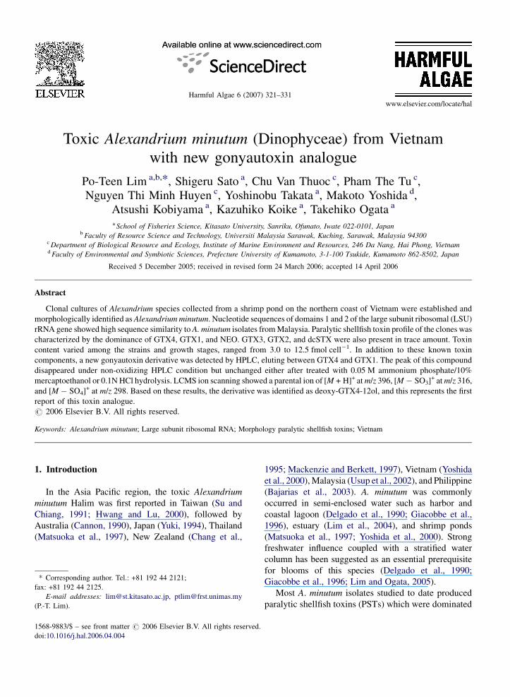
A. minutum cells used in this study were isolated
from shrimp ponds in Do Son, Hai Phong, Vietnam in
October 2004 (Fig. 1). The cultures were initially
established in Daigo’s IMK medium for Marine
Microalgae (Daigo, Tokyo, Japan). The cultures were
later transferred to Laboratory of Aquatic Microbiol-
Fig. 1. Map showing location of Do Son, Hai Phong
ogy, Kitasato University and maintained in ES medium
(Kokinos and Anderson, 1995), at 25 8C under a
14:10 h light:dark cycle at 140 mmol photons m�2 s�1.
Seawater of 33 PSU from Okkirai Bay was used as the
medium base. Salinity of the medium was adjusted to
15.0 � 1.0 PSU by addition of deionized distilled
water.
For species identification, mid exponential phase
cultures were harvested by centrifugation at 2000 � g
for 5 min and preserved in 4% formaldehyde solution.
Cells were then stained with 1% Calcofluor White M2R
(Sigma Aldrich Co. Ltd., Gillingham, UK) (Fritz and
Triemer, 1985) and observed under an Olympus BX51
epi-fluorescence microscope (Olympus, Tokyo, Japan).
Digital images under UVexcitation were captured using
a Pixera Penguin 600XL cooled CCD camera (Pixera
Corporation, Los Gatos, CA, USA). A total of 50
randomly selected cells were measured with a mean
determined.
2.2. DNA extraction, amplication and sequencing
Mid-exponential batch cultures were harvested by
centrifugation at 3000 � g for 5 min. Total DNA was
extracted using ISOGEN (Nippongene, Tokyo, Japan)
according to the manufacturer instruction. Approxi-
mately 700 bp of domains 1 and 2 (D1–D2) of LSU
rRNA gene was amplified by polymerase chain reaction
(PCR) using primers D1R and D2C (Scholin et al.,
1994) from Sigma Genosys (Sigma, The Woodland,
TX, USA). PCR was carried out on an ASTEC PC707
thermacycler (ASETC, Kanagawa, Japan). Purified
products were stored at �20 8C. DNA sequencing
was performed using dideoxy terminator (DYEnamic
ET terminator cycle sequencing kit, Amersham
Bioscience, Sweden) on an ABI 377 automated DNA
sequencer (Applied Biosystem, CA, USA).
in Vietnam from where samples were collected.
P.-T. Lim et al. / Harmful Algae 6 (2007) 321–331 323
2.3. Molecular phylogenetic analysis
Sequences obtained were aligned using the Clustal-
X program (Thompson et al., 1997). These and
previously published (Table 1) sequences were used
in the phylogenetic analysis. Phylogenetic analyses
were carried out using PAUP* Ver. 4.0b10 (Swofford,
1998) with maximum parsimony and likelihood
algorithms. Maximum parsimony was performed by
heuristic search of 1000 random additions and TBR
branch swapping. The MODELTEST Ver. 3.06 program
(Posada and Crandall, 1998) was used to determine the
best model of evolution. The best fit evolutionary model
selected for the sequence data set was the general time
reversible model with gamma distribution (GTR + G)
and estimated base frequencies of A = 0.2693,
C = 0.1526, G = 0.2580, T = 0.3201; base substitution
rates of [G � T] = 1.0000, [A � G] = 2.3324,
[A � T] = 1.0000, [C � G] = 1.0000, [C � T] = 6.8275
and [G � T] = 1.0000, a G distribution shape of 0.5178,
and zero proportion of invariable sites. The GTR + G
model and maximum likelihood parameters were then
used in the maximum likelihood analysis with the
previous parsimony tree as the starting tree.
2.4. Toxin extraction and analysis
Fifteen mililitres of clonal cultures (AmSp01, 03, 04,
05 and 07) at early and late exponential phases were
harvested by centrifugation at 2000 � g for 15 min. One
mililitre of duplicate subsamples were taken for cell
counts. The cells were preserved in Lugol’s solution.
Cell pellets for toxin extraction were then sonicated
with ultrasonic homogenizer UH-50 (SMT Co. Ltd.,
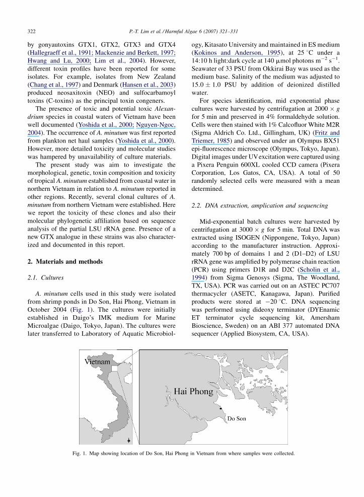
Table 1
Strains of Alexandrium species used in the phylogenetic analysis, with orig
Strain Species Origin
AmKB01, AmKB06 A. minutum Malaysia, Tumpat
AL3T A. minutum Italy, Gulf of Trieste
AMAD06 A. minutum Australia, Port River
X20 A. minutum France, the Rance
3.9h A. minutum England, Fleet Lagoon
95/4 A. minutum France, Bay of Concarne
CAWD13 A. minutum New Zealand, Malboroug
– A. minutum New Zealand, Anakoha B
GHmin04 A. minutum Denmark, Korsor Nor
Al1V A. minutum Spain, Galicia
SZN29 Alexandrium tamutum Italy
AI104 Alexandrium insuetum Japan
K0287 Alexandrium ostenfeldii Denmark, Limfjordan
AlMS02 Alexandrium leei Malaysia, Malacca
X12 Alexandrium margalefi France, Bay of Concarne
Japan) in 0.5 M acetic acid (AcOH) for 2 min on ice.
Cell debris was removed by centrifugation at
12,000 � g for 10 min. The supernatant was collected
and kept in �20 8C until further analysis.
Toxins analysis was carried out by HPLC using the
isocratic, post-column derivatization method of Oshima
(1995b) on a JASCO HPLC system (JASCO, Japan)
fitted with post-column system and fluorescence
detector. The samples were run through a Wakosil
C18 column (Ø 4.6 mm � 150 mm; Wako, Osaka,
Japan). The post-column temperature was kept at 70 8Cfor all runs. Detection wavelengths were set at 330 nm
excitation and 390 nm emissions. Authentic toxins
provided by Dr. Y. Oshima, Tohoku University, Japan
were used as toxin references. Further toxin verification
was carried out in non-oxidizing post-column condition
by replacing the oxidizing reagent with distilled water
and the reaction coil was kept in the ice bath during the
analysis. Hydrolysis of sample was carried out by
boiling the extract in 0.1N of HCl for 10 min (Hall and
Reichardt, 1984).
2.5. Isolation and fractionation of potentially new
toxin analogue
Twenty litres of A. minutum clonal culture at late
exponential phase was harvested by sieving at 10 mm
mesh size sieve and followed by centrifugation. Toxins
were extracted according to the procedure as described
above. The extract was then proceeded to lyophiliza-
tion. The freeze-dried sample was treated with 0.05 M
ammonium phosphate/10% (v/v) mercaptoethanol
(ME) and boiled for 10 min to remove the sulformoyl
moiety of GTXs (Sakamoto et al., 2000; Sato and
in of isolate, GenBank accession number, and citation
GenBank accession number Reference
AY566185, AY566187 Leaw et al. (2005)
AJ535353 John et al. (2003)
U44936 Scholin et al. (1994)
AF318232 Guillou et al. (2002)
AY705869 Nascimento et al. (2005)
au AF318264 Guillou et al. (2002)
h Sounds AY338751 Direct submission
ay AF033532 Walsh et al. (1998)
AY294613 Hansen et al. (2003)
L38626 Zardoya et al. (1995)
AJ535372 John et al. (2003)
AB088248 Direct submission
AJ535356 John et al. (2003)
AY566183 Leaw et al. (2005)
au AF318230 Guillou et al. (2002)
P.-T. Lim et al. / Harmful Algae 6 (2007) 321–331324
Kodama, 2003). Conversion of GTX1–4 to STXs (STX
and NEO) was confirmed by HPLC analysis. The sample
was then purified using a Bio-Gel P-2 column (fine;
15 mm � 450 mm) (BioRad, Hercules, CA, USA)
equilibrated with deionized distilled water. The sample
was loaded to the column and eluted with 0.2 M AcOH at
a flow rate of 0.5 mL min�1. Five-milliliter fractions
were collected using Redifrac fraction collector (Phar-
macia Biotech, New Jersey, USA). Individual fractions
were further analysed by HPLC. Fractions containing
compound of interest were combined, lyophilized and
then dissolved in 0.05 M AcOH. Samples were kept
frozen at �20 8C until further analysis.
2.6. Liquid chromatography–mass
spectrophotometry (LCMS/MS) analysis
An Agilent 1100 LC system (Agilent Technologies,
CA, USA) and a API 2000 quadru-pole MS/MS system
(Applied Biosystems, CA, USA) were used to analyse
the purified compound. Chromatographic separation
was performed using a column of Wakosil Navi 5C-18
(2 mm � 150 mm; Wako, Japan) with a linear gradient
system that was run from 0.2% heptafluorobutyric acid
(HFBA) to 30% acetonitrile containing 0.2% HFBA in
12 min at a flow rate of 0.2 mL min�1. The electrospray
ionization interface (ESI) was operated in positive
mode. The mass spectrometer was operated in both Q1
scan and product ion scan mode in which N2 was used as
Fig. 2. Epi-fluorescent micrographs of Alexandrium minutum from Vietnam
the first apical plate (10), a long sixth precingular plate (600) and the position
complex (Apc); (E and F) wide posterior sulcal plate (s.p.). Scale bar: 10
desoluvation, cone and collision gas (curtain gas 50 psi;
ion spray voltage 5500 V; ion-source gas 1, 40 psi; ion-
source gas 2, 60 psi; declustering potential 10 V;
collision energy 30 V; collision cell exit potential 15 V).
3. Results
3.1. Morphology and molecular phylogenetic
analysis
Cells from Vietnam were oval in shape, small with
transdiameter between 20 and 28 mm (Fig. 2A). Gamete
was approximately half the size of vegetative cell
(Fig. 2B). The first apical plate (10) was rhomboidal with
ventral pore (vp) located on the anterior right margin of
the plate (Fig. 2B and C). Some cells showed long and
narrow 10, with almost parallel right and left margins
(Fig. 2A). Apical pore complex (APC) was comma in
shape without anterior attachment pore (Fig. 2D). Sixth
precingular plate (600) was longer than wide (length
width ratio = 1.5–1.8). Posterior sulcal plate (s.p.) was
wider than long (Fig. 2E and F).
Nucleotide sequences of domain 1 and 2 of LSU
ribosomal RNA gene were obtained for four strains
(AmSp01, AmSp03, AmSp05, and AmSp17) with
sequence length of 657 bp. The aligned sequences
contained 626 characters (including gaps) for 18 taxa.
Of these, 393 were constant, 154 were variable but
parsimony uninformative, and 79 were parsimony
. (A) An oval vegetative cell and gamete (B); (C) ventral view showing
of ventral pore (vp); (D) apical view showing the shape of apical pore
mm.
P.-T. Lim et al. / Harmful Algae 6 (2007) 321–331 325
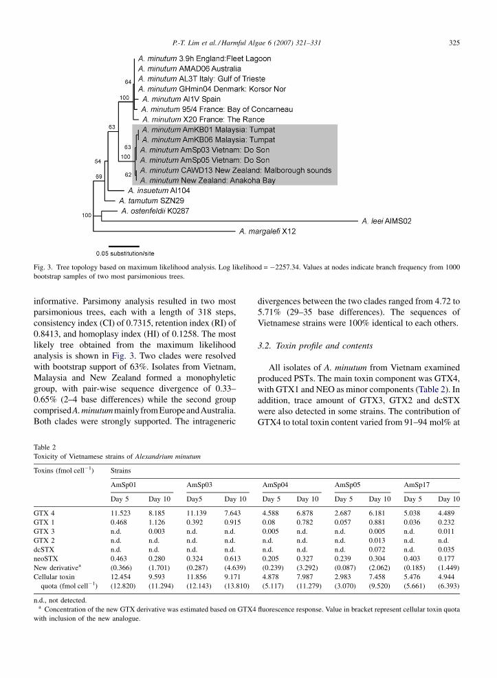
Fig. 3. Tree topology based on maximum likelihood analysis. Log likelihood = �2257.34. Values at nodes indicate branch frequency from 1000
bootstrap samples of two most parsimonious trees.
informative. Parsimony analysis resulted in two most
parsimonious trees, each with a length of 318 steps,
consistency index (CI) of 0.7315, retention index (RI) of
0.8413, and homoplasy index (HI) of 0.1258. The most
likely tree obtained from the maximum likelihood
analysis is shown in Fig. 3. Two clades were resolved
with bootstrap support of 63%. Isolates from Vietnam,
Malaysia and New Zealand formed a monophyletic
group, with pair-wise sequence divergence of 0.33–
0.65% (2–4 base differences) while the second group
comprisedA.minutummainlyfromEuropeandAustralia.
Both clades were strongly supported. The intrageneric
Table 2
Toxicity of Vietnamese strains of Alexandrium minutum
Toxins (fmol cell�1) Strains
AmSp01 AmSp03
Day 5 Day 10 Day5 Day 10
GTX 4 11.523 8.185 11.139 7.643
GTX 1 0.468 1.126 0.392 0.915
GTX 3 n.d. 0.003 n.d. n.d.
GTX 2 n.d. n.d. n.d. n.d.
dcSTX n.d. n.d. n.d. n.d.
neoSTX 0.463 0.280 0.324 0.613
New derivativea (0.366) (1.701) (0.287) (4.639)
Cellular toxin
quota (fmol cell�1)
12.454
(12.820)
9.593
(11.294)
11.856
(12.143)
9.171
(13.810)
n.d., not detected.a Concentration of the new GTX derivative was estimated based on GTX4
with inclusion of the new analogue.
divergences between the two clades ranged from 4.72 to
5.71% (29–35 base differences). The sequences of
Vietnamese strains were 100% identical to each others.
3.2. Toxin profile and contents
All isolates of A. minutum from Vietnam examined
produced PSTs. The main toxin component was GTX4,
with GTX1 and NEO as minor components (Table 2). In
addition, trace amount of GTX3, GTX2 and dcSTX
were also detected in some strains. The contribution of
GTX4 to total toxin content varied from 91–94 mol% at
AmSp04 AmSp05 AmSp17
Day 5 Day 10 Day 5 Day 10 Day 5 Day 10
4.588 6.878 2.687 6.181 5.038 4.489
0.08 0.782 0.057 0.881 0.036 0.232
0.005 n.d. n.d. 0.005 n.d. 0.011
n.d. n.d. n.d. 0.013 n.d. n.d.
n.d. n.d. n.d. 0.072 n.d. 0.035
0.205 0.327 0.239 0.304 0.403 0.177
(0.239) (3.292) (0.087) (2.062) (0.185) (1.449)
4.878
(5.117)
7.987
(11.279)
2.983
(3.070)
7.458
(9.520)
5.476
(5.661)
4.944
(6.393)
fluorescence response. Value in bracket represent cellular toxin quota
P.-T. Lim et al. / Harmful Algae 6 (2007) 321–331326
early exponential phase to 83–91 mol% at late
exponential phase. Cellular toxin quota varied among
strains and growth stages with the range of 3.0–
12.5 fmol PST cell�1.
3.3. Existence of a new toxin analogue
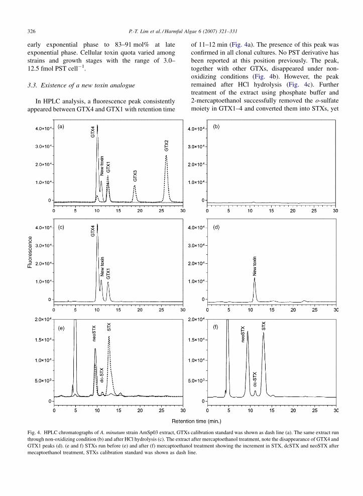
In HPLC analysis, a fluorescence peak consistently
appeared between GTX4 and GTX1 with retention time
Fig. 4. HPLC chromatographs of A. minutum strain AmSp03 extract, GTXs
through non-oxidizing condition (b) and after HCl hydrolysis (c). The extract
GTX1 peaks (d). (e and f) STXs run before (e) and after (f) mercaptoethano
mecaptoethanol treatment, STXs calibration standard was shown as dash l
of 11–12 min (Fig. 4a). The presence of this peak was
confirmed in all clonal cultures. No PST derivative has
been reported at this position previously. The peak,
together with other GTXs, disappeared under non-
oxidizing conditions (Fig. 4b). However, the peak
remained after HCl hydrolysis (Fig. 4c). Further
treatment of the extract using phosphate buffer and
2-mercaptoethanol successfully removed the o-sulfate
moiety in GTX1–4 and converted them into STXs, yet
calibration standard was shown as dash line (a). The same extract run
after mercaptoethanol treatment, note the disappearance of GTX4 and
l treatment showing the increment in STX, dcSTX and neoSTX after
ine.
P.-T. Lim et al. / Harmful Algae 6 (2007) 321–331 327
the peak was unaffected (Fig. 4D–F). The compound
also displayed significant changes over the growth
stages (Table 2). The concentration of this compound
was estimated based on the fluorescence response of
GTX4 with the assumption that it gave similar response
as GTX4 in the HPLC analysis. The concentration was
low (2.4–4.7 mol%) at the early exponential phase but
increased significantly to 15–33.6 mol% in all the
strains at the late exponential phase (day 10).
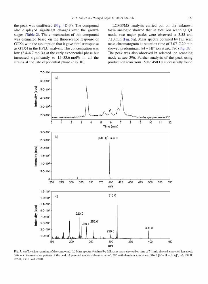
Fig. 5. (a) Total ion scanning of the compound. (b) Mass spectra obtained by
396. (c) Fragmentation pattern of the peak. A parental ion was observed at
255.0, 238.1 and 220.0.
LCMS/MS analysis carried out on the unknown
toxin analogue showed that in total ion scanning Q1
mode, two major peaks were observed at 3.55 and
7.10 min (Fig. 5a). Mass spectra obtained by full scan
mass chromatogram at retention time of 7.07–7.29 min
showed predominant [M + H]+ ion at m/z 396 (Fig. 5b).
The peak was also observed in selected ion scanning
mode at m/z 396. Further analysis of the peak using
product ion scan from 150 to 450 Da successfully found
full scans mass at retention time of 7.1 min showed a parental ion at m/z
m/z 396 with daughter ions at m/z 316.0 [M + H � SO3]+, m/z 299.0,
P.-T. Lim et al. / Harmful Algae 6 (2007) 321–331328
similar GTXs fragmentation (Fig. 5c). In the mass
spectrum of the peak, ion at m/z 316 corresponding to
[M + H � SO3]+, a structure that eliminated SO3 moiety
from the site chain, was predominant. The results
clearly showed the presence of o-sulfate side chain in
the compound. Other fragment ions at m/z 255, 238 and
220 were also observed.
4. Discussion
Identification of A. minutum based on morpholo-
gical thecal plates tabulation was a straight forward
task. The size and shape of the cells, position of ventral
pore (vp), the shape of posterior sulcal plate (s.p.) and
other sulcal plates observed in all the isolates from
Vietnam fitted well with Balech’s description (Balech,
1989, 1995). However, some morphological differ-
ences have been reported from the European strains.
Some of the European strains were reported to have
reticulation on hypotheca (Montresor et al., 1990) and
some do not have vp (Hansen et al., 2003). In contrast,
most of the Asia Pacific strains reported thus far
possessed smooth cell surface with the presence of vp
(Usup et al., 2002).
In this study, A. minutum was genetically separated
into two main groups based on partial LSU rRNA gene
analysis. The Vietnamese isolates were clustered
together with the Malaysian and New Zealand isolates,
forming a monophyletic group with large sequence
divergences compared to the European and southern/
werstern Australian isolates. Several previous studies
have also shown the biogeographical separation of A.
minutum populations, i.e. the Asia Pacific clade and
European clade (including southern/western Australia)
(Hansen et al., 2003; Lilly et al., 2005). Two discrete
ribotypes were also reported among the Australian A.
minutum (De Salas et al., 2001). In the present study,
high sequence homogeneity (>99.5%) was observed
between the Vietnamese and Malaysian strains of A.
minutum, in relative to other strains reported in the
region. The similarity between both populations
suggested the presence of gene flow. This could be
explained by the monsoon currents pattern occurred
annually in the South China Sea. During the winter
monsoon, northeast current flows through the coastal
waters of Vietnam and eventually ended in the Gulf of
Thai and northeastern of Peninsula Malaysia.
The cellular toxin quota (Qt) of Vietnamese A.
minutum isolates ranged from 3.0 to 13.8 fmol PST
cell�1 (Table 1), which was at comparable level with the
strains from Malaysia (4.0–12.0 fmol cell�1; Lim and
Ogata, 2005), New Zealand (3.4–10.1 fmol cell�1;
Mackenzie and Berkett, 1997), and Spain (1.0–
18.0 fmol cell�1; Franco et al., 1994). GTX4 and
GTX1 were reported as predominant toxin congeners
in most of the strains from Asia Pacific regions, including
strains from Taiwan (Hwang and Lu, 2000), New Zealand
(Mackenzie and Berkett, 1997), Thailand (Piumsomboon
et al., 2001) and Malaysia (Lim et al., 2004). Similar
toxin composition was also observed for the Vietnamese
isolates. However, some of the New Zealand strains were
reported with NEO as principle toxin congener (Chang
et al., 1997). Toxin profile of A. minutum from Europe
was far more diverse compared to the Asia Pacific strains.
Some European strains such as those from France (Belin,
1993) and UK (Percy et al., 2002) possessed different
toxin composition with GTX3 and/or GTX2 as the
predominant toxin congeners. In contrast, GTX4 and
GTX1 were the main toxin congeners for Portugal
(Cembella et al., 1987) and Spain strains (Franco et al.,
1994; Carreto et al., 2001). The Danish strains, on the
other hand, were reported with predominant C1 and C2
(Hansen et al., 2003). Recently, a strain with GTX3 and
STX as the major toxin congeners has been reported from
Fleet Lagoon, UK (Nascimento et al., 2005). None-
theless, geographical divergence of the toxin profiles has
been reported in many other Alexandrium species
(Cembella et al., 1987; Anderson et al., 1994; Cembella
and Destombe, 1996; Yoshida et al., 2001) and
Gymnodinium catenatum (Oshima et al., 1993).
In this study, a new GTX analogue was found to occur
naturally in the Vietnamese strains of A. minutum. In our
HPLC analysis, a fluorescent peak appeared consistently
between the peaks of GTX4 and GTX1. As a matter of
fact, some non-PST compounds also gave false
fluorescence signals in the post-column reaction system
(Gulavita et al., 1988; Onodera et al., 1996; Sato and
Shimizu, 1998) and remained under non-oxidizing
condition. In our analysis, however, the peak disappeared
under the non-oxidizing condition. The results showed
that the compound reacted similarly to oxidizing reagent
as other GTXs. In addition, the retention time of the peak
showed that it might possess similar polarity and ion state
as other GTXs. Furthermore, the peak remained when
hydrolyzed with HCl. This indicated that N-sulfocarba-
moyl moiety is absent from this compound.
Results of LCMS/MS showed a parental ion [M + H]+
at m/z 396 for the compound. The molecular weight was
differed from GTX4/GTX1 ([M + H]+ = 412) by 16, but
identical to the m/z values of GTX3 and GTX2. This
results indicated the absence of one oxygen atom in this
compound compared to GTX1 or GTX4. In this study, the
compound did not react with ME and this indicated that
the atom oxygen was most probably absent at the C12
P.-T. Lim et al. / Harmful Algae 6 (2007) 321–331 329
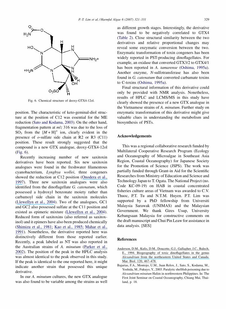
Fig. 6. Chemical structure of deoxy-GTX4-12ol.
position. The characteristic of keto-geminal-diol struc-
ture at the position of C12 was essential for the ME
reduction (Sato and Kodama, 2003). On the other hand,
fragmentation pattern at m/z 316 was due to the loss of
SO3 from the [M + H]+ ion, clearly evident in the
presence of o-sulfate side chain at R2 or R3 (C11)
position. These result strongly suggested that the
compound is a new GTX analogue, deoxy-GTX4-12ol
(Fig. 6).
Recently increasing number of new saxitoxin
derivatives have been reported. Six new saxitoxin
analogues were found in the freshwater filamentous
cyanobacterium, Lyngbya wollei, three congeners
showed the reduction at C12 position (Onodera et al.,
1997). Three new saxitoxin analogues were also
identified from the dinoflagellate G. catenatum, which
possessed a hydroxyl benzonate moiety rather than
carbamoyl side chain in the saxitoxin molecules
(Llewellyn et al., 2004). Two of the analogues, GC1
and GC2 also possessed sulfate at the C11 position and
existed as epimeric mixture (Llewellyn et al., 2004).
Reduced form of saxitoxins (also referred as saxitox-
inol) and it epimers have also been produced chemically
(Shimizu et al., 1981; Kao et al., 1985; Mahar et al.,
1991). Nonetheless, the derivative reported here was
distinctively different from those reported earlier.
Recently, a peak labeled as NT was also reported in
the Australian strains of A. minutum (Parker et al.,
2002). The position of the peak in the HPLC analysis
was almost identical to the peak observed in this study.
If the peak is identical to the one reported here, it might
indicate another strain that possessed this unique
derivative.
In our A. minutum cultures, the new GTX analogue
was also found to be variable among the strains as well
as different growth stages. Interestingly, the derivative
was found to be negatively correlated to GTX4
(Table 2). Close structural similarity between the two
derivatives and relative proportional changes may
reveal some enzymatic conversion between the two.
Enzymatic transformation of toxin congeners has been
widely reported in PST-producing dinoflagellates. For
example, an oxidase that converted GTX3/2 to GTX4/1
has been reported in A. tamarense (Oshima, 1995a).
Another enzyme, N-sulfotransferase has also been
found in G. catenatum that converted carbamate toxins
to C-toxins (Oshima, 1995a).
Final structural information of this derivative could
only be provided with NMR analysis. Nonetheless,
results of HPLC and LCMS/MS in this study have
clearly showed the presence of a new GTX analogue in
the Vietnamese strains of A. minutum. Further study on
enzymatic transformation of this derivative might give
valuable clues in understanding the metabolism and
biosynthesis of PSTs.
Acknowledgements
This was a regional collaborative research funded by
Multilateral Cooperative Research Program (Ecology
and Oceanography of Microalgae in Southeast Asia
Region, Coastal Oceanography) for Japanese Society
for the Promotion of Science (JSPS). The work was
partially funded through Grant-in Aid for the Scientific
Researches from Ministry of Education and Science and
Technology Japan to T. Ogata. The National Project (no.
Code KC-09-19) on HAB in coastal concentrated
fisheries culture areas of Vietnam was awarded to C.V.
Thuoc, P.T. Tu and N.T.M. Huyen. P.T. Lim was
supported by a PhD fellowship from Universiti
Malaysia Sarawak (UNIMAS) and the Malaysian
Government. We thank Gires Usup, University
Kebangsaan Malaysia for constructive comments on
the draft manuscript and Chui Pin Leaw for assistance in
data analysis. [SES]
References
Anderson, D.M., Kulis, D.M., Doucette, G.J., Gallagher, J.C., Balech,
E., 1994. Biogeography of toxic dinoflagellates in the genus
Alexandrium from the northeastern United States and Canada.
Mar. Biol. 120, 467–478.
Bajarias, F.A., Montojo, U.M., Juan Relox, J., Sato, S., Kodama, M.,
Yoshida, M., Fukuyo, Y., 2003. Paralytic shellfish poisoning due to
Alexandrium minutum Halim in northwestern Philippines. In: The
First Joint Seminar on Coastal Oceanography, Chiang Mai, Thai-
land, p. 18.
P.-T. Lim et al. / Harmful Algae 6 (2007) 321–331330
Balech, E., 1989. Redescription of Alexandrium minutum Halim
(Dinophyceae) type species of the genus Alexandrium. Phycologia
28, 206–211.
Balech, E., 1995. The Genus Alexandrium Halim (Dinoflagellata).
Sherkin Island Marine Station, Cork, Ireland, p.151.
Belin, C., 1993. Distribution of Dinophysis spp. and Alexandrium
minutum along French coasts since 1984 and their DSP and PSP
toxicity levels. In: Smayda, T.J., Shimizu, Y. (Eds.), Toxic
Phytoplankton Blooms in the Sea. Elsevier, New York, pp.
469–474.
Cannon, J.A., 1990. Development and dispersal of red tides in the Port
River, south Australia. In: Graneli, E., Sundstrom, B., Edler, L.,
Anderson, D.M. (Eds.), Toxic Marine Phytoplankton. Elsevier,
New York, pp. 110–115.
Carreto, J.I., Carignan, M.O., Montoya, N.G., 2001. Comparative
studies on mycosporine-like amino acids, paralytic shellfish toxins
and pigment profiles of the toxic dinoflagellates Alexandrium
tamarense, A. catenella and A. minutum. Mar. Ecol. Prog. Ser.
223, 49–60.
Cembella, A.D., Destombe, C., 1996. Genetic differentiation among
Alexandrium populations from eastern Canada. In: Yasumoto, T.,
Oshima, Y., Fukuyo, Y. (Eds.), Harmful and Toxic Algal Blooms.
Intergovernmental Oceanographic Commission of UNESCO,
Paris, pp. 447–450.
Cembella, A.D., Sullivan, J.J., Boyer, G.L., Taylor, F.J.R., Andersen,
R.J., 1987. Variation in paralytic shellfish toxin composition
within the Protogonyaulax tamarensis/catenella species complex;
red tide dinoflagellates. Biochem. Syst. Ecol. 15, 171–186.
Chang, F.H., Anderson, D.M., Kulis, D.M., Till, D.G., 1997. Toxin
production of Alexandrium minutum (Dinophyceae) from the Bay
of Plenty, New Zealand. Toxicon 35, 393–409.
Chang, F.H., Mackenzie, L., Till, D., Hannah, D., Rhodes, L., 1995.
The first toxic shellfish outbreaks and the associated phytoplank-
ton blooms in early 1993 in New Zealand. In: Lassus, P., Arzul,
G., Erard-Le Denn, E., Gentien, P., Marcaillou-Le Baut, C.
(Eds.), Harmful Marine Algal Blooms. Lavoisier, Paris, pp.
145–150.
De Salas, M.F., Van Emmerick, M.J., Hallegraeff, G.M., Negri, A.P.,
Vaillancourt, R.E., Bolch, C.J., 2001. Toxic Australian Alexan-
drium dinoflagellates: introduced or indigenous? In: Hallegraeff,
G.M., Bolch, C.J., Lewis, R.J. (Eds.), Harmful Algal Blooms
2001. IOC of UNESCO, pp. 214–217.
Delgado, M., Estrada, M., Camp, J., Fernandez, J.J., Santmarti, M.,
Lleti, C., 1990. Development of a toxic Alexandrium minutum
Halim (Dinophyceae) bloom in the harbour of Sant Charles de la
Rapita (Ebro Delta northwestern Mediterranean). Sci. Mar. 54, 1–
7.
Franco, J.M., Fernandez, P., Reguera, B., 1994. Toxin profiles of
natural population and cultures of Alexandrium minutum Halim
from Galician (Spain) coastal waters. J. Appl. Phycol. 6, 275–279.
Fritz, L., Triemer, R.E., 1985. A rapid technique utilizing Calcofluor
White M2R for the visualization of dinoflagellate tehcal plates. J.
Phycol. 21, 662–664.
Giacobbe, M.G., Oliva, F.D., Maimone, G., 1996. Environmental
factors and seasonal occurrence of the dinoflagellate Alexandrium
minutum, a PSP potential producer, a Mediterranean Lagoon. Est.
Coast. Shelf Sci. 42, 539–549.
Guillou, L., Nezan, E., Cueff, V., Erand-Le Denn, E., Cambon-
Bonavita, M.-A., Gentien, P., Barbier, G., 2002. Genetic diversity
and molecular detection of three toxic dinoflagellate genera
(Alexandrium, Dinophysis, and Karenia) from French coasts.
Protist 153, 223–238.
Gulavita, N., Hori, A., Shimizu, Y., 1988. Aphanorphine, a novel
tricyclic alkaloid from the blue-green alga Aphanizomenon flos-
aqua. Tetrahedron Lett. 29, 4381–4384.
Hall, S., Reichardt, P.B., 1984. Cryptic paralytic shellfish toxins. In:
Ragelis, E.P. (Ed.), Seafood Toxins. ACS Symposium Series 262.
American Chemical Society, Washington, DC, pp. 113–123.
Hallegraeff, G.M., Bolch, C.J., Blackburn, S.I., Oshima, Y., 1991.
Species of the toxigenic dinoflagellate genus Alexandrium in
southern Australian waters. Bot. Mar. 34, 575–587.
Hansen, G., Daugbjerg, N., Franco, J.M., 2003. Morphology, toxin
composition and LSU rDNA phylogeny of Alexandrium minutum
(Dinophyceae) from Denmark, with some morphological obser-
vations on other European strains. Harmful Algae 2, 317–335.
Hwang, D.F., Lu, Y.H., 2000. Influence of environmental and nutri-
tional factors on growth, toxicity, and toxin profile of dinoflagel-
late Alexandrium minutum. Toxicon 38, 1491–1503.
John, U., Fensome, R.A., Medlin, L.K., 2003. The application of a
molecular clock based on molecular sequences and the fossil
record to explain biogeographic distributions within the Alexan-
drium tamarense ‘‘species complex’’ (Dinophyceae). Mol. Biol.
Evol. 20, 1015–1027.
Kao, C.Y., Kao, P.N., James-Krace, M.R., Koehn, F.E., Wichmann,
C.F., Schnoes, H.K., 1985. Actions of epimers of 12-(OH)-reduced
saxitoxin and of 11-(OSO3)-saxitoxin on squid axon. Toxicon 23,
647–655.
Kokinos, J.P., Anderson, D.M., 1995. Morphological development of
resting cysts in cultures of the marine dinoflagellate Lingulodi-
nium polyedrum (=L. machaerophorum). Palynology 19, 143–166.
Leaw, C.P., Lim, P.T., Ng, B.K., Cheah, M.Y., Ahmad, A., Usup, G.,
2005. Phylogenetic analysis of Alexandrium species and Pyrodi-
nium bahamense (Dinophyceae) based on theca morphology and
nuclear ribosomal gene sequence. Phycologia 44, 550–565.
Lilly, E.L., Halanych, K.M., Anderson, D.M., 2005. Phylogeny,
biogeography, and species boundaries within the Alexandrium
minutum group. Harmful Algae 4, 1004–1020.
Lim, P.T., Leaw, C.P., Usup, G., 2004. First incidence of paralytic
shellfish poisoning on the east coast of Peninsular Malaysia. In:
Phang, S.M., Chong, V.C., Ho, S.S., Mokhtar, N., Ooi, J.L.S.
(Eds.), Marine Science into the New Millennium: New Perspec-
tives and Challenges. University of Malaya Maritime Research
Centre, Kuala Lumpur, Malaysia, pp. 661–667.
Lim, P.T., Ogata, T., 2005. Salinity effect on growth and toxin
production of four tropical Alexandrium species (Dinophyceae).
Toxicon 45, 699–710.
Llewellyn, L., Negri, A., Quilliam, M., 2004. High affinity for the rat
brain sodium channel of newly discovered hydroxybenzoate sax-
itoxin analogues from the dinoflagellate Gymnodinium catenatum.
Toxicon 43, 101–104.
Mackenzie, L., Berkett, N., 1997. Cell morphology and PSP-toxn
profiles of Alexandrium minutum in the Marlborough Sounds, New
Zealand. N. Z. J. Mar. Freshwater Res. 31, 403–409.
Mahar, J., Lukacs, G.L., Li, Y., Hall, S., Moczydlowski, E., 1991.
Pharmacological and biochemical properties of saxiphilin, a
soluble saxitoxin-binding protein from the bullfrog (Rana cates-
beiana). Toxicon 29, 53–71.
Matsuoka, K., Fukuyo, Y., Yoshida, M., 1997. Alexandrium minutum
Halim collected from aquaculture ponds in tropical and subtro-
pical coastal waters. In: Sudara, S. (Ed.), Marine Conservation and
Resource Rehabilitation. Chulalongkorn University, Chiangrai,
Thailand, pp. 85–94.
Montresor, M., Marino, D., Zingone, A., Dafnis, G., 1990. Three
Alexandrium species from coastal Tyrrhenian waters (Mediterra-
P.-T. Lim et al. / Harmful Algae 6 (2007) 321–331 331
nean Sea). In: Graneli, E., Sundstrom, B., Edler, L., Anderson,
D.M. (Eds.), Toxic Marine Phytoplankton. Elsevier, New York,
pp. 82–87.
Nascimento, S.M., Purdie, D.A., Lilly, E.L., Larsen, J., Morris, S.,
2005. Toxin profile, pigment composition, and large subunit rDNA
phylogenetic analysis of an Alexandrium minutum (Dinophyceae)
strain isolated from the Fleet Lagoon, United Kingdom. J. Phycol.
41, 343–353.
Nguyen-Ngoc, L., 2004. An autecological study of the potentially
toxic dinoflagellate Alexandrium affine isolated from Vietnamese
waters. Harmful Algae 3, 117–129.
Onodera, H., Oshima, Y., Watanabe, M.F., Watanabe, M., Bolch, C.J.,
Blackburn, S., Yasumoto, T., 1996. Screening of paralytic shellfish
toxins in freshwater cyanobacteria and chemical confirmation of
the toxins in cultured Anabana circinalis from Australia. In:
Yasumoto, T., Oshima, Y., Fukuyo, Y. (Eds.), Harmful and Toxic
Algal Blooms. Intergovernmental Oceanographic Commission of
UNESCO, Paris, pp. 563–566.
Onodera, H., Satake, M., Oshima, Y., Yasumoto, T., Carmichael,
W.W., 1997. New saxitoxin analogues from the freshwater fila-
mentous cyanobacterium Lyngbya wollei. Nat. Toxins 5, 146–151.
Oshima, Y., 1995a. Chemical and enzymatic transformation of paraly-
tic shellfish toxins in marine organisms. In: Lassus, P., Arzul, G.,
Denn, E.E.-L., Gentien, P., Baut, C.M.-L. (Eds.), Harmful Marine
Algal Blooms. Londres, Paris, pp. 475–480.
Oshima, Y., 1995b. Postcolumn derivatization liquid chromatography
method for paralytic shellfish toxins. J. AOAC Int. 78, 528–532.
Oshima, Y., Blackburn, S.I., Hallegraeff, G.M., 1993. Comparative
study on paralytic shellfish toxin profiles of the dinoflagellate
Gymnodinium catenatum from three different countries. Mar. Biol.
116, 471–476.
Parker, N.S., Negri, A.P., Frampton, D.M.F., Rodolfi, L., Tredici,
M.R., Blackburn, S.I., 2002. Growth of the toxic dinoflagellate
Alexandrium minutum (Dinophyceae) using high biomass culture
systems. J. Appl. Phycol. 14, 313–324.
Percy, L., Lewis, J., Morris, S., Stone, D.M., Higman, W., 2002. The
relationship between Alexandrium species in water and PSP toxins
in shellfish from the Fal estuary, United Kingdom. In: Steidinger,
K.A. (Ed.), Tenth International Conference on Harmful Algae.
Book of Abstracts, St. Petersburg, FL, p. 229.
Piumsomboon, A., Songroop, C., Kungsuwan, A., Polpunthin, P.,
2001. Species of the dinoflagellate genus Alexandrium (Gonyau-
lacales) in the Gulf of Thailand. In: Hallegraeff, G.M., Blackburn,
S.I., Bolch, C.J., Lewis, R.J. (Eds.), Harmful Algal Blooms 2000.
Intergovernmetal Oceanographic Commission of UNESCO,
Hobart, Australia, pp. 12–15.
Posada, D., Crandall, K., 1998. MODELTEST: testing the model of
DNA substitution. Bioinformatics 14, 817–818.
Sakamoto, S., Sato, S., Ogata, T., Kodama, M., 2000. Formation of
intermediate conjugates in the reductive transformation of
gonyautoxins to saxitoxins by thiol compounds. Fish. Sci. 66,
136–141.
Sato, S., Kodama, M., 2003. Chemical and biological transformation
of paralytic shellfish poisoning toxins. In: Fingerman, M., Na-
gabhushanam, R. (Eds.), Recent Advances in Marine Biotechnol-
ogy, vol. 8. Bioremediation, Science Publisher, Enfield, NH, USA,
pp. 319–334.
Sato, S., Shimizu, Y., 1998. Purification of a fluorescent product from
the bacterium, Moraxella: a neosaxitoxin impostor. In: Reguera,
B., Blanco, J., Fernandez, M.L., Wyatt, T. (Eds.), Harmful Algae.
Xunta de Galicia and UNESCO Intergovernmental Oceanographic
Commission, Grafisant, Santiago de Compostela, Spain, pp. 465–
467.
Scholin, C.A., Herzog, M., Sogin, M., Anderson, D.M., 1994. Identi-
fication of group- and strain-specific genetic markers for globally
distributed Alexandrium (Dinophyceae). II. Sequence analysis of a
fragment of the LSU rRNA gene. J. Phycol. 30, 999–1011.
Shimizu, Y., Hsu, C.-P., Genenah, A., 1981. Structure of saxitoxin in
solutions and stereochemistry of dihydrosaxitoxins. J. Am. Chem.
Soc. 103, 605–609.
Su, H.-M., Chiang, Y.-M., 1991. Dinoflagellates collected from
aquaculture ponds in southern Taiwan. Jpn. J. Phycol. 39, 227–
237.
Swofford, D.L., 1998. PAUP*: Phylogenetic Analysis using Parsi-
mony (* and other methods), Version 4. Sinauer Associates,
Sunderland, Massachusetts, p.140.
Thompson, J.D., Gibson, T.J., Plewniak, F., Jeanmougin, F., Higgins,
D.G., 1997. The CLUSTAL �Windows interface: flexible stra-
tegies for multiple sequence alignment aided by quality analysis
tools. Nucl. Acids Res. 25, 4876–4882.
Usup, G., Leaw, C.P., Ahmad, A., Lim, P.T., 2002. Alexandrium
(Dinophyceae) species in Malaysian waters. Harmful Algae 1,
265–275.
Walsh, D., Reeves, R.A., Saul, D.J., Gray, R.D., Mackenzie, L.,
Bergquist, P.R., Bergquist, P.L., 1998. Heterogeneity of SSU
and LSU rDNA sequences of Alexandrium species. Biochem.
Syst. Ecol. 26, 495–509.
Yoshida, M., Ogata, T., Thuoc, C.V., Matsuoka, K., Fukuyo, Y., Hoi,
N.C., Kodama, M., 2000. The first finding of toxic dinoflagellate
Alexandrium minutum in Vietnam. Fish. Sci. 66, 177–179.
Yoshida, T., Sako, Y., Uchida, A., 2001. Geographic differences in
paralytic shellfish poisoning toxin profiles among Japanese popu-
lations of Alexandrium tamarense and A. catenella (Dinophyceae).
Phycol. Res. 49, 13–22.
Yuki, K., 1994. First report of Alexandrium minutum Halim (Dino-
phyceae) from Japan. Jpn. J. Phycol. 42, 425–430.
Zardoya, R., Costas, E., Lopez-Rodas, V., Garrido-Pertierra, A.,
Bautista, J.M., 1995. Revised dinoflagellate phylogeny inferred
from molecular analysis of large-subunit ribosomal RNA gene
sequences. J. Mol. Evol. 41, 637–645.
Related Documents


