Towards Omni-Tomography—Grand Fusion of Multiple Modalities for Simultaneous Interior Tomography Ge Wang 1,5 *, Jie Zhang 2 , Hao Gao 3 , Victor Weir 4 , Hengyong Yu 5 , Wenxiang Cong 1 , Xiaochen Xu 6 , Haiou Shen 1 , James Bennett 1 , Mark Furth 7 , Yue Wang 8 , Michael Vannier 9 1 Biomedical Imaging Division, VT-WFU School of Biomedical Engineering and Sciences, Blacksburg, Virginia Tech, Blacksburg, Virginia, United States of America, 2 Department of Radiology, University of Kentucky, Lexington, Kentucky, United States of America, 3 Department of Mathematics, University of California Los Angeles, Los Angeles, California, United States of America, 4 Department of Medical Physics and Radiation Safety, Baylor Health Care System, Dallas, Texas, United States of America, 5 Biomedical Imaging Division, VT-WFU School of Biomedical Engineering and Sciences, Wake Forest University Health Sciences, Winston-Salem, North Carolina, United States of America, 6 Medical Business Unit, Texas Instruments Inc., Dallas, Texas, United States of America, 7 Comprehensive Cancer Center, Wake Forest University Health Sciences, Winston-Salem, North Carolina, United States of America, 8 Department of Electrical and Computer Engineering, Virginia Tech, Blacksburg, Virginia, United States of America, 9 Department of Radiology, University of Chicago, Chicago, Illinois, United States of America Abstract We recently elevated interior tomography from its origin in computed tomography (CT) to a general tomographic principle, and proved its validity for other tomographic modalities including SPECT, MRI, and others. Here we propose ‘‘omni- tomography’’, a novel concept for the grand fusion of multiple tomographic modalities for simultaneous data acquisition in a region of interest (ROI). Omni-tomography can be instrumental when physiological processes under investigation are multi-dimensional, multi-scale, multi-temporal and multi-parametric. Both preclinical and clinical studies now depend on in vivo tomography, often requiring separate evaluations by different imaging modalities. Over the past decade, two approaches have been used for multimodality fusion: Software based image registration and hybrid scanners such as PET- CT, PET-MRI, and SPECT-CT among others. While there are intrinsic limitations with both approaches, the main obstacle to the seamless fusion of multiple imaging modalities has been the bulkiness of each individual imager and the conflict of their physical (especially spatial) requirements. To address this challenge, omni-tomography is now unveiled as an emerging direction for biomedical imaging and systems biomedicine. Citation: Wang G, Zhang J, Gao H, Weir V, Yu H, et al. (2012) Towards Omni-Tomography—Grand Fusion of Multiple Modalities for Simultaneous Interior Tomography. PLoS ONE 7(6): e39700. doi:10.1371/journal.pone.0039700 Editor: Xiaoyuan Chen, NIH, United States of America Received February 15, 2012; Accepted May 24, 2012; Published June 29, 2012 Copyright: ß 2012 Wang et al. This is an open-access article distributed under the terms of the Creative Commons Attribution License, which permits unrestricted use, distribution, and reproduction in any medium, provided the original author and source are credited. Funding: This work was partially supported by National Institutes of Health/National Institute of Biomedical Imaging and Bioengineering (NIH/NIBIB) grant EB011785 and National Institutes of Health (NIH)/National Heart, Lung, and Blood Institute (NIH/NHLBI) grant HL098912. The funders had no role in study design, data collection and analysis, decision to publish, or preparation of the manuscript. No additional external funding received for this study. Competing Interests: Dr. Xiaochen Xu (co-author) is a current, full-time employee of Texas Instruments Inc, Medical Systems Division. This does not alter the authors’ adherence to all the PLoS ONE policies on sharing data and materials. * E-mail: [email protected] Introduction The physiome concept was first presented to the International Union of Physiological Sciences (IUPS) in 1993, and later designated as a strategic area by IUPS in 2001 [1–3]. Physiome describes physiological units and their interactions from the genome scale to complex organisms in a systematic fashion. The IUPS Physiome Project supports a worldwide repository of models and datasets, and represents an integral component of systems biomedicine. Biomedical imaging has been instrumental for the Physiome Project, and is yet to be improved for much richer functional, cellular and molecular information. In the medical imaging field, efforts are being made to link molecular assays with diagnostic imaging [4,5]; however, success to date has been rather limited. One reason is that current medical imaging scanners do not individually offer a wide enough spectrum of information. For example, current x-ray CT produces a limited amount of information from gray-scale images based on differences in linear attenuation coefficients of various tissues. On the other hand, an information explosion is seen from genetic and epigenetic profiling. This imbalance between phenotypic information (e.g., CT images) and genome-level information (e.g., RNA data) demands more capabilities from the in vivo imaging side. Indeed, the medical imaging field is rapidly trending in this direction. Turning again to x-ray CT as an example, the transition has started from gray-scale to true-color images with energy-sensitive, photon-counting detector technol- ogy [6]. Another area of advancement is x-ray phase-contrast and dark-field imaging [7,8]. Overall, both imaging modalities and contrast agents are being rapidly improved. The holy grail of biomedical imaging is an integrated system capable of producing tomographic, simultaneous, dynamic obser- vations of highly complex biological phenomena in vivo. The multimodality fusion, or multimodal fusion, approach has made significant strides towards meeting this challenge, as demonstrated by the popularity of PET-CT and other hybrid systems [9–13]. We envision that tomography will evolve beyond current modality fusion and towards grand fusion, a large-scale fusion of many imaging modalities, which is called omni-tomography/multi- tomography [14]. Unlike modality fusion, omni-tomography is for truly simultaneous and often local reconstruction of multiple PLoS ONE | www.plosone.org 1 June 2012 | Volume 7 | Issue 6 | e39700

Welcome message from author
This document is posted to help you gain knowledge. Please leave a comment to let me know what you think about it! Share it to your friends and learn new things together.
Transcript
Towards Omni-Tomography—Grand Fusion of MultipleModalities for Simultaneous Interior TomographyGe Wang1,5*, Jie Zhang2, Hao Gao3, Victor Weir4, Hengyong Yu5, Wenxiang Cong1, Xiaochen Xu6,
Haiou Shen1, James Bennett1, Mark Furth7, Yue Wang8, Michael Vannier9
1 Biomedical Imaging Division, VT-WFU School of Biomedical Engineering and Sciences, Blacksburg, Virginia Tech, Blacksburg, Virginia, United States of America,
2 Department of Radiology, University of Kentucky, Lexington, Kentucky, United States of America, 3 Department of Mathematics, University of California Los Angeles, Los
Angeles, California, United States of America, 4 Department of Medical Physics and Radiation Safety, Baylor Health Care System, Dallas, Texas, United States of America,
5 Biomedical Imaging Division, VT-WFU School of Biomedical Engineering and Sciences, Wake Forest University Health Sciences, Winston-Salem, North Carolina, United
States of America, 6 Medical Business Unit, Texas Instruments Inc., Dallas, Texas, United States of America, 7 Comprehensive Cancer Center, Wake Forest University Health
Sciences, Winston-Salem, North Carolina, United States of America, 8 Department of Electrical and Computer Engineering, Virginia Tech, Blacksburg, Virginia, United States
of America, 9 Department of Radiology, University of Chicago, Chicago, Illinois, United States of America
Abstract
We recently elevated interior tomography from its origin in computed tomography (CT) to a general tomographic principle,and proved its validity for other tomographic modalities including SPECT, MRI, and others. Here we propose ‘‘omni-tomography’’, a novel concept for the grand fusion of multiple tomographic modalities for simultaneous data acquisition ina region of interest (ROI). Omni-tomography can be instrumental when physiological processes under investigation aremulti-dimensional, multi-scale, multi-temporal and multi-parametric. Both preclinical and clinical studies now depend on invivo tomography, often requiring separate evaluations by different imaging modalities. Over the past decade, twoapproaches have been used for multimodality fusion: Software based image registration and hybrid scanners such as PET-CT, PET-MRI, and SPECT-CT among others. While there are intrinsic limitations with both approaches, the main obstacle tothe seamless fusion of multiple imaging modalities has been the bulkiness of each individual imager and the conflict of theirphysical (especially spatial) requirements. To address this challenge, omni-tomography is now unveiled as an emergingdirection for biomedical imaging and systems biomedicine.
Citation: Wang G, Zhang J, Gao H, Weir V, Yu H, et al. (2012) Towards Omni-Tomography—Grand Fusion of Multiple Modalities for Simultaneous InteriorTomography. PLoS ONE 7(6): e39700. doi:10.1371/journal.pone.0039700
Editor: Xiaoyuan Chen, NIH, United States of America
Received February 15, 2012; Accepted May 24, 2012; Published June 29, 2012
Copyright: � 2012 Wang et al. This is an open-access article distributed under the terms of the Creative Commons Attribution License, which permitsunrestricted use, distribution, and reproduction in any medium, provided the original author and source are credited.
Funding: This work was partially supported by National Institutes of Health/National Institute of Biomedical Imaging and Bioengineering (NIH/NIBIB) grantEB011785 and National Institutes of Health (NIH)/National Heart, Lung, and Blood Institute (NIH/NHLBI) grant HL098912. The funders had no role in study design,data collection and analysis, decision to publish, or preparation of the manuscript. No additional external funding received for this study.
Competing Interests: Dr. Xiaochen Xu (co-author) is a current, full-time employee of Texas Instruments Inc, Medical Systems Division. This does not alter theauthors’ adherence to all the PLoS ONE policies on sharing data and materials.
* E-mail: [email protected]
Introduction
The physiome concept was first presented to the International
Union of Physiological Sciences (IUPS) in 1993, and later
designated as a strategic area by IUPS in 2001 [1–3]. Physiome
describes physiological units and their interactions from the
genome scale to complex organisms in a systematic fashion. The
IUPS Physiome Project supports a worldwide repository of models
and datasets, and represents an integral component of systems
biomedicine. Biomedical imaging has been instrumental for the
Physiome Project, and is yet to be improved for much richer
functional, cellular and molecular information.
In the medical imaging field, efforts are being made to link
molecular assays with diagnostic imaging [4,5]; however, success
to date has been rather limited. One reason is that current
medical imaging scanners do not individually offer a wide
enough spectrum of information. For example, current x-ray CT
produces a limited amount of information from gray-scale images
based on differences in linear attenuation coefficients of various
tissues. On the other hand, an information explosion is seen from
genetic and epigenetic profiling. This imbalance between
phenotypic information (e.g., CT images) and genome-level
information (e.g., RNA data) demands more capabilities from the
in vivo imaging side. Indeed, the medical imaging field is rapidly
trending in this direction. Turning again to x-ray CT as an
example, the transition has started from gray-scale to true-color
images with energy-sensitive, photon-counting detector technol-
ogy [6]. Another area of advancement is x-ray phase-contrast
and dark-field imaging [7,8]. Overall, both imaging modalities
and contrast agents are being rapidly improved.
The holy grail of biomedical imaging is an integrated system
capable of producing tomographic, simultaneous, dynamic obser-
vations of highly complex biological phenomena in vivo. The
multimodality fusion, or multimodal fusion, approach has made
significant strides towards meeting this challenge, as demonstrated
by the popularity of PET-CT and other hybrid systems [9–13].
We envision that tomography will evolve beyond current modality
fusion and towards grand fusion, a large-scale fusion of many
imaging modalities, which is called omni-tomography/multi-
tomography [14]. Unlike modality fusion, omni-tomography is
for truly simultaneous and often local reconstruction of multiple
PLoS ONE | www.plosone.org 1 June 2012 | Volume 7 | Issue 6 | e39700
imaging mechanisms such as CT, MRI, PET, SPECT, US, and
optical imaging.
Since the advent of diagnostic imaging, there has been a
powerful push to combine imaging modalities in a single
coordinate system. Modality fusion began with PET-CT which
revolutionized medical diagnosis. A PET-CT scanner sequentially
acquires CT and PET images in an integrated gantry. As a result,
metabolic processes from PET can be co-registered with anatomic
information from CT. As such, PET-CT has redefined the fields of
oncology, surgical planning, and radiation therapy. There are
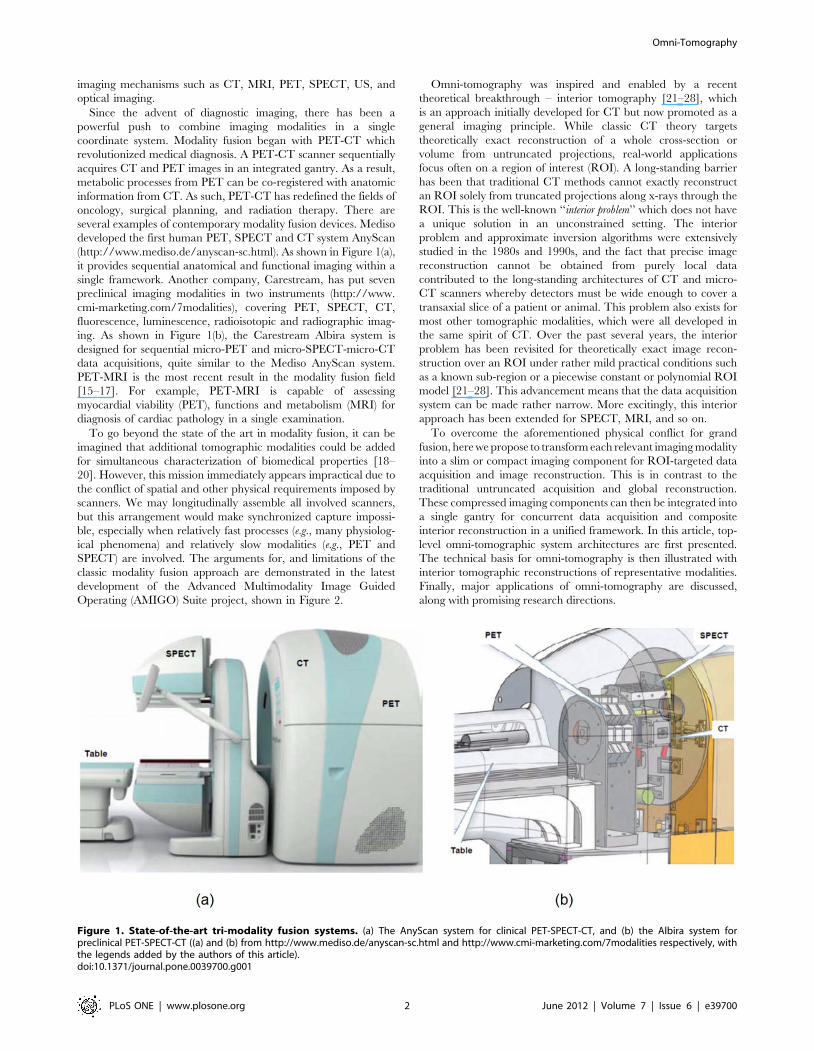
several examples of contemporary modality fusion devices. Mediso
developed the first human PET, SPECT and CT system AnyScan
(http://www.mediso.de/anyscan-sc.html). As shown in Figure 1(a),
it provides sequential anatomical and functional imaging within a
single framework. Another company, Carestream, has put seven
preclinical imaging modalities in two instruments (http://www.
cmi-marketing.com/7modalities), covering PET, SPECT, CT,
fluorescence, luminescence, radioisotopic and radiographic imag-
ing. As shown in Figure 1(b), the Carestream Albira system is
designed for sequential micro-PET and micro-SPECT-micro-CT
data acquisitions, quite similar to the Mediso AnyScan system.
PET-MRI is the most recent result in the modality fusion field
[15–17]. For example, PET-MRI is capable of assessing
myocardial viability (PET), functions and metabolism (MRI) for
diagnosis of cardiac pathology in a single examination.
To go beyond the state of the art in modality fusion, it can be
imagined that additional tomographic modalities could be added
for simultaneous characterization of biomedical properties [18–
20]. However, this mission immediately appears impractical due to
the conflict of spatial and other physical requirements imposed by
scanners. We may longitudinally assemble all involved scanners,
but this arrangement would make synchronized capture impossi-
ble, especially when relatively fast processes (e.g., many physiolog-
ical phenomena) and relatively slow modalities (e.g., PET and
SPECT) are involved. The arguments for, and limitations of the
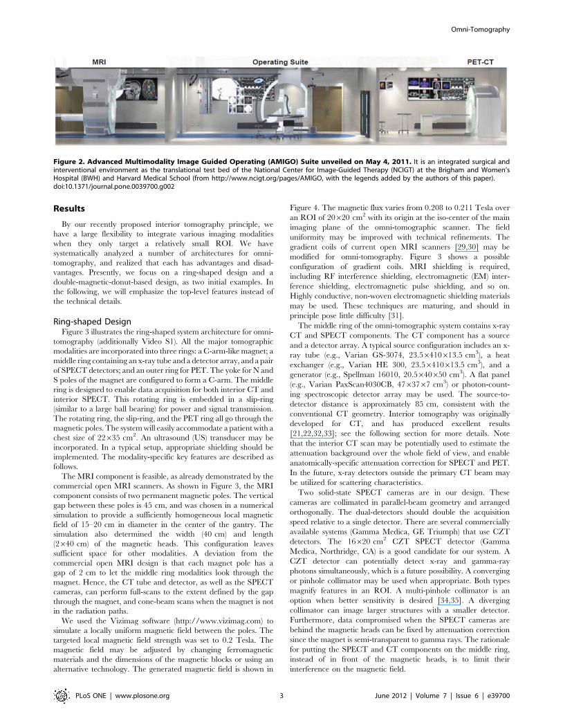
classic modality fusion approach are demonstrated in the latest
development of the Advanced Multimodality Image Guided
Operating (AMIGO) Suite project, shown in Figure 2.
Omni-tomography was inspired and enabled by a recent
theoretical breakthrough – interior tomography [21–28], which
is an approach initially developed for CT but now promoted as a
general imaging principle. While classic CT theory targets
theoretically exact reconstruction of a whole cross-section or
volume from untruncated projections, real-world applications
focus often on a region of interest (ROI). A long-standing barrier
has been that traditional CT methods cannot exactly reconstruct
an ROI solely from truncated projections along x-rays through the
ROI. This is the well-known ‘‘interior problem’’ which does not have
a unique solution in an unconstrained setting. The interior
problem and approximate inversion algorithms were extensively
studied in the 1980s and 1990s, and the fact that precise image
reconstruction cannot be obtained from purely local data
contributed to the long-standing architectures of CT and micro-
CT scanners whereby detectors must be wide enough to cover a
transaxial slice of a patient or animal. This problem also exists for
most other tomographic modalities, which were all developed in
the same spirit of CT. Over the past several years, the interior
problem has been revisited for theoretically exact image recon-
struction over an ROI under rather mild practical conditions such
as a known sub-region or a piecewise constant or polynomial ROI
model [21–28]. This advancement means that the data acquisition
system can be made rather narrow. More excitingly, this interior
approach has been extended for SPECT, MRI, and so on.
To overcome the aforementioned physical conflict for grand
fusion, here we propose to transform each relevant imaging modality
into a slim or compact imaging component for ROI-targeted data
acquisition and image reconstruction. This is in contrast to the
traditional untruncated acquisition and global reconstruction.
These compressed imaging components can then be integrated into
a single gantry for concurrent data acquisition and composite
interior reconstruction in a unified framework. In this article, top-
level omni-tomographic system architectures are first presented.
The technical basis for omni-tomography is then illustrated with
interior tomographic reconstructions of representative modalities.
Finally, major applications of omni-tomography are discussed,
along with promising research directions.
Figure 1. State-of-the-art tri-modality fusion systems. (a) The AnyScan system for clinical PET-SPECT-CT, and (b) the Albira system forpreclinical PET-SPECT-CT ((a) and (b) from http://www.mediso.de/anyscan-sc.html and http://www.cmi-marketing.com/7modalities respectively, withthe legends added by the authors of this article).doi:10.1371/journal.pone.0039700.g001
Omni-Tomography
PLoS ONE | www.plosone.org 2 June 2012 | Volume 7 | Issue 6 | e39700
Results
By our recently proposed interior tomography principle, we
have a large flexibility to integrate various imaging modalities
when they only target a relatively small ROI. We have
systematically analyzed a number of architectures for omni-
tomography, and realized that each has advantages and disad-
vantages. Presently, we focus on a ring-shaped design and a
double-magnetic-donut-based design, as two initial examples. In
the following, we will emphasize the top-level features instead of
the technical details.
Ring-shaped DesignFigure 3 illustrates the ring-shaped system architecture for omni-
tomography (additionally Video S1). All the major tomographic
modalities are incorporated into three rings: a C-arm-like magnet; a
middle ring containing an x-ray tube and a detector array, and a pair
of SPECT detectors; and an outer ring for PET. The yoke for N and
S poles of the magnet are configured to form a C-arm. The middle
ring is designed to enable data acquisition for both interior CT and
interior SPECT. This rotating ring is embedded in a slip-ring
(similar to a large ball bearing) for power and signal transmission.
The rotating ring, the slip-ring, and the PET ring all go through the
magnetic poles. The system will easily accommodate a patient with a
chest size of 22635 cm2. An ultrasound (US) transducer may be
incorporated. In a typical setup, appropriate shielding should be
implemented. The modality-specific key features are described as
follows.
The MRI component is feasible, as already demonstrated by the
commercial open MRI scanners. As shown in Figure 3, the MRI
component consists of two permanent magnetic poles. The vertical
gap between these poles is 45 cm, and was chosen in a numerical
simulation to provide a sufficiently homogeneous local magnetic
field of 15–20 cm in diameter in the center of the gantry. The
simulation also determined the width (40 cm) and length
(2640 cm) of the magnetic heads. This configuration leaves
sufficient space for other modalities. A deviation from the
commercial open MRI design is that each magnet pole has a
gap of 2 cm to let the middle ring modalities look through the
magnet. Hence, the CT tube and detector, as well as the SPECT
cameras, can perform full-scans to the extent defined by the gap
through the magnet, and cone-beam scans when the magnet is not
in the radiation paths.
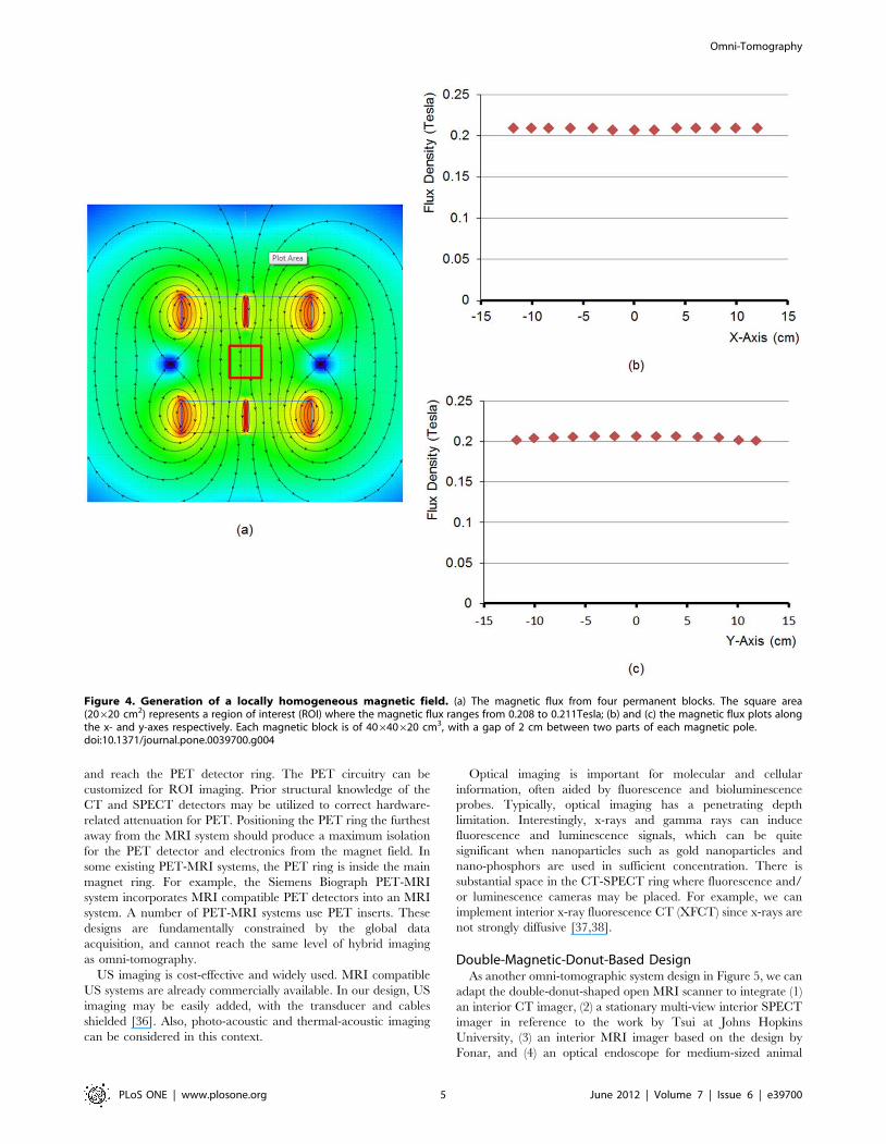
We used the Vizimag software (http://www.vizimag.com) to
simulate a locally uniform magnetic field between the poles. The
targeted local magnetic field strength was set to 0.2 Tesla. The
magnetic field may be adjusted by changing ferromagnetic
materials and the dimensions of the magnetic blocks or using an
alternative technology. The generated magnetic field is shown in
Figure 4. The magnetic flux varies from 0.208 to 0.211 Tesla over
an ROI of 20620 cm2 with its origin at the iso-center of the main
imaging plane of the omni-tomographic scanner. The field
uniformity may be improved with technical refinements. The
gradient coils of current open MRI scanners [29,30] may be
modified for omni-tomography. Figure 3 shows a possible
configuration of gradient coils. MRI shielding is required,
including RF interference shielding, electromagnetic (EM) inter-
ference shielding, electromagnetic pulse shielding, and so on.
Highly conductive, non-woven electromagnetic shielding materials
may be used. These techniques are maturing, and should in
principle pose little difficulty [31].
The middle ring of the omni-tomographic system contains x-ray
CT and SPECT components. The CT component has a source
and a detector array. A typical source configuration includes an x-
ray tube (e.g., Varian GS-3074, 23.56410613.5 cm3), a heat
exchanger (e.g., Varian HE 300, 23.56410613.5 cm3), and a
generator (e.g., Spellman 16010, 20.5640650 cm3). A flat panel
(e.g., Varian PaxScan4030CB, 4763767 cm3) or photon-count-
ing spectroscopic detector array may be used. The source-to-
detector distance is approximately 85 cm, consistent with the
conventional CT geometry. Interior tomography was originally
developed for CT, and has produced excellent results
[21,22,32,33]; see the following section for more details. Note
that the interior CT scan may be potentially used to estimate the
attenuation background over the whole field of view, and enable
anatomically-specific attenuation correction for SPECT and PET.
In the future, x-ray detectors outside the primary CT beam may
be utilized for scattering characteristics.
Two solid-state SPECT cameras are in our design. These
cameras are collimated in parallel-beam geometry and arranged
orthogonally. The dual-detectors should double the acquisition
speed relative to a single detector. There are several commercially
available systems (Gamma Medica, GE Triumph) that use CZT
detectors. The 16620 cm2 CZT SPECT detector (Gamma
Medica, Northridge, CA) is a good candidate for our system. A
CZT detector can potentially detect x-ray and gamma-ray
photons simultaneously, which is a future possibility. A converging
or pinhole collimator may be used when appropriate. Both types
magnify features in an ROI. A multi-pinhole collimator is an
option when better sensitivity is desired [34,35]. A diverging
collimator can image larger structures with a smaller detector.
Furthermore, data compromised when the SPECT cameras are
behind the magnetic heads can be fixed by attenuation correction
since the magnet is semi-transparent to gamma rays. The rationale
for putting the SPECT and CT components on the middle ring,
instead of in front of the magnetic heads, is to limit their
interference on the magnetic field.
Figure 2. Advanced Multimodality Image Guided Operating (AMIGO) Suite unveiled on May 4, 2011. It is an integrated surgical andinterventional environment as the translational test bed of the National Center for Image-Guided Therapy (NCIGT) at the Brigham and Women’sHospital (BWH) and Harvard Medical School (from http://www.ncigt.org/pages/AMIGO, with the legends added by the authors of this paper).doi:10.1371/journal.pone.0039700.g002
Omni-Tomography
PLoS ONE | www.plosone.org 3 June 2012 | Volume 7 | Issue 6 | e39700

The PET detector ring of 120 cm internal diameter consists of
LYSO crystals but it could be built out of CZT or other solid-state
materials. In LYSO crystal-based systems, the scintillation
emission can be detected with avalanche photodiode detectors
(APDs). The detector units are 464620 mm3. There are 4 units
per detector block, 471 detector blocks per ring, and 20 rings in
total. The axial extent is 16 cm. Another part is a coincidence
timing or time-of-flight (TOF) analysis circuit, which is commer-
cially available. The 511KeV PET photons are sufficiently
energetic to pass through the solid-state CT and SPECT detectors,
Figure 3. Ring-shaped design for omni-tomography. (a) A 3D rendering of the top-level design, (b) a partial rendering, (c) an in-plane view,and (d) a through-plane view. There are two static rings and one rotating ring for omni-tomography. While the red C-arm is a permanent magnet andthe yellow outer ring contains PET crystals, the blue ring supports a CT tube, a CT detector and a pair of SPECT camera. The blue CT-SPECT ring is on agreen slip ring (like a large ball bearing) as the interface for power and data. The CT-SPECT ring, the slip-ring, and the PET ring all go through themagnetic poles.doi:10.1371/journal.pone.0039700.g003
Omni-Tomography
PLoS ONE | www.plosone.org 4 June 2012 | Volume 7 | Issue 6 | e39700
and reach the PET detector ring. The PET circuitry can be
customized for ROI imaging. Prior structural knowledge of the
CT and SPECT detectors may be utilized to correct hardware-
related attenuation for PET. Positioning the PET ring the furthest
away from the MRI system should produce a maximum isolation
for the PET detector and electronics from the magnet field. In
some existing PET-MRI systems, the PET ring is inside the main
magnet ring. For example, the Siemens Biograph PET-MRI
system incorporates MRI compatible PET detectors into an MRI
system. A number of PET-MRI systems use PET inserts. These
designs are fundamentally constrained by the global data
acquisition, and cannot reach the same level of hybrid imaging
as omni-tomography.
US imaging is cost-effective and widely used. MRI compatible
US systems are already commercially available. In our design, US
imaging may be easily added, with the transducer and cables
shielded [36]. Also, photo-acoustic and thermal-acoustic imaging
can be considered in this context.
Optical imaging is important for molecular and cellular
information, often aided by fluorescence and bioluminescence
probes. Typically, optical imaging has a penetrating depth
limitation. Interestingly, x-rays and gamma rays can induce
fluorescence and luminescence signals, which can be quite
significant when nanoparticles such as gold nanoparticles and
nano-phosphors are used in sufficient concentration. There is
substantial space in the CT-SPECT ring where fluorescence and/
or luminescence cameras may be placed. For example, we can
implement interior x-ray fluorescence CT (XFCT) since x-rays are
not strongly diffusive [37,38].
Double-Magnetic-Donut-Based DesignAs another omni-tomographic system design in Figure 5, we can
adapt the double-donut-shaped open MRI scanner to integrate (1)
an interior CT imager, (2) a stationary multi-view interior SPECT
imager in reference to the work by Tsui at Johns Hopkins
University, (3) an interior MRI imager based on the design by
Fonar, and (4) an optical endoscope for medium-sized animal
Figure 4. Generation of a locally homogeneous magnetic field. (a) The magnetic flux from four permanent blocks. The square area(20620 cm2) represents a region of interest (ROI) where the magnetic flux ranges from 0.208 to 0.211Tesla; (b) and (c) the magnetic flux plots alongthe x- and y-axes respectively. Each magnetic block is of 40640620 cm3, with a gap of 2 cm between two parts of each magnetic pole.doi:10.1371/journal.pone.0039700.g004
Omni-Tomography
PLoS ONE | www.plosone.org 5 June 2012 | Volume 7 | Issue 6 | e39700
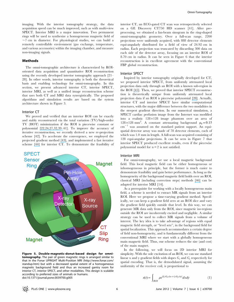
imaging. With the interior tomography strategy, the data
acquisition speed can be much improved, such as with multi-view
SPECT. Interior MRI is a major innovation. Two permanent
rings will be used to synthesize a homogeneous magnetic field of
,7 cm in diameter. For physiological studies, we can build a
remotely controllable environment (gas exchange, temperature,
and various accessories) within the imaging chamber, and measure
non-imaging signals.
Methods
The omni-tomographic architecture is characterized by ROI-
oriented data acquisition and quantitative ROI reconstruction
using the recently developed interior tomography approach [21–
28]. In other words, interior tomography is both the theoretical
basis and enabling technology for omni-tomography. In this
section, we present advanced interior CT, interior SPECT,
interior MRI, as well as a unified image reconstruction scheme
that uses both CT and MRI data synergistically. The proposed
algorithms and simulation results are based on the system
architecture shown in Figure 3.
Interior CTWe proved and verified that an interior ROI can be exactly
and stably reconstructed via the total variation (TV)/high-order
TV (HOT) minimization if the ROI is piecewise constant or
polynomial [22,24,27,32,39–41]. To improve the accuracy of
iterative reconstruction, we recently derived a new re-projection
scheme [42]. To accelerate the convergence, we employed the
projected gradient method [43], and implemented a fast iterative
scheme [44] for interior CT. To demonstrate the feasibility of
interior CT, an ECG-gated CT scan was retrospectively selected
on a GE Discovery CT750 HD scanner [41]. After pre-
processing, we obtained a fan-beam sinogram in the ring-shaped
omni-tomographic geometry. Over a full-scan range, 2200
projections were uniformly acquired, with 888 detector elements
equi-angularly distributed for a field of view of 24.92 cm in
radius. Each projection was truncated by discarding 300 data on
each side of the detector array, focusing on an interior ROI of
8.70 cm in radius. It can be seen in Figure 6 that the interior
reconstruction is in excellent agreement with the conventional
FBP global reconstruction.
Interior SPECTInspired by interior tomography originally developed for CT,
we proposed interior SPECT, from uniformly attenuated local
projection data only through an ROI with a known sub-region in
the ROI [45]. Then, we proved that interior SPECT reconstruc-
tion is theoretically unique from uniformly attenuated local
projection data if an ROI is piecewise polynomial [28]. Actually,
interior CT and interior SPECT have similar computational
structures, with the major difference between the two modalities in
the steepest gradient direction. In our numerical simulation, a
SPECT cardiac perfusion image from the Internet was modified
into a realistic 1286128 image phantom over an area of
1286128 mm2. A constant attenuating background m0~0:15cm21 was assumed on the standard patient support. An equi-
spatial detector array was made of 78 detector elements, each of
which was 1.0 mm in length. A full-scan was acquired consisting of
128 equi-angular projections. It can be seen in Figure 7 that
interior SPECT produced excellent results, even if the piecewise
polynomial model for n = 2 is not satisfied.
Interior MRIFor omni-tomography, we use a local magnetic background
field. This local magnetic field can be either homogeneous or
inhomogeneous in principle, but the former is much easier to
demonstrate feasibility and gain better performance. As long as the
homogeneity of the background magnetic field holds over an ROI,
classical MRI (including correction steps) methods [46] can be
adapted for interior MRI [14].
As a prerequisite for working with a locally homogeneous main
field, a scheme is needed to extract MR signals from an interior
ROI. Here we propose a time-varying gradient method. Specif-
ically, we can keep a gradient field zero at an ROI slice and vary
the gradient field quickly outside that level. In this way, we can
generate MR data only from the ROI, since magnetic iso-regions
outside the ROI are incoherently excited and negligible. A similar
strategy can be used to collect MR signals from a volume of
interest. The key idea is to take advantage of regions with equal
magnetic field strength, or ‘‘level sets’’, in the background field for
spatial localization. This approach accommodates a certain degree
of field non-homogeneity, and is fundamentally different from the
conventional MRI where we start with a globally homogeneous
main magnetic field. Thus, our scheme reduces the size (and cost)
of the main magnet.
In the following, we will focus on 2D interior MRI for
simplicity. With the sole excitation of an ROI, we can use standard
linear x and y gradient fields with slopes Gx and Gy respectively for
spatial encoding. That is, the demodulated signal, assuming the
uniformity of the receiver coil, is proportional to
s(t)~
ðROI
rei½(cGxt)xz(cGyt)y�dxdy ð1Þ
Figure 5. Double-magnetic-donut-based design for omni-tomography. The pair of green magnetic rings is arranged similar tothat in the Fonar UPRIGHT Multi-Position MRI (http://www.fonar.com/standup.htm) but with a decreased spatial extent of a homogeneousmagnetic background field and thus an increased gantry room forinterior CT, interior SPECT, and other modalities. This design is scalableaccording to preferred sizes of animals or humans.doi:10.1371/journal.pone.0039700.g005
Omni-Tomography
PLoS ONE | www.plosone.org 6 June 2012 | Volume 7 | Issue 6 | e39700
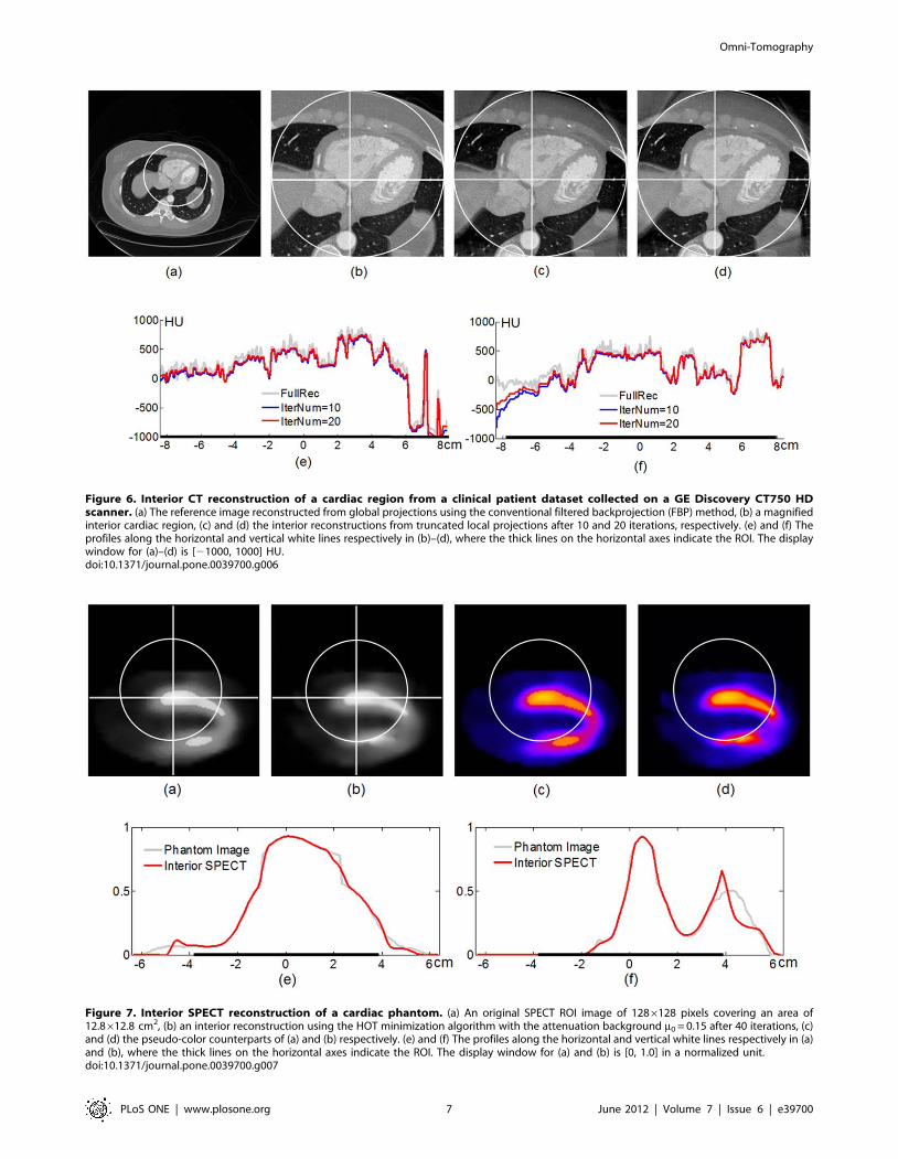
Figure 6. Interior CT reconstruction of a cardiac region from a clinical patient dataset collected on a GE Discovery CT750 HDscanner. (a) The reference image reconstructed from global projections using the conventional filtered backprojection (FBP) method, (b) a magnifiedinterior cardiac region, (c) and (d) the interior reconstructions from truncated local projections after 10 and 20 iterations, respectively. (e) and (f) Theprofiles along the horizontal and vertical white lines respectively in (b)–(d), where the thick lines on the horizontal axes indicate the ROI. The displaywindow for (a)–(d) is [21000, 1000] HU.doi:10.1371/journal.pone.0039700.g006
Figure 7. Interior SPECT reconstruction of a cardiac phantom. (a) An original SPECT ROI image of 1286128 pixels covering an area of12.8612.8 cm2, (b) an interior reconstruction using the HOT minimization algorithm with the attenuation background m0 = 0.15 after 40 iterations, (c)and (d) the pseudo-color counterparts of (a) and (b) respectively. (e) and (f) The profiles along the horizontal and vertical white lines respectively in (a)and (b), where the thick lines on the horizontal axes indicate the ROI. The display window for (a) and (b) is [0, 1.0] in a normalized unit.doi:10.1371/journal.pone.0039700.g007
Omni-Tomography
PLoS ONE | www.plosone.org 7 June 2012 | Volume 7 | Issue 6 | e39700
where r represents a 2D MR image to be reconstructed. On the
discretized Cartesian grid, we rewrite the MR signal equation by
Ar~s ð2Þ
where A is the system matrix discretized from Eq. (1). Under the
least-square data fidelity and the TV regularization, interior MRI
can be formulated as
r~ argr
min Ar{sk k22zl rk kTV ð3Þ
where l is the regularization parameter. Here we apply the split
Bregman method [47] as an efficient solver of Eq. (3).
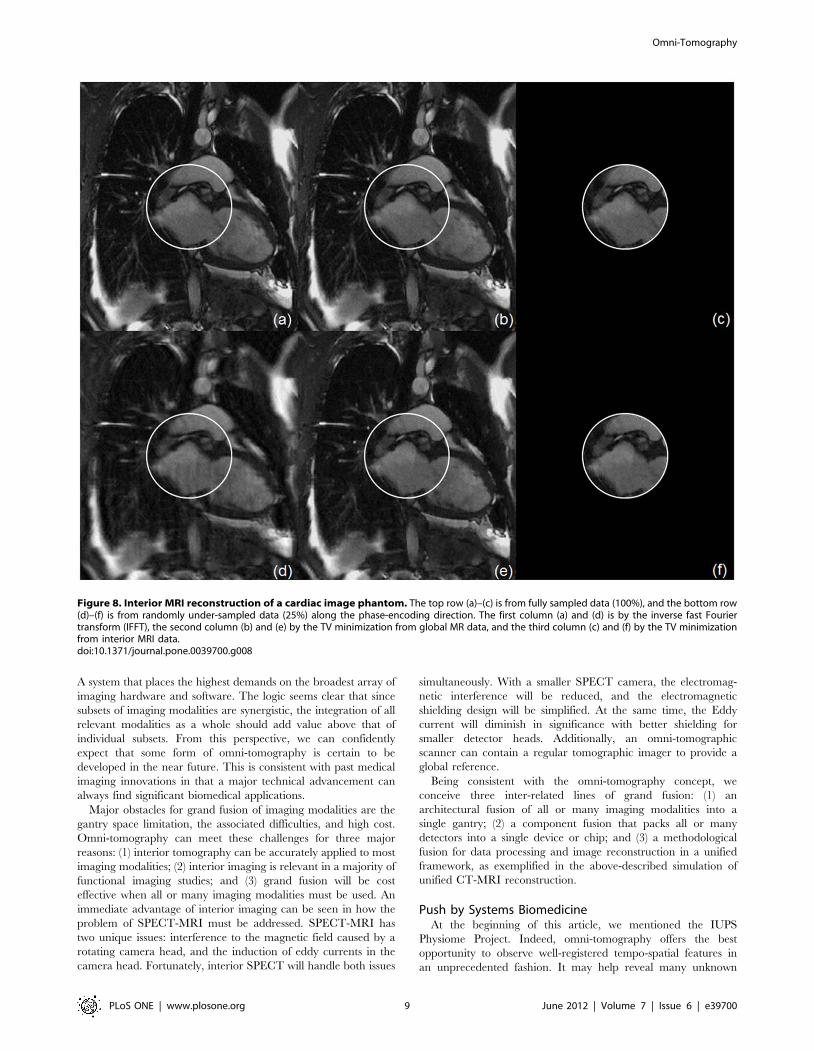
We used an MRI cardiac image as the phantom. As shown in
Figure 8, the proposed interior MRI method generated accurate
interior reconstruction, based on a locally homogeneous main
magnetic field. In this example, we compared the results with L2
regularization, TV regularization, and TV regularization for
interior reconstruction with fully sampled k-space data and under-
sampled k-space data respectively. In reference to the practical
sampling pattern, we under-sampled the phase encoding direction
and regularly sampled the frequency encoding direction.
Unified Interior CT-MRIAlthough various modalities have different contrast mecha-
nisms, the physiological process to be reconstructed is the same.
Consequently, there is strong correlation among these different
types of data. To optimize the composite image quality, our
unified omni-tomographic approach is based on inter-modality
coherence, which is important prior information unavailable in the
traditional reconstruction approach when image reconstruction is
separately considered for each modality. In light of lowered
sampling rate using temporal/spectral image coherence [48,49],
we hypothesize that inter-modality coherence can be utilized for
omni-tomography to reduce data requirements further, while
giving the same image quality.
In general, the multi-modal imaging model is
Pi(Aixi)~yi,iƒN ð4Þ
where Pi, Ai, xi, and yi are an under-sampling operator, an imaging
system matrix, an image, and data respectively, for the ith
modality. Then, we have the matrix model
AX~Y ð5Þ
where the ith column of X (Y) corresponds to xi (yi), and A should
be understood in terms of Eq. (4).First, we have the rank-sparsity
decomposition
X~XLzXS ð6Þ
where XL and XS are low-rank and sparse components
respectively. Then, we have the optimization problem
(XL,XS)~ arg min(XL ,XS )
DDA(XLzXS){Y DD2zl�DDTL(XL)DD�
zl1DDTS(XS)DD1
ð7Þ
where ||N||* is the nuclear norm to enforce the inter-modality
coherence of XL after a transform TL with a parameter l�, and
||N||1 is the L1 norm to promote the sparsity of XS after a
transform TS with a parameter l1. In this study, TL was the
identity transform, and TS a linear framelet transform. Eq. (7) was
solved using the split Bregman method [47].
In this pilot study, we selected a set of MRI and CT head scans
on the same human subject from the NIH Visible Human Project
(http://www.nlm.nih.gov/research/visible), including MRI T1,
T2, proton density, and CT images. The data were under-sampled
with a factor of 8. The MR Cartesian k-space data were pseudo-
randomly under-sampled along the phase encoding direction [50].
The fan-beam CT data were under-sampled using the dynamical
strategy [49]. As shown in Figure 9, the unified reconstruction
improved the image quality significantly. Clearly, this unified
reconstruction framework can be extended to cover more imaging
modalities in support of omni-tomography.
Discussion
Trend of Hybrid ImagingTomography is widely used for preclinical and clinical imaging
to characterize morphology and to a limited extent physiology.
Given current technology, we have been forced to accept several
intrinsic limitations, especially the necessity to acquire images
sequentially on the same subject. This time-wise separation
impairs our ability to decipher correlated dynamic biological
functions. For example, a cardiac infarct commonly begins with
decreased perfusion, then tissue hypoxia and eventually cell death.
These stages evolve in a continuum over a short period relative to
the time to acquire multimodality data.
If technology could be developed to simultaneously image
physiome of dynamic complexity with multiple modalities, rapidly
evolving physiological processes will become transparent. Such
processes have temporal evolution at many intervals, including
sub-seconds, seconds, minutes or longer, and may or may not be
reversible. A single session imaging is important to study processes
such as ischemia, drug interactions, radiation effects, apoptosis,
and many others. To some extent, this has partly been
accomplished with PET-CT and MRI-PET systems. Although
many of these multimodality imaging systems still acquire data
sequentially, the delay in data acquisition is improved relative to
single-modality predecessors.
With the Physiome Project, the need to simultaneously acquire
and unify multimodal images has become more important than
ever before. There are critical and immediate needs to remove the
limitations inherent in today’s tomographic imaging approaches to
the extent that complex biological processes can be studied in vivo
in real-time using multiple modalities. In a recent review article
entitled ‘‘Multimodality Imaging: Beyond PET/CT and SPECT/CT’’
[18], Dr. Simon Cherry wrote that ‘‘Multimodality imaging with
PET/CT and SPECT/CT has become commonplace in clinical practice and
in preclinical and basic medical research. Do other combinations of imaging
modalities have a similar potential to impact medical science and clinical
medicine? The combination of PET or SPECT with MRI is an area of active
research at the present time, while other, perhaps less obvious combinations,
including CT/MR and PET/optical also are being studied.’’ Later, Dr.
Cherry raised a question that could represent the thought
processes of experts in the field: ‘‘Is the fusion of PET and SPECT
with CT the ultimate answer in multimodality imaging, or is it just the first
example of a more general trend towards harnessing the complementary nature
of the different modalities on integrated imaging platforms?’’.
Our answer is the tightest integration of all relevant modalities
for fusion of information from each technology. We intend to
reach this target by applying the latest insights from interior
tomography to guide the design of the omni-tomographic system –
Omni-Tomography
PLoS ONE | www.plosone.org 8 June 2012 | Volume 7 | Issue 6 | e39700
A system that places the highest demands on the broadest array of
imaging hardware and software. The logic seems clear that since
subsets of imaging modalities are synergistic, the integration of all
relevant modalities as a whole should add value above that of
individual subsets. From this perspective, we can confidently
expect that some form of omni-tomography is certain to be
developed in the near future. This is consistent with past medical
imaging innovations in that a major technical advancement can
always find significant biomedical applications.
Major obstacles for grand fusion of imaging modalities are the
gantry space limitation, the associated difficulties, and high cost.
Omni-tomography can meet these challenges for three major
reasons: (1) interior tomography can be accurately applied to most
imaging modalities; (2) interior imaging is relevant in a majority of
functional imaging studies; and (3) grand fusion will be cost
effective when all or many imaging modalities must be used. An
immediate advantage of interior imaging can be seen in how the
problem of SPECT-MRI must be addressed. SPECT-MRI has
two unique issues: interference to the magnetic field caused by a
rotating camera head, and the induction of eddy currents in the
camera head. Fortunately, interior SPECT will handle both issues
simultaneously. With a smaller SPECT camera, the electromag-
netic interference will be reduced, and the electromagnetic
shielding design will be simplified. At the same time, the Eddy
current will diminish in significance with better shielding for
smaller detector heads. Additionally, an omni-tomographic
scanner can contain a regular tomographic imager to provide a
global reference.
Being consistent with the omni-tomography concept, we
conceive three inter-related lines of grand fusion: (1) an
architectural fusion of all or many imaging modalities into a
single gantry; (2) a component fusion that packs all or many
detectors into a single device or chip; and (3) a methodological
fusion for data processing and image reconstruction in a unified
framework, as exemplified in the above-described simulation of
unified CT-MRI reconstruction.
Push by Systems BiomedicineAt the beginning of this article, we mentioned the IUPS
Physiome Project. Indeed, omni-tomography offers the best
opportunity to observe well-registered tempo-spatial features in
an unprecedented fashion. It may help reveal many unknown
Figure 8. Interior MRI reconstruction of a cardiac image phantom. The top row (a)–(c) is from fully sampled data (100%), and the bottom row(d)–(f) is from randomly under-sampled data (25%) along the phase-encoding direction. The first column (a) and (d) is by the inverse fast Fouriertransform (IFFT), the second column (b) and (e) by the TV minimization from global MR data, and the third column (c) and (f) by the TV minimizationfrom interior MRI data.doi:10.1371/journal.pone.0039700.g008
Omni-Tomography
PLoS ONE | www.plosone.org 9 June 2012 | Volume 7 | Issue 6 | e39700

physiological, pathological, pharmaceutical and interventional
interactions in vivo, significantly improving the sensitivity and
specificity of basic research, diagnosis and intervention. This is the
major hypothesis that can only be tested with a prototype omni-
tomographic system.
Imaging technology development must target important
biomedical problems. For example, biotechnology and bioinfor-
matics have been developed to decode:
(a) Genomic/epigenetic signals at the DNA level associated
with various forms of genomic signatures (e.g., single
mutations, rare mutations, SNPs, copy number changes,
indels, genomic instability index, etc.);
(b) Gene expressions at mRNA, miRNA, shRNA, gene, exon,
and splicing levels, and their various functions;
(c) Protein expressions (protein complexes, metabolic features,
etc.);
(d) Complicated interactions and networks among these players
on multi-scales.
Many of these studies are currently limited to cells (tissue
samples, cell lines, etc.) and rarely go beyond studies in vitro, yet
imaging in vivo should facilitate or guide translation to organ,
system, and body levels.
Systems biology should be a great driver for omni-tomography.
Let us consider a biological subject as a system with inputs,
Figure 9. Unified CT-MRI reconstruction using inter-modality coherence. An MRI-CT head scan consisted of MR T1 (the 1st column), T2(the 2nd column), proton density images (the 3rd column), and a CT image (the 4th column). The top row shows the phantom images, the middlerow the images separately reconstructed using the conventional FFT or FBP method, and the bottom row the images simultaneouslyreconstructed in the unified rank-sparsity decomposition framework.doi:10.1371/journal.pone.0039700.g009
Omni-Tomography
PLoS ONE | www.plosone.org 10 June 2012 | Volume 7 | Issue 6 | e39700

circuitry with feedback loops, and outputs. Our omni-tomographic
system, with the help of probes, can image some components of
such a system in multiple dimensions (e.g., time, space, charac-
teristics). One can imagine multiple ways in which this imaging
system would be attractive and even indispensable. In the
following, let us examine some potential applications of omni-
tomography.
Hope for Major ApplicationsFluoroscopy guided cardiac catheterization has been the
original clinical standard for identifying stenotic lesions in the
coronary arteries. Recently, other diagnostic imaging techniques
are being researched and applied for this application, such as CT,
MRI, PET, SPECT, intra-coronary ultrasound, and optical
coherence tomography. The latest emphasis is to improve
understanding of pathobiology and genetics behind coronary
artery diseases. There are demands for better diagnostic perfor-
mance to achieve this goal. For example, the ability to model high
risk atherosclerotic lesions would be clinically invaluable. The
development of imaging methods and computational models is
needed to identify and predict high-risk lesions that may rupture
leading to coronary thrombosis and myocardial infarction. If our
proposed system could be used to study preclinical infarction
models, and predict outcomes, it would be instrumental for
research and healthcare.
Oncological imaging already uses all the imaging modalities.
Lung cancer imaging is a good example. CT defines air-tissue
interfaces, detects nodules and tumors, and provides quantitatively
accurate information. MRI measures airway functions with
hyperpolarized Helium-3. PET and SPECT improve lung cancer
diagnosis and staging with radiotracers. Omni-tomography could
potentially quantify malignancy without biopsy. Also, a futuristic
feature might use omni-tomographic data to create a knowledge
depository linked to genetic/epigenetic information and patient
histories.
The omni-tomographic system may be first developed for drug
development in animal models, with aid of multimodality probes.
This application is not subject to lengthy FDA approval, and will
set a stage for translational research and clinical trials. Engineering
probes visible to multiple modalities is a hot topic in the field of
bio-nanotechnology, and will have a profound impact on drug
development and molecular medicine [51]. Recently, exciting
work was reported on a combination of multiple nano-millimeter-
sized components to facilitate multimodality imaging and even
enable new imaging modes [52].
It is acknowledged that not all imaging tasks can be reduced to
interior imaging. To address this inherent ROI limitation, our
interiorized omni-tomography approach may be extended for
globalized omni-tomography. One idea is to simplify each data
acquisition chain for sparse sampling that makes space for
interlacing various imaging modalities. In a substantial sense, the
above-described omni-tomographic imager is a special case of this
more general plan. Further details are beyond the scope of this
paper.
Concluding RemarkOmni-tomography is a fresh concept with its interior recon-
struction feasibility demonstrated for CT, SPECT, MRI, and
other modalities. Although it may be more complex and costly
than any conventional scanner, we expect a prototype can be built
in the near future to demonstrate its instrumentation feasibility
and biomedical utility. We share our excitement towards omni-
tomography, and believe that it will find important applications
especially for systems biomedicine. We are committed to working
along this direction.
Supporting Information
Video S1 Virtual reality video clip showing the ‘ring-shaped’
omni-tomographic system design.
(WMV)
Acknowledgments
We would like to thank Drs. Tiange Zhuang, Jun Zhao, Yang Lv, Erik
Ritman, Erwei Bai, Robert Kraft, Craig Hamilton, Youngkyoo Jung,
Wenbing Yun, Yantian Zhang, Alexander Katsevich, and others for
helpful discussions.
Author Contributions
Performed the experiments: JZ HG VW HY WC XX HS JB GW. Wrote
the paper: GW JZ HG VW HY WC XX HS JB MF YW MV.
References
1. Hunter P, Robbins P, Noble D (2002) The IUPS human physiome project.
Pflugers Archiv European Journal of Physiology 445: 1–9.
2. Hunter P, Smith N, Fernandez J, Tawhai M (2005) Integration from proteins to
organs: the IUPS Physiome Project. Mechanisms of Ageing and Development126: 187–192.
3. Hunter PJ, Borg TK (2003) Integration from proteins to organs: the PhysiomeProject. Nat Rev Mol Cell Biol 4: 237–243.
4. Rutman AM, Kuo MD (2009) Radiogenomics: Creating a link betweenmolecular diagnostics and diagnostic imaging. European Journal of Radiology
70: 232–241.
5. van Houten VMM, Tabor MP, van den Brekel MWM, Denkers F, WishauptRGA, et al. (2000) Molecular Assays for the Diagnosis of Minimal Residual
Head-and-Neck Cancer: Methods, Reliability, Pitfalls, and Solutions. ClinicalCancer Research 6: 3803–3816.
6. Xu Q, Yu HY, Bennett J, He P, Zianon R, et al. (2012) Image reconstruction fora hybrid true-color micro-CT system. IEEE Transactions on Biomedical
Engineering: To appear.
7. Cong WX, Pfeiffer F, Bech M, Wang G (2012) X-ray dark-field imaging
modeling. Journal of the Optical Society of America A: To appear.
8. Cong WX, Yang JS, Wang G (2012) Differential phase-contrast interiortomography. Phys Med Biol 57: 2905–2914.
9. Ahn PH, Garg MK (2008) Positron Emission Tomography/ComputedTomography for Target Delineation in Head and Neck Cancers. Seminars in
Nuclear Medicine 38: 141–148.
10. Bockisch A, Freudenberg LS, Schmidt D, Kuwert T (2009) Hybrid Imaging by
SPECT/CT and PET/CT: Proven Outcomes in Cancer Imaging. Seminars in
Nuclear Medicine 39: 276–289.
11. Delbeke D, Schoder H, Martin WH, Wahl RL (2009) Hybrid Imaging (SPECT/
CT and PET/CT): Improving Therapeutic Decisions. Seminars in Nuclear
Medicine 39: 308–340.
12. Even-Sapir E, Keidar Z, Bar-Shalom R (2009) Hybrid Imaging (SPECT/CT
and PET/CT)–Improving the Diagnostic Accuracy of Functional/Metabolic
and Anatomic Imaging. Seminars in Nuclear Medicine 39: 264–275.
13. Kaufmann PA, Di Carli MF (2009) Hybrid SPECT/CT and PET/CT Imaging:
The Next Step in Noninvasive Cardiac Imaging. Seminars in Nuclear Medicine
39: 341–347.
14. Wang G, Zhang J, Gao H, Weir V, Yu HY, et al. (2011) Omni-tomography/
Multi-tomography-Integrating Multiple Modalities for Simultaneous Imaging.
arXiv:11062124 [physicsmed-ph].
15. Boss A, Bisdas S, Kolb A, Hofmann M, Ernemann U, et al. (2010) Hybrid PET/
MRI of Intracranial Masses: Initial Experiences and Comparison to PET/CT.
J Nucl Med 51: 1198–1205.
16. Boss A, Stegger L, Bisdas S, Kolb A, Schwenzer N, et al. (2011) Feasibility of
simultaneous PET/MR imaging in the head and upper neck area. European
Radiology 21: 1439–1446.
17. Pichler BJ, Wehrl HF, Kolb A, Judenhofer MS (2008) Positron Emission
Tomography/Magnetic Resonance Imaging: The Next Generation of Multi-
modality Imaging? Seminars in Nuclear Medicine 38: 199–208.
18. Cherry SR (2009) Multimodality Imaging: Beyond PET/CT and SPECT/CT.
Seminars in Nuclear Medicine 39: 348–353.
19. Mah D, Chen CC (2008) Image Guidance in Radiation Oncology Treatment
Planning: The Role of Imaging Technologies on the Planning Process. Seminars
in Nuclear Medicine 38: 114–118.
Omni-Tomography
PLoS ONE | www.plosone.org 11 June 2012 | Volume 7 | Issue 6 | e39700

20. Patton JA, Townsend DW, Hutton BF (2009) Hybrid Imaging Technology:
From Dreams and Vision to Clinical Devices. Seminars in Nuclear Medicine 39:247–263.
21. Ye YB, Yu HY, Wei YC, Wang G (2007) A general local reconstruction
approach based on a truncated Hilbert transform. International Journal ofBiomedical Imaging 2007: 63634.
22. Yu HY, Wang G (2009) Compressed sensing based Interior tomography. PhysMed Biol 54: 2791–2805.
23. Wang G, Yu HY, Ye YB (2009) A scheme for multi-source interior tomography.
Med Phys 36: 3575–3581.24. Yang JS, Yu HY, Jiang M, Wang G (2010) High-order total variation
minimization for interior tomography. Inverse Problems 26: 035013.25. Courdurier M, Noo F, Defrise M, Kudo H (2008) Solving the interior problem
of computed tomography using a priori knowledge. Inverse Problems 24:065001.
26. Kudo H, Courdurier M, Noo F, Defrise M (2008) Tiny a priori knowledge solves
the interior problem in computed tomography. Phys Med Biol 53: 2207–2231.27. Wang G, Yu HY (2010) Can interior tomography outperform lambda
tomography? Proceedings of the National Academy of Sciences of the UnitedStates of America 107: E92–E93.
28. Yang JS, Yu HY, Jiang M, Wang G (2011) High Order Total Variation
Minimization for Interior SPECT. Inverse Problems 28: 015001.29. Moon CH, Park HW, Cho MH, Lee SY (2000) Design of convex-surface
gradient coils for a vertical-field open MRI system. Meas Sci Technol 11: N89–N94.
30. While PT, Forbes LK, Crozier S (2010) 3D gradient coil design for open MRIsystems. Journal of Magnetic Resonance 207: 124–133.
31. Laskaris ET (1995) Open MRI magnet wth superconductive shielding. United
States: General Electric Company.32. Yu HY, Yang JS, Jiang M, Wang G (2009) Supplemental analysis on
compressed sensing based interior tomography. Phys Med Biol 54: N425–N432.33. Yu HY, Ye YB, Wang G (2008) Local Reconstruction Using the Truncated
Hilbert Transform via Singular Value Decomposition. Journal of X-Ray Science
and Technology 16: 243–251.34. Boles CD, Boser BE, Hasegawa BH, Heanue JA (1998) A multimode digital
detector readout for solid-state medical imaging detectors. Solid-State Circuits,IEEE Journal of 33: 733–742.
35. Tumer TO, Cajipe VB, Clajus M, Hayakawa S, Volkovskii A (2006) Multi-Channel Front-End Readout IC for Position Sensitive Solid-State Detectors.
Nuclear Science Symposium Conference Record (NSS/MIC), 2006 IEEE 1:
384–388.36. Annie M. Tang DFK, Edmund Y. Lam, Michael Brodsky, Ferenc A. Jolesz,
Edward S. Yang (2007 ) Multi-modal Imaging: Simultaneous MRI and
Ultrasound Imaging for Carotid Arteries Visualization. 29th Annual Interna-
tional Conference of the IEEE Engineering in Medicine and Biology Society.Lyon.
37. Jones BL, Cho SH (2011) The feasibility of polychromatic cone-beam x-ray
fluorescence computed tomography (XFCT) imaging of gold nanoparticle-loaded objects: a Monte Carlo study. Physics in Medicine and Biology 56: 3719–
3730.38. Cheong S-K, Jones BL, Siddiqi AK, Liu F, Manohar N, et al. (2010) X-ray
fluorescence computed tomography (XFCT) imaging of gold nanoparticle-
loaded objects using 110 kVp x-rays. Phys Med Biol 55: 647–666.39. Han W, Yu HY, Wang G (2009) A total variation minimization theorem for
compressed sensing based tomography. International Journal of BiomedicalImaging 2009: 125871.
40. Katsevich E, Katsevich A, Wang G (2012) Stability of the interior problem for apolynomial region of interest. Inverse Problems: To appear.
41. Yu HY, Wang G, Hsieh J, Entrikin D, Ellis S, et al. (2011) Compressive sensing-
based interior tomography: preliminary clinical application. Journal of computerassisted tomography 35: 762–764.
42. Yu HY, Wang G (2012) Finite detector based projection model for high spatialresolution. Journal of X-ray Science and Technology: To appear.
43. Daubechies I, Fornasier M, Loris I (2008) Accelerated Projected Gradient
Method for Linear Inverse Problems with Sparsity Constraints. Journal OfFourier Analysis And Applications 14: 764–792.
44. Beck A, Teboulle M (2009) A Fast Iterative Shrinkage-Thresholding Algorithmfor Linear Inverse Problems. Siam Journal on Imaging Sciences 2: 183–202.
45. Yu HY, Yang JS, Jiang M, Wang G (2009) Interior SPECT-exact and stableROI reconstruction from uniformly attenuated local projections. Communica-
tions in Numerical Methods in Engineering 25: 693–710.
46. Fessler JA (2010) Model-Based Image Reconstruction for MRI. IEEE SignalProcessing Magazine 27: 81–89.
47. Goldstein T, Osher S (2009) The split Bregman algorithm for l1 regularizedproblems. SIAM J Imaging Sci 2: 323–343.
48. Gao H, Yu HY, Osher S, Wang G (2011) Multi-energy CT based on a prior
rank, intensity and sparsity model (PRISM). Inverse Problems 27: 115012.49. Gao H, Cai JF, Shen ZW, Zhao HK (2011) Robust principal component
analysis-based four-dimensional computed tomography. Physics in Medicineand Biology 56: 3181–3198.
50. Bieri O, Markl M, Scheffler K (2005) Analysis and compensation of eddycurrents in balanced SSFP. Magnetic Resonance in Medicine 54: 129–137.
51. Chen XY, editor (2011) Nanoplatform-based molecular imaging. Hoboken,
New Jersey: John Wiley & Sons.52. Jin Y, Jia C, Huang S-W, O’Donnell M, Gao X (2010) Multifunctional
nanoparticles as coupled contrast agents. Nature Communications 1: 41.
Omni-Tomography
PLoS ONE | www.plosone.org 12 June 2012 | Volume 7 | Issue 6 | e39700
All in-text references underlined in blue are linked to publications on ResearchGate, letting you access and read them immediately.
Related Documents


![An In Vitro Study for the Detection of Breast Cancer by ...as Computed Tomography (CT) contrast agents [14-16]. Computed Tomography is one of the most common imaging modalities in](https://static.cupdf.com/doc/110x72/60016d073c43147177729a83/an-in-vitro-study-for-the-detection-of-breast-cancer-by-as-computed-tomography.jpg)