research papers Acta Cryst. (2011). B67, 535–551 doi:10.1107/S0108768111042868 535 Acta Crystallographica Section B Structural Science ISSN 0108-7681 Towards crystal structure prediction of complex organic compounds – a report on the fifth blind test David A. Bardwell, a * Claire S. Adjiman, b Yelena A. Arnautova, c Ekaterina Bartashevich, d Stephan X. M. Boerrigter, e Doris E. Braun, f Aurora J. Cruz-Cabeza, a,g,h Graeme M. Day, i Raffaele G. Della Valle, j Gautam R. Desiraju, k Bouke P. van Eijck, l Julio C. Facelli, m,n Marta B. Ferraro, o Damian Grillo, o Matthew Habgood, f Detlef W. M. Hofmann, p,q Fridolin Hofmann, q,r K. V. Jovan Jose, s Panagiotis G. Karamertzanis, b Andrei V. Kazantsev, b John Kendrick, t Liudmila N. Kuleshova, p Frank J. J. Leusen, t Andrey V. Maleev, u Alston J. Misquitta, v Sharmarke Mohamed, f Richard J. Needs, v Marcus A. Neumann, w Denis Nikylov, d Anita M. Orendt, m Rumpa Pal, k Constantinos C. Pantelides, b Chris J. Pickard, x Louise S. Price, f Sarah L. Price, f Harold A. Scheraga, c Jacco van de Streek, w Tejender S. Thakur, k Siddharth Tiwari, k Elisabetta Venuti j and Ilia K. Zhitkov u a Cambridge Crystallographic Data Centre, 12 Union Road, Cambridge CB2 1EZ, England, b Imperial College London, England, c Cornell University, USA, d South Ural State University, Russian Federation, e SSCI, An Aptuit Company, USA, f Department of Chemistry, University College London, England, g The Pfizer Institute for Pharmaceutical Materials Science, University Chemical Laboratory, University of Cambridge, England, h University of Amsterdam, The Neth- erlands, i Department of Chemistry, University of Cambridge, England, j Universita ` di Bologna, Italy, k Indian Institute of Science, India, l University of Utrecht, The Netherlands, m Center for High Performance Computing, University of Utah, USA, n Department of Biomedical Infor- matics, University of Utah, USA, o Universidad de Buenos Aires, Argentina, p Parco Scientifico e Technologico, Italy, q FlexCryst, Germany, r University Erlangen–Nu ¨ rnberg, Germany, s Ruhr-Universita ¨t Bochum, Germany, t University of Bradford, England, u Vladimir State Humanitarian University, Russian Federation, v Cavendish Laboratory, England, w Avant-garde Materials Simulation, Germany, and x Department of Physics and Astronomy, University College London, England Correspondence e-mail: [email protected] Following on from the success of the previous crystal structure prediction blind tests (CSP1999, CSP2001, CSP2004 and CSP2007), a fifth such collaborative project (CSP2010) was organized at the Cambridge Crystallographic Data Centre. A range of methodologies was used by the participating groups in order to evaluate the ability of the current computational methods to predict the crystal structures of the six organic molecules chosen as targets for this blind test. The first four targets, two rigid molecules, one semi-flexible molecule and a 1:1 salt, matched the criteria for the targets from CSP2007, while the last two targets belonged to two new challenging categories – a larger, much more flexible molecule and a hydrate with more than one polymorph. Each group submitted three predictions for each target it attempted. There was at least one successful prediction for each target, and two groups were able to successfully predict the structure of the large flexible molecule as their first place submission. The results show that while not as many groups successfully predicted the structures of the three smallest molecules as in CSP2007, there is now evidence that methodologies such as dispersion-corrected density functional theory (DFT-D) are able to reliably do so. The results also highlight the many challenges posed by more complex systems and show that there are still issues to be overcome. Received 1 August 2011 Accepted 16 October 2011 1. Introduction This paper reports on the results of the fifth blind test of crystal structure prediction (CSP), an international test hosted periodically by the Cambridge Crystallographic Data Centre (CCDC). We refer to this fifth blind test as CSP2010. Over the last several decades there has been much research in the field of crystal structure prediction. The grand aim is to develop the ability to reliably predict, by computational methods, how a molecule will crystallize in the solid state, with only the chemical diagram and the crystallization conditions known. This would allow for the prediction of solid-state properties before the molecule or molecules in question had even been synthesized, and could also help determine the likelihood that different polymorphic forms, or as yet unseen polymorphs of currently known structures, exist. This appli- cation is of particular importance in the pharmaceutical industry where the presence of different polymorphs can lead to very different and potentially undesirable physical prop- erties of new drugs. For the last decade the CCDC has held periodic blind tests to assess the current reliability and capabilities of the techni- ques available in the field. Four blind tests, starting in 1999 and every 2 or 3 years thereafter, have previously been held. Each

Welcome message from author
This document is posted to help you gain knowledge. Please leave a comment to let me know what you think about it! Share it to your friends and learn new things together.
Transcript
research papers
Acta Cryst. (2011). B67, 535–551 doi:10.1107/S0108768111042868 535
Acta Crystallographica Section B
StructuralScience
ISSN 0108-7681
Towards crystal structure prediction of complexorganic compounds – a report on the fifth blind test
David A. Bardwell,a* Claire S.Adjiman,b Yelena A. Arnautova,c
Ekaterina Bartashevich,d Stephan X. M.Boerrigter,e Doris E. Braun,f Aurora J.Cruz-Cabeza,a,g,h Graeme M. Day,i
Raffaele G. Della Valle,j Gautam R.Desiraju,k Bouke P. van Eijck,l Julio C.Facelli,m,n Marta B. Ferraro,o DamianGrillo,o Matthew Habgood,f
Detlef W. M. Hofmann,p,q FridolinHofmann,q,r K. V. Jovan Jose,s
Panagiotis G. Karamertzanis,b
Andrei V. Kazantsev,b John Kendrick,t
Liudmila N. Kuleshova,p Frank J. J.Leusen,t Andrey V. Maleev,u Alston J.Misquitta,v Sharmarke Mohamed,f
Richard J. Needs,v Marcus A.Neumann,w Denis Nikylov,d Anita M.Orendt,m Rumpa Pal,k Constantinos C.Pantelides,b Chris J. Pickard,x Louise S.Price,f Sarah L. Price,f Harold A.Scheraga,c Jacco van de Streek,w
Tejender S. Thakur,k Siddharth Tiwari,k
Elisabetta Venutij and Ilia K. Zhitkovu
aCambridge Crystallographic Data Centre, 12
Union Road, Cambridge CB2 1EZ, England,bImperial College London, England, cCornell
University, USA, dSouth Ural State University,
Russian Federation, eSSCI, An Aptuit Company,
USA, fDepartment of Chemistry, University
College London, England, gThe Pfizer Institute
for Pharmaceutical Materials Science, University
Chemical Laboratory, University of Cambridge,
England, hUniversity of Amsterdam, The Neth-
erlands, iDepartment of Chemistry, University of
Cambridge, England, jUniversita di Bologna,
Italy, kIndian Institute of Science, India,lUniversity of Utrecht, The Netherlands, mCenter
for High Performance Computing, University of
Utah, USA, nDepartment of Biomedical Infor-
matics, University of Utah, USA, oUniversidad
de Buenos Aires, Argentina, pParco Scientifico e
Technologico, Italy, qFlexCryst, Germany,rUniversity Erlangen–Nurnberg, Germany,sRuhr-Universitat Bochum, Germany,tUniversity of Bradford, England, uVladimir State
Humanitarian University, Russian Federation,vCavendish Laboratory, England, wAvant-garde
Materials Simulation, Germany, andxDepartment of Physics and Astronomy,
University College London, England
Correspondence e-mail: [email protected]
Following on from the success of the previous crystal structure
prediction blind tests (CSP1999, CSP2001, CSP2004 and
CSP2007), a fifth such collaborative project (CSP2010) was
organized at the Cambridge Crystallographic Data Centre. A
range of methodologies was used by the participating groups
in order to evaluate the ability of the current computational
methods to predict the crystal structures of the six organic
molecules chosen as targets for this blind test. The first four
targets, two rigid molecules, one semi-flexible molecule and a
1:1 salt, matched the criteria for the targets from CSP2007,
while the last two targets belonged to two new challenging
categories – a larger, much more flexible molecule and a
hydrate with more than one polymorph. Each group
submitted three predictions for each target it attempted.
There was at least one successful prediction for each target,
and two groups were able to successfully predict the structure
of the large flexible molecule as their first place submission.
The results show that while not as many groups successfully
predicted the structures of the three smallest molecules as in
CSP2007, there is now evidence that methodologies such as
dispersion-corrected density functional theory (DFT-D) are
able to reliably do so. The results also highlight the many
challenges posed by more complex systems and show that
there are still issues to be overcome.
Received 1 August 2011
Accepted 16 October 2011
1. Introduction
This paper reports on the results of the fifth blind test of
crystal structure prediction (CSP), an international test hosted
periodically by the Cambridge Crystallographic Data Centre
(CCDC). We refer to this fifth blind test as CSP2010.
Over the last several decades there has been much research
in the field of crystal structure prediction. The grand aim is to
develop the ability to reliably predict, by computational
methods, how a molecule will crystallize in the solid state, with
only the chemical diagram and the crystallization conditions
known. This would allow for the prediction of solid-state
properties before the molecule or molecules in question had
even been synthesized, and could also help determine the
likelihood that different polymorphic forms, or as yet unseen
polymorphs of currently known structures, exist. This appli-
cation is of particular importance in the pharmaceutical
industry where the presence of different polymorphs can lead
to very different and potentially undesirable physical prop-
erties of new drugs.
For the last decade the CCDC has held periodic blind tests
to assess the current reliability and capabilities of the techni-
ques available in the field. Four blind tests, starting in 1999 and
every 2 or 3 years thereafter, have previously been held. Each
has required the identification of a set of molecules with
known but previously unpublished crystal structures to use as
targets for the participants to predict using the various tech-
niques they have developed. This approach is similar to that
adopted to monitor and test advances in other areas of
predictive modelling, such as protein structure prediction
(Moult et al., 2007). Recently there has also been a blind test
for search methods for the crystal structure prediction of
purely inorganic systems (Oganov, 2010). Repeating the blind
test periodically helps to evaluate advances that have been
made in methodologies since the last test, as well as establish
the reliability of the techniques which have been successful in
previous tests for a given category of target; the small number
of targets in any one blind test introduces the possibility of a
slightly easier or harder molecule (whose difficulty cannot be
easily judged prior to commencement of the test) influencing
the results.
This fifth blind test was therefore held to assess the repro-
ducibility of the good results (Neumann et al., 2008; Day et al.,
2009) from the previous blind test, CSP2007, and also to assess
the developments in methodologies when applied to more
challenging targets than the relatively simple rigid molecules
mostly studied thus far. These additional targets better
represent cases that would be more likely to be encountered in
the pharmaceutical industry.
2. Organization and approach
The organization for this latest blind test, CSP2010, was
similar to that used for the previous four evaluations of the
field, the results of which have been previously published:
CSP1999 (Lommerse et al., 2000), CSP2001 (Motherwell et al.,
2002), CSP2004 (Day et al., 2005) and CSP2007 (Day et al.,
2009). Invitations to participate were sent to 24 research
groups known to be active in the field. The test was also
advertised through various websites and meetings.
The previous blind test puts forward targets for prediction
in the following four categories:
(1) Small, rigid molecules; only the elements C, H, N and O;
Z0 = 1 in any space group; up to 25 atoms.
(2) Rigid molecules; unusual functional groups or elements
such as halogens, S, P and B; Z0 = 1 in any space group; up to 30
atoms.
(3) Moderately flexible molecule with 2–4 internal degrees
of freedom; Z0 = 1 in any space group; up to 40 atoms.
(4) Multiple independent rigid molecules, e.g. solvates, co-
crystals, salts or Z0 = 2 structures; any space group; up to 30
atoms.
These four categories were left the same as those used in
CSP2007 so as to facilitate comparison of results. In addition,
it was decided to add two new categories that would provide
greater challenges:
(5) Molecule with 4–8 internal degrees of freedom; Z0 � 2 in
any space group; 50–60 atoms.
(6) Molecule for which more than one polymorph is known,
and which roughly falls into one of the first four
categories.
The new fifth category presents a much greater challenge in
terms of flexibility than previously encountered in earlier
blind tests, with a large flexible molecule intended to represent
those often associated with modern pharmaceuticals. The new
sixth category gives an opportunity to study the challenging
effects of polymorphism by introducing a molecule for which
more than one polymorph is known.
Crystallographers were contacted in August 2009 with a
request for unpublished crystal structures that matched one or
more of the six categories for the fifth blind test. Crystal
structures were collected at the CCDC and assessed for the
possibility of inclusion in one of the six possible categories. To
be suitable, a crystal structure had to be of high quality and
have all atoms located with no disorder. The crystal structure
had to be unpublished and the donor crystallographer had to
agree to postpone any publication for the duration of the blind
test. Collection of suitable candidates for all six categories
proved exceptionally difficult, especially for category 1, where
the target molecule is very small with a very restricted set of
constituent elements, and also for category 6 where few
suitable candidates were available that were not of sufficient
interest to be withheld from publication for the duration of
this test. Almost 30 submitted crystal structures had to be
rejected either due to not conforming to any of the six cate-
gories, or the presence of refinement issues such as disorder.
After considerable effort, one candidate was collected for
category 1, four for category 2, eight for category 3, three for
category 4, three for category 5 and one for category 6. For
those categories where there was more than one candidate, the
final target choice was made randomly.
For category 6, the one candidate that was submitted was
gallic acid monohydrate, for which two new polymorphs had
been found. These complemented the two previously
published polymorphs for gallic acid monohydrate, which are
located in the Cambridge Structural Database (CSD; Allen,
2002) under the KONTIQ CSD reference code family. For the
purposes of this blind test, these known forms are referred to
as forms (1) and (2). Of the two new forms submitted as
candidates for prediction, one [form (4), as recently published
by Clarke et al., 2011] had one formula unit in the asymmetric
unit (i.e. one gallic acid and one water molecule). The other,
form (3), was originally solved with two formula units per
asymmetric unit. However, analysis after the blind test
submissions showed that this solution contained a disordered
hydrogen-bonding network and the crystal structure could
also be described with an ordered hydrogen-bonding network
by doubling the unit cell, as now published (Clarke et al.,
2011). For the purposes of this blind test, form (3) was
therefore deemed inappropriate as a target crystal structure.
The main aim for this category, then, was to predict form (4),
whose structure has been recently independently published
(Demirtas et al., 2011) and see where (if at all) forms (1) and
(2) appeared in the ranked list of predictions.
The molecular diagrams and crystallization conditions were
sent by e-mail to 15 participant groups on 16 November 2009.
Immediately after circulation of the target crystal structures
we were made aware that the crystal structure of the molecule
research papers
536 David A. Bardwell et al. � Fifth blind test Acta Cryst. (2011). B67, 535–551
selected for category 1 (4-ethynylbenzonitrile) had been
solved, was undergoing publication and so would soon be in
the public domain. The decision was therefore made to
remove this candidate for category 1 and attempt to locate a
suitable replacement. Thankfully a suitable candidate was
quickly provided and the revised list of target molecules, as
detailed in Table 1, was distributed to participants on 23
November 2009. Following the numbering used in the
previous blind tests we refer to these molecules
by the Roman numerals (XVI)–(XXI).
The format of this blind test was kept broadly
the same as the last blind test, with the exception
that a greater length of time was allowed before
submission of results. Participants were requested
to forward their three ‘official’ predictions for
each target molecule to the CCDC, where the
experimentally determined crystal structures
were held for the duration of the test. As well as
these three main predictions, participants were
urged to submit an extended list of the crystal
structures they generated in order to help post-
analysis and to provide insight into the perfor-
mance of the various methods. The deadline for
submissions was 20 August 2010. The experi-
mentally determined crystal structures for all six
categories were then circulated to all participants
on 23 August 2010 to allow post-analysis of their
predictions. Lastly, a workshop was held at the
CCDC mid-September 2010 to discuss the results.
We present here results from the 14 partici-
pating groups that agreed to publish their results.
Details of these 14 participating groups, together
with a summary of which targets they attempted
and if a match with the experimental structure
was observed in their submission, are presented
in Table 2(a).
3. Methodologies
Methodologies for the participating research
groups vary significantly. A summary of the
techniques used by each of the groups is
presented in Table 2(b), together with key refer-
ences for most of the methods used. More
detailed descriptions are also provided in the
associated supplementary material.1
In general, each of the methods employed
involved three general steps:
(i) building a three-dimensional molecular
structure from the supplied two-dimensional
chemical diagrams;
(ii) searching for plausible crystal packing
arrangements of the molecule;
(iii) ranking the generated crystal structures in
order of likelihood of formation.
3.1. Methods of generating the molecular structure
There are two main approaches that can be used for treating
the molecular structure during crystal structure prediction.
Firstly, the molecule can be treated as rigid throughout the
research papers
Acta Cryst. (2011). B67, 535–551 David A. Bardwell et al. � Fifth blind test 537
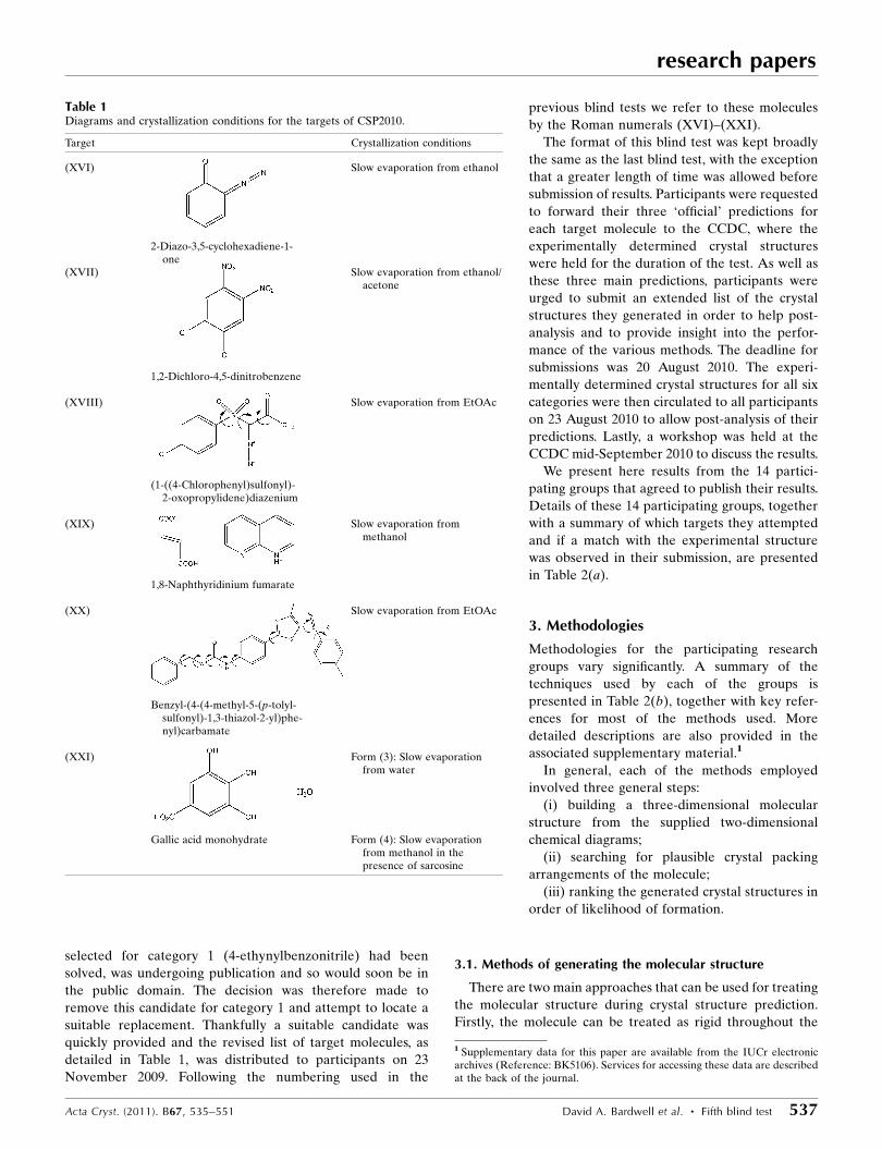
Table 1Diagrams and crystallization conditions for the targets of CSP2010.
Target Crystallization conditions
(XVI) Slow evaporation from ethanol
2-Diazo-3,5-cyclohexadiene-1-one
(XVII) Slow evaporation from ethanol/acetone
1,2-Dichloro-4,5-dinitrobenzene
(XVIII) Slow evaporation from EtOAc
(1-((4-Chlorophenyl)sulfonyl)-2-oxopropylidene)diazenium
(XIX) Slow evaporation frommethanol
1,8-Naphthyridinium fumarate
(XX) Slow evaporation from EtOAc
Benzyl-(4-(4-methyl-5-(p-tolyl-sulfonyl)-1,3-thiazol-2-yl)phe-nyl)carbamate
(XXI) Form (3): Slow evaporationfrom water
Gallic acid monohydrate Form (4): Slow evaporationfrom methanol in thepresence of sarcosine
1 Supplementary data for this paper are available from the IUCr electronicarchives (Reference: BK5106). Services for accessing these data are describedat the back of the journal.
research papers
538 David A. Bardwell et al. � Fifth blind test Acta Cryst. (2011). B67, 535–551
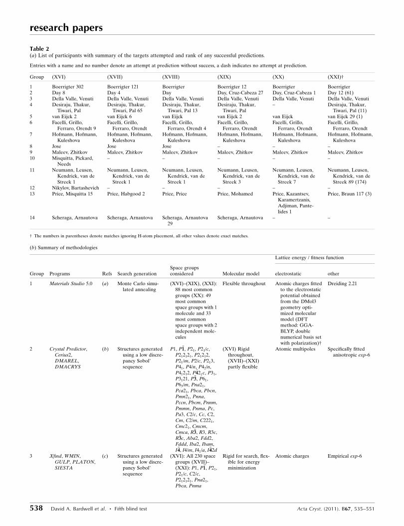
Table 2(a) List of participants with summary of the targets attempted and rank of any successful predictions.
Entries with a name and no number denote an attempt at prediction without success, a dash indicates no attempt at prediction.
Group (XVI) (XVII) (XVIII) (XIX) (XX) (XXI)†
1 Boerrigter 302 Boerrigter 121 Boerrigter Boerrigter 12 Boerrigter Boerrigter2 Day 8 Day 4 Day Day, Cruz-Cabeza 27 Day, Cruz-Cabeza 1 Day 12 (61)3 Della Valle, Venuti Della Valle, Venuti Della Valle, Venuti Della Valle, Venuti Della Valle, Venuti Della Valle, Venuti4 Desiraju, Thakur,
Tiwari, PalDesiraju, Thakur,
Tiwari, Pal 65Desiraju, Thakur,
Tiwari, Pal 13Desiraju, Thakur,
Tiwari, Pal– Desiraju, Thakur,
Tiwari, Pal (11)5 van Eijck 2 van Eijck 6 van Eijck van Eijck 2 van Eijck van Eijck 29 (1)6 Facelli, Grillo,
Ferraro, Orendt 9Facelli, Grillo,
Ferraro, OrendtFacelli, Grillo,
Ferraro, Orendt 4Facelli, Grillo,
Ferraro, OrendtFacelli, Grillo,
Ferraro, OrendtFacelli, Grillo,
Ferraro, Orendt7 Hofmann, Hofmann,
KuleshovaHofmann, Hofmann,
KuleshovaHofmann, Hofmann,
KuleshovaHofmann, Hofmann,
KuleshovaHofmann, Hofmann,
KuleshovaHofmann, Hofmann,
Kuleshova8 Jose Jose Jose – – –9 Maleev, Zhitkov Maleev, Zhitkov Maleev, Zhitkov Maleev, Zhitkov Maleev, Zhitkov Maleev, Zhitkov10 Misquitta, Pickard,
Needs– – – – –
11 Neumann, Leusen,Kendrick, van deStreek 1
Neumann, Leusen,Kendrick, van deStreek 1
Neumann, Leusen,Kendrick, van deStreek 1
Neumann, Leusen,Kendrick, van deStreek 3
Neumann, Leusen,Kendrick, van deStreek 7
Neumann, Leusen,Kendrick, van deStreek 89 (174)
12 Nikylov, Bartashevich – – – – –13 Price, Misquitta 15 Price, Habgood 2 Price, Price Price, Mohamed Price, Kazantsev,
Karamertzanis,Adjiman, Pante-lides 1
Price, Braun 117 (3)
14 Scheraga, Arnautova Scheraga, Arnautova Scheraga, Arnautova29
Scheraga, Arnautova – –
† The numbers in parentheses denote matches ignoring H-atom placement, all other values denote exact matches.
(b) Summary of methodologies
Lattice energy / fitness function
Group Programs Refs Search generationSpace groupsconsidered Molecular model electrostatic other
1 Materials Studio 5.0 (a) Monte Carlo simu-lated annealing
(XVI)–(XIX), (XXI):88 most commongroups (XX): 49most commonspace groups with 1molecule and 33most commonspace groups with 2independent mole-cules
Flexible throughout Atomic charges fittedto the electrostaticpotential obtainedfrom the DMol3geometry opti-mized molecularmodel (DFTmethod: GGA-BLYP, doublenumerical basis setwith polarization)†
Dreiding 2.21
2 Crystal Predictor,Cerius2,DMAREL,DMACRYS
(b) Structures generatedusing a low discre-pancy Sobol’sequence
P1, P�11, P21, P21/c,P212121, P21212,P21/m, P2/c, P213,P41, P4/n, P42/n,P41212, P�4421c, P31,P3121, P�33, P61,P63/m, Pna21,Pca21, Pbca, Pbcn,Pmn21, Pnna,Pccn, Pbcm, Pnnm,Pmmn, Pnma, Pc,Pa3, C2/c, Cc, C2,Cm, C2/m, C2221,Cmc21, Cmcm,Cmca, R�33, R3, R3c,R�33c, Aba2, Fdd2,Fddd, Iba2, Ibam,I �44, I4/m, I41/a, I �442d
(XVI) Rigidthroughout,(XVII)–(XXI)partly flexible
Atomic multipoles Specifically fittedanisotropic exp-6
3 Xfind, WMIN,GULP, PLATON,SIESTA
(c) Structures generatedusing a low discre-pancy Sobol’sequence
(XVI): All 230 spacegroups (XVII)–(XXI): P1, P�11, P21,P21/c, C2/c,P212121, Pna21,Pbca, Pnma
Rigid for search, flex-ible for energyminimization
Atomic charges Empirical exp-6
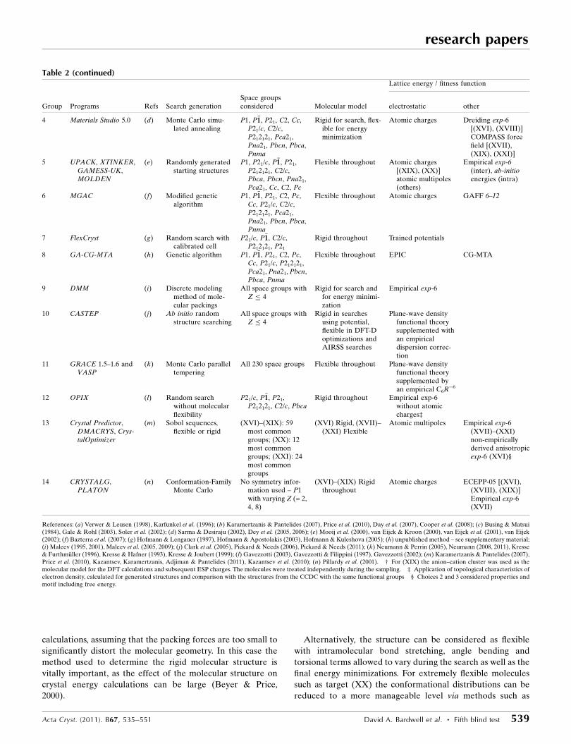
calculations, assuming that the packing forces are too small to
significantly distort the molecular geometry. In this case the
method used to determine the rigid molecular structure is
vitally important, as the effect of the molecular structure on
crystal energy calculations can be large (Beyer & Price,
2000).
Alternatively, the structure can be considered as flexible
with intramolecular bond stretching, angle bending and
torsional terms allowed to vary during the search as well as the
final energy minimizations. For extremely flexible molecules
such as target (XX) the conformational distributions can be
reduced to a more manageable level via methods such as
research papers
Acta Cryst. (2011). B67, 535–551 David A. Bardwell et al. � Fifth blind test 539
Table 2 (continued)Lattice energy / fitness function
Group Programs Refs Search generationSpace groupsconsidered Molecular model electrostatic other
4 Materials Studio 5.0 (d) Monte Carlo simu-lated annealing
P1, P�11, P21, C2, Cc,P21/c, C2/c,P212121, Pca21,Pna21, Pbcn, Pbca,Pnma
Rigid for search, flex-ible for energyminimization
Atomic charges Dreiding exp-6[(XVI), (XVIII)]COMPASS forcefield [(XVII),(XIX), (XXI)]
5 UPACK, XTINKER,GAMESS-UK,MOLDEN
(e) Randomly generatedstarting structures
P1, P21/c, P�11, P21,P212121, C2/c,Pbca, Pbcn, Pna21,Pca21, Cc, C2, Pc
Flexible throughout Atomic charges[(XIX), (XX)]atomic multipoles(others)
Empirical exp-6(inter), ab-initioenergies (intra)
6 MGAC (f) Modified geneticalgorithm
P1, P�11, P21, C2, Pc,Cc, P21/c, C2/c,P212121, Pca21,Pna21, Pbcn, Pbca,Pnma
Flexible throughout Atomic charges GAFF 6–12
7 FlexCryst (g) Random search withcalibrated cell
P21/c, P�11, C2/c,P212121, P21
Rigid throughout Trained potentials
8 GA-CG-MTA (h) Genetic algorithm P1, P�11, P21, C2, Pc,Cc, P21/c, P212121,Pca21, Pna21, Pbcn,Pbca, Pnma
Flexible throughout EPIC CG-MTA
9 DMM (i) Discrete modelingmethod of mole-cular packings
All space groups withZ � 4
Rigid for search andfor energy minimi-zation
Empirical exp-6
10 CASTEP (j) Ab initio randomstructure searching
All space groups withZ � 4
Rigid in searchesusing potential,flexible in DFT-Doptimizations andAIRSS searches
Plane-wave densityfunctional theorysupplemented withan empiricaldispersion correc-tion
11 GRACE 1.5–1.6 andVASP
(k) Monte Carlo paralleltempering
All 230 space groups Flexible throughout Plane-wave densityfunctional theorysupplemented byan empirical C6R�6
12 OPIX (l) Random searchwithout molecularflexibility
P21/c, P�11, P21,P212121, C2/c, Pbca
Rigid throughout Empirical exp-6without atomiccharges‡
13 Crystal Predictor,DMACRYS, Crys-talOptimizer
(m) Sobol sequences,flexible or rigid
(XVI)–(XIX): 59most commongroups; (XX): 12most commongroups; (XXI): 24most commongroups
(XVI) Rigid, (XVII)–(XXI) Flexible
Atomic multipoles Empirical exp-6(XVII)–(XXI)non-empiricallyderived anisotropicexp-6 (XVI)§
14 CRYSTALG,PLATON
(n) Conformation-FamilyMonte Carlo
No symmetry infor-mation used – P1with varying Z (= 2,4, 8)
(XVI)–(XIX) Rigidthroughout
Atomic charges ECEPP-05 [(XVI),(XVIII), (XIX)]Empirical exp-6(XVII)
References: (a) Verwer & Leusen (1998), Karfunkel et al. (1996); (b) Karamertzanis & Pantelides (2007), Price et al. (2010), Day et al. (2007), Cooper et al. (2008); (c) Busing & Matsui(1984), Gale & Rohl (2003), Soler et al. (2002); (d) Sarma & Desiraju (2002), Dey et al. (2005, 2006); (e) Mooij et al. (2000), van Eijck & Kroon (2000), van Eijck et al. (2001), van Eijck(2002); (f) Bazterra et al. (2007); (g) Hofmann & Lengauer (1997), Hofmann & Apostolakis (2003), Hofmann & Kuleshova (2005); (h) unpublished method – see supplementary material;(i) Maleev (1995, 2001), Maleev et al. (2005, 2009); (j) Clark et al. (2005), Pickard & Needs (2006), Pickard & Needs (2011); (k) Neumann & Perrin (2005), Neumann (2008, 2011), Kresse& Furthmuller (1996), Kresse & Hafner (1993), Kresse & Joubert (1999); (l) Gavezzotti (2003), Gavezzotti & Filippini (1997), Gavezzotti (2002); (m) Karamertzanis & Pantelides (2007),Price et al. (2010), Kazantsev, Karamertzanis, Adjiman & Pantelides (2011), Kazantsev et al. (2010); (n) Pillardy et al. (2001). † For (XIX) the anion–cation cluster was used as themolecular model for the DFT calculations and subsequent ESP charges. The molecules were treated independently during the sampling. ‡ Application of topological characteristics ofelectron density, calculated for generated structures and comparison with the structures from the CCDC with the same functional groups § Choices 2 and 3 considered properties andmotif including free energy.
analysis of conformational preferences using software such as
Mogul (Bruno et al., 2004).
3.2. Generating trial crystal structures
There are many diverse methods for generating crystal
packing arrangements in order to achieve a variety of plau-
sible packing arrangements. Most participants in this blind test
opted to generate large numbers of crystal structures with
random or quasi-random variables such as unit-cell para-
meters and positions and orientations of the molecules.
Several groups also elected to use a low-discrepancy Sobol’
sequence (Sobol’, 1967; Press et al., 1992). This helps ensure a
more uniform and thus efficient sampling and avoids the
problems of gaps and clusters that purely random sampling
can exhibit. Other groups used Monte Carlo types of search,
genetic algorithms, grid-based systematic searches or first-
principles ab initio random structure searching which allows
the possibility of a change in covalent bonding (Pickard &
Needs, 2006, 2011).
For the majority of these methods, space-group symmetry is
used. These methods search each space group and Z0 sepa-
rately and so in order to help reduce the computing time
required, many groups chose to restrict their search to only the
most commonly adopted space groups. This blind test saw two
groups electing to search all 230 space groups for some or all
of their predictions. Other groups used the alternative
approach of generating P1 crystal structures with varying
numbers of independent molecules (up to 8) in the unit cell.
Space-group symmetry was then identified in the resulting
crystal structures, after energy minimization, using packages
such as PLATON (Spek, 2009).
3.3. Ranking of crystal structures
The final ranking of the crystal structures is still almost
exclusively based on the calculated lattice energies of the
structures generated by the crystal structure search. Often
tens, if not hundreds, of possible structures can exist within a
few kJ mol�1 of the calculated global minimum (Day et al.,
2004) and therefore extreme accuracy is needed. One
successful approach to generating these lattice energies is the
DFT-D method, which can give more accurate lattice energies
(Neumann & Perrin, 2005) or re-minimization of the struc-
tures with more sophisticated force fields such as distributed
multipoles (Stone, 2005) and additional flexibility (Kazantsev,
Karamertzanis, Adjiman & Pantelides, 2011; Day & Cooper,
2010; Gorbitz et al., 2010). Moreover, additional or alternative
criteria may be used to discrimi-
nate between likely and unlikely
crystal structures. Such approa-
ches include lattice dynamic
contributions (van Eijck, 2001;
Anghel et al., 2002) or compar-
isons to known crystal structures
in the CSD (Dey et al., 2006),
exploiting any isostructurality
relationships (Asmadi et al.,
2010a,b).
4. Results
This paper is accompanied by a
large amount of supplementary
material: the coordinates of the
experimental crystal structures,
lists of predicted crystal structures
by each participant, as well as
detailed descriptions of metho-
dology, results and post-analysis
by most of the participating
research groups. Before discussing
the results of the predictions, the
crystal packings in the X-ray
determined crystal structures of
the six categories are described.
4.1. Experimental crystal struc-tures
4.1.1. Molecule (XVI). 2-Diazo-
3,5-cyclohexadiene-1-one (C6H4-
research papers
540 David A. Bardwell et al. � Fifth blind test Acta Cryst. (2011). B67, 535–551
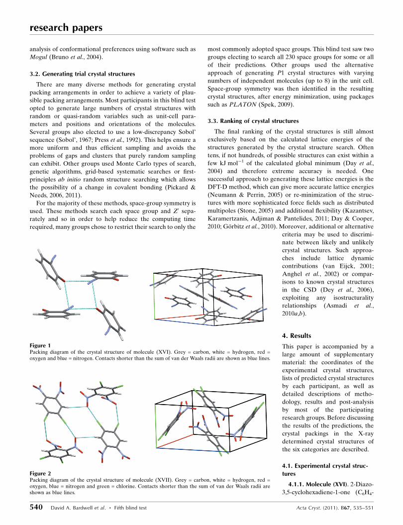
Figure 1Packing diagram of the crystal structure of molecule (XVI). Grey = carbon, white = hydrogen, red =oxygen and blue = nitrogen. Contacts shorter than the sum of van der Waals radii are shown as blue lines.
Figure 2Packing diagram of the crystal structure of molecule (XVII). Grey = carbon, white = hydrogen, red =oxygen, blue = nitrogen and green = chlorine. Contacts shorter than the sum of van der Waals radii areshown as blue lines.
N2O) was chosen as the blind test target for category 1 after
the initial target, 4-ethynylbenzonitrile, was found to have
been previously solved. Molecule (XVI) was crystallized by
slow evaporation from ethanol and the crystal structure was
solved from X-ray diffraction data collected at 174 K (Britton,
2010). The molecule crystallizes with Z0 = 1 in the orthor-
hombic space group Pbca. The crystal packing shows diazide-
carbonyl and CH� � �O interactions (Fig. 1).
4.1.2. Molecule (XVII). 1,2-Dichloro-4,5-dinitrobenzene
(C6H2Cl2N2O4) was chosen as the blind test target for category
2, although it deviates somewhat from the criteria for this
category as the molecule is not truly rigid; the nitro groups
allow for some degree of rotational freedom. Crystals were
obtained by slow evaporation of methanol and X-ray
diffraction data were collected at 174 K (Britton, 2010). The
molecule crystallizes in the monoclinic space group P21/c with
Z0 = 1 (Fig. 2).
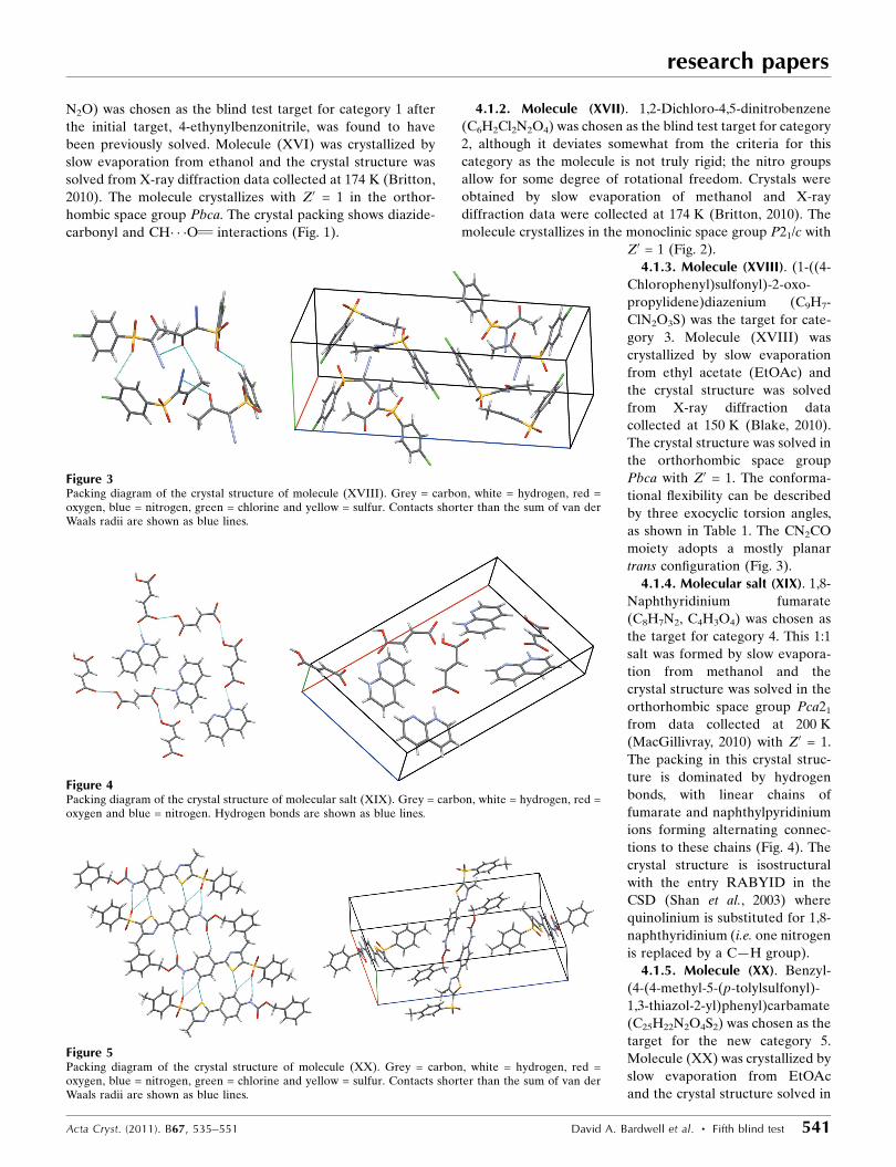
4.1.3. Molecule (XVIII). (1-((4-
Chlorophenyl)sulfonyl)-2-oxo-
propylidene)diazenium (C9H7-
ClN2O3S) was the target for cate-
gory 3. Molecule (XVIII) was
crystallized by slow evaporation
from ethyl acetate (EtOAc) and
the crystal structure was solved
from X-ray diffraction data
collected at 150 K (Blake, 2010).
The crystal structure was solved in
the orthorhombic space group
Pbca with Z0 = 1. The conforma-
tional flexibility can be described
by three exocyclic torsion angles,
as shown in Table 1. The CN2CO
moiety adopts a mostly planar
trans configuration (Fig. 3).
4.1.4. Molecular salt (XIX). 1,8-
Naphthyridinium fumarate
(C8H7N2, C4H3O4) was chosen as
the target for category 4. This 1:1
salt was formed by slow evapora-
tion from methanol and the
crystal structure was solved in the
orthorhombic space group Pca21
from data collected at 200 K
(MacGillivray, 2010) with Z0 = 1.
The packing in this crystal struc-
ture is dominated by hydrogen
bonds, with linear chains of
fumarate and naphthylpyridinium
ions forming alternating connec-
tions to these chains (Fig. 4). The
crystal structure is isostructural
with the entry RABYID in the
CSD (Shan et al., 2003) where
quinolinium is substituted for 1,8-
naphthyridinium (i.e. one nitrogen
is replaced by a C—H group).
4.1.5. Molecule (XX). Benzyl-
(4-(4-methyl-5-(p-tolylsulfonyl)-
1,3-thiazol-2-yl)phenyl)carbamate
(C25H22N2O4S2) was chosen as the
target for the new category 5.
Molecule (XX) was crystallized by
slow evaporation from EtOAc
and the crystal structure solved in
research papers
Acta Cryst. (2011). B67, 535–551 David A. Bardwell et al. � Fifth blind test 541
Figure 4Packing diagram of the crystal structure of molecular salt (XIX). Grey = carbon, white = hydrogen, red =oxygen and blue = nitrogen. Hydrogen bonds are shown as blue lines.
Figure 5Packing diagram of the crystal structure of molecule (XX). Grey = carbon, white = hydrogen, red =oxygen, blue = nitrogen, green = chlorine and yellow = sulfur. Contacts shorter than the sum of van derWaals radii are shown as blue lines.
Figure 3Packing diagram of the crystal structure of molecule (XVIII). Grey = carbon, white = hydrogen, red =oxygen, blue = nitrogen, green = chlorine and yellow = sulfur. Contacts shorter than the sum of van derWaals radii are shown as blue lines.
the monoclinic space group P21/n with Z0 = 1 (Blake, 2010).
The conformational flexibility can be described with eight
exocyclic torsion angles (Table 1). The molecule adopts an
elongated S shape, with the central part of the molecule mostly
planar, the greatest deviation from planarity being between
the phenyl and thiazol groups with an angle of 13�. The mostly
planar mid-section of the molecule forms stacks via a series of
weak interactions with CH and NH� � �OS as well as CH� � �OC
atom–atom contacts (i.e. shorter than the sum of van der
Waals radii), as shown in Fig. 5.
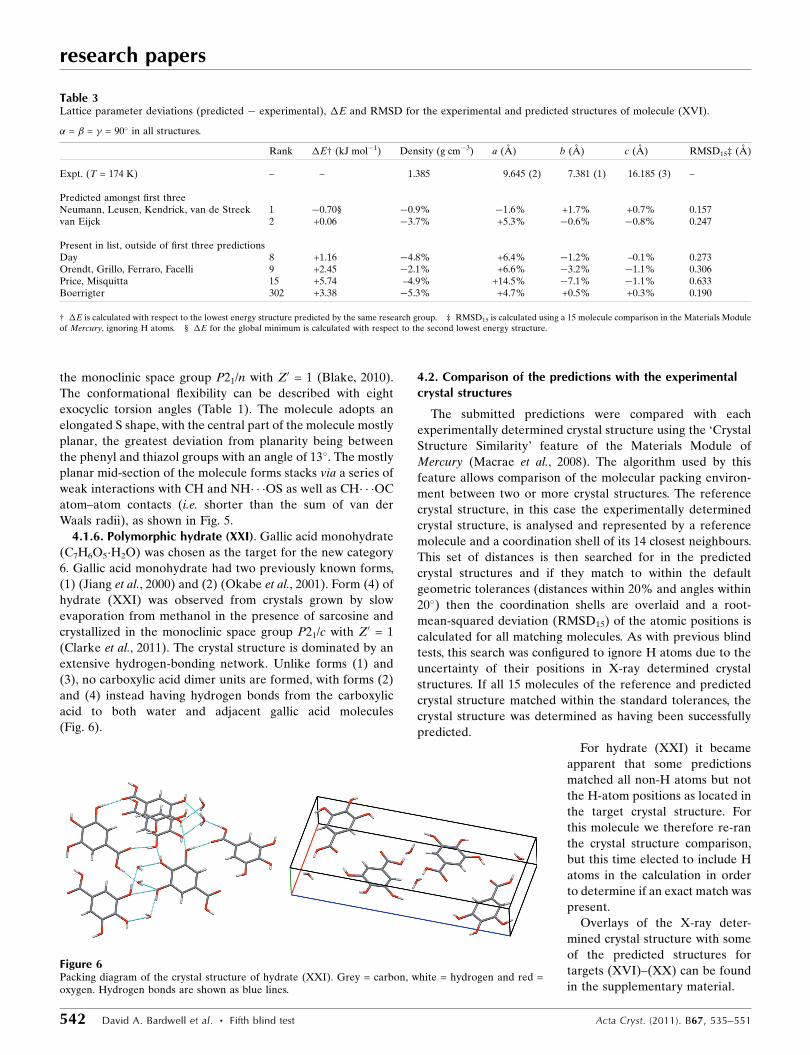
4.1.6. Polymorphic hydrate (XXI). Gallic acid monohydrate
(C7H6O5�H2O) was chosen as the target for the new category
6. Gallic acid monohydrate had two previously known forms,
(1) (Jiang et al., 2000) and (2) (Okabe et al., 2001). Form (4) of
hydrate (XXI) was observed from crystals grown by slow
evaporation from methanol in the presence of sarcosine and
crystallized in the monoclinic space group P21/c with Z0 = 1
(Clarke et al., 2011). The crystal structure is dominated by an
extensive hydrogen-bonding network. Unlike forms (1) and
(3), no carboxylic acid dimer units are formed, with forms (2)
and (4) instead having hydrogen bonds from the carboxylic
acid to both water and adjacent gallic acid molecules
(Fig. 6).
4.2. Comparison of the predictions with the experimentalcrystal structures
The submitted predictions were compared with each
experimentally determined crystal structure using the ‘Crystal
Structure Similarity’ feature of the Materials Module of
Mercury (Macrae et al., 2008). The algorithm used by this
feature allows comparison of the molecular packing environ-
ment between two or more crystal structures. The reference
crystal structure, in this case the experimentally determined
crystal structure, is analysed and represented by a reference
molecule and a coordination shell of its 14 closest neighbours.
This set of distances is then searched for in the predicted
crystal structures and if they match to within the default
geometric tolerances (distances within 20% and angles within
20�) then the coordination shells are overlaid and a root-
mean-squared deviation (RMSD15) of the atomic positions is
calculated for all matching molecules. As with previous blind
tests, this search was configured to ignore H atoms due to the
uncertainty of their positions in X-ray determined crystal
structures. If all 15 molecules of the reference and predicted
crystal structure matched within the standard tolerances, the
crystal structure was determined as having been successfully
predicted.
For hydrate (XXI) it became
apparent that some predictions
matched all non-H atoms but not
the H-atom positions as located in
the target crystal structure. For
this molecule we therefore re-ran
the crystal structure comparison,
but this time elected to include H
atoms in the calculation in order
to determine if an exact match was
present.
Overlays of the X-ray deter-
mined crystal structure with some
of the predicted structures for
targets (XVI)–(XX) can be found
in the supplementary material.
research papers
542 David A. Bardwell et al. � Fifth blind test Acta Cryst. (2011). B67, 535–551
Table 3Lattice parameter deviations (predicted � experimental), �E and RMSD for the experimental and predicted structures of molecule (XVI).
� = � = � = 90� in all structures.
Rank �E† (kJ mol�1) Density (g cm�3) a (A) b (A) c (A) RMSD15‡ (A)
Expt. (T = 174 K) – – 1.385 9.645 (2) 7.381 (1) 16.185 (3) –
Predicted amongst first threeNeumann, Leusen, Kendrick, van de Streek 1 �0.70§ �0.9% �1.6% +1.7% +0.7% 0.157van Eijck 2 +0.06 �3.7% +5.3% �0.6% �0.8% 0.247
Present in list, outside of first three predictionsDay 8 +1.16 �4.8% +6.4% �1.2% –0.1% 0.273Orendt, Grillo, Ferraro, Facelli 9 +2.45 �2.1% +6.6% �3.2% �1.1% 0.306Price, Misquitta 15 +5.74 –4.9% +14.5% �7.1% �1.1% 0.633Boerrigter 302 +3.38 �5.3% +4.7% +0.5% +0.3% 0.190
† �E is calculated with respect to the lowest energy structure predicted by the same research group. ‡ RMSD15 is calculated using a 15 molecule comparison in the Materials Moduleof Mercury, ignoring H atoms. § �E for the global minimum is calculated with respect to the second lowest energy structure.
Figure 6Packing diagram of the crystal structure of hydrate (XXI). Grey = carbon, white = hydrogen and red =oxygen. Hydrogen bonds are shown as blue lines.
4.3. Predictions results
4.3.1. Molecule (XVI). All of the participating research
groups attempted predictions for molecule (XVI), two of
whom predicted the observed crystal structure within their
three predictions (Table 3). One of these successes (Neumann,
Leusen, Kendrick and van de Streek) was submitted as the
group’s first prediction, while the other (van Eijck) was
submitted as the participant’s second prediction. Both of these
successful predictions gave RMSD15 deviations from the
experimentally determined crystal structure of less than
0.25 A.
Outside of the three official predictions, the observed
crystal structure was present in the extended lists of five other
research groups. The success rates here are comparable to the
first three blind tests, while not quite as high as the results
observed in the fourth blind test. This may be attributed to
some methods having difficulties with many structures close in
energy. The very small �E in Table 3, even when the observed
structure is found outside of the first three predictions, shows
how closely spaced the energies are for this molecule, and the
accuracy in lattice energy required for a successful
prediction.
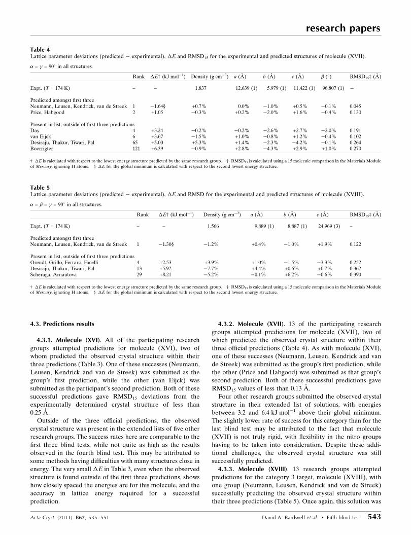
4.3.2. Molecule (XVII). 13 of the participating research
groups attempted predictions for molecule (XVII), two of
which predicted the observed crystal structure within their
three official predictions (Table 4). As with molecule (XVI),
one of these successes (Neumann, Leusen, Kendrick and van
de Streek) was submitted as the group’s first prediction, while
the other (Price and Habgood) was submitted as that group’s
second prediction. Both of these successful predictions gave
RMSD15 values of less than 0.13 A.
Four other research groups submitted the observed crystal
structure in their extended list of solutions, with energies
between 3.2 and 6.4 kJ mol�1 above their global minimum.
The slightly lower rate of success for this category than for the
last blind test may be attributed to the fact that molecule
(XVII) is not truly rigid, with flexibility in the nitro groups
having to be taken into consideration. Despite these addi-
tional challenges, the observed crystal structure was still
successfully predicted.
4.3.3. Molecule (XVIII). 13 research groups attempted
predictions for the category 3 target, molecule (XVIII), with
one group (Neumann, Leusen, Kendrick and van de Streek)
successfully predicting the observed crystal structure within
their three predictions (Table 5). Once again, this solution was
research papers
Acta Cryst. (2011). B67, 535–551 David A. Bardwell et al. � Fifth blind test 543
Table 5Lattice parameter deviations (predicted � experimental), �E and RMSD for the experimental and predicted structures of molecule (XVIII).
� = � = � = 90� in all structures.
Rank �E† (kJ mol�1) Density (g cm�3) a (A) b (A) c (A) RMSD15‡ (A)
Expt. (T = 174 K) – – 1.566 9.889 (1) 8.887 (1) 24.969 (3) –
Predicted amongst first threeNeumann, Leusen, Kendrick, van de Streek 1 �1.30§ �1.2% +0.4% �1.0% +1.9% 0.122
Present in list, outside of first three predictionsOrendt, Grillo, Ferraro, Facelli 4 +2.53 +3.9% +1.0% �1.5% �3.3% 0.252Desiraju, Thakur, Tiwari, Pal 13 +5.92 �7.7% +4.4% +0.6% +0.7% 0.362Scheraga, Arnautova 29 +8.21 �5.2% �0.1% +6.2% �0.6% 0.390
† �E is calculated with respect to the lowest energy structure predicted by the same research group. ‡ RMSD15 is calculated using a 15 molecule comparison in the Materials Moduleof Mercury, ignoring H atoms. § �E for the global minimum is calculated with respect to the second lowest energy structure.
Table 4Lattice parameter deviations (predicted � experimental), �E and RMSD15 for the experimental and predicted structures of molecule (XVII).
� = � = 90� in all structures.
Rank �E† (kJ mol�1) Density (g cm�3) a (A) b (A) c (A) � (�) RMSD15‡ (A)
Expt. (T = 174 K) – – 1.837 12.639 (1) 5.979 (1) 11.422 (1) 96.807 (1) �
Predicted amongst first threeNeumann, Leusen, Kendrick, van de Streek 1 �1.64§ +0.7% 0.0% �1.0% +0.5% �0.1% 0.045Price, Habgood 2 +1.05 �0.3% +0.2% �2.0% +1.6% �0.4% 0.130
Present in list, outside of first three predictionsDay 4 +3.24 �0.2% �0.2% �2.6% +2.7% �2.0% 0.191van Eijck 6 +3.67 �1.5% +1.0% �0.8% +1.2% �0.4% 0.102Desiraju, Thakur, Tiwari, Pal 65 +5.00 +5.3% +1.4% �2.3% �4.2% �0.1% 0.264Boerrigter 121 +6.39 �0.9% +2.8% �4.3% +2.9% +1.0% 0.270
† �E is calculated with respect to the lowest energy structure predicted by the same research group. ‡ RMSD15 is calculated using a 15 molecule comparison in the Materials Moduleof Mercury, ignoring H atoms. § �E for the global minimum is calculated with respect to the second lowest energy structure.
submitted as this group’s first submission, with an RMSD15
from the observed crystal structure of just 0.12 A.
Three other groups also reported the correct crystal struc-
ture in their extended lists of solutions, with one group
(Orendt, Grillo, Ferraro and Facelli) close to having a
successful prediction as their number 4 structure is a close
match to the experimental structure with an RMSD15 value of
0.252 A.
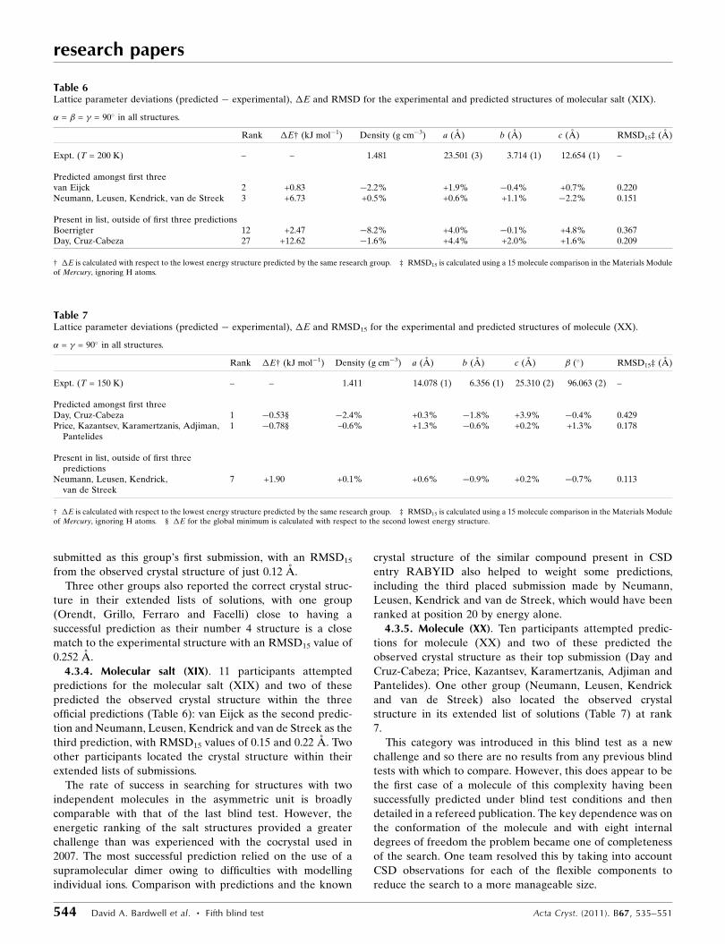
4.3.4. Molecular salt (XIX). 11 participants attempted
predictions for the molecular salt (XIX) and two of these
predicted the observed crystal structure within the three
official predictions (Table 6): van Eijck as the second predic-
tion and Neumann, Leusen, Kendrick and van de Streek as the
third prediction, with RMSD15 values of 0.15 and 0.22 A. Two
other participants located the crystal structure within their
extended lists of submissions.
The rate of success in searching for structures with two
independent molecules in the asymmetric unit is broadly
comparable with that of the last blind test. However, the
energetic ranking of the salt structures provided a greater
challenge than was experienced with the cocrystal used in
2007. The most successful prediction relied on the use of a
supramolecular dimer owing to difficulties with modelling
individual ions. Comparison with predictions and the known
crystal structure of the similar compound present in CSD
entry RABYID also helped to weight some predictions,
including the third placed submission made by Neumann,
Leusen, Kendrick and van de Streek, which would have been
ranked at position 20 by energy alone.
4.3.5. Molecule (XX). Ten participants attempted predic-
tions for molecule (XX) and two of these predicted the
observed crystal structure as their top submission (Day and
Cruz-Cabeza; Price, Kazantsev, Karamertzanis, Adjiman and
Pantelides). One other group (Neumann, Leusen, Kendrick
and van de Streek) also located the observed crystal
structure in its extended list of solutions (Table 7) at rank
7.
This category was introduced in this blind test as a new
challenge and so there are no results from any previous blind
tests with which to compare. However, this does appear to be
the first case of a molecule of this complexity having been
successfully predicted under blind test conditions and then
detailed in a refereed publication. The key dependence was on
the conformation of the molecule and with eight internal
degrees of freedom the problem became one of completeness
of the search. One team resolved this by taking into account
CSD observations for each of the flexible components to
reduce the search to a more manageable size.
research papers
544 David A. Bardwell et al. � Fifth blind test Acta Cryst. (2011). B67, 535–551
Table 7Lattice parameter deviations (predicted � experimental), �E and RMSD15 for the experimental and predicted structures of molecule (XX).
� = � = 90� in all structures.
Rank �E† (kJ mol�1) Density (g cm�3) a (A) b (A) c (A) � (�) RMSD15‡ (A)
Expt. (T = 150 K) – – 1.411 14.078 (1) 6.356 (1) 25.310 (2) 96.063 (2) –
Predicted amongst first threeDay, Cruz-Cabeza 1 �0.53§ �2.4% +0.3% �1.8% +3.9% �0.4% 0.429Price, Kazantsev, Karamertzanis, Adjiman,
Pantelides1 �0.78§ –0.6% +1.3% �0.6% +0.2% +1.3% 0.178
Present in list, outside of first threepredictions
Neumann, Leusen, Kendrick,van de Streek
7 +1.90 +0.1% +0.6% �0.9% +0.2% �0.7% 0.113
† �E is calculated with respect to the lowest energy structure predicted by the same research group. ‡ RMSD15 is calculated using a 15 molecule comparison in the Materials Moduleof Mercury, ignoring H atoms. § �E for the global minimum is calculated with respect to the second lowest energy structure.
Table 6Lattice parameter deviations (predicted � experimental), �E and RMSD for the experimental and predicted structures of molecular salt (XIX).
� = � = � = 90� in all structures.
Rank �E† (kJ mol�1) Density (g cm�3) a (A) b (A) c (A) RMSD15‡ (A)
Expt. (T = 200 K) – – 1.481 23.501 (3) 3.714 (1) 12.654 (1) –
Predicted amongst first threevan Eijck 2 +0.83 �2.2% +1.9% �0.4% +0.7% 0.220Neumann, Leusen, Kendrick, van de Streek 3 +6.73 +0.5% +0.6% +1.1% �2.2% 0.151
Present in list, outside of first three predictionsBoerrigter 12 +2.47 �8.2% +4.0% �0.1% +4.8% 0.367Day, Cruz-Cabeza 27 +12.62 �1.6% +4.4% +2.0% +1.6% 0.209
† �E is calculated with respect to the lowest energy structure predicted by the same research group. ‡ RMSD15 is calculated using a 15 molecule comparison in the Materials Moduleof Mercury, ignoring H atoms.
research papers
Acta Cryst. (2011). B67, 535–551 David A. Bardwell et al. � Fifth blind test 545
Table 8(a) Lattice parameter deviations (predicted � experimental), �E and RMSD15 for the experimental and predicted structures of hydrate (XXI) (withmatching hydrogen placement).
� = � = 90� in all structures.
Rank �E† (kJ mol�1) Density (g cm�3) a (A) b (A) c (A) � (�) RMSD15‡ (A)
Expt. (T = 150 K) – – 1.639 9.790 (7) 3.609 (3) 21.583 (16) 91.462 (14) –
Present in list, outside of first three predictionsDay 12 +2.96 +2.4% �2.9% �0.4% +1.1% +0.3% 0.159van Eijck 29 +12.47 +10.1% �4.9% �2.4% �2.5% +0.8% 0.208Neumann, Leusen,
Kendrick, van deStreek
81 +8.85 +2.0% �2.8% +1.2% �0.4% �0.5% 0.228
Price, Braun 117 +12.69 +3.7% �1.5% �1.9% �0.1% +0.8% 0.108
† �E is calculated with respect to the lowest energy structure predicted by the same research group. ‡ RMSD15 is calculated using a 15 molecule comparison in the Materials Moduleof Mercury, ignoring H atoms.
(b) Lattice parameter deviations (predicted� experimental), �E and RMSD15 for the predicted structures of hydrate (XXI) with alternative H-atom placement tothe experimental structure. � = � = 90� in all structures.
Rank �E† (kJ mol�1) Density (g cm�3) a (A) b (A) c (A) � (�) RMSD15‡ (A)
Expt. (T = 150 K) – – 1.639 9.790 (7) 3.609 (3) 21.583 (16) 91.462 (14) –
Predicted amongst first threeVan Eijck 1 �2.43§ +13.1% �5.4% �3.8% �2.8% +1.2% 0.232Price, Braun 3 +1.08 +5.8% �1.0% �4.0% �0.6% +0.1% 0.224
Present in list, outside of first three predictionsDesiraju, Thakur, Tiwari, Pal 11 +0.19 +6.2% +0.6% �3.3% �3.2% �0.4% 0.642Day 61 +6.96 +4.5% �1.0% �1.4% �2.1% �1.6% 0.218Neumann, Leusen, Kendrick,
van de Streek174 +11.30 +2.9% �2.7% �0.2% +0.1% �0.4% 0.192
† �E is calculated with respect to the lowest energy structure predicted by the same research group. ‡ RMSD15 is calculated using a 15 molecule comparison in the Materials Moduleof Mercury, ignoring H atoms. § �E for the global minimum is calculated with respect to the second lowest energy structure.
(c) Lattice parameter deviations (predicted� experimental), �E and RMSD15 for the experimental and predicted structures of KONTIQ [form (1)]. �= � = 90� inall structures.
Rank �E† (kJ mol�1) Density (g cm�3) a (A) b (A) c (A) � (�) RMSD15‡ (A)
Expt. (T = 150 K) – – 1.599 5.794 (4) 4.719 (5) 28.688 (5) 95.080 (30) –
Present in listNeumann, Leusen, Kendrick,
van de Streek19 +0.22 +3.6% +1.3% �5.4% +0.8% +0.6% 0.211
Day 48 +6.61 +3.1% +4.3% �6.3% +0.2% �0.6% 0.295van Eijck 74 +18.80 +10.8% +6.1% �17.0% +2.2% �5.0% 0.690Desiraju, Thakur, Tiwari, Pal 143 +12.11 +7.6% �4.4% �3.3% +1.1% +2.9% 0.442Boerrigter 282 +13.18 �2.6% �8.42% +10.2% +2.8% +4.7% 0.631Price, Braun 338 +11.87 +1.9% +11.0% �14.6% +5.0% +6.8% 0.683
† �E is calculated with respect to the lowest energy structure predicted by the same research group. ‡ RMSD15 is calculated using a 15 molecule comparison in the Materials Moduleof Mercury, ignoring H atoms.
(d) Lattice parameter deviations (predicted� experimental), �E and RMSD15 for the experimental and predicted structures of KONTIQ01 [form (2)]. �= � = 90�
in all structures.
Rank �E† (kJ mol�1) Density (g cm�3) a (A) b (A) c (A) � (�) RMSD15‡ (A)
Expt. (T = 150 K) – – 1.636 14.150 (10) 3.622 (9) 15.028 (10) 97.520 (70) –
Present in listvan Eijck 9 +8.44 +11.0% �2.2% �2.7% �5.2% +0.5% 0.206Price, Braun 23 +4.83 +3.9% +0.7% �3.6% �0.6% +0.5% 0.186Neumann, Leusen, Kendrick,
van de Streek49 +0.34 +2.0% �1.0% �0.1% �2.2% +0.1% 0.090
Day 53 +6.68 +4.7% �1.4% �0.6% �2.1% +1.7% 0.135Desiraju, Thakur, Tiwari, Pal 126 +10.93 +4.9% 1.4% �2.1% �0.7% +2.2% 0.228
† �E is calculated with respect to the lowest energy structure predicted by the same research group. ‡ RMSD15 is calculated using a 15 molecule comparison in the Materials Moduleof Mercury, ignoring H atoms.
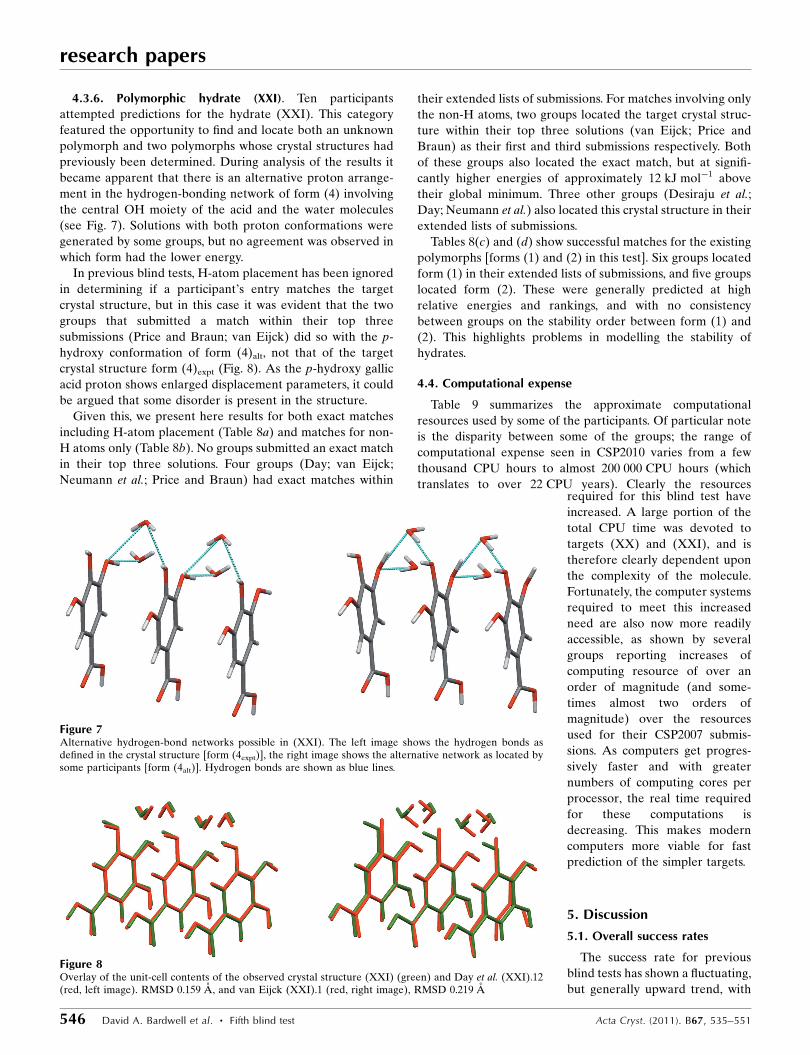
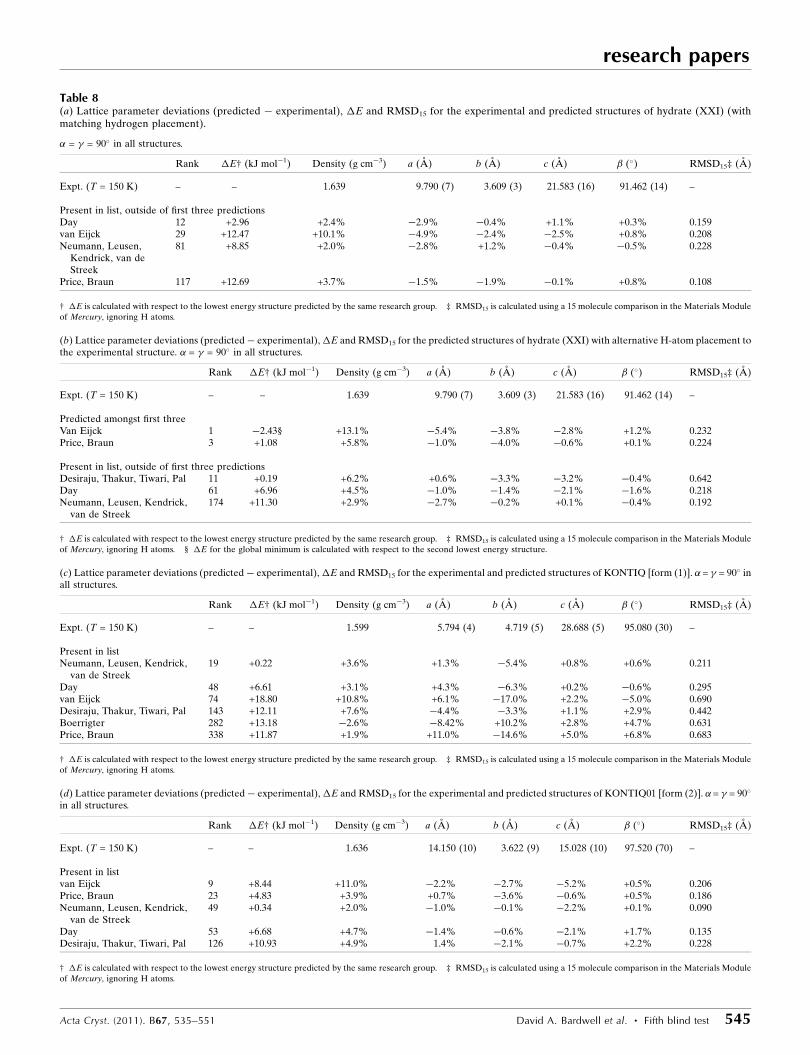
4.3.6. Polymorphic hydrate (XXI). Ten participants
attempted predictions for the hydrate (XXI). This category
featured the opportunity to find and locate both an unknown
polymorph and two polymorphs whose crystal structures had
previously been determined. During analysis of the results it
became apparent that there is an alternative proton arrange-
ment in the hydrogen-bonding network of form (4) involving
the central OH moiety of the acid and the water molecules
(see Fig. 7). Solutions with both proton conformations were
generated by some groups, but no agreement was observed in
which form had the lower energy.
In previous blind tests, H-atom placement has been ignored
in determining if a participant’s entry matches the target
crystal structure, but in this case it was evident that the two
groups that submitted a match within their top three
submissions (Price and Braun; van Eijck) did so with the p-
hydroxy conformation of form (4)alt, not that of the target
crystal structure form (4)expt (Fig. 8). As the p-hydroxy gallic
acid proton shows enlarged displacement parameters, it could
be argued that some disorder is present in the structure.
Given this, we present here results for both exact matches
including H-atom placement (Table 8a) and matches for non-
H atoms only (Table 8b). No groups submitted an exact match
in their top three solutions. Four groups (Day; van Eijck;
Neumann et al.; Price and Braun) had exact matches within
their extended lists of submissions. For matches involving only
the non-H atoms, two groups located the target crystal struc-
ture within their top three solutions (van Eijck; Price and
Braun) as their first and third submissions respectively. Both
of these groups also located the exact match, but at signifi-
cantly higher energies of approximately 12 kJ mol�1 above
their global minimum. Three other groups (Desiraju et al.;
Day; Neumann et al.) also located this crystal structure in their
extended lists of submissions.
Tables 8(c) and (d) show successful matches for the existing
polymorphs [forms (1) and (2) in this test]. Six groups located
form (1) in their extended lists of submissions, and five groups
located form (2). These were generally predicted at high
relative energies and rankings, and with no consistency
between groups on the stability order between form (1) and
(2). This highlights problems in modelling the stability of
hydrates.
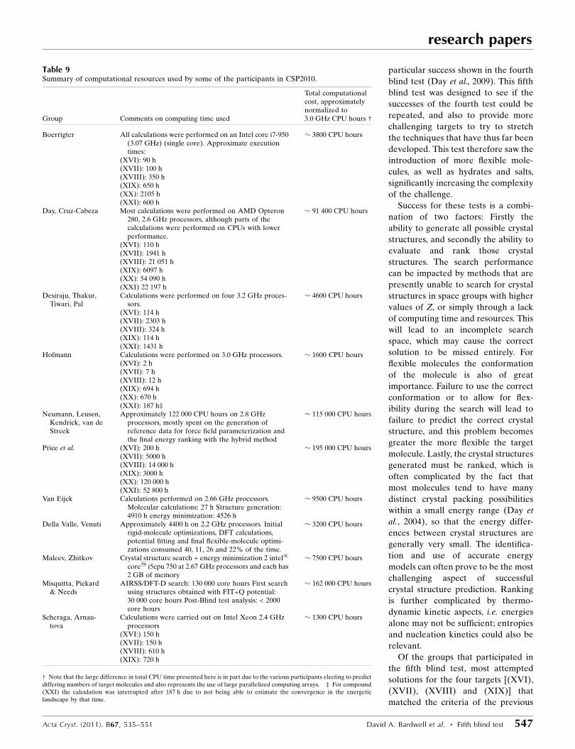
4.4. Computational expense
Table 9 summarizes the approximate computational
resources used by some of the participants. Of particular note
is the disparity between some of the groups; the range of
computational expense seen in CSP2010 varies from a few
thousand CPU hours to almost 200 000 CPU hours (which
translates to over 22 CPU years). Clearly the resourcesrequired for this blind test have
increased. A large portion of the
total CPU time was devoted to
targets (XX) and (XXI), and is
therefore clearly dependent upon
the complexity of the molecule.
Fortunately, the computer systems
required to meet this increased
need are also now more readily
accessible, as shown by several
groups reporting increases of
computing resource of over an
order of magnitude (and some-
times almost two orders of
magnitude) over the resources
used for their CSP2007 submis-
sions. As computers get progres-
sively faster and with greater
numbers of computing cores per
processor, the real time required
for these computations is
decreasing. This makes modern
computers more viable for fast
prediction of the simpler targets.
5. Discussion
5.1. Overall success rates
The success rate for previous
blind tests has shown a fluctuating,
but generally upward trend, with
research papers
546 David A. Bardwell et al. � Fifth blind test Acta Cryst. (2011). B67, 535–551
Figure 8Overlay of the unit-cell contents of the observed crystal structure (XXI) (green) and Day et al. (XXI).12(red, left image). RMSD 0.159 A, and van Eijck (XXI).1 (red, right image), RMSD 0.219 A
Figure 7Alternative hydrogen-bond networks possible in (XXI). The left image shows the hydrogen bonds asdefined in the crystal structure [form (4expt)], the right image shows the alternative network as located bysome participants [form (4alt)]. Hydrogen bonds are shown as blue lines.
particular success shown in the fourth
blind test (Day et al., 2009). This fifth
blind test was designed to see if the
successes of the fourth test could be
repeated, and also to provide more
challenging targets to try to stretch
the techniques that have thus far been
developed. This test therefore saw the
introduction of more flexible mole-
cules, as well as hydrates and salts,
significantly increasing the complexity
of the challenge.
Success for these tests is a combi-
nation of two factors: Firstly the
ability to generate all possible crystal
structures, and secondly the ability to
evaluate and rank those crystal
structures. The search performance
can be impacted by methods that are
presently unable to search for crystal
structures in space groups with higher
values of Z, or simply through a lack
of computing time and resources. This
will lead to an incomplete search
space, which may cause the correct
solution to be missed entirely. For
flexible molecules the conformation
of the molecule is also of great
importance. Failure to use the correct
conformation or to allow for flex-
ibility during the search will lead to
failure to predict the correct crystal
structure, and this problem becomes
greater the more flexible the target
molecule. Lastly, the crystal structures
generated must be ranked, which is
often complicated by the fact that
most molecules tend to have many
distinct crystal packing possibilities
within a small energy range (Day et
al., 2004), so that the energy differ-
ences between crystal structures are
generally very small. The identifica-
tion and use of accurate energy
models can often prove to be the most
challenging aspect of successful
crystal structure prediction. Ranking
is further complicated by thermo-
dynamic kinetic aspects, i.e. energies
alone may not be sufficient; entropies
and nucleation kinetics could also be
relevant.
Of the groups that participated in
the fifth blind test, most attempted
solutions for the four targets [(XVI),
(XVII), (XVIII) and (XIX)] that
matched the criteria of the previous
research papers
Acta Cryst. (2011). B67, 535–551 David A. Bardwell et al. � Fifth blind test 547
Table 9Summary of computational resources used by some of the participants in CSP2010.
Group Comments on computing time used
Total computationalcost, approximatelynormalized to3.0 GHz CPU hours †
Boerrigter All calculations were performed on an Intel core i7-950(3.07 GHz) (single core). Approximate executiontimes:
� 3800 CPU hours
(XVI): 90 h(XVII): 100 h(XVIII): 350 h(XIX): 650 h(XX): 2105 h(XXI): 600 h
Day, Cruz-Cabeza Most calculations were performed on AMD Opteron280, 2.6 GHz processors, although parts of thecalculations were performed on CPUs with lowerperformance.
� 91 400 CPU hours
(XVI): 110 h(XVII): 1941 h(XVIII): 21 051 h(XIX): 6097 h(XX): 54 090 h(XXI) 22 197 h
Desiraju, Thakur,Tiwari, Pal
Calculations were performed on four 3.2 GHz proces-sors.
� 4600 CPU hours
(XVI): 114 h(XVII): 2303 h(XVIII): 324 h(XIX): 114 h(XXI): 1431 h
Hofmann Calculations were performed on 3.0 GHz processors. � 1600 CPU hours(XVI): 2 h(XVII): 7 h(XVIII): 12 h(XIX): 694 h(XX): 670 h(XXI): 187 h‡
Neumann, Leusen,Kendrick, van deStreek
Approximately 122 000 CPU hours on 2.8 GHzprocessors, mostly spent on the generation ofreference data for force field parameterization andthe final energy ranking with the hybrid method
� 115 000 CPU hours
Price et al. (XVI): 200 h � 195 000 CPU hours(XVII): 5000 h(XVIII): 14 000 h(XIX): 3000 h(XX): 120 000 h(XXI): 52 800 h
Van Eijck Calculations performed on 2.66 GHz processors.Molecular calculations: 27 h Structure generation:4910 h energy minimization: 4526 h
� 9500 CPU hours
Della Valle, Venuti Approximately 4400 h on 2.2 GHz processors. Initialrigid-molecule optimizations, DFT calculations,potential fitting and final flexible-molecule optimi-zations consumed 40, 11, 26 and 22% of the time.
� 3200 CPU hours
Maleev, Zhitkov Crystal structure search + energy minimization 2 intel1
core2 i5cpu 750 at 2.67 GHz processors and each has2 GB of memory
� 7500 CPU hours
Misquitta, Pickard& Needs
AIRSS/DFT-D search: 130 000 core hours First searchusing structures obtained with FIT+Q potential:30 000 core hours Post-Blind test analysis: < 2000core hours
� 162 000 CPU hours
Scheraga, Arnau-tova
Calculations were carried out on Intel Xeon 2.4 GHzprocessors
� 1300 CPU hours
(XVI:) 150 h(XVII): 150 h(XVIII): 610 h(XIX): 720 h
† Note that the large difference in total CPU time presented here is in part due to the various participants electing to predictdiffering numbers of target molecules and also represents the use of large parallelized computing arrays. ‡ For compound(XXI) the calculation was interrupted after 187 h due to not being able to estimate the convergence in the energeticlandscape by that time.
blind test. Overall, the success rates for these four targets were
a little lower than for CSP2007, but generally at least as good if
not better than the results obtained for CSP1999, CSP2001
and CSP2004. What these results do show, however, is that just
as in CSP2007, the method adopted by Neumann, Leusen,
Kendrick and van de Streek again excelled, with this group
able to successfully predict the crystal structures of the first
three categories with their number 1 submission, as well as the
fourth category with their number 3 submission. They were
the only participants able to generate all target crystal struc-
tures within their extended list of submissions. They did so
with the lowest RMSD15 values for all except the hydrate
crystal structure. This demonstrates the reliability of DFT-D
methods to predict the crystal structures of small organic
molecules (Asmadi et al., 2009; Chan et al., 2011). For the
fourth category, complete crystal-structure prediction studies
were performed for (XIX) and for model compound
RABYID from the CSD. The energy landscapes of these two
systems were analysed and showed significant similarities.
Based on these similarities, it had to be concluded that the
experimental structure of (XIX) could be isostructural to the
experimental structure of RABYID, and this structure, even
though it was ranked 20th by energy (22nd for RABYID), was
submitted as the third candidate structure (Kendrick et al.,
2011).
More complex systems such as salts continue to provide
some challenge, perhaps suggesting that a salt should be
considered a new, more challenging category than the current
‘cocrystal’ definition of category four.
This test also introduced two new categories that provided
much greater challenges to the participants and 11 out of the
participating groups attempted at least one of the targets (XX)
and (XXI). Particularly encouraging was that two groups
(Price et al.; Day et al.) successfully predicted the crystal
structure for the largest, most flexible molecule to be included
in this series of blind tests. The hydrate target (XXI) proved to
be a considerable challenge, even to methods that have been
successful for hydrates of o-dihydroxybenzoic acids (Braun et
al., 2011), and highlights the many difficulties that such a
system can pose to characterization as well as successful
structure prediction. However, this is a system that needs to be
tackled; water is one of the most complex solvents to model,
yet it is also one of the most important.
This blind test has also once again highlighted that the use
of generic standard force fields does not lead to good crystal
structure prediction results. We have also observed that the
more extensive search methods are adequate within the
limitations (Z0, no disorder etc.) implicit in the blind test
categories, but have to assume the covalent bonding in the
chemical diagram and rely on a sufficient number of search
structures being refined by the more accurate and expensive
model for the lattice energy. Successful prediction of small
molecule crystal structures has been shown to require both
accuracy of energies and the ability to coordinate the inter-
and intramolecular force field contributions. The methods that
gave the greatest success were varied and were modified to
take on these tougher challenges.
5.2. Challenges faced
Molecule (XVI), while the simplest of the rigid molecule
targets, proved to have many crystal structures close in energy.
Transferable empirical potentials had difficulty coping with
diazide–carbonyl interactions with induction being a problem.
Simple point-charge models used by some groups failed
completely for this molecule, although van Eijck did find in
post-analysis that one set of charges predicted the observed
structure.
This system is the first to be tackled by an ab initio random
search method (Misquitta, Pickard and Needs) which does not
fix the chemical bonding and uses electronic structure
methods during the search, although this approach results in a
significant increase in the computing resources required when
compared with the methods employed by the other partici-
pating groups. This method failed as the search was not
extended to eight formula units in the cell. Many of the
numerous minima, including the global minimum, corre-
sponded to an isomer with the formation of a bond to give a
heterocyclic ring with the two N atoms, showing the promise of
this method for cases, such as tautomers, where the covalent
bonding is uncertain.
Molecule (XVII) was perhaps not well selected as a target
for its category as the molecule was not truly rigid; the
orientation of the nitro groups may be affected by inter-
molecular interactions in the crystal structure. As a result
participants were forced to first consider how to deal with this
flexibility. The electrostatic potentials of the nitro groups also
proved to be unusually challenging to model successfully,
although the dispersion proved to be a very important
contribution to the lattice energy. These additional issues lead
to molecule (XVII) being a significantly more difficult
problem than previous targets in this category. Despite these
extra challenges, the success rate for this category was good
compared with previous blind tests.
For molecule (XVIII) flexibility proved to be the key to
successfully locating the crystal structure in the search. Some
searches missed the crystal structure, with the most funda-
mental reason being the wrong conformation of the
C(N2)C(O) bond. The relative energies of the cis and trans
configurations were sufficiently sensitive to the methods being
used to cause mis-assignment.
For the salt (XIX), there were again some difficulties with
flexibility with the relative orientation of the two fragments;
for the acid there is considerable conformational flexibility
and the calculated stability of the conformers alters between
the gas and solid.
All groups encountered significant problems with devel-
oping suitable methods of evaluating the relative lattice
energies of structures containing the different conformers.
Plane-wave ab initio methods do not cope well with isolated
ions in a vacuum, causing problems with ion-specific reference
data calculated with DFT-D methods for force-field para-
meterization. Induction and charge transfer, which are
stronger in molecular salts, limited the transferability of exp-6
potentials which had been fitted to crystal structures of neutral
research papers
548 David A. Bardwell et al. � Fifth blind test Acta Cryst. (2011). B67, 535–551
molecules. Successful prediction based on energy (van Eijck)
was achieved by the use of a supramolecular dimer, rather
than two ions as individual molecules.
Other groups noted the similarity of an existing crystal
structure in the CSD, RABYID, the crystal structure of which
consisted of the same fumarate ion but with a quinolinium
counterion instead of 1,8-naphthyridinium (i.e. where the
unprotonated nitrogen is instead a CH moiety). The energy
landscapes generated for both salts proved similar enough to
encourage the speculation that the two crystal structures could
be isostructural and one group (Neumann et al.) submitted a
successful prediction based on this approach (Kendrick et al.,
2011).
Molecule (XX) was the first large flexible molecule to
feature in the blind tests and proved a considerable challenge.
For such highly flexible molecules there is a key dependence
on the conformation of the molecule and successful prediction
involved succeeding at this early step. One of the main diffi-
culties is the computing power required to make a complete
search for all available space groups with a flexible molecule;
when all standard orientations about the exocyclic single
bonds are considered, there are over one thousand possible
conformations. The two successful strategies (Day and Cruz-
Cabeza; Price, Kazantsev, Karamertzanis, Adjiman and
Pantelides) reduced the search space to a more manageable
level, producing innovations in methodology that have been
described and contrasted in detail elsewhere (Kazantsev,
Karamertzanis, Adjiman, Pantelides, Price, Galek, Day &
Cruz-Cabeza, 2011). Day et al. used geometry data for similar
systems from the CSD to help limit the search further and
considered a set of predefined conformations. Price et al.
identified likely ranges of values for the flexible torsions and
used an extension to the CrystalPredictor methodology and
databases of the ab initio calculations on the isolated molecule
to allow the crystal structures and conformations to be
simultaneously refined (Kazantsev, Karamertzanis, Adjiman
& Pantelides, 2011). Neumann et al. employed a fully flexible
molecule, allowing all conformations to be explored during the
crystal structure generation step. Use of multipoles and
empirical potentials performed better than DFT-D in this case,
with both groups using this method (Day et al.; Price et al.)
successfully predicting the crystal structure in first
place.
Hydrate (XXI) proved to be one of the most challenging
systems in the blind test. For this molecule two known poly-
morphs already existed. However, the difficulty in predicting
this crystal structure was not due to the availability of two
already known polymorphs, but rather that the representation
of water–water and water–gallic acid interactions is extremely
difficult to model, making the successful prediction of even the
known polymorphs a difficult task.
As a hydrate, the hydrogen-bonding network enabled by
the water molecules and the various hydrogen-bond donors
and acceptors in the acid proved key to successfully predicting
the crystal structure, but it is also obvious that the sheer
number of different possible hydrogen-bond networks make
the problem a difficult one. The results obtained for form (4)
show that with the same placement of non-H atoms there is
more than one set of hydrogen positions that is possible.
Energetically, the OH conformation observed in the experi-
mental structure is not the most favourable in isolation and,
given the nature of X-ray diffraction, the positions of these
protons cannot be deemed as unequivocally determined.
Indeed, there is evidence of large displacement parameters for
the protons involved in the two alternative hydrogen-bond
networks. This leads us to consider that the structure is best
described as disordered with respect to which network is
present. This matter would only be resolved with an in-depth
temperature-dependent X-ray and NMR study. A post-blind
test polymorphism screen (Braun, Personal communication)
showed that the ordered form (2) structure is the most stable
polymorph at room temperature.
Overall, the systems that gave the most difficulty are those
where the molecules can adopt very different low-energy
conformations, where current methods may not accurately
reflect the energy differences between the conformations in
the solid state. Work on improving the estimates of poly-
morphic energy differences in challenging cases where the
polymorphs have different numbers of inter- and intramole-
cular hydrogen bonds (Karamertzanis et al., 2008) shows that
improving the theoretical basis of the methods used to eval-
uate the lattice energies will lead to further progress.
6. Conclusion
This fifth blind test has built upon the successes of previous
blind tests and shows that a state-of-the-art method for crystal
structure prediction is able to reliably predict crystal struc-
tures of small rigid and slightly flexible molecules, and
methods are emerging that are able to tackle larger more
flexible molecules and complex systems such as salts and
hydrates.
For each of the six target crystal structures, there was at
least one successful prediction under the criteria stated for
success at the start of the test [although for the hydrate (XXI)
certain protons were incorrectly placed]. The number of
successful predictions for each of the first four categories was
broadly comparable with the first three blind tests, but slightly
less than the great successes observed with CSP2007. This may
be due to the difficulty of easily gauging a target’s difficulty
based on its molecular diagram alone – several of the targets in
this blind test showed additional challenges not faced in
CSP2007 even though the target molecule met the same
selection criteria. One observation that is easily made,
however, is that the DFT-D method continues to perform very
well for these molecule types, although it does not yet supply a
comprehensive solution, as observed by the inability to predict
some targets [such as (XIX)] by energy methods alone. Most
other successes were based on using realistic models for the
intermolecular forces (Stone, 1996), which included a
distributed multipole representation of the molecular charge
distribution.
For the large flexible molecule [target (XX)] it is promising
that two groups were able to successfully predict the crystal
research papers
Acta Cryst. (2011). B67, 535–551 David A. Bardwell et al. � Fifth blind test 549
structure as their first place entry. In both cases success was
achieved by systematic reduction of the problem to more
manageable proportions, such as through the use of CSD
geometry data to determine the more likely conformations in
the experimental crystal structure. More methodological and
program development should allow the most thermo-
dynamically stable crystal structures to be computed more
readily for molecules of this complexity in the future. The best
approach for such complex systems may well be the use of
experimental data, including polymorph screening, alongside
the calculations to move towards a predictive technology for
the understanding and anticipation of polymorphism.
The difficulties faced in this blind test have helped push the
participating teams to adopt novel approaches in an attempt
to successfully predict the experimental crystal structures.
While some challenges remain, such as the need to include a
direct consideration of temperature (thermodynamics
prescribes that relative stability is a function of temperature),
the results achieved in this blind test demonstrate that crystal
structure prediction can now be performed reliably for small
molecules using a state-of-the-art method. Furthermore,
results on the large molecule [target (XX)] as well as the salt
[target (XIX)] and the hydrate [target (XXI)] provide
encouragement that crystal structure prediction can move on
from prediction of small rigid molecules to more complex
systems, while highlighting deficiencies in current methods
where key developments are still required.
We are grateful to the crystallographers who supplied
candidate structures: Professor Doyle Britton [molecules
(XVI) and (XVII)], Professor Alexander Blake [molecules
(XVIII) and (XX)], Professor Leonard MacGillivray [salt
(XIX)] and Professor Michael Zaworotko [molecule (XXI)].
SLP group developments were funded by CPOSS Basic
Technology EP/F03573X, http://www.cposs.org.uk. DEB was
funded by the Austrian Science fund (FWF) J2897-N17. The
Imperial College team (AVK, PGK, CSA and CCP) gratefully
acknowledge the Engineering and Physical Sciences Research
Council (EPSRC) under the Molecular Systems Engineering
grant (EP/E016340) for financial support and the High
Performance Computing Cluster at Imperial College London
for providing resources for calculations. GMD thanks the
Royal Society for funding. AJCC thanks the Pfizer Institute
for Pharmaceutical Materials Science for funding. ST, RP and
TST thank the Indian Institute of Science for fellowships.
GRD thanks the DST for the award of a JC Bose fellowship.
BvE thanks Paul Ruttink. The University of Utah team
gratefully acknowledges an allocation of computer time from
the Center for High Performance Computing at the Univeristy
of Utah. RJN and AJM would like to acknowledge EPSRC
grant EP/F032773/1 for funding.
References
Allen, F. H. (2002). Acta Cryst. B58, 380–388.Anghel, A. T., Day, G. M. & Price, S. L. (2002). CrystEngComm, 4,
348–355.
Asmadi, A., Kendrick, J. & Leusen, F. J. J. (2010a). Chem. Eur. J. 16,12701–12709.
Asmadi, A., Kendrick, J. & Leusen, F. J. J. (2010b). Phys. Chem.Chem. Phys. 12, 8571–8579.
Asmadi, A., Neumann, M. A., Kendrick, J., Girard, P., Perrin, M. A. &Leusen, F. J. J. (2009). J. Phys. Chem. B, 113, 16303–16313.
Bazterra, V. E., Thorley, M., Ferraro, M. B. & Facelli, J. C. (2007). J.Chem. Theory Comput. 3, 201–209.
Beyer, T. & Price, S. L. (2000). CrystEngComm, 2, 183–190.Blake, A. (2010). Personal communication.Braun, D. E., Karamertzanis, P. G. & Price, S. L. (2011). Chem.
Commun. 47, 5443–5445.Britton, D. (2010). Personal communication.Bruno, I. J., Cole, J. C., Kessler, M., Luo, J., Motherwell, W. D., Purkis,
L. H., Smith, B. R., Taylor, R., Cooper, R. I., Harris, S. E. & Orpen,A. G. (2004). J. Chem. Inf. Comput. Sci. 44, 2133–2144.
Busing, W. R. & Matsui, M. (1984). Acta Cryst. A40, 532–538.Chan, H. C. S., Kendrick, J. & Leusen, F. J. J. (2011). Angew. Chem.
Int. Ed. 50, 2979–2981.Clark, S. J., Segall, M. D., Pickard, C. J., Hasnip, P. J., Probert, M. J.,
Refson, K. & Payne, M. C. (2005). Z. Kristallogr. 220, 567.Clarke, H. D., Arora, K. K., Wojtas, L. & Zaworotko, M. J. (2011).
Cryst. Growth Des. 11, 964–966.Cooper, T. G., Hejczyk, K. E., Jones, W. & Day, G. M. (2008). J. Chem.
Theory Comput. 4, 1795–1805.Day, G. M., Chisholm, J., Shan, N., Motherwell, W. D. S. & Jones, W.
(2004). Cryst. Growth Des. 4, 1327–1340.Day, G. M. & Cooper, T. G. (2010). CrystEngComm, 12, 2443–2453.Day, G. M. et al. (2005). Acta Cryst. B61, 511–527.Day, G. M. et al. (2009). Acta Cryst. B65, 107–125.Day, G. M., Motherwell, W. D. S. & Jones, W. (2007). Phys. Chem.
Chem. Phys. 9, 1693–1704.Demirtas, G., Dege, N. & Buyukgungor, O. (2011). Acta Cryst. E67,
o1509–o1510.Dey, A., Kirchner, M. T., Vangala, V. R., Desiraju, G. R., Mondal, R.
& Howard, J. A. (2005). J. Am. Chem. Soc. 127, 10545–10559.Dey, A., Pati, N. N. & Desiraju, G. R. (2006). CrystEngComm, 8, 751–
755.Eijck, B. P. van (2001). J. Comput. Chem. 22, 816–826.Eijck, B. P. van (2002). J. Comput. Chem. 23, 456–462.Eijck, B. P. van & Kroon, J. (2000). Acta Cryst. B56, 535–542.Eijck, B. P. van, Mooji, W. T. M. & Kroon, J. (2001). J. Comput. Chem.
22, 805–815.Gale, J. D. & Rohl, A. L. (2003). Mol. Simul. 29, 291–341.Gavezzotti, A. (2002). Modell. Simul. Mater. Sci. Eng. 10, R1.Gavezzotti, A. (2003). OPiX. University of Milano, Italy.Gavezzotti, A. & Filippini, G. (1997). Energetic Aspects of Crystal
Packing: Experiment and Computer Simulations in TheoreticalAspects and Computer Modeling of the Molecular Solid State.Chichester: Wiley and Sons.
Gorbitz, C. H., Dalhus, B. & Day, G. M. (2010). Phys. Chem. Chem.Phys. 12, 8466–8477.
Hofmann, D. W. M. & Apostolakis, J. (2003). J. Mol. Struct.THEOCHEM, 647, 17–39.
Hofmann, D. W. M. & Kuleshova, L. (2005). J. Appl. Cryst. 38, 861–866.
Hofmann, D. W. M. & Lengauer, T. (1997). Acta Cryst. A53, 225–235.
Jiang, R.-W., Ming, D.-S., But, P. P. H. & Mak, T. C. W. (2000). ActaCryst. C56, 594–595.
Karamertzanis, P. G., Day, G. M., Welch, G. W. A., Kendrick, J.,Leusen, F. J. J., Neumann, M. A. & Price, S. L. (2008). J. Chem.Phys. 128, 244708.
Karamertzanis, P. G. & Pantelides, C. C. (2007). Mol. Phys. 105, 273–291.
Karfunkel, H. R., Wu, Z. J., Burkhard, A., Rihs, G., Sinnreich, D.,Buerger, H. M. & Stanek, J. (1996). Acta Cryst. B52, 555–561.
research papers
550 David A. Bardwell et al. � Fifth blind test Acta Cryst. (2011). B67, 535–551

Kazantsev, A. V., Karamertzanis, P. G., Adjiman, C. S. & Pantelides,C. C. (2011). J. Chem. Theory Comput. 7, 1998–2016.
Kazantsev, A. V., Karamertzanis, P. G., Adjiman, C. S., Pantelides,C. C., Price, S. L., Galek, P. T. A., Day, G. M. & Cruz-Cabeza, A. J.(2011). Int. J. Pharm. 218, 168–178.
Kazantsev, A. V., Karamertzanis, P. G., Pantelides, C. C. & Adjiman,C. S. (2010). Molecular System Engineering, edited by C. S.Adjiman & A. Galindo, Vol. 6, pp. 1–42. Weinheim: Wiley-VCHVerlag GmbH and Co.
Kendrick, J., Leusen, F. J. J., Neumann, M. A. & van de Streek, J.(2011). Chem. Eur. J. 17, 10736–10744.
Kresse, G. & Furthmuller, J. (1996). Phys. Rev. B, 54, 11169–11186.
Kresse, G. & Hafner, J. (1993). Phys. Rev. B, 47, 558–561.Kresse, G. & Joubert, D. (1999). Phys. Rev. B, 59, 1758–1775.Lommerse, J. P. M., Motherwell, W. D. S., Ammon, H. L., Dunitz, J. D.,
Gavezzotti, A., Hofmann, D. W. M., Leusen, F. J. J., Mooij, W. T. M.,Price, S. L., Schweizer, B., Schmidt, M. U., van Eijck, B. P., Verwer,P. & Williams, D. E. (2000). Acta Cryst. B56, 697–714.
MacGillivray, L. R. (2010). Personal communication.Macrae, C. F., Bruno, I. J., Chisholm, J. A., Edgington, P. R., McCabe,
P., Pidcock, E., Rodriguez-Monge, L., Taylor, R., van de Streek, J. &Wood, P. A. (2008). J. Appl. Cryst. 41, 466–470.
Maleev, A. V. (1995). Crystallogr. Rep. 40, 354–356.Maleev, A. V. (2001). Crystallogr. Rep. 46, 13–18.Maleev, A. V., Zhitkov, I. K. & Rau, V. G. (2005). Crystallogr. Rep. 50,
727–734.Maleev, A. V., Zhitkov, I. K. & Rau, V. G. (2009). J. Struct. Chem. 50,
S1–S7.Mooij, W. T. M., van Eijck, B. P. & Kroon, J. (2000). J. Am. Chem. Soc.
122, 3500–3505.Motherwell, W. D. S. et al. (2002). Acta Cryst. B58, 647–661.Moult, J., Fidelis, K., Kryshtafovych, A., Rost, B., Hubbard, T. &
Tramontano, A. (2007). Proteins, 69, 3–9.Neumann, M. A. (2008). J. Phys. Chem. B, 112, 9810–9829.
Neumann, M. A. (2011). GRACE. Avant-garde Materials SimulationGmbH, Germany, http://www.avmatsim.eu.
Neumann, M. A., Leusen, F. J. J. & Kendrick, J. (2008). Angew. Chem.Int. Ed. 47, 2427–2430.
Neumann, M. A. & Perrin, M. A. (2005). J. Phys. Chem. B, 109,15531–15541.
Oganov, A. R. (2010). Editor. Modern Methods of Crystal StructurePrediction. Weinheim: Wiley-VCH Verlag GmbH and Co.
Okabe, N., Kyoyama, H. & Suzuki, M. (2001). Acta Cryst. E57, o764–o766.
Pickard, C. J. & Needs, R. J. (2006). Phys. Rev. Lett. 97, 045504.Pickard, C. J. & Needs, R. J. (2011). J. Phys. Condens. Matter, 23,
053201.Pillardy, J., Arnautova, Y. A., Czaplewski, C., Gibson, K. D. &
Scheraga, H. A. (2001). Proc. Natl Acad. Sci. USA, 98,12351.
Press, W. H., Teukolsky, S. A., Vetterling, W. T. & Flannery, B. P.(1992). Numerical Recipes in FORTRAN. Cambridge UniversityPress.
Price, S. L., Leslie, M., Welch, G. W. A., Habgood, M., Price, L. S.,Karamertzanis, P. G. & Day, G. M. (2010). Phys. Chem. Chem. Phys.12, 8478–8490.
Sarma, J. A. R. P. & Desiraju, G. R. (2002). Cryst. Growth Des. 2, 93–100.
Shan, N., Batchelor, E. & Jones, W. (2003). Acta Cryst. E59, o397–o398.
Sobol’, I. M. (1967). Comput. Math. Math. Phys. 7, 86–112.Soler, J. M., Artacho, E., Gale, J. D., Garcıa, A., Junquera, J., Ordejon,
P. & Sanchez-Porta, D. (2002). J. Phys. Condens. Matter, 14, 2745–2779.
Spek, A. L. (2009). Acta Cryst. D65, 148–155.Stone, A. J. (1996). The Theory of Intermolecular Forces. Clarendon
Press: Oxford.Stone, A. J. J. (2005). J. Chem. Theory Comput. 1, 1128–1132.Verwer, P. & Leusen, F. J. J. (1998). Rev. Comput. Chem. 12, 327–365.
research papers
Acta Cryst. (2011). B67, 535–551 David A. Bardwell et al. � Fifth blind test 551
Related Documents


