JOURNAL OF THE MECHANICAL BEHAVIOR OF BIOMEDICAL MATERIALS 6 (2012) 166–173 Available online at www.sciencedirect.com journal homepage: www.elsevier.com/locate/jmbbm Technical note Towards child versus adult brain mechanical properties S. Chatelin a , J. Vappou a , S. Roth a , J.S. Raul a,b , R. Willinger a,∗ a University of Strasbourg, IMFS-CNRS, 2rue Boussingault, 67000 Strasbourg, France b Institut de Médecine Légale, 11 rue Humann, 67085 Strasbourg, France ARTICLE INFO Article history: Received 9 February 2010 Received in revised form 21 September 2011 Accepted 23 September 2011 Published online 12 October 2011 Keywords: Brain biomechanics Viscoelasticity Child neurotrauma ABSTRACT The characterization of brain tissue mechanical properties is of crucial importance in the development of realistic numerical models of the human head. While the mechanical behavior of the adult brain has been extensively investigated in several studies, there is a considerable paucity of data concerning the influence of age on mechanical properties of the brain. Therefore, the implementation of child and infant head models often involves restrictive assumptions like properties scaling from adult or animal data. The present study presents a step towards the investigation of the effects of age on viscoelastic properties of human brain tissue from a first set of dynamic oscillatory shear experiments. Tests were also performed on three different locations of brain (corona radiata, thalamus and brainstem) in order to investigate regional differences. Despite the limited number of child brain samples a significant increase in both storage and loss moduli occurring between the age of 5 months and the age of 22 months was found, confirmed by statistical Student’s t-tests (p = 0.104, 0.038 and 0.054 for respectively corona radiata, thalamus and brain stem samples locations respectively). The adult brain appears to be 3–4 times stiffer than the young child one. Moreover, the brainstem was found to be approximately 2–3 times stiffer than both gray and white matter from corona radiata and thalamus. As a tentative conclusion, this study provides the first rheological data on the human brain at different ages and brain regions. This data could be implemented in numerical models of the human head, especially in models concerning pediatric population. c ⃝ 2011 Elsevier Ltd. All rights reserved. 1. Introduction According to epidemiological studies reported in several countries, traumatic brain injury (TBI) is a major cause of death and disability among the young pediatric population (Kraus et al., 1990; Tsai et al., 2004) with an incidence ranging from 155 to 350 per 100,000 children (Durkin et al., 1998; Bruns and Hauser, 2003; Rutland-Brown et al., 2006). In general, most frequent causes of TBI are vehicle traffic accidents, falls ∗ Corresponding author. Tel.: +33 0 368852923; fax: +33 0 368852936. E-mail address: [email protected] (R. Willinger). and mishandling (Viano et al., 1997). Numerical modeling of the human head is a widely used method that aims at understanding how TBI is related to externally-applied mechanical loads and at improving protective devices. In order to perform realistic simulations, material properties of the components of the head need to be defined, especially brain tissue. In particular, the development of child head models requires the mechanical behavior of pediatric brain tissue to be characterized. Whereas mechanical properties 1751-6161/$ - see front matter c ⃝ 2011 Elsevier Ltd. All rights reserved. doi:10.1016/j.jmbbm.2011.09.013

Welcome message from author
This document is posted to help you gain knowledge. Please leave a comment to let me know what you think about it! Share it to your friends and learn new things together.
Transcript

J O U R N A L O F T H E M E C H A N I C A L B E H AV I O R O F B I O M E D I C A L M A T E R I A L S 6 ( 2 0 1 2 ) 1 6 6 – 1 7 3
Available online at www.sciencedirect.com
journal homepage: www.elsevier.com/locate/jmbbm
Technical note
Towards child versus adult brain mechanical properties
S. Chatelina, J. Vappoua, S. Rotha, J.S. Raula,b, R. Willingera,∗
aUniversity of Strasbourg, IMFS-CNRS, 2rue Boussingault, 67000 Strasbourg, Franceb Institut de Médecine Légale, 11 rue Humann, 67085 Strasbourg, France
A R T I C L E I N F O
Article history:
Received 9 February 2010
Received in revised form
21 September 2011
Accepted 23 September 2011
Published online 12 October 2011
Keywords:
Brain biomechanics
Viscoelasticity
Child neurotrauma
A B S T R A C T
The characterization of brain tissue mechanical properties is of crucial importance in the
development of realistic numerical models of the human head. While the mechanical
behavior of the adult brain has been extensively investigated in several studies, there is
a considerable paucity of data concerning the influence of age on mechanical properties
of the brain. Therefore, the implementation of child and infant head models often involves
restrictive assumptions like properties scaling from adult or animal data. The present study
presents a step towards the investigation of the effects of age on viscoelastic properties of
human brain tissue from a first set of dynamic oscillatory shear experiments. Tests were
also performed on three different locations of brain (corona radiata, thalamus and brainstem)
in order to investigate regional differences. Despite the limited number of child brain
samples a significant increase in both storage and loss moduli occurring between the age
of 5 months and the age of 22 months was found, confirmed by statistical Student’s t-tests
(p = 0.104,0.038 and 0.054 for respectively corona radiata, thalamus and brain stem samples
locations respectively). The adult brain appears to be 3–4 times stiffer than the young
child one. Moreover, the brainstem was found to be approximately 2–3 times stiffer than
both gray and white matter from corona radiata and thalamus. As a tentative conclusion,
this study provides the first rheological data on the human brain at different ages and
brain regions. This data could be implemented in numerical models of the human head,
especially in models concerning pediatric population.c⃝ 2011 Elsevier Ltd. All rights reserved.
d
1. Introduction
According to epidemiological studies reported in severalcountries, traumatic brain injury (TBI) is a major cause ofdeath and disability among the young pediatric population(Kraus et al., 1990; Tsai et al., 2004) with an incidence rangingfrom 155 to 350 per 100,000 children (Durkin et al., 1998; Brunsand Hauser, 2003; Rutland-Brown et al., 2006). In general,most frequent causes of TBI are vehicle traffic accidents, falls
∗ Corresponding author. Tel.: +33 0 368852923; fax: +33 0 368852936.E-mail address: [email protected] (R. Willinger).
1751-6161/$ - see front matter c⃝ 2011 Elsevier Ltd. All rights reservedoi:10.1016/j.jmbbm.2011.09.013
and mishandling (Viano et al., 1997). Numerical modelingof the human head is a widely used method that aimsat understanding how TBI is related to externally-appliedmechanical loads and at improving protective devices. Inorder to perform realistic simulations, material properties ofthe components of the head need to be defined, especiallybrain tissue. In particular, the development of child headmodels requires the mechanical behavior of pediatric braintissue to be characterized. Whereas mechanical properties
.

J O U R N A L O F T H E M E C H A N I C A L B E H AV I O R O F B I O M E D I C A L M A T E R I A L S 6 ( 2 0 1 2 ) 1 6 6 – 1 7 3 167
Fig. 1 – Dynamic frequency sweep experiments overview in terms of storage and loss moduli.
of adult brain tissue have been extensively investigated onseveral species as shown in Fig. 1 (Fallenstein et al., 1969;Shuck and Advani, 1972; Bilston et al., 1997; Brands et al.,1999; Miller and Chinzei, 2002; Nicolle et al., 2005; Hrapkoet al., 2006), there is a considerable lack of data concerningthe pediatric population. Only a few studies have investigatedage-related changes inmechanical behavior of brain tissue onanimal species: Thibault and Margulies (1998) compared themechanical behavior of porcine adult brains (1 year-old) withimmature brains (2–3 day-old). Prange and Margulies (2002)also performed characterization experiments on porcinespecies for two age classes (5 day-old and 4 week-old),and Gefen et al. (2003) investigated age-dependence of ratbrain mechanical properties on 4 age groups. The absenceof data concerning pediatric human brain leads to the useof infant porcine data in existing FE models of the pediatrichuman head (De Santis Klinich and Hulbert, 2002; Rothet al., 2007, 2010; Coats et al., 2007), on the basis of existingsimilarities between both species in terms of changes inbrain tissue composition during maturation (Thibault andMargulies, 1998). Other investigations concerning human age-dependence brain tissue have been performed more recentlyin vivo using Magnetic Resonance Elastography. It was pointedout in 2009 by Sack et al. on 55 healthy volunteers aged from18 to 88 how physiological aging can contribute to modifyelasticity and viscosity of the human brain (Sack et al., 2009).The shear storage modulus was found to be between 1.2and 1.8 times higher in 18 year-old compared to 88 year-old subjects while the shear loss modulus was found to be1.4–1.6 times higher. In order to describe the properties ofbrain matter in its linear behavior range, a strain limit hadto be established. Brands et al. (1999) had established thislinearity limit using dynamic strain sweep experiments tobe equal to 1% strain for frequencies from 0.1 to 16 Hz. In1997 the same value was already proposed by Peters et al. andconfirmed in 2004 by Nicolle et al. (Peters et al., 1997; Nicolle
et al., 2005). Some recent studies aim at characterizing braintissue at larger strain, i.e. at strain values where it exhibitsnonlinear behavior (Darvish and Crandall, 2001; Franceschiniet al., 2006; Coats and Margulies, 2006; Miller and Chinzei,2002; Velardi et al., 2006). Although the nonlinear behavior ofbrain tissue has been repeatedly demonstrated, the influenceof age on these mechanical properties remains poorly known,even at small deformations. In this context, the presentpreliminary study aims at evaluating the dependence on ageof linear viscoelastic properties of child compared to adulthuman brain tissue using dynamic oscillatory experimentscarried out on four age groups. An investigation is alsoperformed on different locations of the brain in order toanalyze the effects of brain tissue structural heterogeneity.
2. Methods
The mechanical behavior of the human brain tissue wasinvestigated in this study. Samples were extracted duringthe autopsy of a limited number of subjects. Excised tissuesamples were immediately stored in a saline solution andrefrigerated at 8 ◦C. The rheological tests were performedwithin 1–2 h after excision at non destructive strain values.The total post-mortem time varied between 24 and 48 h.Experiments were performed on seven subjects whose ageranged from 2 months to 55 years. Only subjects that didnot present obvious brain damage were considered, excludingtherefore cases such as brain oedema. In order to investigatelocal mechanical properties, samples were extracted fromthree different locations of brain, namely white matter (coronaradiata), gray matter (thalamus), and brain stem. It has to bementioned that only gray matter from thalamus is examinedin this study, without taking gray matter from cortex thatseems to have different mechanical properties accordingto Coats and Margulies (2006). Homogeneous cylindrically-shaped tissue samples had approximately a diameter of
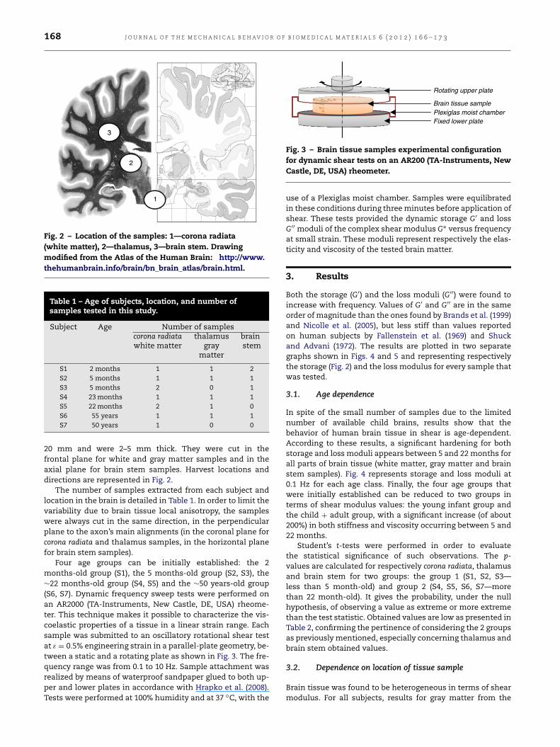
168 J O U R N A L O F T H E M E C H A N I C A L B E H AV I O R O F B I O M E D I C A L M A T E R I A L S 6 ( 2 0 1 2 ) 1 6 6 – 1 7 3
Fig. 2 – Location of the samples: 1—corona radiata(white matter), 2—thalamus, 3—brain stem. Drawingmodified from the Atlas of the Human Brain: http://www.thehumanbrain.info/brain/bn_brain_atlas/brain.html.
Table 1 – Age of subjects, location, and number ofsamples tested in this study.
Subject Age Number of samplescorona radiatawhite matter
thalamusgray
matter
brainstem
S1 2 months 1 1 2S2 5 months 1 1 1S3 5 months 2 0 1S4 23months 1 1 1S5 22months 2 1 0S6 55 years 1 1 1S7 50 years 1 0 0
20 mm and were 2–5 mm thick. They were cut in thefrontal plane for white and gray matter samples and in theaxial plane for brain stem samples. Harvest locations anddirections are represented in Fig. 2.
The number of samples extracted from each subject andlocation in the brain is detailed in Table 1. In order to limit thevariability due to brain tissue local anisotropy, the sampleswere always cut in the same direction, in the perpendicularplane to the axon’s main alignments (in the coronal plane forcorona radiata and thalamus samples, in the horizontal planefor brain stem samples).
Four age groups can be initially established: the 2months-old group (S1), the 5 months-old group (S2, S3), the∼22 months-old group (S4, S5) and the ∼50 years-old group(S6, S7). Dynamic frequency sweep tests were performed onan AR2000 (TA-Instruments, New Castle, DE, USA) rheome-ter. This technique makes it possible to characterize the vis-coelastic properties of a tissue in a linear strain range. Eachsample was submitted to an oscillatory rotational shear testat ε = 0.5% engineering strain in a parallel-plate geometry, be-tween a static and a rotating plate as shown in Fig. 3. The fre-quency range was from 0.1 to 10 Hz. Sample attachment wasrealized by means of waterproof sandpaper glued to both up-per and lower plates in accordance with Hrapko et al. (2008).Tests were performed at 100% humidity and at 37 ◦C, with the
Fig. 3 – Brain tissue samples experimental configurationfor dynamic shear tests on an AR200 (TA-Instruments, NewCastle, DE, USA) rheometer.
use of a Plexiglas moist chamber. Samples were equilibratedin these conditions during threeminutes before application ofshear. These tests provided the dynamic storage G′ and lossG′′ moduli of the complex shear modulus G∗ versus frequencyat small strain. These moduli represent respectively the elas-ticity and viscosity of the tested brain matter.
3. Results
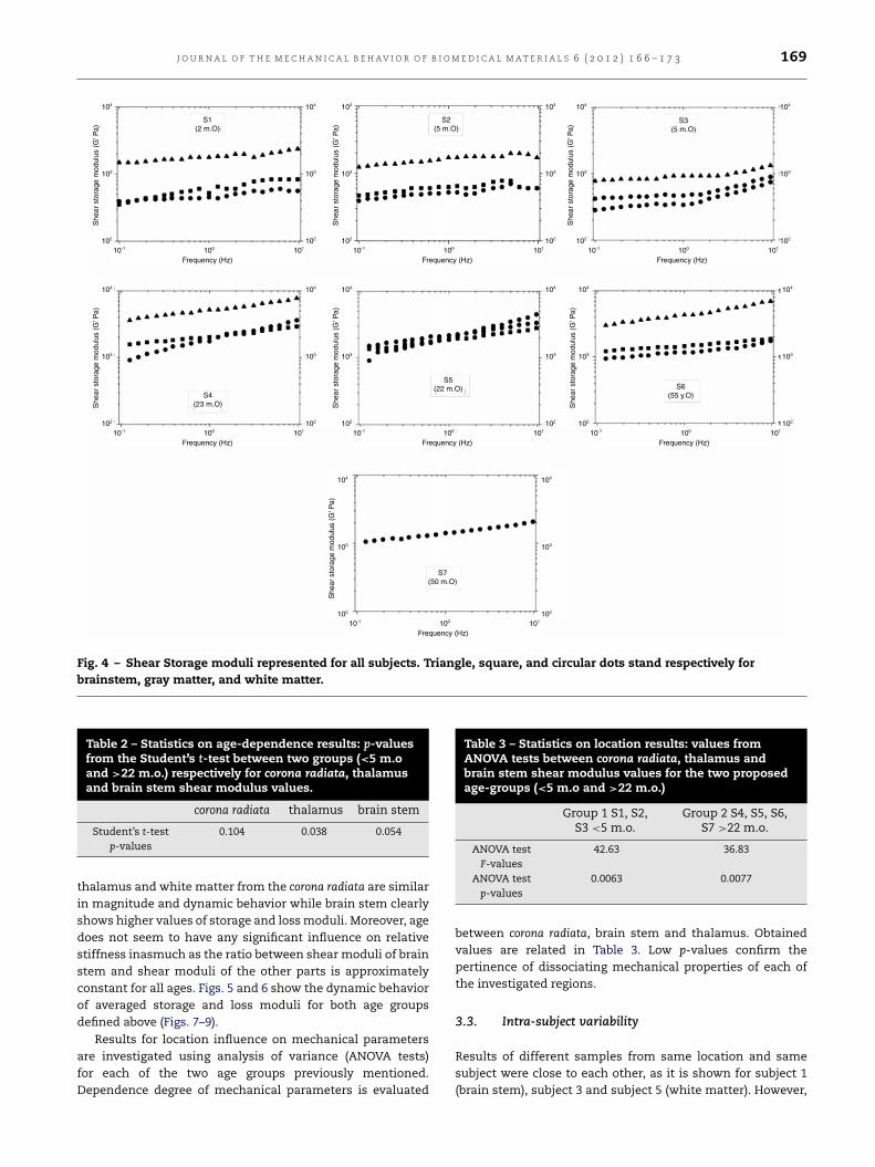
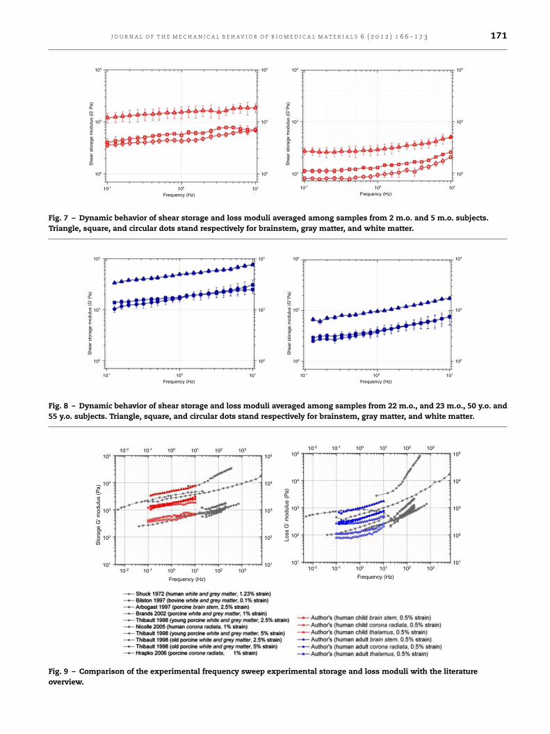
Both the storage (G′) and the loss moduli (G′′) were found toincrease with frequency. Values of G′ and G′′ are in the sameorder of magnitude than the ones found by Brands et al. (1999)and Nicolle et al. (2005), but less stiff than values reportedon human subjects by Fallenstein et al. (1969) and Shuckand Advani (1972). The results are plotted in two separategraphs shown in Figs. 4 and 5 and representing respectivelythe storage (Fig. 2) and the loss modulus for every sample thatwas tested.
3.1. Age dependence
In spite of the small number of samples due to the limitednumber of available child brains, results show that thebehavior of human brain tissue in shear is age-dependent.According to these results, a significant hardening for bothstorage and loss moduli appears between 5 and 22months forall parts of brain tissue (white matter, gray matter and brainstem samples). Fig. 4 represents storage and loss moduli at0.1 Hz for each age class. Finally, the four age groups thatwere initially established can be reduced to two groups interms of shear modulus values: the young infant group andthe child + adult group, with a significant increase (of about200%) in both stiffness and viscosity occurring between 5 and22 months.
Student’s t-tests were performed in order to evaluatethe statistical significance of such observations. The p-values are calculated for respectively corona radiata, thalamusand brain stem for two groups: the group 1 (S1, S2, S3—less than 5 month-old) and group 2 (S4, S5, S6, S7—morethan 22 month-old). It gives the probability, under the nullhypothesis, of observing a value as extreme or more extremethan the test statistic. Obtained values are low as presented inTable 2, confirming the pertinence of considering the 2 groupsas previouslymentioned, especially concerning thalamus andbrain stem obtained values.
3.2. Dependence on location of tissue sample
Brain tissue was found to be heterogeneous in terms of shearmodulus. For all subjects, results for gray matter from the
J O U R N A L O F T H E M E C H A N I C A L B E H AV I O R O F B I O M E D I C A L M A T E R I A L S 6 ( 2 0 1 2 ) 1 6 6 – 1 7 3 169
Fig. 4 – Shear Storage moduli represented for all subjects. Triangle, square, and circular dots stand respectively forbrainstem, gray matter, and white matter.
Table 2 – Statistics on age-dependence results: p-valuesfrom the Student’s t-test between two groups (<5 m.oand >22 m.o.) respectively for corona radiata, thalamusand brain stem shear modulus values.
corona radiata thalamus brain stem
Student’s t-testp-values
0.104 0.038 0.054
thalamus and white matter from the corona radiata are similarin magnitude and dynamic behavior while brain stem clearlyshows higher values of storage and lossmoduli. Moreover, agedoes not seem to have any significant influence on relativestiffness inasmuch as the ratio between shear moduli of brainstem and shear moduli of the other parts is approximatelyconstant for all ages. Figs. 5 and 6 show the dynamic behaviorof averaged storage and loss moduli for both age groupsdefined above (Figs. 7–9).
Results for location influence on mechanical parametersare investigated using analysis of variance (ANOVA tests)for each of the two age groups previously mentioned.Dependence degree of mechanical parameters is evaluated
Table 3 – Statistics on location results: values fromANOVA tests between corona radiata, thalamus andbrain stem shear modulus values for the two proposedage-groups (<5 m.o and >22 m.o.)
Group 1 S1, S2,S3 <5 m.o.
Group 2 S4, S5, S6,S7 >22 m.o.
ANOVA testF-values
42.63 36.83
ANOVA testp-values
0.0063 0.0077
between corona radiata, brain stem and thalamus. Obtainedvalues are related in Table 3. Low p-values confirm thepertinence of dissociating mechanical properties of each ofthe investigated regions.
3.3. Intra-subject variability
Results of different samples from same location and samesubject were close to each other, as it is shown for subject 1(brain stem), subject 3 and subject 5 (white matter). However,

170 J O U R N A L O F T H E M E C H A N I C A L B E H AV I O R O F B I O M E D I C A L M A T E R I A L S 6 ( 2 0 1 2 ) 1 6 6 – 1 7 3
Fig. 5 – Shear Loss moduli represented for all subjects. Triangle, square, and circular dots stand respectively for brainstem,gray matter, and white matter.
Fig. 6 – Averaged values of shear storage (G′) and loss (G′′) moduli at f = 0.1 Hz for the four different initial age groups.Error bars stand for calculated standard deviations. White, gray and dark bars stand for white matter, gray matter, andbrain stem, respectively.
due to the limited number of samples no further conclusionsare possible at this level. This result consolidates assumptionthat the intra subject local variability of the shear modulusis not significant as long as samples are excised in the samedirection according to Nicolle et al. (2005).
4. Discussion
This study aimed at analyzing the dependence of ageon the mechanical properties of human brain tissue. Thedifficulty in obtaining human samples partially explains the
J O U R N A L O F T H E M E C H A N I C A L B E H AV I O R O F B I O M E D I C A L M A T E R I A L S 6 ( 2 0 1 2 ) 1 6 6 – 1 7 3 171
Fig. 7 – Dynamic behavior of shear storage and loss moduli averaged among samples from 2 m.o. and 5 m.o. subjects.Triangle, square, and circular dots stand respectively for brainstem, gray matter, and white matter.
Fig. 8 – Dynamic behavior of shear storage and loss moduli averaged among samples from 22 m.o., and 23 m.o., 50 y.o. and55 y.o. subjects. Triangle, square, and circular dots stand respectively for brainstem, gray matter, and white matter.
Fig. 9 – Comparison of the experimental frequency sweep experimental storage and loss moduli with the literatureoverview.

172 J O U R N A L O F T H E M E C H A N I C A L B E H AV I O R O F B I O M E D I C A L M A T E R I A L S 6 ( 2 0 1 2 ) 1 6 6 – 1 7 3
Table 4 – Mean stiffness ratio of the storage, loss and complex shear moduli between youngest and old brain tissuesamples for the 3 considered locations.
Stiffness ratio brain stem corona radiata white matter thalamus gray matter
G′
(2 and 5 month-old)/G′
(2 and 50 year-old)3.25 3.70 3.08
G′′
(2 and 5 month-old)/G′′
(2 and 50 year-old)3.28 4.06 3.05
G(2 and 5 month-old)/G(2 and 50 year-old) 3.25 3.71 3.08
paucity of data in the literature concerning human brainmechanical properties when compared with other species.Similar restrictions are observed in the present study due tothe limited number of available brain samples. Experimentson human subjects involve considerable drawbacks. Forexample, there is a significant disparity in post-mortem timebetween different subjects, resulting in various possiblebrain matter degradation states. This phenomenon leads toadditional uncertainty in mechanical parameters, accordingto Metz and Weber (1982). Experiments can also frequently berestricted to small strain tests in order to avoid destructionof the sample. Changes in viscoelastic properties of the brainduring early maturation have only been studied on pigs andon rats (Thibault and Margulies, 1998; Prange and Margulies,2002; Gefen et al., 2003). Although age-equivalence betweenthese species and humans can be assumed on the basisof existing similarities of brain composition changes duringmaturation, the experiments reported in the present studyare essential in order to determine whether or not thesechanges in composition correspond with the alteration of itsmechanical properties.
A significant difference seems to appear between the(2 month-old, 5 month-old) group and the (2 year-old, 50 year-old) group. The adult brain tissue seems to be 3–4 times stifferthan the immature young child one, as presented in Table 4.It is possible therefore to conclude from our preliminaryresults that a significant increase in viscoelastic values ofhuman brain tissue occurs between 5 and 22 months. Thedifference between the less than 5 month-old and more than22 month-old group was found to be statistically significantfor the three locations, namely white matter, grey matterand brain stem. This assertion is in good agreement with thealteration concerning biological constituents as it is reportedby Thibault and Margulies (1998) from Dobbing (1981). Inparticular, it appears that water content seems to reach adefinitive plateau around the age of 1 year. This significantchange in material properties is also in good agreement withthe results of Thibault and Margulies (1998) on pigs, showingthat at small strains, 2–3 day old pig brains (equivalent toa 1 m.o. human if age correspondence is assumed) are lessstiff than one year old pig brains (equivalent to more than a4 year-old human). A more accurate comprehension of thisdramatic increase in brain stiffness would require additionalexperimental data at the age region of alteration, i.e. aroundthe age of one year for human species.
Effects of heterogeneity of brain tissue on its materialproperties were also investigated in this study. Brain stemwasfound to be stiffer (approximately 2–3 times) than gray andwhite matter. Previous studies also report higher values ofelasticity moduli of brainstem compared with other cerebraltissue locations under same testing conditions (Arbogastand Margulies, 1997, 1998). As a consequence, numerical
models of the head should clearly distinguish brain stemfrom the other parts of the brain in terms of mechanicalproperties. It must be mentioned though that the nonnegligible effects of structural anisotropy of brain tissue onits material properties, according to Arbogast and Margulies(1998), were not examined in this study since all samplesbelonging to the same region were excised in the samedirection. Another main limitation of this work is the fact thatrheometric experiments were performed only at small strainvalues (0.5%), whereas the influence of strain magnitudeon age-dependent mechanical behavior of brain tissue hasbeen previously demonstrated on animal brain. Thibault andMargulies (1998) reported an increase in shear moduli duringmaturation at small strains (2.5%) but no significant change atlarge strains (5%), and Prange and Margulies (2002) observedthat immature pig brain tissue was stiffer than adult pigat larger strains (50%). Consequently, it seems necessary toinvestigate further nonlinear mechanical properties of braintissue and their dependence on age.
5. Conclusion
This preliminary study provides first dynamic viscoelasticdata of human brain tissue at different ages, based on afirst set of samples due to the limited number of availablechild brains. It is shown that at small strains, human braintissue seems to undergo a significant increase in stiffness(of about 200%) between 5 and 22 months. Brain stem isalso found to be significantly stiffer than gray matter andwhite matter. Further research should investigate a highernumber of samples with a focus on the 1 year old childbrain. Only then it will be possible to implement realistic childmechanical properties into child head finite element models.
R E F E R E N C E S
Arbogast, K.B., Margulies, S.S., 1997. Regional differences inmechanical properties of the central nervous system. In: StappCar Crash Conference Proceedings. pp. 293–300.
Arbogast, K.B., Margulies, S.S., 1998. Material characterizationof the brainstem from oscillatory shear tests. Journal ofBiomechanics 31, 801–807.
Bilston, L.E., Liu, Z., Phan-Thien, N., 1997. Linear viscoelasticproperties of bovine brain tissue in shear. Biorheology 34,377–385.
Brands, D.W.A., Bovendeerd, P.H.M., Peters, G.W.M., Wism0ans,J.S.H.M., Paas, M.H.J.W., Van Bree, J.L.M.J., 1999. Comparisonof the dynamic behaviour of brain tissue and two modelmaterials. In: Stapp Car Crash Conference Proceedings,pp. 313–320.
Bruns Jr., J., Hauser, A., 2003. The epidemiology of traumatic braininjury: a review. Epilepsia 44, 2–10.

J O U R N A L O F T H E M E C H A N I C A L B E H AV I O R O F B I O M E D I C A L M A T E R I A L S 6 ( 2 0 1 2 ) 1 6 6 – 1 7 3 173
Coats, B., Margulies, S.S., 2006. Material properties of porcineparietal cortex. Journal of Biomechanics 39, 2521–2525.
Coats, B., Margulies, S.S., Ji, S., 2007. Parametric study ofhead impact in the infant. Stapp Car Crash Journal 51,1–15.
Darvish, K.K., Crandall, J.R., 2001. Non linear viscoelastic effectsin oscillatory shear deformation of brain tissue. MedicalEngineering and Physics 23 (9), 633–645.
De Santis Klinich, K.D., Hulbert, G.M., 2002. Estimating infant headinjury criteria and impact response using crash reconstructionand finite element modelling. Stapp Car Crash Journal 46,165–194.
Dobbing, J., 1981. The later development of the brain andits vulnerability. In: Davis, J.A., Dobbing, J. (Eds.), ScientificFoundations of Paediatrics. Heinemann Medical, London.
Durkin, M.S., Olsen, S., Barlow, B., Virella, A., Connolly Jr.,E.S., 1998. The epidemiology of urban pediatric neurologicaltrauma: evaluation of, and implications for, injury preventionprograms. Neurosurgery 42, 300–310.
Fallenstein, G.T., Hulce, V.D., Melvin, J.W., 1969. Dynamicmechanical properties of human brain tissue. Journal ofBiomechanics 2, 217–226.
Franceschini, G., Bigoni, D., Regitnig, P., Holzapfel, G.A., 2006.Brain tissue deforms similarly to filled elastomers and followsconsolidation theory. Journal of the Mechanics and Physics ofSolids 54, 2592–2620.
Gefen, A., Gefen, N., Zhu, Q., Raghupathi, R., Margulies, S.S.,2003. Age-dependent changes in material properties of thebrain and braincase of the rat. Journal of Neurotrauma 20,1163–1177.
Hrapko, M., Van Dommelen, J.A.W., Peters, G.W.M., Wismans,J.S.H.M., 2006. The mechanical behaviour of brain tissue: largestrain response and constitutive modelling. Biorheology 43,623–636.
Hrapko, M., van Dommelen, J.A.W., Peters, G.W.M., Wismans,J.S.H.M., 2008. The influence of test conditions on characteri-zation of the mechanical properties of brain tissue. Journal ofBiomechanical Engineering 130 (3), 1–10.
Kraus, J.F., Rock, A., Hemyari, P., 1990. Brain injuries amonginfants, children, adolescents, and young adults. AmericanJournal of Diseases of Children 144, 684–691;Miller, K., Chinzei, K., 2002. Mechanical properties of braintissue in tension. Journal of Biomechanics 35, 483–490.
Metz, H., Weber, D., 1982. Interpretations of impact responses ofa 3-year-old child dummy relative to child injury potential. In:Proc. of the 9th Int. Conf. on Experimental Safety Vehicles.
Miller, K., Chinzei, K., 2002. Mechanical properties of brain tissuein tension. Journal of Biomechanics 35, 483–490.
Nicolle, S., Lounis, M., Willinger, R., Palierne, J.-F., 2005. Shearlinear behavior of brain tissue over a large frequency range.Biorheology 42, 209–223.
Peters, G.W.M., Meulman, J.H., Sauren, A.A.H.J., 1997. Theapplicability of the time/temperature superposition principleto the brain tissue. Biorheology 34 (2), 127–138.
Prange, M.T., Margulies, S.S., 2002. Regional, directional, andage-dependent properties of the brain undergoing largedeformation. Journal of Biomechanical Engineering 124,244–252.
Roth, S., Raul, J.-S., Ludes, B., Willinger, R., 2007. Finiteelement analysis of impact and shaking inflicted to a child.International Journal of Legal Medicine 121, 223–228.
Roth, S., Raul, J.-S., Willinger, R., 2010. Finite element modeling ofpediatric head impact: global validation against experimentaldata. Computer Methods and Programs in Biomedicine 99 (1),25–33.
Rutland-Brown, W., Langlois, J.A., Thomas, K.E., Xi, Y.L., 2006.Incidence of traumatic brain injury in the United States, 2003.Journal of Head Trauma Rehabilitation 21, 544–548.
Sack, I., Beierbach, B., Wuerfel, J., Klatt, D., Hamhaber, U.,Papazoglou, S., Martus, P., Braun, J., 2009. The impact of ageingand gender on brain viscoelasticity. NeuroImage 46, 652–657.
Shuck, L., Advani, S., 1972. Rheological response of human braintissue in shear. Journal of Basic Engineering 94, 905–911.
Thibault, K.L., Margulies, S.S., 1998. Age-dependent materialproperties of the porcine cerebrum: effect on pediatric inertialhead injury criteria. Journal of Biomechanics 31, 1119–1126.
Tsai, W.-C., Chiu, W.-T., Chiou, H.-Y., Choy, C.-S., Hung, C.-C., Tsai,S.-H., 2004. Pediatric traumatic brain injuries in Taiwan: an 8-year study. Journal of Clinical Neuroscience 11, 126–129.
Velardi, F., Fraternali, F., Angelillo, M., 2006. Anisotropicconstitutive equations and experimental tensile behavior ofbrain tissue. Biomechanics and Modeling in Mechanobiology5, 53–61.
Viano, D., von Holst, H., Gordon, E., 1997. Serious brain injuryfrom traffic related causes: priorities for primary prevention.Accident Analysis & Prevention 29, 811–816.
Related Documents