http://informahealthcare.com/nan ISSN: 1743-5390 (print), 1743-5404 (electronic) Nanotoxicology, 2015; 9(S1): 118–132 ! 2015 Informa UK Ltd. DOI: 10.3109/17435390.2014.991431 REVIEW ARTICLE Towards an alternative testing strategy for nanomaterials used in nanomedicine: Lessons from NanoTEST M. Dusinska 1 , S. Boland 2 , M. Saunders 3 , L. Juillerat-Jeanneret 4 , L. Tran 5 , G. Pojana 6,7 , A. Marcomini 7 , K. Volkovova 8 , J. Tulinska 8 , L. E. Knudsen 9 , L. Gombau 10 , M. Whelan 11 , A. R. Collins 12 , F. Marano 2 , C. Housiadas 13 , D. Bilanicova 6,7 , B. Halamoda Kenzaoui 4,11 , S. Correia Carreira 14 , Z. Magdolenova 1 , L. M. Fjellsbø 1 , A. Huk 1 , R. Handy 15 , L. Walker 16 , M. Barancokova 8 , A. Bartonova 1 , E. Burello 11,17 , J. Castell 10 , H. Cowie 5 , M. Drlickova 8,18 , R. Guadagnini 2 , G. Harris 11 , M. Harju 1 , E. S. Heimstad 1 , M. Hurbankova 8 , A. Kazimirova 8 , Z. Kovacikova 8 , M. Kuricova 8 , A. Liskova 8 , A. Milcamps 11 , E. Neubauerova 8 , T. Palosaari 11 , P. Papazafiri 19 , M. Pilou 14 , M. S. Poulsen 9 , B. Ross 5 , E. Runden-Pran 1 , K. Sebekova 20 , M. Staruchova 8 , D. Vallotto 6,7 , and A. Worth 11 1 Health Effects Laboratory-MILK, NILU – Norwegian Institute for Air Research, Kjeller, Norway, 2 Unit of Functional and Adaptive Biology (BFA), Laboratory of Molecular and Cellular Responses to Xenobiotics (RMCX)), Univ Paris Diderot, Sorbonne Paris Cite ´, UMR 8251 CNRS, Paris, France, 3 Department of Medical Physics & Bioengineering, BIRCH, Bioengineering, Innovation & Research Hub, St. Michael’s Hospital, University Hospitals Bristol NHS Foundation Trust, Bristol, United Kingdom, 4 University Institute of Pathology, Lausanne, Switzerland, 5 Institute of Occupational Medicine, Riccarton, Edinburgh, UK, 6 DFBC – Department of Philosophy and Cultural Heritage, University Ca’ Foscari Venice, Venice, Italy, 7 DAIS – Department of Environmental Sciences, Informatics and statistics, University Ca’ Foscari Venice, Venice, Italy, 8 Faculty of Medicine, Slovak Medical University, Bratislava, Slovakia, 9 Faculty of Health and Medicinal Sciences, Institute of Public Health, University of Copenhagen, Copenhagen, Denmark, 10 Leitat Technological Center, Scientific Park, Barcelona, Spain, 11 Institute for Health and Consumer Protection, European Commission Joint Research Centre, Ispra (VA), Italy, 12 Department of Nutrition, University of Oslo, Oslo, Norway, 13 Thermal Hydraulics and Multiphase Flows Laboratory, Institute of Nuclear & Radiological Sciences & Technology, Energy & Safety, NCSR ‘‘Demokritos’’, Agia Paraskevi, Greece, 14 Bristol Centre for Functional Nanomaterials, University of Bristol, Bristol, UK, 15 School of Biomedical and Biological Sciences, Plymouth University, Plymouth, UK, 16 Bristol Heart Institute, School of Clinical Sciences, University of Bristol, Bristol, UK, 17 Computational Chemistry Group, RAPID Department (Risk Analysis of Products in Development), TNO, Zeist, The Netherlands, 18 Centre for Chemical Substances and Preparations, Bratislava, Slovakia, 19 Department of Biology, University of Athens, University Campus, Athens, Greece, and 20 Medical Faculty, Institute of Molecular Biomedicine, Comenius University, Bratislava, Slovakia Abstract In spite of recent advances in describing the health outcomes of exposure to nanoparticles (NPs), it still remains unclear how exactly NPs interact with their cellular targets. Size, surface, mass, geometry, and composition may all play a beneficial role as well as causing toxicity. Concerns of scientists, politicians and the public about potential health hazards associated with NPs need to be answered. With the variety of exposure routes available, there is potential for NPs to reach every organ in the body but we know little about the impact this might have. The main objective of the FP7 NanoTEST project (www.nanotest-fp7.eu) was a better understanding of mechanisms of interactions of NPs employed in nanomedicine with cells, tissues and organs and to address critical issues relating to toxicity testing especially with respect to alternatives to tests on animals. Here we describe an approach towards alternative testing strategies for hazard and risk assessment of nanomaterials, highlighting the adaptation of standard methods demanded by the special physicochemical features of nanomaterials and bioavailability studies. The work has assessed a broad range of toxicity tests, cell models and NP types and concentrations taking into account the inherent impact of NP properties and the effects of changes in experimental conditions using well-characterized NPs. The results of the studies have been used to generate recommendations for a suitable and robust testing strategy which can be applied to new medical NPs as they are developed. Abbreviations: AFM: atomic force microscopy; BBB: blood–brain barrier; BET: Brunauer– Emmett–Teller; BSA: bovine serum albumin; CBMN: cytokinesis-block micronucleus; CS: calf serum; CNS: central nervous system; DCFH-DA: 2,7-dichlorodihydro-fluorescein diacetate; DLS: dynamic light scattering; DNA: deoxyribonucleic acid; ELISA: enzyme-linked immunosorbent assay; EDX/EDS: energy-dispersive X-ray spectroscopy; FBS: fetal bovine serum; Fl-25 SiO 2 : fluorescent 25 nm silica; GM-CSF: granulocyte macrophage colony-stimulating factor; H 2 AX: H2A histone family member X; HE: hydroethidine; HTS: high-throughput screening; IL: Keywords Hazard assessment, in vitro, nanoparticles, NanoTEST, testing strategy History Received 14 August 2014 Revised 2 November 2014 Accepted 19 November 2014 Published online 29 April 2015 Correspondence: Dr Maria Dusinska, Health Effects Laboratory, Norwegian Institute for Air Research (NILU), Instituttveien 18, 2007 Kjeller, Norway. Tel: +4763898157. E-mail: [email protected] Nanotoxicology Downloaded from informahealthcare.com by 95.102.179.39 on 04/29/15 For personal use only.

Welcome message from author
This document is posted to help you gain knowledge. Please leave a comment to let me know what you think about it! Share it to your friends and learn new things together.
Transcript

http://informahealthcare.com/nanISSN: 1743-5390 (print), 1743-5404 (electronic)
Nanotoxicology, 2015; 9(S1): 118–132! 2015 Informa UK Ltd. DOI: 10.3109/17435390.2014.991431
REVIEW ARTICLE
Towards an alternative testing strategy for nanomaterials used innanomedicine: Lessons from NanoTEST
M. Dusinska1, S. Boland2, M. Saunders3, L. Juillerat-Jeanneret4, L. Tran5, G. Pojana6,7, A. Marcomini7, K. Volkovova8,J. Tulinska8, L. E. Knudsen9, L. Gombau10, M. Whelan11, A. R. Collins12, F. Marano2, C. Housiadas13, D. Bilanicova6,7,B. Halamoda Kenzaoui4,11, S. Correia Carreira14, Z. Magdolenova1, L. M. Fjellsbø1, A. Huk1, R. Handy15, L. Walker16,M. Barancokova8, A. Bartonova1, E. Burello11,17, J. Castell10, H. Cowie5, M. Drlickova8,18, R. Guadagnini2, G. Harris11,M. Harju1, E. S. Heimstad1, M. Hurbankova8, A. Kazimirova8, Z. Kovacikova8, M. Kuricova8, A. Liskova8, A. Milcamps11,E. Neubauerova8, T. Palosaari11, P. Papazafiri19, M. Pilou14, M. S. Poulsen9, B. Ross5, E. Runden-Pran1, K. Sebekova20,M. Staruchova8, D. Vallotto6,7, and A. Worth11
1Health Effects Laboratory-MILK, NILU – Norwegian Institute for Air Research, Kjeller, Norway, 2Unit of Functional and Adaptive Biology (BFA),
Laboratory of Molecular and Cellular Responses to Xenobiotics (RMCX)), Univ Paris Diderot, Sorbonne Paris Cite, UMR 8251 CNRS, Paris, France,3Department of Medical Physics & Bioengineering, BIRCH, Bioengineering, Innovation & Research Hub, St. Michael’s Hospital, University Hospitals
Bristol NHS Foundation Trust, Bristol, United Kingdom, 4University Institute of Pathology, Lausanne, Switzerland, 5Institute of Occupational
Medicine, Riccarton, Edinburgh, UK, 6DFBC – Department of Philosophy and Cultural Heritage, University Ca’ Foscari Venice, Venice, Italy, 7DAIS –
Department of Environmental Sciences, Informatics and statistics, University Ca’ Foscari Venice, Venice, Italy, 8Faculty of Medicine, Slovak Medical
University, Bratislava, Slovakia, 9Faculty of Health and Medicinal Sciences, Institute of Public Health, University of Copenhagen, Copenhagen,
Denmark, 10Leitat Technological Center, Scientific Park, Barcelona, Spain, 11Institute for Health and Consumer Protection, European Commission
Joint Research Centre, Ispra (VA), Italy, 12Department of Nutrition, University of Oslo, Oslo, Norway, 13Thermal Hydraulics and Multiphase Flows
Laboratory, Institute of Nuclear & Radiological Sciences & Technology, Energy & Safety, NCSR ‘‘Demokritos’’, Agia Paraskevi, Greece, 14Bristol Centre
for Functional Nanomaterials, University of Bristol, Bristol, UK, 15School of Biomedical and Biological Sciences, Plymouth University, Plymouth, UK,16Bristol Heart Institute, School of Clinical Sciences, University of Bristol, Bristol, UK, 17Computational Chemistry Group, RAPID Department
(Risk Analysis of Products in Development), TNO, Zeist, The Netherlands, 18Centre for Chemical Substances and Preparations, Bratislava, Slovakia,19Department of Biology, University of Athens, University Campus, Athens, Greece, and 20Medical Faculty, Institute of Molecular Biomedicine,
Comenius University, Bratislava, Slovakia
Abstract
In spite of recent advances in describing the health outcomes of exposure to nanoparticles(NPs), it still remains unclear how exactly NPs interact with their cellular targets. Size, surface,mass, geometry, and composition may all play a beneficial role as well as causing toxicity.Concerns of scientists, politicians and the public about potential health hazards associated withNPs need to be answered. With the variety of exposure routes available, there is potential forNPs to reach every organ in the body but we know little about the impact this might have. Themain objective of the FP7 NanoTEST project (www.nanotest-fp7.eu) was a better understandingof mechanisms of interactions of NPs employed in nanomedicine with cells, tissues and organsand to address critical issues relating to toxicity testing especially with respect to alternatives totests on animals. Here we describe an approach towards alternative testing strategies forhazard and risk assessment of nanomaterials, highlighting the adaptation of standard methodsdemanded by the special physicochemical features of nanomaterials and bioavailability studies.The work has assessed a broad range of toxicity tests, cell models and NP types andconcentrations taking into account the inherent impact of NP properties and the effects ofchanges in experimental conditions using well-characterized NPs. The results of the studieshave been used to generate recommendations for a suitable and robust testing strategy whichcan be applied to new medical NPs as they are developed.
Abbreviations: AFM: atomic force microscopy; BBB: blood–brain barrier; BET: Brunauer–Emmett–Teller; BSA: bovine serum albumin; CBMN: cytokinesis-block micronucleus; CS: calfserum; CNS: central nervous system; DCFH-DA: 2,7-dichlorodihydro-fluorescein diacetate; DLS:dynamic light scattering; DNA: deoxyribonucleic acid; ELISA: enzyme-linked immunosorbentassay; EDX/EDS: energy-dispersive X-ray spectroscopy; FBS: fetal bovine serum; Fl-25 SiO2:fluorescent 25 nm silica; GM-CSF: granulocyte macrophage colony-stimulating factor; H2AX:H2A histone family member X; HE: hydroethidine; HTS: high-throughput screening; IL:
Keywords
Hazard assessment, in vitro, nanoparticles,NanoTEST, testing strategy
History
Received 14 August 2014Revised 2 November 2014Accepted 19 November 2014Published online 29 April 2015
Correspondence: Dr Maria Dusinska, Health Effects Laboratory, Norwegian Institute for Air Research (NILU), Instituttveien 18, 2007 Kjeller, Norway.Tel: +4763898157. E-mail: [email protected]
Nan
otox
icol
ogy
Dow
nloa
ded
from
info
rmah
ealth
care
.com
by
95.1
02.1
79.3
9 on
04/
29/1
5Fo
r pe
rson
al u
se o
nly.

interleukin; LDH: lactate dehydrogenase; LTT: lymphocyte transformation test; mBBr:monobromobimane; MTT: 3-(4,5-dimethyl-thiazol-2-yl)-2,5-diphenyl-tetrazolium bromide;NaFlu: sodium fluorescein; NP: nanoparticle; NTA: nanoparticle tracking analysis; OC-Fe3O4:Na-oleate-coated iron oxide; PBMC: peripheral blood mononuclear cells; PBPK: physiologicallybased pharmacokinetic; PI: propidium iodide; PLGA-PEO: polylactic-co-glycolic acid-Poly polyethylene oxide; (Q)SAR: quantitative structure–activity relationship; ROS: reactive oxygenspecies; RTqPCR: quantitative real time RT-PCR; SANS: small angle neutron scattering; SB: strandbreaks; SD: stock dispersion; SEM: scanning electron microscopy; SLS: static light scattering;SOPs: standard operating procedures; SP-ICP-MS: single particle inductively coupled plasma-mass spectrometry; TEM: transmission electron microscopy; U-Fe3O4: uncoated iron oxide;WST1: 2-(4 -iodophenyl)-3-(4-nitrophenyl)-5-(2,4-disulphophenyl)-2H-tetrazolium;
Introduction
The rapid and enormous development of nanotechnology has beenaccompanied by a deep concern about the effects that nanopar-ticles (NPs) may have on human health and the environment.However, the knowledge gaps in our understanding of thebehaviour of NPs, their transformation and fate in differentenvironments including biological systems, make it difficult toevaluate their toxic effects and to perform adequate hazard andrisk assessment. Selection of the best endpoints and methods inappropriate cell models and adaptation, standardization andvalidation of methods are still needed.
NPs can potentially enter the human body through a range ofexposure routes (Elsaesser & Howard 2012; Hagens et al., 2007)including intravenous injection, inhalation and ingestion via thedigestive tract. They can then translocate to the blood from wherethey can reach most organs and possibly accumulate, before beingeliminated through processes that are not yet clearly understood(Oberdorster et al., 2005).
In the respiratory tract, NPs interact with bronchial andalveolar epithelial cells, inducing cell activation and reaction.Very small NPs may translocate through the lung epithelium andthe endothelium of the blood vessels into the blood and lymphcirculation. Similar mechanical properties as in the respiratorytract can be expected in the digestive tract cells in which NPs caninduce cell activation and tissue reaction, oxidative stress and lossof the barrier functions of the epithelium. In the blood, NPs caninteract with circulating cells, inducing cell activation, increasedadhesion of the NPs to each other or endothelial cells. Interactionwith cells of the vascular wall can induce vascular reaction,activation and vascular leakage, and uptake of NPs by endothelialcells and perivascular cells. The liver is the major site forbiotransformation and defence against foreign materials and
xenobiotics, and this is very likely also true for NPs, possiblyinducing hepatocyte and/or sinusoidal endothelial and Kupffercell activation. The kidney transports and excretes NPs from theblood to the urine, or reabsorbs them from urine. The centralnervous system (CNS) is separated from the blood by the blood–brain barrier (BBB), represented by a very specialized vascularsystem consisting of endothelial cells, pericytes and astrocytes,but limiting access to the brain. NPs may induce the activation ofbrain endothelial, astroglial and microglial cells. The placenta is abiological barrier of particular interest in relation to the sensitivenature of the foetus and NPs may induce placental inflammationassociated with foetal defects. Representative cells and cell linesoriginating from these organs were used to test NPs selected in theNanoTEST project (Tables 1 and 2) and to select the best in vitromodels to determine modes of action for hazard assessment(Juillerat-Jeanneret et al., 2015).
A strategy for in vitro toxicity testing in a regulatory contextrequires a battery of tests addressing different mechanisms andcovering all main important toxicity endpoints. Thus, to identifyrelevant short-term hazard models, we used several standardtoxicity assays for different markers such as cell viability, pro-inflammatory response, oxidative stress, genotoxicity, immuno-toxicity, cell uptake and transport. OECD recommended methodswere chosen where possible, such as in the case of genotoxicity(Magdolenova et al., 2012a,b) and when necessary methods wereadapted for NP testing (Guadagnini et al., 2015a) and fullydocumented in relevant publications or NanoTEST protocols(www.nanotest-fp7.eu). The specific focus of our biomarkervalidation strategy was to identify the most suitable conditions fordetecting a significant response and, by including relevantpositive and negative controls, to ensure that the method isreliable and gives reproducible results.
Table 1. Selected nanoparticles (NPs).
Name Abbreviation Source Comment
Nanomagnetite coated withNa-oleate
OC-Fe3O4 PlasmaChem (Hamburg, D)
Nanomagnetite uncoated U-Fe3O4 PlasmaChem (Hamburg, D)Polylactic-co-glycolic acid PLGA-PEO Advancell (Barcelona, E)Red-Fluorescent 25 nm
silicaFl-25 SiO2 Microspheres-nanospheres
Red-Fluorescent 50 nmsilica
Fl-50 SiO2 Microspheres-nanospheres
Nanosilica powder, 20 nm SiO2 NM-203 Reference Material, Joint Research Centre,(Ispra, Italy)
Nano-sized titanium diox-ide AeroxideP25; ana-tase/rutile powder of21 nm (nominal size)
TiO2 NM-105, P25-Degussa-Evonik (Essen, Germany),obtained from Joint Research Centre (Ispra, Italy)
Reference NPs, positive control
Dextran-coated ferumoxide Endorem Guerbet (Paris, F) Negative control, in clinics
DOI: 10.3109/17435390.2014.991431 An alternative testing strategy for nanomaterials 119
Nan
otox
icol
ogy
Dow
nloa
ded
from
info
rmah
ealth
care
.com
by
95.1
02.1
79.3
9 on
04/
29/1
5Fo
r pe
rson
al u
se o
nly.
To verify the suitability of in vitro models, in vivo studieswere carried out. A single i.v. administration of TiO2 orNa-oleate-coated iron oxide (OC-Fe3O4) NPs (0.1, 1 and 10%of LD50) to young female rats did not elicit overt acute orsubacute toxicity (Sebekova et al., 2014; Volkovova et al., 2015)but seemed to have an immunomodulatory effect. The in vitromodel of human peripheral blood cells generally reflected in vivoresponses of peripheral blood immune cells to TiO2 and OC-Fe3O4 NPs in exposed rats and proved the reliability of our panelof immune assays proposed as biomarkers for assessment ofimmunotoxicity in vitro (Tulinska et al. in preparation). There wasalso a good correlation in genotoxicity tests between in vitro/in vivo micronucleus tests and the comet assay for both TiO2 andOC-Fe3O4 NPs (Kazimirova et al., in preparation). In addition, insilico methods were considered and a new model for predictingthe oxidative stress potential of oxide NPs was proposed (Burello& Worth, 2011, 2015). The in vitro and in silico methodsdeveloped for NPs used in nanomedicine can also be utilised forthe assessment of health effects of NPs used and applied in otherareas and thus can have a wider impact on all 3 R’s (replacement,reduction and refinement of animals) for toxicity testing.
The overall aim of NanoTEST was to provide testing strategiesfor hazard identification and risk assessment of NPs, and topropose recommendations for evaluating potential risks asso-ciated with new medical NPs. The specific objective was todevelop a set of master standard operating procedures (SOPs) forat least two assays for each type of toxicity (including cellviability, pro-inflammatory response, oxidative stress, genotoxi-city, immunotoxicity, cell uptake and transport). The mostadvanced and standardised techniques would be adapted forautomation and prepared for validation.
The project addressed the factors responsible for variability inthe results of nanotoxicity studies – namely, the source and type ofNP, method of preparation or synthesis, stabilizers used, disper-sion method, state of agglomeration, presence of impurities;as well as variations in experimental conditions such as pH,temperature and sonication. The treatment regime is critical;results can depend on cell type used, exposure time, dose, the
assay method used, and possible interference with the detectionsystem (Dusinska et al., 2011, 2012, 2013; Guadagnini et al.,2015a, 2015b) as discussed below. The overall goal of the currentpaper is to summarise the artefacts and issues identified innanotoxicology, into a coherent set of tables and guidelines foruse by the research community in the design of testing strategiesand to suggest modifications of assays where appropriate.
Characterization of NPs
A recommended list of physico-chemical properties to becharacterized when testing specific manufactured nanomaterialsfor human health and environmental safety, has been proposed bythe OECD (Report no. 36ENV/JM/MONO(2012)40, 2012). Itincludes particle size distribution (in solid and in liquid media),shape, agglomeration/aggregation, water solubility/dispersability,as well as parameters occasionally measured such as octanol–water partition coefficient (where relevant), redox potential andradical formation potential. It is now clear that discrepanciesbetween reported toxicity results are caused not only partly bydifferent intrinsic properties, both physical (size, shape, etc.) andchemical (crystal structure, surface chemistry, etc.) of nominallysimilar, or identical, NPs, but also by the application of differenttesting conditions of NPs in physiological media, which couldaffect transport kinetics in the investigated fluids (Kato et al.,2009; Magdolenova et al., 2012a). Clearly, a testing strategy fornanomaterials needs to include a comprehensive characterization(Bouwmeester et al., 2011), including in particular a determin-ation of the main physical and chemical properties of NPs, and theproperties pertaining to NP behaviour in biological media used forevaluating toxicological effects. The most frequently employedtechniques are scanning and transmission electron microscopy(SEM and TEM, respectively), Brunauer–Emmett–Teller (BET),dynamic or static light scattering (DLS and SLS, respectively),NP tracking analysis (NTA) and small angle neutron scattering(SANS; Hassellov & Kaegi, 2009). No single technique couldadequately characterize a selected NP (Warheit, 2008); only aproper combination of various techniques is able to describe the
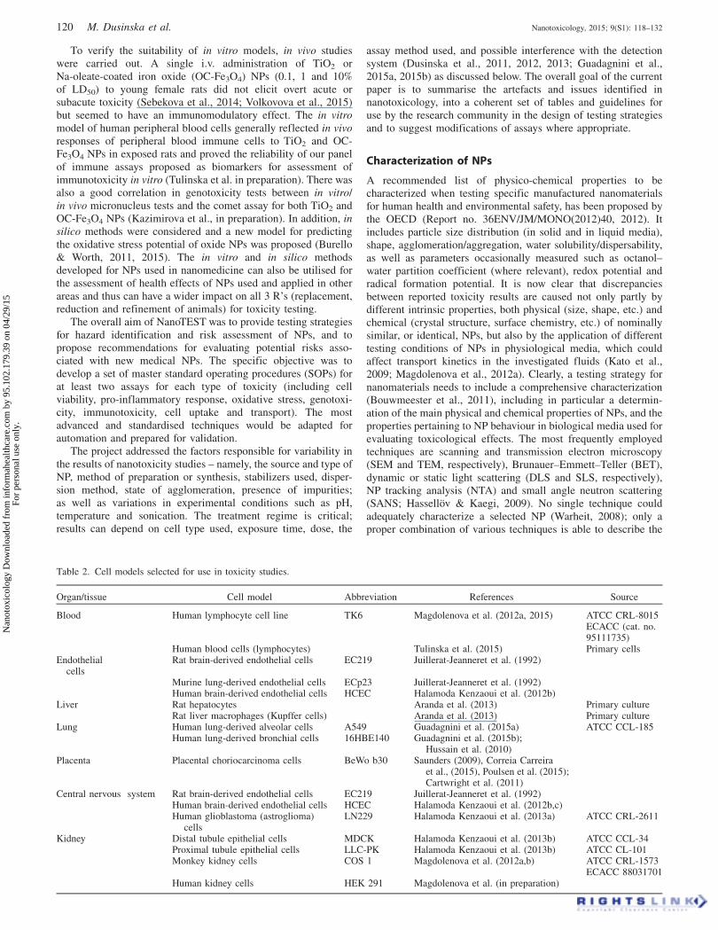
Table 2. Cell models selected for use in toxicity studies.
Organ/tissue Cell model Abbreviation References Source
Blood Human lymphocyte cell line TK6 Magdolenova et al. (2012a, 2015) ATCC CRL-8015ECACC (cat. no.95111735)
Human blood cells (lymphocytes) Tulinska et al. (2015) Primary cellsEndothelial
cellsRat brain-derived endothelial cells EC219 Juillerat-Jeanneret et al. (1992)
Murine lung-derived endothelial cells ECp23 Juillerat-Jeanneret et al. (1992)Human brain-derived endothelial cells HCEC Halamoda Kenzaoui et al. (2012b)
Liver Rat hepatocytes Aranda et al. (2013) Primary cultureRat liver macrophages (Kupffer cells) Aranda et al. (2013) Primary culture
Lung Human lung-derived alveolar cells A549 Guadagnini et al. (2015a) ATCC CCL-185Human lung-derived bronchial cells 16HBE140 Guadagnini et al. (2015b);
Hussain et al. (2010)Placenta Placental choriocarcinoma cells BeWo b30 Saunders (2009), Correia Carreira
et al., (2015), Poulsen et al. (2015);Cartwright et al. (2011)
Central nervous system Rat brain-derived endothelial cells EC219 Juillerat-Jeanneret et al. (1992)Human brain-derived endothelial cells HCEC Halamoda Kenzaoui et al. (2012b,c)Human glioblastoma (astroglioma)
cellsLN229 Halamoda Kenzaoui et al. (2013a) ATCC CRL-2611
Kidney Distal tubule epithelial cells MDCK Halamoda Kenzaoui et al. (2013b) ATCC CCL-34Proximal tubule epithelial cells LLC-PK Halamoda Kenzaoui et al. (2013b) ATCC CL-101Monkey kidney cells COS 1 Magdolenova et al. (2012a,b) ATCC CRL-1573
ECACC 88031701Human kidney cells HEK 291 Magdolenova et al. (in preparation)
120 M. Dusinska et al. Nanotoxicology, 2015; 9(S1): 118–132
Nan
otox
icol
ogy
Dow
nloa
ded
from
info
rmah
ealth
care
.com
by
95.1
02.1
79.3
9 on
04/
29/1
5Fo
r pe
rson
al u
se o
nly.

NP properties driving the observed toxicological behaviour. Thereis no consensus yet on the strategy to identify an optimal set oftechniques and procedures, mainly because of the rapidlyincreasing variety of available NPs and the limited comparativeevaluations carried out so far on the advantages and constraints ofeach analytical method and technique applied to date in toxico-logical testing (Stone et al., 2010; Zuin et al., 2007).
Preparation of NP dispersions for treatment of cells
An accurate characterization of NPs additionally to primarycharacteristics at different stages of testing (i.e. as supplied,before/after administration, during the course of experiments) isessential to find a meaningful correlation between NP structuralproperties and toxicity (Jiang et al., 2009; Oberdorster et al.,2005; Powers et al., 2007). Properties of nanomaterials changedepending on the surrounding environment. NPs tend to precipi-tate, agglomerate and aggregate, which can affect their toxicpotential and the tendency for agglomeration/aggregation hasalready been proposed as a key property for the interpretation of(eco)toxicological results (Kato et al., 2009). The stability of thedispersion depends on the effect of various forces (electrostaticand steric hindrance, Van der Waals forces, magnetic attractionforce), which are determined mainly by the properties of theparticle and the dispersing medium (as mentioned above) andparticle surface properties, i.e. surface chemistry (OECD Reportno. 36ENV/JM/MONO(2012)40, 2012). Most proposed protocolsso far are simply derived from protocols previously developed forstandard chemicals, and rarely cope with the intrinsic instabilityof NPs in biological media (Handy et al., 2012). Differences inhandling procedures and dispersion protocols for NPs haverecently been demonstrated to strongly affect the overall toxico-logical behaviour of NPs (Magdolenova et al., 2012a).A satisfactory stability of dispersion in culture medium is,
however, sometimes extremely difficult to achieve because of theintrinsic properties of some NPs and the selected experimentalconditions (Handy et al., 2012; Ramirez-Garcia et al., 2011).Within NanoTEST, primary and secondary characteristics of NPswere published by Guadagnini et al. (2015a). Properties of NPsand their toxic effects can also be influenced by the differentphysical and chemical properties of solvents used for dispersing ordissolving them. Factors such as pH, salinity, water hardness,temperature and the presence of dissolved or natural organicparticles can influence the biological reactivity of NPs. Thus, theymight behave differently in water, culture medium, PBS and othersolvents (Handy et al., 2012), with pronounced effects on theiruptake, cellular localization and hence the observed toxicresponse. For in vitro toxicity testing, it is essential to characterizeNPs in the treatment medium immediately before and if possiblealso after treatment. Particle size, state of agglomeration, surfaceproperties and stability of the dispersion stock solution as well asof the NPs dispersed in the final treatment medium should bemeasured. However, methods to follow the transformation andfate of NPs are not yet fully developed. It is recommended tomeasure particle size distribution using at least two methods[OECD Report no. 36ENV/JM/MONO(2012)40, 2012]. InNanoTEST, a wide range of techniques were employed includingSEM, TEM, atomic force microscopy (AFM) and DLS. Theexperience from NanoTEST showed that the NP dispersion shouldalways be freshly prepared, i.e. immediately before the experi-ment, as the stability of NP suspensions is in most cases limited(Table 3). Most common dispersion protocols include bovineserum albumin (BSA) or fetal bovine or calf serum (FBS or CS),as the presence of proteins prevents agglomeration. The data onstabilities of TiO2 NPs in various culture media showed that thepreparation of stock dispersion and use of serum proteins in stockdispersion as well as in final medium have impact on NP size anddispersion stability. While a stock dispersion prepared without
Table 3. Average hydrodynamic diameters of TiO2 NPs dispersed in stock dispersion SD-TB and SD-TC then added to investigated biological media(TiO2 NPs concentration in media: 0.3 mg/ml) and measured by dynamic light scattering (DLS) after 30 min and 48 h.
Biological medium
Hydrodynamicdiameter (nm) after
30 min forSD-TB
Hydrodynamicdiameter (nm) after
30 min forSD-TC
Hydrodynamicdiameter (nm)
after 48 hfor SD-TB
Hydrodynamicdiameter (nm)
after 48 h for SD-TC
DMEM Large agglomeratesb 85 ± 14/246 ± 54 Large agglomeratesb 109 ± 22/363 ± 64DMEM + 10% FBSa 752 ± 397 112 ± 20/296 ± 55 Large agglomeratesb 125 ± 27/366 ± 65DMEM-HG Large agglomeratesb 100 ± 15/266 ± 50 Large agglomeratesb 108 ± 21/318 ± 69DMEM-HG + 10% FBSa 642 ± 283 80 ± 18/276 ± 73 Large agglomeratesb 94 ± 17/283 ± 69RPMI-1640 Large agglomeratesb 92 ± 18/270 ± 69 Large agglomeratesb 114 ± 22/371 ± 82RPMI-1640 + 10% FBSa 779 ± 382 102 ± 15/285 ± 67 Large agglomeratesb 116 ± 14/352 ± 49DMEM-F12-HAMc 2130 ± 1160 92 ± 18/270 ± 69 Large agglomeratesb 110 ± 13/286 ± 45DMEM-F12-HAMc + 10% FBSa 756 ± 422 84 ± 13/245 ± 55 Large agglomeratesb 123 ± 20/360 ± 63
TiO2 NPs, an anatase/rutile powder of 21 nm (nominal size), NM-105. Sub-samples of NM-105 were packed under Good Laboratory Practiceconditions and preserved under argon in the dark until use.
TiO2 NPs dispersion protocol SD-TB. Stock solutions of TiO2 NPs were made by weighing 20 mg of TiO2 NPs and suspending in 10 ml of culturemedium containing 15 mM Hepes buffer without FBS in a 15 ml plastic tube. The suspensions were sonicated using an ultrasonic probe sonicator(Labsonic, Sartorius) for 3 min at 60 W (on ice and water mixture to allow the cooling down of the solution). Within 2 min after sonication anddirectly after 10 sec of vortexing, the solution was divided into 10 microcentrifuge tubes and stored at �20 �C for further use. Immediately before useTiO2 NPs were thawed, vortexed for 10 s before being immediately sonicated for 1 min (on ice and water mixture) at 60 W, and added to cell culturemedium to achieve a 0.3 mg/ml working solution.
TiO2 NPs dispersion protocol SD-TC. Stock solutions at 5 mg/ml of TiO2 NPs were prepared fresh each time. To prepare 1 ml of stock solution, 1 ml of20% foetal bovine serum (FBS) in PBS was added to 5 mg of TiO2 NPs in a microcentrifuge tube. The dispersion was sonicated with a UP200S probesonicator by Hielscher Ultrasonic Technology (Teltow, Germany) for 15 min at 100 Watt (cycle: 100%). The dispersion was cooled during sonicationwith an ice/water bath in order to prevent heating of the dispersion. The resulting stock suspension was added to cell culture medium to achieve a0.3 mg/ml working suspension.
All media were purchased from Sigma-Aldrich RPMI – 1640 cat.no. R8758; DMEM cat.no.D6046; DMEM-HG cat.no. D5796; DMEM-F12-HAMcat.no. D6421
aFor ethical reasons, only one type of FBS was used: Sigma-Aldrich cat.no.F9665.bFormation of very large agglomerates not detectable by DLS technique, unstable dispersion.cDMEM-F12-HAM was supplements with 1% Amphotericin B (cat. no. A2942) + 1% L-Glutamine–Penicillin–Streptomycin solution (cat. no. G6784).
DOI: 10.3109/17435390.2014.991431 An alternative testing strategy for nanomaterials 121
Nan
otox
icol
ogy
Dow
nloa
ded
from
info
rmah
ealth
care
.com
by
95.1
02.1
79.3
9 on
04/
29/1
5Fo
r pe
rson
al u
se o
nly.
serum resulted in large agglomerates, preparation with FBS gave amore stable (up to 48 h) bimodal dispersion with two peaks moreor less in the nanosized range (Table 3). However, the proteincorona that forms around NPs affects their toxicological proper-ties (Lundqvist et al., 2011; Mahon et al., 2012; Magdolenovaet al., 2012a; Mortensen et al., 2013; Yang et al., 2013).Sonication of the dispersion also protects against agglomerationand is widely used. However, severe sonication can affect theproperties of nanomaterials (Taurozzi et al., 2011).
It is also important to note that the in vitro treatment mediumshould mimic real in vivo conditions as closely as possible, e.g.addition of serum proteins is conceivable for endothelial or bloodcells but not for respiratory cell cultures for which surfactantcomponents could be used to achieve good dispersions; compos-ition and proportion of proteins and other components should besimilar to those present in the organism.
Expression of concentrations (metrics)
The concentration of NPs is commonly expressed in mass units –[mg/ml], [mg/cm2] or [mg/cell]. The relationship between the massunits can vary depending on the type of culture plates, amount ofmedium and number of cells used. In addition, concentrations canbe expressed as number of NPs per ml, per cm2 or per cell as wellas surface area of NPs per ml, per cm2 or per cell. In theNanoTEST project, we recommended that concentrations beexpressed in at least two different units, not only as mass but alsoas number of NPs or as surface area, since surface properties andsize are among those physicochemical properties of NPs that mayimpact on toxicity and thus these units might be more informativefor the comparative evaluation of toxicity of different NPs.Primary particle size and agglomerate size of the suspensionsshould thus be measured which will also allow calculation of thenumber concentration if this is not determined experimentally(calculation using nominal values should be avoided). Thesurface area should also be determined experimentally whenpossible as porosity and roughness will influence the actualsurface area of the particles. The expression of concentration percell seems most appropriate for NP testing and should beconsidered in in vitro toxicity testing. In the NanoTEST experi-ments, concentrations were expressed in mg/ml and in mg/cm2 andaspects of experimental set up such as the plate surface area,number of cells, volume of medium used, for all toxicity testswere the same whenever possible. In the future, concentration percell could or should be verified using emerging methods such assingle particle inductively coupled plasma-mass spectrometryanalysis (SP-ICP-MS) or imaging with energy-dispersive X-rayspectroscopy (EDX/EDS) subject to particle composition(Laborda et al., 2013).
Concentrations used should be realistic, i.e. relevant topossible human exposures. For some assays, notably the cometassay, recommended concentrations should range from non-toxicto around 80% cell viability, since breakage of deoxyribonucleicacid (DNA) can be a secondary effect of cytotoxicity and so theuse of cytotoxic concentrations could give false positive results. Insome tests (micronucleus assay), the toxicity range is normallyfrom non-toxic to around 50% viability.
NPs have a tendency to agglomerate and therefore theconcentration of NPs should not exceed the level at whichagglomeration is enhanced. The stability of the dispersiondecreases with increasing concentration. When agglomerationoccurs, it is difficult to quantify exposure as it varies and is mostlikely reduced either due to changes in concentration mass,reduced particle count or surface area. Agglomeration of NPsaffects their bioavailability to the cell and thus might lead to falsepositive/negative results. High concentrations can also give rise to
overload effects that can be misinterpreted as evidence ofcytotoxicity (Wittmaack, 2011).
Exposure conditions: time of treatment andconcentration range
The exposure time is crucial. For testing ordinary chemicalsin vitro, 3–6 h and 24 h exposures are usually recommended. NPsmay need more time to enter the cells. NP uptake in cells withmacrocytic activity is usually shorter than in most of the other celltypes. Liver macrophages (Kupffer cells) but not hepatocyteswere able to internalize silica NPs after 4 h (Aranda et al., 2013).
For NP toxicity studies in NanoTEST, both shorter (1–3 h) aswell as longer (at least 24–72 h) treatments were used dependingon the endpoint studied; a longer treatment was preferred toensure uptake by cells.
For certain tests, such as the micronucleus assay, 24 htreatment is necessary to cover at least 1–1.5 cell cycles, assome compounds including NPs might be active only at a specificcell cycle stage and also access to nuclear DNA will be facilitatedby the absence of nuclear membrane during mitosis. Theconcentration range of nanomaterials should ensure adequateexposure that reflects possible exposure scenarios and theconcentrations used need to be scientifically justified.
Positive and negative controls and reference standards
Positive and negative controls are integral parts of the testingprocedure that are always included in experiments, for thepurpose of quality control, to demonstrate correct performance ofthe assay and to ensure reproducibility. Negative controls consistof dispersion solutions without NPs but otherwise processedidentically to NP dispersions (e.g. same sonication schedule, etc.).A positive control (an agent inducing toxicity appropriate to theparticular assay and cell type) is included in each experiment tocheck that the assay is performing correctly and giving theexpected positive response. In the case of metal NPs, metal ionsshould be used as an additional control, since metal ions releasedfrom NPs can cause production of reactive oxygen species (ROS)via Fenton-like reactions and so it is important to test whether thepresence of these ions, rather than the NPs, is inducing toxicity.
Coating materials or NP stabilizers can also cause toxicity andthus should also be tested and included in the experimental set-upas additional reference material (control). NPs are good carriers,and if a stabilizer or coating is toxic, low, normally non-toxicconcentrations can cause damage due to their enhanced intern-alization into cells. Within NanoTEST, OC-Fe3O4 NPs weretested and Na-oleate was included in the genotoxicity testing aswell as other tests of cell stress (Magdolenova et al., 2015; Schutzet al., 2014). These additional controls to discriminate betweencoating/solvent/stabiliser effects and effects of NPs are of utmostimportance.
A challenge for nanotoxicity studies is the choice of nano-specific positive/negative controls. In the NanoTEST project,dextran-coated iron oxide Endorem� was used as negative control(Cowie et al., 2015). There are several initiatives currentlyfocusing on selection of nanomaterials with appropriate propertiesto be recommended as reference standards (Stone et al., 2010;reviewed by Stefaniak et al., 2013). ZnO NPs were suggested as apositive control for the comet assay in the EU NanoGenotoxproject report (http://www.nanogenotox.eu/files/PDF/nanogen-otox_web.pdf); however, results were not reproducible, beingparticularly affected by the type of cell used. Certified nano-specific reference standards for use as positive controls areurgently needed. The NanoTEST project also suggested severalpositive controls for each toxicity endpoint as discussed below.Reproducibility is crucial for any test method but especially for
122 M. Dusinska et al. Nanotoxicology, 2015; 9(S1): 118–132
Nan
otox
icol
ogy
Dow
nloa
ded
from
info
rmah
ealth
care
.com
by
95.1
02.1
79.3
9 on
04/
29/1
5Fo
r pe
rson
al u
se o
nly.

NPs with so many factors that may contribute to variabilitybetween and within tests. Thus, building historical positive andnegative controls (average values from all experiments performedin the laboratory with particular cell model and test over period ofseveral years) as used in regulatory toxicology is good practice forquality assurance and evaluation of safety of nanomaterials.
Bioavailability of nanomaterial: uptake, subcellularlocalization and NP release
For evaluation of toxicity generally, knowledge of bioavailabilityof the tested compound is essential. In the case of NPs, uptakestudies are needed to show whether NPs are able to reach andenter the cells. The internalization of NPs is highly size-dependent; however, uptake might not follow commonly definedsize limits, and kinetics of uptake for the same type of NPs variesin the different cell types (dos Santos et al., 2011). NP transport ismost affected by tightness of the cell barrier, with transportincreasing in the order: brain5placenta5kidney after 2 h andbrain5kidney5placenta after 24 h exposure as shown fromtransport studies of OC-Fe3O4 NPs utilising different cell types(Figure 1; Correia Carreira et al., 2015; Halamoda Kenzaouiet al., 2012a, 2013b). The different order in extent of transportwith time is likely to be due to changes in cell growth in eachmodel and will reflect differences in the tightness of the barrierformed.
Uptake and subcellular localization of NanoTEST NPswere extensively studied in different cell types (Correia Carreiraet al., 2015; Halamoda Kenzaoui et al., 2012a,b,c, 2013a,b;Magdolenova et al., 2015; Poulsen et al., 2015). If toxicity testinggives negative results, toxic effects cannot be excluded unlessuptake of NPs has been demonstrated. On the other hand, ademonstration of non-uptake does not necessarily imply non-toxicity, since NPs may act indirectly via oxidative stress (Hussainet al., 2010) or inflammation, in which case they do not need to beinternalized.
Studies of transport and release of NPs are limited to labelledNPs and NPs that can be detected at low concentration in buffersand to NPs which do not agglomerate under such cell cultureconditions. They are also limited due to lack of analytical methods
and by the physical properties of the membranes used to developtwo-chamber models as Transwell� inserts. Permeable mem-branes are available from a number of manufacturers and candiffer widely in terms of composition, coating and pore size, all ofwhich have the potential to introduce artefacts in cell seeding andNP interactions (Ragnaill et al., 2011; Saunders, 2009).
Selection of cell models and assays
Appropriate cellular model systems were selected in theNanoTEST project, representing different target organs andretaining organ-specific functions including cell activation andmetabolic modification.
Criteria for selection of the best cell models (either primarycells or cell lines) include (a) their commercial availability, (b)their growth in culture media with minimal addition of growthfactors which could be absorbed by the NPs, (c) expression oforgan specific functions and (d) their stability under cultureconditions. The initial selection and evaluation of cells (underNanoTEST) resulted in the adoption of a range of cell models aslaid out in Table 2.
NP-induced toxicity may primarily result from direct inter-action of particles with cells and cell organelles such asmitochondria, or DNA, or indirectly through the enhancedproduction of ROS by cellular constituents in response to theirinteraction with the particles (Magdolenova et al., 2014). Bothpathways may depend on surface properties, the presence oftransition metals, intracellular iron mobilization and lipidperoxidation processes. ROS can also be the cause of thesecondary toxicity, via the inflammatory response of host cells.Oxidative stress has often been described as a key mechanismunderlying the ability of NPs to cause cellular injury includingDNA damage (Karlsson, 2010).
The broad range of toxicity assays tested under NanoTEST,including cytotoxicity, oxidative stress, inflammatory stress,immunotoxicity, genotoxicity, uptake and transport assays, aredescribed in more detail in (Aranda et al., 2013; Correia Carreiraet al., 2015; Guadagnini et al., 2015a, 2015b; Halamoda Kenzaouiet al., 2012a,b,c, 2013a,b; Harris et al., 2015; Kazimirova et al.,2012; Magdolenova et al., 2012a,b, 2015; Poulsen et al., 2015;Tulinska et al., 2015). SOPs for each selected model and assay,detailed culture conditions, exposure to the NPs and experimentalprotocols are described in a database, available from the projectwebsite (www.nanotest-fp7.eu).
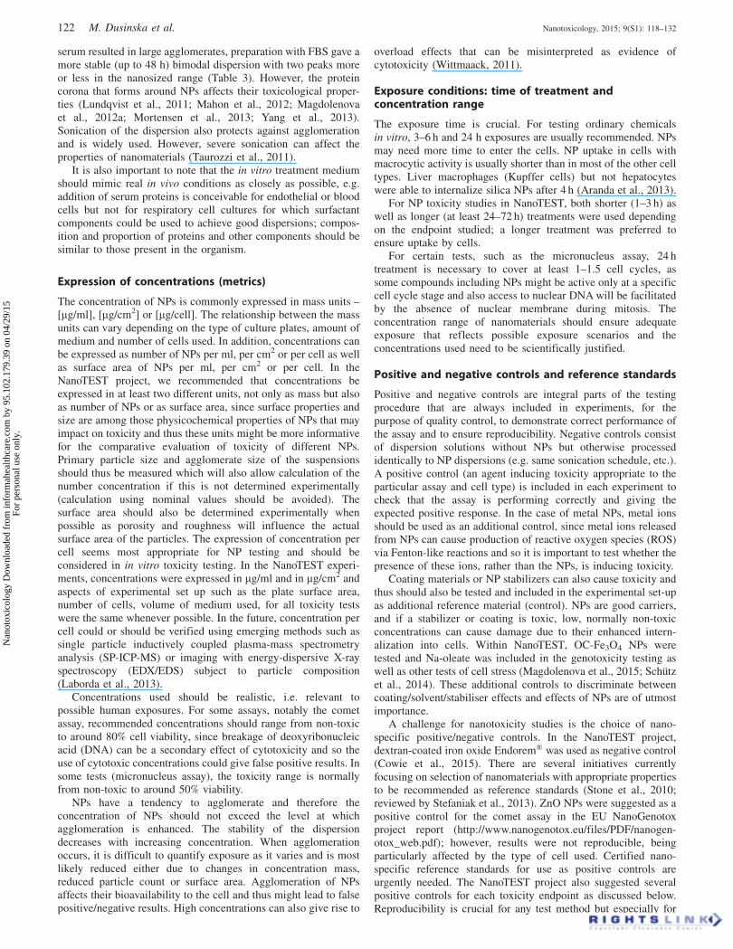
We have evaluated statistically the results of experimentscomparing cells representing different organs. Cytotoxic effectsinduced by NPs depend on the test used, exposure conditionsand the cell type (Aranda et al., 2013; Correia Carreira et al.,2015; Guadagnini et al., 2015; Halamoda Kenzaoui et al.,2012a,c, 2013a,b; Harris et al., 2015; Kazimirova et al., 2012;Magdolenova et al., 2012a, 2015; Poulsen et al., 2015; Tulinskaet al., 2015). The data also suggest that while there aredifferences between the cell lines, the strongest effect is fromthe NPs as seen with the OC-Fe3O4 NPs results (Figure 2). Forgenotoxicity screening of NPs, the various cell types used giveconsistent results but with different sensitivity, allowing thestudy of target organ specificity and cell type sensitivity(Cowie et al., 2015).
Technical limitations of the assays: possible interferenceof nanomaterial with the test
Properties of NPs such as adsorption capacity, optical properties,hydrophobicity, chemical composition, surface charge and surfaceproperties, catalytic activities as well as agglomeration can resultin interference with standard toxicity tests (Aranda et al., 2013;Guadagnini et al., 2015a; Kroll et al., 2012). The interference of
Figure 1. Cell line and exposure-dependent transport of OC-Fe3O4 NPsacross cell barriers. The appearance of OC-Fe3O4 NPs in the basalchamber was determined over the course of 2 h (open bars) and 24 h(filled bars). The initial applied amount of iron of 50 mg was added to theapical chamber (100 mg/mL) and results are expressed as the percentageof the iron added to the apical chamber as detected in the basal chamber,quantified using the Prussian blue reaction and normalised to the amounttransported across the Transwell insert (3mm Costar polyester membrane)in the absence of cells.
DOI: 10.3109/17435390.2014.991431 An alternative testing strategy for nanomaterials 123
Nan
otox
icol
ogy
Dow
nloa
ded
from
info
rmah
ealth
care
.com
by
95.1
02.1
79.3
9 on
04/
29/1
5Fo
r pe
rson
al u
se o
nly.
NPs with specific assays was observed for metallic oxide solidcore NPs and was demonstrated with a range of in vitro cellviability assays [MTT (3-(4,5-dimethyl-thiazol-2-yl)-2,5-diphe-nyl-tetrazolium bromide], LDH (lactate dehydrogenase), WST-1(2-(4-iodophenyl)-3-(4-nitrophenyl)-5-(2,4-disulphophenyl)-2H-tetrazolium), Annexin V/PI (propidium iodide), neutral red,caspase activation, propidium iodide, 3H-thymidine incorpor-ation, automated cell counting], inflammatory responses (ELISAfor granulocyte macrophage colony-stimulating factor (GM-CSF),interleukin (IL)-6 and IL-8] and oxidative stress detection[monoBromoBimane (mBBr), dichlorodihydro-fluoresceindiace-tate (DCFH-DA), NO assays; Guadagnini et al., 2015a; Krollet al., 2012]. Interferences found were assay as well as NP-specific. Thus, the evaluation of possible interference is requiredto ensure reliable results. This is mainly relevant for cytotoxicityassays, oxidative stress responses of cells and the production bythe cells of bio-molecules such as peptides, proteins or others(Guadagnini et al., 2015a). It is clear that for nanotoxicity testingmost of the assays need to be adapted and modified to avoidmeasuring artefacts. Aranda et al. (2013) showed that despite thequenching effect of NPs on DCFH-DA assay, it can be consideredas a useful tool for quantitative measurement of NPs-inducedoxidative stress by minor modifications of the standardizedprotocol. Additional standards need to be included as controls forthe interference. For genotoxicity, interference was reported so farwith the micronucleus test (Gonzalez et al., 2011; Magdolenovaet al., 2012b) and the comet assay (Karlsson, 2010; Stone et al.,2009). The protocol for the micronucleus assay needed modifi-cation as cytochalasin B (used in this assay) inhibits endocytosisand may prevent uptake of NPs (Gonzalez et al., 2011;Magdolenova et al., 2012b). Using the comet assay, with 6 NPs,we found no interference. (Magdolenova et al., 2012b). However,to prevent false-negative/false-positive results, we recommendtesting for possible interference of NPs in the gel, using bothuntreated cells and cells exposed to a known genotoxic compound(causing DNA strand breaks as well as oxidized DNA lesions).This would be a sensible precaution to be sure that nooverestimation or underestimation of damage is occurring.
Testing strategy
We investigated whether tests used in the NanoTEST are reliable,give reproducible results and are suitable for NP testing.In addition, we set out to validate a battery of tests covering all
important toxic endpoints (Table 4). Methodological consider-ation of these tests has been addressed in Guadagnini et al.(2015a) for cytotoxicity, oxidative stress and inflammatorymarkers and for genotoxicity in Magdolenova et al. (2012b).
As mentioned above, one of the main obstacles for assessingthe toxicity of nanomaterials is the lack of knowledge of howphysicochemical properties relate to the interaction of NPs withbiological systems and the mechanism of toxicity. It is clear thatphysical and chemical properties can influence NP behaviour andmay have an impact on toxicity; they must therefore be an integralpart of toxicity testing. This is one of the key aspects of toxicityscreening strategies (Dusinska & NanoTEST Consortium, 2009;Dusinska et al., 2011, 2012, 2013). Both primary and secondarycharacterizations of tested NPs are crucial, including in situcharacterization during exposure. The physico-chemical proper-ties that should be considered for assessing toxic effects ofnanomaterials include as a minimum chemical composition,particle size, shape, surface properties, size distribution, agglom-eration state and crystal structure. Regarding the likelihood ofbiomolecular corona formation, it is also important to set upexperimental conditions that can mimic exposure in humans. AsNPs change their properties depending on the surrounding milieu,we recommend at least two different exposure conditions fortesting the NP’s effects (Magdolenova et al., 2012a).
An important question is whether the commonly used assaysfor chemicals could be applied to NPs. Our results show that it isnot always possible to use these assays without careful adaptationbecause of possible interference (Guadagnini et al., 2015a),especially between NPs, the dye and the optical detection or withthe assay components during the experiment (Tables 4–6). It istherefore of crucial importance to test possible interference of allstudied NPs with the foreseen methods prior to evaluating cellularresponses to NPs. To avoid these interferences, special adapta-tions of standard toxicity tests are also proposed [refer Tables 4and 5, and Guadagnini et al. (2015a) for more detailed descrip-tion]. Furthermore, all the assays do not have the same sensitivityand it is important to choose the most sensitive appropriate assay.From our results, for the oxidative stress markers, the thioldepletion and induction of antioxidant enzymes seem to be moresensitive than the measure of ROS (Guadagnini et al., 2015b).Our proposal for further evaluation of testing strategies is toperform first a battery of assays for validation of the effects ofa representative set of well-characterized NPs on the targetcells; then if appropriate and available, to screen larger banks of
Figure 2. Cell viability of EC219, HCEC, LN229 or N11 cells exposed to (A) Si-25 or (B) OC-Fe3O4 NPs for 72 h as measured by the MTT assay.Values represent average % of untreated control ± SD of three separate experiments for each exposure.
124 M. Dusinska et al. Nanotoxicology, 2015; 9(S1): 118–132
Nan
otox
icol
ogy
Dow
nloa
ded
from
info
rmah
ealth
care
.com
by
95.1
02.1
79.3
9 on
04/
29/1
5Fo
r pe
rson
al u
se o
nly.

Tab
le4
.Im
ple
men
tati
on
of
stan
dar
din
vitr
oto
xic
ity
assa
ys
for
nan
om
ater
ial
test
ing
.
Ass
ay(S
OP
)E
nd
po
int
Mo
dif
icat
ion
sin
assa
yfo
rN
Ps
ver
sus
stan
dar
dch
emic
als
Inte
rfer
ence
/tec
hn
ical
pro
b-
lem
sw
ith
NP
sH
igh
thro
ug
hp
ut
Au
tom
atio
nC
ost
-ef
fect
ive
Use
r-fr
ien
dly
Sp
ecif
iceq
uip
men
tn
eed
ed
Cy
toto
xic
ity
WS
T1
(mea
sure
din
sup
ern
atan
ts)
Cy
toto
xic
ity
Yes
(mea
sure
men
to
nsu
p-
tern
atan
ts;
spin
/fil
ter
ou
tN
Ps)
NP
sco
uld
incr
ease
or
dec
reas
eO
Dm
easu
re-
men
ts(u
sesu
per
nat
ants
)
Yes
Po
ssib
leY
esY
esM
ult
iwel
lp
late
read
er(a
bso
rban
ce)
Lac
tate
deh
yd
rogen
ase
(LD
H)
Cy
toto
xic
ity
Yes
(en
sure
colo
rim
etri
cco
ntr
ols
are
incl
ud
edan
dte
stad
sorp
tio
no
fL
DH
on
NP
s)
NP
sco
uld
incr
ease
or
dec
reas
eO
Dm
easu
re-
men
tsan
dL
DH
may
be
abso
rbed
by
som
eN
Ps
Yes
Po
ssib
leY
esY
esM
ult
iwel
lp
late
read
er(a
bso
rban
ce)
PI
up
tak
eby
flow
cyto
met
ryC
yto
tox
icit
yY
es(g
atin
gto
excl
ud
efr
eeN
Ps
fro
man
alysi
san
dto
acco
un
tfo
rch
anges
infl
uo
resc
ence
by
NP
s)
NP
sco
uld
incr
ease
or
dec
reas
efl
uo
resc
ence
(ad
apt
gat
ing
)
Med
ium
-th
rou
gh
pu
tD
iffi
cult
Yes
No
Flo
wcy
tom
eter
Pla
tin
gef
fici
ency
Cy
toto
xic
ity
Yes
/No
(on
lyw
hen
auto
-m
atic
met
ho
dfo
rco
un
t-in
gce
lls
isu
sed
)
Ag
glo
mer
ated
NP
sca
nin
terf
ere
wit
hau
tom
atic
cell
cou
nti
ng
Med
ium
-th
rou
gh
pu
tif
auto
mat
icce
llco
un
ter
Po
ssib
leY
esY
esN
o;
Lig
ht
mic
rosc
op
e;au
tom
atic
cou
nte
r(o
pti
on
al)
Rel
ativ
eg
row
thac
tiv
ity
Cy
toto
xic
ity
Yes
/No
(on
lyw
hen
auto
-m
atic
met
ho
dfo
rco
un
t-in
gce
lls
isu
sed
)
Ag
glo
mer
ated
NP
sca
nin
terf
ere
wit
hau
tom
atic
cell
cou
nti
ng
Med
ium
-th
rou
gh
pu
tif
auto
mat
icce
llco
un
ter
Po
ssib
leY
esY
esN
o;
lig
ht
mic
rosc
op
e;au
tom
atic
cou
nte
r(o
pti
on
al)
Try
pan
blu
eex
clu
sio
nC
yto
tox
icit
yY
es/N
o(o
nly
wh
enau
to-
mat
icm
eth
od
for
cou
nt-
ing
cell
sis
use
d)
Ag
glo
mer
ated
NP
sca
nin
terf
ere
wit
hau
tom
atic
cell
cou
nti
ng
Med
ium
-th
rou
gh
pu
tif
auto
mat
icce
llco
un
ter
Po
ssib
leY
esY
esN
o;
lig
ht
mic
rosc
op
e;au
tom
atic
cou
nte
r(o
pti
on
al)
MT
TC
yto
tox
icit
yY
es(v
erif
yin
terf
eren
ce)
NP
sco
uld
incr
ease
or
dec
reas
eO
Dm
easu
re-
men
tsan
dN
Ps
cou
ldad
sorb
MT
T
Yes
Po
ssib
leb
ut
oft
enin
terf
eren
cew
ith
NP
s
Yes
Yes
Mu
ltiw
ell
pla
tere
ader
(ab
sorb
ance
)
Pro
-in
flam
mat
ory
resp
on
seE
LIS
AP
ro-i
nfl
amm
ato
ryre
spo
nse
Yes
(tes
tad
sorp
tio
no
fcy
tok
ines
on
NP
s)N
Ps
cou
ldad
sorb
secr
eted
cyto
kin
esY
esP
oss
ible
bu
to
ften
inte
rfer
ence
wit
hN
Ps
No
Yes
Mu
ltiw
ell
pla
tere
ader
(ab
sorb
ance
)
RT
qP
CR
Pro
-in
flam
mat
ory
resp
on
seY
es(u
seg
uan
idin
ium
thio
-cy
anat
e–phen
ol–
chlo
ro-
form
RN
Aex
trac
tio
np
roto
col)
NP
sco
uld
adso
rbR
NA
(co
mp
are
reco
ver
ym
eth
od
san
dch
oo
seex
trac
tio
nm
eth
od
that
reco
ver
sal
lR
NA
)
No
Dif
ficu
ltN
oN
oR
eal
tim
eP
CR
inst
rum
ent
Ox
idat
ive
stre
ssR
OS
det
ecti
on
by
DH
Eu
sin
gp
late
read
ers
Ox
idat
ive
stre
ssY
es(v
erif
yin
terf
eren
ce)
NP
sco
uld
incr
ease
or
dec
reas
efl
uo
resc
ence
Yes
Po
ssib
leY
esY
esM
ult
iwel
lp
late
read
er(f
luo
resc
ence
)R
OS
det
ecti
on
by
car-
box
y-f
luo
resc
ein
usi
ng
pla
tere
ader
s
Ox
idat
ive
stre
ssY
es(v
erif
yin
terf
eren
ce)
NP
sco
uld
incr
ease
or
dec
reas
efl
uo
resc
ence
Yes
Po
ssib
leY
esY
esM
ult
iwel
lp
late
read
er(f
luo
resc
ence
)
Th
iols
det
ecti
on
by
bro
-m
ob
iman
eu
sin
gp
late
read
ers
Ox
idat
ive
stre
ssY
es(v
erif
yin
terf
eren
ce)
NP
sco
uld
incr
ease
or
dec
reas
efl
uo
resc
ence
Yes
Po
ssib
leY
esY
esM
ult
iwel
lp
late
read
er(f
luo
resc
ence
)
RO
Sd
etec
tio
nby
HE
usi
ng
flow
cyto
met
ryO
xid
ativ
est
ress
Yes
(ad
apt
gat
ing
toex
clu
de
free
NP
sfr
om
anal
ysi
san
dto
acco
un
t
NP
sco
uld
incr
ease
or
dec
reas
efl
uo
resc
ence
and
NP
agg
lom
erat
es
Med
ium
-th
rou
gh
pu
tD
iffi
cult
Yes
No
Flo
wcy
tom
eter (c
on
tin
ued
)
DOI: 10.3109/17435390.2014.991431 An alternative testing strategy for nanomaterials 125
Nan
otox
icol
ogy
Dow
nloa
ded
from
info
rmah
ealth
care
.com
by
95.1
02.1
79.3
9 on
04/
29/1
5Fo
r pe
rson
al u
se o
nly.

Tab
le4
.C
on
tin
ued
Ass
ay(S
OP
)E
nd
po
int
Mo
dif
icat
ion
sin
assa
yfo
rN
Ps
ver
sus
stan
dar
dch
emic
als
Inte
rfer
ence
/tec
hnic
alpro
b-
lem
sw
ith
NP
sH
igh
thro
ug
hp
ut
Au
tom
atio
nC
ost
-ef
fect
ive
Use
r-fr
ien
dly
Sp
ecif
iceq
uip
men
tn
eed
ed
for
chan
ges
infl
uo
res-
cen
ceby
NP
s)co
uld
be
cou
nte
das
cell
so
rd
ebri
sR
Tq
PC
RO
xid
ativ
est
ress
Yes
(use
gu
anid
iniu
mth
io-
cyan
ate–
phen
ol–
chlo
ro-
form
RN
Aex
trac
tio
np
roto
col)
NP
sco
uld
adso
rbR
NA
(co
mp
are
reco
ver
ym
eth
od
san
dch
oo
seex
trac
tio
nm
eth
od
that
reco
ver
sal
lR
NA
)
No
Dif
ficu
ltN
oN
oR
eal
tim
eP
CR
inst
rum
ent
Up
take
Sid
esc
atte
ro
fla
ser
lig
ht
infl
ow
cyto
met
ry
Up
take
No
tu
sed
for
chem
ical
sC
ano
nly
be
use
dfo
rN
Ps
wh
ich
scat
ter
lig
ht;
gat
ing
nee
ded
toex
clu
de
free
NP
san
dd
ead
cell
s;im
agin
gfl
ow
cyto
met
ryn
eed
edto
dis
tin
gu
ish
NP
sb
ou
nd
on
cell
surf
ace
Med
ium
-th
rou
gh
pu
tD
iffi
cult
Yes
No
Flo
wcy
tom
eter
/im
agin
gfl
ow
cyto
met
er
Cel
lfl
uo
resc
ence
qu
an-
tifi
edby
flow
cyto
met
ry
Up
take
Yes
(ad
apt
gat
ing
toex
clu
de
free
NP
sfr
om
anal
ysi
s)
Can
on
lyb
eu
sed
for
NP
sw
hic
har
efl
uo
resc
ent;
gat
ing
nee
ded
toex
clu
de
free
NP
s;u
seo
fqu
ench
ers
or
imag
ing
flow
cyto
met
ryn
eed
edto
dis
tin
gu
ish
NP
sb
ou
nd
on
cell
surf
ace
Med
ium
-th
rou
gh
pu
tD
iffi
cult
Yes
No
Flo
wcy
tom
eter
/im
agin
gfl
ow
cyto
met
er
Tra
nsm
issi
on
elec
tro
nm
icro
sco
py
(TE
M)
Up
take
No
Ver
ific
atio
nre
qu
ired
that
ob
ject
sse
enar
eac
tual
lyN
Ps
e.g
.by
ED
S/E
DX
No
No
Yes
No
Ele
ctro
nm
icro
sco
pe
Gen
oto
xic
ity
Co
met
assa
yS
ing
lean
dd
ou
ble
stra
nd
bre
aks
No
En
sure
NP
sin
gel
do
no
tin
terf
ere
by
usi
ng
bo
thu
ntr
eate
dce
lls
and
cell
sex
po
sed
tok
now
ngen
o-
tox
icco
mp
ou
nd
Yes
Po
ssib
leY
esY
esF
luo
resc
ence
mic
ro-
sco
pe,
imag
ean
aly
sis
Co
met
assa
yw
ith
rep
air
enzy
me
Gen
oto
xic
ity
:b
ase
DN
Ad
amag
eN
oN
orm
ally
NP
sd
on
ot
com
ein
con
tact
wit
hen
zym
e:H
ow
ever
,ad
dit
ion
alco
ntr
ol,
cell
sw
ith
ox
i-d
ized
lesi
on
san
dN
Ps
ingel
,ca
nb
ein
clu
ded
.
Yes
Po
ssib
leY
esY
esF
luo
resc
ence
mic
ro-
sco
pe,
imag
ean
aly
sis
Cy
tok
ines
is-b
lock
mic
ron
ucl
eus
assa
yG
eno
tox
icit
y/
mu
tagen
icit
yY
es(c
yto
chal
asin
B2
4h
afte
rN
Ps)
Cy
toch
alas
inB
can
inh
ibit
NP
up
take
Yes
Po
ssib
leY
esY
esL
igh
tm
icro
sco
pe,
imag
ean
alysi
sT
ran
spo
rtB
arri
erp
erm
eab
ilit
y(N
aFlu
)B
arri
erin
teg
rity
Yes
(rin
seb
arri
erw
ith
PB
Sp
rio
rto
asse
ssm
ent)
Po
ten
tial
inte
rfer
ence
of
NP
sw
ith
flu
ore
scen
ceP
oss
ible
No
Yes
Yes
Pla
tere
ader
;T
ran
swel
ls
Bar
rier
tran
spo
rto
fN
Pin
Tra
nsw
ells
Tra
nsp
ort
of
NP
sac
ross
bar
rier
Yes
(po
rou
sm
emb
ran
en
eed
sto
be
op
tim
ised
for
NP
may
adh
ere
toT
ran
swel
lm
emb
ran
ean
dre
ten
tio
no
fN
Ps
by
po
rou
s
Po
ssib
leN
oY
esY
esP
late
read
er;
Tra
nsw
ells
126 M. Dusinska et al. Nanotoxicology, 2015; 9(S1): 118–132
Nan
otox
icol
ogy
Dow
nloa
ded
from
info
rmah
ealth
care
.com
by
95.1
02.1
79.3
9 on
04/
29/1
5Fo
r pe
rson
al u
se o
nly.

NPs using automated procedures on a predefined set of repre-sentative cells. Appropriate statistical analyses must always beimplemented.
For the evaluation of the different cell models, depending onroute of exposure and use of NPs there should be several organmodels used for testing. Blood is an important model both as adirect target as well as surrogate target tissue and gives anindication of toxicity. Peripheral blood lymphocytes are suitablecells but unfortunately not always accessible; the TK6 (lympho-blastic) cell line is an alternative. We additionally propose thatcommercially available human cell lines for each representativeorgan be included in the testing strategy, e.g. for lung cellsavailable cell lines (A549 cells is one alternative), CaCo2 cells(colon), LN229 cells (glioblastoma) and HEK293 or MDCK(human or porcine kidney, respectively).
For short-term hazard assessment, the testing strategy shouldinclude all important toxicity endpoints such as cytotoxicity,oxidative stress, genotoxicity or immunotoxicity to investigate themode of action of NPs in biological systems. In vitro experimentswith cells representing different organ targets revealed thatoxidative stress and toxic effects induced by NPs depend on theNPs’ properties, the test used and the cell type. Polylactic-co-glycolic acid (PLGA-PEO) NPs induced little or no oxidativestress in any cell type compared with solid-core metallic NPswhich generally produced ROS (Guadagnini et al., 2015b;Halamoda Kenzaoui et al., 2012c, 2013b). Genotoxicity inducedby NPs depends on the NPs, the dispersion protocol and themeasured endpoint (Magdolenova et al., 2012a). All cells testedwere able to detect the positive response but with differentsensitivity showing tissue specific effects (Cowie et al., 2015).
In the initial stages of testing, cytotoxicity assays should beused to identify non-cytotoxic concentrations of the NPs for morespecific in vitro studies. Moreover, inclusion of nanospecificpositive and negative controls is strongly recommended.
The strategy proposed for a battery of in vitro tests is explainedas follows.(1) To determine possible cytotoxicity and induction of oxidative
stress.– For cytotoxicity studies, basal cellular toxicity tests such as
relative growth activity and plating efficiency and the MTTand WST-1 assays and a time course of 24 and 72 h, usingOC-Fe3O4 NPs as positive control NPs and PLGA-PEO NPsas negative control NPs.
– For oxidative stress, the thiol depletion measured bymonobromobimane assay (and possibly DCFH-DA; Arandaet al., 2013) and the induction of antioxidant enzyme mRNAexpression measured by RT-qPCR (Guadagini et al., 2015b),4 and 24 h time-course, using uncoated iron oxide (U-Fe3O4)NPs and TiO2 NPs as positive control NPs and PLGA-PEONPs as negative control NPs.
(2) to determine the uptake and possible release, followinguptake, of the NPs by relevant cells of the different organs, atnon-cytotoxic concentrations of the NPs.
– For uptake, TEM and depending on NP properties flowcytometry (if light scattering or fluorescent) or analyticalmethods (ICP-MS). Analytical chemistry on cell super-natants could be used to study the release of NPs (dos Santoset al. 2011; Elsaesser et al, 2011).
– For uptake and release studies, 24 h uptake followed by 24 hrelease, using U-Fe3O4 NPs as positive control NPs(Halamoda Kenzaoui et al., 2012b, 2013b).
– For transport studies, 24 h time-course, using OC-Fe3O4
NPs as positive control NPs, limiting these experiments toNPs which do not agglomerate in the culture conditions(Correia Carreira et al., 2015; Halamoda Kenzaoui et al,2013b).
par
ticl
ety
pe
and
cell
typ
e)m
emb
ran
ew
ill
red
uce
tran
spo
rt:a
sses
sN
Pin
tera
ctio
nw
ith
mem
-b
ran
efi
lter
and
NP
tran
spo
rt.
NP
det
ecti
on
met
ho
ds
nee
dto
be
refi
ned
Imm
un
oto
xic
ity
Ph
ago
cyti
cac
tiv
ity
and
resp
irat
ory
bu
rst
of
leu
ko
cyte
sby
flow
cyto
met
ry
Imm
un
oto
xic
ity
Yes
(ad
apt
gat
ing
toex
clu
de
free
NP
sfr
om
anal
ysi
san
dto
acco
un
tfo
rch
anges
infl
uo
res-
cen
ceb
yN
Ps)
Po
ten
tial
inte
rfer
ence
of
NP
sw
ith
flu
ore
scen
ceM
ediu
m-t
hro
ug
hp
ut
Dif
ficu
ltY
esN
oF
low
cyto
met
er
Nat
ura
lk
ille
rce
llac
tiv-
ity
by
flow
cyto
met
ryIm
mu
no
tox
icit
yY
es(a
dap
tgat
ing
toex
clu
de
free
NP
sfr
om
anal
ysi
san
dto
acco
un
tfo
rch
anges
infl
uo
res-
cen
ceb
yN
Ps)
Po
ten
tial
inte
rfer
ence
of
NP
sw
ith
flu
ore
scen
ceM
ediu
m-t
hro
ug
hp
ut
Dif
ficu
ltN
oN
oF
low
cyto
met
er
Pro
life
rati
ve
resp
on
seo
fly
mp
ho
cyte
sby
3H
thy
mid
ine
inco
rpo
rati
on
Imm
un
oto
xic
ity
Yes
(ver
ify
inte
rfer
ence
)N
Ps
cou
ldin
terf
ere
wit
hco
un
tsp
erm
inu
tes
mea
sure
men
ts
Po
ssib
leD
iffi
cult
No
Yes
Liq
uid
scin
till
atio
nco
un
ter
DOI: 10.3109/17435390.2014.991431 An alternative testing strategy for nanomaterials 127
Nan
otox
icol
ogy
Dow
nloa
ded
from
info
rmah
ealth
care
.com
by
95.1
02.1
79.3
9 on
04/
29/1
5Fo
r pe
rson
al u
se o
nly.

Tab
le5
.N
ano
mat
eria
lp
rop
erti
esw
hic
hco
uld
be
pro
ble
mat
icfo
rsp
ecif
icte
stm
eth
od
s.
Ass
ayA
gg
lom
erat
ion
Ad
sorp
tio
no
fp
rote
ins
Ad
sorp
tio
no
fd
yes
Flu
ore
scen
ceA
bso
rpti
on
/sca
tter
-in
go
fli
gh
tA
ffin
ity
tola
bw
are
mat
eria
lS
tab
ilit
y/s
olu
bil
ity
of
par
ticl
es
Cy
toto
xic
ity
PI
up
take
by
flow
cyto
met
ryN
eed
filt
rati
on
bef
ore
anal
ysi
sO
KO
Kif
adju
stg
atin
gP
rob
lem
(dep
end
so
nw
avel
eng
th)
OK
ifad
just
gat
ing
OK
OK
LD
HO
KP
rob
lem
Pro
ble
mP
rob
lem
(dep
end
so
nw
avel
eng
th)
Pro
ble
mO
KO
K
Pla
tin
gef
fici
ency
Pro
ble
mif
auto
mat
icco
un
tin
gO
KO
KO
KO
KO
KO
K
Rel
ativ
eg
row
thac
tiv
ity
Pro
ble
mif
auto
mat
icco
un
tin
gO
KO
KO
KO
KO
KO
K
Up
take
Qu
anti
fica
tio
no
fce
ll-a
sso
-ci
ated
lig
ht
scat
teri
ng
NM
sby
flow
cyto
met
ry
Nee
dfi
ltra
tio
nb
efo
rean
alysi
sO
KO
KP
rob
lem
(dep
end
-en
tso
nw
ave-
len
gth
asP
Ist
ain
ing
nee
ded
)
Nee
dN
Ms
wh
ich
scat
ter
lig
ht
OK
Pro
ble
m
Qu
anti
fica
tio
no
fce
ll-a
sso
-ci
ated
flu
ore
scen
tN
Ms
by
flow
cyto
met
ry
Nee
dfi
ltra
tio
nb
efo
rean
alysi
sO
KO
KN
eed
flu
ore
scen
tN
Ms
(pro
ble
mif
ble
ach
ing
or
qu
ench
ing
of
flu
ore
scen
ce)
Pro
ble
m(a
dju
stgat
ing
)O
KP
rob
lem
Qu
anti
fica
tio
no
fce
llas
so-
ciat
edir
on
con
tain
ing
NM
sby
pla
tere
ader
OK
OK
OK
Pro
ble
m(d
epen
ds
on
wav
elen
gth
)P
rob
lem
Pro
ble
mO
K
Qu
anti
fica
tio
no
fce
ll-a
sso
-ci
ated
flu
ore
scen
tN
Ms
by
pla
tere
ader
OK
OK
OK
Nee
dfl
uo
resc
ent
NM
s(p
rob
lem
ifb
leac
hin
go
rqu
ench
ing
of
flu
ore
scen
ce)
Pro
ble
mP
rob
lem
Pro
ble
m
Tra
nsm
issi
on
elec
tro
nm
icro
s-co
py
(TE
M)
OK
OK
OK
OK
OK
OK
Pro
ble
m
Pro
-in
flam
mat
ory
resp
on
seE
LIS
AO
KP
rob
lem
OK
OK
OK
OK
OK
RT
qP
CR
OK
Pro
ble
mif
adso
rbe
RN
A(a
dap
tex
trac
tio
nte
chn
iqu
e)
OK
OK
OK
OK
OK
Ox
idat
ive
stre
ssO
xid
ativ
est
ress
mea
sure
men
tsby
flu
ore
scen
td
yes
(DC
FH
-DA
,H
E,
mB
Br
etc)
usi
ng
flow
cyto
met
ry
Nee
dfi
ltra
tio
nb
efo
rean
alysi
sO
KO
Kif
adju
stg
atin
gP
rob
lem
(dep
end
ing
on
wav
elen
gth
)O
Kif
adju
stg
atin
gO
KO
K
RT
qP
CR
OK
Pro
ble
mif
adso
rbe
RN
A(a
dap
tex
trac
tio
nte
chn
iqu
e)
OK
OK
OK
OK
OK
Ox
yd
ativ
est
ress
mea
sure
men
tsby
flu
ore
scen
td
yes
(DC
FH
-D
A,
HE
,m
BB
ret
c)u
sin
gp
late
read
er
OK
OK
Pro
ble
mP
rob
lem
(dep
end
ing
on
wav
elen
gh
)P
rob
lem
Pro
ble
mif
inte
rfer
-en
cew
ith
det
ecti
on
OK
128 M. Dusinska et al. Nanotoxicology, 2015; 9(S1): 118–132
Nan
otox
icol
ogy
Dow
nloa
ded
from
info
rmah
ealth
care
.com
by
95.1
02.1
79.3
9 on
04/
29/1
5Fo
r pe
rson
al u
se o
nly.
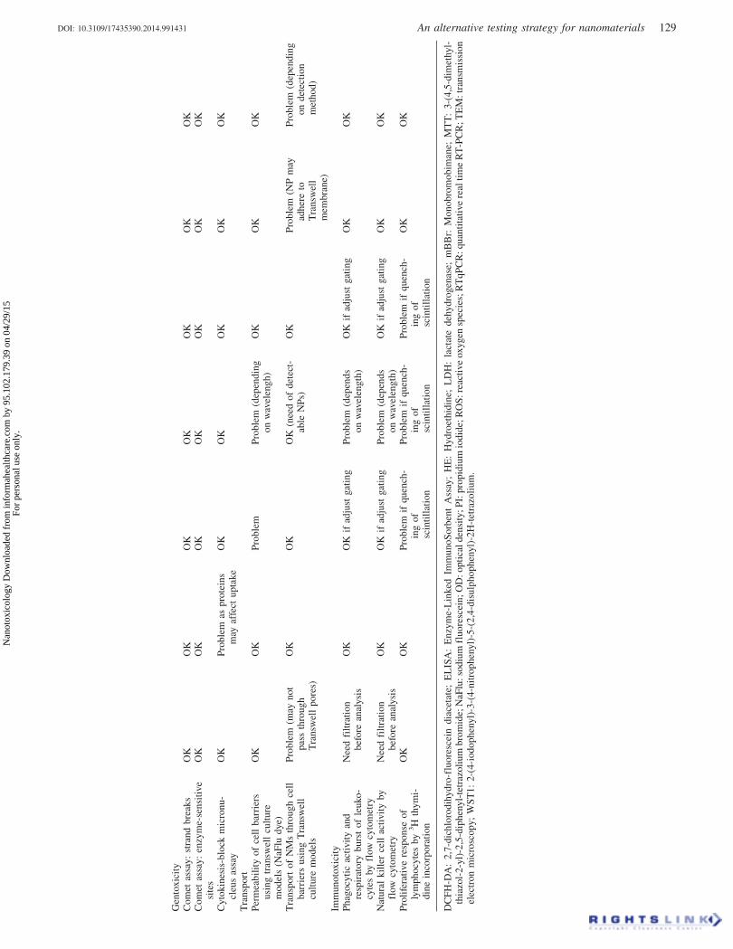
Gen
tox
icit
yC
om
etas
say
:st
ran
db
reak
sO
KO
KO
KO
KO
KO
KO
KC
om
etas
say
:en
zym
e-se
nsi
tive
site
sO
KO
KO
KO
KO
KO
KO
K
Cy
tok
ines
is-b
lock
mic
ron
u-
cleu
sas
say
OK
Pro
ble
mas
pro
tein
sm
ayaf
fect
up
tak
eO
KO
KO
KO
KO
K
Tra
nsp
ort
Per
mea
bil
ity
of
cell
bar
rier
su
sin
gtr
answ
ell
cult
ure
mo
del
s(N
aFlu
dye)
OK
OK
Pro
ble
mP
rob
lem
(dep
end
ing
on
wav
elen
gh
)O
KO
KO
K
Tra
nsp
ort
of
NM
sth
rou
gh
cell
bar
rier
su
sin
gT
ran
swel
lcu
ltu
rem
od
els
Pro
ble
m(m
ayn
ot
pas
sth
rou
gh
Tra
nsw
ell
po
res)
OK
OK
OK
(nee
do
fd
etec
t-ab
leN
Ps)
OK
Pro
ble
m(N
Pm
ayad
her
eto
Tra
nsw
ell
mem
bra
ne)
Pro
ble
m(d
epen
din
go
nd
etec
tio
nm
eth
od
)
Imm
un
oto
xic
ity
Ph
ago
cyti
cac
tiv
ity
and
resp
irat
ory
bu
rst
of
leu
ko
-cy
tes
by
flow
cyto
met
ry
Nee
dfi
ltra
tio
nb
efo
rean
alysi
sO
KO
Kif
adju
stgat
ing
Pro
ble
m(d
epen
ds
on
wav
elen
gth
)O
Kif
adju
stgat
ing
OK
OK
Nat
ura
lk
ille
rce
llac
tiv
ity
by
flow
cyto
met
ryN
eed
filt
rati
on
bef
ore
anal
ysi
sO
KO
Kif
adju
stgat
ing
Pro
ble
m(d
epen
ds
on
wav
elen
gth
)O
Kif
adju
stgat
ing
OK
OK
Pro
life
rati
ve
resp
on
seo
fly
mp
ho
cyte
sby
3H
thy
mi-
din
ein
corp
ora
tio
n
OK
OK
Pro
ble
mif
qu
ench
-in
go
fsc
inti
llat
ion
Pro
ble
mif
qu
ench
-in
go
fsc
inti
llat
ion
Pro
ble
mif
qu
ench
-in
go
fsc
inti
llat
ion
OK
OK
DC
FH
-DA
:2
,7-d
ich
loro
dih
yd
ro-f
luo
resc
ein
dia
ceta
te;
EL
ISA
:E
nzy
me-
Lin
ked
Imm
un
oS
orb
ent
Ass
ay;
HE
:H
yd
roet
hid
ine;
LD
H:
lact
ate
deh
yd
rogen
ase;
mB
Br:
Mo
no
bro
mo
bim
ane;
MT
T:
3-(
4,5
-dim
ethyl-
thia
zol-
2-y
l)-2
,5-d
iph
enyl-
tetr
azo
liu
mb
rom
ide;
NaF
lu:
sod
ium
flu
ore
scei
n;
OD
:o
pti
cal
den
sity
;P
I:p
rop
idiu
mio
did
e;R
OS
:re
acti
ve
ox
ygen
spec
ies;
RT
qP
CR
:qu
anti
tati
ve
real
tim
eR
T-P
CR
;T
EM
:tr
ansm
issi
on
elec
tro
nm
icro
sco
py;
WS
T1
:2
-(4
-io
do
ph
eny
l)-3
-(4
-nit
rop
hen
yl)
-5-(
2,4
-dis
ulp
ho
ph
enyl)
-2H
-tet
razo
liu
m.
DOI: 10.3109/17435390.2014.991431 An alternative testing strategy for nanomaterials 129
Nan
otox
icol
ogy
Dow
nloa
ded
from
info
rmah
ealth
care
.com
by
95.1
02.1
79.3
9 on
04/
29/1
5Fo
r pe
rson
al u
se o
nly.

(3) to determine possible genotoxic effect (Magdolenova et al.,2012b,2014).
– For DNA damage, cells exposed for 24 h to NPs, using thecomet assay for DNA strand breaks (SB) and oxidized DNAlesions (TiO2 or OC-Fe3O4 as positive control NPs andPLGA-PEO NPs as negative control NPs, at non-cytotoxicconcentrations of NPs).
– For mutagenicity and clastogenicity, cytokinesis-blockmicronucleus (CBMN) modified protocol for NPs genotoxi-city testing. However, positive and negative controls shouldbe further specified.
– The gH2AX (H2A histone family, member X) assay is aninteresting end-point for automated procedures, but littleinformation so far exists about predicting NP-inducedgenotoxicity using this test.
– Gene mutation assays (in either tk or hprt locus) have notonly been tested within NanoTEST, but should also beimplemented into genotoxicity strategy to cover all geno-toxicity endpoints.
(4) To determine immunosafety of NPs, human peripheral wholeblood or isolated peripheral blood mononuclear cells(PBMC) as representatives of human blood cell model areproposed for in vitro screening of the immunotoxic potentialof nanoproducts. The main strength is the complexity of themodel containing several cell types and components in arelatively intact environment. Testing strategy for assessmentof immunotoxic effect of NPs should contain a panel ofimmune assays to cover several aspects of the immuneresponse. Cellular immune response: phagocytic activity andrespiratory burst of leukocytes, natural killer cell activity,proliferative activity of lymphocytes in vitro stimulated withmitogens and/or antigens (LTT). Humoral immune response:production/expression of cytokines.
– For lymphocyte transformation test (LTT), fluorescent 25 nmsilica (Fl-25 SiO2) NPs as immunosuppressive control andTiO2 NPs as possible candidate for immunostimulatorycontrol.
– For phagocytic activity and respiratory burst assay, U-Fe3O4
as stimulatory control and OC-Fe3O4 as suppressive nano-control.
– For natural killer cell activity, OC-Fe3O4NPs as suppressivecontrol and Fl-25 SiO2NPs as stimulatory control.
– For cytokine gene expression, OC-Fe3O4NPs as suppressivecontrol for IL-6.
(5) Finally, to perform the more selective evaluations, asrequired by the particular characteristics of the organ-representative cells, such as cytokine production, cellularlocalization of NPs inside cells using techniques such asconfocal or electronic microscopy techniques, etc.
Tables 5 and 6 are intended to help choosing the best testingstrategy (toxicity test/dispersion media/adaptation of standardmethods) depending on the physico-chemical characteristics ofthe NPs.
Several assays appeared to be suitable for high throughputscreening and automation (Table 4) and their implementation ina testing strategy is desirable for a large number of NPs.Increased throughput of the comet assay for detection of SBsand specific DNA lesions is suggested for robust testingtogether with automation and high throughput of assays forcytotoxicity (measuring cell count, nuclear intensity and nuclearsize) and alternatively also for genotoxicity (gH2AX assay;Harris et al., 2015). It is recommended to use a multi-parametric analysis (high content imaging – HCI) whichprovides more information and can allow determination of thecause of the cytotoxic effect. High-throughput screening (HTS)assays provide several benefits, including an upscaling ofnumber of NPs to be tested; for optimization and precision ofthe assays; as a support to validations; and if applicable alsofor industrial use.
The quantitative structure–activity relationship [(Q)SAR]models promise to be valuable tools for future testing strategies.The theoretical model predicting oxidative stress potential led tothe prioritisation of metal oxide NPs for further evaluation. Theproposed model could be used to guide the development of morerational and efficient screening strategies; in addition, it cancreate a more coherent conceptual framework when additionaltoxicity related physicochemical properties (e.g. agglomerationand solubility in water) are included (Burelo & Worth, 2011,2015).
With proper refinement of computational models and meth-odologies, physiologically based pharmacokinetic (PBPK) mod-elling may serve as an alternative testing strategy in future (Pilouet al., 2015), replacing experiments that are expensive both intime and resources.
Table 6. General problems which may arise from specific nanomaterial properties and should be taken into consideration to choose the best testingstrategy and for correct interpretation of results.
AgglomerationAdsorption of
proteins
Adsorption ofdyes/assayreagents Fluorescence
Absorption/scat-tering of light
Affinity to lab-ware material
Stability/solubilityof particles
Problem for flowcytometry
Change size Interference withtest methods
Interference withread-out system
Interference withread-outsystem
Interference withread-outsystem andtest methods
Problem for dosage/detection
Change settling Change uptake Change of surfacecharacteristics ifnot core-labelled
Changebioavailability
Change size
Pipetting (problemfor correct dosingandreproducibility)
Change intracel-lular transport
Problem of detectionif leaching orbleaching
Problem fordosage
Change uptake
Change uptake Changetranslocation
Problem if interfer-ence with dyes
Change intracellulartransport
Change intracellulartransport
Change inmetabolism
Changetranslocation
Biomarkerinterference
130 M. Dusinska et al. Nanotoxicology, 2015; 9(S1): 118–132
Nan
otox
icol
ogy
Dow
nloa
ded
from
info
rmah
ealth
care
.com
by
95.1
02.1
79.3
9 on
04/
29/1
5Fo
r pe
rson
al u
se o
nly.
Conclusions and remarks
A strategy for short-term hazard assessment and toxicity testing ina regulatory context requires a battery of tests addressing differentmechanisms and covering all main important toxicity endpointsincluding oxidative stress, genotoxicity, inflammation andimmunotoxicity. Toxicity tests should be performed in anappropriate treatment medium and uptake should be an integralpart of the testing. Treatment should be long enough for the NPsto enter the cells and entry and uptake should be confirmed andquantified where possible (Elsaesser et al., 2011).
Appropriate controls and reference standards should beincluded in tests. The solvent and, if NPs are coated, the coatingmaterial need to be tested separately. Historical control data (foreach cell type used) are valuable references for quality control.Toxicity tests must be accompanied by extensive characterizationof NPs, including in test-media (before and after exposure), andcovering primary and secondary properties of NPs. Number ofcells, cell culture plate format and volume of treatment mediumon the plate are important factors in expressing concentration andcan impact on the effect of NPs, and so they should be constantwithin the study, especially when different NPs are compared.Concentration range and exposure time are also crucial aspects intoxicity testing and care must be taken to express concentrationsin at least two different units, not only in mass but also in numberof NPs or in surface area as this might be more representative forevaluation of toxicity of different NPs than using mass units.Determination of actual size and surface area is important asnumber or surface concentrations are sometimes based on thenominal particle size and may not reflect the actual particle size/surface.
We propose that for full assessment of NP toxicity, at least 2–3cytotoxicity tests (the MTT, WST-1 and plating efficiency assaysor relative growth activity), a set of at least 2–3 representativecells and five NP concentrations should be used. Initially, thecytotoxicity response to the NPs must be determined, thenoxidative stress response using at least two assays. A testingstrategy for assessment of immunotoxic effects of NPs shouldcontain assays covering several aspects of the immune response(inducible proliferative response, phagocytic activity and respira-tory burst of leukocytes, natural killer cell activity, production/expression of cytokines). For genotoxicity, the modified cometassay for DNA damage (strand breaks as well as oxidised DNAlesions) should be included in the testing strategy together withthe micronucleus assay and a gene mutation test with the option ofthe gH2AX assay in automated procedures. The evaluation ofinternalization of NPs by cells should be an integral part of testingstrategy but is not always feasible, and the analytical methods andthe devices designed to evaluate NPs transport across cell layersneed improvements in technology.
Declaration of interest
The authors declare that there is no conflict of interests. The workwas supported by EC FP7 NanoTEST [Health-2007-1.3-4], Contractno: 201335, EC FP7 QualityNano [INFRA-2010-1.131], Contract no:214547-2, EC FP7 NANoREG, [NMP.2012.1.3-3], Contract no: 310584,EC FP7 NanoTOES [PITN-GA-2010-264506] and by NILU internalprojects 106179. The work by UH Bristol was carried out with the supportof the Bristol Centre for Nanoscience and Quantum Information,University of Bristol.
References
Aranda A, Sequedo L, Tolosa L, Quintas G, Burello E, Castell JV,Gombau L. 2013. Dichloro-dihydro-fluorescein diacetate (DCFH-DA)assay: a quantitative method for oxidative stress assessment ofnanoparticle-treated cells. Toxicol in Vitro 27:954–63.
Bouwmeester H, Lynch I, Marvin HJP, Dawson KA, Berges M, BraguerD, et al. 2011. Minimal analytical characterization of engineerednanomaterials needed for hazard assessment in biological matrices.Nanotoxicology 5:1–11.
Burello E, Worth A. 2011. A theoretical framework for predicting theoxidative stress potential of oxide nanoparticles. Nanotoxicology 5:228–35.
Burello E, Worth A. 2015. A rule for designing safer nanomaterials: donot interfere with the cellular redox equilibrium. Nanotoxicology9(S1):116–117.
Cartwright L, Poulsen MS, Nielsen HM, Pojana G, Knudsen LE,Saunders M, Rytting E. 2011. In vitro placental model optimization fornanoparticle transport studies. Int J Nanomed 7:497–510.
Correia Carreira S, Walker L, Paul K, Saunders M. 2015. The toxicity,transport and uptake of nanoparticles in the in vitro BeWo b30placental cell barrier model used within NanoTEST. Nanotoxicology9(S1):66–78.
Cowie H, Magdolenova Z, Saunders M, Drlickova M, Correia Carreira S,Halamoda Kenzaoiu B, et al. 2015. Suitability of human andmammalian cells of different origin for the assessment of genotoxicityof metal and polymeric engineered nanoparticles. Nanotoxicology9(S1):57–65.
dos Santos T, Varela J, Lynch I, Salvati A, Dawson KA. 2011.Quantitative assessment of the comparative nanoparticle-uptake effi-ciency of arrange of cell lines. Small 7:3341–9.
Dusinska M, Fjellsbø LM, Magdolenova Z, Ravnum S, Rinna A, Runden-Pran E. 2011. Chapter 11. Safety of nanomaterial in nanomedicine. In:Hunter RJ, Preedy VR, eds. Nanomedicine in Health and Disease. NewHampshire: Science Publishers [CRC Press], 203–26.
Dusinska M, Magdolenova Z, Fjellsbø LM. 2013. Toxicologicalaspects for nanomaterial in humans. Chapter 1. In: Oupicky D, OgrisM, eds. Nanotechnology for Nucleic Acid Delivery, Methods inMolecular Biology. New York: Humana Press, Springer BWF Book948, 1–12.
Dusinska M, NanoTEST consortium. 2009. Safety of nanoparticles usedin medical application. Development of alternative testing strategies fortoxicity testing. Sci Technol Public Serv Rev 4:126–7.
Dusinska M, Runden-Pran E, Carreira SC, Saunders M. 2012. In vitro andin vivo toxicity test methods. Chapter 4. Critical Evaluation of ToxicityTests. In: Fadeel B, Pietroiusti A, Shvedova A, eds. Adverse Effects ofEngineered Nanomaterials: Exposure, Toxicology and Impact onHuman Health. New York: Elsevier, 63–84.
Elsaesser A, Barnes CA, McKerr G, Salvati A, Lynch I, Dawson KA,Howard CV. 2011. Quantification of nanoparticle uptake by cells usingan unbiased sampling method and electron microscopy. Nanomedicine(Lond) 6(7):1189–98.
Elsaesser A, Howard CV. 2012. Toxicology of nanoparticles. Adv DrugDeliv Rev 64:129–37.
Gonzalez L, Corradi S, Thomassen LC, Martens JA, Cundari E, Lison D,Kirsch-Volders M. 2011. Methodological approaches influencingcellular uptake and cyto-(geno) toxic effects of nanoparticles. JBiomed Nanotechnol 7:3–5.
Guadagnini R, Halamoda Kenzaoui B, Cartwright L, Pojana G,Magdolenova Z, Bilanicova D, et al. 2015a. Toxicity screenings ofnanomaterials: challenges due to interference with assay processes andcomponents of classic in vitro tests. Nanotoxicology 9(S1):13–24.
Guadagnini R, Moreau K, Hussain S, Marano F, Boland S. 2015b.Toxicity evaluation of engineered nanoparticles for medical applica-tions for the pulmonary system. Nanotoxicology 9(S1):25–32.
Hagens WI, Oomen AG, de Jong WH, Cassee FR, Sips AJ. 2007. Whatdo we (need to) know about the kinetic properties of nanoparticles inthe body? Regul Toxicol Pharmacol 49:217–29.
Halamoda Kenzaoui B, Angeloni S, Overstolz T, Niedermann P,ChapuisBernasconi C, Liley M, Juillerat-Jeanneret L. 2013a. Transferof ultra small iron oxide nanoparticles from human brain-derivedendothelial cells to human glioblastoma cells. ACS Appl MaterInterfaces 5:3581–6.
Halamoda Kenzaoui B, Bernasconi C, Hofmann H, Juillerat-Jeanneret L.2012a. Evaluation of uptake and transport of ultra small superparamagnetic iron oxide nanoparticles by human brain-derived endo-thelial cells. Nanomedicine 7:39–53.
Halamoda Kenzaoui B, Chapuis Bernasconi C, Guney-Ayra S, Juillerat-Jeanneret L. 2012c. Induction of oxidative stress, lysosome activationand autophagy by nanoparticles in human brain endothelial cells.Biochem J 441:813–21.
DOI: 10.3109/17435390.2014.991431 An alternative testing strategy for nanomaterials 131
Nan
otox
icol
ogy
Dow
nloa
ded
from
info
rmah
ealth
care
.com
by
95.1
02.1
79.3
9 on
04/
29/1
5Fo
r pe
rson
al u
se o
nly.
Halamoda Kenzaoui B, Chapuis Bernasconi C, Juillerat-Jeanneret L.2013b. Stress-reaction of kidney epithelial cells to inorganic solid-corenanoparticles. Cell Biol Toxicol 29:39–58.
Halamoda Kenzaoui B, Vila MR, Miquel JM, Cengelli F, Juillerat-Jeanneret L. 2012b. Evaluation of uptake and transport of cationic andanionic ultrasmall iron oxide nanoparticles by human colon cells. Int JNanomed, 7:1275–86.
Handy RD, van den Brink N, Chappell M, Muhling M, Behra R, DusinskaM, et al. 2012. Practical considerations for conducting ecotoxicity testmethods with manufactured nanomaterials: what have we learnt so far?Ecotoxicology 21:933–72.
Harris G, Palosaari T, Magdolenova Z, Mennecozzi M, Gineste JM,Saavedra L, et al. 2015. Iron oxide nanoparticle toxicity testing usinghigh throughput analysis and high content imaging. Nanotoxicology9(S1):87–94.
Hassellov M, Kaegi R. 2009. Analysis and characterization ofmanufactured nanoparticles in aquatic environments. In: Lead JR,Smith E, eds. Environmental and Human Health Impacts ofNanotechnology. West Sussex, United Kingdom: BlackwellPublishing Ltd, 211–66.
Hussain S, Thomassen LCJ, Ferecatu I, Borot C, Andreau K, Martens JA,et al. 2010. Carbon black and titanium dioxide nanoparticles elicitdistinct apoptotic pathways in bronchial epithelial cells. Part FibreToxicol 7:10.
Jiang J, Oberdorster G, Biswas P. 2009. Characterization of size, surfacecharge, and agglomeration state of nanoparticle dispersions fortoxicological studies. J Nanopart Res 11:77–89.
Juillerat-Jeanneret L, Dusinska M, Fjellesbo LM, Collins AR, Handy R,Riediker M. 2015. Biological impact assessment of nanomaterialused in nanomedicine. Introduction to the NanoTEST project.Nanotoxicology 9(S1):5–12.
Juillerat-Jeanneret L, Aguzzi A, Wiestler OD, Darekar P, Janzer RC.1992. Dexamethasone selectively regulates the activity of enzymaticmarkers of cerebral endothelial cell lines. In Vitro Cell Dev Biol28A:537–43.
Karlsson HL. 2010. The comet assay in nanotoxicology research. AnalBioanal Chem 398:651–66.
Kato H, Suzuki M, Fujita K, Horie M, Endoh S, Yoshida Y, et al. 2009.Reliable size determination of nanoparticles using dynamic lightscattering method for in vitro toxicology assessment. Toxicol in Vitro23:927–34.
Kazimirova A, Magdolenova Z, Barancokova M, Staruchova M,Volkovova K, Dusinska M. 2012. Genotoxicity testing of PLGA-PEOnanoparticles in TK6 cells by the comet assay and the cytokinesis-block micronucleus assay. Mutat Res 748:42–7.
Kroll A, Pillukat MH, Hahn D, Schnekenburger J. 2012. Interference ofengineered nanoparticles with in vitro toxicity assays. Arch Toxicol 86:1123–36.
Laborda F, Jimenez-lamana J, Bolea E, Castillo JR. 2013. Criticalconsiderations for the determination of nanoparticle number concen-trations, size and number size distributions by single particle ICP-MS.J Anal Atomic Spectrom 28:1220–32.
Lundqvist M, Stigler J, Cedervall T, Berggard T, Flanagan MB, Lynch I,et al. 2011. The evolution of the protein corona around nanoparticles: atest study. ACS Nano 5:7503–9.
Magdolenova Z, Bilanicova D, Pojana G, Fjellsbø LM, Hudecova A,Hasplova K, et al. 2012a. Impact of agglomeration and different disper-sions of titanium dioxide nanoparticles on the human related in vitrocytotoxicity and genotoxicity. J Environ Monit 14:455–64.
Magdolenova Z, Lorenzo Y, Collins A, Dusinska M. 2012b. Can standardgenotoxicity tests be applied to nanoparticles? J Toxicol EnvironHealth A 75:800–6.
Magdolenova Z, Collins AR, Kumar A, Dhawan A, Stone V, Dusinska M.2014. Mechanisms of genotoxicity. Review of recent in vitro and in vivostudies with engineered nanoparticles. Nanotoxicology 8:233–78.
Magdolenova Z, Drlickova M, Henjum K, Runden-Pran E, Tulinska J,Bilanicova D, et al. 2015. Coating-dependent induction of cytotoxicityand genotoxicity of iron oxide nanoparticles. Nanotoxicology9(S1):44–56.
Mahon E, Salvati A, Baldelli Bombelli F, Lynch I, Dawson KA. 2012.Designing the nanoparticle-biomolecule interface for ‘‘targeting andtherapeutic delivery". J Control Release 161:164–74.
Mortensen NP, Hurst GB, Wang W, Foster CM, Nallathamby PD, RettererST. 2013. Dynamic development of the protein corona on silicananoparticles: composition and role in toxicity. Nanoscale 5:6372–80.
Oberdorster G, Maynard A, Donaldson K, Castranova V, Fitzpatrick J,Ausman K, et al. 2005. Principles for characterizing the potentialhuman health effects from exposure to nanomaterials: elements of ascreening strategy. Part FibreToxicol 2:8.
OECD. 2012. Environment, health and safety publications series on thesafety of manufactured nanomaterials No. 36. Guidance on samplepreparation and dosimetry for the safety testing of manufacturednanomaterials. ENV/JM/MONO(2012) 40:1–93.
Pilou M, Mavrofrydi O, Housiadas C, Eleftheriadis K, Papazafiri P. 2015.Computational modeling as part of an alternative testing strategies inthe respiratory and cardiovascular systems: inhaled nanoparticle dosemodeling based on representative aerosol measurements and corres-ponding toxicological analysis. Nanotoxicology 9(S1):106–115.
Poulsen MS, Mose T, Maroun LL, Mathiesen L, Knudsen LE, Rytting E.2015. Kinetics of silica nanoparticles in the human placenta.Nanotoxicology 9(S1):79–86.
Powers KW, Palazuelos M, Moudgil BM, Roberts SM. 2007.Characterization of the size, shape, and state of dispersion ofnanoparticles for toxicological studies. Nanotoxicology 1:42–51.
Ragnaill MN, Brown M, Ye D, Bramini M, Callanan S, Lynch I, DawsonKA. 2011. Internal bench-marking of a human blood–brain barrier cellmodelling for screening of nanoparticle uptake and transcytosis. Eur JPharm Biopharm 77:360–7.
Ramirez-Garcia S, Chen L, Morris MA, Dawson KA. 2011. A newmethodology for studying nanoparticle interactions in biologicalsystems: dispersing titania in biocompatible media using chemicalstabilisers. Nanoscale 3:4617–24.
Saunders M. 2009. Transplacental transport of nanomaterials. WileyInterdiscip Rev Nanomed Nanobiotechnol 1:671–84.
Schutz CA, Staedler D, Crosbie-Staunton K, Movia D, ChapuisBernasconi C, Halamoda Kenzaoui B, et al. 2014. Differential stressreaction of human colon cells to oleic acid-stabilized and unstabilizedultra small iron oxide nanoparticles. Int J Nanomed 9:3481–98.
Sebekova K, Dusinska M, Simon Klenovics K, Kollarova R, Boor P, et al.2014. Comprehensive assessment of nephrotoxicity of intravenouslyadministered sodium-oleate-coated ultra small super paramagnetic ironoxide (USPIO) and titanium dioxide (TiO2) nanoparticles in rats.Nanotoxicology 8:142–57.
Stefaniak AB, Hackley VA, Roebben G, Ehara K, Hankin S, Postek MT,et al. 2013. Nanoscale reference materials for environmental, healthand safety measurements: needs, gaps and opportunities.Nanotoxicology 7:1325–37.
Stone V, Johnston H, Schins RPF. 2009. Development of in vitro systemsfor nanotoxicology: methodological considerations. Crit Rev Toxicol39:613–26.
Stone V, Nowack B, Baun A, van den Brink N, von der Kammer F,Dusinska M, et al. 2010. Nanomaterials for environmental studies:classification, reference material issues, and strategies for physico-chemical characterization. Sci Total Environ 408:1745–54.
Taurozzi JS, Hackley AH, Wiesner MR. 2011. Ultrasonic dispersion ofnanoparticles for environmental, health and safety assessment – issuesand recommendations. Nanotoxicology 5:711–29.
Tulinska J, Kazimirova A, Kuricova M, Barancokova M, Liskova A,Neubauerova E, et al. 2015. Immunotoxicity and genotoxicity testingof PLGA-PEO nanoparticles in human blood cell model.Nanotoxicology 9(S1):33–43.
Volkovova K, Handy R, Ulicna O, Kucharska J, Staruchova M, Kebis A,et al. 2015. Health effects of selected nanoparticles in vivo: Liverfunction and hepatotoxicity following intravenous injection of titaniumdioxide and Na-oleate coated iron oxide nanoparticles in rodents.Nanotoxicology 9(S1):95–105.
Warheit DB. 2008. How meaningful are the results of nanotoxicity studiesin the absence of adequate material characterization? Toxicol Sci 101:183–5.
Wittmaack K. 2011. Excessive delivery of nanostructured matter tosubmersed cells caused by rapid gravitational settling. ACS Nano 5:3766–78.
Yang ST, Liu Y, Wang YW, Cao A. 2013. Biosafety and bioapplication ofnanomaterials by designing protein-nanoparticle interactions. Small 9:1635–53.
Zuin S, Pojana, G, Marcomini, A. 2007. Effect-oriented physicochemicalcharacterization of nanomaterials. In: Monteiro-Riviere NA, Tran CL,eds. Nanotoxicology: Characterization, Dosing and Health Effects.1st ed. New York: Informa Healthcare, 19–57.
132 M. Dusinska et al. Nanotoxicology, 2015; 9(S1): 118–132
Nan
otox
icol
ogy
Dow
nloa
ded
from
info
rmah
ealth
care
.com
by
95.1
02.1
79.3
9 on
04/
29/1
5Fo
r pe
rson
al u
se o
nly.
Related Documents