Vol:.(1234567890) Brain Topography (2019) 32:926–942 https://doi.org/10.1007/s10548-019-00744-6 1 3 REVIEW Towards a Universal Taxonomy of Macro-scale Functional Human Brain Networks Lucina Q. Uddin 1,2 · B. T. Thomas Yeo 3 · R. Nathan Spreng 4,5 Received: 30 July 2019 / Accepted: 2 November 2019 / Published online: 9 November 2019 © Springer Science+Business Media, LLC, part of Springer Nature 2019 Abstract The past decade has witnessed a proliferation of studies aimed at characterizing the human connectome. These projects map the brain regions comprising large-scale systems underlying cognition using non-invasive neuroimaging approaches and advanced analytic techniques adopted from network science. While the idea that the human brain is composed of multiple macro-scale functional networks has been gaining traction in cognitive neuroscience, the field has yet to reach consensus on several key issues regarding terminology. What constitutes a functional brain network? Are there “core” functional networks, and if so, what are their spatial topographies? What naming conventions, if universally adopted, will provide the most utility and facilitate communication amongst researchers? Can a taxonomy of functional brain networks be delineated? Here we survey the current landscape to identify six common macro-scale brain network naming schemes and conventions utilized in the literature, highlighting inconsistencies and points of confusion where appropriate. As a minimum recommendation upon which to build, we propose that a scheme incorporating anatomical terminology should provide the foundation for a taxonomy of functional brain networks. A logical starting point in this endeavor might delineate systems that we refer to here as “occipital”, “pericentral”, “dorsal frontoparietal”, “lateral frontoparietal”, “midcingulo-insular”, and “medial fron- toparietal” networks. We posit that as the field of network neuroscience matures, it will become increasingly imperative to arrive at a taxonomy such as that proposed here, that can be consistently referenced across research groups. Keywords Coactivation · Functional connectivity · Human connectome · Network neuroscience Introduction As fields of science mature, they formalize by adopting standardized terminology. In biology, for example, the tax- onomic categories of kingdom, phylum, class, order, fam- ily, genus and species are universally accepted and utilized to communicate new research findings. Such classification systems are grounded in accepted principles specific to a given field, and their consistent usage greatly facilitates discovery and progress in scientific inquiry. In the imaging neurosciences, the adoption of standardized 3-dimensional coordinate systems such as those utilized in the Talairach atlas (Talairach and Tournoux 1988) and later the Montreal Neurological Institute (MNI) atlas (Collins et al. 1994) revo- lutionized neuroimaging by providing a means for research- ers to compare results across different studies using common reference points. The emerging field of network neuroscience aims to understand the principles and mechanisms underlying cogni- tion and behavior by studying structural and functional brain networks (Bassett and Sporns 2017). Improved neuroimag- ing data acquisition protocols, computational advances, and population neuroscience data sharing initiatives have con- tributed significant insights over the past decade (Van Essen et al. 2013; Zhou et al. 2019). Yet, theoretical advances have not always kept pace with these methodological innovations and achievements. As an example, the notion of “large-scale neurocognitive networks” describing the neural architecture subserving cognition and behavior has persisted for nearly Handling Editor: Christoph M. Michel. This is one of several papers published together in Brain Topography on the “Special Issue: Current Opinions in Brain Imaging Methods and Applications”. * Lucina Q. Uddin [email protected] * R. Nathan Spreng [email protected] Extended author information available on the last page of the article Author's personal copy

Welcome message from author
This document is posted to help you gain knowledge. Please leave a comment to let me know what you think about it! Share it to your friends and learn new things together.
Transcript
Vol:.(1234567890)
Brain Topography (2019) 32:926–942https://doi.org/10.1007/s10548-019-00744-6
1 3
REVIEW
Towards a Universal Taxonomy of Macro-scale Functional Human Brain Networks
Lucina Q. Uddin1,2 · B. T. Thomas Yeo3 · R. Nathan Spreng4,5
Received: 30 July 2019 / Accepted: 2 November 2019 / Published online: 9 November 2019 © Springer Science+Business Media, LLC, part of Springer Nature 2019
AbstractThe past decade has witnessed a proliferation of studies aimed at characterizing the human connectome. These projects map the brain regions comprising large-scale systems underlying cognition using non-invasive neuroimaging approaches and advanced analytic techniques adopted from network science. While the idea that the human brain is composed of multiple macro-scale functional networks has been gaining traction in cognitive neuroscience, the field has yet to reach consensus on several key issues regarding terminology. What constitutes a functional brain network? Are there “core” functional networks, and if so, what are their spatial topographies? What naming conventions, if universally adopted, will provide the most utility and facilitate communication amongst researchers? Can a taxonomy of functional brain networks be delineated? Here we survey the current landscape to identify six common macro-scale brain network naming schemes and conventions utilized in the literature, highlighting inconsistencies and points of confusion where appropriate. As a minimum recommendation upon which to build, we propose that a scheme incorporating anatomical terminology should provide the foundation for a taxonomy of functional brain networks. A logical starting point in this endeavor might delineate systems that we refer to here as “occipital”, “pericentral”, “dorsal frontoparietal”, “lateral frontoparietal”, “midcingulo-insular”, and “medial fron-toparietal” networks. We posit that as the field of network neuroscience matures, it will become increasingly imperative to arrive at a taxonomy such as that proposed here, that can be consistently referenced across research groups.
Keywords Coactivation · Functional connectivity · Human connectome · Network neuroscience
Introduction
As fields of science mature, they formalize by adopting standardized terminology. In biology, for example, the tax-onomic categories of kingdom, phylum, class, order, fam-ily, genus and species are universally accepted and utilized to communicate new research findings. Such classification systems are grounded in accepted principles specific to a given field, and their consistent usage greatly facilitates
discovery and progress in scientific inquiry. In the imaging neurosciences, the adoption of standardized 3-dimensional coordinate systems such as those utilized in the Talairach atlas (Talairach and Tournoux 1988) and later the Montreal Neurological Institute (MNI) atlas (Collins et al. 1994) revo-lutionized neuroimaging by providing a means for research-ers to compare results across different studies using common reference points.
The emerging field of network neuroscience aims to understand the principles and mechanisms underlying cogni-tion and behavior by studying structural and functional brain networks (Bassett and Sporns 2017). Improved neuroimag-ing data acquisition protocols, computational advances, and population neuroscience data sharing initiatives have con-tributed significant insights over the past decade (Van Essen et al. 2013; Zhou et al. 2019). Yet, theoretical advances have not always kept pace with these methodological innovations and achievements. As an example, the notion of “large-scale neurocognitive networks” describing the neural architecture subserving cognition and behavior has persisted for nearly
Handling Editor: Christoph M. Michel.
This is one of several papers published together in Brain Topography on the “Special Issue: Current Opinions in Brain Imaging Methods and Applications”.
* Lucina Q. Uddin [email protected] * R. Nathan Spreng [email protected] author information available on the last page of the article
Author's personal copy
927Brain Topography (2019) 32:926–942
1 3
30 years. Even before the widespread use of non-invasive neuroimaging, neurologists theorized based on lesion stud-ies that cognitive processes including attention, language, and memory rely on distributed processing within “multi-focal neural systems” rather than specific anatomical sites (Mesulam 1990). However, we have yet to arrive at a clear definition of what precisely constitutes a large-scale neu-rocognitive network. Contemporary network neuroscience is fragmented due to the lack of consistent naming conven-tions. Consider the three statements below:
“The cingulo-opercular network includes the anterior prefrontal cortex, anterior insula/frontal operculum, dorsal anterior cingulate cortex and thalamus” (Dosen-bach et al. 2008).“The anterior insular cortex is thought to be a key node of a salience network that also includes the dorsal ante-rior cingulate cortex and other subcortical and limbic structures” (Uddin 2015).“Core regions of the ventral [attention] network include temporoparietal junction…and ventral fron-tal cortex, including parts of the middle frontal gyrus, inferior frontal gyrus, frontal operculum, and anterior insula” (Corbetta et al. 2008).In all of these cases, the authors refer to a functional
brain network that includes the anterior insula. In these three instances, the authors use specific terms to refer to the networks of interest (“cingulo-opercular network”, “sali-ence network”, and “ventral attention network”) and go on to ascribe different—if partially overlapping—functions to them. This proliferation of terminology is particularly prob-lematic when one attempts to integrate information across multiple empirical investigations. Indeed, one can imagine a scenario in which a researcher might search for studies investigating the role of the anterior insula in the ventral attention network but be completely unaware of relevant publications using the salience or cingulo-opercular network terminology.
If we were to synthesize the claims implicit in these descriptions, we might posit that a single brain region, the anterior insula, participates in multiple functional net-works. The anterior insula example highlights the ubiquitous “many-to-many mapping” dilemma which arises when we consider structure–function mapping in the brain. The fact that the anterior insula is thought to participate in multiple large-scale brain networks is perhaps not surprising consid-ering that this region shows diverse patterns of co-activa-tion (Uddin et al. 2014), dynamic functional connectivity (Nomi et al. 2016), and structural connectivity (Nomi et al. 2018) consistent with this capacity. Yet, network naming conventions that have been widely adopted by researchers in the field have yet to sufficiently capture this complexity. Some have acknowledged this explicitly, conceding both
that individual brain regions participate in many functions, and that many functions are carried out by multiple brain regions (Pessoa 2014). Others have posited the existence of domain general, distributed structure–function mappings that account for a range of cognitive phenomena (Barrett and Satpute 2013). Alongside these conceptualizations, the “neural context” hypothesis—the idea that the functional relevance of a brain area depends on the status of other con-nected areas (McIntosh 2004)—provides another illustration of the difficulties inherent to structure–function mapping in the brain. The neural context hypothesis has been extended to the whole-brain level, where it has been shown that static localized networks are superordinate approximations of underlying dynamic states (Ciric et al. 2017). In light of these considerations, any attempt to derive a universal tax-onomy of functional brain networks must balance the need for communication amongst researchers investigating simi-lar phenomena with the desire to accurately represent the dynamic, hierarchical nature of the brain.
While there are complex dynamics at play when we observe large-scale neurocognitive systems, a remarkable degree of consensus has been obscured by disparate net-work characterizations. We begin by briefly surveying how functional brain networks are currently (inconsistently and incompletely) defined. We explore the question of how many networks are thought to exist at the macro-scale, as well as their purported anatomical configurations and dynamic properties. Finally, we outline a proposal for a suggested universal network naming scheme, or taxonomy, that should facilitate future cross-study comparisons, meta-analyses, and empirical investigations.
How is a Functional Brain Network Defined? How Many Functional Brain Networks Are There?
A fundamental construct in neuroscience is the definition of a brain area. Brain areas are defined by their functional specificity, connectivity, architectonics and topographic organization (Felleman and Van Essen 1991; Van Essen and Glasser 2018; Eickhoff et al. 2018b). Not all four criteria are met in defining brain areas. Substantial effort by neuropsy-chologists and cognitive neuroscientists has delineated the putative functions of many regions of the brain. Intercon-nected brain areas form large-scale networks, observable at the macro-scale. What constitutes a connection between brain regions for a functional network is typically a statisti-cal dependency, such as a correlation or covariance (Friston 1994). Stable functional networks are likely underpinned by mono- or poly- synaptic white matter connections (Lu et al. 2011). Critically, the functional interactions of the brain regions comprising a network, both within the network and
Author's personal copy
928 Brain Topography (2019) 32:926–942
1 3
with the rest of the brain, lead to the emergence of complex behavior that is likely more than additive of the discrete computations of each region alone (Mesulam 1990).
Brain networks are characterized in graph theory as com-prising nodes (brain regions) and edges (connections). By examining patterns of pairwise associations and network-level properties, graph theory has been extremely success-ful in characterizing the architecture of the brain. However, not all graphs, or brain networks characterized by network statistics, are equal. A common approach to studying the functional network architecture of the brain is to examine the functional connectivity between approximately equally-sized segments of cortex. However, nodes (parcels, vertices or voxels) rarely constitute brain areas as defined above (Wig et al. 2011). Network neuroscientists have been successful in delineating large-scale systems, as well as some of their functional attributes (Betzel and Bassett 2017). Regrettably, much of the fine-grained information related to the function of specific areas is lost as the pieces are broken up and put back together agnostically without consideration of any of their relevant functional or structural properties. A critical way forward in determining the cognitive network neurosci-ence architecture of the human brain will be to assemble networks from brain regions. For this reason, our proposed solution to the number of networks will be low. There is structure within every level of analysis in the human brain, and solutions depend upon the unit of measurement and analysis, as we discuss next. This “resolution issue” will account for some of the variability in characterizing the number of brain networks based on analytic approaches. Here we focus on functional brain networks, though similar issues of node and edge definition arise when considering structural properties (Eickhoff et al. 2018a, b).
It is important to note that different network definitions and node selection procedures can create confusion in the literature. Different naming conventions for brain areas and idiosyncratic seed (region-of-interest, ROI) selection dur-ing network construction can further contribute to apparent inconsistencies in the network neuroscience literature.
The question How many functional brain networks are there? is ultimately ill-posed given the hierarchies inher-ent to the network architecture of the brain. Organization is observed at multiple levels of analysis in neuroscience (Sejnowski et al. 1988). A multi-resolution decomposition of large-scale functional networks into functional areas with hierarchical ordering has recently been demonstrated (Urchs et al. 2019). Coarse- and fine-grained network parcellations both provide valid solutions for network analysis. Never-theless, low model order independent component analysis (ICA), meta-analysis of task-fMRI (Smith et al. 2009), and whole-brain parcellation studies (Yeo et al. 2011) provide the basis for our claim that six networks represent a rea-sonable starting point for taxonomy building. Rather than
continue with a proliferation of network names based on idi-osyncratic findings, we suggest that the field should embrace a common nomenclature to provide a basis for integration of findings across a fractionated literature.
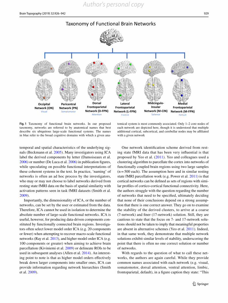
Some of the earliest resting state fMRI studies to deline-ate multiple macro-scale networks arrived at 5 (De Luca et al. 2006) and 10 (Damoiseaux et al. 2006) networks, respectively. These earlier works, combined with evidence from parcellation studies for seven (Yeo et al. 2011) and five (Doucet et al. 2019) networks observable at the macro-scale, were considered in our current proposal. As it will likely be easier for the community to agree upon a small set of core networks rather than a larger number, our proposal here centers around six functional brain networks that appear ubiquitously in both task and resting state fMRI investiga-tions. In an effort towards standardization, here we call these the occipital network (ON), pericentral network (PN), dorsal frontoparietal network (D-FPN), lateral frontoparietal net-work (L-FPN), midcingulo-insular network (M-CIN), and medial frontoparietal network (M-FPN) (Fig. 1).
Our proposed taxonomy is cortico-centric at present, though subcortical and cerebellar structures associated with each network are delineated where adequate information permits. Subcortical and cerebellar nodes are clearly associ-ated with each of the networks we discuss. In the interest of focusing on the issue of network nomenclature, rather than network composition, we refer the reader to several previ-ous works that have carefully delineated these components (Buckner et al. 2011; Choi et al. 2012; Ji et al. 2019).
At higher resolution, the six networks identified here will fractionate into subsystems (for example, the dissociation of the language network from the medial frontoparietal default network, and primary from secondary visual regions (Ji et al. 2019)). It is important to remember that fractionated sys-tems will likely show greater functional affiliation within the broader macro-scale network than between macro-scale networks, though time-varying analysis may reveal dynamic affiliations of brain regions with areas outside their core net-works (Uddin 2014).
Resting-State Functional Connectivity
Large-scale brain networks have been successfully deline-ated using an approach referred to as resting-state functional connectivity (RSFC). This approach examines synchro-nized patterns of spontaneous oscillations in blood-oxygen level dependent (BOLD) signal measured at rest with MRI (Biswal et al. 1995) (see Fox and Raichle 2007 for review). Some of the earliest studies using RSFC to delineate macro-scale functional brain networks used ICA. ICA is a model-free approach that decomposes neuroimaging datasets into a set of independent one-dimensional time series and associated three-dimensional spatial maps that describe the
Author's personal copy
929Brain Topography (2019) 32:926–942
1 3
temporal and spatial characteristics of the underlying sig-nals (Beckmann et al. 2005). Many investigators using ICA label the derived components by letter (Damoiseaux et al. 2006) or number (De Luca et al. 2006) in publication figures, while speculating on possible functional interpretations of these coherent systems in the text. In practice, ‘naming’ of networks is often an ad hoc process by the investigators, who may or may not choose to label networks derived from resting state fMRI data on the basis of spatial similarity with activation patterns seen in task fMRI datasets (Smith et al. 2009).
Importantly, the dimensionality of ICA, or the number of networks, can be set by the user or estimated from the data. Therefore, ICA cannot be used in isolation to determine the absolute number of large-scale functional networks. ICA is useful, however, for producing data-driven components con-stituted by functionally connected brain regions. Investiga-tors often select lower model order ICA (e.g. 20 components or fewer) when attempting to recover macro-scale functional networks (Ray et al. 2013), and higher model order ICA (e.g. 100 components or greater) when aiming to achieve brain parcellation (Kiviniemi et al. 2009) or delineate ROIs to be used in subsequent analyses (Allen et al. 2014). An interest-ing point to note is that as higher model orders effectively break down larger components into smaller ones, ICA can provide information regarding network hierarchies (Smith et al. 2009).
One network identification scheme derived from rest-ing state fMRI data that has been very influential is that proposed by Yeo et al. (2011). Yeo and colleagues used a clustering algorithm to parcellate the cortex into networks of functionally coupled brain regions using two large samples (n = 500 each). The assumption here and in similar resting state fMRI parcellation work (e.g. Power et al. 2011) is that cortical networks can be defined as sets of regions with simi-lar profiles of cortico-cortical functional connectivity. Here, the authors struggle with the question regarding the number of networks that need to be specified, ultimately deciding that none of their conclusions depend on a strong assump-tion that there is one correct answer. They go on to examine the stability of the derived clusters, to arrive at a coarse (7-network) and finer (17-network) solution. Still, they are cautious to state that the focus on 7- and 17-network solu-tions should not be taken to imply that meaningful properties are absent in alternative schemes (Yeo et al. 2011). Indeed, in that same work, they demonstrate that multiple network solutions exhibit similar levels of stability, underscoring the point that there is often no one correct solution or number of networks.
With regards to the question of what to call these net-works, the authors are again careful. While they provide common names associated with each network (e.g. visual, somatomotor, dorsal attention, ventral attention, limbic, frontoparietal, default), in a figure caption they state: “This
Fig. 1 Taxonomy of functional brain networks. In our proposed taxonomy, networks are referred to by anatomical names that best describe six ubiquitous large-scale functional systems. The names in blue refer to the broad cognitive domains with which a given ana-
tomical system is most commonly associated. Only 1–2 core nodes of each network are depicted here, though it is understood that multiple additional cortical, subcortical, and cerebellar nodes may be affiliated with a given network
Author's personal copy
930 Brain Topography (2019) 32:926–942
1 3
should not be taken to mean that our estimated networks correspond exactly to those in the literature or that the networks code solely for functions associated with their assigned name. As examples of limitations of heuristic ref-erence labels, the violet ventral attention network is likely an aggregate of (or closely adjacent to) multiple networks in the literature variably referred to as the salience (Seeley et al. 2007) and cingulo-opercular networks (Dosenbach et al. 2007), and the red default network can be fractionated (e.g. Andrews-Hanna et al. 2010).”
Similar caveats can also be seen in other work. For exam-ple Farrant and Uddin (2015) note in their work that while some investigators see the high degree of functional and anatomical overlap between the ventral attention network and salience network as evidence that they are part of the same system (Kucyi et al. 2012), others have conceptualized these networks as distinct entities (Power et al. 2011; Cole et al. 2013). Unfortunately, this type of nuance is not always evident in the broad network neuroscience literature. As no universally accepted network naming convention currently exists, researchers continue to adopt their own preferred nomenclature in publications, contributing to a greater pro-liferation of network naming schemes.
Task-Activation and Meta-analysis
Another way to define functional networks is by examin-ing patterns of task co-activation, and amalgamating these results through meta-analyses to discover reliable network nodes. The first such successful meta-analytic approach led to the discovery of the ubiquitous medial frontoparietal net-work. Reliable decreases in blood flow were found during active visual tasks in posterior cingulate, inferior parietal cortex, medial prefrontal cortex, and other regions (Shulman et al. 1997). It was only later that this constellation of regions was described as active “by default” (Raichle et al. 2001), and subsequently referred to as the “default mode network” upon demonstration of functional connectivity between its key nodes (Greicius et al. 2003). This network has been reliably observed to be suppressed during many tasks that require visuospatial attention and has been referred to as the “task-negative network” for its antiphase and largely antago-nistic relationship with the dorsal frontoparietal attention network (Fox et al. 2005) and other lateral frontoparietal net-works (Sridharan et al. 2008) (see also Dixon et al. (2017)). This unfortunate “task-negative” nomenclature has obscured the active role of the medial frontoparietal default network in numerous forms of cognition. Meta-analytic evidence sug-gests that this network is involved in memory processes, such as recollection, as well as social reasoning (Spreng et al. 2009). However, inquiry into cognition is often siloed into discrete domains of research, and a common set of co-active brain regions have been named for discrete cognitive
functions, with limited cross-talk and enriched understand-ing of how these seemingly diverse set of functions may rely on core mechanisms. For example:
“Recollection - retrieval of qualitative information about a past event - is associated with enhanced neural activity in a consistent set of neural regions (the ‘core recollection network’)…including the hippocampus, angular gyrus, medial prefrontal cortex, retrosplenial/posterior cingulate cortex, and middle temporal gyrus” (Thakral et al. 2017).“The mentalizing system - consisting of the temporo-parietal junction, the medial prefrontal cortex and the precuneus - is activated when behavior that enables inferences to be made about goals, beliefs or moral issues presented in abstract terms” (Van Overwalle and Baetens 2009).“The neural systems specialized for storage and retrieval of semantic knowledge are widespread and occupy a large proportion of the cortex in the human brain. The areas implicated in these processes can be grouped into three broad categories: posterior hetero-modal association cortex (AG, MTG, and fusiform gyrus), specific subregions of heteromodal prefrontal cortex (dorsal, ventromedial, and inferior prefrontal cortex), and medial paralimbic regions with strong connections to the hippocampal formation (parahip-pocampus and posterior cingulate gyrus)” (Binder et al. 2009).While describing different cognitive processes, the cor-
responding functional neuroanatomy in these three examples point to the medial frontoparietal default network. Similar to our earlier example centered on the anterior insula, a common nomenclature for the underlying network archi-tecture could enrich the cognitive characterization of these systems (e.g. Spreng and Andrews-Hanna 2015). Some of the imprecision in the field is explained by an incomplete correspondence between RSFC networks and task coactiva-tion patterns. While there is a broad convergence between task-evoked networks (Smith et al. 2009; Laird et al. 2011; Yeo et al. 2016) and resting-state fMRI derived networks (Yeo et al. 2011), there is not a perfect match. In many cases, resting state networks appear to be more broadly distrib-uted across the cortex, whereas task-evoked networks often appear more circumscribed (Yeo et al. 2016). One specula-tion based on this observation is that resting state networks might represent the full functional repertoire of brain modes, from which tasks engage subsets of regions as revealed by subtraction of tightly-matched control conditions. Projects such as the “cognitive atlas” (Poldrack et al. 2011) and the “Cognitive Paradigm Ontology” (Turner and Laird 2012) that aim to systematically characterized mental processes provide critical empirical data with which one can begin
Author's personal copy
931Brain Topography (2019) 32:926–942
1 3
to delineate task-evoked networks. Of note, the ubiquitous antagonistic brain activation/deactivation pattern between dorsal frontoparietal attention and medial frontoparietal default network brain regions, discussed above, can be reca-pitulated using such meta-analytic approaches (Bolt et al. 2017b; Toro et al. 2008). While an in depth discussion of this issue is beyond the scope of the current work, it is worth bearing in mind that the degree of correspondence between rest and task functional network configurations is a topic of ongoing investigation (Cole et al. 2014; Krienen et al. 2014; Bolt et al. 2017a).
Complex cognition may also evoke multiple, and interact-ing, networks. For example, working memory for visuospa-tial information will engage both the lateral frontoparietal control and dorsal frontoparietal attention networks, whereas working memory for mnemonic information will evoke activity in both the lateral frontoparietal control network and medial frontoparietal default networks (Spreng et al. 2010, 2012). Inter-regional patterns of RSFC have revealed that particular regions within the lateral frontoparietal con-trol network are more functionally aligned with either the medial default or dorsal frontopariatal attention networks (Spreng et al. 2013). These observations are consistent with a fractionation of the extended lateral frontoparietal control network into subsystems, with differing functional alignment depending upon task demands (Dixon et al. 2018). Depend-ing on the spatial scale, it is likely that all large-scale neuro-cognitive networks will fractionate, revealing both network hierarchies and dissociable cognitive functions.
Partially owing to these complexities, network neurosci-ence has largely sidestepped several key issues with regards to terminology. A common nomenclature based upon shared neuroanatomy would greatly facilitate the integration of novel discoveries within a cognitive network neuroscience framework. Such integration has the potential to deeply enrich our understanding of the macro-scale network archi-tecture of the human brain and ensure that findings from disparate subdisciplines can be more readily accessed and incorporated into theory.
Outline of a Universal Taxonomy of Functional Brain Networks
As reviewed above, the emerging field of network neurosci-ence currently suffers from the lack of a consistent network taxonomy. This is particularly problematic in that it hinders successful interfacing with decades of findings from cogni-tive neuroscience. The only way we see to remedy this is to formally propose a consensus nomenclature, closely tied to human neuroanatomy. This proposal synthesizes observa-tions from RSFC MRI, reliable patterns of functional coacti-vation from task-based fMRI, and cross-modal convergence
where available. We suggest common nomenclature for six reliable macro-scale brain networks composed of specific core brain regions. For each, we provide a primary anatomi-cal label, as well as a secondary, and necessarily broad, cog-nitive label. We note that these cognitive labels may need to be continuously revised as newer investigations suggest-ing previously unidentified functionality emerge. For this reason, we emphasize a priority to the anatomical network label. We name the core regions which comprise each net-work, noting that additional brain regions may participate in any given network through processes including dynamic affiliation (Pessoa 2014).
Because most networks are spatially distributed across cortical regions, anatomical labels reflect core regions for each network. Across studies, the extent of connectivity and coactivation can vary for many regions as a function of analytic approach, temporal signal-to-noise, and idiosyn-cratic task-dependent coactivation patterns. We denote these as zones that are less reliably characterized where appro-priate. In addition, we point out cases in which networks appear to break down further into separable subsystems. In some instances, the nodes we delineate as central to a given network can be characterized as “core” or “hub” nodes as defined in the graph theoretical sense (van den Heuvel and Sporns 2011). Our proposed taxonomy includes brief sum-maries of the cognitive functions associated with each net-work, and previously used terms for the network to aid in organization of prior observations.
The proposal is that going forward, network neuroscien-tists and cognitive neuroscientists should endeavor to use the following nomenclature whenever possible in order to provide a common reference point for other investigators interested in similar questions. As discussed, the core net-works we describe can often fractionate into multiple sub-systems that may not yet be fully described or agreed upon. In the interest of parsimony, we recommend that research-ers may benefit from using the broad anatomical network names suggested here, before further elaborating on the extent to which any given set of findings warrants the usage of additional nomenclature to more completely describe the network structure observed. For illustration purposes, we show examples from the literature of networks derived from resting-state fMRI parcellations (Yeo et al. 2011; Gordon et al. 2017c; Ji et al. 2019) and task-based fMRI (Toro et al. 2008; Smith et al. 2009; Corbetta and Shulman 2011; Nien-dam et al. 2012) that guide our taxonomy building project.
Anatomical Name: Occipital Network (ON)
Cognitive Domain: Visual Network
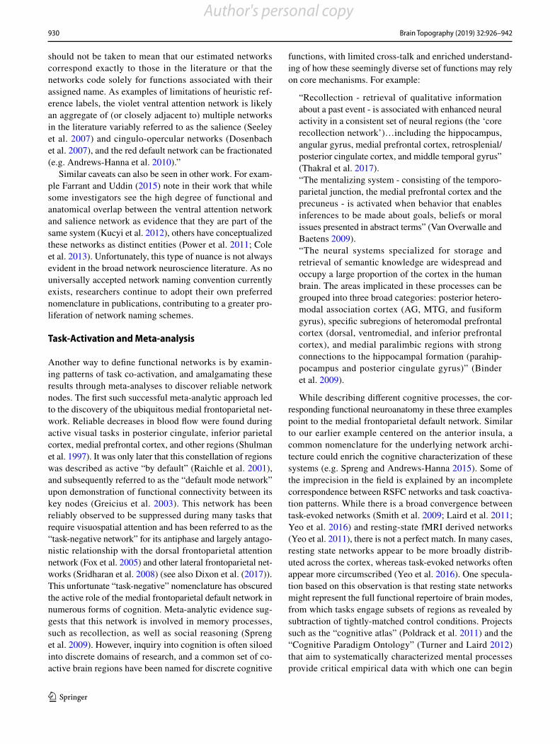
Core regions are the occipital lobe, including striate and extrastriate cortex (Fig. 2). This network also likely
Author's personal copy
932 Brain Topography (2019) 32:926–942
1 3
includes the lateral geniculate nucleus of the thalamus. The cognitive label “visual” is applied to this network, as the system is robustly observed to be involved in visual processing.
Figure 2 illustrates several examples of occipital net-works. In searching for correspondence between task-activation and ICA-derived resting state networks, Smith and colleagues observe three maps corresponding to medial, occipital pole, and lateral visual areas. Parcella-tions derived solely based on RSFC provide evidence for two visual networks, medial and lateral (Yeo et al. 2011; Gordon et al. 2017c). Taken together, these parcellation studies provided evidence for at least two subsystems associated with the ON, one situated more medially, asso-ciated with primary visual cortex along the calcarine sul-cus, and another more laterally encompassing extrastriate areas involved in visual processing (Haxby et al. 1994).
Note that the dorsal and ventral visual streams (Goodale and Milner 1992) likely originate from this core occipital network. These streams have been referred to as the “where” and “what” pathways for visual object perception (Ungerleider and Haxby 1994).
Anatomical Name: Pericentral Network (PN)
Cognitive Domain: Somatomotor Network
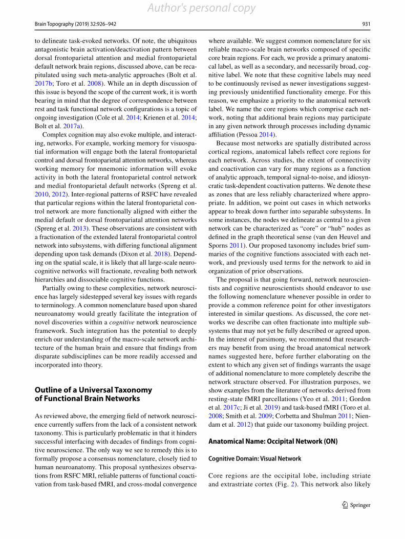
Core regions are motor and somatomotor cortices, anterior and posterior to the central sulcus. Regions of the pericen-tral network additionally include the juxtapositional lobule (supplementary motor area) (Fig. 3). Less well characterized zones include auditory cortex of the superior temporal gyrus, which is often encapsulated within this network in studies using RSFC. The cognitive label “somatomotor” is applied to this network for the system’s well-documented involve-ment in motor processes and somatosensory processing.
At least two subsystems are likely associated with the PN. Left and right separation can be observed using high model order ICA (Smith et al. 2009), and dorsal (hand) and ventral (face) subsystems appear in some parcellations (Yeo et al. 2011) 17-network; (Gordon et al. 2017c). At higher resolu-tion MRI, auditory and somatosensory face areas can also be separated (Kong et al. 2019). Note that the PN serves as the cortical component of both primary sensory and motor pathways.
Fig. 2 Occipital network. a Medial (120), occipital pole (220), and lat-eral (320) visual areas (Smith et al. 2009). RSN resting state network, BM BrainMap meta-analytic activation maps. b Purple and red visual
networks in 17-network parcellation (Yeo et al. 2011). c Medial (tan) and lateral (blue) visual networks (Gordon et al. 2017c)
Author's personal copy
933Brain Topography (2019) 32:926–942
1 3
Anatomical Name: Dorsal Frontoparietal Network (D-FPN)
Cognitive Domain: Attention Network
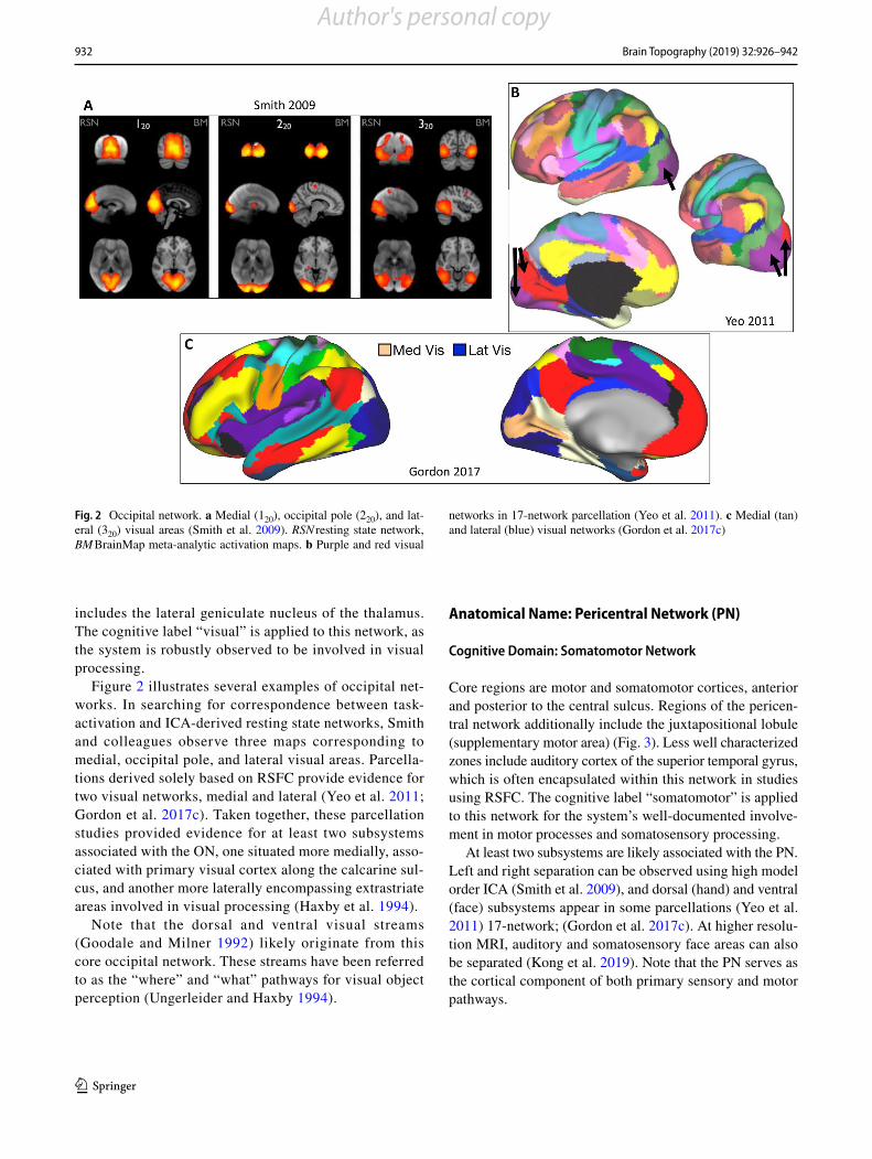
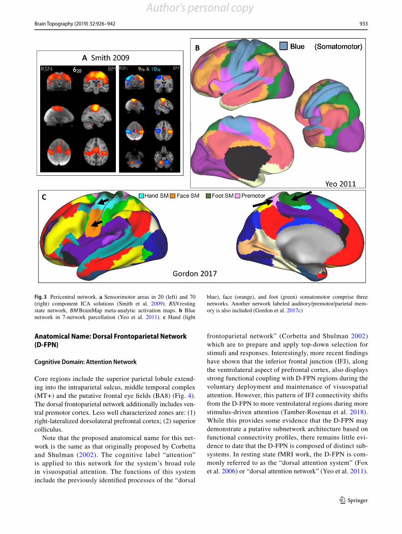
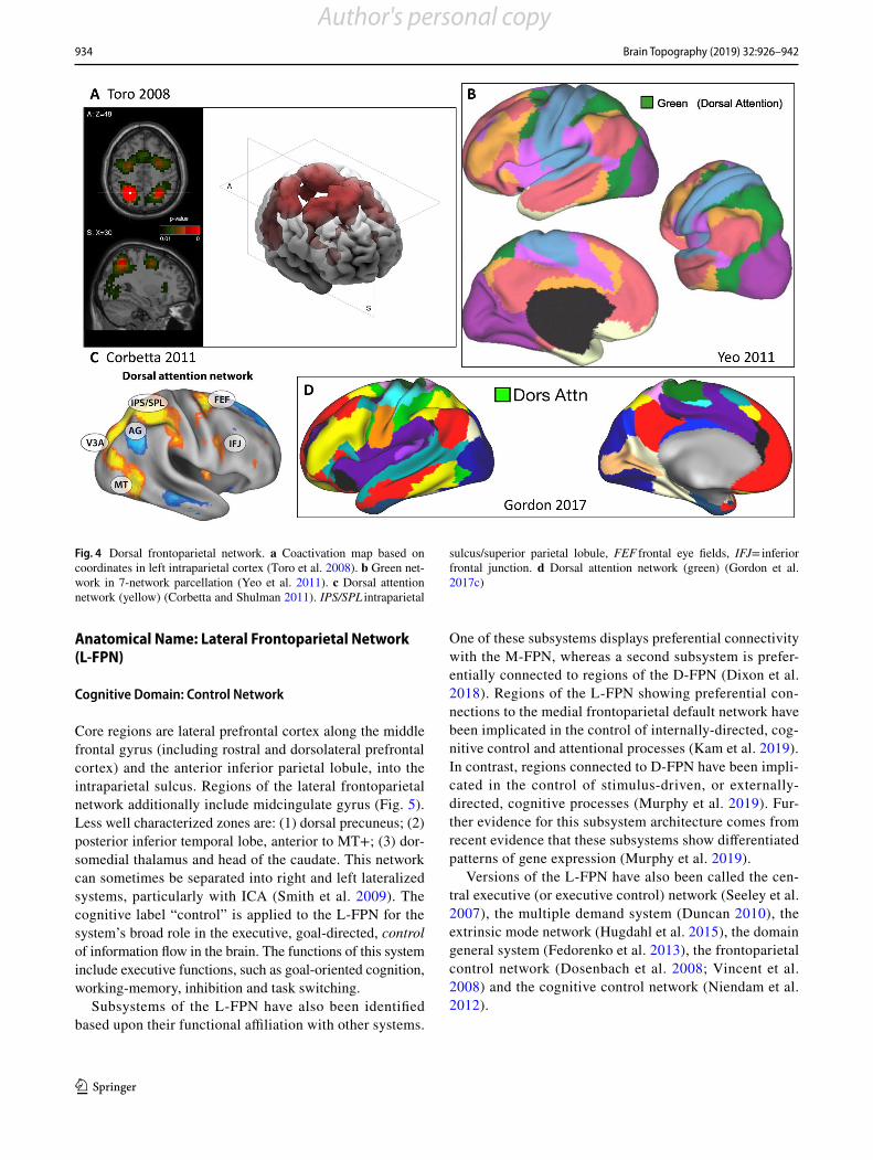
Core regions include the superior parietal lobule extend-ing into the intraparietal sulcus, middle temporal complex (MT+) and the putative frontal eye fields (BA8) (Fig. 4). The dorsal frontoparietal network additionally includes ven-tral premotor cortex. Less well characterized zones are: (1) right-lateralized dorsolateral prefrontal cortex; (2) superior colliculus.
Note that the proposed anatomical name for this net-work is the same as that originally proposed by Corbetta and Shulman (2002). The cognitive label “attention” is applied to this network for the system’s broad role in visuospatial attention. The functions of this system include the previously identified processes of the “dorsal
frontoparietal network” (Corbetta and Shulman 2002) which are to prepare and apply top-down selection for stimuli and responses. Interestingly, more recent findings have shown that the inferior frontal junction (IFJ), along the ventrolateral aspect of prefrontal cortex, also displays strong functional coupling with D-FPN regions during the voluntary deployment and maintenance of visuospatial attention. However, this pattern of IFJ connectivity shifts from the D-FPN to more ventrolateral regions during more stimulus-driven attention (Tamber-Rosenau et al. 2018). While this provides some evidence that the D-FPN may demonstrate a putative subnetwork architecture based on functional connectivity profiles, there remains little evi-dence to date that the D-FPN is composed of distinct sub-systems. In resting state fMRI work, the D-FPN is com-monly referred to as the “dorsal attention system” (Fox et al. 2006) or “dorsal attention network” (Yeo et al. 2011).
Fig. 3 Pericentral network. a Sensorimotor areas in 20 (left) and 70 (right) component ICA solutions (Smith et al. 2009). RSN resting state network, BM BrainMap meta-analytic activation maps. b Blue network in 7-network parcellation (Yeo et al. 2011). c Hand (light
blue), face (orange), and foot (green) somatomotor comprise three networks. Another network labeled auditory/premotor/parietal mem-ory is also included (Gordon et al. 2017c)
Author's personal copy
934 Brain Topography (2019) 32:926–942
1 3
Anatomical Name: Lateral Frontoparietal Network (L-FPN)
Cognitive Domain: Control Network
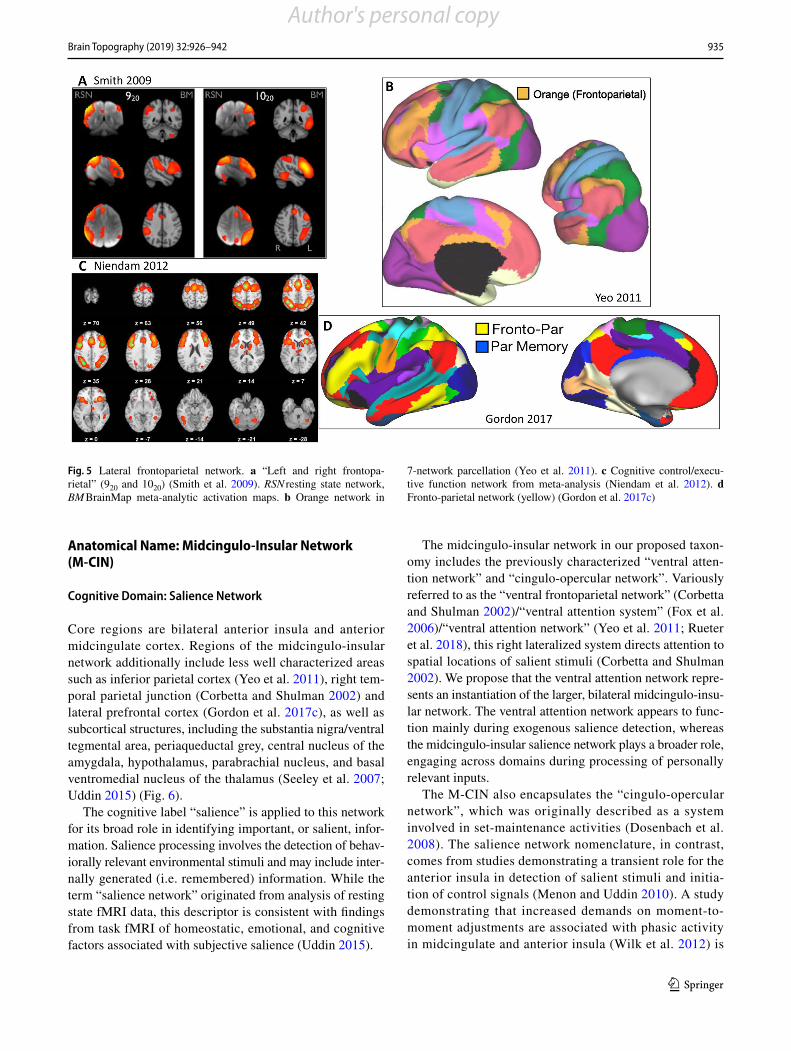
Core regions are lateral prefrontal cortex along the middle frontal gyrus (including rostral and dorsolateral prefrontal cortex) and the anterior inferior parietal lobule, into the intraparietal sulcus. Regions of the lateral frontoparietal network additionally include midcingulate gyrus (Fig. 5). Less well characterized zones are: (1) dorsal precuneus; (2) posterior inferior temporal lobe, anterior to MT+; (3) dor-somedial thalamus and head of the caudate. This network can sometimes be separated into right and left lateralized systems, particularly with ICA (Smith et al. 2009). The cognitive label “control” is applied to the L-FPN for the system’s broad role in the executive, goal-directed, control of information flow in the brain. The functions of this system include executive functions, such as goal-oriented cognition, working-memory, inhibition and task switching.
Subsystems of the L-FPN have also been identified based upon their functional affiliation with other systems.
One of these subsystems displays preferential connectivity with the M-FPN, whereas a second subsystem is prefer-entially connected to regions of the D-FPN (Dixon et al. 2018). Regions of the L-FPN showing preferential con-nections to the medial frontoparietal default network have been implicated in the control of internally-directed, cog-nitive control and attentional processes (Kam et al. 2019). In contrast, regions connected to D-FPN have been impli-cated in the control of stimulus-driven, or externally-directed, cognitive processes (Murphy et al. 2019). Fur-ther evidence for this subsystem architecture comes from recent evidence that these subsystems show differentiated patterns of gene expression (Murphy et al. 2019).
Versions of the L-FPN have also been called the cen-tral executive (or executive control) network (Seeley et al. 2007), the multiple demand system (Duncan 2010), the extrinsic mode network (Hugdahl et al. 2015), the domain general system (Fedorenko et al. 2013), the frontoparietal control network (Dosenbach et al. 2008; Vincent et al. 2008) and the cognitive control network (Niendam et al. 2012).
Fig. 4 Dorsal frontoparietal network. a Coactivation map based on coordinates in left intraparietal cortex (Toro et al. 2008). b Green net-work in 7-network parcellation (Yeo et al. 2011). c Dorsal attention network (yellow) (Corbetta and Shulman 2011). IPS/SPL intraparietal
sulcus/superior parietal lobule, FEF frontal eye fields, IFJ= inferior frontal junction. d Dorsal attention network (green) (Gordon et al. 2017c)
Author's personal copy
935Brain Topography (2019) 32:926–942
1 3
Anatomical Name: Midcingulo-Insular Network (M-CIN)
Cognitive Domain: Salience Network
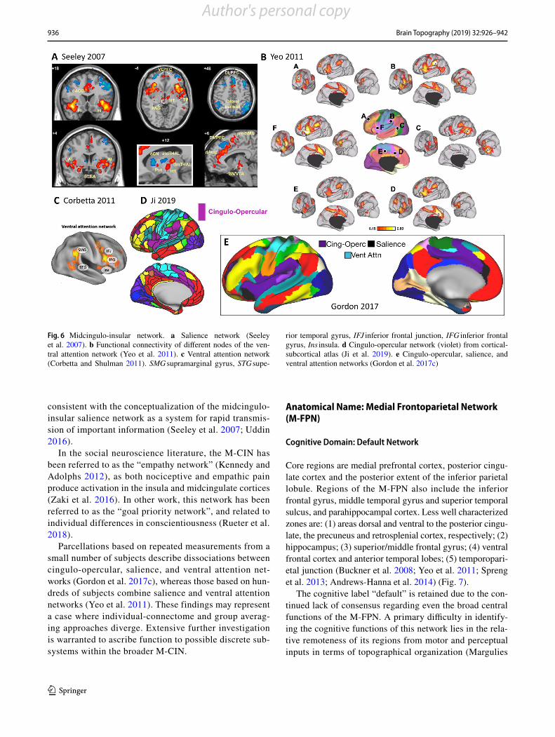
Core regions are bilateral anterior insula and anterior midcingulate cortex. Regions of the midcingulo-insular network additionally include less well characterized areas such as inferior parietal cortex (Yeo et al. 2011), right tem-poral parietal junction (Corbetta and Shulman 2002) and lateral prefrontal cortex (Gordon et al. 2017c), as well as subcortical structures, including the substantia nigra/ventral tegmental area, periaqueductal grey, central nucleus of the amygdala, hypothalamus, parabrachial nucleus, and basal ventromedial nucleus of the thalamus (Seeley et al. 2007; Uddin 2015) (Fig. 6).
The cognitive label “salience” is applied to this network for its broad role in identifying important, or salient, infor-mation. Salience processing involves the detection of behav-iorally relevant environmental stimuli and may include inter-nally generated (i.e. remembered) information. While the term “salience network” originated from analysis of resting state fMRI data, this descriptor is consistent with findings from task fMRI of homeostatic, emotional, and cognitive factors associated with subjective salience (Uddin 2015).
The midcingulo-insular network in our proposed taxon-omy includes the previously characterized “ventral atten-tion network” and “cingulo-opercular network”. Variously referred to as the “ventral frontoparietal network” (Corbetta and Shulman 2002)/“ventral attention system” (Fox et al. 2006)/“ventral attention network” (Yeo et al. 2011; Rueter et al. 2018), this right lateralized system directs attention to spatial locations of salient stimuli (Corbetta and Shulman 2002). We propose that the ventral attention network repre-sents an instantiation of the larger, bilateral midcingulo-insu-lar network. The ventral attention network appears to func-tion mainly during exogenous salience detection, whereas the midcingulo-insular salience network plays a broader role, engaging across domains during processing of personally relevant inputs.
The M-CIN also encapsulates the “cingulo-opercular network”, which was originally described as a system involved in set-maintenance activities (Dosenbach et al. 2008). The salience network nomenclature, in contrast, comes from studies demonstrating a transient role for the anterior insula in detection of salient stimuli and initia-tion of control signals (Menon and Uddin 2010). A study demonstrating that increased demands on moment-to-moment adjustments are associated with phasic activity in midcingulate and anterior insula (Wilk et al. 2012) is
Fig. 5 Lateral frontoparietal network. a “Left and right frontopa-rietal” (920 and 1020) (Smith et al. 2009). RSN resting state network, BM BrainMap meta-analytic activation maps. b Orange network in
7-network parcellation (Yeo et al. 2011). c Cognitive control/execu-tive function network from meta-analysis (Niendam et al. 2012). d Fronto-parietal network (yellow) (Gordon et al. 2017c)
Author's personal copy
936 Brain Topography (2019) 32:926–942
1 3
consistent with the conceptualization of the midcingulo-insular salience network as a system for rapid transmis-sion of important information (Seeley et al. 2007; Uddin 2016).
In the social neuroscience literature, the M-CIN has been referred to as the “empathy network” (Kennedy and Adolphs 2012), as both nociceptive and empathic pain produce activation in the insula and midcingulate cortices (Zaki et al. 2016). In other work, this network has been referred to as the “goal priority network”, and related to individual differences in conscientiousness (Rueter et al. 2018).
Parcellations based on repeated measurements from a small number of subjects describe dissociations between cingulo-opercular, salience, and ventral attention net-works (Gordon et al. 2017c), whereas those based on hun-dreds of subjects combine salience and ventral attention networks (Yeo et al. 2011). These findings may represent a case where individual-connectome and group averag-ing approaches diverge. Extensive further investigation is warranted to ascribe function to possible discrete sub-systems within the broader M-CIN.
Anatomical Name: Medial Frontoparietal Network (M-FPN)
Cognitive Domain: Default Network
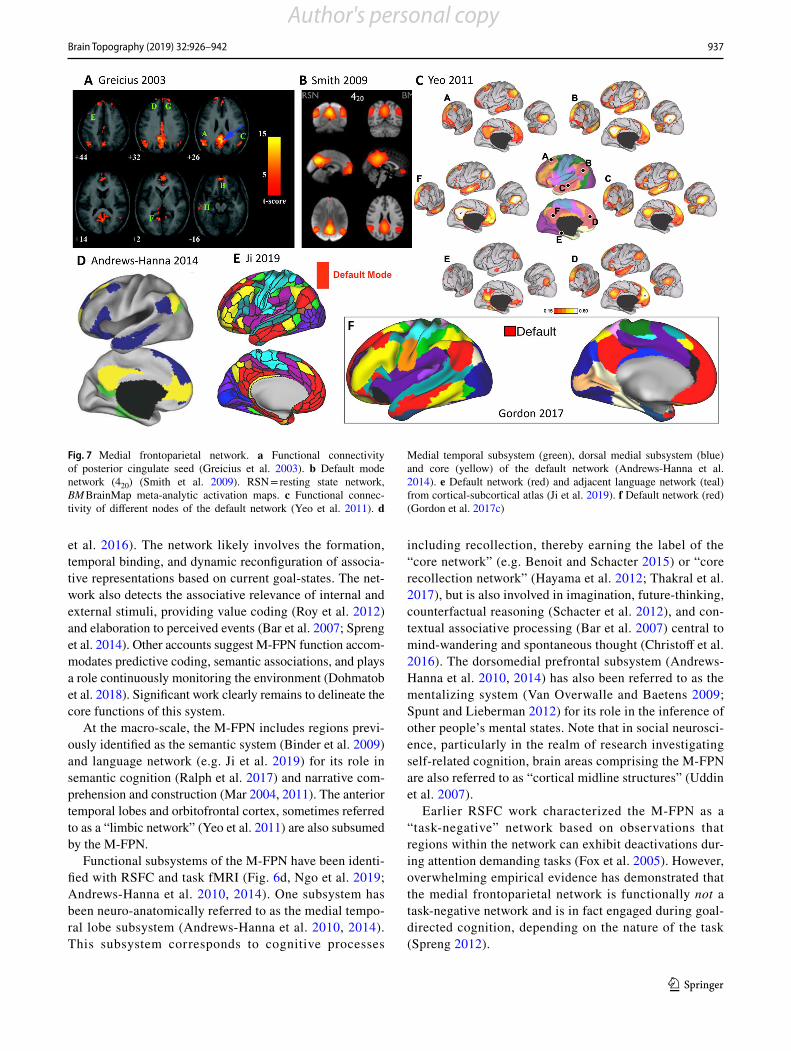
Core regions are medial prefrontal cortex, posterior cingu-late cortex and the posterior extent of the inferior parietal lobule. Regions of the M-FPN also include the inferior frontal gyrus, middle temporal gyrus and superior temporal sulcus, and parahippocampal cortex. Less well characterized zones are: (1) areas dorsal and ventral to the posterior cingu-late, the precuneus and retrosplenial cortex, respectively; (2) hippocampus; (3) superior/middle frontal gyrus; (4) ventral frontal cortex and anterior temporal lobes; (5) temporopari-etal junction (Buckner et al. 2008; Yeo et al. 2011; Spreng et al. 2013; Andrews-Hanna et al. 2014) (Fig. 7).
The cognitive label “default” is retained due to the con-tinued lack of consensus regarding even the broad central functions of the M-FPN. A primary difficulty in identify-ing the cognitive functions of this network lies in the rela-tive remoteness of its regions from motor and perceptual inputs in terms of topographical organization (Margulies
Fig. 6 Midcingulo-insular network. a Salience network (Seeley et al. 2007). b Functional connectivity of different nodes of the ven-tral attention network (Yeo et al. 2011). c Ventral attention network (Corbetta and Shulman 2011). SMG supramarginal gyrus, STG supe-
rior temporal gyrus, IFJ inferior frontal junction, IFG inferior frontal gyrus, Ins insula. d Cingulo-opercular network (violet) from cortical-subcortical atlas (Ji et al. 2019). e Cingulo-opercular, salience, and ventral attention networks (Gordon et al. 2017c)
Author's personal copy
937Brain Topography (2019) 32:926–942
1 3
et al. 2016). The network likely involves the formation, temporal binding, and dynamic reconfiguration of associa-tive representations based on current goal-states. The net-work also detects the associative relevance of internal and external stimuli, providing value coding (Roy et al. 2012) and elaboration to perceived events (Bar et al. 2007; Spreng et al. 2014). Other accounts suggest M-FPN function accom-modates predictive coding, semantic associations, and plays a role continuously monitoring the environment (Dohmatob et al. 2018). Significant work clearly remains to delineate the core functions of this system.
At the macro-scale, the M-FPN includes regions previ-ously identified as the semantic system (Binder et al. 2009) and language network (e.g. Ji et al. 2019) for its role in semantic cognition (Ralph et al. 2017) and narrative com-prehension and construction (Mar 2004, 2011). The anterior temporal lobes and orbitofrontal cortex, sometimes referred to as a “limbic network” (Yeo et al. 2011) are also subsumed by the M-FPN.
Functional subsystems of the M-FPN have been identi-fied with RSFC and task fMRI (Fig. 6d, Ngo et al. 2019; Andrews-Hanna et al. 2010, 2014). One subsystem has been neuro-anatomically referred to as the medial tempo-ral lobe subsystem (Andrews-Hanna et al. 2010, 2014). This subsystem corresponds to cognitive processes
including recollection, thereby earning the label of the “core network” (e.g. Benoit and Schacter 2015) or “core recollection network” (Hayama et al. 2012; Thakral et al. 2017), but is also involved in imagination, future-thinking, counterfactual reasoning (Schacter et al. 2012), and con-textual associative processing (Bar et al. 2007) central to mind-wandering and spontaneous thought (Christoff et al. 2016). The dorsomedial prefrontal subsystem (Andrews-Hanna et al. 2010, 2014) has also been referred to as the mentalizing system (Van Overwalle and Baetens 2009; Spunt and Lieberman 2012) for its role in the inference of other people’s mental states. Note that in social neurosci-ence, particularly in the realm of research investigating self-related cognition, brain areas comprising the M-FPN are also referred to as “cortical midline structures” (Uddin et al. 2007).
Earlier RSFC work characterized the M-FPN as a “task-negative” network based on observations that regions within the network can exhibit deactivations dur-ing attention demanding tasks (Fox et al. 2005). However, overwhelming empirical evidence has demonstrated that the medial frontoparietal network is functionally not a task-negative network and is in fact engaged during goal-directed cognition, depending on the nature of the task (Spreng 2012).
Fig. 7 Medial frontoparietal network. a Functional connectivity of posterior cingulate seed (Greicius et al. 2003). b Default mode network (420) (Smith et al. 2009). RSN = resting state network, BM BrainMap meta-analytic activation maps. c Functional connec-tivity of different nodes of the default network (Yeo et al. 2011). d
Medial temporal subsystem (green), dorsal medial subsystem (blue) and core (yellow) of the default network (Andrews-Hanna et al. 2014). e Default network (red) and adjacent language network (teal) from cortical-subcortical atlas (Ji et al. 2019). f Default network (red) (Gordon et al. 2017c)
Author's personal copy
938 Brain Topography (2019) 32:926–942
1 3
Outstanding Issues and Future Directions
Several important considerations and outstanding issues should be acknowledged with regards to our proposed net-work taxonomy. Here we focus on macro-scale functional networks, with a strong emphasis on converging evidence from RSFC and task-activation fMRI studies. In this final section, we note that continued development in functional connectivity dynamics, accounting for inter-individual variability, and incomplete delineation remain significant challenges as we move forward in the development and adoption of a universal taxonomy.
As we have alluded to throughout, a simplifying assumption is that static macro-scale human brain net-works can be delineated and described. However, recent work emphasizes the time-varying nature of functional connectivity and the importance of considering temporal properties of brain networks (Hutchison et al. 2013). Early observations of this phenomenon include work by Chang and Glover, who demonstrated that the posterior cingulate cortex, a primary node of the M-FPN, exhibits variable functional connectivity with the rest of the brain such that commonly observed negative correlations between the M-FPN and other frontoparietal networks should not be viewed as static (Chang and Glover 2010). Some have proposed methods for leveraging time-varying properties of functional networks for parcellation, uncovering “rep-resentative dominant patterns” (Preti and Van De Ville 2017), though these approaches await further validation. Controversies surrounding the interpretation of dynamic functional connectivity notwithstanding (Hindriks et al. 2016; Laumann et al. 2017; Liégeois et al. 2017), consid-eration of brain dynamics remains an intriguing direction for future research aimed at network taxonomy delineation.
There is substantial variability across humans in the precise spatial location of functional brain areas (Stevens et al. 2015). Subject-specific functional localization of brain regions using task fMRI provides one solution to determining broader network affiliation, which can mean-ingfully predict individual differences in behavior (Stevens et al. 2017). Several researchers have noted that the size, location, and spatial arrangement of individual-specific brain networks vary substantially across participants (Wang et al. 2015; Harrison et al. 2015; Laumann et al. 2015; Glasser et al. 2016; Gordon et al. 2017a, b, c; Braga and Buckner 2017) Recent studies have suggested that the spatial arrangement (e.g. topography) and size of individ-ual-specific networks can be predictive of demographics (e.g., sex) and behavior (Bijsterbosch et al. 2018; Salehi et al. 2018; Cui et al. 2019; Kong et al. 2019; Li et al. 2019a, b; Seitzman et al. 2019). Whole brain approaches to better estimate and delineate inter-subject variability in
functional brain regions comprising large-scale brain net-works are being developed in earnest. These whole brain approaches determine individual locations of functional brain areas from patterns of RSFC (Chong et al. 2017), and may be more sensitive to detecting RSFC associa-tions with individual differences in behavior (Mwilambwe-Tshilobo et al. 2019). Furthermore, some have suggested that network subsystems might be fully dissociable, rather than simply overlapping, when examined within-subject and with high-resolution data (Braga and Buckner 2017; Braga et al. 2019). In one such example, the dorsal, lateral and medial frontoparietal networks were found to each represent two fully dissociable networks when examined within-subject (Braga and Buckner 2017), an idea that warrants further investigation. Finally, analysis of task fMRI reveals that inter-subject and task-condition vari-ability can be influenced by the resolution of the data, such that when moving from lower to higher resolution, variance in activation maps explained by between-person differences increases while variance explained by task conditions decreases (Bolt et al. 2019). All of these con-siderations surrounding inter-subject variability must be considered in future iterations of the taxonomy proposed here.
Within the proposed six-network taxonomy, many regions of cortex are not classified, and subcortical regions are not fully incorporated at this time. Our classification most prominently excludes ventral temporal cortex. These regions have been characterized as “ventral multi-modal” zones in a recent parcellation scheme (Ji et al. 2019). Future work further incorporating whole brain characterization, includ-ing subcortical, cerebellar and brainstem structures will be essential.
While the task of creating a universally accepted taxon-omy of human brain networks is daunting, we are optimistic that it will be realized in the coming decade. The six-net-work scheme outlined here is based on a synthesis of prac-tices and assumptions that are already in place. The critical contribution of the current proposal is the introduction of a consistent, anatomically-grounded naming convention that will enable researchers investigating the same brain systems to communicate more effectively. If we can agree on a basic set of regions, their rough boundaries, and the conditions under which they interact to form a network, then we may move forward as a field with a consensus on the macro-scale neurocognitive networks of the human brain.
There are multiple approaches for defining large-scale networks (Eickhoff et al. 2018a, b). In order to create a tax-onomy that has a broad impact and is universally adopted, a larger group within the community of researchers must be engaged. Inspiration for this endeavor could come from sim-ilar efforts within the neuorimaging community, such as the Time Varying Working Group, which has worked towards
Author's personal copy
939Brain Topography (2019) 32:926–942
1 3
consensus with regards to issues surrounding the measure-ment and interpretation of dynamic functional connectivity (Lurie et al. 2018). Other groups have worked towards stand-ardized definitions of functional and effective connectivity (Reid et al. 2019), and best practices in MRI data analysis and sharing (Nichols et al. 2017). A working group devoted to standardization of network naming conventions could be assembled to follow up the initial effort presented here, potentially drawing upon more varied network characteriza-tion, such as structural measures.
As it is unlikely that these naming conventions will immediately replace those that have been used up to this point, we propose a simple process by which a transition to the taxonomy proposed here might be adopted. In future studies, some researchers may still wish to name their net-works of interest using their favorite nomenclature for the sake of continuity with previous work. However, we would urge that they should also include the terminology we intro-duce here. For example, in future papers using the “sali-ence network” term, we would hope that the authors include “midcingulo-insular network” as a keyword in the publica-tion so that their work will be accessible in the future. Mov-ing forward, we suggest that networks not be named for a favored cognitive function, particularly when based solely on task-evoked activation patterns. The principle of many-to-many mapping suggests that ascribing a singular function to a brain region or network is likely erroneous, and serves to deepen the silos of an already fractionated literature. We hope that the proposal outlined here is adopted in the fields of network neuroscience and cognitive neuroscience, and that we will see many more studies examining functional properties of the occipital, pericentral, dorsal frontoparietal, lateral frontoparietal, midcingulo-insular, and medial fron-toparietal networks in the years to come.
Acknowledgements LQU is supported by the National Institute of Mental Health (R01MH107549), the Canadian Institute for Advanced Research, and a University of Miami Gabelli Senior Scholar Award. RNS is supported by the Natural Sciences and Engineering Research Council of Canada and Canadian Institutes of Health Research, and is a Research Scholar supported by Fonds de recherche du Québec – Santé. BTTY is supported by the Singapore National Research Foundation (NRF) Fellowship (Class of 2017). The authors gratefully acknowledge Roberto Toro and Evan Gordan for assistance with figures.
References
Allen EA, Damaraju E, Plis SM et al (2014) Tracking whole-brain con-nectivity dynamics in the resting state. Cereb Cortex 24:663–676
Andrews-Hanna JR, Reidler JS, Sepulcre J et al (2010) Functional-anatomic fractionation of the brain’s default network. Neuron 65:550–562
Andrews-Hanna JR, Smallwood J, Spreng RN (2014) The default net-work and self-generated thought: component processes, dynamic control, and clinical relevance. Ann N Y Acad Sci 1316:29–52
Bar M, Aminoff E, Mason M, Fenske M (2007) The units of thought. Hippocampus 17:420–428
Barrett LF, Satpute AB (2013) Large-scale brain networks in affec-tive and social neuroscience: towards an integrative functional architecture of the brain. Curr Opin Neurobiol. 23:361–372
Bassett DS, Sporns O (2017) Network neuroscience. Nat Neurosci 20:353–364
Beckmann CF, DeLuca M, Devlin JT, Smith SM (2005) Investiga-tions into resting-state connectivity using independent com-ponent analysis. Philos Trans R Soc Lond B 360:1001–1013
Benoit RG, Schacter DL (2015) Specifying the core network sup-porting episodic simulation and episodic memory by activation likelihood estimation. Neuropsychologia 75:450–457
Betzel RF, Bassett DS (2017) Multi-scale brain networks. Neuroim-age 160:73–83
Bijsterbosch JD, Woolrich MW, Glasser MF et al (2018) The rela-tionship between spatial configuration and functional con-nectivity of brain regions. eLife. https ://doi.org/10.7554/eLife .32992
Binder JR, Desai RH, Graves WW, Conant LL (2009) Where is the semantic system? A critical review and meta-analysis of 120 functional neuroimaging studies. Cereb Cortex 19:2767–2796
Biswal B, Yetkin FZ, Haughton VM, Hyde JS (1995) Functional connectivity in the motor cortex of resting human brain using echo-planar MRI. Magn Reson Med 34:537–541
Bolt T, Nomi JS, Rubinov M, Uddin LQ (2017a) Correspondence between evoked and intrinsic functional brain network configu-rations. Hum Brain Mapp 38:1992–2007
Bolt T, Nomi JS, Yeo BTT, Uddin LQ (2017b) Data-Driven extrac-tion of a nested model of human brain function. J Neurosci 37:7263–7277
Bolt T, Nomi JS, Bainter SA et al (2019) The situation or the person? Individual and task-evoked differences in BOLD activity. Hum Brain Mapp 40:2943–2954
Braga RM, Buckner RL (2017) Parallel interdigitated distributed net-works within the individual estimated by intrinsic functional connectivity. Neuron 95:457–471.e5
Braga RM, Van Dijk KRA, Polimeni JR et al (2019) Parallel distrib-uted networks resolved at high resolution reveal close juxta-position of distinct regions. J Neurophysiol 121:1513–1534
Buckner RL, Andrews-Hanna JR, Schacter DL (2008) The brain’s default network: anatomy, function, and relevance to disease. Ann N Y Acad Sci 1124:1–38
Buckner RL, Krienen FM, Castellanos A et al (2011) The organiza-tion of the human cerebellum estimated by intrinsic functional connectivity. J Neurophysiol 106:2322–2345
Chang C, Glover GH (2010) Time-frequency dynamics of resting-state brain connectivity measured with fMRI. Neuroimage 50:81–98
Choi EY, Yeo BTT, Buckner RL (2012) The organization of the human striatum estimated by intrinsic functional connectivity. J Neuro-physiol 108:2242–2263
Chong M, Bhushan C, Joshi AA et al (2017) Individual parcellation of resting fMRI with a group functional connectivity prior. Neu-roimage 156:87–100
Christoff K, Irving ZC, Fox KCR et al (2016) Mind-wandering as spontaneous thought: a dynamic framework. Nat Rev Neurosci 17:718–731
Ciric R, Nomi JS, Uddin LQ, Satpute AB (2017) Contextual connectiv-ity: a framework for understanding the intrinsic dynamic archi-tecture of large-scale functional brain networks. Sci Rep 7:6537
Cole MW, Reynolds JR, Power JD et al (2013) Multi-task connectiv-ity reveals flexible hubs for adaptive task control. Nat Neurosci 16:1348–1355
Cole MW, Bassett DS, Power JD et al (2014) Intrinsic and task-evoked network architectures of the human brain. Neuron 83:238–251
Author's personal copy
940 Brain Topography (2019) 32:926–942
1 3
Collins DL, Neelin P, Peters TM, Evans AC (1994) Automatic 3D intersubject registration of MR volumetric data in standardized Talairach space. J Comput Assist Tomogr 18:192–205
Corbetta M, Shulman GL (2002) Control of goal-directed and stimulus-driven attention in the brain. Nat Rev Neurosci 3:201–215
Corbetta M, Shulman GL (2011) Spatial neglect and attention net-works. Annu Rev Neurosci 34:569–599
Corbetta M, Patel G, Shulman GL (2008) The reorienting system of the human brain: from environment to theory of mind. Neuron 58:306–324
Cui Z, Li H, Xia CH, et al (2019) Individual variation in control net-work topography supports executive function in youth
Damoiseaux JS, Rombouts SA, Barkhof F et al (2006) Consistent resting-state networks across healthy subjects. Proc Natl Acad Sci USA 103:13848–13853
De Luca M, Beckmann CF, De Stefano N et al (2006) fMRI resting state networks define distinct modes of long-distance interactions in the human brain. Neuroimage 29:1359–1367
Dixon ML, Andrews-Hanna JR, Spreng RN et al (2017) Interactions between the default network and dorsal attention network vary across default subsystems, time, and cognitive states. Neuroim-age 147:632–649
Dixon ML, De La Vega A, Mills C et al (2018) Heterogeneity within the frontoparietal control network and its relationship to the default and dorsal attention networks. Proc Natl Acad Sci USA 115(7):E1598–E1607
Dohmatob E, Dumas G, Bzdok D (2018) Dark control: towards a unified account of default mode function by Markov decision processes
Dosenbach NU, Fair DA, Miezin FM et al (2007) Distinct brain net-works for adaptive and stable task control in humans. Proc Natl Acad Sci USA 104:11073–11078
Dosenbach NU, Fair DA, Cohen AL et al (2008) A dual-networks architecture of top-down control. Trends Cogn Sci 12:99–105
Doucet GE, Lee WH, Frangou S (2019) Evaluation of the spatial vari-ability in the major resting-state networks across human brain functional atlases. Hum Brain Mapp 40:4577–4587
Duncan J (2010) The multiple-demand (MD) system of the primate brain: mental programs for intelligent behaviour. Trends Cogn Sci 14:172–179
Eickhoff SB, Constable RT, Yeo BTT (2018a) Topographic organiza-tion of the cerebral cortex and brain cartography. Neuroimage 170:332–347
Eickhoff SB, Yeo BTT, Genon S (2018b) Imaging-based parcellations of the human brain. Nat Rev Neurosci 19:672–686
Farrant K, Uddin LQ (2015) Asymmetric development of dorsal and ventral attention networks in the human brain. Dev Cogn Neu-rosci 12:165–174
Fedorenko E, Duncan J, Kanwisher N (2013) Broad domain generality in focal regions of frontal and parietal cortex. Proc Natl Acad Sci USA 110:16616–16621
Felleman DJ, Van Essen DC (1991) Distributed hierarchical processing in the primate cerebral cortex. Cereb Cortex 1:1–47
Fox MD, Raichle ME (2007) Spontaneous fluctuations in brain activity observed with functional magnetic resonance imaging. Nat Rev Neurosci 8:700–711
Fox MD, Snyder AZ, Vincent JL et al (2005) The human brain is intrinsically organized into dynamic, anticorrelated functional networks. Proc Natl Acad Sci USA 102:9673–9678
Fox MD, Corbetta M, Snyder AZ et al (2006) Spontaneous neuronal activity distinguishes human dorsal and ventral attention systems. Proc Natl Acad Sci USA 103:10046–10051
Friston K (1994) Functional and effective connectivity in neuroimag-ing: a synthesis. Hum Brain Mapp 2:56–78
Glasser MF, Coalson TS, Robinson EC et al (2016) A multi-modal parcellation of human cerebral cortex. Nature 536:171–178
Goodale MA, Milner AD (1992) Separate visual pathways for per-ception and action. Trends Neurosci 15:20–25
Gordon EM, Laumann TO, Adeyemo B et al (2017a) Individual-specific features of brain systems identified with resting state functional correlations. NeuroImage 146:918–939
Gordon EM, Laumann TO, Adeyemo B, Petersen SE (2017b) Indi-vidual variability of the system-level organization of the human brain. Cereb Cortex 27:386–399
Gordon EM, Laumann TO, Gilmore AW et al (2017c) Precision func-tional mapping of individual human brains. Neuron 95:791–807.e7
Greicius MD, Krasnow B, Reiss AL, Menon V (2003) Func-tional connectivity in the resting brain: a network analysis of the default mode hypothesis. Proc Natl Acad Sci USA 100:253–258
Harrison SJ, Woolrich MW, Robinson EC et al (2015) Large-scale probabilistic functional modes from resting state fMRI. Neuro-image 109:217–231
Haxby JV, Horwitz B, Ungerleider LG et al (1994) The functional organization of human extrastriate cortex: a PET-rCBF study of selective attention to faces and locations. J Neurosci 14:6336–6353
Hayama HR, Vilberg KL, Rugg MD (2012) Overlap between the neu-ral correlates of cued recall and source memory: evidence for a generic recollection network? J Cogn Neurosci 24:1127–1137
Hindriks R, Adhikari MH, Murayama Y et al (2016) Can sliding-window correlations reveal dynamic functional connectivity in resting-state fMRI? Neuroimage 127:242–256
Hugdahl K, Raichle ME, Mitra A, Specht K (2015) On the existence of a generalized non-specific task-dependent network. Front Hum Neurosci 9:430
Hutchison RM, Womelsdorf T, Allen EA et al (2013) Dynamic func-tional connectivity: promise, issues, and interpretations. Neuro-image 80:360–378
Ji JL, Spronk M, Kulkarni K et al (2019) Mapping the human brain’s cortical-subcortical functional network organization. Neuroim-age 185:35–57
Kam JWY, Lin JJ, Solbakk A-K et al (2019) Default network and frontoparietal control network theta connectivity supports inter-nal attention. Nat Hum Behav. https ://doi.org/10.1038/s4156 2-019-0717-0
Kennedy DP, Adolphs R (2012) The social brain in psychiatric and neurological disorders. Trends Cogn Sci 16:559–572
Kiviniemi V, Starck T, Remes J et al (2009) Functional segmentation of the brain cortex using high model order group PICA. Hum Brain Mapp 30:3865–3886
Kong R, Li J, Orban C et al (2019) Spatial topography of individual-specific cortical networks predicts human cognition, personality, and emotion. Cereb Cortex 29:2533–2551
Krienen FM, Yeo BTT, Buckner RL (2014) Reconfigurable task-dependent functional coupling modes cluster around a core func-tional architecture. Philos Trans R Soc B 369:20130526
Kucyi A, Hodaie M, Davis KD (2012) Lateralization in intrinsic functional connectivity of the temporoparietal junction with salience- and attention-related brain networks. J Neurophysiol 108:3382–3392
Laird AR, Fox PM, Eickhoff SB et al (2011) Behavioral interpre-tations of intrinsic connectivity networks. J Cogn Neurosci 23:4022–4037
Laumann TO, Gordon EM, Adeyemo B et al (2015) Functional system and areal organization of a highly sampled individual human brain. Neuron 87:657–670
Laumann TO, Snyder AZ, Mitra A et al (2017) On the stability of BOLD fMRI correlations. Cereb Cortex 27:4719–4732
Li J, Bolt T, Bzdok D et al (2019a) Topography and behavioral rel-evance of the global signal in the human brain. Sci Rep 9:14286
Author's personal copy
941Brain Topography (2019) 32:926–942
1 3
Li M, Wang D, Ren J et al (2019b) Performing group-level functional image analyses based on homologous functional regions mapped in individuals. PLoS Biol 17:e2007032
Liégeois R, Laumann TO, Snyder AZ et al (2017) Interpreting tem-poral fluctuations in resting-state functional connectivity MRI. Neuroimage 163:437–455
Lu J, Liu H, Zhang M et al (2011) Focal pontine lesions provide evi-dence that intrinsic functional connectivity reflects polysynaptic anatomical pathways. J Neurosci 31:15065–15071
Lurie D, Kessler D, Bassett D et al (2018) On the nature of resting fMRI and time-varying functional connectivity
Mar RA (2004) The neuropsychology of narrative: story comprehen-sion, story production and their interrelation. Neuropsychologia 42:1414–1434
Mar RA (2011) The neural bases of social cognition and story compre-hension. Annu Rev Psychol 62:103–134
Margulies DS, Ghosh SS, Goulas A et al (2016) Situating the default-mode network along a principal gradient of macroscale cortical organization. Proc Natl Acad Sci USA 113:12574–12579
McIntosh AR (2004) Contexts and catalysts: a resolution of the locali-zation and integration of function in the brain. Neuroinformatics 2:175–182
Menon V, Uddin LQ (2010) Saliency, switching, attention and con-trol: a network model of insula function. Brain Struct Funct 214:655–667
Mesulam MM (1990) Large-scale neurocognitive networks and dis-tributed processing for attention, language, and memory. Ann Neurol 28:597–613
Murphy AC, Bertolero MA, Papadopoulos L et al (2019) Multiscale and multimodal network dynamics underpinning working memory
Mwilambwe-Tshilobo L, Ge T, Chong M et al (2019) Loneliness and meaning in life are reflected in the intrinsic network architecture of the brain. Soc Cogn Affect Neurosci 14:423–433
Ngo GH, Eickhoff SB, Nguyen M et al (2019) Beyond consensus: embracing heterogeneity in curated neuroimaging meta-analysis. Neuroimage 200:142–158
Nichols TE, Das S, Eickhoff SB et al (2017) Best practices in data analysis and sharing in neuroimaging using MRI. Nat Neurosci 20:299–303
Niendam TA, Laird AR, Ray KL et al (2012) Meta-analytic evidence for a superordinate cognitive control network subserving diverse executive functions. Cogn Affect Behav Neurosci 12:241–268
Nomi JS, Farrant K, Damaraju E et al (2016) Dynamic functional network connectivity reveals unique and overlapping profiles of insula subdivisions. Hum Brain Mapp 37:1770–1787
Nomi JS, Schettini E, Broce I et al (2018) Structural connections of functionally defined human insular subdivisions. Cereb Cortex 28:3445–3456
Pessoa L (2014) Understanding brain networks and brain organization. Phys Life Rev 11:460–461
Poldrack RA, Kittur A, Kalar D et al (2011) The cognitive atlas: toward a knowledge foundation for cognitive neuroscience. Front Neu-roinformatics 5:17
Power JD, Cohen AL, Nelson SM et al (2011) Functional network organization of the human brain. Neuron 72:665–678
Preti MG, Van De Ville D (2017) Dynamics of functional connectivity at high spatial resolution reveal long-range interactions and fine-scale organization. Sci Rep 7
Raichle ME, MacLeod AM, Snyder AZ et al (2001) A default mode of brain function. Proc Natl Acad Sci USA 98:676–682
Ralph MAL, Lambon Ralph MA, Jefferies E et al (2017) The neural and computational bases of semantic cognition. Nat Rev Neu-rosci 18:42–55
Ray KL, McKay DR, Fox PM et al (2013) ICA model order selection of task co-activation networks. Front Neurosci 7:237
Reid AT, Headley DB, Mill RD et al (2019) Advancing functional con-nectivity research from association to causation. Nat Neurosci 22:1751–1760
Roy M, Shohamy D, Wager TD (2012) Ventromedial prefrontal-sub-cortical systems and the generation of affective meaning. Trends Cogn Sci 16:147–156
Rueter AR, Abram SV, MacDonald AW 3rd et al (2018) The goal priority network as a neural substrate of conscientiousness. Hum Brain Mapp 39:3574–3585
Salehi M, Karbasi A, Shen X et al (2018) An exemplar-based approach to individualized parcellation reveals the need for sex specific functional networks. Neuroimage 170:54–67
Schacter DL, Addis DR, Hassabis D et al (2012) The future of memory: remembering, imagining, and the brain. Neuron 76:677–694
Seeley WW, Menon V, Schatzberg AF et al (2007) Dissociable intrin-sic connectivity networks for salience processing and executive control. J Neurosci 27:2349–2356
Seitzman BA, Gratton C, Laumann TO et al (2019) Trait-like variants in human functional brain networks. Proc Natl Acad Sci USA. https ://doi.org/10.1073/pnas.19029 32116
Sejnowski TJ, Koch C, Churchland PS (1988) Computational neurosci-ence. Science 241:1299–1306
Shulman GL, Fiez JA, Corbetta M et al (1997) Common blood flow changes across visual tasks: II. Decreases in cerebral cortex. J Cogn Neurosci 9:648–663
Smith SM, Fox PT, Miller KL et al (2009) Correspondence of the brain’s functional architecture during activation and rest. Proc Natl Acad Sci USA 106:13040–13045
Spreng RN (2012) The fallacy of a “task-negative” network. Front Psychol 3:145
Spreng RN, Andrews-Hanna JR (2015) The default network and social cognition. Brain Mapp 3:165–169
Spreng RN, Mar RA, Kim AS (2009) The common neural basis of autobiographical memory, prospection, navigation, theory of mind, and the default mode: a quantitative meta-analysis. J Cogn Neurosci 21:489–510
Spreng RN, Stevens WD, Chamberlain JP et al (2010) Default network activity, coupled with the frontoparietal control network, sup-ports goal-directed cognition. Neuroimage 53:303–317
Spreng RN, Schacter DL (2012) Default network modulation and large-scale network interactivity in healthy young and old adults. Cereb Cortex 22:2610–2621
Spreng RN, Sepulcre J, Turner GR et al (2013) Intrinsic architecture underlying the relations among the default, dorsal attention, and frontoparietal control networks of the human brain. J Cogn Neu-rosci 25:74–86
Spreng RN, DuPre E, Selarka D et al (2014) Goal-congruent default network activity facilitates cognitive control. J Neurosci 34:14108–14114
Spunt RP, Lieberman MD (2012) Dissociating modality-specific and supramodal neural systems for action understanding. J Neurosci 32:3575–3583
Sridharan D, Levitin DJ, Menon V (2008) A critical role for the right fronto-insular cortex in switching between central-exec-utive and default-mode networks. Proc Natl Acad Sci USA 105:12569–12574
Stevens WD, Tessler MH et al (2015) Functional connectivity con-strains the category-related organization of human ventral occipi-totemporal cortex. Hum Brain Mapp 36:2187–2206
Stevens WD, Kravitz DJ et al (2017) Privileged functional connectivity between the visual word form area and the language system. J Neurosci 37:5288–5297
Talairach J, Tournoux P (1988) Co-planar stereotaxic atlas of the human brain. Theime, New York
Tamber-Rosenau BJ, Asplund CL, Marois R (2018) Functional dissociation of the inferior frontal junction from the dorsal
Author's personal copy
942 Brain Topography (2019) 32:926–942
1 3
attention network in top-down attentional control. J Neurophysiol 120:2498–2512
Thakral PP, Wang TH, Rugg MD (2017) Decoding the content of recol-lection within the core recollection network and beyond. Cortex 91:101–113
Toro R, Fox PT, Paus T (2008) Functional coactivation map of the human brain. Cereb Cortex 18:2553–2559
Turner JA, Laird AR (2012) The cognitive paradigm ontology: design and application. Neuroinformatics 10:57–66
Uddin LQ (2014) Dynamic connectivity and dynamic affiliation. Com-ment on “Understanding brain networks and brain organization” by L. Pessoa. Phys Life Rev 11:460–461
Uddin LQ (2015) Salience processing and insular cortical function and dysfunction. Nat Rev Neurosci 16:55–61
Uddin LQ (2016) Salience network of the human brain. Academic Press, Cambridge
Uddin LQ, Iacoboni M, Lange C, Keenan JP (2007) The self and social cognition: the role of cortical midline structures and mirror neu-rons. Trends Cogn Sci 11:153–157
Uddin LQ, Kinnison J, Pessoa L, Anderson ML (2014) Beyond the tripartite cognition-emotion-interoception model of the human insular cortex. J Cogn Neurosci 26:16–27
Ungerleider LG, Haxby JV (1994) “What”and “where”in the human brain. Curr Opin Neurobiol 4:157–165
Urchs S, Armoza J, Moreau C et al (2019) MIST: a multi-resolution parcellation of functional brain networks. MNI Open Res
van den Heuvel MP, Sporns O (2011) Rich-club organization of the human connectome. J Neurosci 31:15775–15786
Van Essen DC, Glasser MF (2018) Parcellating cerebral cortex: how invasive animal studies inform noninvasive mapmaking in humans. Neuron 99:640–663
Van Essen DC, Smith SM, Barch DM et al (2013) The WU-Minn human connectome project: an overview. Neuroimage 80:62–79
Van Overwalle F, Baetens K (2009) Understanding others’ actions and goals by mirror and mentalizing systems: a meta-analysis. Neu-roimage 48:564–584
Vincent JL, Kahn I, Snyder AZ et al (2008) Evidence for a frontopari-etal control system revealed by intrinsic functional connectivity. J Neurophysiol 100:3328–3342
Wang D, Buckner RL, Fox MD et al (2015) Parcellating cortical func-tional networks in individuals. Nat Neurosci 18:1853–1860
Wig GS, Schlaggar BL, Petersen SE (2011) Concepts and principles in the analysis of brain networks. Ann N Y Acad Sci 1224:126–146
Wilk HA, Ezekiel F, Morton JB (2012) Brain regions associated with moment-to-moment adjustments in control and stable task-set maintenance. Neuroimage 59:1960–1967
Yeo BT, Krienen FM, Sepulcre J et al (2011) The organization of the human cerebral cortex estimated by intrinsic functional connec-tivity. J Neurophysiol 106:1125–1165
Yeo BTT, Krienen FM, Eickhoff SB, Yaakub SN, Fox PT, Buckner RL, Asplund CL, Chee MWL (2016) Functional specializa-tion and flexibility in human association cortex. Cereb Cortex 25:3654–3672
Zaki J, Wager TD, Singer T et al (2016) The anatomy of suffering: understanding the relationship between nociceptive and empathic pain. Trends Cogn Sci 20:249–259
Zhou Y, Zhao L, Zhou N et al (2019) Predictive big data analytics using the UK Biobank data. Sci Rep 9:6012
Publisher’s Note Springer Nature remains neutral with regard to jurisdictional claims in published maps and institutional affiliations.
Affiliations
Lucina Q. Uddin1,2 · B. T. Thomas Yeo3 · R. Nathan Spreng4,5
1 Department of Psychology, University of Miami, P.O. Box 248185-0751, Coral Gables, FL 33124, USA
2 Neuroscience Program, University of Miami Miller School of Medicine, Miami, FL, USA
3 Department of Electrical & Computer Engineering, Clinical Imaging Research Centre, Centre for Sleep & Cognition, N.1 Institute for Health & Memory Network Program, National University of Singapore, Singapore, Singapore
4 Laboratory of Brain and Cognition, Department of Neurology and Neurosurgery, Montreal Neurological Institute, McGill University, 3801 University St, Montreal, QC H3A 2B4, Canada
5 Departments of Psychiatry and Psychology, McGill University, Montreal, QC, Canada
Author's personal copy
Related Documents