Tone-deafness – a new disconnection syndrome? Psyche Loui, David Alsop, and Gottfried Schlaug Beth Israel Deaconess Medical Center and Harvard Medical School Abstract Communicating with one’s environment requires efficient neural interaction between action and perception. Neural substrates ofsound perception and production are connected by the arcuate fasciculus (AF). While AF is known to be involved in language, its roles in non-linguistic functions are unexplored. Here we show that tone-deaf people, with impaired sound perception and production, have reduced AF connectivity. Diffusion tensor tractography and psychophysics were assessed in tone-deaf individuals and matched controls. Abnormally-reduced AF connectivity was observed in the tone-deaf. Furthermore, we observed relationships between AF and auditory-motor behavior: superior and inferior AF branches predict psychophysically-assessed pitch-discrimination and sound production-perception abilities respectively. This neural abnormality suggests that tone-deafness leads to a reduction in connectivity resulting in pitch-related impairments. Results support a dual- stream anatomy of sound production and perception implicated in vocal communications. By identifying white-matter differences and their psychophysical correlates, results contribute to our understanding of how neural connectivity subserves behavior. Keywords diffusion imaging; auditory-motor interactions; arcuate fasciculus; pitch; auditory perception; sound production Introduction Survival depends upon the brain’s ability to perceive and act upon various features of the environment. Neural computations that enable humans to interact with their environment require efficient connectivity in white matter fibers of the brain, specifically between primary sensory areas and action-selection and execution areas. While brain-imaging and neuropsychological evidence converge upon a coupling between frontal motor-related areas and primary sensory cortices in maintaining the perception-action network, the anatomical pathways of communication within the network are as yet unidentified. One prominent candidate highway of white matter connectivity in the human brain is the arcuate fasciculus (AF), a fiber tract connecting temporal and frontal brain regions (Catani and Mesulam, 2008). Right-handed individuals who suffer disrupted connectivity (e.g., due to ischemic stroke, hemorrhage, or brain trauma) to the left AF, known as conduction aphasics, are unable to repeat words/phrases spoken to them (Lichtheim, 1885; Parker et al., 2005), implicating a vital role of the AF in language function. However, other roles of the AF, including its control of feedforward and feedback mechanisms (Tourville et al., 2008) and non-linguistic functions, are yet unexplored. Address all correspondence to: Psyche Loui, Department of Neurology, Beth Israel Deaconess Medical Center and Harvard Medical School, 330 Brookline Avenue, Boston, MA 02215, [email protected]. NIH Public Access Author Manuscript J Neurosci. Author manuscript; available in PMC 2010 February 19. Published in final edited form as: J Neurosci. 2009 August 19; 29(33): 10215–10220. doi:10.1523/JNEUROSCI.1701-09.2009. NIH-PA Author Manuscript NIH-PA Author Manuscript NIH-PA Author Manuscript

Welcome message from author
This document is posted to help you gain knowledge. Please leave a comment to let me know what you think about it! Share it to your friends and learn new things together.
Transcript

Tone-deafness – a new disconnection syndrome?
Psyche Loui, David Alsop, and Gottfried SchlaugBeth Israel Deaconess Medical Center and Harvard Medical School
AbstractCommunicating with one’s environment requires efficient neural interaction between action andperception. Neural substrates ofsound perception and production are connected by the arcuatefasciculus (AF). While AF is known to be involved in language, its roles in non-linguistic functionsare unexplored. Here we show that tone-deaf people, with impaired sound perception and production,have reduced AF connectivity. Diffusion tensor tractography and psychophysics were assessed intone-deaf individuals and matched controls. Abnormally-reduced AF connectivity was observed inthe tone-deaf. Furthermore, we observed relationships between AF and auditory-motor behavior:superior and inferior AF branches predict psychophysically-assessed pitch-discrimination and soundproduction-perception abilities respectively. This neural abnormality suggests that tone-deafnessleads to a reduction in connectivity resulting in pitch-related impairments. Results support a dual-stream anatomy of sound production and perception implicated in vocal communications. Byidentifying white-matter differences and their psychophysical correlates, results contribute to ourunderstanding of how neural connectivity subserves behavior.
Keywordsdiffusion imaging; auditory-motor interactions; arcuate fasciculus; pitch; auditory perception; soundproduction
IntroductionSurvival depends upon the brain’s ability to perceive and act upon various features of theenvironment. Neural computations that enable humans to interact with their environmentrequire efficient connectivity in white matter fibers of the brain, specifically between primarysensory areas and action-selection and execution areas. While brain-imaging andneuropsychological evidence converge upon a coupling between frontal motor-related areasand primary sensory cortices in maintaining the perception-action network, the anatomicalpathways of communication within the network are as yet unidentified. One prominentcandidate highway of white matter connectivity in the human brain is the arcuate fasciculus(AF), a fiber tract connecting temporal and frontal brain regions (Catani and Mesulam,2008). Right-handed individuals who suffer disrupted connectivity (e.g., due to ischemicstroke, hemorrhage, or brain trauma) to the left AF, known as conduction aphasics, are unableto repeat words/phrases spoken to them (Lichtheim, 1885; Parker et al., 2005), implicating avital role of the AF in language function. However, other roles of the AF, including its controlof feedforward and feedback mechanisms (Tourville et al., 2008) and non-linguistic functions,are yet unexplored.
Address all correspondence to: Psyche Loui, Department of Neurology, Beth Israel Deaconess Medical Center and Harvard MedicalSchool, 330 Brookline Avenue, Boston, MA 02215, [email protected].
NIH Public AccessAuthor ManuscriptJ Neurosci. Author manuscript; available in PMC 2010 February 19.
Published in final edited form as:J Neurosci. 2009 August 19; 29(33): 10215–10220. doi:10.1523/JNEUROSCI.1701-09.2009.
NIH
-PA Author Manuscript
NIH
-PA Author Manuscript
NIH
-PA Author Manuscript

While known white matter disconnection syndromes (such as conduction aphasia) can beprofoundly debilitating to everyday life, deficits in pitch perception and production, also knownas tone-deafness or congenital amusia, are accompanied by normal peripheral hearing and onlysubtle anomalies in speech perception (Patel et al., 2008). Tone-deaf individuals exhibitimpaired pitch perception, inaccurate pitch production, and notable mismatches betweenperception and production abilities, suggesting an impairment in the action-perception brainnetwork (Peretz et al., 2002; Foxton et al., 2004; Loui et al., 2008). This perception-actionnetwork is implicated in various domains including vision (Goodale and Milner, 1992), hearing(Kohler et al., 2002), speech (Tourville et al., 2008), music (Lahav et al., 2007), and language(Pulvermüller, 2005), and may be a critical component of the putative mirror-neuron networkin the mammalian brain (Rizzolatti et al., 1996).
Recent studies on the tone-deaf brain have implicated structural and functional anomalies inhigh-order perceptual processes in superior temporal brain areas, as well as in action-selectionregions in the inferior frontal cortex (Peretz et al., 2005; Hyde et al., 2007; Mandell et al.,2007). Simultaneously observed abnormalities in grey and white matter in these two regionsmay result from abnormal connectivity between the regions, or from one abnormally developedregion (e.g., as a result of a migration disorder) affecting the other via mutually connectingwhite matter pathways.
We hypothesized that tone-deaf individuals may have structural abnormalities in their action-perception network as observable by disrupted connectivity in branches of the AF and bycorrelations between AF tract volume and psychophysically-defined perception and productionabilities. Diffusion scans were obtained from tone-deaf individuals (n = 10) and matchedcontrols (n = 10), and tractography was performed using atlas-defined(Mori and van Zijl,2007; Lawes et al., 2008) seed regions of interest in the endpoints of the AF in each hemisphere:posterior superior temporal gyrus (pSTG), posterior middle temporal gyrus (pMTG), andposterior inferior frontal gyrus (pIFG).
Materials and MethodsParticipants
Ten tone-deaf and ten matched control adult subjects participated in the study. Each of thetone-deaf and control groups consisted of 5 males and 5 females with an age range of 21 to 55(tone-deaf group: 22–55; control group: 21–55). All subjects were right-handed and had normalaudiometry. Mean IQ, as assessed by Shipley’s verbal and abstract scaled composite score(Shipley, 1940), was equal in normal and tone-deaf individuals (normals: mean = 117, range= 109–123; tone-deaf: mean = 118, range = 105–124). Subjects were recruited from onlineadvertisements and were screened using a psychophysical three-up-one-down adaptivestaircase procedure for pitch discrimination. All participants provided written informedconsent, which was approved by the Institutional Review Board of Beth Israel DeaconessMedical Center.
Tone-deaf individuals showed an average pitch-discrimination threshold of 51Hz around acenter frequency of 500Hz (SD = 3.4Hz), significantly larger than control subjects’ averagethreshold of 3Hz (SD = 0.27Hz) as confirmed by a two-tailed t-test comparing pitch-discrimination thresholds between groups: t(18) = 4.0, p < 0.001. Pitch interval productionthresholds were also collected in all subjects using the three-up-one-down psychophysicalprocedure combined with online pitch-tracking (same as (Loui et al., 2008)). Results replicatedour previous report in showing a mismatch between perception and production abilities in tone-deaf individuals, but no mismatch in non-tone-deaf individuals (Loui et al., 2008).
Loui et al. Page 2
J Neurosci. Author manuscript; available in PMC 2010 February 19.
NIH
-PA Author Manuscript
NIH
-PA Author Manuscript
NIH
-PA Author Manuscript

To confirm that these subjects were tone-deaf using multiple measures, the contour subtest ofthe Montreal Battery of Amusia (Peretz et al., 2003) was also administered. Resultsreconfirmed the pitch-discrimination test by showing that the tone-deaf subjects scored anaverage of 66%, over two standard deviations below the published norm and below the cutoffof 71% for congenital amusia. Thus, the current group of subjects met both psychophysicaland psychometric criteria for tone-deafness.
ProcedureStructural MRI with DTI was performed using a 3-Tesla General Electric scanner. Anatomicimages were acquired using a T1-weighted, three-dimensional, magnetization-prepared, rapid-acquisition, gradient-echo (MPRAGE) volume acquisition with a voxel resolution of0.93×0.93×1.5mm. DTI was performed using a diffusion-weighted, single-shot, spin-echo,echo-planar, imaging sequence (TE1 = 86.9ms, TR = 10000 milliseconds, FOV = 240 mm,matrix size 94×94 voxels, slice thickness = 2.5 mm, no skip, NEX = 1, axial acquisition). Thirtynoncollinear directions with a b-value of 1000 s/mm2 and six volumes with a b-value of 0 s/mm2 were acquired. Fractional anisotropy (FA) values, a measure of the degree of directionalpreference of water diffusion (Basser, 1995), were calculated within each brain voxel.
Data analysisTractography was applied to the DTI data to reconstruct white matter tracts by successivelyfollowing the path of preferred direction of water diffusion (Jones et al., 1999; Mori et al.,1999; Basser et al., 2000). Using MedINRIA software version 1.7 (Fillard et al., 2006) diffusiontensors were calculated from all voxels within the brain. Fiber tracts were calculated byconnecting adjacent voxels with similar principal eigenvectors, using a threshold FA value of0.2 and a smoothness factor (a parameter ranging from 0 to 1 corresponding to the straightnessof each fiber(Weinstein et al., 1999)) of 0.2 for continuous fiber reconstruction(Thomas et al.,2005). Only fibers with lengths of >10 mm were included. These parameters are similar tothose used by others who applied a fiber assignment by continuous tracking algorithm(Weinstein et al., 1999; Schaechter et al., 2008).
To constrain fiber tracts and to determine regional FA values, regions of interest were drawnon one sagittal slice for each hemisphere on each brain by a single coder, who was blind to thestatus of the participants, in white matter underlying the posterior superior temporal gyrus(pSTG), posterior middle temporal gyrus (pMTG), and posterior inferior frontal gyrus (pIFG).See Figure 1 for region location and region size. Fibers were reconstructed using the pSTG/pMTG and each of the ipsilateral pIFG ROIs in two stages. First, voxels of the pSTG or pMTGserved as seeds and the pIFG ROIs served as the target; then voxels of the pIFG ROI servedas seeds and the pSTG or pMTG served as targets. These regions of interest were definedaccording to published DTI atlases(Wakana et al., 2004;Lawes et al., 2008), and wereidentifiable in all individuals in the sagittal slice of the FA-weighted image. As the AF hasbeen identified as a large fiber tract connecting the posterior inferior frontal gyrus (pIFG) toboth the posterior superior temporal gyrus (pSTG) and middle temporal gyrus (pMTG)(Moriand van Zijl, 2007;Glasser and Rilling, 2008;Sundaram et al., 2008), we labeled the connectionbetween the pIFG and pSTG as the superior arcuate fasciculus (superior AF), and theconnection between pIFG and pMTG as the inferior arcuate fasciculus (inferior AF).
A mean FA value was calculated for each ROI of each subject by averaging the mean FA-values in all voxels. Region-of-interest volume was calculated by multiplying the number ofvoxels included in each ROI by the voxel size. Tone-deaf and control groups were matched involumes of each ROI (mean volume: tone-deaf = 214mm3, control = 202mm3, two-tailed t-test t(18) = 0.72, p = 0.48) and regional FA-values (mean: TD = 0.27, control = 0.28, two-tailedt-test t(18) = 1.11, p = 0.28). After applying tractography, the identified fiber bundles were
Loui et al. Page 3
J Neurosci. Author manuscript; available in PMC 2010 February 19.
NIH
-PA Author Manuscript
NIH
-PA Author Manuscript
NIH
-PA Author Manuscript
compared for tract volume and regional FA values. While the exact relationship between axonalcounts and identified tracts is yet unclear, volume and FA data of identified tracts provideinformation on myelination and white matter connectivity of fiber tracts within neural tissue.
ResultsUnidentified superior arcuate fasciculus among tone-deaf individuals
Fiber tracking reliably identified both the superior and inferior AF bilaterally in normalsubjects. In contrast, only the inferior AF was identifiable bilaterally in all tone-deaf individuals(Fig. 2). Within the tone-deaf group, the superior AF was unidentifiable in the right hemispherefor 9 out of 10 subjects and in the left hemisphere for 1 subject. A chi-square test for tractdetection (detected vs. undetected) by group (tone-deaf vs. control) was highly significant(Χ2 = 55.0, p < 0.0001), confirming that, using the imaging parameters mentioned below, tone-deaf individuals had a lower rate of successful detection of the superior AF by blindedobservers. A three-way ANOVA with AF fiber volume (in mm3) as the dependent variable,with factors of group (tone-deaf vs. control), tract (superior vs. inferior), and hemisphere (leftvs. right) revealed a significant effect of group (F(1,84) = 10.3, p = 0.002), confirming thattone-deaf subjects had less fiber volume overall than controls. Additionally, a three-wayinteraction between the factors of group, hemisphere, and tract was significant (F(1,84) = 23.2,p < 0.0001), supporting the observation that the right superior AF was diminished in TDindividuals relative to controls. FA (Fractional Anisotropy) values in a region of interest drawnbilaterally around the midpoint of the AF (see Materials and Methods) were significantly lowerin TD individuals than in controls (t(18) = 3.16, p < 0.01), confirming that the structure of theAF is disrupted in tone-deaf individuals, resulting in an impaired perception-action network.
In addition to the main effect of group (tone-deaf vs. control) on tract volume, a main effectof hemisphere was also observed in the AF of all subjects, (F(1,84) = 9.4, p = 0.003), withmore fiber volume in left hemisphere than in the right hemisphere. This is consistent withprevious studies showing larger AF volume in the left hemisphere regardless of gender,handedness, and functional lateralization of language (Vernooij et al., 2007). A robust effectof tract was also observed (F(1,84) = 76.0, p < 0.0001), showing more volume in the inferiorbranch overall; this is also consistent with previous studies on AF tractography (Glasser andRilling, 2008), and converges with existing models of dorsal and ventral streams in languageprocessing (Hickok and Poeppel, 2004).
Abnormal right-superior projections in tone-deafnessTo further explore the pathology of an altered or possibly missing right-superior AF in thetone-deaf population, tractography was initiated with only one region of interest in the rightpSTG. Most resultant fibers in normal individuals projected towards the ipsilateral inferiorfrontal gyrus (IFG). In contrast, tracts in the tone-deaf group projected dorsally towards theparietal lobe and/or trans-callosally to the left hemisphere, but not towards the ipsilateral IFG(see Fig. 3a,b). This suggests that the right pSTG, as an endpoint of the AF, is not deficient inconnections per se, but rather lacking in its connectivity specifically towards the frontal lobe.A similar one-ROI analysis initiated from the right pIFG revealed massive posterior projectionstowards the temporal lobe (Fig. 3c,d), suggesting that the pSTG, not the pIFG, is the mainorigin of the pathology.
Behavioral correlates of superior and inferior AF tract volumeHaving identified the bilateral branches of the arcuate fasciculus and their abnormalities intone-deaf individuals, we sought to define behavioral correlates of these tracts to investigatethe hypothesis that branches of the AF subserve behavioral differences indicative of tone-deafness. We correlated the logarithmic pitch-discrimination threshold with the volume of fiber
Loui et al. Page 4
J Neurosci. Author manuscript; available in PMC 2010 February 19.
NIH
-PA Author Manuscript
NIH
-PA Author Manuscript
NIH
-PA Author Manuscript

tracts in the AF. The resultant correlation was significantly negative between right superiorAF volume and logarithmic pitch-discrimination threshold (Spearman rank-order correlationrs = −0.53, t(18) = 2.66, p = 0.016 two-tailed). In a subsequent analysis, tone-deaf individualswhose thresholds were beyond one semitone (n=8) were excluded as their right superior AFfiber volumes were all at a floor level of zero, which would have resulted in a biased correlation(Vul et al., in press). For the remaining subset of normal (n=10) and borderline tone-deafindividuals (n=2), results again showed a significant negative correlation of rs = −0.80(Spearman rank-order correlation, t(10) = −4.21, p = 0.0018 two-tailed; Fig. 4a) between rightsuperior AF volumes and pitch-discrimination thresholds. In other words, considering both theentire set and an unbiased subset of participants, individuals with larger fiber tracts in thesuperior AF had better pitch-discrimination abilities (smaller thresholds), suggesting that thesuperior AF is a neural correlate of conscious pitch discrimination.
To further investigate the dichotomy between production and conscious perception, pitchproduction thresholds were obtained to assess the smallest interval at which subjects couldcorrectly reproduce the direction of intervals. Previous work has shown that tone-deaf subjectshave a perception-production mismatch as demonstrated by smaller production thresholds thanperceptual thresholds (Loui et al., 2008), suggesting separate streams of processing for intervalproduction and conscious pitch perception. We developed a metric of this perception-production mismatch by taking the absolute value of the difference between logarithmicproduction and perception thresholds (|log2(perceptual threshold) − log2(productionthreshold)|). The resultant metric is low for normal individuals (whose pitch perception andproduction abilities are evenly matched) and higher for more severely tone-deaf individuals,resulting in a psychophysically defined index of production-perception mismatch in tone-deafness (Loui et al., 2008). Correlating this index with tract volumes in the AF revealed asignificant negative correlation between the right inferior AF volume and the tone-deafnessindex (Spearman rank-order correlation rs = −0.49, t(18) = 2.39, p = 0.028 two-tailed),confirming that individuals with a greater degree of perception-action mismatch tend to havea smaller inferior AF. The same correlation was observed using the same subset of subjectsthat was included in the aforementioned relationship between pitch-discrimination and superiorAF volume (Spearman rank-order correlation rs = −.77, t(11) = −3.78, p = 0.0036 two-tailed;Fig. 4b). No other correlations were observed between AF volume and pitch and productionmeasurements obtained. Taken together, the findings of two brain-behavior correlations: 1)between superior AF and pitch-perception threshold, and 2) between inferior AF andperception-production mismatch, implicate a dual-stream model of auditory function, wherethe superior branch is responsible for fine-grained discrimination, whereas the inferior branchis responsible for the automatic matching of a sound output to its target. Both the superior andinferior branches of AF are necessary for accurate perception and production.
DiscussionThe study of vocal pitch abilities as they relate to pitch perception abilities is a relativelyneglected area of investigation. While previous documentation exists of individuals who cansing accurately despite poor perception (Ayotte et al., 2002), a systematic mismatch betweenperception and production has only recently been reported in a group of tone-deaf individuals(Loui et al., 2008). Using diffusion tensor tractography combined with perception andproduction psychophysics, we identified neuroanatomical disconnections in the tone-deafpopulation. Here we show that people who are tone-deaf are affected by a previously unknowndisconnection of the AF. Diffusion tensor tractography (Mori et al., 1999) and psychophysicalassessments (Patel et al., 2005) were performed on tone-deaf individuals (who have impairedpitch perception (Foxton et al., 2004) and vocal pitch production (Loui et al., 2008) and matchedcontrols. We identify a lack of neural connectivity in the tone-deaf brain: robust decreases inAF volume were observed in tone-deaf individuals relative to matched controls. Furthermore,
Loui et al. Page 5
J Neurosci. Author manuscript; available in PMC 2010 February 19.
NIH
-PA Author Manuscript
NIH
-PA Author Manuscript
NIH
-PA Author Manuscript

we demonstrate for the first time that the AF is a neural correlate of auditory-motor behavior:its superior and inferior branches predict psychophysically-assessed pitch-discrimination andsound production-perception abilities respectively. This newly identified neural abnormalitysuggests that tone-deafness is a new disconnection syndrome resulting in impaired pitchperception and vocal sound production. Results support a dual-stream anatomy of soundproduction and perception (Hickok and Poeppel, 2007) that may be required for all types ofvocal communication. By identifying behavioral correlates of white matter differencesobserved via diffusion tensor imaging (Jones, 2008), results contribute to our understandingof the role of neural connectivity in human behavior.
Results from the present study converge with previous morphometric studies (Hyde et al.,2007; Mandell et al., 2007) showing fronto-temporal gray matter differences between tone-deaf and control individuals. The finding of abnormal projections in the right hemisphere fitswith Hyde et al.’s study (Hyde et al., 2006) showing white matter morphometric abnormalitieson the right hemisphere, although voxel-based morphometric studies focusing on gray matterdifferences have either shown strong left-hemisphere abnormalities in the fronto-temporalnetwork (Mandell et al., 2007) or found evidence for cortical thickness abnormalities in bothhemispheres (Hyde et al., 2007). Although most of the observed effects in the current studyare on the right hemisphere, the observed significance levels may be due to fiber volumes beingsmaller overall on the right (and thus to differences being more easily detectable) and thus maynot be indicative of an exclusively right-hemispheric network subserving pitch perception andproduction. Future studies are needed to further explore the relationships between gray andwhite matter abnormalities within and between hemispheres in this interesting developmentaldisorder. Compared to the normal population, tone-deaf individuals possessed fewer fibers inthe arcuate fasciculus, with at least one of the superior AF branches being unidentified withthe current imaging parameters. Furthermore, superior AF tract volume was a significantpredictor of conscious pitch discrimination ability, whereas inferior AF volume predicted thedegree of action-perception mismatch. Both of these correlations were observed even whileexcluding profoundly tone-deaf individuals with pitch-discrimination thresholds of over onesemitone so as to minimize the chances of an artificially-boosted correlation. This may suggestthat the neural correlates of conscious pitch discrimination and action-perception identifiedhere are applicable not only to tone-deaf individuals but can be generalized to the normalpopulation.
Fiber tracking with DTI is not directly analogous to studies using tracers such as horseradishperoxidase where the connectivity of axons is assured; here the real number of axons within atract relative to the number of reconstructed fibers is unknown (Johansen-Berg and Behrens,2006). However, fiber tracking can be used to identify sufficiently large tracts of axonstraveling in the same general direction, and tract-based statistics have been applied in variousnormal and diseased populations as a proxy for structural integrity and hemispheric asymmetryof major white matter pathways (Johansen-Berg and Behrens, 2006; Leh et al., 2006;Schaechter et al., 2008; Sundaram et al., 2008). In our results, the diminished number andvolume of detectable superior AF fibers may not necessarily imply a complete absence of thetract, but rather that the preferred directions of diffusion along the path readily detected incontrols are discontinuous in TD subjects; this may be indicative of reduced fiber volume,decreased myelination along the tract direction, or abnormal development of fiber projectionsoriginating from one of the regions. Thus, the origin of this disorder could be in regionallyabnormal neuronal migration leading to reduced structural connectivity, similar to what isobserved in congenital prosopagnosia (Thomas et al., 2009), dyslexia (Deutsch et al., 2005) orglobal developmental delay (Sundaram et al., 2008). Furthermore, reduced connectivity intone-deaf controls may not represent a categorical difference from the normal population, butrather a quantitative difference in which the tone-deaf arcuate fasciculus represents the lowend of a continuum. Relative to controls, the structural integrity of white-matter tracts is so
Loui et al. Page 6
J Neurosci. Author manuscript; available in PMC 2010 February 19.
NIH
-PA Author Manuscript
NIH
-PA Author Manuscript
NIH
-PA Author Manuscript

compromised in tone-deaf individuals as to preclude successful tracking of the superior branchof the arcuate fasciculus using currently available neuroimaging and analysis techniques.
Methodologically, the present data suggest that tractography methods can reveal structuralintegrity not only of white matter in general, but of tracts that connect distributed nodes withina structural brain network (Jones et al., 1999). The directionality of these fiber tracts remainsto be determined, and presents a challenge for current diffusion tractographic methods. Anothercaveat in this relatively novel technique lies in the statistical inference of fiber volume fromdiffusion data. In that regard, the present analysis methods, involving the validation of fibertracts connecting seed regions of interest, parallel those of recent studies comparing diffusiondata with dissected post-mortem brain tissue or across various neurological populations suchas patients of diffuse axonal injury (Wang et al., 2008), children with global developmentaldelay (Sundaram et al., 2008), congenital prosopagnosics (Thomas et al., 2009), andhemispherectomized patients with and without blindsight (Leh et al., 2006).
Another question concerns directionality of the newly-identified disconnection syndrome:whether the abnormality of tone-deafness originates from the temporal or frontal regions. Asa two-region of interest analysis does not yield information about whether fiber tracts wereoriginating from or projecting to the temporal and frontal regions (Leh et al., 2006), we includeda one-region of interest analysis, with seed regions placed at the endpoints of the arcuatefasciculus, to identify all tracts passing to or from the pSTG or pIFG. Resultant fibers showedselective hypoconnectivity from the pSTG to the pIFG, suggesting that the pSTG is the originof the disorder (Figure 3); however as tract volume using one-ROI analyses cannot beconstrained to specific branches of the fiber bundle, we report tract volume statistics from two-ROI analyses as the more accurate measure of specific branches of fiber tracts. Test-retestreliability of diffusion tractography has been established by comparing tracts obtained fromthe same subjects using multiple sets of imaging parameters (Loui and Schlaug, 2009).Combined with threshold data for perception and vocal production obtained from each subject,the present results show for the first time that AF volume can be an effective predictor of vocalbehavior.
Although pitch perception and production is a basic ability in humans and other animals, anestimated 17% of the normal population self-identifies as tone-deaf (Cuddy et al., 2005). Thepresent results show that this seemingly normal population possesses disrupted pathwaysbridging perception and action areas in the brain, resulting in an inability to discriminate andproduce pitches in order to sing in tune. The data suggest that action and perception may arisefrom dissociated dorsal and ventral neural pathways that may be differentially affected in tone-deafness. As the posterior inferior frontal gyrus (specifically Broca’s area) has shown to berecruited in visuospatial processing involving sequentially manipulating mental objects(Sluming et al., 2007), the present finding of diminished connectivity to the posterior IFG mightbe related to recent reports of impaired mental rotation among tone-deaf people (Douglas andBilkey, 2007). Furthermore, the dorsal and ventral pathways may be responsible for automatic,category-based sound analysis and conscious access to perceptual information respectively,both of which are important in vocal communication. Results parallel existing dual-streammodels of speech, language, and auditory processing (Hickok and Poeppel, 2004; Sridharan etal., 2007; Glasser and Rilling, 2008; Griffiths, 2008), and may provide the neurological basisfor a domain-general neural framework that subsumes auditory and motor systems in the humanbrain.
AcknowledgmentsWe thank Arup Chakrabarti and Xin Zheng for his help in setting up the pipeline for DTI data analysis, Nadia Ouhibfor her help with analyzing some of the DTI data, and the anonymous reviewers for their helpful comments. This work
Loui et al. Page 7
J Neurosci. Author manuscript; available in PMC 2010 February 19.
NIH
-PA Author Manuscript
NIH
-PA Author Manuscript
NIH
-PA Author Manuscript

was supported by grants from the National Science Foundation (BCS-0518837) and the NIH (R01 DC008796, R01DC009823-01) to G.S. and from the Grammy Foundation to P.L.
ReferencesAyotte J, Peretz I, Hyde K. Congenital amusia: a group study of adults afflicted with a music-specific
disorder. Brain 2002;125:238–251. [PubMed: 11844725]Basser PJ. Inferring microstructural features and the physiological state of tissues from diffusion-
weighted images. NMR Biomed 1995;8:333–344. [PubMed: 8739270]Basser PJ, Pajevic S, Pierpaoli C, Duda J, Aldroubi A. In vivo fiber tractography using DT-MRI data.
Magn Reson Med 2000;44:625–632. [PubMed: 11025519]Catani M, Mesulam M. The arcuate fasciculus and the disconnection theme in language and aphasia:
history and current state. Cortex 2008;44:953–961. [PubMed: 18614162]Cuddy LL, Balkwill LL, Peretz I, Holden RR. Musical difficulties are rare: a study of “tone deafness”
among university students. Ann N Y Acad Sci 2005;1060:311–324. [PubMed: 16597781]Deutsch GK, Dougherty RF, Bammer R, Siok WT, Gabrieli JD, Wandell B. Children’s reading
performance is correlated with white matter structure measured by diffusion tensor imaging. Cortex2005;41:354–363. [PubMed: 15871600]
Douglas KM, Bilkey DK. Amusia is associated with deficits in spatial processing. Nat Neurosci2007;10:915–921. [PubMed: 17589505]
Fillard, P.; Toussaint, N.; Pennec, X. Similar Tensor Workshop. Las Palmas; Spain: 2006. MedINRIA:DT-MRI Processing and Visualization Software.
Foxton JM, Dean JL, Gee R, Peretz I, Griffiths TD. Characterization of deficits in pitch perceptionunderlying ‘tone deafness’. Brain 2004;127:801–810. [PubMed: 14985262]
Glasser MF, Rilling JK. DTI Tractography of the Human Brain’s Language Pathways. Cereb Cortex2008;11:2471–2482. [PubMed: 18281301]
Goodale MA, Milner AD. Separate visual pathways for perception and action. Trends in Neurosciences1992;15:20–25. [PubMed: 1374953]
Griffiths TD. Sensory systems: auditory action streams? Curr Biol 2008;18:R387–388. [PubMed:18460320]
Hickok G, Poeppel D. Dorsal and ventral streams: a framework for understanding aspects of the functionalanatomy of language. Cognition 2004;92:67–99. [PubMed: 15037127]
Hickok G, Poeppel D. The cortical organization of speech processing. Nat Rev Neurosci 2007;8:393–402. [PubMed: 17431404]
Hyde KL, Zatorre RJ, Griffiths TD, Lerch JP, Peretz I. Morphometry of the amusic brain: a two-sitestudy. Brain 2006;129:2562–2570. [PubMed: 16931534]
Hyde KL, Lerch JP, Zatorre RJ, Griffiths TD, Evans AC, Peretz I. Cortical thickness in congenital amusia:when less is better than more. J Neurosci 2007;27:13028–13032. [PubMed: 18032676]
Johansen-Berg H, Behrens TE. Just pretty pictures? What diffusion tractography can add in clinicalneuroscience. Curr Opin Neurol 2006;19:379–385. [PubMed: 16914977]
Jones DK. Studying connections in the living human brain with diffusion MRI. Cortex 2008;44:936–952. [PubMed: 18635164]
Jones DK, Simmons A, Williams SC, Horsfield MA. Non-invasive assessment of axonal fiberconnectivity in the human brain via diffusion tensor MRI. Magn Reson Med 1999;42:37–41.[PubMed: 10398948]
Kohler E, Keysers C, Umilta MA, Fogassi L, Gallese V, Rizzolatti G. Hearing sounds, understandingactions: action representation in mirror neurons. Science 2002;297:846–848. [PubMed: 12161656]
Lahav A, Saltzman E, Schlaug G. Action representation of sound: audiomotor recognition network whilelistening to newly acquired actions. Journal of Neuroscience 2007;27:308–314. [PubMed: 17215391]
Lawes INC, Barrick TR, Murugam V, Spierings N, Evans DR, Song M, Clark CA. Atlas-basedsegmentation of white matter tracts of the human brain using diffusion tensor tractography andcomparison with classical dissection. NeuroImage 2008;39:62–79. [PubMed: 17919935]
Leh SE, Johansen-Berg H, Ptito A. Unconscious vision: new insights into the neuronal correlate ofblindsight using diffusion tractography. Brain 2006;129:1822–1832. [PubMed: 16714319]
Loui et al. Page 8
J Neurosci. Author manuscript; available in PMC 2010 February 19.
NIH
-PA Author Manuscript
NIH
-PA Author Manuscript
NIH
-PA Author Manuscript
Lichtheim L. On aphasia. Brain 1885;7:433–484.Loui P, Schlaug G. Investigating Musical Disorders with Diffusion Tensor Imaging: a Comparison of
Imaging Parameters. Annals of the New York Annual Academy of Sciences. 2009in pressLoui P, Guenther FH, Mathys C, Schlaug G. Action-perception mismatch in tone-deafness. Current
Biology 2008;18:R331–332. [PubMed: 18430629]Mandell J, Schulze K, Schlaug G. Congenital amusia: An auditory-motor feedback disorder? Restorative
Neurology and Neuroscience 2007;25:323–334. [PubMed: 17943009]Mori S, van Zijl P. Human white matter atlas. Am J Psychiatry 2007;164:1005. [PubMed: 17606649]Mori S, Crain BJ, Chacko VP, van Zijl PC. Three-dimensional tracking of axonal projections in the brain
by magnetic resonance imaging. Ann Neurol 1999;45:265–269. [PubMed: 9989633]Parker GJ, Luzzi S, Alexander DC, Wheeler-Kingshott CA, Ciccarelli O, Lambon Ralph MA.
Lateralization of ventral and dorsal auditory-language pathways in the human brain. Neuroimage2005;24:656–666. [PubMed: 15652301]
Patel AD, Foxton JM, Griffiths TD. Musically tone-deaf individuals have difficulty discriminatingintonation contours extracted from speech. Brain Cogn 2005;59:310–313. [PubMed: 16337871]
Patel AD, Wong M, Foxton J, Lochy A, Peretz I. Speech Intonation Perception Deficits in Musical ToneDeafness (Congenital Amusia). Music Perception 2008;25:357–368.
Peretz I, Champod AS, Hyde K. Varieties of musical disorders. The Montreal Battery of Evaluation ofAmusia. Ann N Y Acad Sci 2003;999:58–75. [PubMed: 14681118]
Peretz I, Brattico E, Tervaniemi M. Abnormal electrical brain responses to pitch in congenital amusia.Ann Neurol 2005;58:478–482. [PubMed: 16130110]
Peretz I, Ayotte J, Zatorre RJ, Mehler J, Ahad P, Penhune VB, Jutras B. Congenital amusia: a disorderof fine-grained pitch discrimination. Neuron 2002;33:185–191. [PubMed: 11804567]
Pulvermüller F. Brain mechanisms linking language and action. Nature Neuroscience 2005;6:576–582.Rizzolatti G, Fadiga L, Gallese V, Fogassi L. Premotor cortex and the recognition of motor actions. Brain
Res Cogn Brain Res 1996;3:131–141. [PubMed: 8713554]Schaechter JD, Perdue KL, Wang R. Structural damage to the corticospinal tract correlates with bilateral
sensorimotor cortex reorganization in stroke patients. Neuroimage 2008;39:1370–1382. [PubMed:18024157]
Shipley WC. A self-administering scale for measuring intellectual impairment and deterioration. Journalof Psychology 1940;9:371–377.
Sluming V, Brooks J, Howard M, Downes JJ, Roberts N. Broca’s area supports enhanced visuospatialcognition in orchestral musicians. J Neurosci 2007;27:3799–3806. [PubMed: 17409244]
Sridharan D, Levitin DJ, Chafe CH, Berger J, Menon V. Neural dynamics of event segmentation in music:converging evidence for dissociable ventral and dorsal networks. Neuron 2007;55:521–532.[PubMed: 17678862]
Sundaram SK, Sivaswamy L, Makki MI, Behen ME, Chugani HT. Absence of arcuate fasciculus inchildren with global developmental delay of unknown etiology: a diffusion tensor imaging study. JPediatr 2008;152:250–255. [PubMed: 18206698]
Thomas B, Eyssen M, Peeters R, Molenaers G, Van Hecke P, De Cock P, Sunaert S. Quantitative diffusiontensor imaging in cerebral palsy due to periventricular white matter injury. Brain 2005;128:2562–2577. [PubMed: 16049045]
Thomas C, Avidan G, Humphreys K, Jung K-j, Gao F, Behrmann M. Reduced structural connectivity inventral visual cortex in congenital prosopagnosia. Nat Neurosci 2009;12:29–31. [PubMed:19029889]
Tourville JA, Reilly KJ, Guenther FH. Neural mechanisms underlying auditory feedback control ofspeech. Neuroimage 2008;39:1429–1443. [PubMed: 18035557]
Vernooij MW, Smits M, Wielopolski PA, Houston GC, Krestin GP, van der Lugt A. Fiber densityasymmetry of the arcuate fasciculus in relation to functional hemispheric language lateralization inboth right- and left-handed healthy subjects: a combined fMRI and DTI study. Neuroimage2007;35:1064–1076. [PubMed: 17320414]
Vul E, Harris C, Winkielman P, Pashler H. Voodoo correlations in social neuroscience. Perspectives onPsychological Science. in press
Loui et al. Page 9
J Neurosci. Author manuscript; available in PMC 2010 February 19.
NIH
-PA Author Manuscript
NIH
-PA Author Manuscript
NIH
-PA Author Manuscript

Wakana S, Jiang H, Nagae-Poetscher LM, van Zijl PCM, Mori S. Fiber Tract-based Atlas of HumanWhite Matter Anatomy. Radiology 2004;230:77–87. [PubMed: 14645885]
Wang JY, Bakhadirov K, Devous MD Sr, Abdi H, McColl R, Moore C, Marquez de la Plata CD, DingK, Whittemore A, Babcock E, Rickbeil T, Dobervich J, Kroll D, Dao B, Mohindra N, Madden CJ,Diaz-Arrastia R. Diffusion tensor tractography of traumatic diffuse axonal injury. Arch Neurol2008;65:619–626. [PubMed: 18474737]
Weinstein DM, Kindlmann GL, Lundberg EC. Tensorlines: Advection-diffusion based propagationthrough diffusion tensor fields. IEEE Visualization 1999:249–254.
Loui et al. Page 10
J Neurosci. Author manuscript; available in PMC 2010 February 19.
NIH
-PA Author Manuscript
NIH
-PA Author Manuscript
NIH
-PA Author Manuscript

Figure 1.Anatomical locations of regions of interest superimposed on a T1 image. Red = posteriorsuperior temporal gyrus (pSTG). Green = posterior middle temporal gyrus (pMTG). Blue =posterior inferior frontal gyrus (pIFG).
Loui et al. Page 11
J Neurosci. Author manuscript; available in PMC 2010 February 19.
NIH
-PA Author Manuscript
NIH
-PA Author Manuscript
NIH
-PA Author Manuscript
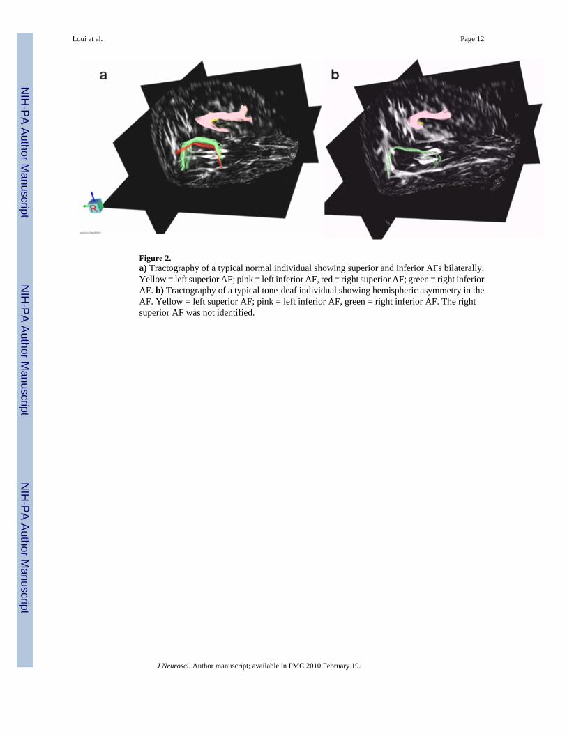
Figure 2.a) Tractography of a typical normal individual showing superior and inferior AFs bilaterally.Yellow = left superior AF; pink = left inferior AF, red = right superior AF; green = right inferiorAF. b) Tractography of a typical tone-deaf individual showing hemispheric asymmetry in theAF. Yellow = left superior AF; pink = left inferior AF, green = right inferior AF. The rightsuperior AF was not identified.
Loui et al. Page 12
J Neurosci. Author manuscript; available in PMC 2010 February 19.
NIH
-PA Author Manuscript
NIH
-PA Author Manuscript
NIH
-PA Author Manuscript
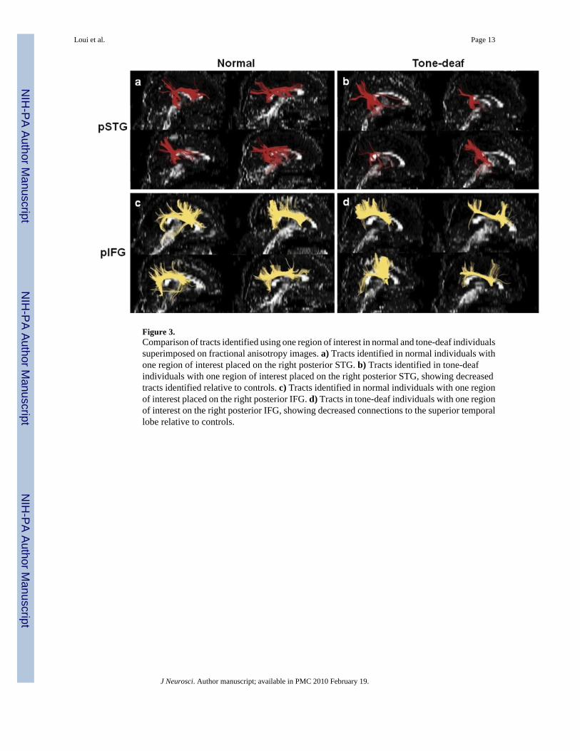
Figure 3.Comparison of tracts identified using one region of interest in normal and tone-deaf individualssuperimposed on fractional anisotropy images. a) Tracts identified in normal individuals withone region of interest placed on the right posterior STG. b) Tracts identified in tone-deafindividuals with one region of interest placed on the right posterior STG, showing decreasedtracts identified relative to controls. c) Tracts identified in normal individuals with one regionof interest placed on the right posterior IFG. d) Tracts in tone-deaf individuals with one regionof interest on the right posterior IFG, showing decreased connections to the superior temporallobe relative to controls.
Loui et al. Page 13
J Neurosci. Author manuscript; available in PMC 2010 February 19.
NIH
-PA Author Manuscript
NIH
-PA Author Manuscript
NIH
-PA Author Manuscript

Figure 4.a) Correlation between volume of fiber tracts in the right superior AF and the pitchdiscrimination threshold. b) Correlation between volume of fiber tracts in the right inferior AFand the action-perception mismatch index.
Loui et al. Page 14
J Neurosci. Author manuscript; available in PMC 2010 February 19.
NIH
-PA Author Manuscript
NIH
-PA Author Manuscript
NIH
-PA Author Manuscript
Related Documents


