Regulation of Cell Fate by c-FLIP Phosphorylation Tomoko Asaoka 2013

Welcome message from author
This document is posted to help you gain knowledge. Please leave a comment to let me know what you think about it! Share it to your friends and learn new things together.
Transcript

Regulation of Cell Fate
by c-FLIP Phosphorylation
Tomoko Asaoka
2013
Tomoko A
saoka R
egulation of Cell Fate by c-FLIP Phosphorylation
2013
ISBN 978-952-12-2988-6Painosalama Oy – Turku, Finland 2013

Regulation of Cell Fate
by c-FLIP Phosphorylation
Tomoko Asaoka
Department of Biosciences, Åbo Akademi University
Turku Cente for Biotechnology, University of Turku and Åbo Akademi Univertity
Turku Doctoral Programme of Biomedical Sciences Finland
2013
From the Department of Biosciences, Åbo Akademi University, Turku Centre for Biotechnology, University of Turku and Åbo Akademi University, and Turku Doctoral Programme of Biomedical Sciences. Supervised by Professor John E Eriksson Department of Biosciences Åbo Akademi University, and Turku Centre for Biotechnology University of Turku and Åbo Akademi Univeristy, Turku, Finland Co-supervised by Doctor Annika Meinander Department of Biosciences Åbo Akademi University Turku, Finland Reviewed by Doctor Markus Rehm Department of Physiology & Medical Physics Royal College of Surgeons in Ireland Dublin, Ireland Docent Ville Hietakangas Institute of Biotechnology University of Helsinki Helsinki, Finland Opponent Professor Marion MacFarlane Medical Research Council Toxicology Unit University of Leicester Leicester, UK
ISBN 978-952-12-2988-6 Painosalama Oy – Turku, Finland 2013
Cover picture: A STED microscopy image of phosphorylated c-FLIP in mitotic cells

From the Department of Biosciences, Åbo Akademi University, Turku Centre for Biotechnology, University of Turku and Åbo Akademi University, and Turku Doctoral Programme of Biomedical Sciences. Supervised by Professor John E Eriksson Department of Biosciences Åbo Akademi University, and Turku Centre for Biotechnology University of Turku and Åbo Akademi Univeristy, Turku, Finland Co-supervised by Doctor Annika Meinander Department of Biosciences Åbo Akademi University Turku, Finland Reviewed by Doctor Markus Rehm Department of Physiology & Medical Physics Royal College of Surgeons in Ireland Dublin, Ireland Docent Ville Hietakangas Institute of Biotechnology University of Helsinki Helsinki, Finland Opponent Professor Marion MacFarlane Medical Research Council Toxicology Unit University of Leicester Leicester, UK
ISBN 978-952-12-2988-6 Painosalama Oy – Turku, Finland 2013

TABLE OF CONTENTS ABSTRACT 6
LIST OF ORIGINAL PUBLICATIONS 7
ABBREVIATIONS 8
INTRODUCTION 10
REVIEW OF THE LITERATURE 11 1. To be, or not to be, that is the question: 11 2. The meaning of death for a cell 12
2.1 Initiation of apoptosis 12 Extrinsic apoptosis signaling commence from death receptors 12 Intrinsic apoptosis pathway is initiated by mitochondria 15
2.2 The killing signal is verified in cell death signaling complexes 16 The DISC formation at the death receptors 16 The deadly Complex II formed by TNFR1 is active in the cytosol 19 Mitochondria-mediated apoptosome formation 20 The PIDDosome induces caspase-2 activation 20 Crosstalk between the extrinsic and intrinsic apoptotic pathways 21
2.3 Execution of apoptosis 22 Activation of the caspase cascade 22 Morphological features of apoptosis 24 A peaceful ending for the apoptotic bodies 26
2.4 Alternative cell deaths 26 3. FLIP - a modulator of cell fate 28
3.1 Viral and mammalian isoforms of FLIP 29 3.2 Regulation of c-FLIP protein expression level 30
c-FLIP abundance regulated by protein synthesis and degradation 31 Subcellular protein localization, a new insight into c-FLIP proteins 33
3.3 Post-translational modifications decipher protein behavior 34 Ubiquitination determines the half-life of c-FLIP 34 Phosphorylation determines the fate of c-FLIP turnover 37 Proteolytic cleavage of c-FLIP by caspase-8 38
4. The dynamics of c-FLIP signaling in cell survival 39 4.1 Anti-apoptotic role of FLIP 39
Inhibition of apoptosis at the DISC 40 c-FILP as a rheostat in the induction of necroptosis 41
4.2 Pro-survival roles of FLIP in signal transduction 42 c-FLIP determines the outcome of NF-κB signaling 42
Regulation of MAPK pathways by c-FLIPL 45 Induced Wnt signaling by overexpression of c-FLIPL 45 PI3K/Akt signaling pathway and c-FLIPL 46 Autophagy regulation by c-FLIP 46 Regulation of caspase-8 in pro-survival signaling 47
4.3 Modeling the dynamic cell death signaling pathway 47 5. Abnormality of c-FLIP in disease 50
5.1 c-FLIP in development 51 5.2 The role of c-FLIP in the immune system and in autoimmune
diseases 52 5.3 Targeting c-FLIP for cancer therapy 54
OUTLINES OF THE STUDY 57
MATERIALS AND METHODS 58
RESULTS AND DISCUSSION 61 1. The role of phosphorylated c-FLIP in death receptor activation 61 1.1 Phosphorylation of c-FLIP on serine 193 (I, III, IV) 61
c-FLIP S193 phosphorylation is mediated by classical PKC 61 c-FLIP S193 phosphorylation is induced upon DR stimulation 63
1.2 PKC-mediated c-FLIP phosphorylation leads to protein stability via regulating ubiquitination (I, III) 63
1.3 c-FLIPL S193 determines protein distribution (III) 66 1.4 c-FLIP protein level is crucial in determining the outcome of
death receptor-mediated apoptosis (I-III) 68 c-FLIP S193 phosphorylation in TRAIL-induced apoptosis 68 Bench-to-Model, quantitative study of c-FLIP behavior 70
2. Formation of c-FLIPL-mediated death effector filaments (unpublished) 73 2.1 Serine 227 is phosphorylated in c-FLIPL 73 2.2 c-FLIPL phosphorylated on S227 form death effector filaments 73 2.3 c-FLIPL death effector filaments are affected by the cytoskeletal
network 75 2.4 Possible function of c-FLIP death effector filaments 76
3. Phosphorylation of c-FLIP in cell proliferation (IV) 78 3.1 Phosphorylated c-FLIP in the cell cycle 78 3.2 Phosphorylation of c-FLIP regulates the outcome of cell
proliferation 80 CONCLUDING REMARKS 85
ACKNOWLEDGEMENTS 86
REFERENCES 88

ABSTRACT Programmed cell death is an important physiological cellular process that maintains homeostasis and protects multicellular organisms from diseases. Apoptosis is the principal mode of cell death, which eliminates unwanted cells and an enormous effort has been made to understand the molecular mechanisms of the signaling pathway and its regulatory systems. Irregular apoptosis often has life-threatening consequences to humans, including cancer, autoimmune diseases and degenerative diseases. In cancer for example, cell death is an attractive target to eradicate uncontrollably proliferating cells that have disregard pro-apoptotic signaling. Targeted therapeutic approaches are not as effective as once expected, since now we know that the cell death pathways are not sole entities in cells, but are highly associated with various cellular processes. Proteins that regulate apoptosis can also control non-apoptotic signaling pathways. For example, c-FLIP is a protein that can either inhibit or promote caspase-8 activation, which is required to induce apoptosis. Not only has c-FLIP opposing effects on initiating apoptosis, but it also regulates various pro-survival signaling pathways in the cell. It is well known that protein expression level is a determinant of how c-FLIP can regulate different signaling pathways, but other regulatory mechanisms potentially affecting the role of c-FLIP are less well understood. This work addresses novel insights into the mechanisms of c-FLIP post-translational modifications and their functional consequences. We have identified that phosphorylation is an important inception for subcellular localization of c-FLIP, thereby dictating which apoptotic and non-apoptotic signaling pathways c-FLIP could regulate to promote cell survival. Furthermore, we have constructed mathematical models to unite independent studies to establish more systematic c-FLIP signaling pathways to understand the dynamics of extrinsically-induced apoptosis.
LIST OF ORIGINAL PUBLICATIONS This thesis is based on the following original publications and manuscripts, which are referred to in the text by their Roman numerals. In addition, unpublished results are included. I Kaunisto A, Kochin V*, Asaoka T*, Mikhailov A, Poukkula M,
Meinander A, Eriksson JE (2009). PKC-mediated phosphorylation regulates c-FLIP ubiquitylation and stability. Cell Death Differ 16(9): 1215-1226.
*Equal contribution. II Toivonen HT, Meinander A, Asaoka T, Westerlund M, Pettersson
F, Mikhailov A, Eriksson JE, Saxén H (2011). Modeling reveals that dynamic regulation of c-FLIP levels determined cell-to-cell distribution of CD95-mediated apoptosis. J Biol Chem 286(21): 18375-82.
III Asaoka T, Joko CA, Toivonen H, Russell J, Wikström V, Meinander
A, Saxén H, Eriksson J. Isoform-specific phosphorylation of c-FLIP proteins determine their transcellular distribution and capacity to direct signaling from the death-inducing signaling complex. Submitted manuscript
IV Asaoka T, Paul P, Wikström V, Russell J, Rajendran S, Meinander
A, Eriksson J. Regulation of cell population size by c-FLIPL phosphorylation. Manuscript
The original publications have been reproduced with permission of the copyright holders.

ABBREVIATIONS AICD Activation-induced cell death AIDS Aquired immunodeficiency
syndrome ALS Amyotrophic lateral sclerosis aPKC Atypical protein kinase C ATP Adenosine triphosphate Atg Autophagy-related Apaf-1 Apoptotic protease activating
factor-1 Bad Bcl-2-associated death protein Bak Bcl-2 homologous antagonistic
killer Bax Bcl-2-associated X protein Bcl-2 B-cell lymphoma gene 2 BH Bcl-2 homology Bid BH3-interacting domain death
agonist Bim BH3-interacting mediator of cell
death BSA Bovine serum albumin CAD Caspase-activated DNase CAM Chorioallantoic membrane CaM Calmodulin CaMKII Calcium/calmodulin-dependent
protein kinase II CARD caspase recruitment domain Caspase Cysteine-dependent aspartate-
specific protease CD95L CD95 ligand CFLAR CASP8 and FADD-like apoptosis
regulator c-FLIP Cellular-FLIP CHX Cycloheximide cPKC Classical protein kinase C DAMP Damage-associated molecular
patterns DAPI 4’,6-diamidino-2-phyenylindole DcR Decoy receptor DD Death domain DED Death effector domain DEDD DED-containing DNA binding
protein DEF Death effector filament DIABLODirect IAP binding protein with
low PI DISC Death inducing signaling
complex DMSO Dimethyl sulfoxide DR Death receptor DTT Dithiothreitol DUB Deubiquitinating enzyme E1 Ubiquitin-activating enzyme E2 Ubiquitin-conjugating enxyme E3 Ubiquitin ligase ECL Enhanced chemiluminescence
ERK Extracellular signal-regulated
protein FACS Fluoresence-activated cell sorting FADD Fas-associated protein with death
domain FCS Fetal calf serum FLICE FADD-like interleukin-1 beta-
converting enzyme FLIP FLICE-inhibitory protein GFP Green fluorescent protein GSK-3β Glycogen synthase kinase-3β HDAC Histone deacetylase HECT Homologous to E6-AP carboxy-
terminus HHV Human herpesvirus HRP Horse radish peroxidase Hsc70 Heat shock cognate protein 70 HtrA2 High temperature requirement
protein IAP Inhibitor of apoptosis ICE interleukin-converting enzyme IκB Inhibitor of κB IKK IκB kinase IF Immunofluorescence IL Interleukin IP Immunoprecipitation JNK c-Jun N-terminal kinase LUBAC Linear ubiquitin chain assembly
complex MAPK Mitogen-activated protein kinase MCV Molluscum contagiosum MEF Mouse embryonic fibroblast MEK MAP kinase miRNA MicroRNA MKK MAP kinase MOMP Mitochondrial outer membrane
permeabilization mRNA Messenger RNA mTOR Mammalian target of rapamycin NEMO NF-κB essential modulator NES Nuclear export sequence NFAT Nuclear factor of activated T cells NF-κB Nuclear factor kappa enhancer
binding protein NK Natural killer NIK NF-κB-inducing kinase NLS Nuclear location sequence NHL Non-Hodgekin’s leukemia nPKC Novel protein kinase C NSCLC Non-small carcinoma lung cell ODE Ordinary differential equation OPG Osteoprotegerin
PAGE Polyacrylamide gel electrophoresis
PARP Poly(ADP-ribose) polymerase PBS Phosphate-buffered saline PEA-15 Phospho-protein enriched in
astrocytes 15 kDa PI Propedium iodide PI3K Phosphatidylinositide-3-kinase PIDD p53-induced protein with a DD PKC Protein kinase C Plk Polo-like kinase PMA Phorbol 12-myristate 13-acetate PMSF Phynylmethanesulfuorid PTM Post-translational modification Puma p53-upregulated modulator of
apoptosis RAIDD RIP-associated Ich-1/CED
homologous protein with DD RING Really interesting new gene RIPK Receptor-interacting protein
kinase S Serine SDS Sodium dodekyl sulphate siRNA Small interfering RNA SLE Systemic lupus erythematous SMAC Second mitochondria-derived
activator of caspases SPOTS Signaling protein oligomeric
transduction structure tBid Truncated Bid TCF/LEFT-cell factor/lymphoid-enhancer
factor TCR T cell receptor TNF Tumor necrosis factor TNFR TNF receptor THD TNF homology domain TPA 12-O-tetradecanoyl-phorbol 13-
acetate TRADD TNFR-associated death domain TRAF TNF receptor-associated factor TRAIL TNF-related apoptosis-inducing
ligand TRAIL-RTRAIL-receptor UBD Ubiquitin-binding domain v-FLIP Viral-FLIP WB Western blot WST Water soluble tetrazolium WT Wild type XIAP X-linked inhibitor of apoptosis

INTRODUCTION
The term cell was first described by Robert C. Hooke in 1665, when he viewed a thinly sliced cork under a crude compound microscope and observed a multitude of small individual compartments. With advances in microscopy, the cell theory was developed by Theodor Schwann and Matthias Jakob Schleiden in 1839. It was adapted by Rudolf Virchow in 1855, when he published the work of Robert Remak, hypothesizing that all living organisms are composed of cells and that they originated from pre-existing cells. Numerous observations of dying cells were made already by the pioneering cell biologists, which contributed to the fundamental understanding of the cell. Carl Vogt was the first to describe the principle of cell death in toad development in 1842, and Walther Flemming described a systematic cell death following tissue injury in 1885. Flemming continued to depict chromatolysis, which today we call apoptosis, as a physiological cellular process but his findings were not appreciated at the time. He himself deviated from the research to study the cell cycle, which was a favored topic of his and many following generations of cell biologists. Thereby, concept of cell death was held in abeyance by developmental biologists for over half a century. The term programmed cell death was introduced by Lockshin and Williams in 1964, and John Kerr and colleagues characterized common morphological features of apoptotic cells in various cell types in 1972. Apoptosis was defined critical for multicellular organisms and the interests in cell death reemerged. Since then, rapid development of the cell death field revealed the mechanisms and functions of apoptosis. Sydney Brenner, Robert Horvitz, and John Sulston jointly received the Nobel Prize in Physiology and Medicine 2002 for their works on genetic regulation of organ development and cell death. Studies continue to demonstrate the importance of cell death in life, which is reflected by over 335,000 articles that have been written on cell death, two-third accounting for publications in the last decade (NCBI –Pubmed). It has been estimated that there is a turnover of over 60 billion cells everyday to maintain a human adult. Emerging studies are revealing just how closely cell death and proliferation signaling pathways crosstalk to restrain irreversible cell death, until it is absolutely required. In this thesis, I focused on how a protein, named c-FLIP, and its cellular localization may regulate the opposing cellular outcomes in response to cell death signals.
REVIEW OF THE LITERATURE 1. To be, or not to be, that is the question: Multicellular organisms originate from a single cell. One cell divides to increase in number and differentiate into more specialized entities to form tissues where they serve precise functions necessary for life. The adult human is estimated to comprise of 1013 cells and there is a turnover of more than 60 billion cells each day to maintain the homeostasis of the normal tissues (Reed, 2002). Such equilibrium is strictly maintained by cellular processes that eliminate cells to balance their mitotic activity, a theory proposed by Ludwig Graper a hundred years ago. The existence of a cell is determined by the status of the cell itself and its surrounding environment. Diverse biological signals must be interpreted and processed by the cell to reflect its overall response. If a cell is damaged or unwanted, death-inducing signals dominate over survival-promoting signals and the cell commits suicide to avoid further injury to the functioning tissue. Cell proliferation and regulated cell death are two antagonizing outcomes that must be adequately balanced, and disturbance of these processes results in devastating physiological consequences, many of which are life-threatening (Fig 1). The decision of cells to stay alive or to eliminate themselves from the body is essential for every multicellular organism from worm to human, and cells are constantly in the state of asking the question to die or not, to prevent themselves from becoming a burden on the organism.
Figure 1. Homeostasis is balanced by cell death and proliferation. Various physiological impairments are caused by uncontrolled increase or decrease in cell death. Diseases may result from accumulation of cells due to lack of cell death (left) or too scarce number of cells due to extreme cell death (right). SLE; systemic lupus erythematous, ALS; amyotrophic lateral sclerosis, AIDS; acquired immunodeficiency syndrome (From Nobel Foundation).
Homeostasis H eostas
Huntington’s disease ALS Shigellosis AIDS Stroke Myocardial infarction
Cancer SLE Rheumatoid arthritis Polycythemia vera

2. The meaning of death for a cell We are destined to live to age, during which we must maintain homeostasis and overcome or adapt to different stresses. Cell death is an important regulatory process that disposes of any unnecessary or harmful cells, which may provoke harm to the organism. There are numerous modes of cell death and they can be classified as genetically programmed, regulated or accidental cell death. Apoptosis is genetically programmed and apoptosis-regulating genes are exceptionally well conserved throughout the evolution (Liu and Hengartner, 1999). Cell death is pivotal in the elimination of unwanted cells during embryonic development and normal cell turnover in proliferating tissues, and therefore the ability for a cell to die on cue must be precise. The degree of how well apoptosis is regulated can be demonstrated in Caenorhabditis elegans, where exactly 131 of the 1090 somatic cells generated during development undergo apoptosis (Horvitz et al., 1994). Likewise, this programmed cell death serves for a definition of finger digits in mammalian embryo, as well as regulation of the immune system in adult. Apoptosis is the best characterized among all cell deaths. It is a distinct form of cell death that is energy-dependent and follows a sequence of genetically predetermined events (Kroemer et al., 1997). If not, cells may be eliminated before they function properly, or may provide resistance to death signals, leading to unwanted cells lingering for an undefined time. On the molecular level, apoptosis can be defined in initiation, decision and execution phases, which will be discussed in the following chapters. 2.1 Initiation of apoptosis Apoptosis is induced by various stimuli and cells have complex mechanisms to sense and respond to death signals. The common modes of early phase apoptosis are extrinsically-induced apoptosis, which occur upon receiving a killing signal from outside the cell and intrinsically-induced apoptosis, which is induced by intracellular stress. Extrinsic apoptosis signaling commence from death receptors The extrinsic apoptotic pathway occurs during normal physiological cell turnover. Killing signals are normally presented in the form of death ligands, typically expressed by cells of the immune system. The extrinsic apoptotic pathway is a common way by which a cell is removed by immune cells, for example to eradicate infected or transformed cells to avoid the development of infection or tumor, respectively. The death ligands belong to the tumor necrosis factor receptor (TNFR) ligand family of type II transmembrane proteins and they contain a conserved
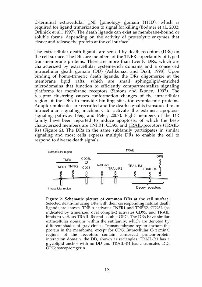
C-terminal extracellular TNF homology domain (THD), which isrequired for ligand trimerization to signal for killing (Bodmer et al., 2002; Orlinick et al., 1997). The death ligands can exist as membrane-bound or soluble forms, depending on the activity of proteolytic enzymes that cleave and release the protein at the cell surface. The extracellular death ligands are sensed by death receptors (DRs) on the cell surface. The DRs are members of the TNFR superfamily of type I transmembrane proteins. There are more than twenty DRs, which are characterized by extracellular cysteine-rich domains and a conserved intracellular death domain (DD) (Ashkenazi and Dixit, 1998). Upon binding of homo-trimeric death ligands, the DRs oligomerize at the membrane lipid rafts, which are small sphingolipid-enriched microdomains that function to efficiently compartmentalize signaling platforms for membrane receptors (Simons and Ikonen, 1997). The receptor clustering causes conformation changes of the intracellular region of the DRs to provide binding sites for cytoplasmic proteins. Adaptor molecules are recruited and the death signal is transduced to an intracellular signaling machinery to activate the extrinsic apoptosis signaling pathway (Feig and Peter, 2007). Eight members of the DR family have been reported to induce apoptosis, of which the best-characterized members are TNFR1, CD95, and TRAIL-receptors (TRAIL-Rs) (Figure 2). The DRs in the same subfamily participates in similar signaling and most cells express multiple DRs to enable the cell to respond to diverse death signals.
Figure 2. Schematic picture of common DRs at the cell surface. Selected death-inducing DRs with their corresponding natural death ligands are shown. TNF-α activates TNFR1 and TNFR2, CD95L (as indicated by trimerized oval complex) activates CD95, and TRAIL binds to various TRAIL-Rs and soluble OPG. The DRs have similar extracellular domains within the subfamily, which are denoted by different shades of gray circles. Transmembrane region anchors the protein in the membrane, except for OPG. Intracellular C-terminal regions of the receptors contain conserved protein-protein interaction domain, the DD, shown as rectangles. TRAIL-R3 has a glycolipid anchor with no DD and TRAIL-R4 has a truncated DD. OPG; osteoprotegerin.
OPG
TRAIL-R1 TRAIL-R2
TRAIL-R3 TRAIL-R4
Intracellular region Decoy receptors
Extracellular region
CD95 TNFR2 TNFR1
CD95L
TRAIL
TNF
2. The meaning of death for a cell We are destined to live to age, during which we must maintain homeostasis and overcome or adapt to different stresses. Cell death is an important regulatory process that disposes of any unnecessary or harmful cells, which may provoke harm to the organism. There are numerous modes of cell death and they can be classified as genetically programmed, regulated or accidental cell death. Apoptosis is genetically programmed and apoptosis-regulating genes are exceptionally well conserved throughout the evolution (Liu and Hengartner, 1999). Cell death is pivotal in the elimination of unwanted cells during embryonic development and normal cell turnover in proliferating tissues, and therefore the ability for a cell to die on cue must be precise. The degree of how well apoptosis is regulated can be demonstrated in Caenorhabditis elegans, where exactly 131 of the 1090 somatic cells generated during development undergo apoptosis (Horvitz et al., 1994). Likewise, this programmed cell death serves for a definition of finger digits in mammalian embryo, as well as regulation of the immune system in adult. Apoptosis is the best characterized among all cell deaths. It is a distinct form of cell death that is energy-dependent and follows a sequence of genetically predetermined events (Kroemer et al., 1997). If not, cells may be eliminated before they function properly, or may provide resistance to death signals, leading to unwanted cells lingering for an undefined time. On the molecular level, apoptosis can be defined in initiation, decision and execution phases, which will be discussed in the following chapters. 2.1 Initiation of apoptosis Apoptosis is induced by various stimuli and cells have complex mechanisms to sense and respond to death signals. The common modes of early phase apoptosis are extrinsically-induced apoptosis, which occur upon receiving a killing signal from outside the cell and intrinsically-induced apoptosis, which is induced by intracellular stress. Extrinsic apoptosis signaling commence from death receptors The extrinsic apoptotic pathway occurs during normal physiological cell turnover. Killing signals are normally presented in the form of death ligands, typically expressed by cells of the immune system. The extrinsic apoptotic pathway is a common way by which a cell is removed by immune cells, for example to eradicate infected or transformed cells to avoid the development of infection or tumor, respectively. The death ligands belong to the tumor necrosis factor receptor (TNFR) ligand family of type II transmembrane proteins and they contain a conserved
C-terminal extracellular TNF homology domain (THD), which isrequired for ligand trimerization to signal for killing (Bodmer et al., 2002; Orlinick et al., 1997). The death ligands can exist as membrane-bound or soluble forms, depending on the activity of proteolytic enzymes that cleave and release the protein at the cell surface. The extracellular death ligands are sensed by death receptors (DRs) on the cell surface. The DRs are members of the TNFR superfamily of type I transmembrane proteins. There are more than twenty DRs, which are characterized by extracellular cysteine-rich domains and a conserved intracellular death domain (DD) (Ashkenazi and Dixit, 1998). Upon binding of homo-trimeric death ligands, the DRs oligomerize at the membrane lipid rafts, which are small sphingolipid-enriched microdomains that function to efficiently compartmentalize signaling platforms for membrane receptors (Simons and Ikonen, 1997). The receptor clustering causes conformation changes of the intracellular region of the DRs to provide binding sites for cytoplasmic proteins. Adaptor molecules are recruited and the death signal is transduced to an intracellular signaling machinery to activate the extrinsic apoptosis signaling pathway (Feig and Peter, 2007). Eight members of the DR family have been reported to induce apoptosis, of which the best-characterized members are TNFR1, CD95, and TRAIL-receptors (TRAIL-Rs) (Figure 2). The DRs in the same subfamily participates in similar signaling and most cells express multiple DRs to enable the cell to respond to diverse death signals.
Figure 2. Schematic picture of common DRs at the cell surface. Selected death-inducing DRs with their corresponding natural death ligands are shown. TNF-α activates TNFR1 and TNFR2, CD95L (as indicated by trimerized oval complex) activates CD95, and TRAIL binds to various TRAIL-Rs and soluble OPG. The DRs have similar extracellular domains within the subfamily, which are denoted by different shades of gray circles. Transmembrane region anchors the protein in the membrane, except for OPG. Intracellular C-terminal regions of the receptors contain conserved protein-protein interaction domain, the DD, shown as rectangles. TRAIL-R3 has a glycolipid anchor with no DD and TRAIL-R4 has a truncated DD. OPG; osteoprotegerin.
OPG
TRAIL-R1 TRAIL-R2
TRAIL-R3 TRAIL-R4
Intracellular region Decoy receptors
Extracellular region
CD95 TNFR2 TNFR1
CD95L
TRAIL
TNF

TNFR1 (also known as DR1, CD120a, p55, and p60) is present in most cells and exert pleiotropic biological activities upon binding of TNF-α, a cytokine predominantly produced by activated macrophages in response to infections. The receptor regulates cellular processes such as inflammation, proliferation, differentiation and cell death (Ashkenazi and Dixit, 1998; Wajant et al., 2003). TNFR1 can induce formation of two distinct signaling complexes via recruiting its adaptor protein TNFR-associated death domain (TRADD) in the intracellular region of the receptor (Hsu et al., 1995). The TNFR1-TRADD complex subsequently transduces either pro-survival or pro-apoptotic signaling by recruiting different cytoplasmic proteins. Inhibition of TNF-TNFR1 signaling pathways has emerged as an effective therapeutic target for autoimmune diseases (Muppidi et al., 2004). TNFR2, on the other hand, lacks the DD and thus has lesser potency and no cytotoxic activity (Baker and Reddy, 1998; Tartaglia et al., 1993). CD95 (also known as Fas, DR2, and APO-1) is expressed in various tissues, whereas CD95 ligand (CD95L) is expressed mainly by lymphocytes to kill target cells (Griffith et al., 1995; Suda et al., 1993). The membrane-bound CD95L is suggested to have more effective cytotoxic activity in vivo compared to the soluble oligomerized ligands (O’Reilly et al., 2009). CD95-mediated apoptosis appears to be necessary in the immune system, for example to eliminate autoreactive lymphocytes, delete activated lymphocytes after an immune response, and restrict immune system access from privileged sites. Additionally, it plays a role in eliminating cancer cells and virally infected cells (Curtin and Cotter, 2003; Krammer, 2000; Kurts et al., 1998; Nagata, 1999). CD95L-CD95 death signaling is transduced into the cell via the death-inducing signaling complex (DISC), a multiprotein platform where the decision to execute apoptosis occurs (Itoh et al., 1991). There are five TRAIL-R members of which two are agonistic, TRAIL-R1 (also known as DR4 and APO-2) and TRAIL-R2 (or DR5, KILLER, TRICK2), while other members are antagonistic decoy receptors, TRAIL-R3/DcR1, TRAIL-R4/DcR2, and osteoprotegerin (Fig 2) (Degli-Esposti et al., 1997; Degli-Esposti et al., 1997; Emery et al., 1998; MacFarlane et al., 1997; Pan et al., 1997; Screaton et al., 1997; Walczak et al., 1997). These antagonistic TRAIL-Rs inhibit TRAIL-mediated cell death by competing with TRAIL-R1 and TRAIL-R2 for the ligand binding (Sheridan et al., 1997). TRAIL posses a strong apoptosis inducing activity in a wide range of human cancer cells while it has minimum cytotoxicity to normal cells, making TRAIL a potential target for cancer therapy (Ashkenazi and Dixit, 1998; Griffith and Lynch, 1998; Lin et al., 2002; Pitti et al., 1996; Walczak et al., 1999). Similar to the CD95 DISC, the TRAIL-R DISC formation is required to initiate the downstream apoptosis.
Despite their name, the DRs are not simply dedicated to induce cell death but also mediate diverse non-apoptotic functions, including cell survival, differentiation, and regulation of the immune response (Locksley et al., 2001). For example, CD95 provide a co-stimulatory signal to T cells upon activation (Alderson et al., 1995). While much of this introduction will be focused on cell death, the non-apoptotic aspect of the DR-mediated signaling will be discussed throughout this book. Intrinsic apoptotic pathway is initiated by mitochondria Apoptosis induced via the intrinsic pathway is another form of programmed cell death, which is also referred to as the mitochondria initiated pathway, since the majority of regulations occur in the mitochondria. When cells sense intracellular stresses such as DNA damage, growth factor deprivation, and hypoxia, they trigger the stress response pathways in attempt to mend subsequent damages. If the stress signal is too severe, however, cells induce the intrinsic apoptotic pathway to eliminate themselves. Upon receiving a death signal, mitochondrial outer membrane permeabilization (MOMP) occurs, leading to efflux of proteins from the mitochondrial intermembrane space to the cytoplasm, which are sensed as killing signals and the apoptotic signaling is initiated (reviewed by Galluzzi et al., 2012). It causes dysfunction of the mitochondria, inducing transmembrane potential dissipation and arrest of adenosine triphosphate (ATP) synthesis. B cell CLL/lymphoma-2 (Bcl-2) proteins, which are the central regulators of the intrinsically-induced apoptosis, respond to multiple intracellular stresses, and determine the mitochondrial integrity. Bcl-2 proteins are either pro-survival or pro-apoptotic to their nature and are categorized into three functional groups (Fig 3) (Adams and Cory, 1998; Antonsson and Martinou, 2000). The anti-apoptotic Bcl-2 proteins, Bcl-xL being the most potent, prevent the initiation of apoptosis and preserve the integrity of the mitochondria to carry out efficient energy metabolism, production of membrane lipids and cell growth (Chen et al., 2005; Willis and Adams, 2005). When a death signal arrives, the anti-apoptotic Bcl-2 proteins are inhibited by Bcl-2 homology-3 (BH3)-only proteins. This leads to the oligomerization of the effector pro-apoptotic Bax (Bcl-2 associated x protein) and Bak (Bcl-2 antagonist killer 1) to create pores on the mitochondrial outer membranes and causes the intermembrane space contents to leak out (reviewed by Hengartner, 2000).

Fig 3. Regulation of the intrinsic apoptotic pathway by the Bcl-2 proteins. The Bcl-2 family comprises of three subfamilies that contain between one and four BH domains (BH1-4). The anti-apoptotic Bcl-2 proteins, including Bcl-2, Bcl-xL and Mcl-1, contain four BH domains. The pro-apoptotic subfamily is subdivided into effector and BH3-only members. The anti-apoptotic Bcl-2 proteins are differentially inhibited by the BH3-only proteins. Some BH3-only proteins interact with all, whereas others interact only with certain anti-apoptotic members (adapted from Taylor et al., 2008).
2.2 The killing signal is verified in cell death signaling complexes Once cells receive enduring killing signals that dominate over pro-survival signalings, they initiate the suicidal process by structuring multiprotein complexes to converge the death signals to activate caspases (cysteine-dependent aspartate-specific protease). Thus, formation of these complexes is a crucial regulatory step in transducing the apoptotic signals (reviewed by Bao and Shi, 2006). There are several complexes that determine if the cell will die or not. The assembly of death signaling complexes relies on protein-protein interactions to effectively facilitate the activation and amplification of the downstream signaling. The members of the DD superfamily contain interaction domains, which have no enzymatic function but their structures allow homotypic binding with other proteins that contain the same domains (Weber and Vincenz, 2001). Such interaction motifsinclude the DDs and the death effector domain (DED) in DR signaling,and caspase recruitment domain (CARD) in the intrinsic cell death pathway (Reed et al., 2004). Some caspases contain DED or CARD prodomains that mediate recruitment of the molecules to adjacent death signaling complexes for their activation. The DISC formation at the death receptors The DISC is critical for initiating the DR signaling for CD95 and TRAIL-Rs. The DISC is assembled by DED-containing proteins, including an adaptor Fas-Associated Death Domain (FADD), procaspase-8 (also known as FADD-like interleukin-1 beta-converting enzyme (FLICE)),
Bcl-2 Bcl-XL Mcl-1
Anti-apoptotic
Bad Bim, Puma
Noxa
Bax Bak Bok
Effector pro-apoptotic
Pro-apoptotic (BH3-only) MOMP
procaspase-10, and the regulatory protein FLICE-inhibitory protein(FLIP). The DEDs are predominantly confined to seven proteins andconsiderable studies focus on FADD, caspase-8 and FLIP that commonly regulate the DISC signaling (Fig 4). These subgroups, which are major components of the DISC, are present in higher quantities compared to the other DED-containing proteins (Schleich et al., 2012). Intriguingly, highly structurally similar DEDs between different species of DED-containing proteins can have opposing effects and they are not functionally interchangeable.
Figure 4. Representation of DED-containing proteins. FADD, procaspases and c-FLIP are fundamental components of the DISC. Although PEA-15, DEDD and DEDD2 have regulatory roles in the extrinsic apoptotic signaling, they are less known. There are also additional proteins that contain variant DEDs, but they are not shown in the figure nor discussed in the text. C-terminal number represents the number of amino acids in each protein (adapted from Yu and Shi, 2008).
FADD (also known as MORT1) is a critical adaptor protein that transduces signaling from the DRs to intracellular signaling machinery. It possesses a C-terminal DD, which binds directly to the DR and a N-terminal DED in turn recruits other DED-containing proteins (Boldin et al., 1996; Fernandes-Alnemri et al., 1996; Muzio et al., 1996). Furthermore, oligomerization of the FADD DED drive homotypic DD interactions, thus functioning as a linker to enhance binding affinity to oligomeric ligand-bound DRs and forming a stable signaling complex (Carrington et al., 2006; Sandu et al., 2006; Thomas et al., 2004). FADD self-association is also implicated in generation of clusters of receptor termed SPOTS (signaling protein oligomeric transduction structure), which rapidly amplifies the apoptotic signal (Kischkel et al., 1995; Siegel et al., 2004). The initiator caspases, namely procaspase-8 (also known as FLICE, MACH and Mch5) and procaspase-10, interact with FADD via their N-terminal tandem DEDs (Reed et al., 2004; Tsukumo and Yonehara, 1999).
206 FADD
DED1 DED2 Pseudo-caspase 480 c-FLIP
DED1 DED2 Caspase 479 Procaspase-10
DED1 DED2 Caspase 496 Procaspase-8
130 PEA-15
DED 318 DEDD
326 DEDD2
DED DD
DED
DED

Cells without caspase-8 are resistant to DR-mediated apoptosis but not to mitochondria-dependent apoptosis, indicating that caspase-8 plays indispensable roles in the extrinsic apoptosis signaling, but is expendable for other modes of cell death (Varfolomeev et al., 1998). The DISC provides a platform where the C-terminal protease domains of the procaspase are recruited and dimerized, which is a prerequisite for caspase activation. Further cleavage of the proteins stabilizes the dimers and increases their activity (Fig 5) (Martin et al., 1998; Muzio et al., 1996; Oberst et al., 2010; Wang et al., 2001). The activation liberates caspase-8 dimers from the DISC into the cytoplasm and in turn activates a downstream caspase cascade to execute cell death (reviewed by Salvesen, 2002). Active caspase-8 and caspase-10 have similar substrate specificities, but caspase-10 cannot functionally substitute caspase-8 in initiating the extrinsic apoptosis signaling pathway (Engels et al., 2005; Kischkel et al., 2001; Milhas et al., 2005; Vincenz and Dixit, 1997). c-FLIP is another DED-containing protein, which can bind to the DISC and modulate caspase-8 activation, thereby determining the outcome of DISC signaling (Fig 5). c-FLIP is the focus of my thesis work and its detailed regulatory mechanisms will be discussed later.
Figure 5. Schematic diagram of the DISC complex. DED-containing proteins form the DISC through homotypic DD and DED interactions in the intracellular region of the DRs. The right side of the DISC: procaspase-8 recruitment via FADD (black) results in full caspase-8 activation and apoptotic signal transduction. The left side: simplified view of c-FLIP inhibiting caspase activation at the DISC by binding to FADD. Numbers on the molecules indicate DED domains, 1; DED1 and 2; DED2. Different shades of grey depict one molecule of procaspase-8.
The receptor-bound DISC can dissociate into the cytoplasm to form DISC complex II, which is composed only of the DED-containing proteins (Jin and El-Deiry, 2006; Lavrik et al., 2008). The CD95 DISC may internalize to amplify the receptor-mediated death signal (Lee et al., 2006), whereas
X
Death ligand
Death receptors Plasma membrane
c-FLIP Procaspase-8
Active caspase-8
1 2 1 2 2 1
FADD FADDFF
internalized TRAIL-R DISC has been shown to activate both apoptotic and survival signaling pathways (Varfolomeev et al., 2005). Other DED-containing proteins include the anti-apoptotic PEA-15 (phospho-protein enriched in astrocytes 15 kDa), which can regulate the DISC signaling (Xiao et al., 2002). Other DED-containing proteins can regulate apoptosis independent of the DISC. For example, DED-containing DNA binding protein (DEDD) in the nucleolus activates caspase-6 and inhibits transcription mediated by RNA polymerase I (Schickling et al., 2001). Its analog DEDD2 can sequester c-FLIP in the nucleoli to regulate the DR-induced apoptosis (Roth et al., 2002). The deadly Complex II formed by TNFR1 is active in the cytosol Whilst CD95 and TRAIL-Rs predominantly activate the extrinsic apoptotic pathway, TNFR1 primarily induces pro-inflammatory and immune-stimulatory activities, and apoptosis is secondary. When TNFR1 is stimulated, TRADD binds at the receptor via DD homotypic interactions, which in turn recruits other proteins to form complex I to induce nuclear factor-κB (NF-κB) activation. The complex may dissociate from the receptor to form two types of complex II (Fig 6) (Micheau and Tschopp, 2003). In complex IIa, FADD and procaspase-8 are assembled to induce apoptosis. Formation of complex IIa is a prerequisite for TNFR1-
Figure 6. TNF-α-TNFR1 can signal for cell survival, apoptosis or necroptosis. TNFR1 signaling primarily induce pro-survival signaling via the formation of complex I at the receptor. If this signaling pathway is incompetent however, either complex IIa or complex IIb is subsequently formed to activate caspase-8 or RIPKs and induce apoptosis or necroptosis, respectively.
Ub Ub Ub
X
Apoptosis
Survival
Necroptosis
Complex I
Complex IIa
Complex IIb
c-FLIP
Caspase-8 molecules

mediated apoptosis, since DED-containing proteins cannot bind directly at the receptors (Harper et al., 2003). Complex IIb, also known as the necrosome, is formed when caspase-8 activation in complex IIa is impaired and do not cleave necrosis inducing kinases, initiating necroptosis, a different form of regulated cell death (Fig 6). Death through TNFR1 complex II is typically a result of inefficient pro-survival signals induced by complex I. Mitochondria-mediated apoptosome formation Upon activation of the intrinsic apoptotic pathway, mitochondria are permeabilized by the Bcl-2 proteins to release various pro-apoptotic molecules and induce a potent cell death (reviewed by Chipuk and Green, 2008). Among these, cytochrome c is a major apoptogen that interacts with a cytoplasmic adaptor receptor Apaf-1 (Apoptotic protease activating factor-1) to form a large molecular weight oligomeric caspase activation complex known as the apoptosome (Fig 7) (Li et al., 1997; Liu et al., 1996; Zou et al., 1997). The apoptosome recruits procaspase-9 via CARD-CARD interaction with Apaf-1 and oligomerization of the procaspases lead to their activation (Adrain and Martin, 2001; Hofmann et al., 1997). This step in the mitochondrial pathway is often implied as the point of no return, since the function of mitochondria deteriorate and downstream signaling of MOMP is irreversible. In addition to the release of cytochrome c, mitochondria release other proteins that aid in amplifying the apoptotic signaling (Barnhart et al., 2003). For example, Smac/Diablo (the second mitochondrial activator of caspases/Direct IAP binding protein with low PI) binds to IAPs (inhibitor of apoptosis proteins), thereby blocking the ability of IAPs to inhibit initiator caspase-9 and downstream effector caspases (Deveraux et al., 1998; Du et al., 2000; Verhagen et al., 2002). Interestingly, XIAP (X-linked IAP) can inhibit caspase-9 activated in the apoptosome, but it can no longer inhibit those activated by downstream effector caspases in a feedback amplification loop (Holcik and Korneluk, 2001). The PIDDosome induces caspase-2 activation In response to genotoxic stress, p53 induces the expression level of PIDD (p53-induced protein with a DD). PIDD forms the core moiety of a large oligomeric complex known as the PIDDosome, where it binds to a CARD-containing protein RAIDD (RIP-associated Ich-1/CED homologous protein with DD). Procaspase-2 also contains a CARD that facilitates its recruitment to the PIDDosome and dimerization activates the caspase to regulate inflammation or apoptosis signaling pathways. Even though caspase-2 was one of the first caspases discovered, its detailed physiological function remains to be established (Janssens and
Tinel, 2011; Zhivotovsky and Orrenius, 2005). Emerging studies have described caspase-2 as a tumor suppressor in vivo, by providing protection against cellular stress and transformation (Puccini et al., 2013).
Figure 7. MOMP and formation of the apoptosome. Following an irreparable cellular stress, pro-apoptotic signaling leads to MOMP and allows the release of mitochondrial intermembrane space proteins to the cytoplasm. The release of cytochrome c (denoted by black circles) promotes Apaf-1 oligomerization and formation of the apoptosome. This complex provides a platform for caspase-9 activation. Other apoptogens, such as Smac/Diablo and Omi/HtrA2 facilitate caspase activation by inhibiting several members of the IAP family. In addition, activated caspase-8 can truncate Bid, a BH3-only pro-apoptotic protein, which in turn directly activate Bak and Bax to form lipoprotein pores in the outer membranes of mitochondria to induce MOMP.
Crosstalk between the extrinsic and intrinsic apoptotic pathways While the extrinsic and intrinsic apoptosis signaling are distinct from each other in the modes of initiation, they may crosstalk to amplify the killing signals and robustly execute apoptosis (Kuwana et al., 1998). A well-known example is the induction of mitochondria-dependent apoptosis by activated caspase-8 (Kantari and Walczak, 2011). Some cells (Type I) are characterized by rapid high level of DISC formation by lipid raft-associated DRs, which efficiently cleave procaspase-8 and directly activate its downstream caspase cascade leading to apoptosis. Other cells (Type II), however, lack such strong initiation of cell death, thus requires an amplification loop. The activated caspase-8 truncate the BH3-only Bcl-2 protein Bid, which in turn promote oligomerization of pro-apoptotic Bcl-2 proteins to initiate the mitochondrial-pathway (Fig 7) (Eskes et al., 2000; Li et al., 1998; Li and Yuan, 2008; Luo et al., 1998). The type II apoptosis pathway can be inhibited by overexpression of Bcl-2 and Bcl-
Cytochrome C Apaf-1
Apoptosome
Mitochondria
Procaspase-9
MOMP
dATP
MOSmac/Diablo Sm
Omi/HtrA2
Active Caspase-9
Cell Stress e.g. UV radiation Etoposide Staurosporin
Cy
MOMOMO
Active caspase-8 (Extrinsic pathway)
blo

xL or inhibiting caspase-9 activity (Scaffidi et al., 1999). Furthermore, once the decision is made to die, cells often shuts down pro-survival signaling pathways to further reinforce the death machinery. 2.3 Execution of apoptosis After cells receive killing signals and the decision is made to transduce the apoptotic signaling, a well-characterized chronological execution of cell suicide follows. The final stages of apoptosis orchestrate and compile the killing signals to successfully eliminate the cells in a highly ordered manner. Activation of the caspase cascade Execution of apoptosis is ultimately carried out by the caspase family. The caspases are an evolutionary conserved family of enzymes that proteolytically cleave their substrates after an aspartic acid residue by using their catalytically active cysteine to either deactivate or activate the targeted proteins (Dales et al., 2001; van Raam and Salvesen, 2012). There are twelve caspases characterized in humans and their proteolytic activity is responsible for many cellular processes (Fig 8). All caspases are synthesized as inactive precursors, called the zymogens or procaspases, with an N-terminal prodomain and C-terminal protease domain, containing a large and a small subunit. When activated, the zymogens are proteolytically cleaved to generate active subunits, which heterodimerize with one another to form a mature heterotetramer caspases (Degterev et al., 2003; Fuentes-Prior and Salvesen, 2004). Caspase-1 (ICE; interleukin-1β conversion enzyme), caspase-4, and caspase-5 are involved in the regulation of inflammatory responses. Upon activation, they form a complex called the inflammasome, which leads to secretion of pro-inflammatory cytokines (Martinon and Tschopp, 2006; Pétrilli et al., 2007). Caspase-14 is required for keratinocyte maturation in the epidermis (Denecker et al., 2007). Other caspases are primarily committed in apoptosis, although at least one non-apoptotic function has been attributed for these caspases. Diverse stimuli cause the activation of caspases and result in various outcomes, but I will only focus on apoptosis-related caspases for the purpose of my thesis. The important role of caspases in apoptosis was discovered in C. elegans, in which the death gene ced-3 was shown to be indispensable for the execution of cell death (Yuan et al., 1993). In mice, caspase-8 knockout has the most dramatic phenotype of all caspase knockouts, demonstrated by earliest embryonic lethality (Varfolomeev et al., 1998). Of note, while some caspases are indispensible in specific apoptosis pathways, others can be compensated, suggesting that caspases induce apoptosis in a
highly context-dependent manner. Mammalian apoptotic caspases can be divided into initiator and executioner caspases according to the sequential activation of the zymogens in the caspase cascade.
Figure 8. Phylogenetic tree and schematic domain presentation of the human caspase family. All caspases have similar structures, consisting of an N-terminal prodomain followed by C-terminal protease domain, a large α subunit (indicated as p20) and a small β subunit (p10). The N-terminal domains, CARD or tandem DEDs,and C-terminal amino acid number are indicated. The structures of either extrinsic or intrinsic initiator caspases (medium grey), and shorter effector caspases (dark grey) are drawn. The lineage is not drawn to scale (adapted from Inoue et al., 2009; Roschitzki-Voser et al., 2012).
As already discussed, initiator caspase-8, caspase-9, and caspase-2 are activated at the DISC, apoptosome, and PIDDosome, respectively. They are first in line of the caspase cascade to be activated, hence called initiator caspases. The initiator procaspases must homodimerize for their autoactivation to occur, and therefore, DEDs or CARD-dependent recruitment of the procaspases to the death signaling complexes are essential. These initiator procaspases are dimerized in death signaling complexes and autocleaved through induced proximity (Chang et al., 2003; Fuentes-Prior and Salvesen, 2004). The autocleavage stabilizes the homodimers and form an enzyme with increased activity. Caspase-8 also cleaves itself between the DEDs and the large subunit, resulting in the release of stable dimers from the DISC to the cytoplasm. These initiator caspases converge death signals to the execution phase by activating the downstream effector caspases.
Caspase-14
Caspase-12 Caspase-4 Caspase-5 Caspase-1 Caspase-8 Caspase-10 Caspase-3 Caspase-7 Caspase-6 Caspase-9 Caspase-2
Prodomain p20 p10
Keratinocyte maturation
Inflammation
Extrinsic
Extrinsic
Apoptosis
Intrinsic
303
377
418
404
479
521
277
303
293
416
435

The executioner or effector caspases, namely caspase-3, caspase-6, and caspase-7, are downstream molecules of the caspase cascade (Shi, 2002). They lack a long N-terminal prodomain and cannot self-activate, thus require activated caspases to process them at the inter-subunit linker. There are other less common ways in which caspases may be activated, although much of the detail is still to be clarified. In the granzyme B pathway, apoptosis-inducing protease granzyme B is excreted by lymphocytes onto their target cells to activate the executioner caspases (Yang et al., 1998). Furthermore, caspases may be activated by the nuclear Pml/PODs/nuclear bodies and converges to a common execution phase of apoptosis. The executioner caspases are considered to be the generator of apoptotic cell death and they are usually more abundantly expressed than their initiator caspases (Budihardjo et al., 1999). Since all initiator caspases can activate caspase-3, it is here that various death stimuli are processed to give one common execution phase (Stennicke et al., 1998). Once activated, the executioner caspases function as enhancers of the apoptosis, creating a growing cascade of apoptotic proteinases by positive feedback activation (Grütter, 2000). Caspase-3 is a potent downstream caspase that cleaves the majority of cellular substrates in apoptotic cells (Porter and Janicke, 1999), although caspase-7 is similar to caspase-3 and has close substrate specificity (Degterev et al., 2003; Fuentes-Prior and Salvesen, 2004). The activation of the effector caspases is considered the hallmark of apoptosis where rapid all-or-none progress for commitment to death is achieved and this is commonly irreversible. Morphological features of apoptosis Original observations of dying cells and early studies that distinguished apoptosis from other forms of cell death were based exclusively on distinct morphological criteria (Kerr et al., 1972). These morphological changes occur with remarkable consistency in an extensive variety of cell types owing to the similarity of the biochemical events in the late phase of apoptosis. Indeed, the proteolytic cleavages by caspases, particularly of cytoplasmic and nuclear structural network, are responsible for inducing precise sequential apoptotic morphological phenotypes (Van Engeland et al., 1997). Over the years, common morphological hallmarks of apoptosis were described, which includes cellular shrinkage with nuclear chromatin condensation (pyknosis), nuclear fragmentation (karyorrhexis), and plasma membrane blebbing (Galluzzi et al., 2007; Kerr et al., 1972; Wyllie, 1981). Cellular changes observed by electron microscope determines a highly organized form of cell death (Cummings, 1997), and the chief reliable technique to determine apoptotic cells remains to be the observation of morphological changes by electron microscopy (Fig 10). The caspase targets include degradation of both nuclear and cytoskeletal structural proteins, as well as
deactivating the proteasomes, and many pro-survival signaling proteins(Earnshaw et al., 1999). Some of the earliest morphological changes in apoptosis appear on the cell surface, as cell adhesion to the extracellular matrix decrease and the plasma membrane loses its asymmetry. The exposure of phosphatidylserine is a well-documented surface modification in apoptosis caused by decreased activity of aminophospholipid transferase and enhanced activation of scramblase. Phosphatidylserine in viable cells is expressed asymmetrically in the inner leaflet of the plasma membrane, but redistributed to the outer leaflet in apoptotic cells. The appearances of many organelles remain unchanged and they continue to function until the last phases of apoptosis. Mitochondria are exceptions as their membrane potential collapse and organelle fission occur (Zamzami et al., 2005). The DNA damage-responsive enzyme poly(ADP-ribose) polymerase (PARPs) and in particular PARP1, which alone accounts for most of the cellular PARP DNA repair activities, is a well-know target for activated caspases (Amé et al., 2004). The ultimate determinant of apoptosis is the homogenous condensation of chromatin and genomic DNA fragmentation. Activation of CAD (Caspase-activated DNase) is dependent on caspase-3 mediated cleavage of its inhibitory subunit, and the release of the catalytic enzyme proceeds to internucleosomal DNA cleavage and fragmentation of genomic DNA at intervals of 180 to 200 base pairs (Fischer et al., 2003). To establish if a cell died by apoptosis, both caspase activation and DNA fragmentation must be demonstrated in most cases (Galluzzi et al., 2012). At later stage of apoptosis, the dying cell is easily distinguished from neighboring live cells due to membrane blebbing where plasma membrane is convoluted and extensions are
Figure 9. Detection of morphological changes occurring during apoptosis. Some morphological changes of a typical dying cell that could be detected with the electron microscope are listed as a timeline (adapted from Huerta et al., 2007). Pictures represent typical appearances of apoptotic HeLa cells under a light microscope (images taken by T Asaoka).
Earlier phases of Apoptosis Later phase
Chromatin condensation
Convolution of nuclear
and cellular outlines
Nuclear fragmentation
Formation of apoptotic
blebs, loosely packed
organelles, intact plasma
membrane
Cytoplasmic and
membrane degradation in lysosomes of apoptotic
bodies
Massive cell hydration

formed (Taatjes et al., 2008; Watanabe et al., 2002; Willingham, 1999; Ziegler and Groscurth, 2004). These extensions disaggregate into a number of membrane-bound compartments known as apoptotic bodies, which are tightly packed and contain well-preserved organelles and fragments of nucleus. A peaceful ending for the apoptotic bodies The purpose of apoptosis is to eliminate dying cells efficiently and prevent damages to the neighboring cells. When cells undergo apoptosis, the process produce various “eat me” signals, which are recognized by phagocytic cells (Edinger and Thompson, 2004; Fadok et al., 1992; Haslett et al., 1995). Such signals include phosphatidylserine exposure on the plasma membrane and release of fragmented intracellular molecules, such as phospholipase A2 to produce lysophatidylcholine (Lauber et al., 2003). Caspase-8-mediated emission of damage-associated molecular patterns (DAMPs) and IL-33 undergoes caspase-dependent proteolysis into the nonimmunological forms (Lüthi et al., 2009; Panaretakis et al., 2009). These processes ensure that the apoptotic cells will not activate any inflammatory and autoimmune responses. TRAIL-R DISC complex II formation and its kinase stimulation are also reported to signal for cell clearance (Varfolomeev et al., 2005). The formation of apoptotic bodies is the final cue for surrounding cells to recognize the dying cells, which must be removed by phagocytosis. The apoptotic bodies are rapidly engulfed by neighboring epithelial cells or macrophages and eventually degraded (Platt et al., 1998; Savill and Fadok, 2000). If apoptotic bodies are not phagocytosed, the corpse will undergo degradation in a process called secondary necrosis with the consequence of inflammation (Lauber et al., 2004). 2.4 Alternative cell deaths Apoptosis may be the most common and efficient mode of cell death, but there are alternative means in which a cell commits to death. Necrosis is an accidental cell death considered to be on the other extreme of continuous spectrum of cell death and often contrasted to apoptosis. Recent studies, however, have demonstrated that molecular regulations in these two pathways are closely connected to each other and this new insight will be discussed here. Other cell death mechanisms are listed and briefly described in Table 1 at the end of this chapter. Necrosis was the first form of cell death identified and it may be induced by extreme physiological conditions such as heat stress, mechanical stress, osmotic shock, and toxin exposure. The cells are rapidly killed and exhibit characteristic morphologies, such as cellular swelling and
collapse, irregular chromatin destruction, and vacuole formation. Ultimately necrotic cells are lysed and release their cellular contents, including DAMPs, thereby activating an inflammatory response (Chollet-Martin et al., 1991). This phenomenon is often referred to as uncontrolled energy-independent mode of cell death and a massive necrosis can be extremely damaging to the organism. The morphological features of necrosis can be induced in a more ordered manner (Vandenabeele et al., 2010). Regulated necrosis, also called necroptosis, can be defined as cell death mediated through a pathway that depends on the receptor-interacting protein kinase (RIPK) complex (He et al., 2009; Holler et al., 2000). When caspase-8 is dysfunctional, DD-containing RIPK1 protein and its homolog RIPK3 are not efficiently degraded and engage in functional interactions that ultimately activate the execution of necrotic cell death (Cho et al., 2009; He et al., 2009; Zhang et al., 2009). Like apoptosis, necroptosis require the formation of multiprotein complexes to initiate the signaling pathway. The concept of necroptosis emerged when some cells died by exhibiting necrotic appearance upon TNFR1-induced apoptosis while caspase activation was blocked (Degterev et al., 2005; Holler et al., 2000; Vercammen et al., 1998). Under conditions where apoptosis is hindered, cells alter their mode of cell death to necroptosis by forming RIPK1/RIPK3/FADD/inactive procaspase-8 necrosome complex (Fig 7) (He et al., 2009; Holler et al., 2000). RIPK1/3 activities are obligatory for necrosome formation and can be prevented by the RIPK1-targeting inhibitor necrostatin-1 (Cho et al., 2009). Another necrosis-inducing complex, the ripoptosome, is currently considered to be a central trigger for necroptosis in cancer cells (Feoktistova et al., 2011; Tenev et al., 2011). Its core components, namely RIPK1, FADD and caspase-8 spontaneously assemble in response to genotoxic-induced depletion of IAPs, which is a negative regulator of the components. The ripoptosome can trigger either caspase-8-mediated apoptosis or caspase-independent necroptosis, depending on the strength of caspase-8 activity in the complex. The induction of necroptosis provokes strong inflammatory responses (Kaczmarek et al., 2013). RIPK-mediated necroptosis is suggested as an important mechanism in the clearance of virus-infection or cancer cells that escaped apoptosis (Li and Beg, 2000; Upton et al., 2010). Viruses express anti-apoptotic proteins that prevent caspase-8-mediated apoptosis to prolong cell viability and to facilitate viral replication (Thome et al., 1997). RIP3-mediated necroptosis may act as a standby mechanisms to clear pathogens by killing infected cells via virus-induced necroptotic cells, which are more immunogenic than apoptotic cells, hence enhance inflammatory responses and better antigen presentation (Weinlich et al., 2011).

Table 1. List of other cell deaths. Some less prevalent cell deaths and their functional features are described. AIF; apoptosis-inducing factor, ENDOG; endonuclease G (adapted from Galluzzi et al., 2012; Kroemer et al., 2008).
3. FLIP - a modulator of cell death and survival Cell death is a complex biological process with specific purposes, and its regulation is crucial for the organism to live. To achieve an accurate execution of targeted cell death, a great degree of molecular regulations occur at many levels, especially at the initiation of caspase activation when the decision is made in the death signaling complexes. The regulatory mechanisms behind programmed cell death are extensively studied and many complicated regulatory circuits have been elegantly clarified. For example, XIAP can directly inhibit executioner caspases and play a critical regulator in type I cells, and anti-apoptotic Bcl-2 proteins can inhibit apoptosis of type II cells by blocking the mitochondrial-dependent pathway. In this thesis, I will focus on the roles and mechanisms of apoptosis signaling modulation by FLIP in DR-mediated apoptosis.
3.1 Viral and mammalian isoforms of FLIP
FLIP was first discovered while searching the genomes for the DED-containing proteins to identify regulators of apoptotic caspases. It was found in several γ-herpesviruses and molluscipoxvirus, and collectively named as viral-FLIP (v-FLIPs) (Bertin et al., 1997; Thome et al., 1997). Since CD95-induced apoptosis can effectively eliminate viral infected cells, viruses have evolved strategies to evade this destiny of the immune response. They express a number of inhibitory proteins to suppress apoptosis of the host cells. v-FLIPs, which consist of two DEDs with alternative C-tails, were shown to bind to the DED of FADD and interfere with caspase-8 interaction, thereby inhibit apoptosis induced by DRs in the infected host cells (Thome and Tschopp, 2001). Furthermore, the crystal structure v-FLIP MC195 (Fig 10) revealed that MC195 disrupt the DISC formation by preventing FADD-DED self-association rather than competing away procaspase-8. Thus, it is proposed that v-FLIPs regulate the DISC signaling at multiple levels (Yu and Shi, 2008).
Figure 10. Crystal structure of the v-FLIP MC159 protein. The DED consists of six α-helices, as numbered, connected by short loops. All FLIP proteins contain two DEDs, tightly associated with each other through a hydrophobic interface. DED1 is divergent from a typical DED fold, but DED2 is an authentic domain. The conserved Arg-X-Asp-Leu motif in the α6 helix is essential for the protein to regulate apoptosis (edited from Yu and Shi, 2008).
The long and short mammalian homologues of v-FLIPs were characterized soon after and were named cellular-FLIP (c-FLIP), also known as Casper, I-FLICE, FLAME-1, CASH, CLARP, MRIT or usurpin (Goltsev et al., 1997; Hu et al., 1997; Inohara et al., 1997; Irmler et al., 1997; Shu et al., 1997). Three isoforms have been detected at the protein level in humans, namely c-FLIP Long (c-FLIPL, 55 kDa), c-FLIP Short (c-FLIPS, 26 kDa), and c-FLIP Raji (c-FLIPR, 24 kDa) (Djerbi et al., 2001). Notably, all three isoforms have the same DEDs, but the sequences of the splicing tails are isoform-specific (Fig 11). c-FLIPL is composed of 480 amino acids and contains tandem DEDs and caspase-like domain, p20 and p12, structurally resembling the initiator procaspase-8 and procaspase-10. The c-FLIPL C-terminal domain lacks catalytic activity due to the replacement of the crucial cysteine residue within the Gln-Ala-Cys-X-Gly motif and a histidine residue within the His-Gly-motif

(Cohen, 1997; Tschopp et al., 1998). The short isoforms of c-FLIP are DED-only splice variants, similar to v-FLIPs, consisting of short C-terminal tails of 19 and 17 amino acids for c-FLIPS and c-FLIPR, respectively (Fig 11). Several properties of the short isoforms are similar, including their protein expression pattern during T cell activation, protein turnover and anti-apoptotic efficiencies (Djerbi et al., 2001; Golks et al., 2005).
Figure 11. Domain structure of c-FLIP isoforms in viruses and mammals. FLIP contains two DEDs (DED1 in lighter and DED2 in darker grey), followed by an interlinking region and a C-terminal tail, which is unique for each isoforms (c-FLIP C-terminal regions are indicated in black). The long isoform of c-FLIP contains caspase-like domain p20 and p12 subunits. MCV, Molluscum contagiosum virus; EHV, equine herpesvirus; HHV, human herpesvirus.
The initial discovery demonstrated that the tandem DEDs of c-FLIP appeared to antagonize the DR signaling in the same manner as v-FLIP by competing with procaspase-8 for FADD interaction at the DISC. Despite some reports that characterized c-FLIPL as an anti-apoptotic protein that functions in a way analogous to v-FLIPs and c-FLIPS/R, others ascribed pro-apoptotic functions in the long isoform, referring to its assistance in the autocatalytic activation of procaspase-8 at the DISC. The detailed functions of c-FLIP will be discussed in the next chapter. 3.2 Regulation of c-FLIP protein expression level The cellular activities are highly dependent on proteins and there are complex regulatory mechanisms that govern the cellular protein levels, which may occur at anytime from the transcription of genes to the degradation of proteins. c-FLIP is a dynamic regulator of caspase-8 activation and elicit diverse cellular responses. Therefore, it is not surprising that the maintenance of c-FLIP level is indispensable. c-FLIPL is widely expressed in mammalian tissues, especially the heart, lymphoid tissue, skeletal muscle, and kidney. The short isoforms of c-FLIP are more specific to the lymphatic tissues (Irmler et al., 1997; Rasper et al., 1998). In some cells, either c-FLIPS or c-FLIPR are expressed, whereas in
v-FLIP (EHV-2, HHV-8)
v-FLIP (MCV)
DED1 DED2
c-FLIPS 221
c-FLIPR 213
c-FLIPL 480 p20 p12 1 75 91 172
others both short isoforms may be detected. In addition, cell lines contain different amounts of the isoforms; c-FLIPL is often more abundantly expressed than the short isoforms (Irmler et al., 1997; Rasper et al., 1998). The c-FLIP level is an important determinant of the DISC signaling outcome in various cell types, and the expression of c-FLIP often directly correlates with the sensitivity to DR-induced cell death. The dynamics of c-FLIP protein levels are critical to determine the outcome of apoptosis and are often regulated in an isoform-specific manner. In accordance with this, the expression of c-FLIP is tightly regulated by a number of different pathways. c-FLIP abundance regulated by protein synthesis and degradation The process of protein synthesis begins by transcribing a gene to messenger RNA (mRNA), which then is translated into a polypeptide of amino acids and folded into a specific conformation to yield a mature protein. The c-FLIP gene called CFLAR (CASP8 and FADD-like apoptosis regulator) is located on the human chromosome 2 q33-q34, in a cluster of approximately 200 kilobases with the initiator procaspase-8 and procaspase-10. This close proximity of gene location suggests that c-FLIP may have evolved as a gene-duplication of procaspase-8 (Han et al., 1997; Inohara et al., 1997; Srinivasula et al., 1997). CFLAR is composed of fourteen exons and initially transcribed preRNA is processed through alternative splicing to yield several mRNA that contain different combinations of exons (Djerbi et al., 2001). Inclusion of CFLAR exon 7 with a stop codon leads to translation of c-FLIPS, whereas c-FLIPL omits exon 7 to produce the full-length protein (Fig 12). In humans, the decision to express c-FLIPS or c-FLIPR is determined by a nucleotide polymorphism in a 3’ splice site of CFLAR. The splice-dead variant produces c-FLIPR at lower protein translation rate (Ueffing et al., 2008). c-FLIPR lacks intron 6 exclusion and a stop codon at the start of the intron results in translation of the shortest c-FLIP isoform (Djerbi et al., 2001). To date, understanding of the regulation of the stability of c-FLIP mRNA is incomplete. In humans, protein-coding genes account for 2% of the genome. The rest are mostly transcribed into non-coding RNA. Protein-coding mRNAs may carry biological activity that is independent of the protein it encodes, which may be be unrelated or paradoxal to the protein function (Holland et al., 2004). Since c-FLIP have many alternative splicing variants and some include introns, studying c-FLIP at RNA level may reveal important regulatory mechanism of c-FLIP expression and signaling.

Figure 12. Splice pattern of the human CFLAR. CFLAR consists of fourteen exons and the c-FLIP isoforms detected at the protein levels are shown. The start (arrowheads) and stop (asterisks) sites for translation are indicated. Black boxes in the short isoforms of c-FLIP represent intron sequences (Adapted from (Djerbi et al., 2001).
Housekeeping genes are transcribed constitutively to produce proteins required for common cellular functions, while inducible genes are transcribed temporary upon stimulation to express proteins required for adaptive responses. The inducible genes are activated by the binding of a stimulated transcription factor to its specific binding site in the promoter region of the gene. The human proteome is dynamic and alters in response to a multitude of stimuli. Cells must converge various signals to activate appropriate transcription factors, which regulate the expression of target genes. c-FLIP is constitutively expressed in a wide array of normal cells, but the CFLAR gene is also a transcriptional target for a number of stimuli (Safa, 2012). Pro-survival singnaling pathways that activate NF-κB (Karin and Lin, 2002; Kreuz et al., 2001; Mayo and Baldwin, 2000; Sevilla et al., 2001), Akt and ERK can upregulate c-FLIP (Nam et al., 2003; Panka et al., 2001; Suhara et al., 2001). These signaling pathways will be discussed more in later chapters. Furthermore, the consensus binding sites of the NFAT (nuclear factor of activated T cells) family is positioned in the CFLAR promoter and is recruited to induce c-FLIP expression (Rao et al., 1997; Zaichuk et al., 2004). Other transcription factors including c-Myc, FOXO3 and c-Fos, on the other hand, binds directly to the promoter of CFLAR and suppresses the expression of c-FLIP (Ricci et al., 2004; Skurk et al., 2004). CaMKII (calcium/calmodulin-dependent protein kinase II) activity induces the expression of both long and short isoforms of c-FLIP and protects cells from CD95-mediated apoptosis (Yang et al., 2003). The tumor suppressor p53 also regulates the expression of c-FLIP by upregulating the transcription or promoting protein degradation (Bartke et al., 2001). c-FLIP mRNA levels are regulated by many cytokines, an IL-2 being the most studied. c-FLIP is suppressed in IL-2-dependent manner following T cell activation, which sensitizes cells to CD95-mediated apoptosis (Refaeli et al., 1998). From the emerging field of miRNA, it has
c-FLIPS
c-FLIPR
c-FLIPL
3 2 1 8 10 9 4 5 6 12 11 13 14 7
*
*
*
DED DED Caspase-like domain
been reported that miR-512-3p negatively regulates c-FLIP expression (Chen et al., 2010). Protein degradation is another process that determines the abundance of proteins in a cell. The 26S proteasome is a eukaryotic ATP-dependent protease complex where targeted proteins are efficiently degraded. Here, the substrate is hydrolyzed into short polypeptides that are released from the proteasome, thereby, providing recyclable cell material (Finley, 2009; Pickart and Eddins, 2004). Many reports have shown that c-FLIP is effectively degraded by the proteasome, which could be rescued using proteasome inhibitors (Fukazawa et al., 2001; Kim et al., 2002). c-FLIPS has a notably shorter half-life than c-FLIPL, although half-lives of the c-FLIP proteins vary in a cell type-specific fashion. C-terminal tails are pivotal in regulating stability of the c-FLIP proteins, especially in the short isoforms (Poukkula et al., 2005). The rate of c-FLIP degradation can be altered by various stimuli. For example, hyperthermia sensitizes activated T cells to CD95-induce apoptosis, primarily due to induced down regulation of c-FLIPS by rapidly enhancing the proteasomal degradation (Meinander et al., 2007). Subcellular protein localization, a new insight into c-FLIP proteins The eukaryotic cells are compartmentalized by a plasma membrane, and endomembrane systems support the organelles inside the cell. These organelles provide distinct regions for a collection of proteins to reside and perform specific cellular processes. It is estimated that half of the proteins synthesized on cytosolic ribosomes must be transported into or across at least one cell membrane to reach their functional destination (Wickner and Schekman, 2005). Compartment-specific reactions can efficiently change concentration and kinetics by confining the protein in a smaller volume and achieve functional diversity and economize on protein design and synthesis (Butler and Overall, 2009). Since the main function of c-FLIP is considered to be anti-apoptotic acting at the intracellular region of plasma membrane, c-FLIP has been assumed to localize in the cytoplasm. One of the new concepts proposed for c-FLIP in the recent years is that c-FLIPL is expressed in both the cytoplasm and nucleus. So far, two groups have shown such observations in various cell lines and claim that two nuclear localization signals (NLSs), short stretches of amino acid sequences that target the protein to the nucleus, are found at the C-terminal tail of c-FLIPL and mutations at these sites obstructed the presence of nuclear c-FLIP (Katayama et al., 2010; Zhang et al., 2009). Additionally, a nuclear export signal (NES) between the two NLSs was identified (Fig 13) (Katayama et al., 2010). Proteins must localize appropriately to function properly in their biochemical reactions. Subcellular compartments have unique

protein compositions and mislocation of these proteins contributes to the pathogenesis of many diseases (Hung and Link, 2011). Further understanding of subcellular distribution of c-FLIP may contribute significantly to their functions. 1 MSAEVIHQVE EALDTDEKEM LLFLCRDVAI DVVPPNVRDL LDILRERGKL SVGDLAELLY 61 RVRRFDLLKR ILKMDRKAVE THLLRNPHLV SDYRVLMAEI GEDLDKSDVS SLIFLMKDYM 121 GRGKISKEKS FLDLVVELEK LNLVAPDQLD LLEKCLKNIH RIDLKTKIQK YKQSVQGAGT 181 SYRNVLQAAI QKSLKDPSNN FRLHNGRSKE QRLKEQLGAQ QEPVKKSIQE SEAFLPQSIP 241 EERYKMKSKP LGICLIIDCI GNETELLRDT FTSLGYEVQK FLHLSMHGIS QILGQFACMP 301 EHRDYDSFVC VLVSRGGSQS VYGVDQTHSG LPLHHIRRMF MGDSCPYLAG KPKMFFIQNY 361 VVSEGQLEDS SLLEVDGPAM KNVEFKAQKR GLCTVHREAD FFWSLCTADM SLLEQSHSSP 421 SLYLQCLSQK LRQERKRPLL DLHIELNGYM YDWNSRVSAK EKYYVWLQHT LRKKLILSYT
NES1 NLS NES2 Fig 13. Amino acid sequence of human c-FLIPL. Amino acid residues of NLSs and NES are shown in bold and sequences are underlined and indicated. Domains are shaded in grey; darker grey, DED1; lighter grey, DED2.
3.3 Post-translational modifications decipher protein behavior A protein is modified after its synthesis, a process known as post-translational modifications (PTMs), in which covalent addition of a functional group or protein, or proteolytic cleavage of regulatory subunits occur. These modifications can appear shortly after translation to regulate the function of the protein, until the end to dictate degradation of the protein. Certain modifications affect distinct downstream responses, thereby increasing the functional diversity of a protein. The modifications are often reversible, providing further sophisticated means of regulation, and it further offers diversity to a protein by potential combinatory modifications. The PTMs play critical roles in the regulation of most cellular processes including apoptosis, proliferation and cell cycle, by altering a proteins conformation, binding properties, changing its activity localization or half-life. c-FLIP protein is indeed modified by PTMs, commonly by ubiquitination, phosphorylation and proteolysis. Ubiquitination determines the half-life of c-FLIP Ubiquitin, as the name suggests, is a widely expressed 8.5-kDa protein in cells throughout evolution. Its 76 amino acid sequence is highly conserved and it is involved in various cellular functions to drastically affect the biochemical properties by covalently being conjugated to target proteins (Hochstrasser, 2009; Schlesinger et al., 1975). Countless evidence demonstrate the involvement of ubiquitin conjugation in apoptosis regulation. For example, ubiquitination of the C-terminus of caspase-8 upon TRAIL-ligand signaling increase its pro-apoptotic potential by
stabilizing the dimer and subsequent cell death, while deubiquitination decrease caspase-8 activity (Jin et al., 2009). The initial covalent conjugation of ubiquitin to a side chain lysine is dependent on a three-step enzyme cascade (Fig 14). ATP-dependent ubiquitin-activating E1 yield an active ubiquitin, which is transferred to an E2 ubiquitin-conjugating enzyme and brings the correct modifier to a suitable E3 ubiquitin ligase (Pickart and Eddins, 2004). With the help of the E3 ligase, the E2 catalyze the binding of ubiquitin to the target protein in a process called ubiquitination. The diversity of the enzymes facilitating ubiquitination is represented in a hierarchical manner and in humans, two E1s, 38 E2s, and approximately 600 to 1000 E3s are expressed (reviewed by Ye and Rape, 2009). E3 ligases provide a flexible means of linking a conserved ubiquitin to potentially thousands of substrates.
Figure 14. Depicted enzymatic cascade of ubiquitination. Ubiquitin (Ub) chains are assembled in a stepwise process that involvesubiquitin-activating E1, ubiquitin-conjugating E2, and ubiquitin-protein E3 ligases. During ubiquitination, the ubiquitin is either transferred from the E2 to the catalytic cysteine of the E3 HECT domain before it is conjugated to the target lysine of the substrate, or E3 RING ligase transfers the ubiquitin directly to the substrate (Rotin and Kumar, 2009). Some examples of ubiquitin linkage chains are classified and shown on the right (Ikeda and Dikic, 2008).
Ubiquitin can modify substrate proteins in its monomeric form, called monoubiquitination, or conjugate proceeding ubiquitin to form polyubiquitin chains (Haglund and Dikic, 2005). Ubiquitin contains seven lysines (K), K6, K11, K27, K29, K33, K48, and K63, as well as the N-terminal methionine, which all function as acceptor site for other ubiquitin molecules. Consequently, conjugated ubiquitin chains of several ubiquitin moieties with various length and linkage types with specific conformation are formed on target proteins (reviewed by Ikeda
ATP AMP + PPi
ATP AMP
C S
O
C S
O Substrate
C S
O C NH
O
C S
O C NH
O O COO
S bS b
X
X
Ub
Ub Ub Ub
Ub
Ub
Ub
Ub Ub
Ub Ub
Ub
Monoubiquitination
Homotypic chains:
Linear chain
K48-linkage
K63-linkage
Mixed chains e.g. K6/11 K29/48

et al., 2010). In general, ubiquitin E3 ligases are important determinants of substrate selection. The majority of know E3s belongs to the HECT (homologous to the E6AP carboxyl terminus) or RING (really interesting new gene) and RING-like E3 ligases. The topology of ubiquitin chain is recognized by the specific ubiquitin receptors containing ubiquitin-binding domains (UBD), which couple the ubiquitin-modified protein to other protein complexes or to a signaling pathway, thereby determining the output of the specific ubiquitination event. For example, K48-linked ubiquitin chains typically regulate protein stability, while K63-linked and M1-linked linear ubiquitin chains have crucial roles in signal transduction in the NF-κB signaling pathway (Gerlach et al., 2011; Hayden and Ghosh, 2008). The cysteine proteases that cleave ubiquitin from substrates to counter E3 ligase actions are called deubiquitinating enzymes (DUBs) (Hochstrasser, 1995). They also regulate the fate of ubiquitinated proteins and maintain ubiquitin homeostasis by producing free ubiquitin from synthesized fused ubiquitin or substrates (reviewed by Komander et al., 2009). The ubiquitin-proteasome system is required to dispose of misfolded and denatured proteins via the proteasome and thus the degradation system cannot be too discriminating. Nevertheless, certain proteins often involved in cell signaling have to be degraded in a selective manner. Typically, K48-linked ubiquitin chain of at least four ubiquitin molecules is required for the ubiquitin receptors on the proteasome to recognize the target for degradation (Thrower et al., 2000). In this way, ubiquitin-mediated 26S proteasomal turnover regulate protein expression level in eukarytotic cells. The c-FLIP protein expression is strongly regulated by the ubiquitin-dependent degradation rate (Fukazawa et al., 2001). Several E3 ligases have been shown to regulate c-FLIP stability. For example, upon TNF-α stimulation, mild induction of JNK activates the E3 ligase Itch, which ubiquitinates the long isoform of c-FLIP through interaction with the caspase-like domain and leads to c-FLIPL-specific proteasomal degradation (Chang et al., 2006). In addition, mTOR (mammalian target of rapamycin) complex 2, which is a rapamycin insensitive complex, stabilizes c-FLIPS. When mTOR complex 2 is inhibited, c-FLIPS expression is decreased by Cbl-dependent ubiquitination and degradation of the protein (Zhao et al., 2013). The c-FLIP proteins share the 202 N-terminal amino acids but differ in their C-terminal regions, and it is these unique tails that dictate their particular stability. K192 and K195 in c-FLIPS play an important role in the ubiquitination and degradation of the protein (Poukkula et al., 2005). Furthermore, DNA repairing protein Ku70 interacts with DED2 of c-FLIP to regulate c-FLIP polyubiquitination, thereby protein stability (Kerr et al., 2012).
Phosphorylation determines the fate of c-FLIP turnover
Protein phosphorylation and dephoshorylation is the most prominent PTM in signal transduction networks in which external stimuli are transmitted into cellular responses. It has been estimated that a third of the proteins in the human proteome are substrates for phosphorylation and are modified at multiple sites (Cohen, 2000). Despite many protein modifications by a phosphate group is common, it occurs only at the side chain of three hydroxyamino acids, serine (S), threonine, and tyrosine, to create phospho-amino acids. They contain a nucleophilic group that transfers the terminal phosphate group of ATP to the amino acid side chain (Fig 15). Protein kinases are enzymes that catalyze the transfer of phosphate group to their substrates, including proteins, lipids, carbohydrates and nucleotides. The human proteome contain more than 500 kinases and 80% of the mammalian kinome is comprised of Serine/Threonine kinases. The relative abundance ratio of phosphorylated Serine:Threonine:Tyrosine residues in a cell is estimated to be 1800:200:1 (Mann et al., 2002). A covalently attached phosphate presents charged properties to the protein surface and acts as a new chemical unit. It causes conformational changes in the phosphorylated protein and regulates the catalytic activity or alters the ability to recruit other proteins. The activities of kinases are often under the control of its own phosphorylation status. Most kinases in their inactive state are dephosphorylated and stimulation-induced phosphorylation activates its regulatory units. Activated kinases then phosphorylate their corresponding downstream substrates, including other kinases, until the response is achieved or signaling is terminated. Protein phosphorylation is reversible and dephosphorylation of the substrate is mediated by protein phosphatases (Fig 15). It is estimated that the human proteome contain 150 phosphatases and together with kinases, facilitate the dynamic nature of phosphorylated proteins in a cell.
Figure 15. Phosphorylation reaction. The kinase adds a phosphate group (P) from ATP to its substrate on its hydroxyl-group. Phosphatases, in turn dephosphorylate their substrates by hydrolysis.
Many kinases have been reported to regulate c-FLIP phosphorylation to modulate apoptosis signaling. Phosphorylation of c-FLIPL by CaMKII
O
Substrate
HO
H2O
P P P P P
P
P HPP
X X
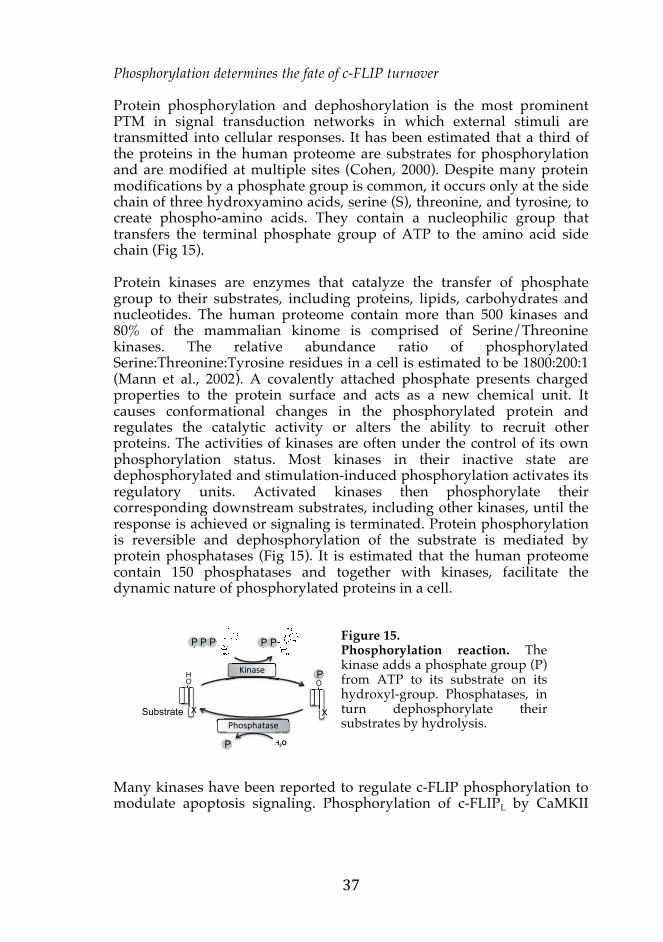

promote recruitment of c-FLIP to the DISC and protects the cell from DR-mediate apoptosis, while PKC and indirect phosphorylation by bile acid glycochenodeoxycholate decrease c-FLIPL affinity for FADD (Higuchi et al., 2003; Xiao et al., 2005; Yang et al., 2003). Furthermore, recent studies have begun to elucidate how the concerted phosphorylation and ubiquitination jointly regulates the stability of c-FLIP. Upon mycobacteria infection-induced TNF-α-mediated apoptosis, S4 and Y211 of murine short c-FLIP isoform are phosphorylated by p38 and c-Abl, respectively (Fig 16). These phosphorylations were required to facilitate the interaction between murine c-FLIPR and the E3 ligase c-Cbl (Kundu et al., 2009). The Akt1-mediated phosphorylation on S273 has been reported to mediate proteasomal degradation of c-FLIPL during macrophage activation (Shi et al., 2009).
Figure 16. TNFα-mediated PTM of murine c-FLIPS. Upon mycobacteria infection, phosphorylation (P) of c-FLIP induces degradative ubiquitination (Ub). Y211 is close to C-terminal tail ubiquitination site, thus may partly explain the regulation (Kundu et al., 2009; Poukkula et al., 2005).
Proteolytic cleavage of c-FLIP by caspase-8 Proteolysis is a process that cleaves target proteins into smaller polypeptides commonly by enzymes called proteases. Proteolytic cleavage of a peptide bond is a thermodynamically favorable reaction and, therefore, the reaction is irreversible. Degradative proteolysis regulates protein concentrations, but proteases can also cleave signal peptides from nascent proteins and activating zymogens, or remove signal sequences after the protein is transported. As mentioned previously, caspases are proteases, which themselves are synthesized as precursor proteins and are cleaved to form their final active structure. c-FLIPL contains two aspartic acid cleavage sites, D198 between DED2 and the caspase-like domain, and D376 between the subunits p12 and p20. They can be cleaved by caspase-8 upon heterodimerization in the DISC (Golks et al., 2006; Irmler et al., 1997; Scaffidi et al., 1999). Proteolytic cleavage of c-FLIPL after D376 (LEVD) release the p10 fragment and generates p43-FLIP, and further cleavage of c-FLIP at D198 generates p22-FLIP. Cleavage of c-FLIPL induces its degradation (Fukazawa et al., 2001). Moreover, these cleaved products play powerful roles in non-apoptotic signaling pathways and the degree of cleavages may influence the survival of a cell. Detailed mechanisms and functions of c-FLIP cleavage will be discussed in the next chapter.
c-FLIPS 221
P S4
K192/195
P Y211
Ub Ub
Ub
DED1 DED2
Figure 17. Cleavage sites of c-FLIPL. c-FLIPL may be cleaved after D376 by caspase-8 to produce the p43-FLIP isoform (43 kDa; cleavage product 1). D198 is found in all c-FLIP isoforms and, hence, caspase-8 can cleave all isoforms to produce p22-FLIP (22 kDa; cleavage product 2).
4. The dynamics of c-FLIP signaling in cell survival The specific regulation of the stability and localization of splice variants provides flexibility in protein functions and enables the cell to adapt to a changing environment. It is important that the expression of c-FLIP is tightly regulated, since vital cellular processes are reported to be influenced by c-FLIP. c-FLIP not only regulates the DISC signaling pathway in an isoform-specific manner, but recent studies have revealed that it is an important regulator in necroptosis. Furthermore, accumulating data show that c-FLIPL can function beyond cell death regulation by directly activating pro-survival signaling pathways. 4.1 The anti-apoptotic role of FLIP The activation of caspase-8 occurs in two phases. Dimerization of procaspases at the DISC results in a cleavage to generate two intermediate subunits p43 and p12, followed by a second cleavage where the prodomain p26 and the fully active enzyme subunits p18 and p10 are produced. The active dimeric monomers oligomerize with one another to form a stable heterotetramer p102-p182 with full substrate processing capacity. These active caspase molecules are released from the DISC to the cytosol and initiate the apoptotic proteolytic cascade (Fig 18, left signaling) (Hughes et al., 2009; Oberst et al., 2010). Since the first discovery of FLIP, a great enthusiasm went into understanding the protein function regulating the caspase-8 activity in apoptotic signaling. Initially, v-FLIP was defined as an anti-apoptotic viral protein that blocked caspase-8 activation at the DISC, thereby inhibiting the mammalian host cell from cell death.
c-FLIPL 480 p20 p12
D198 D376
DED1 DED2
p43-FLIP
p22-FLIP
Cleavage products
1 2

Inhibition of apoptosis at the DISC
c-FLIP proteins are recruited to the DISC via DED interactions (Irmler et al., 1997; Scaffidi et al., 1999). Short c-FLIP isoforms effectively block DR-induced apoptosis via binding to the DISC and inhibiting procaspase-8 processing into its active form, thereby functioning as dominant-negative regulators (Fig 18, right signaling) (Golks et al., 2005; Krueger et al., 2001). c-FLIPL was simultaneously identified by several groups, who inconsistently described either anti- or pro-apoptotic regulatory role in the DISC. Anti-apoptotic behavior of c-FLIPL was shown at high levels of ectopic expression, by competing with procaspase-8 for the DED binding sites of FADD (Scaffidi et al., 1999). Furthermore, c-FLIP knockout mouse embryonic fibroblasts (MEFs) are highly sensitive to caspase-8 mediated apoptosis compared to the wild-type MEFs (Yeh et al., 2000).
Figure 18. Different signaling pathways initiated at the DISC by modulation of c-FLIP. Homodimeric procaspase-8 is processes to its activated form by autocleavage to generate prodomains and a heterotetramer of two p10 and two p18 active subunits. The prodomains of caspase-8 molecules stay in the DISC, whereas the active subunit dissociates to the cytoplasm (left signaling). Hetero-dimerization of procaspase-8 and c-FLIPL permits partial processing to its p43/p10 cleavage fragment, caspase-8 remains associated within the complex and apoptosis is inhibited (middle signaling). Short isoforms of c-FLIP prevent caspase-8 activation (right signaling). Furthermore, at high concentration of c-FLIP, procaspase-8 is competed out for binding to FADD at the DISC.
X X XXXXXX
p18
p18
p10
p10
XXXX
p18
p20
p10
p12
X
Inhibition of caspase-8 activation
Caspase-8 activation ion
Partial caspase-8 activation
c-FLIP concentration
p12 p10
FADD c-FLIPS/R c-FLIPL
DRs
Caspase-8
On the contrary, endogenous c-FLIPL heterodimerizes with procaspase-8 to promote partial processing of caspase-8 for non-apoptotic functions (Micheau et al., 2002). Heterodimerization of procaspase-8 and c-FLIPL allows an activation loop in the C-terminal caspase-like domain of c-FLIPL to overlap and expose the enzymatic region of procaspase-8 permitting the first cleavage to occur (Chang et al., 2002). The second cleavage of caspase-8 is inhibited however, resulting in a release of the p10 fragment and partially active p43-caspase-8 molecule (Fig 18, middle signaling). The heterodimer is proteolytically active, but has a restricted substrate repertoire due to the low caspase-8 catalytic activity at the DISC. Formation of the heterodimers do not initiate apoptotic signaling, but result in a low level activation directed against local substrates, which include themselves and the necroptosis mediator RIPKs (Irmler et al., 1997; Kavuri et al., 2011; Micheau et al., 2002; Scaffidi et al., 1999). Interestingly, this heterodimerization of c-FLIP and caspase-8 occur with higher affinity and tighter interaction via hydrogen bonds than procaspase-8 homodimerization (Boatright et al., 2004; Jong et al., 2009). Therefore, at physiological c-FLIPL levels, heterodimerization occur more readily and this provides further refining regulation of caspase-8 activation in the DISC signaling pathways. In such situation, it is only after caspase-8 in heterodimer is cleaved by an external protease, that caspase-8 becomes pro-apoptotic. c-FLIP as a rheostat in the induction of necroptosis Recent advances in the study of death inducing complexes revealed that c-FLIP could shift the outcome of cell death between apoptosis and necroptosis in an isoform-specific manner. The formation of procaspase-8 homodimers within multiprotein complexes fully activates caspase-8 and trigger apoptosis. In contrast, heterodimerization of procaspase-8 and c-FLIPL results in limited catalytic activity of the caspase, not sufficient to initiate apoptosis, but enough to cleave RIPKs, thereby inhibiting the induction of necroptosis (O’Donnell et al., 2011; Oberst et al., 2011). This regulation by c-FLIP can be observed in TNF-α−TNFR1-mediated formation of complex IIa and complex IIb. When the activation of caspase-8 is prevented in complex IIa to initiate apoptosis, RIPK1 and RIPK3 accumulate, leading to increased formation of the necrosome (complex IIb) and consequently initiate necroptosis (Fig 6). Another example of c-FLIP-mediated cell death decision occurs in the ripoptosome, which is a cytosolic death-inducing complex. The ripoptosome is spontaneously formed upon depletion of cIAP1/2, which function as E3 ligases and degrades RIP1, and accumulated RIPK1, FADD and procaspase-8 are assembled, thereby activating RIPK or caspase-8 (Tenev et al., 2011). The ripoptosome can stimulate caspase-8-mediated apoptosis as well as caspase-independent necroptosis, and the

outcome of the complex formation is regulated by c-FLIP in an isoform-specific manner. The presence of c-FLIPL will partially activate caspase-8 with enough catalytic action to cleave and deactivate RIPK1. When procaspase-8 and c-FLIPS heterodimerization predominates, on the other hand, caspase activation and RIPK1 cleavage are prevented, and if caspase-8 activity remains absent, active RIPK will eventually execute the cell death by necroptosis. In this situation, apoptosis is blocked and the mode of cell death is instead switched to necroptosis (Feoktistova et al., 2011). Thus far, c-FLIP is the only protein identified that can prevent RIPK or caspase-8 full activation to cell death. Taken together, caspase-8-c-FLIPL heterodimers are important in maintaining homeostasis by negatively regulating necroptotic cell death. 4.2 Pro-survival roles of FLIP in signal transduction Accumulative studies have revealed that the apoptosis signaling pathway is regulated by various mediators and that there is a dynamic crosstalk with other signaling pathways. The expression of inducible genes coordinated by various transcription factors is an important determinant in regulating the level of apoptotic proteins. In contrast to c-FLIPS/R, the biological functions of c-FLIPL extend beyond being a regulator of DR-mediated apoptosis. In addition to its function in cell death, c-FLIPL is implicated in cell survival and proliferation. c-FLIPL itself is processed by caspase-8 after D379 to produce p43-FLIP, a cleaved product with survival promoting properties. p43-FLIP functions by recruiting the adaptor proteins involved in different pro-survival signaling pathways. c-FLIP clearly has multiple roles in pro-survival pathway, but further studies are required to clarify the source of variations between different studies. c-FLIP determines the outcome of NF-κB signaling NF-κB proteins are an evolutionary conserved family of nuclear transcription factors that regulate approximately three hundred genes, including those regulating inflammation, immune response to infections, apoptosis, and cell proliferation (Bonizzi and Karin, 2004; Gerondakis et al., 1999; Gilmore, 2006; Hayden and Ghosh, 2004; Pasparakis et al., 2006; Sen and Baltimore, 1986). In unstimulated cells, NF-κB is sequestered by inhibitor of κB (IκB) that binds and retains NF-κB in the cytoplasm (Ghosh and Karin, 2002). In the conventional pro-inflammatory TNF-α signaling pathway, TNFR1 recruit TRADD and NF-κB activating components to form complex I. The multiprotein complex is an ubiquitin-dependent signaling platform. It is modified by K63-linked ubiquitination of RIPK1 by the E3 ligases TNF receptor-associated factor 2 (TRAF2) and cIAP1/2, and linear ubiquitination of the IκB kinase (IKK)
complex by linear ubiquitin chain assembly complex (LUBAC), which provides stability to the complex for efficient activation of the IKK complex regulatory unit, NF-κB essential modulator (NEMO) (Haas et al., 2009; Poyet et al., 2000; Zhang et al., 2000). The IKK complex is postulated to be a gatekeeper of NF-κB activation as it phosphorylates IκB, which subsequently ubiquitinates and leads to degradation of IκB by the 26S proteasome (reviewed by Perkins, 2007). Freed NF-κB exposes its NLS and translocates to the nucleus, where it regulate gene expression by binding to the κB sites (Fig 19) (Hayden and Ghosh, 2004). Survival pathways induced via DRs are primarily mediated via the TNFR, although low-level stimulations of CD95 and TRAIL signaling also have been reported to convey survival signaling properties (Falschlehner et al., 2007; Siegel et al., 2000). Activation of the canonical NF-κB pathway induces resistance to both extrinsic and intrinsic apoptotic pathways by upregulating the expression level of anti-apoptotic and pro-survival proteins (Van Antwerp et al., 1996). The regulatory circuit of NF-κB signaling reinforces the decision-making, which leads to a rapid amplification and dominates the survival signaling. c-FLIP is one potent and early target of NF-κB and indeed contributes greatly to NF-κB-mediated regulation of death signals. Expression of c-FLIP under the control of the NF-κB results in increased resistance to the DR-mediated apoptosis (Kreuz et al., 2001; Micheau et al., 2001). Deubiquitination of RIPK1 drives the conversion of the complex I to II, where the complex dissociate into the cytoplasm to form other signaling platforms; complex IIa to initiate apoptosis by recruiting FADD and procaspase-8, or complex IIb to initiate necroptosis by activating RIPK1 and RIPK3 (Fig 6) (Gerlach et al., 2011; Ikeda et al., 2011). c-FLIP takes part in the regulatory circuit by inhibiting the activation of caspase-8 in complex IIa (Fig 19) (Kreuz et al., 2001; Micheau et al., 2001; Yeh et al., 2000). c-FLIPL acts as a dual functioning molecular switch in complex II by regulating both the apoptotic and necrotic programmed mode of cell death (Fig 2). If the c-FLIP expression is low, it is inadequate to inhibit the activation of caspase-8 induced by complex IIa (Micheau and Tschopp, 2003). For example, TNFα-mediated JNK activation antagonizes NF-κB activation by increasing the turnover of c-FLIPL (Chang et al., 2006). Furthermore, overexpression of c-FLIPL in various cell lines has been shown to trigger strong activation of the NF-κB signaling pathway in a DED-dependent manner (Chaudhary et al., 2000; Hu et al., 2000; Kataoka et al., 2000). Procaspase-8 proteolytic cleavage of the flexible intersubunit linker within the c-FLIPL protease-like domain produces p43-FLIP and promotes the recruitment of TRAF2 and RIP1. This leads to the RIP1 ubiquitination and interaction with NEMO, thereby activating NF-κB efficiently (Dohrman et al., 2005; Kataoka and Tschopp, 2004). This phenomenon was further supported by v-FLIPs that potently activate the pathway via interacting with TRAF2 (Chaudhary et al., 1999; Field et al., 2003; Guasparri et al., 2004). p43-FLIP cleaved by procaspase-8 generates

a p22-FLIP isoform that only consists of its amino terminal tandem DEDs and directly interacts with NEMO in non-apoptotic cells (Golks et al., 2006). In addition, c-FLIP was shown to interact with p105, a precursor of the NF-κB subunit and atypical IκB molecule, and inhibited the processing of p105 into p50 and IκB-γ (Li et al., 2003). Furthermore, c-FLIP regulates DR-induced NF-κB activity. Some studies indeed show induced NF-κB activation upon DR stimulation in the presence of c-FLIP, while other studies convincingly show that c-FLIP strongly inhibits NF-κB in the presence of the same DRs (Iyer et al., 2011; Kataoka et al., 2000; Kavuri et al., 2011; Micheau and Tschopp, 2003; Wachter et al., 2004).
Figure 19. Simplified canonical NF-κB signaling pathway with emphasis on the regulatory roles of c-FLIP. Stimulation of TNFR1 by TNF-α induces the formation of complex I. Activation of the IKK complex (depicted as the regulatory component, NEMO) releases NF-κB from IκB. Activated NF-κB rapidly transcribes a number of anti-apoptotic genes including c-FLIP. Some components of complex I form complex IIa and induce apoptosis. c-FLIP can augment the survival signaling by p43-FLIP association with RIP1, TRAF2 and NEMO to promote NF-κB activation, or inhibit apoptosis in complex II, as indicated with dotted lines.
κP P
Ub Ub Ub
X X XXX
p43-FLIP
c-FLIPL
Active caspase-8
Anti-apoptotic
AA titiA t
TNFR1
TNFα�
Complex I
Complex IIa
Regulation of MAPK pathways by c-FLIPL The mitogen-activated protein kinase (MAPK) family transmits signaling from extracellular stimuli and regulates pro-survival processes, such as cell differentiation, inflammation, and immune regulations. The MAPK family consists of different signaling cascades including the extracellular signal-regulated kinase (ERK), c-Jun N-terminal kinase (JNK), and the p38 kinase pathway. Various c-FLIP-mediated alterations have been observed in MAPK signaling pathways. The protein kinase Raf, MAPK kinase (MEK) and ERK constitute the classical ERK pathway. This kinase signaling cascade transmits mitogenic signals from Ras, which is activated by the cell surface receptors, to the nucleus and regulates the transcription of genes that drives cell proliferation and differentiation. The ERK activity has been shown to directly inhibit DR-induced apoptosis by preventing the caspase activation (Holmström et al., 2000; Tran et al., 2001). c-FLIP recruitment to the TRAIL-R DISC inhibits caspase-8 cleavage and couples NF-κB and ERK for cell survival in TRAIL-resistant cells (Song and Lee, 2008). Overexpression of c-FLIPL has been shown to augment ERK in T cells upon TCR activation (Kataoka et al., 2000). Furthermore, c-FLIP-induced ERK activation is dependent on the presence of PI3K in B cell receptor signaling (Fang et al., 2004). On the contrary, non-apoptotic CD95 stimulation-induced ERK1/2 and p38 activations are inhibited by high expression of all c-FLIP isoforms (Kober et al., 2011). Although c-FLIP is not required for JNK activation (Yeh et al., 2000), c-FLIPL inhibits JNK in a TNF-α-mediated signaling by directly binding to MAPK kinase (MKK)-7 (Nakajima et al., 2006). Induced Wnt signaling by overexpression of c-FLIPL The Wnt signaling pathway is important for the embryonic development, where it regulates proper formation of various tissues and organs. The canonical Wnt signaling pathway is dependent on β-catenin, a signal transducer that also plays a role in cell-cell adhesion. In unstimulated cells, free cytosolic β-catenin is maintained at low level by phosphorylation-induced proteasomal degradation regulated by a multiprotein complex containing GSK-3β (glycogen synthase kinase-3β). Upon the activation of Wnt, β-catenin degradative ubiquitination is inhibited, causing β-catenin to accumulate and translocate to the nucleus. Here, β-catenin interacts with transcription factors of the TCF/LEF (T-cell factor/lymphoid-enhancer factor) family, which in turn activate their target genes important in development to promote the cell growth (Clevers, 2006; Logan and Nusse, 2004).

Overexpressed FLIP proteins are prone to aggregation and cause impairment to the ubiquitin-proteasome system, thereby accumulating short-lived proteins in the cell. β-catenin is one of such proteins, and as a consequence, the downstream Wnt signaling pathway is enhanced (Ishioka et al., 2007; Naito et al., 2004; Nakagiri et al., 2005). Conversely, depletion of the endogenous c-FLIP protein reduces the signaling (Naito et al., 2004). Moreover, a report further described that nuclear c-FLIPL has a positive role in Wnt signaling by directly modulating the β-catenin-mediated gene expression (Katayama et al., 2010). PI3K/Akt signaling pathway and c-FLIPL Akt (also known as protein kinase B) is a serine/threonine kinase, which plays a role in cell viability by promoting survival and inhibiting apoptosis. Akt is activated via phospho-inositides, which in turn are generated by phosphatidylinositide-3-kinase (PI3K) upon mitogen-induced activation of Ras (Toker, 2000). The PI3K/Akt pathway upregulates the protein level of c-FLIP to inhibit DR-mediated apoptosis (Moriyama and Yonehara, 2007). Respectively, hindering PI3K or Akt activity reduces the expression of c-FLIP and sensitizes cells to DR-mediated apoptosis (Suhara et al., 2001). c-FLIPL, in turn, has been demonstrated to mediate the activation of PI3K/Akt pathway. c-FLIPL can enhance the anti-apoptotic activity of Akt by modulating GSK-3β (Quintavalle et al., 2010). CD95 activates several survival signaling pathways, including NF-κB and PI3K/Akt. The latter has an inhibitory effect on NF-κB activation upon CD95 stimulation and c-FLIPL has been suggested to regulate the crosstalk by augmenting phosphorylation of Akt in the presence of CD95L (Iyer et al., 2011). Furthermore, c-FLIP can directly interact with Akt and acts as a substrate for the kinase upon the activation of macrophage (Shi et al., 2009). Autophagy regulation by c-FLIP Macroautophagy, or simply referred to as autophagy, is a homeostatic response in which intracellular constituents are engulfed to form double-membrane autophagosomes, which are delivered to lysosomes for a bulk catabolic degradation. The degradation mobilizes nutrient products and the process is also utilized as a response to stress. Autophagy has cytoprotective physiological roles in starvation adaptation, protein and organelle clearance, and elimination of microorganisms and apoptotic cells. A cell death as the result of progressive cellular consumptions has been attributed to excessive autophagy.
Emerging evidences indicate that both viral and cellular FLIPs suppress the initiation of autophagosome biogenesis by binding to Atg (Autophagy-related)-3 E2-like enzyme and preventing Atg3 from processing LC3 ubiquitin-like proteins. LC3 must be inserted into the membrane of autophagic vesicle for expansion, and therefore, short c-FLIP isoforms prevent autophagosome formation. Subsequently, the autophagic killing was rescued by overexpression of FLIP (Lee et al., 2009; Liang, 2010). Furthermore, LC3 is rapidly translocated to death cell-containing phagosomes, but the role of c-FLIP is yet to be determined. Regulation of caspase-8 in pro-survival signaling The activity of caspase-8 also has functions outside the extrinsic apoptotic signaling. Caspase-8 is sequestered in different cellular compartments following an initiation of cell proliferation or death, thereby profoundly altering the access of caspase-8 to its substrates. A diverse role of caspase-8 has been reported. For example, caspase-8 can initiate the NF-κB activation independently of its proteolytic activity (Jun et al., 2004). Furthermore, it can interact with integrins to regulate cell migration (Barbero et al., 2009). More recent studies have shown that caspase-8 is activated in the inflammasome to induce apoptosis (Sagulenko et al., 2013), or to modulate the production of IL-1β upon macrophage activation (Man et al., 2013). c-FLIP is an important regulator that can fine-tune the degree of caspase-8 activation at the DISC. Therefore, c-FLIP may further influence various signaling pathways indirectly via regulating caspase-8 activity in its diverse functions. The role of c-FLIP is yet to be defined in many of these atypical caspase-8 signaling pathways. For these arising questions to be answered, quantification analysis of isoform-specific c-FLIP functions, such as absolute subcellular protein concentrations and rate of reactions, would advance the understanding of c-FLIP signaling. 4.3 Modeling the dynamic cell death signaling pathway The biochemical properties of key proteins in apoptosis are well understood and much of the introduction focused on the qualitative descriptions of c-FLIP protein and its related aspects. However, not all the information reported on c-FLIP is referred to, since it is simply overwhelming to read and fit all the c-FLIP data available into one thesis. Conjointly, understanding quantitative descriptions, which are highly influential in a biological system, makes it difficult to determine the exact context-dependent outcome of c-FLIP signaling. Comprehending the network-based interplay and resulting outcome of c-FLIP signaling is

therefore necessary. To understand such highly complex biological responses on a system biology scale mathematical modeling approach is increasingly being employed. It adds significance to the qualitative signaling pathways by analyzing the dynamic co-regulation of components and interactions, and describes the statistical properties of the network as a summary of biological system. To construct a mathematical model, a scheme that depicts the signaling pathway of interest is designed and a set of mathematical equations for each reaction is fitted. Sensitivity analysis and parameter estimation identify the important components in the modeled signaling pathway and to determine the parameter values and the fit of simulated model. A model is usually trained against data obtained by laboratory experiments for validation so they can be applied to deterministic biochemical models (Fig 20). Measurements and quantitative data is an important task in computational cell biology. Although the development of biologically representative models of mammalian cells for computation remains a challenge, with the development of quantitative molecular-biological techniques, mathematical modeling is becoming more reliable for biological use. It is through the development and analysis of such models that we will be able to understand the basic design principles of a cell and it enables the identification of components that are critical in the signaling system. Owing to the amount of information available from diverse cell death studies, development of mathematical models has been expanding since the first mathematical model, merging caspase function and apoptotic pathway was published (Fussenegger et al., 2000). Various mathematical models of apoptotic signaling are cumulatively generated to convey the cell death circulatory (Rehm and Prehn, 2013). Such model equations include ordinary differential equations (ODEs) (Albeck et al., 2008; Bentele et al., 2004; Fricker et al., 2010; Fussenegger et al., 2000; Neumann et al., 2010; Rehm et al., 2006), Boolean modeling (Calzone et al., 2010; Schlatter et al., 2009), Petri nets (Chen et al., 2007; Heiner et al., 2004; Heiner, 2009), cellular automata (Düssmann et al., 2009), and agent-based models (Schleich et al., 2012).
Figure 20. Workflow for creating a mathematical model. First, the signaling pathway of interest is determined and mathematical equation is applied to build a model. Sensitivity analysis and parameter estimation is performed to determine each reaction, which are compared with quantitative experimental data to fit the model (adapted from Cho et al., 2003).
Advance in quantitative modeling of extrinsic apoptotic signaling pathway, especially of the kinetics of DR-induced caspase-8 activation, has given new insight into the cell death mechanisms. When cells are stimulated with death ligands, there is a rapid activation of upstream signaling, followed by a duration in which cells enforce their decision to die or survive. Should a cell decide to die, a sudden activation of downstream signaling occurs to insure an efficient removal of the cell (Rehm et al., 2002; Tyas et al., 2000; Vaughan et al., 2002). Modeling has revealed that this duration of delayed responses varies between cells due to differences in the activities of receptor-proximal biochemical reactions (Albeck et al., 2008). The source of cellular heterogenicity has been demonstrated by collective modeling studies. The variability in the timing and probability for DR-mediated apoptosis to occur within a population of cells is mainly due to naturally occurring differences in the levels and status of proteins (Spencer et al., 2009). The cell fate is transiently inherited as the protein status is transmitted to daughter cells after cell division (Rosenfeld et al., 2005). Later, however, protein synthesis promotes divergence and therefore sister cells no longer respond similarly to the same death ligand (Kaufmann et al., 2007; Sigal et al., 2006). The stoichiometry of the DISC components is highly crucial in determining how the DR signal will be transduced in the cell. The CD95-FADD homotypic DD complex predominantly assembles at a ratio of 5:5 (Esposito et al., 2010; Wang et al., 2010). An endogenous DISC structural model proposes that DED-dependent caspase-8 chain assembly upon DR ligation is common for inducing apoptosis by TRAIL
Translation Simulation
Yes
No

and CD95 (Dickens et al., 2012; Schleich et al., 2012). In silico comparison of amplification of caspase cascades revealed that the balance of caspase-8 dimerization and dissociation was an important regulatory mechanism that avoids unwanted cell death (Würstle et al., 2010). A great interest focuses on elucidating the crosstalk mechanisms by using models that integrate consistent data from literatures on DR-mediated apoptosis versus its opposing survival signaling (Calzone et al., 2010). NF-κB and apoptosis signaling pathways are closely integrated and they can be induced from the same DRs (Shishodia and Aggarwal, 2002). Neumann and colleagues predicted that the CD95-mediated pathways diverge at the DISC, and the balance between c-FLIPL and caspase-8 is highly influential. The model postulated a direct interaction between p43-FLIP and the IKK complex, which was later validated with experimental data (Neumann et al., 2010). Their continuous work proposed an underlying mechanism for the dual role of c-FLIPL upon the DR stimulation. They demonstrated that c-FLIP displays its pro-apoptotic behavior when c-FLIPL is expressed at the physiological level and the cells receive strong receptor stimulation, or when the short isoforms of c-FLIP are present at high levels (Fricker et al., 2010). The apoptotic signaling pathway has a complex dynamic behavior involving several reactions with regulatory feedback loops. Therefore, it is often difficult to infer its overall response by examining segments of the signaling pathway separately. On average, more than fifty articles are published everyday on apoptosis. Mathematical modeling and simulation would provide valuable insights into the behavior of apoptosis by incorporating new data and identifying outlier studies. Additionally, models enable the identification of components that are the most susceptible to biological interventions. This could identify new targets within the cell for therapeutic interventions for diseases, as well as predicting patient outcome and responsiveness to personalized treatment strategies. For example, an ODE system biological model, APOPTO-CELL, was developed to simulate the ability of cancer cells to undergo mitochondria-mediated apoptosis (Rehm et al., 2006). The model has been extended to provide the proof-of-concept for a clinical setting to colorectal cancer patients in assessing individual patient responses to novel targeted therapeutics (Hector et al., 2012). 5. Abnormality of c-FLIP in disease Apoptosis is not only necessary for normal tissue development (Wyllie, 1981), but it is also a distinctive pathological process leading to various diseases once misregulated. Therefore, detection of apoptosis together with investigation of regulatory mechanisms that augment or inhibit
apoptosis is of importance for clinical purposes. It is palpable that defects in DR-mediated apoptosis have detrimental effects on a wide range of implications in human diseases. The loss-of-function of a DED-containing protein demonstrates the clearest phenotype to support its role in the extrinsic apoptosis pathway, as well as in the embryonic development and the adult tissue maintenance. c-FLIP profoundly affects various cell signaling networks and a collapse of c-FLIP signaling can lead to pathological conditions, including developmental abnormalities, autoimmune diseases, and cancer. 5.1 c-FLIP in development Programmed cell death is common during embryonic development and the mechanism is well conserved among various species. During organogenesis and tissue remodeling, cells are produced in excess only to be eliminated to obtain the optimal state (Penaloza et al., 2006). It is proposed that cell death is the default state during the development of a multicellular organism, and for the cells to stay alive, they must compete for sufficient survival factors in their specific microenvironment (reviewed by Raff, 1992). Therefore, it was speculated that deficiency of key apoptotic proteins would result in accumulation of cells. The knockout animals of key pro-apoptotic genes presented phenotypes consistent with failure to eliminate cells. Interestingly however, the key components of DR pathway suffered early embryonic lethality. Deficiency of FADD, caspase-8 or c-FLIP gene in mice caused lethality at embryonic gestation day 12.5 and 10.5, respectively. They presented similar phenotypic effects, including defects in myocardial development and extreme abdominal hemorrhage (Rasper et al., 1998; Varfolomeev et al., 1998; Yeh et al., 2000). Recent studies show that the lethality caused by deficiency of FADD or caspase-8 occur due to unregulated RIPK1/3-dependent necroptosis, whereas c-FLIP carries a role in embryogenesis distinct from that of other DED-containing proteins, to which apoptotic cell death proposed to be the reason (Dillon et al., 2012). It has been suggested that caspase-8-FLIPL is a catalytically active complex that protects mice by suppressing necrotic death. Defects in T cell development in the caspase-8 knockout mice can be rescued by catalytically active caspase-8 that is not processed (Leverrier et al., 2010). This unprocessed caspase-8 activity with different substrate specificity from the initiator activity in apoptosis, functions to preserve the embryonic development (Pop et al., 2011). Of note, there are discrepancies between mouse and human studies; caspase-8 is not essential in human embryogenesis due to the presence of caspase-10, which is not found in the mouse genome (Chun et al., 2002; Kischkel et al., 2001). Moreover, murine caspase-8 only has one cleavage site, and therefore, dimerization studies in mice may be different when studying

the function of caspase-8-c-FLIPL heterodimers in humans (van Raam and Salvesen, 2012). In addition to the embryonic development, c-FLIP has been shown to play a vital role in the heart development, cardiac myocyte survival during injury, and pathological cardiac hypertrophy. Highest levels of c-FLIP expression are found in cardiac tissue and the importance of c-FLIP in regulation of susceptibility of cardiac myocytes to apoptotic stimuli have been demonstrated in a number of studies. c-FLIP play a indispensable role in cardiac remodeling following myocardial infarction by interrupting JNK1/2 signaling and augmenting Akt signaling, thereby promoting cardioprotection (Imanishi et al., 2000; Steenbergen et al., 2003). In line with this, reduced c-FLIP expression at the end-stage of failing human hearts has been reported. Apoptosis is important in atherogenesis and vascular remodeling (Han et al., 1995), and studies indicate that c-FLIP is involved in these cellular processes. c-FLIP plays a role in anti-apoptotic resistance of vascular smooth muscle cells (Wang et al., 2002). Moreover, the c-FLIP protein expression is induced in tunica intima and media after artery injury and remodeling (Imanishi et al., 2000). 5.2 The role of c-FLIP in the immune system and in autoimmune diseases We are constantly exposed to potentially harmful substances, such as microorganisms and toxins, from the environment. Fortunately, our immune system effectively defends off such destructive elements. The innate immune system includes the physiological barriers and phagocytic cells, where as the adaptive immune response functions via the lymphocytes to specifically recognize particular antigens by lymphocytes. B cells are antibody-producing cells, while T cells directly kill the infected host cells or assist other lymphocytes. The foundation of the adaptive immune response relies on its ability of clonal expansion of antigen-specific lymphocytes upon infection to clear out the antigens. Regulation of the lymphocyte population is important to maintain a functional immune system, and impaired homeostasis is often associated with both immunodeficiency and autoimmune diseases. c-FLIP signaling in the immune system is thoroughly studied physiological process, as it demonstrates the most biological relevance of the protein. In a broad range of lymphocytes, induced high level of c-FLIP rescues the cells from death ligand-mediated apoptosis. Following clonal expansion of the antigen-specific lymphocytes, subsequent efficient elimination of these activated cells by apoptosis occur, a process known as activation-induced cell death (AICD). AICD relies on CD95L-CD95-mediated apoptosis to terminate the immune response and in the
induction of peripheral T cell self-tolerance. Following the TCR stimulation, the level of c-FLIPS/R is upregulated, which is primarily responsible for the resistance towards CD95-mediated apoptosis by limiting caspse-8 activation in the activated T cells (Park et al., 2005). After clonal expansion, the c-FLIP levels decrease in an IL-2-dependent manner (Algeciras-Schimnich et al., 1999; Refaeli et al., 1998), and sensitize T cells towards CD95L-stimulation (Kirchhoff et al., 2000; Schmitz et al., 2004). The short c-FLIP isoforms also inhibit NF-κB activity, resulting in decreased T cell activation and survival. In vitro restimulation of preactivated primary human T cells induces the expression of c-FLIPS, rendering the proportion of cells that survive another AICD (Kirchhoff et al., 2000). The relative levels of c-FLIP might therefore provide a crucial balance of caspase and NF-κB activities in the effector T cells. In addition to the regulation of DR-mediated apoptosis, the core DISC components are crucial in T cell survival, proliferation, and activation (reviewed by Budd, 2002). T cell stimulation activates caspase-8 at the lipid rafts, and blocking the caspase activation in turn prevents T cell activation and proliferation (Alam et al., 1999; Simons and Toomre, 2000; Simons and Ikonen, 1997). c-FLIP is also recruited to the lipid rafts, where it is rapidly cleaved to p43-FLIP (Misra et al., 2007). The phenotype of c-FLIP deficient T cells is closely parallel that of caspase-8 deficient T cells (Salmena et al., 2003). The loss of c-FLIP profoundly affects thymocyte development, and the proliferation of c-FLIP deficient T cells is greatly reduced in response to IL-2 and TCR crosslinking stimulations (Chau et al., 2005; Zhang and He, 2005). Overexpression of c-FLIPL in Jurkat cells produces more IL-2 and increases proliferation following TCR stimulation via NF-κB and ERK signaling pathways (Kataoka et al., 2000; Lens et al., 2002). The c-FLIP expression in transgenic mice causes the T cell hyperproliferation upon activation, but they may be more sensitive to cell death, owing to simultaneously augmented caspase activity (Dohrman et al., 2005). Taken together, increased expression of c-FLIP in T cells cause the accumulation of lymphocytes or resist CD95-mediated apoptosis. These characteristics may attribute in various autoimmune diseases, multiple sclerosis, rheumatoid arthritis, Graves’ disease, and inflammatory arthritis (Budd et al., 2006; Stassi et al., 2000). c-FLIP also function in other immune cells, such as antigen-presenting cells. The c-FLIP level alteration occurs during differentiation of peripheral blood monocytes into macrophages. Monocytes contain very low levels of c-FLIP and are highly sensitive to CD95-induced cell death, but macrophages and immature DCs express substantial amounts of c-FLIP and are, therefore, resistant to the same stimulus (Ashany et al., 1999; Perlman et al., 1999). Activation of B cell receptors through the PI3K/Akt pathway upregulates c-FLIP expression and protects the B

cells from CD95-induced cell death (Moriyama and Yonehara, 2007). Hemin-differentiated K562 cells show reduced c-FLIP expression and higher sensitivity to TRAIL-induced apoptosis than undifferentiated cells (Hietakangas et al., 2003). Microglia are resident macrophages in the immune privileged organs that are important for an active immune defense. TGF-β is produced in such immune privileged sites and it can increase c-FLIP levels in an MKK-dependent pathway to protect cells from CD95-mediated apoptosis (Schlapbach et al., 2000). c-FLIP inhibits HIV-1 replication in lymphocytes by preventing apoptotic cell death induced by HIV-1 infection and upregulating the expression of viral restriction factors (Tan et al., 2013; Wang et al., 2010). 5.3 Targeting c-FLIP for cancer therapy Cancer is a leading cause of death. On the worldwide scale, death from cancer was accounted for 7.6 million (around 13% of all deaths) in 2008 and it is predicted to rise to over 13 million by 2030 (World Health Organization). Cancer is collective of many diseases, each with distinct characteristics and therapeutic options. However, all cancers confer survival advantages that contribute to uncontrollable cell proliferation and major molecular changes arising in tumorigenesis are well understood. The change in expression level of proteins involved in apoptosis regulation and impaired ability to undergo programmed cell death in response to death stimuli is one of the hallmarks of cancer (Hanahan and Weinberg, 2000). In cells, apoptosis by metabolic stress is crucial to suppress tumorigenesis and inability to die acquires cancer cells a selective advantage for progression and metastasis, as well as resistance to chemotherapies. Overcoming this resistance to conventional and targeted therapies remains a key challenge in the fight against cancer and interest in developing therapeutics that kill cancer cells via the extrinsic apoptotic pathway has emerged. Many cancer cells are more susceptible to TRAIL than normal cells due to an impaired expression of decoy receptors in cancer cells (Pan et al., 1997; Sheridan et al., 1997). Some of the potential examples of targeting TRAIL for cancer therapy include: TRM-1, a TRAIL-R1 monoclonal antibody, which has completed the phase II clinical trials for relapsed non-small carcinoma lung cancer (NSCLC) and non-Hodgkin’s lymphoma (NHL) (Human genome sciences); and AMG 951 for NSCLC, NHL and colorectal cancer, which have completed early phases of the clinical trials (Amgen and Genentech). However, highly malignant tumors are resistant to apoptosis induced by TRAIL and some cancers that were originally sensitive can acquire the resistance. During the evolution of tumors in vivo, selection occurs for malignant clones capable of withstanding immune attack, thus successful therapy depends on restoring the competence of DR-mediated apoptosis. Apoptosis
regulatory proteins are intensively studied as potential targets to restore the responsiveness to chemotherapy. c-FLIP is highly expressed in various tumor cells and often correlates with resistant to the DR-mediated apoptosis (Table 2) (Verbrugge et al., 2009). Furthermore, higher c-FLIP levels are shown to correlate with more aggressive tumors (Shirley and Micheau, 2010). c-FLIP silencing restores sensitivity to death ligands in various cancer cells, and therefore, c-FLIP would be an important target for TRAIL targeted therapy. Indeed, small interfering RNAs (siRNAs) can be used to knockdown c-FLIP and enhance TRAIL-induced DISC formation and caspase-8-dependent apoptosis in cancer cells (Mathas et al., 2004).
κ
Table 2. Tumors with elevated expression level of c-FLIP. Some of the tumors with high levels of c-FLIP are listed here together with the signaling pathway responsible for the overexpression or relative prognosis, if known.

The PI3K/Akt signaling pathway predominantly regulates the c-FLIP expression level in tumor cells and it is one major signaling target for sensitizing cancer cells to TRAIL (Garg and Aggarwal, 2002; Karin et al., 2002). Several small molecule inhibitors have the potential to downregulate c-FLIP proteins by disruption of the PI3K/Akt pathway (Fulda et al., 2000). Some of the most recent studies include the therapeutic potential of Sorafenib and histone deacetylase (HDAC) for gastrointestinal cancer (Hamed et al., 2013), tocotrienols for breast cancer (Sylvester et al., 2013), tectorigenin for paclitaxel-resistant ovarian cancer (Yang et al., 2012), Bortezomib for myelodysplastic (Huang et al., 2011), and LY294002 for gastric cancer (Liu et al., 2011). Furthermore, other signaling pathways may be targeted to diminish the anti-apoptotic behavior of c-FLIP in cancer. CDDO-Me, which is in the phase II clinical trial as a combination therapy for lung cancer, triggers induction of apoptosis by targeting c-FLIPs to ubiquitin-dependent proteasomal degradation (Safa et al., 2008). Flavopiridol, an inhibitor of cyclin-dependent kinase shown to sensitize breast tumor cells to TRAIL by inducing proteasome-dependent degradation of c-FLIP (Palacios et al., 2006). Other therapeutic targets involving c-FLIP regulations include several HDAC inhibitors significantly downregulate c-FLIP synthesis in various cancer cells (Lucas et al., 2010; Yerbes and López-Rivas, 2012). Similarly the endoplasmic reticulum stress-inducing agents sensitize cells to TRAIL-mediated apoptosis by altering different apoptosis-related proteins including decline in the level of c-FLIP (Martín-Pérez et al., 2012). Etoposide-mediated depletion of IAPs led to spontaneous assembly the ripoptosome (Tenev et al., 2011). c-FLIP is the determinant that regulates caspase-8 activity in ripoptosome, thereby it could influence the outcome of cell death via RIP3-dependent nectoptosis or caspase-dependent apoptosis (Feoktistova et al., 2011). It is fascinating to see how two structurally similar proteins, procaspase-8 and c-FLIP, can have similar effects as well as very opposing functions. Furthermore, the gross overall structural conformations of homodimers and heterodimers of caspase-8 and c-FLIPL are similar. Thus, restricted substrate specificity of the heterodimers is likely to be caused by higher regulatory mechanisms. c-FLIP plays an important role in determining cell survival, but often observations contradict. Further molecular understanding of c-FLIP is required to translate our knowledge to more effective clinical uses.
OUTLINES OF THE STUDY The aim of this thesis was to determine the regulation of PTMs of c-FLIP and how they in turn modulate the c-FLIP behavior. When I joined the laboratory, two phosphorylation sites were identified in c-FLIP at serine 193 (S193) and serine 227 (S227). My thesis focuses on the role of phosphorylation of c-FLIP in DR-mediated apoptosis. S193 is phosphorylated in all isoforms of c-FLIP. To investigate the mechanism underlying phosphorylation of S193, we first identified classical PKCs as the major regulators of the phosphorylation site in all isoforms. Since c-FLIP has anti-apoptotic functions and PKC can be activated by TRAIL, we defined that TRAIL stimulation can induce phosphorylation of c-FLIP. Finally, we demonstrated that c-FLIP S193 phosphorylation cause cytoplasmic accumulation of c-FLIP, thereby contributing to the DISC signaling outcome upon TRAIL-R activation. With accumulating data on cell death and the DISC activation, various mathematical models are utilized to provide an alternative method to comprehend the cell death signaling. Another aspect of my thesis was to establish interdisciplinary collaborations with chemical engineers from Åbo Akademi University to construct mathematical models that revealed the importance of c-FLIP expression levels upon the DR-mediated DISC signaling. My continuous work extends from observations made during the first studies. When the distribution of phosphorylated c-FLIP was analyzed by confocal microscopy, we observed distinct localizations of c-FLIP in a phospho-dependent manner. Furthermore, we classified the subcellular localizations of differentially phosphorylated c-FLIP and their functions in non-extrinsic apoptosis signaling. We identified that phosphorylation on S227 is required for c-FLIPL to form filamentous structures, which was previously reported not feasible. Separately, the global phosphorylation of c-FLIP was accumulated at the centrosomes throughout the cell cycle, and c-FLIPL phosphorylation was necessary to sustain c-FLIPL-induced increase in cell population via hyperproliferation.
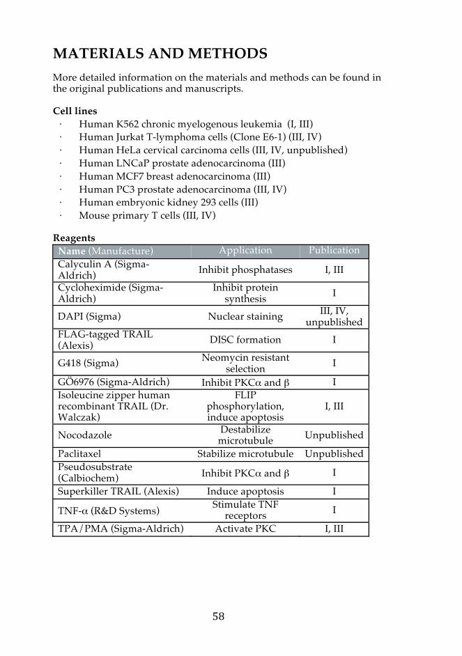
MATERIALS AND METHODS More detailed information on the materials and methods can be found in the original publications and manuscripts. Cell lines ⋅ Human K562 chronic myelogenous leukemia (I, III) ⋅ Human Jurkat T-lymphoma cells (Clone E6-1) (III, IV) ⋅ Human HeLa cervical carcinoma cells (III, IV, unpublished) ⋅ Human LNCaP prostate adenocarcinoma (III) ⋅ Human MCF7 breast adenocarcinoma (III) ⋅ Human PC3 prostate adenocarcinoma (III, IV) ⋅ Human embryonic kidney 293 cells (III) ⋅ Mouse primary T cells (III, IV)
Reagents
Name (Manufacture) Application Publication Calyculin A (Sigma-Aldrich) Inhibit phosphatases I, III
Cycloheximide (Sigma-Aldrich)
Inhibit protein synthesis I
DAPI (Sigma) Nuclear staining III, IV, unpublished
FLAG-tagged TRAIL (Alexis) DISC formation I
G418 (Sigma) Neomycin resistant selection I
GÖ6976 (Sigma-Aldrich) Inhibit PKCα and β I Isoleucine zipper human recombinant TRAIL (Dr. Walczak)
FLIP phosphorylation, induce apoptosis
I, III
Nocodazole Destabilize microtubule Unpublished
Paclitaxel Stabilize microtubule Unpublished Pseudosubstrate (Calbiochem) Inhibit PKCα and β I
Superkiller TRAIL (Alexis) Induce apoptosis I
TNF-α (R&D Systems) Stimulate TNF receptors I
TPA/PMA (Sigma-Aldrich) Activate PKC I, III
Antibodies Target (Manufacture) Application Publication Caspase-3 (BD Pharmingen) WB III Caspase-8 (clone C-15, Alexis) WB I, III DR4 (Alexis) IP I DR5 (Alexis) IP I c-FLIP (clone NF6, Alexis) WB I, III, IV FLAG M2 (Sigma-Aldrich) IP I GFP (clone JL-8, Clontech) WB I HA (Santa Cruz Biotechnology) IP I Hsc 70 (SPA-810, StressGen) WB I, III, IV PARP1 (clone C-2-10, Sigma-Aldrich) WB I, III
Pericentrin (Abcam) IF IV PKCα IF III PKCβ IF III Ubiquitin (clone FK-1, BioMol) WB I
Abbreviations: WB, Western blot; IP, immunoprecipiration Preparation of anti-FLIP phospho-specific antibodies (I, III, IV, unpublished): Anti-pS193 phosphopeptide: AIQK(pS)LKDPS Anti-pS227 phosphopeptide: EPVKK(pS)IQES Secondary antibodies: HRP-conjugated secondary antibodies were used for WB (I, III, IV). Fluoresence-conjugated secondary antibodies were used for IF (III, IV, unpublished). Plasmid constructs ⋅ FLAG-tagged c-FLIPL and c-FLIPS, kind gift from Dr. Jürg Tschopp
(I, III, IV) ⋅ FLAG-tagged c-FLIPR was constructed by PCR using FLAG-tagged
c-FLIPL as a template with following primers: Forward: 5’-ACAGTTGAATTCATGTCTGCTGAAGTC-3’ Reverse: 5’-TCTAGACTCGAGTCATGCTGGGATTCCATTCCATA TGTTTTCTCCAGACTCACCCTGAAGTTATTTGAAGG-3’
⋅ c-FLIP point mutations were made using the QuikChange site-directed mutagenesis kit (Stratagene) (I, III, IV)
⋅ HA-tagged ubiquitin, kindly provided by Dr. Dirk Bohmann (I) ⋅ GFP-tagged KD PKCα/β, kind gift from Dr. Christer Larsson (I)

SiRNA ⋅ ON-TARGETplus Human CFLAR siRNA and ON-TARGETplus
Non-targeting Pool were purchased from Thermo Scientific (III) Methods Name Publication CAM IV Cell culture I, III, IV, unpublished Densitometry quantification I, III DISC analysis III, IV FLAG Immunoprecipitation I Image analysis I, III, IV, unpublished Ki67 staining IV Luminometric caspase-8 activity assay I Microscopy III, IV, unpublished Nuclear fractionation assay III Propedium Iodide staining IV SDS-PAGE and Western blotting I, III, IV Stable cell lines I, III Statistical analysis I, III, IV Transient transfections III, IV Trypan blue exclusion assay III, IV Tunel assay (Invetrogen) IV Ubiquitination assay I WST-1 (Roche) IV
RESULTS AND DISCUSSION 1 Phosphorylated c-FLIP in death receptor activation 1.1 Phosphorylation of c-FLIP on serine 193 (I, III, IV) Before this thesis was initiated, little was known about c-FLIP phosphorylation and its mechanisms and biological implications. To provide insights into the PTM of c-FLIP, phosphorylation sites were identified. In brief, K562 erythroleukemic cells stably overexpressing c-FLIPL or c-FLIPS were treated with the phosphatase inhibitor calyculin A and 32P in vivo orthophosphate labeling was performed to detect phosphorylation by electrophoresis and autoradiography (Fig 1A, Article I). Following the observation that c-FLIP is indeed phosphorylated in our system, phosphopeptide mapping and mass spectrometric analysis were performed to identify S193 as a novel phosphorylation site common to all isoforms of c-FLIP (Fig 1B-G, I). A polyclonal antibody against the phosphorylation was developed to provide a tool for biochemical analysis (Fig 2A, I and Supplementary Fig 2, III). In the first article, immunoprecipitated exogenous c-FLIP in K562 cells was used to principally understand the mechanisms of c-FLIP phosphorylation. Endogenous phosphorylation of S193 was observed in all untreated cell lines thus far being tested. They include Jurkat T cell leukemia, K562 cells, PC3 prostate carcinoma cells, HeLa cervical adenocarcinoma cells, human embryonic kidney 293 cells and mouse primary T cells (Fig 2 and supplementary data 2, III, and unpublished observations). c-FLIP S193 phosphorylation is mediated by classical PKC Bioinformatics analysis was performed to find kinase consensus site on c-FLIP and revealed a number of candidate kinases for S193 phosphorylation. To identify the kinase phosphorylating c-FLIP S193, we manipulated the activities of various kinases with pharmacological activators and inhibitors, as well as genetic exploitation to observe if any would alter the status of c-FLIP S193 phosphorylation. 1 MSAEVIHQVE EALDTDEKEM LLFLCRDVAI DVVPPNVRDL LDILRERGKL SVGDLAELLY 61 RVRRFDLLKR ILKMDRKAVE THLLRNPHLV SDYRVLMAEI GEDLDKSDVS SLIFLMKDYM 121 GRGKISKEKS FLDLVVELEK LNLVAPDQLD LLEKCLKNIH RIDLKTKIQK YKQSVQGAGT 181 SYRNVLQAAI QKSLKDPSNN FRMITPYAHC PDLKILGNCS M
* * * Fig 21. Amino acid sequence of human c-FLIPS. The domains are shaded in grey; darker grey, DED1; lighter grey, DED2. c-FLIPS-specific C-terminal tail is underlined. Relevant PTM sites for this study are marked with asterisks; S193 phosphorylation site (S) and Lysine 192 and 195 ubiquitination sites (K). The numbers on the left indicate starting amino acid residues of each line.

When we treated K562 cells with GÖ6976, an inhibitor of the classical protein kinase C (cPKC) isoforms α and β, S193 phosphorylation was considerably reduced (Fig 2B, I). Likewise, treatment with pseudosubstrate of PKCα and PKCβ provided similar results (data not shown). Moreover, when the PKC activity was induced with phorbol ester PMA (or TPA in article I), phosphorylation of c-FLIP S193 was significantly increased (Fig 2C, I). PMA-mediated phosphorylation of c-FLIPS was mostly abolished by GÖ6976 pretreatment, indicating that the phosphorylation of c-FLIPS S193 is mainly mediated by PKCα and PKCβ. Similar results were obtained with c-FLIPR (Fig 2D-E, I). GÖ6976 pretreatment in turn showed less effect on the phosphorylation of c-FLIPL S193, implying that c-FLIPL may be subject to phosphorylation by a broader range of kinases (Fig 2D, I). To ensure the specificity of results obtained by the pharmacological PKC inhibitors, we transfected c-FLIP overexpressing K562 cells with kinase-dead PKCα and PKCβ, which act as dominant negative inhibitors of the endogenous PKCs. They indeed interfered with the induction of PMA-induced c-FLIP phosphorylation of both c-FLIPS and c-FLIPL (Fig 2F, I). Calmodulin (CaM) is a major calcium sensing protein, which upon binding to calcium, undergoes a major conformational change and the complex interacts with a variety of target proteins. Direct interaction between CaM and c-FLIPL was demonstrated to regulate CD95-induced signaling (Pawar et al., 2008). CaMKII, which is regulated by the calcium/CaM complex, has also been shown to phosphorylated c-FLIP. c-FLIP proteins get threonine-phosphorylated by CaMKII, which blocks recruitment of all isoforms to the DISC (Yang et al., 2003). In our system, CaMKII inhibition did not affect S193 phosphorylation (data not shown). PKC is a well-understood kinase that transduces cell surface signals by phosphorylating substrate proteins. Once activated upon cell stimulation, cytosolic PKC is translocated to membranes, or sometimes to the nucleus, where it exhibits catalytic activity. Eleven isoforms of PKC has been identified in mammalian cells, which are assorted as cPKC, novel PKC (nPKC) and atypical PKC (aPKC). cPKC are Ca2+-dependent and PMA-responsive, nPKC that are Ca2+-independent and aPKC not responseive to phorbol esters. Initially, we used prolonged treatment of PMA to identify PKC as the mediators of c-FLIP S193 phosphorylation (Article I). Since PKC signaling is rapid, we continued by investigating if PMA-mediated S193 phosphorylation could be a rapid response. Upon 10 minute PMA treatment, activated PKCα proteins were found mostly in the cytosol of Jurkat cells (unpublished observation). In accordance with this, endogenous c-FLIP phosphorylation was indeed increased in the cytosol (Fig 3A, III). After one hour of treatment, activated PKCs were found also in the nucleus, and c-FLIP phosphorylation was increased throughout the cell (Fig 3B, III). These data indicate that PKC-mediated c-FLIP S193 phosphorylation is an acute signaling mechanism.
c-FLIP S193 phosphorylation is induced upon DR stimulation After identifying cPKC as the chief kinases that phosphorylates c-FLIP S193, we wanted to elucidate its purpose in the cells. Since c-FLIP is critical in cell death, we first investigated its relevance in the extrinsic apoptotic pathway. In the context of cell death, PKC has been reported to modulate the DR signaling both positively and negatively. PKC plays a protective role by interfering with the formation of the DISC by blocking FADD recruitment and subsequent caspase-8 activation (de Thonel et al., 2001; Gomez-Angelats and Cidlowski, 2003; Harper et al., 2003; Leroy et al., 2005) (Supplementary Fig 6, III). Furthermore, DR-mediated PKC activation has been observed in various cell lines. TRAIL at killing concentration induces PKC activation rapidly although the activation mechanism is yet to be determined. We determined S193 phosphorylation upon the DR stimulations and found in a K562 c-FLIP overexpressed system that TRAIL and TNF-α indeed increased S193 phosphorylation (Fig 5A-B, I). In more acute activation of the DRs, Jurkat cells were treated with TRAIL, CD95 or TNF-α for 10 minutes and endogenous phosphorylation of S193 was detected. Intriguingly, TRAIL and CD95 induced S193 phosphorylation by 15% or more, whereas no change was observed with TNF-α (Fig 7A, III and unpublished data). Taken together, the DR stimulation indeed affect the phosphorylation status of c-FLIP, speculatively via PKC activated by signals transmitted from the DR signaling complexes. How PKC is activated via the DR stimulation is not yet described and the physiological relevance of the acute DR-mediated PKC activation is unknown. It is conceivable from our acute DR stimulation experiment that decision to phosphorylate c-FLIP occurs when death signaling is initiated. Phosphorylation of c-FLIP occur rapidly upon TRAIL and CD95-mediated signaling, while there is a delay in TNF-α-mediated signaling, since the complex II must be formed before apoptosis signaling can be initiated via TNFR1 stimulation. With this notion we continued our study to understand the role of TRAIL-mediated c-FLIP S193 phosphorylation in cell death. 1.2 PKC-mediated c-FLIP phosphorylation leads to protein stability via regulating ubiquitination (I, III) The c-FLIP S193 residue is positioned in a region that is crucial for c-FLIP ubiquitination, between K192 and K195 ubiquitin acceptor sites in c-FLIPS (Poukkula et al., 2005). Many E3 ligases typically recognize substrates that are phosphorylated on specific serine or threonine residues. We were thus prompted to investigate whether c-FLIP S193 phosphorylation influenced this ubiquitination, all of which are PTMs found in every isoform of c-FLIP. We first performed S193 site-directed
mutagenesis to produce a non-phosphorylatable c-FLIP at S193 by replacing the serine with alanine (S193A), or with aspartic acid (S193D) to mimic constitutively phosphorylated c-FLIP. The inhibition of phosphorylation by the S193A mutation increased the ubiquitination of all isoforms of c-FLIP, while the S193D efficiently decreased the ubiquitination (Fig 3A-C, I). Furthermore, when PKC was inhibited by GÖ6976, wild type (WT) c-FLIP was more ubiquitinated, whilst ubiquitination of c-FLIPR S193D was not affected by the same treatment (Fig 2E, I). Hence, S193 phosphorylation does regulate the ubiquitination in all c-FLIP isoforms, although it does not exhibit the typical phosphorylation-induced ubiquitination. Next, we produced the DED-only c-FLIP (1-202) S193A mutant, which can be ubiquitinated but lacks the crucial C-terminal tail for degradation. In the absence of the C-terminal tail, the protein could not be modified by ubiquitin, but S193 phosphorylation was not affected in vivo, indicating that the C-terminal part of c-FLIP is required for ubiquitination but not for PKC kinase recruitment (Fig 2D, I and data not shown). Finally, we introduced S193A into c-FLIPS mutant that could not be ubiquitinated at K192 and 195. The phosphorylation mutant still increased ubiquitination, which signifies that the dephosphorylation impose strong destabilizing effect on c-FLIP even when the principal ubiquitin targeted lysines are unavailable. Taken together, c-FLIP ubiquitination is determined prominently by phosphorylation of S193. Ubiquitin modification functions to target proteins to various cellular processes, protein degradation by K48-linked ubiquitin conjugation being the commonly described. Since majority of reported ubiquitin modification of c-FLIP function in protein degradation, we continued to decipher the role of S193 phosphorylation on the stability of c-FLIP. We overexpressed c-FLIP S193 WT and phospho-mutants for each isoforms and inhibited the protein synthesis by cycloheximide to compare their half-lives (Fig 4A-C, I). S193A mutants of c-FLIPS/R had shorter half-lives, whereas S193D mutant prolonged their half-lives (Fig 4A-B, I). These results signify that phosphorylation on S193 alone can specifically affect the stability of the short c-FLIP isoforms. To further reveal the relevance of S193 phosphorylation-mediated ubiquitination, we induced PKC activity by PMA and observed that prolonged treatment indeed increased the total amount of both isoforms of endogenous c-FLIP in Jurkat cells (Fig 3C, III). The c-FLIPS/R turnover is determined by S193 phosphorylation and degradative ubiquitination. Since the short isoforms have short half-lives, PTMs could ensure the quick change of c-FLIP levels in the cells and provides the opportunity to generate rapid response to the DR-mediated signaling (Fig 22). Furthermore, endogenous phosphorylation of c-FLIP S193 is observed in various unstimulated cells, indicating that these
PTMs occur possibly to maintain the c-FLIP level by protecting the proteins from being degraded.
Fig 22. Interplay of phosphorylation and ubiquitination on protein stability of c-FLIPS. S193 phosphorylation affects the ubiquitination of all c-FLIP isoforms, but specifically regulates the stability of the short c-FLIP proteins. Rapid PTM can alter c-FLIPS/R concentration thus the outcome of cell death and survival (adapted from Asaoka et al., 2011).
c-FLIP proteins are short-lived, with the half-lives of c-FLIPS/R and c-FLIPL were estimated to be 40 and 120 minutes, respectively. Upon sensitization of activated T cells with the protein synthesis inhibitor cycloheximide, c-FLIPL was slowly downregulated while c-FLIPS becomes undetectable (Meinander et al., 2007; Schmitz et al., 2004). This differential stability is determined by degradative ubiquitin chains. The mutation of K192 and K195 in the linker area between DED2 and the splicing tail significantly decreased c-FLIPS ubiquitination, but it did not affect the overall c-FLIPL ubiquitination in K562 cells (Poukkula et al., 2005). c-FLIP S193 phosphorylation counteracted ubiquitination of unidentified sites of c-FLIPL, although mutation of S193 alone did not affect the protein stability (Fig 3C and 4C, I). A structural model predicts that the K192 and K195 ubiquitination region in the interlinker of tandem DEDs and caspase-like domains of the long isoform would be masked by S193 phosphorylation (Fig 3D, III). Furthermore, PMA-induced PKC activation increased c-FLIPL protein level (Fig 3B, III). Therefore, c-FLIPL protein stability is under the regulation of PKC-induced phosphorylation and S193 may be one of the determinants, although it would not be the sole PTM regulation that we observed in c-FLIPS/R. Ubiquitination tends to be rather promiscuous, and it is often difficult to pinpoint one lysine residue that is entirely responsible for accepting
Caspase-8 Caspase-8
Caspase-8 c-FLIPL
Apoptosis
NF-κB/ ERK
Proteasomal degradation
c-FLIPS
K192/195
P S193
Ub Ub
Ub
DED1 DED2
Regulatory domain Catalytic domain
cPKC Phorbol/DAG Ca2+ ATP Substrate binding
C1 C2 C3 C4

ubiquitin. Moreover, cysteine, serine, and threonine residues have been shown to function as polyubiquitin chain acceptor sites (Cadwell and Coscoy, 2005; Wang et al., 2007). The isoform-specific difference we have observed in c-FLIP ubiquitination may be caused by the variant tails of c-FLIP that create dissimilar protein conformations and docking sites for E3 ligases, thereby affecting the efficiency or type of ubiquitination. Alternatively, the rate of polyubiquitination is determined by the activities of E2, E3 ligases and DUBs. The faster ubiquitinated proteins, called processive substrates, are polyubiquitinated with only one encounter with the ligase and are efficiently degraded. The distributive substrates need several binding events for polyubiquitin chain long enough for destruction. Therefore, the distributive substrates are not efficiently modified before the processive substrates have been ubiquitinated. This processivity gradient can discriminate between substrates that have to be degraded in a specific timely and sequential order (Hochstrasser, 2006; Rape et al., 2006). c-FLIP may function in such system, where the short isoforms could be processive substrates and the long isoform a distributive substrate. 1.3 c-FLIPL S193 determines protein distribution (III) During this thesis work, studies reported that c-FLIPL was found in the nucleus (Katayama et al., 2010; Zhang et al., 2009). In the reports, it was stated that the C-terminal tail of c-FLIPL express two NLS and one NES, which were conserved throughout mammalian c-FLIP (Fig 1A, III). At first, this evidence seemed apprehensive, since c-FLIP was always assumed as a cytoplasmic protein that regulated cytosolic signaling pathways. However, our data on various cell lines also supported the observation of nuclear c-FLIP. Initially, endogenous c-FLIP was detected in the nucleus by confocal microscopy (Fig 1B and supplementary Fig 1, III). We then performed subcellular fractionation on Jurkat cells to separate the nucleus and cytoplasm fractions. Western blot analysis revealed that most of endogenous c-FLIPL indeed resided in the nucleus, whereas endogenous c-FLIPS was found only in the cytoplasmic fraction (Fig 1C, III). The transient overexpression of c-FLIP did not distinguish the localization of c-FLIP, and even the exogenous FLAG-tagged c-FLIPS was found in the nucleus (data not shown). Stably overexpressed c-FLIP proteins, however, showed protein distribution that mimics the nuclear c-FLIPL observation previously observed (Fig 1D, III). Fascinatingly, when Jurkat cells were stained for c-FLIP and protein localization was analyzed by imaging, most of the c-FLIP phosphorylated at S193 was localized outside the nucleus, which was defined by DAPI co-staining (Fig 2A, III). To strengthen our hypothesis that c-FLIP S193 phosphorylation resides mostly in the cytoplasm, we stably overexpressed c-FLIPL WT and S193 phospho-mutants in Jurkat
cells and performed subcellular fractionation. The phospho-mimicking c-FLIPL S193D was found more in the cytoplasm compared to WT or S193A (Fig 2A, III). Furthermore, we treated Jurkat cells with PMA to phosphorylate c-FLIP on S193. PMA induced phosphorylation of S193 in endogenous c-FLIP, and after a four-hour prolonged treatment, the amount of c-FLIPL in the cytoplasmic fraction was increased in the detection by Western blot (Fig 3, III). Our data revealed that c-FLIPL is mostly expressed in the nucleus, and this localization can be regulated by c-FLIPL S193; when phosphorylated, c-FLIPL preferentially resides in the cytoplasm. The S193 residue is located in the linker region between the second DED and the C-terminal domain of c-FLIPL, where as the NLS and NES are located in the C-terminal tail (Fig 2C, III). Structural modeling by Pymol predicted that phosphorylation of S193 can influence the folding of the interlinker region and thus, expose the NLS and NES that determines the nuclear localization of the protein (Fig 2D, III). DEDD contains a NLS, and when diubiquitinated, it shuttles out of the nucleus (Lee et al., 2002). Therefore, it is possible that localization of c-FLIPL may be regulated by ubiquitination, as S193 phosphorylation does inhibit ubiquitination of the protein (Fig 4C, I). It would be interesting to investigate if c-FLIPL ubiquitination may be indicative for a nuclear localization, especially as it did not affect the half-life. DEDD2 is a minor DED-containing protein and upon overexpression, it sequesters c-FLIP in the nucleoli (Roth et al., 2002). Here, we observed c-FLIPL WT in the nucleoplasm, thus the regulatory mechanisms of c-FLIP is probably independent of DEDD2. Studies have shown that pro-apoptotic DED-containing proteins can enter the nucleus under certain conditions. For example, FADD S194 phosphorylation regulates subcellular distribution, where a bulk of the protein resides in the nucleus (Gomez-Angelats and Cidlowski, 2003; Screaton et al., 2003). FADD may translocate to cytoplasm in response to apoptotic insults that lead to cell death via DISC formation, or alternatively to the nucleus to regulate cell cycle progression. Hence the DISC formation via homophilic interaction of DEDs for induction of apoptosis probably would not occur in the nucleus. Furthermore, it has been shown that sumoylation of caspase-8 can translocate the protein to the nucleus (Besnault-Mascard et al., 2005), and activated caspase-8 enters the nucleus to directly cleave PARP to amplify the cell death signaling (Benchoua et al., 2002). Interestingly, we observed p43-FLIP fragments in the nucleus fraction upon apoptosis induction, indicating that c-FLIPL might also be cleaved in the nucleus (unpublished data). c-FLIPS/R is not localized in the nucleus, thus the nuclear localization is not likely to be dependent of the DED region, which is consistent with the suggested NLS location. Here, we introduce for the first time that a phosphorylation modification on c-FLIP, independent of a regulatory role in protein stability via ubiquitination, is an important determinant for subcellular localization.
1.4 c-FLIP protein level is crucial in determining the outcome of death receptor-mediated apoptosis (I-III) Our studies so far indicated that c-FLIP S193 phosphorylation is important in mediating c-FLIP concentration in the cytoplasm and this may play a role in the DR-mediated signaling. Since c-FLIP is a potent inhibitor of caspase-8 activation at the DISC, it is critical to determine if S193 phosphorylation could regulate the DR signaling outcome. c-FLIP S193 phosphorylation in TRAIL-induced apoptosis Phosphorylation has been proposed as a mechanism that regulates the recruitment of c-FLIP to the DISC. Phosphorylation of c-FLIP by CaMKII promotes c-FLIPL recruitment to the DISC. In contrast, phosphorylation of c-FLIPL by other kinases, such as PKC or bile acid glycocheondeoxycholate results in reduced c-FLIPL recruitment by lowering its affinity to FADD leading to sensitization to TRAIL-mediated apoptosis (Higuchi et al., 2003; Yang et al., 2003). We did not detect any changes in the recruitment efficiency of c-FLIP to the TRAIL-DISC by c-FLIP S193 phosphorylation modification alone (Fig 5C, I). Furthermore, caspase-8 activity was not altered by S193 phosphorylation of any c-FLIP isoforms. However, cycloheximide-induced degradation of the WT or S193A c-FLIPS showed that the phospho-mutant overexpressing cells died earlier than in the WT (Fig 5E, I). These data suggest that S193 phosphorylation does not directly affect the potential of c-FLIP to inhibit apoptosis, however, the effects are likely to have profound consequences on cell death sensitivity by regulating the stability of c-FLIPS and therefore the anti-apoptotic protein availability at the DISC (Fig 22). Since we have established that c-FLIPS/R S193 phosphorylation stabilized the protein to increase its protein concentration that could influence the DISC signaling, we wanted to investigate if c-FLIPL S193 phosphorylation also played an anti-apoptotic effect by translocating the protein to the cytoplasm. To this end, we constructed a mathematical model to simulate TRAIL-induced PKC activation, which in turn phosphorylate c-FLIP S193 and translocate the nuclear c-FLIPL to the cytoplasm (Fig 4, III). Simultaneously, we generated a c-FLIPS S193 phosphorylation signal that stabilized the short isoform upon TRAIL-mediated PKC activation. Experimental data validated this model as acute TRAIL treatment indeed translocated c-FLIPL from the nucleus to cytoplasm, probably as well as stabilizing the protein (Fig 8, III). Following an acute TRAIL treatment in Jurkat cells, we observed that processing of procaspase-8 reaches a plateau at 15 minutes until 45 minutes, then rapidly cleaved once again (Fig 5C, III). Nuclear c-FLIPL started to translocate to the cytoplasm around the same time when
caspase-8 activation was slowed (Fig 6 and 8, III). The dissociation kinetic rate of the heterodimer of caspase-8 and c-FLIPL is significantly lower compared to homodimer of caspase-8, hence the heterodimerization is presumed to be formed preferentially upon DR stimulation (Boatright et al., 2004; Donepudi et al., 2003). The concentration of c-FLIP is limited and upon persistent death-inducing ligand stimulation, such as in our experiments, the homodimers of caspase-8 will form to induce apoptosis. The caspase activation plateau may represent a period when c-FLIP is recruited to the DISC to regulate type II cells that require mitochondria-dependent cell death activation. This early caspase-8 activation period when the cells were making decisions were difficult to model with accuracy. A model published by Shi and coworkers on TRAIL-induced apoptosis in HeLa cells also showed difficulties in mimicking the in vitro experiments of early caspase-8 activation, where it exhibited transient decrease in activity, together with c-FLIP upregulation (Shi et al., 2013). We tried to show that shuttling of c-FLIPL was important for TRAIL-induced apoptosis by utilizing stable Jurkat cells overexpressing c-FLIPL mutants. To our surprise, c-FLIPL S193D, which had more c-FLIPL in the cytosol, died faster than the WT or phospho-mutant S193A (data not shown). This is probably due to the presence of high cytoplasmic level of c-FLIP in S193D at basal level, which encourage the activation of procaspase-8 cleavage, mimicking previously reported situations when c-FLIPL is overexpressed at high level. S193A may not shuttle out upon TRAIL treatment and relatively low concentration of cytoplasmic c-FLIP are maintained compared to the WT or S193D mutant c-FLIP, when these exogenous proteins are expressed at equal amount. The model revealed that c-FLIP concentration at the time of TRAIL-induced apoptotic signaling is vital, and that phosphorylation of c-FLIP induced by TRAIL is indeed important in determining the time-to-death. The model predicted that 10-fold higher rate of c-FLIPL phosphorylation prolong plateau lag phase by approximately 5 minutes, which is equivalent to more than 10 % delay for the full activation of caspase-8 to occur (Fig 7E, III). Accordingly, the model showed that a decrease in phosphorylation cause slower translocation of c-FLIPL from the nucleus and less processing to p43-FLIP (Fig 7C-D, III). Consequently, caspase-8 activation occurred more rapidly. Since Jurkat cells have significantly lower amount of c-FLIPS, the model suggested that it is not a strong determinant. In cells with higher amount of short isoforms of c-FLIP, however, S193 phosphorylation-mediated stability would be of an importance in TRAIL-mediated apoptosis (data not shown). Mathematical modeling and in silico analysis of biological systems have become important tools for simulation-based hypothesis testing. To date, many mathematical modeling studies still remain merely in silico exercises, often with limited practical relevance to biologists. Close interdisciplinary collaboration allowed us to employ integrated

modeling and experimental approach to predict a signaling pathway otherwise tricky to evaluate endogenously. Cellular components often dynamically shuttle between cellular organelles and our compartmental model treat the same molecule in different compartments as distinct species instead of assuming that they are regulated in the same manner. One of the difficult experimental challenges is the measurement of localization-specific chemical kinetics parameters and molecular concentrations in vivo. Our model will help to guide our future experimental design and integrate newly emerging data to store, visualize and interact, as well as further advance the models and will contribute to the development of models in the field of apoptosis. Bench-to-Model, quantitative study of c-FLIP behavior The DR sensitivity is dynamically regulated in response to environmental stimuli and the intracellular status of cells, and differing observations have been reported. The DISC is a central mediator of the CD95 and TRAIL signaling pathway and it can be comprised of several isoforms of initial procaspases and c-FLIP. Caspase-8 is often expressed at high abundance compared to c-FLIP, but c-FLIP has higher affinity to the DISC (Chang et al., 2002), primarily to delay or inhibit the DR-mediated apoptosis. c-FLIP protein expression differ in isoforms and in quantity, thus numerous modes of the DISC complex can be formed simply by altering the expression of c-FLIP (Supplementary Fig 2, I). In accordance with our previous results and data published by others, we know that c-FLIPS is a potent inhibitor of caspase-8, whereas c-FLIPL permits the activation of caspase-8 in the DISC (Fig 5D, I). With such complexity, many mathematical models are emerging to combine experimental data to understand the behavior of the DISC in apoptosis. The expression level of c-FLIP is an important determinant in the outcome of DR-mediated apoptosis. Bentele and colleagues have established a detailed extrinsic apoptosis signaling model to verify a threshold for CD95 stimulation (Bentele et al., 2004). The model presented that outcome of CD95 signaling depends on the concentration of c-FLIP in the DISC and initial concentration of CD95L. Since the dynamic turnover of the c-FLIP protein level is readily altered in an isoform-specific manner, we investigated how c-FLIP stability regulates apoptotic signaling in both individual cells and population using in silico analysis. The short isoforms of c-FLIP contain unique amino acid sequences at their C termini and these sequences contain crucial lysine residues that promote their ubiquitination and proteasomal degradation, a responsible mechanism for a rapid turnover of c-FLIPS/R compared to c-FLIPL. By inhibiting de novo synthesis, the half-lives of c-FLIPR and c-FLIPL in K562 cells were determined to be 40 and 120 minutes, respectively (Poukkula
et al., 2005). We integrated the regulatory mechanism of c-FLIP synthesis and isoform-specific degradation to the framework of the DISC model presented, to demonstrate the importance of c-FLIP expression as a regulatory mechanism in extrinsically-induced apoptosis (Bentele et al., 2004). Our modified model simulated that the dynamic regulation of c-FLIP protein levels certainly plays a role for DR-mediated apoptosis. The apoptotic outcome was determined largely by the concentration of c-FLIP at the time of death receptor activation, and this concentration is influenced by the overall degradation of c-FLIP but not significantly by protein synthesis (Fig 1, II). Such observation has already been made in biological studies, such as during heat shock that causes c-FLIP protein level to decrease via proteasomal degradation without affecting the protein synthesis rate (Meinander et al., 2007). Our model, in addition, predicted that the threshold of ligand concentration that triggers apoptosis correlates with an increase in the initial c-FLIP concentration, as qualitatively observed by many reports (Fig 2, II). Within a population of cells, the distribution of protein concentration fluctuates (Feinerman et al., 2008; Sigal et al., 2006), and individual cells differ widely in their responsiveness to the uniform physiological stimuli (Albeck et al., 2008). In case of apoptosis mediated by TRAIL, a striking divergence in apoptotic sensitivity and signaling outcome is demonstrated, where it is common for some cells in a clonal population to die while others survive. Furthermore, among the cells that die, the time between the death ligand exposure and caspase activation is highly variable. The original model described the behavior of a single cell with specific protein concentration and kinetic parameters, thus it was not compatible for experimental studies. To account for differences between the behaviors of different cells in a population and to transform the model applicable to experimental settings, we introduced distributions in the initial concentration of proteins and reaction rates of the biochemical network. The model indicated that the time-to-apoptosis was well defined when the intercellular parameter variation was moderate, but when the parameter variations were large, the outcome was highly variable (Fig 3, II). Despite the statistical distributions, a noticeable threshold behavior for cell death was observed with regard to the initial ligand concentration (Fig 4, II). Furthermore, the average time-to-apoptosis was a nearly linear function of the logarithm of the ligand concentration (Fig 5, II). In experimental studies, the apoptotic process is often shown as a proportion of dying cells obtained at given times after a ligand exposure. By depicting the proportion of apoptotic events as a function of time, we obtained the stochastic simulations that can represent the proportion of cells that are predicted to undergo apoptosis within a given time interval (Fig 6, II). Further simulation verified that the model was able to reproduce qualitative experimental observations presented in the literature, showing that the proportion of death ligand-induced apoptosis occurring during a given time depends on ligand

concentration (Fig 7, II). Modifications that were introduced to the original model provided it to be applied to qualitatively predict results obtained from others. Indeed, we were able to simulate heat-shock-induced destabilization of c-FLIP and its effect on apoptosis sensitivity as an example to validate the concept and transfer empirical data to the model (Fig 8 and 9, II). Our model predicts that variations among any given cell population is largely as a result of inter-cell variability in the c-FLIP levels. The results of this study and the developed model provide insight into how small system perturbations may have pronounced effects on cell sensitivity. Previous studies either disregarded protein turnover kinetics, or only adjusted initial c-FLIP levels (Laussmann et al., 2012). Inclusion of the protein turnover was a key advance over previous models that described the influence of c-FLIP in apoptosis initiation. Many anti-cancer drugs exhibit functional killing, in which each round of therapy kills some but not all of the cells in a tumor (Berenbaum, 1972). As a consequence, selected drug-resistant cells undergo clonal expansion to form more aggressive tumor. The efficiency of DR-mediated killing of cancer cells could be increased by reducing the impact of cell-to-cell viability, which arises from natural differences in protein levels (Spencer et al., 2009). The DR-mediated signaling resulting in life or death decisions is made by a complex dynamic system rather than by the influence of a single protein. There are various models, which focus different regulatory pathways in both extrinsic and intrinsic apoptosis signaling, as well as related pro-survival signaling pathways. The feasibility of combining mathematical models to produce a dynamic biology approach will reveal global behavior of cell population upon DR-mediated signaling. Abnormality of cell death is often associated with diseases and more integrated studies are in need to understand the consequence of c-FLIP defects on a higher system level. Successful construction of quantitative and predictive computational models would be significant for research and clinical perspectives (Spencer and Sorger, 2011).
2 Formation of c-FLIPL-mediated death effector filaments (unpublished) 2.1 Serine 227 is phosphorylated in c-FLIPL Along with the identification of the c-FLIP S193 phosphorylation site, the S227 residue was also detected to be phosphorylated (Fig 23) (Vitaly Kochin, PhD thesis). This modification was long isoform specific, since the short isoforms are spliced too early for them to carry the amino acid residue. S227 is positioned before the first caspase-8 cleavage site D376, thus p43-FLIP also includes S227 and is, thus, potentially phosphorylated. 1 MSAEVIHQVE EALDTDEKEM LLFLCRDVAI DVVPPNVRDL LDILRERGKL SVGDLAELLY 61 RVRRFDLLKR ILKMDRKAVE THLLRNPHLV SDYRVLMAEI GEDLDKSDVS SLIFLMKDYM 121 GRGKISKEKS FLDLVVELEK LNLVAPDQLD LLEKCLKNIH RIDLKTKIQK YKQSVQGAGT 181 SYRNVLQAAI QKSLKDPSNN FRLHNGRSKE QRLKEQLGAQ QEPVKKSIQE SEAFLPQSIP 241 EERYKMKSKP LGICLIIDCI GNETELLRDT FTSLGYEVQK FLHLSMHGIS QILGQFACMP 301 EHRDYDSFVC VLVSRGGSQS VYGVDQTHSG LPLHHIRRMF MGDSCPYLAG KPKMFFIQNY 361 VVSEGQLEDS SLLEVDGPAM KNVEFKAQKR GLCTVHREAD FFWSLCTADM SLLEQSHSSP 421 SLYLQCLSQK LRQERKRPLL DLHIELNGYM YDWNSRVSAK EKYYVWLQHT LRKKLILSYT
Fig 23. The amino acid sequence of human c-FLIPL. The DEDs are shaded in grey; darker grey, DED1; lighter grey, DED2. The c-FLIPL-specific C-terminal tail is underlined; single line, p20 subunit; double line, p12 subunit. The S227 phosphorylation site unique only to the long isoform is in bold (S).
Polyclonal antibodies against phosphorylated c-FLIPL S227 and corresponding phospho-nonspecific c-FLIPL were produced to obtain a tool to study this PTM on c-FLIPL. 2.2 c-FLIPL phosphorylated on S227 form death effector filaments Our initial study of S227 phosphorylated c-FLIPL by immunofluorescence imaging analysis revealed unexpected organization of c-FLIPL in HeLa cell line. In some cells, we observed distinct high-intensity filamentous structures in the cytosol, particularly around the nucleus (Fig 24). In unstimulated cells, approximately 30% of the total population contained the clear bright filamentous structures in HeLa cells.
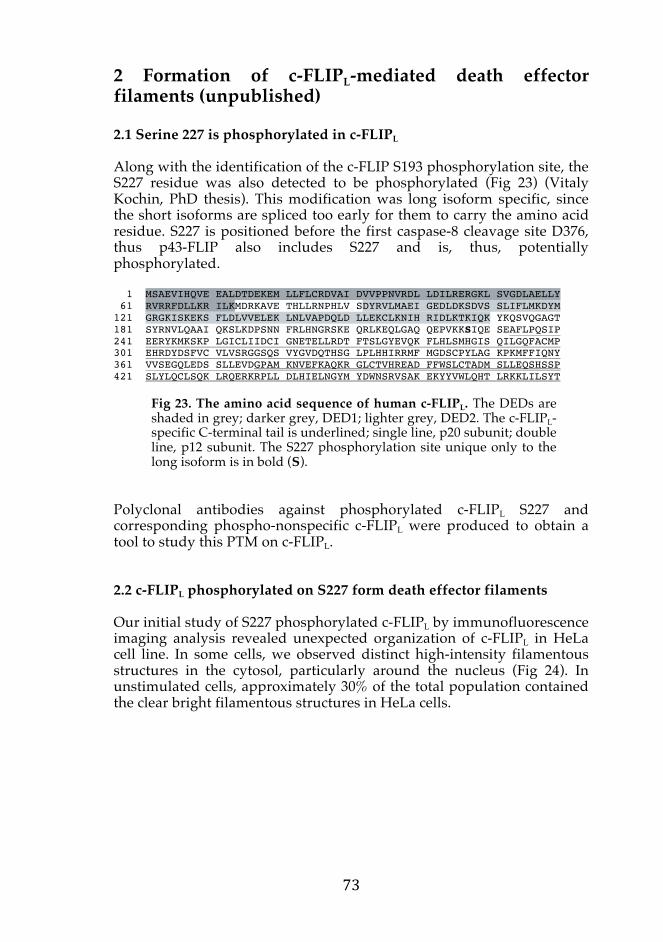

Fig 24. Endogenous c-FLIPL phosphorylation at S227 form filamentous structure in some cells. (A) Immunofluorescence image of HeLa cells stained for c-FLIPL (left panel) or phosphorylated S227 (right panel), and their merged images with DAPI are shown below. The scale bar indicates 50 μm. (B) Magnified HeLa cell with c-FLIPL S227 phosphorylated filamentous structure (green) and co-stained with DAPI (blue). The scale bar indicates 25 μm.
c-FLIPL Phospho-S227 M
erge
d A
B
Phospho-S227 DAPI
Siegel and colleagues have demonstrated that DEDs of FADD and procaspase-8 can self-oligomerize and form filaments upon overexpression. They called this highly ordered oligomerization of pro-apoptotic DEDs death effector filaments (DEFs) (Siegel et al., 1998). Overexpression of the c-FLIP DED region, however, did not self-associate to form the structure, but instead blocked the formation of caspase-8 chain by recruiting to the DEFs (Garvey et al., 2002; Siegel et al., 1998). Additionally, isolated tandem DEDs of v-FLIP proteins were found to exist as monomerics and did not form filament structures (Li et al., 2006; Yang et al., 2005). Our data suggest that C-terminal tail of c-FLIPL and phosphorylation of S227 is a requirement for c-FLIP to form DEFs. Interestingly, reported pro-apoptotic DEFs assembled into cage or lariat-like structures, which resembled cytoskeletal filaments. These descriptions are similar to what we have observed with endogenous filaments of c-FLIPL phosphorylated at S227. I believe our c-FLIP DEF observation is not due to artifacts, since we have verified the specificity of our antibodies (in article III), and the c-FLIP DEFs are restricted to certain cells. Nonetheless, further control experiments are idyllic, for example by using null background cells, to eliminate unspecific immunereactivity. Our observation of c-FLIP DEFs was surprising and may provide an important insight into the function of C-terminal caspase-like domain of the long isoform of c-FLIP. 2.3 c-FLIPL death effector filaments are affected by the cytoskeletal network Following our observation on c-FLIP DEFs, we next investigated if the filamentous structures of phosphorylated c-FLIPL may be related to the structural proteins in the cytosol. DEDs of procaspase-8, when overexpressed, were found to interact with stable microtubule structures, such as the centrosomes and midbodies, and correspondingly, a microtubule-binding motif was identified in the procaspase-8 DED2 (Mielgo et al., 2009). In the same study, they showed that DEDs of c-FLIP did not bind to microtubule structure. Microtubules are tubulin polymers involved in intracellular structural organization and organelle transport as well as a key role in regulating normal cell division. Paclitaxel promotes stabilization of microtubule structures and induce apoptosis in cancer cells. It has been reported that caspase-8 DEFs are stabilized upon paclitaxel treatment via DED association with microtubules thereby providing a platform where procaspase-8 can oligomerize and induce close proximity activation (Mielgo et al., 2009). The study demonstrated that c-FLIP overexpression can rescue this mode of cell death, and therefore we investigated if microtubule organization can influence the c-FLIP DEFs that we observed. We treated HeLa cells

Fig 24. Endogenous c-FLIPL phosphorylation at S227 form filamentous structure in some cells. (A) Immunofluorescence image of HeLa cells stained for c-FLIPL (left panel) or phosphorylated S227 (right panel), and their merged images with DAPI are shown below. The scale bar indicates 50 μm. (B) Magnified HeLa cell with c-FLIPL S227 phosphorylated filamentous structure (green) and co-stained with DAPI (blue). The scale bar indicates 25 μm.
c-FLIPL Phospho-S227
Mer
ged
A
B
Phospho-S227 DAPI
Siegel and colleagues have demonstrated that DEDs of FADD and procaspase-8 can self-oligomerize and form filaments upon overexpression. They called this highly ordered oligomerization of pro-apoptotic DEDs death effector filaments (DEFs) (Siegel et al., 1998). Overexpression of the c-FLIP DED region, however, did not self-associate to form the structure, but instead blocked the formation of caspase-8 chain by recruiting to the DEFs (Garvey et al., 2002; Siegel et al., 1998). Additionally, isolated tandem DEDs of v-FLIP proteins were found to exist as monomerics and did not form filament structures (Li et al., 2006; Yang et al., 2005). Our data suggest that C-terminal tail of c-FLIPL and phosphorylation of S227 is a requirement for c-FLIP to form DEFs. Interestingly, reported pro-apoptotic DEFs assembled into cage or lariat-like structures, which resembled cytoskeletal filaments. These descriptions are similar to what we have observed with endogenous filaments of c-FLIPL phosphorylated at S227. I believe our c-FLIP DEF observation is not due to artifacts, since we have verified the specificity of our antibodies (in article III), and the c-FLIP DEFs are restricted to certain cells. Nonetheless, further control experiments are idyllic, for example by using null background cells, to eliminate unspecific immunereactivity. Our observation of c-FLIP DEFs was surprising and may provide an important insight into the function of C-terminal caspase-like domain of the long isoform of c-FLIP. 2.3 c-FLIPL death effector filaments are affected by the cytoskeletal network Following our observation on c-FLIP DEFs, we next investigated if the filamentous structures of phosphorylated c-FLIPL may be related to the structural proteins in the cytosol. DEDs of procaspase-8, when overexpressed, were found to interact with stable microtubule structures, such as the centrosomes and midbodies, and correspondingly, a microtubule-binding motif was identified in the procaspase-8 DED2 (Mielgo et al., 2009). In the same study, they showed that DEDs of c-FLIP did not bind to microtubule structure. Microtubules are tubulin polymers involved in intracellular structural organization and organelle transport as well as a key role in regulating normal cell division. Paclitaxel promotes stabilization of microtubule structures and induce apoptosis in cancer cells. It has been reported that caspase-8 DEFs are stabilized upon paclitaxel treatment via DED association with microtubules thereby providing a platform where procaspase-8 can oligomerize and induce close proximity activation (Mielgo et al., 2009). The study demonstrated that c-FLIP overexpression can rescue this mode of cell death, and therefore we investigated if microtubule organization can influence the c-FLIP DEFs that we observed. We treated HeLa cells
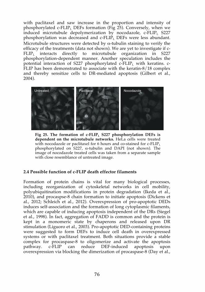
with paclitaxel and saw increase in the proportion and intensity of phosphorylated c-FLIPL DEFs formation (Fig 25). Conversely, when we induced microtubule depolymerization by nocodazole, c-FLIPL S227 phosphorylation was decreased and c-FLIPL DEFs were less abundant. Microtubule structures were detected by α-tubulin staining to verify the efficacy of the treatments (data not shown). We are yet to investigate if c-FLIPL interacts directly to microtubule organization in S227 phosphorylation-dependent manner. Another speculation includes the potential interaction of S227 phosphorylated c-FLIPL with keratins. c-FLIP has been demonstrated to associate with the keratin-8/18 complex and thereby sensitize cells to DR-mediated apoptosis (Gilbert et al., 2004).
Fig 25. The formation of c-FLIPL S227 phosphorylation DEFs is dependent on the microtubule networks. HeLa cells were treated with nocodazole or paclitaxel for 6 hours and co-stained for c-FLIPL phosphorylated on S227, α-tubulin and DAPI (not shown). The image of nocodazole treated cells was taken from a separate sample with close resemblance of untreated image.
2.4 Possible function of c-FLIP death effector filaments Formation of protein chains is vital for many biological processes, including reorganization of cytoskeletal networks in cell mobility, polyubiquitination modifications in protein degradation (Ikeda et al., 2010), and procaspse-8 chain formation to initiate apoptosis (Dickens et al., 2012; Schleich et al., 2012). Overexpression of pro-apoptotic DEDs induces self-association and the formation of long cytoplasmic filaments, which are capable of inducing apoptosis independent of the DRs (Siegel et al., 1998). In fact, aggregation of FADD is common and the protein is kept in a monomeric state by chaperons and released upon DR stimulation (Liguoro et al., 2003). Pro-apoptotic DED-containing proteins were suggested to form DEFs to induce cell death in overexpressed systems or with paclitaxel treatment. Both situations provide a stable complex for procaspase-8 to oligomerize and activate the apoptosis pathway. c-FLIP can reduce DEF-induced apoptosis upon overexpression via blocking the dimerization of procaspase-8 (Day et al.,
Untreated Paclitaxel Nocodazole
2006). On the other hand, transient overexpression of c-FLIPL can enhance cell death probably due to excessive amount of DED-containing proteins that results in caspase-8 DEF formation (Siegel et al., 1998). Our unpublished data strongly suggest that for c-FLIP to form DEFs, the caspase-like domain of c-FLIPL is required and the assembly is regulated by phosphorylation at S227. Interestingly, overexpression of S194/S203 double phosphorylation-mimetic mutant form of FADD strongly induced caspase-8-mediated DEF formation whereas unphosphorylated mutant could not (Jang et al., 2011), further suggesting that phosphorylation status of the DED-containing proteins are important in the formation of DEFs. Understanding the c-FLIP filamentous structure may unearth yet another role of c-FLIP in regulating a different mode of cell death, and therefore continuing studies would be important to understand the mechanism and role of S227 phosphorylation-mediated c-FLIPL DEFs.

3 Phosphorylation of c-FLIP in cell proliferation (IV) The DED-containing proteins are essential for cells to sustain viability. Overexpression of c-FLIPL activates many regulatory proteins in cell proliferation and survival, augmenting the associated signaling pathways upon survival stimulation at molecular level. In this study, we provide evidence that the phosphorylation status of c-FLIPL may be a critical factor that determines the pro-apoptotic or anti-apoptotic function of the protein. 3.1 Phosphorylated c-FLIP in the cell cycle During previous studies, we noticed that c-FLIP phosphorylation of both S193 and S227 were significantly increased in certain cells. By close examination of their nuclear DAPI staining, we identified that they were mitotic cells. The cell cycle functions to pass on life from one cell to two progenitor cells, which is on the extreme contrast to what cell death is in biology. The cell cycle is divided into phases; a cell grows and duplicates its DNA in interphase and divides into two individual daughter cells in mitosis (Fig 26). The cell cycle is an important process for the survival of a healthy cell, where detection and repair of genetic damage occur. The regulation is crucial and unidirectional, with various checkpoints for correct cell cycle progression.
Fig 26. The passage through a cell cycle. In eukaryotic cells, the cell cycle is distributed into interphase (G1, S and G2), during which the cell grows with duplication of DNA, the mitotic (M) phase, when the cell divides and finally cytokinesis to produce two daughter cells. Cellular components and organelles are labeled in italic.
Interphase (G1)
Metaphase
Prometaphase Interphase (G2) Prophase
Anaphase
Telophase
Centrosome
Nucleus Chromosome
Microtubule
Generally, global protein phosphorylation is most abundant during mitosis since numerous kinases phosphorylate a large number of substrates to transit through mitosis (reviewed by Medema and Lindqvist, 2011). We have observed that base level of c-FLIP phosphorylation is low in interphase, but towards metaphase both S193 and S227 phosphorylation increase rapidly compared to the total protein amount (Fig 1A, IV). Upon mitotic arrest by paclitaxel, c-FLIP phosphorylation is significantly increased on both S193 and S227 residues (Fig 1B, IV). Further observation revealed that every cell contained one or two distinct spots when we stained Jurkat cells with S193 or S227 phospho-specific antibodies (Fig 2, IV). This was also observed in various cell lines including HeLa cells, although less noticeable (Fig 1, IV). Both S193 and S227 phospho-spots appeared to occupy space where centrosome positions would be expected. For example, when the chromosomes are aligned in the metaphase plate, phospho-spots are found on the both sides of the chromosomes, and when the cell is divided into two progenitor cells, the spots were located symmetrical to each daughter cell (Fig 2A, IV). The centrosome is important for bipolar spindle assembly during mitosis by nucleating microtubules and organizing their dynamics (reviewed by Delaval and Doxsey, 2010). The centrosome is composed of a pair of centrioles and pericentiolar material, which is a matrix that is composed of proteins required for centrosome-associated functions. Pericentrin is an integral component of the centrosome that serves as a multifunctional scaffold for anchoring numerous protein complexes. Upon co-staining phosphorylated c-FLIP and pericentrin, which was used as a centrosomal marker, all c-FLIP phospho-spots were found to inhabit the same cellular space (Fig 2B, IV). Colocalization analysis using image software indeed revealed that c-FLIP phospho-spots were found at the centrosome. It has been previously shown that FADD phosphorylation is highest at G2/M and low in G1/S phase at S194 (Scaffidi et al., 2000), and that it accumulates at the centrosome during the early phase of mitosis. The FADD phosphorylation is CKIα-mediated and promotes cell cycle progression of T cells (Alappat et al., 2005; Hua et al., 2003). Our observation is similar as accumulated c-FLIP phosphorylation is found at the mitotic spindles. However, the c-FLIP phospho-spot is present in all cells, including those in the interphase. Pericentrin is a scaffold that tethers PKCβII to centrosome. Disruption of this interaction by expression of the interacting region of pericentrin results in release of PKC from the centrosome, microtubule disorganization and cytokinesis failure (Chen et al., 2004). PKC is also a candidate for c-FLIP S227 phosphorylation by analyzing kinase consensus sequences. PKCβ is activated during mitosis and closely resemble c-FLIP phosphorylation, thus an ideal candidate as a kinase. Moreover, at metaphase-anaphase

transition, CaMKII is localized to the centrosomes and between the spindle poles. After anaphase, CaMKII translocated to the area between separating chromosomes (Skelding et al., 2011). CaMKII inhibition cause cells to accumulate in the G2 or M phases, but further analysis is required to identify the kinases involved in the global c-FLIP phosphorylation during mitosis. Aurora A is a well-known mitotic kinase and it is activated at low level during interphase and is increased during mitosis. Aurora A phosphorylates S203 of FADD, which serves to prime FADD for polo-like kinase 1 (Plk-1)-mediated phosphorylation at S194 (Jang et al., 2011), which is required for cells to exit from G2/M phase. Both S193 and S227 are predicted to have the consensus phosphorylation sequences for aurora kinases. Cellular distribution of phosphorylation of c-FLIP during mitosis, especially at the midbody where Aurora B would be activated in cytokinesis, suggest aurora kinases to be a strong candidate for mitotic phosphorylation of c-FLIP. Taken together, many of the kinases suggested to regulate c-FLIP phosphorylation are involved in the same phases of the cell cycle. 3.2 Phosphorylation of c-FLIP regulates the outcome of cell proliferation In highly proliferating cells with a propensity for malignant transformation, close monitoring and ability to respond to cell death signaling would be beneficial, while terminally differentiated cells may downregulate the pro-apoptotic pathways. In T cells, high levels of c-FLIP are detected in the resting cells, whereas the levels are lowered when cells progress into S phase of cell cycle (Algeciras-Schimnich et al., 1999). Many studies elucidated that c-FLIP expression level strongly influence cell proliferating signaling pathways. We wanted to investigate if phosphorylation of c-FLIP is required in cell cycle and proliferation. To study the effect of c-FLIP phosphorylation modifications, we constructed c-FLIP S193/227 phospho-double mutant where the serine residues were mutated to alanine (c-FLIPL-AA), representing a state of non-phosphorable c-FLIPL. Transient transfection of WT c-FLIPL in HeLa cells proliferated faster compared to its control mock that only contained the endogenous c-FLIP (Fig 3A, IV). The similar trend of c-FLIPL-induced cell proliferation was also obtained in transfected Jurkat cells, but to a lesser magnitude possibly due to lower transfection efficiency (data not shown). Interestingly, c-FLIPL double phospho-mutant c-FLIPL-AA did not augment cell proliferation, although its exogenous expression level was equivalent to the WT (Fig 3A, IV). In line with this, when we analyzed cell viability by WST assay, the cells overexpressing c-FLIPL WT had higher cell count compared to the mock or double phospho-mutant (data not shown).
We next investigated the mechanism underlying the reduced proliferative capacity of the c-FLIPL phospho-double mutant. The DNA content of transfected cells was analyzed by flow cytometry of propedium iodide (PI) staining to determine the cell cycle phases but no significant differences were observed between the transfected cells. Likewise, Ki67 staining was used to determine the proportion of proliferating cells, but we could not see any alteration in c-FLIPL overexpressed cells (data not shown). These negative results on cell proliferation lead us to look at the opposing effect of c-FLIPL, which is cell death. To determine the effect of c-FLIP phosphorylation in spontaneous cell death, the apoptotic DNA fragmentation was assessed in the transfected cells by TUNEL assay. Cells overexpressing c-FLIPL WT had lower number of TUNEL positive cells and, therefore, less spontaneous cell death. Intriguingly, the number of cell death was significantly higher in c-FLIPL-AA samples compared to the WT overexpressed cells (Fig 4, IV). Since the cell count was similar for the phospho-mutant and mock, it may be plausible that cell proliferation is increased upon transfection with the double phospho-mutant, but simultaneously increases cell death and keeps the cell number low. There is an increase in the spontaneous in vivo T cell proliferation in c-FLIPL-transgenic mice (Lens et al., 2002). However, c-FLIPL-transgenic mice have apparent similarity to WT mice in the numbers of thymocytes and total peripheral T cells due to concomitant increase in T cells apoptosis owing to augmented caspase-8 activity (Dohrman et al., 2005). In our system, overexpressed c-FLIP proteins were rapidly cleaved to p43-FLIP upon transient expression in HeLa cells and the processing of the WT and double phospho-mutant was similar (Fig 4, IV). This indicates that caspase-8 activity may be similar. In the recent years, many non-apoptotic roles of DED-containing proteins have emerged. Murine FADD S191D mutant is phenotypically identical to those from FADD-deficient mice (Hua et al., 2003). We report that c-FLIP phosphorylation is required for cells to efficiently proliferate and sustain viability. Further investigation will include the identification of the mechanism underlying the effect of c-FLIP on induced cell proliferation, as well as identifying the mode of cell death induced by the inhibition of phosphorylation. T cells are more sensitive to caspase-mediated cell death and clonal expansion of antigen-specific T cells in c-FLIPL-transgenic mice is followed by rapidly accelerated deletion. In parallel, loss-of-function studies using conditional deletion of c-FLIP in the T-cell compartment or complementation of Rag1-deficient blastocytes with c-FLIP-deficient embryonic cells/Raf chimeric mutant mice have revealed the importance of c-FLIP in T cells. Peripheral T cell numbers and proliferation in response to IL-2 and TCR crosslinking were greatly reduced (Chau et al., 2005; Zhang and He, 2005). FLIP mediated T-cell proliferation is reported

not to be mediated through the NF-κB pathway (Zhang et al., 2008) and the exact function of FLIP in T cell proliferation remains elusive. Our data support some of the previous studies and suggest that phosphorylation of c-FLIP is important in these observations. Mitotic catastrophe refers to cases of cell death that are triggered by aberrant mitosis and executed wither during mitosis or in the subsequent interphase (Galluzzi et al., 2012). After aberrant mitosis, cells frequently exhibit gross nuclear attractions, for example micro- and multinucleation, which have been used as morphological markers for the detection of mitotic catastrophe. Although apoptosis seems to be the main mechanism by which cells die after an extended mitotic arrest, Z-vad-fmk or Bcl-2 overexpression failed to completely prevent cell death, indicating that other mechanism may have a role in mitotic cell death (Manchado et al., 2012). It would be of interest to investigate if this mode of cell death play a role in eliminating c-FLIP overexpressed cells where phosphorylation status play a strong role in keeping the cell number high. The hallmarks of cancer include sustained proliferative signaling, resistance to cell death, ability to replicate immortally, induced angiogenesis, evasion of growth suppressors, and activated invasion and metastasis (Hanahan and Weinberg, 2000). In addition, genomic instability and inflammatory features are common in advanced tumors (Hanahan and Weinberg, 2011). The apoptosis-resistant potential of cancer cells in correlation to their proliferative dynamics profoundly affects malignant phenotypes. Accumulating studies have established that pathways governing cell proliferation and cell death are closely interconnected. c-FLIPS anti-apoptotic role is well established and we indeed demonstrated repeatedly that overexpression of c-FLIPL protects Jurkat cells from DR-induced apoptosis (I-III). To examine the effect of c-FLIPL overexpression on cell proliferation, we used the chorioallantoic membrane (CAM) assay. The chick CAM is an extraembryonic membrane, which serves embryonic development functions, and we applied this non-mammalian in vivo system to study the growth of tumors overexpressing exogenous c-FLIP proteins. HeLa cells overexpressing c-FLIPL WT established tumors more rapidly compared to the control mock or c-FLIPS overexpressing cells (Fig 27). The established tumors that contained high expression of c-FLIPL showed accelerated growth and weighed almost as twice as the mock or c-FLIPS expressing tumors (data not shown).
Weight 8.8 mg 13.5 mg 9.6 mg Fig 27. Overexpression of c-FLIPL alone promotes faster tumor growth. Fertilized chicken eggs (LSK Poultry Oy, Finland) were prepared for the CAM assay. In brief, the fertilized chicken eggs were incubated for 10 days and HeLa cells transiently transfected with c-FLIPL, c-FLIPS or mock plasmids were implanted in a plastic ring placed on the CAM at embryo development day (EDD) 11. The tumor growth was continuously visualized every day from the windows cut at the top of the eggs. The grown tumors (indicated by arrows) were harvested from the CAM and weighed on EDD 13.
The chick CAM model allows for rapid and simple evaluation of the implanted samples and is commonly utilized to study angiogenesis. Interestingly, we noticed that eggs implanted with c-FLIPL overexpressing cells developed a more advanced vascular system. Disruption of blood vessels is one of the characteristics reported in the c-FLIP knockout studies in mice. c-FLIP levels may affect the blood vessel growth, since inhibiting angiogenesis downregulate c-FLIP in epithelial cells and induce apoptosis (Kamphaus et al., 2000). In addition, the c-FLIP level is reduced in epithelial cells with impaired blood vessel growth. Reversely, various angiogenic factors augment PI3K/Akt pathway and cells become resistant to CD95-mediated cell death (Papapetropoulos et al., 2000; Sata et al., 2000). Although we do not have further evidence to suggest angiogenesis is directly induced by c-FLIPL overexpression, our data suggest strongly that the tumor development in these cells is more efficient. Our work indicates that c-FLIP further aid in establishing tumors by increasing cell proliferation and enhancing angiogenesis to support metabolically demanding cells. Chromosome 2q33 where the initiator caspase genes reside is a commonly deleted
Mock c-FLIPL c-FLIP
S
EDD 12
EDD 13

region in some cancers (Nishizuka et al., 1998; Otsuka et al., 1996). c-FLIP may also be mutated in some cancers and it would be interesting to screen if c-FLIP phospho-sites are altered in certain cancer cells, which could correlate with development and progression of cancer. Taken together, this thesis work reveals significant importance of phosphorylation as a mode of PTM to regulate c-FLIP cellular distribution. Phosphorylation of c-FLIP S193 contributes to the anti-apoptotic behavior of c-FLIP in DR-mediated cell death, phosphorylation of c-FLIPL S227 induce filamentous formation, and global phosphorylation of the protein is required for cells to sustain its survival. These observations suggest that the functional diversity of c-FLIP may be achieved in a phosphorylation-dependent manner. Furthermore, our studies demonstrate that c-FLIPL overexpression promotes faster cell growth and therefore culture passages will be different compared to mock transfected cells. This means that stable c-FLIP overexpression may indirectly influence the outcome of cellular processes simply by aging the cells faster (from article IV). However, one must also take into account that transient transfection studies of c-FLIP may not mimic physiological localization of c-FLIP and, therefore its native functions (from article III).
CONCLUDING REMARKS Advances in the knowledge of c-FLIP signaling and its interesting concepts emerged from various research groups while I was carrying out my thesis work. The old schemes of singular anti-apoptotic behavior do not apply for c-FLIP anymore, since it can determine fate of a cell by influencing various cellular signaling pathways and, therefore, processes. Thus far, impact on subcellular localization of c-FLIP has been largely ignored in the regulation of apoptosis susceptibility. Our studies strongly suggest that the phosphorylation status of c-FLIP determines its cellular distribution and reveal c-FLIP isoform-specific subcellular localizations may explain how the c-FLIP proteins are involved in various signaling pathways, ranging from cell death to survival. Differential c-FLIP distribution would provide an efficient way to regulate the protein concentration in different organelles, where specific biological functions are allocated. For example, c-FLIP at the lipid rafts would regulate the DISC signaling, whereas c-FLIP at the centrosome may be involved in cell proliferation. Altering the protein localization via phosphorylation could also provide more rapid responses for c-FLIP to regulate fast signals, which would not be feasible by protein synthesis and degradation. Therefore, the absolute quantification of c-FLIP in their subcellular localization and systematic study of the protein in cell death and proliferation holds a key to accurately understand the dynamic c-FLIP signaling. The isoform-specific roles of c-FLIP in apoptosis and necroptosis indicate that c-FLIPS/R may act as an all-or-none signaling switch, while the action of c-FLIPL is gradually deciphered according to other components present in the multiprotein complex at the time of signaling. Furthermore, many studies imply that relative abundance of the DED-containing proteins and their interactions with one another must be considered to truly appreciate the significance of each protein, particularly in the context of caspase-8 activation. The protective role of c-FLIP in keeping caspase-8 as a survival factor to prevent necroptosis, as well as c-FLIP-mediated regulatory functions in many non-apoptotic signaling pathways suggest that the dominant function of c-FLIPL is pro-survival more so than its canonical function as an inhibitor of apoptosis.

ACKNOWLEDGEMENTS This work was performed at the Centre of Biotechnology Turku (CBT), Univeristy of Turku and Åbo Akademi University, and the Department of Biosciences, Åbo Akademi University. I am grateful to Professor Riita Lahesmaa at CBT, and Professor Erik Bonsdorff and Professor Kid Törnquist at the Department of Biosciences for providing excellent research facilities and intellectually inspiring working atmosphere. I would like to thank the technical staffs at CBT and the Cell Imaging Core for their expertise. Professor Olli Lassila and the Turku Doctoral Programme of Biomedical Sciences (TuBS) are thanked for structuring my study and providing a place to meet fellow doctoral students. I owe my utmost appreciation to Professor John Eriksson for providing me with irreplaceable opportunities throughout my doctoral studies. Thank you for your critical advises, encouragements and your trust. I learnt many valuable lessons. I would also like to thank Doctor Annika Meinander, for sharing your visions and being a multi-purpose counselor from the first time we met. Your interest in my work has been of extreme importance. Professor Lea Sistonen is acknowledged for your everlasting enthusiasm. I am deeply thankful to Professor Ralph C. Budd for being a part of our Death squad in Turku and showing me how to progress through a project. Professor Juha Klefström is acknowledged for acting as a thesis committee member and providing me with critical comments in times of need. I would like to express my gratitude to Doctor Markus Rehm and Docent Ville Hietakangas, my external thesis reviewers, for your thorough and valuable advices on my thesis. I want to thank my collaborators for sharing opinions and protocols to make my doctoral work possible. I want to especially thank Professor Hannu Toivonen and Professor Henrik Saxén, for our close collaborations. Although it was challenging for me to understand your philosophy at first, I truly enjoyed our meetings that opened my mind up to different scientific ideas. Without being surrounded by passionate and talented colleagues, pipetting would not have been as pleasant. I want to acknowledge all the past and present members of Eriksson and Sistonen groups for creating a great lab environment. I am privileged to be one of the FLIP girls, and had three talented mentors to follow. Thank you Aura Kaunisto from the bottom of my heart for taking me under your wings when I joined the lab. You not only trained me how to grow cells and do blots (almost blindfolded), but taught me tricks of the trade for me to have a happier life in the Biocity. In times of terror, you were there to comfort me, and your sense of humor never failed to make me laugh (with you). I would like to thank Annika Meinander once more, for taking time out of your busy schedule to advice me on anything and everything. Although I did not have the honor to work with Minna Poukkula in the lab, I am delighted that the protein kept us close. I would like to thank our current c-FLIP team; Preethy Paul for being the other half in the last stretch of my thesis work; Alia Joko, without you, our interdisciplinary collaboration would have been impossible; and my FLIP boy minions with great pair of hands; John Russell for his hard work and
dealing my mood swings like a man; and Vilhelm Wikström for your enthusiasm and sharing the bench summer after summer. I will miss working with all of you. I would like to thank Johanna Ahlskog and Julia Lindqvist for being my lab angels; Rose Chang for our lively discussions; Julius Anckar for sharing your great wisdom; Emmi Peuhu for some serious fun in mökki; Claire Hyder for taking care of me; Senthilkumar Rajendran for all the pancakes and eggs; and for all senior students, including Johanna Björk, Ville Hietakangas, Hanna-Mari Pallari, and Diana Toivonen, for passing down your manuals on how to survive through this era; and Saima Ferraris and Zhanna Chitikova for all the laughs and cries we shared together. I am also grateful for all the help Helana Saarento, Beata Paziewska, and Gunilla Henriksson provided to keep me in one piece in the lab. I would like to thank Professor Susanna Hourani and Doctor George Kass for providing advices at early stages of my study in Surrey. I would like to acknowledge Professor Arleen Rifkind and her lab members, Silvia Diani-Moore, Erin Hall, and Payal Ram, for introducing me to scientific research and sharing many happy memories in New York. I would like to show my gratitude to Professor Pascal Meier and Professor Henning Walzcak for allowing me to drag you to my posters during various conferences and your advices on forthcoming scientific career. I would like to acknowledge Doctor Fumiyo Ikeda for providing me a great inspiration to write. I am grateful to all my friends who were around when I needed cheering up here and there. I would like to especially thank the following; Ghaffar Muharram, Irina Grouneva, Justyna Zdrojewska, and the rest of the Saturday night gangs for keeping me alive during dark Finnish winters with plenty of wine and food; Arianna Colosio and her family for providing me a home to get away for weekends; and last but not least, to Olivia Pitts, Charlotte Smith and Philippa Walden for our everlasting friendships. This book is dedicated to my family; especially to my parents, who spoiled me every step of the way by providing everything I wanted and flying me anywhere I wished to go; and to my brothers, who brought me great diversities at times when all I can worry about were unexplainable bands (not the musical kind). Knowing all the support I had from the family day and night, I was able to explore and overcome obstacles unimaginable if I was on my own. This work was supported financially by K. Albin Johanssons Stiftelse, Medicinska Understödsföreningen Liv och Hälsa r.f., Magnus Ehrnrooth Foundation, Ida Montinin Säätiö, European Molecular Biology Organization, TuBS and the rector of Åbo Akademi University. All are gratefully acknowledged. Turku, November 2013 Tomoko Asaoka

REFERENCEAdams, J.M., and Cory, S. (1998). The Bcl-2
protein family: arbiters of cell survival. Science 281, 1322-1326.
Adrain, C., and Martin, S.J. (2001). The mitochondrial apoptosome: a killer unleashed by the cytochrome seas. Trends Biochem. Sci. 26, 390-397.
Alam, A., Cohen, L.Y., Aouad, S., and Sékaly, R. (1999). Early activation of caspases during T lymphocyte stimulation results in selective substrate cleavage in nonapoptotic cells. J. Exp. Med. 190, 1879-1890.
Alappat, E.C., Feig, C., Boyerinas, B., Volkland, J., Samuels, M., Murmann, A.E., Thorburn, A., Kidd, V.J., Slaughter, C.A., and Osborn, S.L. (2005). Phosphorylation of FADD at serine 194 by CKIα regulates its nonapoptotic activities. Mol. Cell 19, 321-332.
Albeck, J.G., Burke, J.M., Spencer, S.L., Lauffenburger, D.A., and Sorger, P.K. (2008). Modeling a snap-action, variable-delay switch controlling extrinsic cell death. PLoS Biology 6, e299.
Alderson, M.R., Tough, T.W., Davis-Smith, T., Braddy, S., Falk, B., Schooley, K.A., Goodwin, R.G., Smith, C., Ramsdell, F., and Lynch, D.H. (1995). Fas ligand mediates activation-induced cell death in human T lymphocytes. J. Exp. Med. 181, 71-77.
Algeciras-Schimnich, A., Griffith, T.S., Lynch, D.H., and Paya, C.V. (1999). Cell cycle-dependent regulation of FLIP levels and susceptibility to Fas-mediated apoptosis. The Journal of Immunology 162, 5205-5211.
Amé, J., Spenlehauer, C., and de Murcia, G. (2004). The PARP superfamily. Bioessays 26, 882-893.
Antonsson, B., and Martinou, J.C. (2000). The Bcl-2 protein family. Exp. Cell Res. 256, 50-57.
Asaoka, T., Kaunisto, A., and Eriksson, J.E. (2011). Regulation of cell death by c-FLIP phosphorylation. In Advances in TNF Family Research, Springer) pp. 625-630.
Ashany, D., Savir, A., Bhardwaj, N., and Elkon, K. Paper presented at ARTHRITIS AND RHEUMATISM.
Ashkenazi, A., and Dixit, V.M. (1998). Death receptors: signaling and modulation. Science 281, 1305-1308.
Baker, S.J., and Reddy, E.P. (1998). Modulation of life and death by the TNF receptor superfamily. Oncogene 17, 3261.
Bao, Q., and Shi, Y. (2006). Apoptosome: a platform for the activation of initiator caspases. Cell Death & Differentiation 14, 56-65.
Barbero, S., Mielgo, A., Torres, V., Teitz, T., Shields, D.J., Mikolon, D., Bogyo, M., Barilà, D., Lahti, J.M., and Schlaepfer, D. (2009). Caspase-8 association with the focal adhesion complex promotes tumor cell migration and metastasis. Cancer Res. 69, 3755-3763.
Barnhart, B.C., Alappat, E.C., and Peter, M.E. Paper presented at Seminars in immunology.
Bartke, T., Siegmund, D., Peters, N., Reichwein, M., Henkler, F., Scheurich, P., and Wajant, H. (2001). p53 upregulates cFLIP, inhibits transcription of NF-kB-regulated genes and induces caspase-8-independent cell death in DLD-1 cells. Oncogene 20, 571-580.
Benchoua, A., Couriaud, C., Guégan, C., Tartier, L., Couvert, P., Friocourt, G., Chelly, J., Ménissier-de Murcia, J., and Onténiente, B. (2002). Active caspase-8 translocates into the nucleus of apoptotic cells to inactivate poly (ADP-ribose) polymerase-2. J. Biol. Chem. 277, 34217-34222.
Bentele, M., Lavrik, I., Ulrich, M., Stösser, S., Heermann, D., Kalthoff, H., Krammer, P., and Eils, R. (2004). Mathematical modeling reveals threshold mechanism in CD95-induced apoptosis. J. Cell Biol. 166, 839-851.
Berenbaum, M.C. (1972). In vivo determination of the fractional kill of human tumor cells by chemotherapeutic agents. Cancer Chemother. Rep. 56, 563-571.
Bertin, J., Armstrong, R., Ottilie, S., Martin, D., Wang, Y., Banks, S., Wang, G., Senkevich, T., Alnemri, E., Moss, B., et al. (1997). Death effector domain-containing herpesvirus and poxvirus proteins inhibit both Fas- and TNFR1-induced apoptosis. Proceedings of the National Academy of Sciences 94, 1172-1176.
Besnault-Mascard, L., Leprince, C., Auffredou, M.T., Meunier, B., Bourgeade, M.F., Camonis, J., Lorenzo, H.K., and Vazquez, A. (2005). Caspase-8 sumoylation is associated with nuclear localization. Oncogene 24, 3268-3273.
Boatright, K., Deis, C., Denault, J., Sutherlin, D., and Salvesen, G. (2004). Activation of caspases-8 and-10 by FLIPL. Biochem. J. 382, 651-657.
Bodmer, J., Schneider, P., and Tschopp, J. (2002). The molecular architecture of the TNF superfamily. Trends Biochem. Sci. 27, 19.
Boldin, M.P., Goncharov, T.M., Goltsev, Y., and Wallach, D. (1996). Involvement of MACH, a novel MORT1/FADD-interacting protease, in Fas/APO-1-and TNF receptor-induced cell death. Cell 85, 803.
Bonizzi, G., and Karin, M. (2004). The two NF-κB activation pathways and their role in innate and adaptive immunity. Trends Immunol. 25, 280-288.
Budd, R.C. (2002). Death receptors couple to both cell proliferation and apoptosis. J. Clin. Invest. 109, 437-442.
Budd, R.C., Yeh, W., and Tschopp, J. (2006). cFLIP regulation of lymphocyte activation and development. Nature Reviews Immunology 6, 196-204.
Budihardjo, I., Oliver, H., Lutter, M., Luo, X., and Wang, X. (1999). Biochemical pathways of caspase activation during apoptosis. Annu. Rev. Cell Dev. Biol. 15, 269-290.
Butler, G.S., and Overall, C.M. (2009). Proteomic identification of multitasking proteins in unexpected locations complicates drug targeting. Nature Reviews Drug Discovery 8, 935-948.
Cadwell, K., and Coscoy, L. (2005). Ubiquitination on nonlysine residues by a viral E3 ubiquitin ligase. Science 309, 127-130.
Calzone, L., Tournier, L., Fourquet, S., Thieffry, D., Zhivotovsky, B., Barillot, E., and Zinovyev, A. (2010). Mathematical modelling of cell-fate decision in response to death receptor engagement. PLoS Computational Biology 6, e1000702.
Carrington, P.E., Sandu, C., Wei, Y., Hill, J.M., Morisawa, G., Huang, T., Gavathiotis, E., Wei, Y., and Werner, M.H. (2006). The structure of FADD and its mode of interaction with procaspase-8. Mol. Cell 22, 599-610.
Chang, D.W., Xing, Z., Capacio, V.L., Peter, M.E., and Yang, X. (2003). Interdimer processing mechanism of procaspase-8 activation. EMBO J. 22, 4132-4142.
Chang, D.W., Xing, Z., Pan, Y., Algeciras-Schimnich, A., Barnhart, B.C., Yaish-Ohad, S., Peter, M.E., and Yang, X. (2002). c-FLIPL
is a dual function regulator for caspase-8 activation and CD95-mediated apoptosis. Science Signaling 21, 3704.
Chang, L., Kamata, H., Solinas, G., Luo, J., Maeda, S., Venuprasad, K., Liu, Y., and Karin, M. (2006). The E3 Ubiquitin Ligase Itch Couples JNK Activation to TNFα-induced Cell Death by Inducing c-FLIP< sub> L</sub> Turnover. Cell 124, 601-613.
Chau, H., Wong, V., Chen, N., Huang, H., Lin, W., Mirtsos, C., Elford, A.R., Bonnard, M., Wakeham, A., and You-Ten, A.I. (2005). Cellular FLICE-inhibitory protein is required for T cell survival and cycling. J. Exp. Med. 202, 405-413.
Chaudhary, P.M., Eby, M.T., Jasmin, A., Kumar, A., Liu, L., and Hood, L. (2000). Activation of the NF-kappaB pathway by caspase 8 and its homologs. Oncogene 19, 4451.
Chaudhary, P.M., Jasmin, A., Eby, M.T., and Hood, L. (1999). Modulation of the NF-kappa B pathway by virally encoded death effector domains-containing proteins. Oncogene 18, 5738-5746.
Chen, D., Purohit, A., Halilovic, E., Doxsey, S.J., and Newton, A.C. (2004). Centrosomal anchoring of protein kinase C βII by pericentrin controls microtubule organization, spindle function, and cytokinesis. J. Biol. Chem. 279, 4829-4839.
Chen, F., Zhu, H., Zhou, L., Wu, S., Wang, J., and Chen, Z. (2010). Inhibition of c-FLIP expression by miR-512-3p contributes to taxol-induced apoptosis in hepatocellular carcinoma cells. Oncol. Rep. 23, 1457-1462.
Chen, L., Qi-Wei, G., Nakata, M., Matsuno, H., and Miyano, S. (2007). Modelling and simulation of signal transductions in an apoptosis pathway by using timed Petri nets. J. Biosci. 32, 113-127.
Chen, L., Willis, S.N., Wei, A., Smith, B.J., Fletcher, J.I., Hinds, M.G., Colman, P.M., Day, C.L., Adams, J.M., and Huang, D. (2005). Differential targeting of prosurvival Bcl-2 proteins by their BH3-only ligands allows complementary apoptotic function. Mol. Cell 17, 393-403.
Chipuk, J.E., and Green, D.R. (2008). How do BCL-2 proteins induce mitochondrial outer membrane permeabilization? Trends Cell Biol. 18, 157-164.
Cho, K., Shin, S., Kolch, W., and Wolkenhauer, O. (2003). Experimental Design in Systems Biology, Based on

Parameter Sensitivity Analysis Using a Monte Carlo Method: A Case Study for the TNFα-Mediated NF-κ B Signal Transduction Pathway. Simulation 79, 726-739.
Cho, Y., Challa, S., Moquin, D., Genga, R., Ray, T.D., Guildford, M., and Chan, F.K. (2009). Phosphorylation-driven assembly of the RIP1-RIP3 complex regulates programmed necrosis and virus-induced inflammation. Cell 137, 1112-1123.
Chollet-Martin, S., Stamatakis, G., Bailly, S., Mery, J., and Gougerot-Pocidalo, M. (1991). Induction of tumour necrosis factor‐alpha during haemodialysis. Influence of the membrane type. Clinical & Experimental Immunology 83, 329-332.
Chun, H.J., Zheng, L., Ahmad, M., Wang, J., Speirs, C.K., Siegel, R.M., Dale, J.K., Puck, J., Davis, J., and Hall, C.G. (2002). Pleiotropic defects in lymphocyte activation caused by caspase-8 mutations lead to human immunodeficiency. Nature 419, 395-399.
Clevers, H. (2006). Wnt/β-catenin signaling in development and disease. Cell 127, 469-480.
Cohen, G. (1997). Caspases: the executioners of apoptosis. Biochem. J. 326, 1-16.
Cohen, P. (2000). The regulation of protein function by multisite phosphorylation–a 25 year update. Trends Biochem. Sci. 25, 596-601.
Cummings, M. (1997). Winterford CM, Walker NI. Apoptosis.Am J Surg Pathol 21, 88-101.
Curtin, J.F., and Cotter, T.G. (2003). Live and let die: regulatory mechanisms in Fas-mediated apoptosis. Cell. Signal. 15, 983-992.
Dales, J.P., Palmerini, F., Devilard, E., Hassoun, J., Birg, F., and Xerri, L. (2001). Caspases: conductors of the cell death machinery in lymphoma cells. Leuk. Lymphoma 41, 247-253.
Davies, C.C., Mason, J., Wakelam, M.J., Young, L.S., and Eliopoulos, A.G. (2004). Inhibition of phosphatidylinositol 3-kinase-and ERK MAPK-regulated protein synthesis reveals the pro-apoptotic properties of CD40 ligation in carcinoma cells. J. Biol. Chem. 279, 1010-1019.
Day, T.W., Najafi, F., Wu, C., and Safa, A.R. (2006). Cellular FLICE-like inhibitory protein (c-FLIP): a novel target for Taxol-induced apoptosis. Biochem. Pharmacol. 71, 1551-1561.
de Thonel, A., Bettaeb, A., Jean, C., Laurent, G., and Quillet-Mary, A. (2001). Role of protein kinase C ζ isoform in Fas resistance
of immature myeloid KG1a leukemic cells. Blood 98, 3770-3777.
Degli-Esposti, M.A., Dougall, W.C., Smolak, P.J., Waugh, J.Y., Smith, C.A., and Goodwin, R.G. (1997). The novel receptor TRAIL-R4 induces NF-κB and protects against TRAIL-mediated apoptosis, yet retains an incomplete death domain. Immunity 7, 813-820.
Degli-Esposti, M.A., Smolak, P.J., Walczak, H., Waugh, J., Huang, C., DuBose, R.F., Goodwin, R.G., and Smith, C.A. (1997). Cloning and characterization of TRAIL-R3, a novel member of the emerging TRAIL receptor family. J. Exp. Med. 186, 1165-1170.
Degterev, A., Boyce, M., and Yuan, J. (2003). A decade of caspases. Oncogene 22, 8543-8567.
Degterev, A., Huang, Z., Boyce, M., Li, Y., Jagtap, P., Mizushima, N., Cuny, G.D., Mitchison, T.J., Moskowitz, M.A., and Yuan, J. (2005). Chemical inhibitor of nonapoptotic cell death with therapeutic potential for ischemic brain injury. Nature Chemical Biology 1, 112-119.
Delaval, B., and Doxsey, S.J. (2010). Pericentrin in cellular function and disease. J. Cell Biol. 188, 181-190.
Denecker, G., Hoste, E., Gilbert, B., Hochepied, T., Ovaere, P., Lippens, S., Van den Broecke, C., Van Damme, P., D'Herde, K., and Hachem, J. (2007). Caspase-14 protects against epidermal UVB photodamage and water loss. Nat. Cell Biol. 9, 666-674.
Deveraux, Q.L., Roy, N., Stennicke, H.R., Van Arsdale, T., Zhou, Q., Srinivasula, S.M., Alnemri, E.S., Salvesen, G.S., and Reed, J.C. (1998). IAPs block apoptotic events induced by caspase-8 and cytochrome c by direct inhibition of distinct caspases. EMBO J. 17, 2215-2223.
Dickens, L.S., Boyd, R.S., Jukes-Jones, R., Hughes, M.A., Robinson, G.L., Fairall, L., Schwabe, J.W., Cain, K., and MacFarlane, M. (2012). A Death Effector Domain Chain DISC Model Reveals a Crucial Role for Caspase-8 Chain Assembly in Mediating Apoptotic Cell Death. Mol. Cell
Dillon, C.P., Oberst, A., Weinlich, R., Janke, L.J., Kang, T., Ben-Moshe, T., Mak, T.W., Wallach, D., and Green, D.R. (2012). Survival Function of the FADD-CASPASE-8-cFLIP< sub> L</sub> Complex. Cell Reports 1, 401-407.
Djerbi, M., Darreh‐Shori, T., Zhivotovsky, B., and Grandien, A. (2001). Characterization of
the Human FLICE‐Inhibitory Protein Locus and Comparison of the Anti‐Apoptotic Activity of Four Different FLIP Isoforms. Scand. J. Immunol. 54, 180-189.
Dohrman, A., Kataoka, T., Cuenin, S., Russell, J.Q., Tschopp, J., and Budd, R.C. (2005). Cellular FLIP (long form) regulates CD8 T cell activation through caspase-8-dependent NF-κB activation. The Journal of Immunology 174, 5270-5278.
Donepudi, M., Sweeney, A.M., Briand, C., and Grütter, M.G. (2003). Insights into the regulatory mechanism for caspase-8 activation. Mol. Cell 11, 543-549.
Du, C., Fang, M., Li, Y., Li, L., and Wang, X. (2000). Smac, a mitochondrial protein that promotes cytochrome c–dependent caspase activation by eliminating IAP inhibition. Cell 102, 33-42.
Du, X., Bao, G., He, X., Zhao, H., Yu, F., Qiao, Q., Lu, J., and Ma, Q. (2009). Journal of Experimental & Clinical Cancer Research. Journal of Experimental & Clinical Cancer Research 28, 24.
Düssmann, H., Rehm, M., Concannon, C., Anguissola, S., Würstle, M., Kacmar, S., Völler, P., Huber, H., and Prehn, J. (2009). Single-cell quantification of Bax activation and mathematical modelling suggest pore formation on minimal mitochondrial Bax accumulation. Cell Death & Differentiation 17, 278-290.
Earnshaw, W.C., Martins, L.M., and Kaufmann, S.H. (1999). Mammalian caspases: structure, activation, substrates, and functions during apoptosis. Annu. Rev. Biochem. 68, 383-424.
Edinger, A.L., and Thompson, C.B. (2004). Death by design: apoptosis, necrosis and autophagy. Curr. Opin. Cell Biol. 16, 663-669.
Emery, J.G., McDonnell, P., Burke, M.B., Deen, K.C., Lyn, S., Silverman, C., Dul, E., Appelbaum, E.R., Eichman, C., and DiPrinzio, R. (1998). Osteoprotegerin is a receptor for the cytotoxic ligand TRAIL. J. Biol. Chem. 273, 14363-14367.
Engels, I.H., Totzke, G., Fischer, U., Schulze-Osthoff, K., and Jänicke, R.U. (2005). Caspase-10 sensitizes breast carcinoma cells to TRAIL-induced but not tumor necrosis factor-induced apoptosis in a caspase-3-dependent manner. Mol. Cell. Biol. 25, 2808-2818.
Eskes, R., Desagher, S., Antonsson, B., and Martinou, J. (2000). Bid induces the oligomerization and insertion of Bax into the outer mitochondrial membrane. Mol. Cell. Biol. 20, 929-935.
Esposito, D., Sankar, A., Morgner, N., Robinson, C.V., Rittinger, K., and Driscoll, P.C. (2010). Solution NMR investigation of the CD95/FADD homotypic death domain complex suggests lack of engagement of the CD95 C terminus. Structure 18, 1378-1390.
Fadok, V.A., Voelker, D.R., Campbell, P.A., Cohen, J.J., Bratton, D.L., and Henson, P.M. (1992). Exposure of phosphatidylserine on the surface of apoptotic lymphocytes triggers specific recognition and removal by macrophages. The Journal of Immunology 148, 2207-2216.
Falschlehner, C., Emmerich, C.H., Gerlach, B., and Walczak, H. (2007). TRAIL signalling: decisions between life and death. Int. J. Biochem. Cell Biol. 39, 1462-1475.
Fang, L., Tai, T., Yu, W., Liao, F., and Lai, M. (2004). Phosphatidylinositide 3-kinase priming couples c-FLIP to T cell activation. J. Biol. Chem. 279, 13-18.
Feig, C., and Peter, M.E. (2007). How apoptosis got the immune system in shape. Eur. J. Immunol. 37, S61-S70.
Feinerman, O., Veiga, J., Dorfman, J.R., Germain, R.N., and Altan-Bonnet, G. (2008). Variability and robustness in T cell activation from regulated heterogeneity in protein levels. Science 321, 1081-1084.
Feoktistova, M., Geserick, P., Kellert, B., Dimitrova, D.P., Langlais, C., Hupe, M., Cain, K., MacFarlane, M., Häcker, G., and Leverkus, M. (2011). cIAPs block Ripoptosome formation, a RIP1/caspase-8 containing intracellular cell death complex differentially regulated by cFLIP isoforms. Mol. Cell 43, 449-463.
Fernandes-Alnemri, T., Armstrong, R.C., Krebs, J., Srinivasula, S.M., Wang, L., Bullrich, F., Fritz, L.C., Trapani, J.A., Tomaselli, K.J., and Litwack, G. (1996). In vitro activation of CPP32 and Mch3 by Mch4, a novel human apoptotic cysteine protease containing two FADD-like domains. Proceedings of the National Academy of Sciences 93, 7464-7469.
Field, N., Low, W., Daniels, M., Howell, S., Daviet, L., Boshoff, C., and Collins, M. (2003). KSHV vFLIP binds to IKK-γ to activate IKK. J. Cell. Sci. 116, 3721-3728.

Finley, D. (2009). Recognition and processing of ubiquitin-protein conjugates by the proteasome. Annu. Rev. Biochem. 78, 477.
Fischer, U., Jänicke, R., and Schulze-Osthoff, K. (2003). Many cuts to ruin: a comprehensive update of caspase substrates. Cell Death & Differentiation 10, 76-100.
Fricker, N., Beaudouin, J., Richter, P., Eils, R., Krammer, P.H., and Lavrik, I.N. (2010). Model-based dissection of CD95 signaling dynamics reveals both a pro-and antiapoptotic role of c-FLIPL. J. Cell Biol. 190, 377-389.
Fuentes-Prior, P., and Salvesen, G.S. (2004). The protein structures that shape caspase activity, specificity, activation and inhibition. Biochem. J. 384, 201.
Fukazawa, T., Fujiwara, T., Uno, F., Teraishi, F., Kadowaki, Y., Itoshima, T., Takata, Y., Kagawa, S., Roth, J.A., and Tschopp, J. (2001). Accelerated degradation of cellular FLIP protein through the ubiquitin-proteasome pathway in p53-mediated apoptosis of human cancer cells. Oncogene 20, 5225-5231.
Fulda, S., Meyer, E., and Debatin, K. (2000). Metabolic inhibitors sensitize for CD95 (APO-1/Fas)-induced apoptosis by down-regulating Fas-associated death domain-like interleukin 1-converting enzyme inhibitory protein expression. Cancer Res. 60, 3947-3956.
Fussenegger, M., Bailey, J.E., and Varner, J. (2000). A mathematical model of caspase function in apoptosis. Nat. Biotechnol. 18, 768-774.
Galluzzi, L., Maiuri, M., Vitale, I., Zischka, H., Castedo, M., Zitvogel, L., and Kroemer, G. (2007). Cell death modalities: classification and pathophysiological implications. Cell Death & Differentiation 14, 1237-1243.
Galluzzi, L., Vitale, I., Abrams, J.M., Alnemri, E.S., Baehrecke, E.H., Blagosklonny, M.V., Dawson, T.M., Dawson, V.L., El-Deiry, W.S., Fulda, S., et al. (2012). Molecular definitions of cell death subroutines: recommendations of the Nomenclature Committee on Cell Death 2012. Cell Death Differ. 19, 107-120.
Gao, S., Wang, H., Lee, P., Melamed, J., Li, C.X., Zhang, F., Wu, H., Zhou, L., and Wang, Z. (2006). Androgen receptor and prostate apoptosis response factor-4 target the c-FLIP gene to determine survival and apoptosis in the prostate gland. J. Mol. Endocrinol. 36, 463-483.
Garg, A., and Aggarwal, B. (2002). Nuclear transcription factor-kappaB as a target for cancer drug development. Leukemia: Official Journal of the Leukemia Society of America, Leukemia Research Fund, UK 16, 1053.
Garvey, T.L., Bertin, J., Siegel, R.M., Wang, G., Lenardo, M.J., and Cohen, J.I. (2002). Binding of FADD and caspase-8 to molluscum contagiosum virus MC159 v-FLIP is not sufficient for its antiapoptotic function. J. Virol. 76, 697-706.
Gerlach, B., Cordier, S.M., Schmukle, A.C., Emmerich, C.H., Rieser, E., Haas, T.L., Webb, A.I., Rickard, J.A., Anderton, H., and Wong, W.W. (2011). Linear ubiquitination prevents inflammation and regulates immune signalling. Nature 471, 591-596.
Gerondakis, S., Grossmann, M., Nakamura, Y., Pohl, T., and Grumont, R. (1999). Genetic approaches in mice to understand Rel/NF-kappaB and IkappaB function: transgenics and knockouts. Oncogene 18, 6888-6895.
Ghosh, S., and Karin, M. (2002). Missing pieces in the NF-κB puzzle. Cell 109, S81-S96.
Gilbert, S., Loranger, A., and Marceau, N. (2004). Keratins modulate c-Flip/extracellular signal-regulated kinase 1 and 2 antiapoptotic signaling in simple epithelial cells. Mol. Cell. Biol. 24, 7072-7081.
Gilmore, T. (2006). Introduction to NF-κB: players, pathways, perspectives. Oncogene 25, 6680-6684.
Golks, A., Brenner, D., Fritsch, C., Krammer, P.H., and Lavrik, I.N. (2005). c-FLIPR, a new regulator of death receptor-induced apoptosis. J. Biol. Chem. 280, 14507-14513.
Golks, A., Brenner, D., Krammer, P.H., and Lavrik, I.N. (2006). The c-FLIP–NH2 terminus (p22-FLIP) induces NF-κB activation. J. Exp. Med. 203, 1295-1305.
Goltsev, Y.V., Kovalenko, A.V., Arnold, E., Varfolomeev, E.E., Brodianskii, V.M., and Wallach, D. (1997). CASH, a novel caspase homologue with death effector domains. J. Biol. Chem. 272, 19641-19644.
Gomez-Angelats, M., and Cidlowski, J. (2003). Molecular evidence for the nuclear localization of FADD. Cell Death & Differentiation 10, 791-797.
Griffith, T.S., Brunner, T., Fletcher, S.M., Green, D.R., and Ferguson, T.A. (1995). Fas ligand-induced apoptosis as a mechanism of immune privilege. Science 270, 1189-1192.
Griffith, T.S., and Lynch, D.H. (1998). TRAIL: a molecule with multiple receptors and control mechanisms. Curr. Opin. Immunol. 10, 559-563.
Grütter, M.G. (2000). Caspases: key players in programmed cell death. Curr. Opin. Struct. Biol. 10, 649-655.
Guasparri, I., Keller, S.A., and Cesarman, E. (2004). KSHV vFLIP is essential for the survival of infected lymphoma cells. J. Exp. Med. 199, 993-1003.
Haag, C., Stadel, D., Zhou, S., Bachem, M.G., Möller, P., Debatin, K., and Fulda, S. (2011). Identification of c-FLIPL and c-FLIPS as critical regulators of death receptor-induced apoptosis in pancreatic cancer cells. Gut 60, 225-237.
Haas, T.L., Emmerich, C.H., Gerlach, B., Schmukle, A.C., Cordier, S.M., Rieser, E., Feltham, R., Vince, J., Warnken, U., and Wenger, T. (2009). Recruitment of the linear ubiquitin chain assembly complex stabilizes the TNF-R1 signaling complex and is required for TNF-mediated gene induction. Mol. Cell 36, 831-844.
Haglund, K., and Dikic, I. (2005). Ubiquitylation and cell signaling. EMBO J. 24, 3353-3359.
Hamed, H.A., Yamaguchi, Y., Fisher, P.B., Grant, S., and Dent, P. (2013). Sorafenib and HDAC inhibitors synergize with TRAIL to kill tumor cells. J. Cell. Physiol. 228, 1996-2005.
Han, D.K., Chaudhary, P.M., Wright, M.E., Friedman, C., Trask, B.J., Riedel, R.T., Baskin, D.G., Schwartz, S.M., and Hood, L. (1997). MRIT, a novel death-effector domain-containing protein, interacts with caspases and BclXL and initiates cell death. Proceedings of the National Academy of Sciences 94, 11333-11338.
Han, D., Haudenschild, C.C., Hong, M.K., Tinkle, B.T., Leon, M.B., and Liau, G. (1995). Evidence for apoptosis in human atherogenesis and in a rat vascular injury model. The American Journal of Pathology 147, 267.
Hanahan, D., and Weinberg, R.A. (2011). Hallmarks of cancer: the next generation. Cell 144, 646-674.
Hanahan, D., and Weinberg, R.A. (2000). The hallmarks of cancer. Cell 100, 57-70.
Harper, N., Hughes, M.A., Farrow, S.N., Cohen, G.M., and MacFarlane, M. (2003). Protein kinase C modulates tumor necrosis
factor-related apoptosis-inducing ligand-induced apoptosis by targeting the apical events of death receptor signaling. J. Biol. Chem. 278, 44338-44347.
Harper, N., Hughes, M., MacFarlane, M., and Cohen, G.M. (2003). FADD and caspase-8 are not recruited to the TNF-R1 signaling complex during TNF-induced apoptosis. J. Biol. Chem.
Haslett, C., Savill, J., Whyte, M., Stern, M., Dransfield, I., and Meagher, L. (1995). Granulocyte apoptosis and the control of inflammation. In The Role of Apoptosis in Development, Tissue Homeostasis and Malignancy, Springer) pp. 91-97.
Hayden, M.S., and Ghosh, S. (2008). Shared principles in NF-κB signaling. Cell 132, 344-362.
Hayden, M.S., and Ghosh, S. (2004). Signaling to NF-κB. Genes Dev. 18, 2195-2224.
He, S., Wang, L., Miao, L., Wang, T., Du, F., Zhao, L., and Wang, X. (2009). Receptor interacting protein kinase-3 determines cellular necrotic response to TNF-α. Cell 137, 1100-1111.
Hector, S., Rehm, M., Schmid, J., Kehoe, J., McCawley, N., Dicker, P., Murray, F., McNamara, D., Kay, E.W., and Concannon, C.G. (2012). Clinical application of a systems model of apoptosis execution for the prediction of colorectal cancer therapy responses and personalisation of therapy. Gut 61, 725-733.
Heiner, M. (2009). Understanding network behavior by structured representations of transition invariants. In Algorithmic Bioprocesses, Springer) pp. 367-389.
Heiner, M., Koch, I., and Will, J. (2004). Model validation of biological pathways using Petri nets—demonstrated for apoptosis. BioSystems 75, 15-28.
Hengartner, M.O. (2000). The biochemistry of apoptosis. Nature 407, 770-776.
Hietakangas, V., Poukkula, M., Heiskanen, K.M., Karvinen, J.T., Sistonen, L., and Eriksson, J.E. (2003). Erythroid differentiation sensitizes K562 leukemia cells to TRAIL-induced apoptosis by downregulation of c-FLIP. Mol. Cell. Biol. 23, 1278-1291.
Higuchi, H., Yoon, J., Grambihler, A., Werneburg, N., Bronk, S.F., and Gores, G.J. (2003). Bile acids stimulate cFLIP phosphorylation enhancing TRAIL-mediated apoptosis. J. Biol. Chem. 278, 454-461.

Hochstrasser, M. (2009). Origin and function of ubiquitin-like proteins. Nature 458, 422-429.
Hochstrasser, M. (2006). Lingering mysteries of ubiquitin-chain assembly. Cell 124, 27-34.
Hochstrasser, M. (1995). Ubiquitin, proteasomes, and the regulation of intracellular protein degradation. Curr. Opin. Cell Biol. 7, 215-223.
Hofmann, K., Bucher, P., and Tschopp, J. (1997). The CARD domain: a new apoptotic signalling motif. Trends Biochem. Sci. 22, 155-156.
Holcik, M., and Korneluk, R.G. (2001). XIAP, the guardian angel. Nature Reviews Molecular Cell Biology 2, 550-556.
Holland, E.C., Sonenberg, N., Pandolfi, P.P., and Thomas, G. (2004). Signaling control of mRNA translation in cancer pathogenesis. Oncogene 23, 3138-3144.
Holler, N., Zaru, R., Micheau, O., Thome, M., Attinger, A., Valitutti, S., Bodmer, J., Schneider, P., Seed, B., and Tschopp, J. (2000). Fas triggers an alternative, caspase-8–independent cell death pathway using the kinase RIP as effector molecule. Nat. Immunol. 1, 489-495.
Holmström, T.H., Schmitz, I., Söderström, T.S., Poukkula, M., Johnson, V.L., Chow, S.C., Krammer, P.H., and Eriksson, J.E. (2000). MAPK/ERK signaling in activated T cells inhibits CD95/Fas-mediated apoptosis downstream of DISC assembly. EMBO J. 19, 5418-5428.
Horvitz, H., Shaham, S., and Hengartner, M. Paper presented at Cold Spring Harbor symposia on quantitative biology.
Hsu, H., Xiong, J., and Goeddel, D.V. (1995). The TNF receptor 1-associated protein TRADD signals cell death and NF-κB activation. Cell 81, 495-504.
Hu, S., Vincenz, C., Ni, J., Gentz, R., and Dixit, V.M. (1997). I-FLICE, a novel inhibitor of tumor necrosis factor receptor-1-and CD-95-induced apoptosis. J. Biol. Chem. 272, 17255-17257.
Hu, W., Johnson, H., and Shu, H. (2000). Activation of NF-κB by FADD, Casper, and caspase-8. J. Biol. Chem. 275, 10838-10844.
Hua, Z.C., Sohn, S.J., Kang, C., Cado, D., and Winoto, A. (2003). A function of Fas-associated death domain protein in cell cycle progression localized to a single amino acid at its C-terminal region. Immunity 18, 513-521.
Huang, J., Ding, T., Yang, M., Liu, H., Sun, X., and Jin, J. (2011). Antitumor activity and
drug interactions of proteasome inhibitor Bortezomib in human high-risk myelodysplastic syndrome cells. Int. J. Hematol. 93, 482-493.
Huerta, S., Goulet, E.J., Huerta-Yepez, S., and Livingston, E.H. (2007). Screening and detection of apoptosis. J. Surg. Res. 139, 143-156.
Hughes, M.A., Harper, N., Butterworth, M., Cain, K., Cohen, G.M., and MacFarlane, M. (2009). Reconstitution of the death-inducing signaling complex reveals a substrate switch that determines CD95-mediated death or survival. Mol. Cell 35, 265-279.
Hung, M., and Link, W. (2011). Protein localization in disease and therapy. J. Cell. Sci. 124, 3381-3392.
Ikeda, F., Crosetto, N., and Dikic, I. (2010). What determines the specificity and outcomes of ubiquitin signaling? Cell 143, 677-681.
Ikeda, F., Deribe, Y.L., Skånland, S.S., Stieglitz, B., Grabbe, C., Franz-Wachtel, M., van Wijk, S.J., Goswami, P., Nagy, V., and Terzic, J. (2011). SHARPIN forms a linear ubiquitin ligase complex regulating NF-[kgr] B activity and apoptosis. Nature 471, 637-641.
Ikeda, F., and Dikic, I. (2008). Atypical ubiquitin chains: new molecular signals. EMBO Rep. 9, 536-542.
Imanishi, T., McBride, J., Ho, Q., O'Brien, K.D., Schwartz, S.M., and Han, D.K. (2000). Expression of cellular FLICE-inhibitory protein in human coronary arteries and in a rat vascular injury model. The American Journal of Pathology 156, 125-137.
Imanishi, T., Murry, C.E., Reinecke, H., Hano, T., Nishio, I., Liles, W.C., Hofsta, L., Kim, K., O'Brien, K.D., and Schwartz, S.M. (2000). Cellular FLIP is expressed in cardiomyocytes and down-regulated in TUNEL-positive grafted cardiac tissues. Cardiovasc. Res. 48, 101-110.
Inohara, N., Koseki, T., Hu, Y., Chen, S., and Núñez, G. (1997). CLARP, a death effector domain-containing protein interacts with caspase-8 and regulates apoptosis. Proceedings of the National Academy of Sciences 94, 10717-10722.
Inoue, S., Browne, G., Melino, G., and Cohen, G. (2009). Ordering of caspases in cells undergoing apoptosis by the intrinsic pathway. Cell Death & Differentiation 16, 1053-1061.
Irmler, M., Thome, M., Hahne, M., Schneider, P., Hofmann, K., Steiner, V., Bodmer, J., Schröter, M., Burns, K., and Mattmann, C. (1997). Inhibition of death receptor signals by cellular FLIP. Nature 388, 190-195.
Ishioka, T., Katayama, R., Kikuchi, R., Nishimoto, M., Takada, S., Takada, R., Matsuzawa, S., Reed, J.C., Tsuruo, T., and Naito, M. (2007). Impairment of the ubiquitin‐proteasome system by cellular FLIP. Genes to Cells 12, 735-744.
Itoh, N., Yonehara, S., Ishii, A., Yonehara, M., Mizushima, S., Sameshima, M., Hase, A., Seto, Y., and Nagata, S. (1991). The polypeptide encoded by the cDNA for human cell surface antigen Fas can mediate apoptosis. Cell 66, 233-243.
Iyer, A.K.V., Azad, N., Talbot, S., Stehlik, C., Lu, B., Wang, L., and Rojanasakul, Y. (2011). Antioxidant c-FLIP inhibits Fas ligand-induced NF-κB activation in a phosphatidylinositol 3-kinase/Akt-dependent manner. The Journal of Immunology 187, 3256-3266.
Jang, M., Lee, S., Kang, N.S., and Kim, E. (2011). Cooperative phosphorylation of FADD by Aur-A and Plk1 in response to taxol triggers both apoptotic and necrotic cell death. Cancer Res. 71, 7207-7215.
Janssens, S., and Tinel, A. (2011). The PIDDosome, DNA-damage-induced apoptosis and beyond. Cell Death & Differentiation 19, 13-20.
Jin, Z., and El-Deiry, W.S. (2006). Distinct signaling pathways in TRAIL-versus tumor necrosis factor-induced apoptosis. Mol. Cell. Biol. 26, 8136-8148.
Jin, Z., Li, Y., Pitti, R., Lawrence, D., Pham, V.C., Lill, J.R., and Ashkenazi, A. (2009). Cullin3-based polyubiquitination and p62-dependent aggregation of caspase-8 mediate extrinsic apoptosis signaling. Cell 137, 721-735.
Jong, W.Y., Jeffrey, P.D., and Shi, Y. (2009). Mechanism of procaspase-8 activation by c-FLIPL. Proceedings of the National Academy of Sciences 106, 8169-8174.
Jun, J., Chung, C., Lee, H., Pyo, J., Lee, K.N., Kim, N., Kim, Y.S., Yoo, H., Lee, T., and Kim, E. (2004). Role of FLASH in caspase-8-mediated activation of NF-κB: dominant-negative function of FLASH mutant in NF-κB signaling pathway. Oncogene 24, 688-696.
Kaczmarek, A., Vandenabeele, P., and Krysko, D.V. (2013). Necroptosis: the release of damage-associated molecular patterns and its physiological relevance. Immunity 38, 209-223.
Kamphaus, G.D., Colorado, P.C., Panka, D.J., Hopfer, H., Ramchandran, R., Torre, A., Maeshima, Y., Mier, J.W., Sukhatme, V.P., and Kalluri, R. (2000). Canstatin, a novel matrix-derived inhibitor of angiogenesis and tumor growth. J. Biol. Chem. 275, 1209-1215.
Kantari, C., and Walczak, H. (2011). Caspase-8 and bid: caught in the act between death receptors and mitochondria. Biochimica Et Biophysica Acta (BBA)-Molecular Cell Research 1813, 558-563.
Karin, M., Cao, Y., Greten, F.R., and Li, Z. (2002). NF-κB in cancer: from innocent bystander to major culprit. Nature Reviews Cancer 2, 301-310.
Karin, M., and Lin, A. (2002). NF-κB at the crossroads of life and death. Nat. Immunol. 3, 221-227.
Kataoka, T., Budd, R., Holler, N., Thome, M., Martinon, F., Irmler, M., Burns, K., Hahne, M., Kennedy, N., and Kovacsovics, M. (2000). The caspase-8 inhibitor FLIP promotes activation of NF-κB and Erk signaling pathways. Current Biology 10, 640-648.
Kataoka, T., and Tschopp, J. (2004). N-terminal fragment of c-FLIP (L) processed by caspase 8 specifically interacts with TRAF2 and induces activation of the NF-κB signaling pathway. Mol. Cell. Biol. 24, 2627-2636.
Katayama, R., Ishioka, T., Takada, S., Takada, R., Fujita, N., Tsuruo, T., and Naito, M. (2010). Modulation of Wnt signaling by the nuclear localization of cellular FLIP-L. J. Cell. Sci. 123, 23-28.
Kaufmann, B.B., Yang, Q., Mettetal, J.T., and van Oudenaarden, A. (2007). Heritable stochastic switching revealed by single-cell genealogy. PLoS Biology 5, e239.
Kavuri, S.M., Geserick, P., Berg, D., Dimitrova, D.P., Feoktistova, M., Siegmund, D., Gollnick, H., Neumann, M., Wajant, H., and Leverkus, M. (2011). Cellular FLICE-inhibitory protein (cFLIP) isoforms block CD95-and TRAIL death receptor-induced gene induction irrespective of processing of caspase-8 or cFLIP in the death-inducing signaling complex. J. Biol. Chem. 286, 16631-16646.
Kerr, E., Holohan, C., McLaughlin, K., Majkut, J., Dolan, S., Redmond, K., Riley, J., McLaughlin, K., Stasik, I., and Crudden, M. (2012). Identification of an acetylation-dependant Ku70/FLIP complex that regulates FLIP expression and HDAC inhibitor-induced apoptosis. Cell Death & Differentiation 19, 1317-1327.
Kerr, J.F., Wyllie, A.H., and Currie, A.R. (1972). Apoptosis: a basic biological phenomenon with wide-ranging implications in tissue kinetics. Br. J. Cancer 26, 239.
Kim, Y., Suh, N., Sporn, M., and Reed, J.C. (2002). An inducible pathway for degradation of FLIP protein sensitizes tumor cells to TRAIL-induced apoptosis. J. Biol. Chem. 277, 22320-22329.
Kirchhoff, S., Müller, W.W., Li‐Weber, M., and Krammer, P.H. (2000). Up‐regulation of c‐FLIPshort and reduction of activation‐induced cell death in CD28‐co‐stimulated human T cells. Eur. J. Immunol. 30, 2765-2774.
Kischkel, F., Hellbardt, S., Behrmann, I., Germer, M., Pawlita, M., Krammer, P., and Peter, M. (1995). Cytotoxicity-dependent APO-1 (Fas/CD95)-associated proteins form a death-inducing signaling complex (DISC) with the receptor. EMBO J. 14, 5579.
Kischkel, F.C., Lawrence, D.A., Tinel, A., LeBlanc, H., Virmani, A., Schow, P., Gazdar, A., Blenis, J., Arnott, D., and Ashkenazi, A. (2001). Death receptor recruitment of endogenous caspase-10 and apoptosis initiation in the absence of caspase-8. J. Biol. Chem. 276, 46639-46646.
Kober, A., Legewie, S., Pforr, C., Fricker, N., Eils, R., Krammer, P., and Lavrik, I. (2011). Caspase-8 activity has an essential role in CD95/Fas-mediated MAPK activation. Cell Death & Disease 2, e212.
Komander, D., Clague, M.J., and Urbé, S. (2009). Breaking the chains: structure and function of the deubiquitinases. Nature Reviews Molecular Cell Biology 10, 550-563.
Krammer, P.H. (2000). CD95's deadly mission in the immune system. Nature 407, 789-795.
Kreuz, S., Siegmund, D., Scheurich, P., and Wajant, H. (2001). NF-κB inducers upregulate cFLIP, a cycloheximide-sensitive inhibitor of death receptor signaling. Mol. Cell. Biol. 21, 3964-3973.
Kroemer, G., Galluzzi, L., Vandenabeele, P., Abrams, J., Alnemri, E.S., Baehrecke, E., Blagosklonny, M., El-Deiry, W., Golstein, P.,
and Green, D. (2008). Classification of cell death: recommendations of the Nomenclature Committee on Cell Death 2009. Cell Death & Differentiation 16, 3-11.
Kroemer, G., Zamzami, N., and Susin, S.A. (1997). Mitochondrial control of apoptosis. Immunol. Today 18, 44-51.
Krueger, A., Schmitz, I., Baumann, S., Krammer, P.H., and Kirchhoff, S. (2001). Cellular FLICE-inhibitory protein splice variants inhibit different steps of caspase-8 activation at the CD95 death-inducing signaling complex. J. Biol. Chem. 276, 20633-20640.
Kundu, M., Pathak, S.K., Kumawat, K., Basu, S., Chatterjee, G., Pathak, S., Noguchi, T., Takeda, K., Ichijo, H., and Thien, C.B. (2009). A TNF-and c-Cbl-dependent FLIPS-degradation pathway and its function in Mycobacterium tuberculosis–induced macrophage apoptosis. Nat. Immunol. 10, 918-926.
Kurts, C., Heath, W.R., Kosaka, H., Miller, J.F., and Carbone, F.R. (1998). The peripheral deletion of autoreactive CD8 T cells induced by cross-presentation of self-antigens involves signaling through CD95 (Fas, Apo-1). J. Exp. Med. 188, 415-420.
Kuwana, T., Smith, J.J., Muzio, M., Dixit, V., Newmeyer, D.D., and Kornbluth, S. (1998). Apoptosis induction by caspase-8 is amplified through the mitochondrial release of cytochrome c. J. Biol. Chem. 273, 16589-16594.
Lauber, K., Blumenthal, S.G., Waibel, M., and Wesselborg, S. (2004). Clearance of apoptotic cells: getting rid of the corpses. Mol. Cell 14, 277-287.
Lauber, K., Bohn, E., Kröber, S.M., Xiao, Y., Blumenthal, S.G., Lindemann, R.K., Marini, P., Wiedig, C., Zobywalski, A., and Baksh, S. (2003). Apoptotic cells induce migration of phagocytes via caspase-3-mediated release of a lipid attraction signal. Cell 113, 717-730.
Laussmann, M.A., Passante, E., Hellwig, C.T., Tomiczek, B., Flanagan, L., Prehn, J.H., Huber, H.J., and Rehm, M. (2012). Proteasome inhibition can impair caspase-8 activation upon submaximal stimulation of apoptotic tumor necrosis factor-related apoptosis inducing ligand (TRAIL) signaling. J. Biol. Chem. 287, 14402-14411.
Lavrik, I.N., Mock, T., Golks, A., Hoffmann, J.C., Baumann, S., and Krammer, P.H. (2008). CD95 stimulation results in the
formation of a novel death effector domain protein-containing complex. J. Biol. Chem. 283, 26401-26408.
Lee, J., Li, Q., Lee, J., Lee, S., Jeong, J.H., Lee, H., Chang, H., Zhou, F., Gao, S., and Liang, C. (2009). FLIP-mediated autophagy regulation in cell death control. Nat. Cell Biol. 11, 1355-1362.
Lee, J.C., Schickling, O., Stegh, A.H., Oshima, R.G., Dinsdale, D., Cohen, G.M., and Peter, M.E. (2002). DEDD regulates degradation of intermediate filaments during apoptosis. J. Cell Biol. 158, 1051-1066.
Lee, K., Feig, C., Tchikov, V., Schickel, R., Hallas, C., Schütze, S., Peter, M.E., and Chan, A.C. (2006). The role of receptor internalization in CD95 signaling. EMBO J. 25, 1009-1023.
Lens, S.M., Kataoka, T., Fortner, K.A., Tinel, A., Ferrero, I., MacDonald, R.H., Hahne, M., Beermann, F., Attinger, A., and Orbea, H. (2002). The caspase 8 inhibitor c-FLIPL modulates T-cell receptor-induced proliferation but not activation-induced cell death of lymphocytes. Mol. Cell. Biol. 22, 5419-5433.
Leroy, I., de Thonel, A., Laurent, G., and Quillet-Mary, A. (2005). Protein kinase C ζ associates with death inducing signaling complex and regulates Fas ligand-induced apoptosis. Cell. Signal. 17, 1149-1157.
Leverrier, S., Salvesen, G., and Walsh, C. (2010). Enzymatically active single chain caspase-8 maintains T-cell survival during clonal expansion. Cell Death & Differentiation 18, 90-98.
Li, F., Jeffrey, P.D., Jong, W.Y., and Shi, Y. (2006). Crystal Structure of a Viral FLIP INSIGHTS INTO FLIP-MEDIATED INHIBITION OF DEATH RECEPTOR SIGNALING. J. Biol. Chem. 281, 2960-2968.
Li, H., Zhu, H., Xu, C., and Yuan, J. (1998). Cleavage of BID by caspase 8 mediates the mitochondrial damage in the Fas pathway of apoptosis. Cell 94, 491-501.
Li, J., and Yuan, J. (2008). Caspases in apoptosis and beyond. Oncogene 27, 6194-6206.
Li, M., and Beg, A.A. (2000). Induction of necrotic-like cell death by tumor necrosis factor alpha and caspase inhibitors: novel mechanism for killing virus-infected cells. J. Virol. 74, 7470-7477.
Li, P., Nijhawan, D., Budihardjo, I., Srinivasula, S.M., Ahmad, M., Alnemri, E.S.,
and Wang, X. (1997). Cytochrome c and dATP-dependent formation of Apaf-1/caspase-9 complex initiates an apoptotic protease cascade. Cell 91, 479-490.
Li, Z., Zhang, J., Chen, D., and Shu, H. (2003). Casper/c-FLIP is physically and functionally associated with NF-κB1 p105. Biochem. Biophys. Res. Commun. 309, 980-985.
Liang, C. (2010). Negative regulation of autophagy. Cell Death & Differentiation 17, 1807-1815.
Liguoro, D., di Jeso, B., Leonardi, A., and Vito, P. (2003). The α-chain of the nascent polypeptide-associated complex binds to and regulates FADD function. Biochem. Biophys. Res. Commun. 303, 1034-1041.
Lin, T., Huang, X., Gu, J., Zhang, L., Roth, J.A., Xiong, M., Curley, S.A., Yu, Y., Hunt, K.K., and Fang, B. (2002). Long-term tumor-free survival from treatment with the GFP-TRAIL fusion gene expressed from the hTERT promoter in breast cancer cells. Oncogene 21, 8020.
Liu, J., Fu, X., Zhou, W., Yu, H., Yu, J., and Luo, H. (2011). LY294002 potentiates the anti-cancer effect of oxaliplatin for gastric cancer via death receptor pathway. World Journal of Gastroenterology: WJG 17, 181.
Liu, Q.A., and Hengartner, M.O. (1999). The molecular mechanism of programmed cell death in C. elegans. Ann. N. Y. Acad. Sci. 887, 92-104.
Liu, X., Kim, C.N., Yang, J., Jemmerson, R., and Wang, X. (1996). Induction of apoptotic program in cell-free extracts: requirement for dATP and cytochrome c. Cell 86, 147-157.
Locksley, R.M., Killeen, N., and Lenardo, M.J. (2001). The TNF and TNF receptor superfamilies: integrating mammalian biology. Cell 104, 487-501.
Logan, C.Y., and Nusse, R. (2004). The Wnt signaling pathway in development and disease. Annu. Rev. Cell Dev. Biol. 20, 781-810.
Lucas, D.M., Alinari, L., West, D.A., Davis, M.E., Edwards, R.B., Johnson, A.J., Blum, K.A., Hofmeister, C.C., Freitas, M.A., and Parthun, M.R. (2010). The novel deacetylase inhibitor AR-42 demonstrates pre-clinical activity in B-cell malignancies in vitro and in vivo. PLoS One 5, e10941.
Luo, X., Budihardjo, I., Zou, H., Slaughter, C., and Wang, X. (1998). Bid, a Bcl2 interacting protein, mediates cytochrome c release from

mitochondria in response to activation of cell surface death receptors. Cell 94, 481-490.
Lüthi, A.U., Cullen, S.P., McNeela, E.A., Duriez, P.J., Afonina, I.S., Sheridan, C., Brumatti, G., Taylor, R.C., Kersse, K., and Vandenabeele, P. (2009). Suppression of interleukin-33 bioactivity through proteolysis by apoptotic caspases. Immunity 31, 84-98.
MacFarlane, M., Ahmad, M., Srinivasula, S.M., Fernandes-Alnemri, T., Cohen, G.M., and Alnemri, E.S. (1997). Identification and molecular cloning of two novel receptors for the cytotoxic ligand TRAIL. J. Biol. Chem. 272, 25417-25420.
Man, S.M., Tourlomousis, P., Hopkins, L., Monie, T.P., Fitzgerald, K.A., and Bryant, C.E. (2013). Salmonella Infection Induces Recruitment of Caspase-8 to the Inflammasome To Modulate IL-1β Production. The Journal of Immunology 1301581.
Manchado, E., Guillamot, M., and Malumbres, M. (2012). Killing cells by targeting mitosis. Cell Death & Differentiation 19, 369-377.
Mann, M., Ong, S., Grønborg, M., Steen, H., Jensen, O.N., and Pandey, A. (2002). Analysis of protein phosphorylation using mass spectrometry: deciphering the phosphoproteome. Trends Biotechnol. 20, 261-268.
Martin, D.A., Siegel, R.M., Zheng, L., and Lenardo, M.J. (1998). Membrane oligomerization and cleavage activates the caspase-8 (FLICE/MACHα1) death signal. J. Biol. Chem. 273, 4345-4349.
Martinon, F., and Tschopp, J. (2006). Inflammatory caspases and inflammasomes: master switches of inflammation. Cell Death & Differentiation 14, 10-22.
Martín-Pérez, R., Niwa, M., and López-Rivas, A. (2012). ER stress sensitizes cells to TRAIL through down-regulation of FLIP and Mcl-1 and PERK-dependent up-regulation of TRAIL-R2. Apoptosis 17, 349-363.
Mathas, S., Lietz, A., Anagnostopoulos, I., Hummel, F., Wiesner, B., Janz, M., Jundt, F., Hirsch, B., Jöhrens-Leder, K., and Vornlocher, H. (2004). c-FLIP mediates resistance of Hodgkin/Reed-Sternberg cells to death receptor–induced apoptosis. J. Exp. Med. 199, 1041-1052.
Mayo, M.W., and Baldwin, A.S. (2000). The transcription factor NF-κB: control of oncogenesis and cancer therapy resistance.
Biochimica Et Biophysica Acta (BBA)-Reviews on Cancer 1470, M55-M62.
McCourt, C., Maxwell, P., Mazzucchelli, R., Montironi, R., Scarpelli, M., Salto-Tellez, M., O'Sullivan, J.M., Longley, D.B., and Waugh, D.J. (2012). Elevation of c-FLIP in Castrate-Resistant Prostate Cancer Antagonizes Therapeutic Response to Androgen Receptor–Targeted Therapy. Clinical Cancer Research 18, 3822-3833.
McLornan, D.P., Barrett, H.L., Cummins, R., McDermott, U., McDowell, C., Conlon, S.J., Coyle, V.M., Van Schaeybroeck, S., Wilson, R., and Kay, E.W. (2010). Prognostic significance of TRAIL signaling molecules in stage II and III colorectal cancer. Clinical Cancer Research 16, 3442-3451.
McLornan, D., Hay, J., McLaughlin, K., Holohan, C., Burnett, A.K., Hills, R.K., Johnston, P.G., Mills, K.I., McMullin, M.F., and Longley, D.B. (2013). Prognostic and therapeutic relevance of c-FLIP in acute myeloid leukaemia. Br. J. Haematol. 160, 188-198.
Medema, R.H., and Lindqvist, A. (2011). Boosting and suppressing mitotic phosphorylation. Trends Biochem. Sci. 36, 578-584.
Meinander, A., Soderstrom, T.S., Kaunisto, A., Poukkula, M., Sistonen, L., and Eriksson, J.E. (2007). Fever-like hyperthermia controls T Lymphocyte persistence by inducing degradation of cellular FLIPshort. J. Immunol. 178, 3944-3953.
Micheau, O., Lens, S., Gaide, O., Alevizopoulos, K., and Tschopp, J. (2001). NF-κB signals induce the expression of c-FLIP. Mol. Cell. Biol. 21, 5299-5305.
Micheau, O., Thome, M., Schneider, P., Holler, N., Tschopp, J., Nicholson, D.W., Briand, C., and Grütter, M.G. (2002). The long form of FLIP is an activator of caspase-8 at the Fas death-inducing signaling complex. J. Biol. Chem. 277, 45162-45171.
Micheau, O., and Tschopp, J. (2003). Induction of TNF receptor I-mediated apoptosis via two sequential signaling complexes. Cell 114, 181-190.
Mielgo, A., Torres, V.A., Clair, K., Barbero, S., and Stupack, D.G. (2009). Paclitaxel promotes a caspase 8-mediated apoptosis through death effector domain association with microtubules. Oncogene 28, 3551-3562.
Milhas, D., Cuvillier, O., Therville, N., Clavé, P., Thomsen, M., Levade, T., Benoist, H., and
Ségui, B. (2005). Caspase-10 triggers Bid cleavage and caspase cascade activation in FasL-induced apoptosis. J. Biol. Chem. 280, 19836-19842.
Misra, R.S., Russell, J.Q., Koenig, A., Hinshaw-Makepeace, J.A., Wen, R., Wang, D., Huo, H., Littman, D.R., Ferch, U., and Ruland, J. (2007). Caspase-8 and c-FLIPL associate in lipid rafts with NF-κB adaptors during T cell activation. J. Biol. Chem. 282, 19365-19374.
Moriyama, H., and Yonehara, S. (2007). Rapid up-regulation of c-FLIP expression by BCR signaling through the PI3K/Akt pathway inhibits simultaneously induced Fas-mediated apoptosis in murine B lymphocytes. Immunol. Lett. 109, 36-46.
Muppidi, J.R., Tschopp, J., and Siegel, R.M. (2004). Life and death decisions: secondary complexes and lipid rafts in TNF receptor family signal transduction. Immunity 21, 461-465.
Muzio, M., Chinnaiyan, A.M., Kischkel, F.C., O'Rourke, K., Shevchenko, A., Ni, J., Scaffidi, C., Bretz, J.D., Zhang, M., and Gentz, R. (1996). FLICE, a novel FADD-homologous ICE/CED-3-like protease, is recruited to the CD95 (Fas/APO-1) death-inducing signaling complex. Cell 85, 817.
Nagata, S. (1999). Fas ligand-induced apoptosis. Annu. Rev. Genet. 33, 29-55.
Naito, M., Katayama, R., Ishioka, T., Suga, A., Takubo, K., Nanjo, M., Hashimoto, C., Taira, M., Takada, S., and Takada, R. (2004). Cellular FLIP inhibits β-catenin ubiquitylation and enhances Wnt signaling. Mol. Cell. Biol. 24, 8418-8427.
Nakagiri, S., Murakami, A., Takada, S., Akiyama, T., and Yonehara, S. (2005). Viral FLIP enhances Wnt signaling downstream of stabilized β-catenin, leading to control of cell growth. Mol. Cell. Biol. 25, 9249-9258.
Nakajima, A., Komazawa-Sakon, S., Takekawa, M., Sasazuki, T., Yeh, W., Yagita, H., Okumura, K., and Nakano, H. (2006). An antiapoptotic protein, c-FLIPL, directly binds to MKK7 and inhibits the JNK pathway. EMBO J. 25, 5549-5559.
Nam, S.Y., Jung, G., Hur, G., Chung, H., Kim, W.H., Seol, D., and Lee, B.L. (2003). Upregulation of FLIPS by Akt, a possible inhibition mechanism of TRAIL‐induced apoptosis in human gastric cancers. Cancer Science 94, 1066-1073.
Neumann, L., Pforr, C., Beaudouin, J., Pappa, A., Fricker, N., Krammer, P.H., Lavrik, I.N., and Eils, R. (2010). Dynamics within the CD95 death-inducing signaling complex decide life and death of cells. Molecular Systems Biology 6,
Nishizuka, S., Tamura, G., Terashima, M., and Satodate, R. (1998). Loss of heterozygosity during the development and progression of differentiated adenocarcinoma of the stomach. J. Pathol. 185, 38-43.
O’Donnell, M.A., Perez-Jimenez, E., Oberst, A., Ng, A., Massoumi, R., Xavier, R., Green, D.R., and Ting, A.T. (2011). Caspase 8 inhibits programmed necrosis by processing CYLD. Nat. Cell Biol. 13, 1437-1442.
O’Reilly, L.A., Tai, L., Lee, L., Kruse, E.A., Grabow, S., Fairlie, W.D., Haynes, N.M., Tarlinton, D.M., Zhang, J., and Belz, G.T. (2009). Membrane-bound Fas ligand only is essential for Fas-induced apoptosis. Nature 461, 659-663.
Oberst, A., Dillon, C.P., Weinlich, R., McCormick, L.L., Fitzgerald, P., Pop, C., Hakem, R., Salvesen, G.S., and Green, D.R. (2011). Catalytic activity of the caspase-8-FLIPL complex inhibits RIPK3-dependent necrosis. Nature 471, 363-367.
Oberst, A., Pop, C., Tremblay, A.G., Blais, V., Denault, J., Salvesen, G.S., and Green, D.R. (2010). Inducible dimerization and inducible cleavage reveal a requirement for both processes in caspase-8 activation. J. Biol. Chem. 285, 16632-16642.
Orlinick, J.R., Elkon, K.B., and Chao, M.V. (1997). Separate domains of the human Fas ligand dictate self-association and receptor binding. J. Biol. Chem. 272, 32221-32229.
Otsuka, T., Kohno, T., Mori, M., Noguchi, M., Hirohashi, S., and Yokota, J. (1996). Deletion mapping of chromosome 2 in human lung carcinoma. Genes, Chromosomes and Cancer 16, 113-119.
Palacios, C., Yerbes, R., and López-Rivas, A. (2006). Flavopiridol Induces Cellular FLICE-Inhibitory Protein Degradation by the Proteasome and Promotes TRAIL–Induced Early Signaling and Apoptosis in Breast Tumor Cells. Cancer Res. 66, 8858-8869.
Pan, G., O'Rourke, K., Chinnaiyan, A.M., Gentz, R., Ebner, R., Ni, J., and Dixit, V.M. (1997). The receptor for the cytotoxic ligand TRAIL. Science 276, 111-113.
Panaretakis, T., Kepp, O., Brockmeier, U., Tesniere, A., Bjorklund, A., Chapman, D.C.,

Durchschlag, M., Joza, N., Pierron, G., and van Endert, P. (2009). Mechanisms of pre-apoptotic calreticulin exposure in immunogenic cell death. EMBO J. 28, 578-590.
Panka, D.J., Mano, T., Suhara, T., Walsh, K., and Mier, J.W. (2001). Phosphatidylinositol 3-kinase/Akt activity regulates c-FLIP expression in tumor cells. J. Biol. Chem. 276, 6893-6896.
Panner, A., James, C.D., Berger, M.S., and Pieper, R.O. (2005). mTOR controls FLIPS translation and TRAIL sensitivity in glioblastoma multiforme cells. Mol. Cell. Biol. 25, 8809-8823.
Papapetropoulos, A., Fulton, D., Mahboubi, K., Kalb, R.G., O'Connor, D.S., Li, F., Altieri, D.C., and Sessa, W.C. (2000). Angiopoietin-1 inhibits endothelial cell apoptosis via the Akt/survivin pathway. J. Biol. Chem. 275, 9102-9105.
Park, S., Schickel, R., and Peter, M.E. (2005). Nonapoptotic functions of FADD-binding death receptors and their signaling molecules. Curr. Opin. Cell Biol. 17, 610-616.
Pasparakis, M., Luedde, T., and Schmidt-Supprian, M. (2006). Dissection of the NF-κB signalling cascade in transgenic and knockout mice. Cell Death & Differentiation 13, 861-872.
Pawar, P., Micoli, K., Ding, H., Cook, W., Kappes, J., Chen, Y., and McDonald, J. (2008). Calmodulin binding to cellular FLICE-like inhibitory protein modulates Fas-induced signalling. Biochem. J. 412, 459-468.
Penaloza, C., Lin, L., Lockshin, R.A., and Zakeri, Z. (2006). Cell death in development: shaping the embryo. Histochem. Cell Biol. 126, 149-158.
Perkins, N.D. (2007). Integrating cell-signalling pathways with NF-κB and IKK function. Nature Reviews Molecular Cell Biology 8, 49-62.
Perlman, H., Pagliari, L.J., Georganas, C., Mano, T., Walsh, K., and Pope, R.M. (1999). FLICE-inhibitory protein expression during macrophage differentiation confers resistance to fas-mediated apoptosis. J. Exp. Med. 190, 1679-1688.
Pétrilli, V., Dostert, C., Muruve, D.A., and Tschopp, J. (2007). The inflammasome: a danger sensing complex triggering innate immunity. Curr. Opin. Immunol. 19, 615-622.
Pickart, C.M., and Eddins, M.J. (2004). Ubiquitin: structures, functions, mechanisms.
Biochimica Et Biophysica Acta (BBA)-Molecular Cell Research 1695, 55-72.
Pitti, R.M., Marsters, S.A., Ruppert, S., Donahue, C.J., Moore, A., and Ashkenazi, A. (1996). Induction of apoptosis by Apo-2 ligand, a new member of the tumor necrosis factor cytokine family. J. Biol. Chem. 271, 12687-12690.
Platt, N., da Silva, R.P., and Gordon, S. (1998). Recognizing death: the phagocytosis of apoptotic cells. Trends Cell Biol. 8, 365-372.
Pop, C., Oberst, A., Drag, M., Van Raam, B., Riedl, S., Green, D., and Salvesen, G. (2011). FLIPL induces caspase 8 activity in the absence of interdomain caspase 8 cleavage and alters substrate specificity. Biochem. J. 433, 447-457.
Porter, A.G., and Janicke, R.U. (1999). Emerging roles of caspase-3 in apoptosis. Cell Death Differ. 6, 99-104.
Poukkula, M., Kaunisto, A., Hietakangas, V., Denessiouk, K., Katajamaki, T., Johnson, M.S., Sistonen, L., and Eriksson, J.E. (2005). Rapid turnover of c-FLIPshort is determined by its unique C-terminal tail. J. Biol. Chem. 280, 27345-27355.
Poyet, J., Srinivasula, S.M., Lin, J., Fernandes-Alnemri, T., Yamaoka, S., Tsichlis, P.N., and Alnemri, E.S. (2000). Activation of the IκB kinases by RIP via IKKγ/NEMO-mediated oligomerization. J. Biol. Chem. 275, 37966-37977.
Puccini, J., Dorstyn, L., and Kumar, S. (2013). Caspase-2 as a tumour suppressor. Cell Death & Differentiation
Quintavalle, C., Incoronato, M., Puca, L., Acunzo, M., Zanca, C., Romano, G., Garofalo, M., Iaboni, M., Croce, C.M., and Condorelli, G. (2010). c-FLIPL enhances anti-apoptotic Akt functions by modulation of Gsk3β activity. Cell Death & Differentiation 17, 1908-1916.
Raff, M.C. (1992). Social controls on cell survival and cell death.
Rao, A., Luo, C., and Hogan, P.G. (1997). Transcription factors of the NFAT family: regulation and function. Annu. Rev. Immunol. 15, 707-747.
Rape, M., Reddy, S.K., and Kirschner, M.W. (2006). The processivity of multiubiquitination by the APC determines the order of substrate degradation. Cell 124, 89-103.
Rasper, D.M., Vaillancourt, J.P., Hadano, S., Houtzager, V.M., Seiden, I., Keen, S., Tawa,
P., Xanthoudakis, S., Nasir, J., and Martindale, D. (1998). Cell death attenuation by'Usurpin', a mammalian DED-caspase homologue that precludes caspase-8 recruitment and activation by the CD-95 (Fas, APO-1) receptor complex. Cell Death Differ. 5, 271.
Reed, J.C. (2002). Apoptosis-based therapies. Nature Reviews Drug Discovery 1, 111-121.
Reed, J.C., Doctor, K.S., and Godzik, A. (2004). The domains of apoptosis: a genomics perspective. Science Signaling 2004, re9.
Refaeli, Y., Van Parijs, L., London, C.A., Tschopp, J., and Abbas, A.K. (1998). Biochemical mechanisms of IL-2-regulated Fas-mediated T cell apoptosis. Immunity 8, 615-623.
Rehm, M., Düßmann, H., Jänicke, R.U., Tavaré, J.M., Kögel, D., and Prehn, J.H. (2002). Single-cell fluorescence resonance energy transfer analysis demonstrates that caspase activation during apoptosis is a rapid process Role of caspase-3. J. Biol. Chem. 277, 24506-24514.
Rehm, M., Huber, H.J., Dussmann, H., and Prehn, J.H. (2006). Systems analysis of effector caspase activation and its control by X-linked inhibitor of apoptosis protein. EMBO J. 25, 4338-4349.
Rehm, M., and Prehn, J.H. (2013). Systems modelling methodology for the analysis of apoptosis signal transduction and cell death decisions. Methods
Ricci, M.S., Jin, Z., Dews, M., Yu, D., Thomas-Tikhonenko, A., Dicker, D.T., and El-Deiry, W.S. (2004). Direct repression of FLIP expression by c-myc is a major determinant of TRAIL sensitivity. Mol. Cell. Biol. 24, 8541-8555.
Roschitzki-Voser, H., Schroeder, T., Lenherr, E.D., Frölich, F., Schweizer, A., Donepudi, M., Ganesan, R., Mittl, P.R., Baici, A., and Grütter, M.G. (2012). Human Caspases< i> in vitro: Expression, Purification and Kinetic Characterization. Protein Expr. Purif.
Rosenfeld, N., Young, J.W., Alon, U., Swain, P.S., and Elowitz, M.B. (2005). Gene regulation at the single-cell level. Science Signaling 307, 1962.
Roth, W., Stenner-Liewen, F., Pawlowski, K., Godzik, A., and Reed, J.C. (2002). Identification and characterization of DEDD2, a death effector domain-containing protein. J. Biol. Chem. 277, 7501-7508.
Rotin, D., and Kumar, S. (2009). Physiological functions of the HECT family of ubiquitin ligases. Nature Reviews Molecular Cell Biology 10, 398-409.
Safa, A.R., Day, T.W., and Wu, C. (2008). Cellular FLICE-like inhibitory protein (C-FLIP): a novel target for cancer therapy. Current Cancer Drug Targets 8, 37-46.
Safa, A.R. (2012). c-FLIP, A MASTER ANTI-APOPTOTIC REGULATOR. Exp. Oncol. 34, 176-184.
Sagulenko, V., Thygesen, S., Sester, D., Idris, A., Cridland, J., Vajjhala, P., Roberts, T., Schroder, K., Vince, J., and Hill, J. (2013). AIM2 and NLRP3 inflammasomes activate both apoptotic and pyroptotic death pathways via ASC. Cell Death & Differentiation
Salmena, L., Lemmers, B., Hakem, A., Matysiak-Zablocki, E., Murakami, K., Au, P.B., Berry, D.M., Tamblyn, L., Shehabeldin, A., and Migon, E. (2003). Essential role for caspase 8 in T-cell homeostasis and T-cell-mediated immunity. Genes Dev. 17, 883-895.
Salvesen, G. (2002). Caspases and apoptosis. Essays Biochem. 38, 9-19.
Sandu, C., Morisawa, G., Wegorzewska, I., Huang, T., Arechiga, A., Hill, J., Kim, T., Walsh, C., and Werner, M. (2006). FADD self-association is required for stable interaction with an activated death receptor. Cell Death & Differentiation 13, 2052-2061.
Sata, M., Maejima, Y., Adachi, F., Fukino, K., Saiura, A., Sugiura, S., Aoyagi, T., Imai, Y., Kurihara, H., and Kimura, K. (2000). A mouse model of vascular injury that induces rapid onset of medial cell apoptosis followed by reproducible neointimal hyperplasia. J. Mol. Cell. Cardiol. 32, 2097-2104.
Savill, J., and Fadok, V. (2000). Corpse clearance defines the meaning of cell death. Nature 407, 784-788.
Scaffidi, C., Schmitz, I., Krammer, P.H., and Peter, M.E. (1999). The role of c-FLIP in modulation of CD95-induced apoptosis. J. Biol. Chem. 274, 1541-1548.
Scaffidi, C., Schmitz, I., Zha, J., Korsmeyer, S.J., Krammer, P.H., and Peter, M.E. (1999). Differential modulation of apoptosis sensitivity in CD95 type I and type II cells. J. Biol. Chem. 274, 22532-22538.
Scaffidi, C., Volkland, J., Blomberg, I., Hoffmann, I., Krammer, P.H., and Peter, M.E. (2000). Phosphorylation of FADD/MORT1 at serine 194 and association with a 70-kDa cell
cycle-regulated protein kinase. The Journal of Immunology 164, 1236-1242.
Schickling, O., Stegh, A., Byrd, J., and Peter, M. (2001). Nuclear localization of DEDD leads to caspase-6 activation through its death effector domain and inhibition of RNA polymerase I dependent transcription. Cell Death Differ. 8, 1157-1168.
Schlapbach, R., Spanaus, K., Malipiero, U., Lens, S., Tasinato, A., Tschopp, J., and Fontana, A. (2000). TGF‐β induces the expression of the FLICE‐inhibitory protein and inhibits Fas‐mediated apoptosis of microglia. Eur. J. Immunol. 30, 3680-3688.
Schlatter, R., Schmich, K., Vizcarra, I.A., Scheurich, P., Sauter, T., Borner, C., Ederer, M., Merfort, I., and Sawodny, O. (2009). ON/OFF and beyond-a boolean model of apoptosis. PLoS Computational Biology 5, e1000595.
Schleich, K., Warnken, U., Fricker, N., Öztürk, S., Richter, P., Kammerer, K., Schnölzer, M., Krammer, P.H., and Lavrik, I.N. (2012). Stoichiometry of the CD95 death-inducing signaling complex: experimental and modeling evidence for a death effector domain chain model. Mol. Cell 47, 306-319.
Schlesinger, D.H., Goldstein, G., and Niall, H.D. (1975). Complete amino acid sequence of ubiquitin, an adenylate cyclase stimulating polypeptide probably universal in living cells. Biochemistry 14, 2214-2218.
Schmitz, I., Weyd, H., Krueger, A., Baumann, S., Fas, S.C., Krammer, P.H., and Kirchhoff, S. (2004). Resistance of short term activated T cells to CD95-mediated apoptosis correlates with de novo protein synthesis of c-FLIPshort. The Journal of Immunology 172, 2194-2200.
Screaton, G.R., Mongkolsapaya, J., Xu, X., Cowper, A.E., McMichael, A.J., and Bell, J.I. (1997). TRICK2, a new alternatively spliced receptor that transduces the cytotoxic signal from TRAIL. Current Biology 7, 693-696.
Screaton, R.A., Kiessling, S., Sansom, O.J., Millar, C.B., Maddison, K., Bird, A., Clarke, A.R., and Frisch, S.M. (2003). Fas-associated death domain protein interacts with methyl-CpG binding domain protein 4: a potential link between genome surveillance and apoptosis. Science Signaling 100, 5211.
Sen, R., and Baltimore, D. (1986). Multiple nuclear factors interact with the immunoglobulin enhancer sequences. Cell 46, 705-716.
Sevilla, L., Zaldumbide, A., Pognonec, P., and Boulukos, K. (2001). Transcriptional regulation of the bcl-x gene encoding the anti-apoptotic Bcl-xL protein by Ets, Rel/NFKB STAT and AP1 transcription factor families.
Sheridan, J.P., Marsters, S.A., Pitti, R.M., Gurney, A., Skubatch, M., Baldwin, D., Ramakrishnan, L., Gray, C.L., Baker, K., and Wood, W.I. (1997). Control of TRAIL-induced apoptosis by a family of signaling and decoy receptors. Science 277, 818-821.
Shi, B., Tran, T., Sobkoviak, R., and Pope, R.M. (2009). Activation-induced degradation of FLIPL is mediated via the phosphatidylinositol 3-Kinase/Akt signaling pathway in macrophages. J. Biol. Chem. 284, 14513-14523.
Shi, Y. (2002). Mechanisms of caspase activation and inhibition during apoptosis. Mol. Cell 9, 459-470.
Shi, Y., Mellier, G., Huang, S., White, J., Pervaiz, S., and Tucker-Kellogg, L. (2013). Computational modelling of LY303511 and TRAIL-induced apoptosis suggests dynamic regulation of cFLIP. Bioinformatics 29, 347-354.
Shirley, S., and Micheau, O. (2010). Targeting c-FLIP in cancer. Cancer Lett.
Shishodia, S., and Aggarwal, B.B. (2002). Nuclear factor-kappaB activation: a question of life or death. Journal of Biochemistry and Molecular Biology 35, 28-40.
Shu, H., Halpin, D.R., and Goeddel, D.V. (1997). Casper is a FADD-and caspase-related inducer of apoptosis. Immunity 6, 751.
Siegel, R.M., Chan, F.K., Chun, H.J., and Lenardo, M.J. (2000). The multifaceted role of Fas signaling in immune cell homeostasis and autoimmunity. Nat. Immunol. 1, 469-474.
Siegel, R.M., Martin, D.A., Zheng, L., Ng, S.Y., Bertin, J., Cohen, J., and Lenardo, M.J. (1998). Death-effector filaments: novel cytoplasmic structures that recruit caspases and trigger apoptosis. J. Cell Biol. 141, 1243-1253.
Siegel, R.M., Muppidi, J.R., Sarker, M., Lobito, A., Jen, M., Martin, D., Straus, S.E., and Lenardo, M.J. (2004). SPOTS signaling protein oligomeric transduction structures are early mediators of death receptor–induced apoptosis at the plasma membrane. J. Cell Biol. 167, 735-744.
Sigal, A., Milo, R., Cohen, A., Geva-Zatorsky, N., Klein, Y., Liron, Y., Rosenfeld, N., Danon, T., Perzov, N., and Alon, U. (2006).
Variability and memory of protein levels in human cells. Nature 444, 643-646.
Simons, K., and Toomre, D. (2000). Lipid rafts and signal transduction. Nature Reviews Molecular Cell Biology 1, 31-39.
Simons, K., and Ikonen, E. (1997). Functional rafts in cell membranes. Nature 387, 569-572.
Skelding, K.A., Rostas, J.A., and Verrills, N.M. (2011). Controlling the cell cycle: the role of calcium/calmodulin-stimulated protein kinases I and II. Cell Cycle 10, 631-639.
Skurk, C., Maatz, H., Kim, H., Yang, J., Abid, M.R., Aird, W.C., and Walsh, K. (2004). The Akt-regulated forkhead transcription factor FOXO3a controls endothelial cell viability through modulation of the caspase-8 inhibitor FLIP. J. Biol. Chem. 279, 1513-1525.
Song, J.J., and Lee, Y.J. (2008). Differential cleavage of Mst1 by caspase-7/-3 is responsible for TRAIL-induced activation of the MAPK superfamily. Cell. Signal. 20, 892-906.
Spencer, S.L., Gaudet, S., Albeck, J.G., Burke, J.M., and Sorger, P.K. (2009). Non-genetic origins of cell-to-cell variability in TRAIL-induced apoptosis. Nature 459, 428-432.
Spencer, S.L., and Sorger, P.K. (2011). Measuring and modeling apoptosis in single cells. Cell 144, 926-939.
Srinivasula, S.M., Ahmad, M., Ottilie, S., Bullrich, F., Banks, S., Wang, Y., Fernandes-Alnemri, T., Croce, C.M., Litwack, G., and Tomaselli, K.J. (1997). FLAME-1, a novel FADD-like anti-apoptotic molecule that regulates Fas/TNFR1-induced apoptosis. J. Biol. Chem. 272, 18542-18545.
Stassi, G., Di Liberto, D., Todaro, M., Zeuner, A., Ricci-Vitiani, L., Stoppacciaro, A., Ruco, L., Farina, F., Zummo, G., and De Maria, R. (2000). Control of target cell survival in thyroid autoimmunity by T helper cytokines via regulation of apoptotic proteins. Nat. Immunol. 1, 483-488.
Steenbergen, C., Afshari, C.A., Petranka, J.G., Collins, J., Martin, K., Bennett, L., Haugen, A., Bushel, P., and Murphy, E. (2003). Alterations in apoptotic signaling in human idiopathic cardiomyopathic hearts in failure. American Journal of Physiology-Heart and Circulatory Physiology 284, H268-H276.
Stennicke, H.R., Jürgensmeier, J.M., Shin, H., Deveraux, Q., Wolf, B.B., Yang, X., Zhou, Q., Ellerby, H.M., Ellerby, L.M., and Bredesen, D. (1998). Pro-caspase-3 is a major
physiologic target of caspase-8. J. Biol. Chem. 273, 27084-27090.
Suda, T., Takahashi, T., Golstein, P., and Nagata, S. (1993). Molecular cloning and expression of the Fas ligand, a novel member of the tumor necrosis factor family. Cell 75, 1169-1178.
Suhara, T., Mano, T., Oliveira, B.E., and Walsh, K. (2001). Phosphatidylinositol 3-kinase/Akt signaling controls endothelial cell sensitivity to Fas-mediated apoptosis via regulation of FLICE-inhibitory protein (FLIP). Circ. Res. 89, 13-19.
Sylvester, P.W., Akl, M.R., Malaviya, A., Parajuli, P., Ananthula, S., Tiwari, R.V., and Ayoub, N.M. (2013). Potential role of tocotrienols in the treatment and prevention of breast cancer. Biofactors
Taatjes, D.J., Sobel, B.E., and Budd, R.C. (2008). Morphological and cytochemical determination of cell death by apoptosis. Histochem. Cell Biol. 129, 33-43.
Tan, J., Wang, X., Devadas, K., Zhao, J., Zhang, P., and Hewlett, I. (2013). Some mechanisms of FLIP expression in inhibition of HIV‐1 replication in Jurkat cells, CD4 T cells and PBMCs. J. Cell. Physiol.
Tartaglia, L.A., Ayres, T.M., Wong, G.H., and Goeddel, D.V. (1993). A novel domain within the 55 kd TNF receptor signals cell death. Cell 74, 845-853.
Taylor, R.C., Cullen, S.P., and Martin, S.J. (2008). Apoptosis: controlled demolition at the cellular level. Nature Reviews Molecular Cell Biology 9, 231-241.
Tenev, T., Bianchi, K., Darding, M., Broemer, M., Langlais, C., Wallberg, F., Zachariou, A., Lopez, J., MacFarlane, M., and Cain, K. (2011). The Ripoptosome, a signaling platform that assembles in response to genotoxic stress and loss of IAPs. Mol. Cell 43, 432-448.
Thomas, L.R., Henson, A., Reed, J.C., Salsbury, F.R., and Thorburn, A. (2004). Direct binding of Fas-associated death domain (FADD) to the tumor necrosis factor-related apoptosis-inducing ligand receptor DR5 is regulated by the death effector domain of FADD. J. Biol. Chem. 279, 32780-32785.
Thome, M., Schneider, P., Hofmann, K., Fickenscher, H., Meinl, E., Neipel, F., Mattmann, C., Burns, K., Bodmer, J., and Schröter, M. (1997). Viral FLICE-inhibitory proteins (FLIPs) prevent apoptosis induced by death receptors.

Thome, M., and Tschopp, J. (2001). Regulation of lymphocyte proliferation and death by FLIP. Nature Reviews Immunology 1, 50-58.
Thrower, J.S., Hoffman, L., Rechsteiner, M., and Pickart, C.M. (2000). Recognition of the polyubiquitin proteolytic signal. EMBO J. 19, 94-102.
Toker, A. (2000). Protein kinases as mediators of phosphoinositide 3-kinase signaling. Mol. Pharmacol. 57, 652-658.
Tran, S.E., Holmström, T.H., Ahonen, M., Kähäri, V., and Eriksson, J.E. (2001). MAPK/ERK overrides the apoptotic signaling from Fas, TNF, and TRAIL receptors. J. Biol. Chem. 276, 16484-16490.
Tschopp, J., Irmler, M., and Thome, M. (1998). Inhibition of fas death signals by FLIPs. Curr. Opin. Immunol. 10, 552.
Tsukumo, S., and Yonehara, S. (1999). Requirement of cooperative functions of two repeated death effector domains in caspase‐8 and in MC159 for induction and inhibition of apoptosis, respectively. Genes to Cells 4, 541-549.
Tyas, L., Brophy, V.A., Pope, A., Rivett, A.J., and Tavaré, J.M. (2000). Rapid caspase-3 activation during apoptosis revealed using fluorescence-resonance energy transfer. EMBO Rep. 1, 266-270.
Ueffing, N., Keil, E., Freund, C., Kühne, R., Schulze-Osthoff, K., and Schmitz, I. (2008). Mutational analyses of c-FLIPR, the only murine short FLIP isoform, reveal requirements for DISC recruitment. Cell Death & Differentiation 15, 773-782.
Ullenhag, G.J., Mukherjee, A., Watson, N.F., Al-Attar, A.H., Scholefield, J.H., and Durrant, L.G. (2007). Overexpression of FLIPL is an independent marker of poor prognosis in colorectal cancer patients. Clinical Cancer Research 13, 5070-5075.
Upton, J.W., Kaiser, W.J., and Mocarski, E.S. (2010). Virus inhibition of RIP3-dependent necrosis. Cell Host & Microbe 7, 302-313.
Valente, G., Manfroi, F., Peracchio, C., Nicotra, G., Castino, R., Nicosia, G., Kerim, S., and Isidoro, C. (2006). cFLIP expression correlates with tumour progression and patient outcome in non‐Hodgkin lymphomas of low grade of malignancy. Br. J. Haematol. 132, 560-570.
Valnet‐Rabier, M., Challier, B., Thiebault, S., Angonin, R., Margueritte, G., Mougin, C., Kantelip, B., Deconinck, E., Cahn, J., and Fest, T. (2005). c‐Flip protein expression in
Burkitt's lymphomas is associated with a poor clinical outcome. Br. J. Haematol. 128, 767-773.
Van Antwerp, D.J., Martin, S.J., Kafri, T., Green, D.R., and Verma, I.M. (1996). Suppression of TNF-α-induced apoptosis by NF-κB. Science 274, 787-789.
Van Engeland, M., Kuijpers, H.J., Ramaekers, F., Reutelingsperger, C.P., and Schutte, B. (1997). Plasma membrane alterations and cytoskeletal changes in apoptosis. Exp. Cell Res. 235, 421-430.
van Raam, B.J., and Salvesen, G.S. (2012). Proliferative versus apoptotic functions of caspase-8: Hetero or homo: The caspase-8 dimer controls cell fate. Biochimica Et Biophysica Acta (BBA)-Proteins and Proteomics 1824, 113-122.
Vandenabeele, P., Galluzzi, L., Berghe, T.V., and Kroemer, G. (2010). Molecular mechanisms of necroptosis: an ordered cellular explosion. Nature Reviews Molecular Cell Biology 11, 700-714.
Varfolomeev, E.E., Schuchmann, M., Luria, V., Chiannilkulchai, N., Beckmann, J.S., Mett, I.L., Rebrikov, D., Brodianski, V.M., Kemper, O.C., and Kollet, O. (1998). Targeted Disruption of the Mouse< i> Caspase 8 Gene Ablates Cell Death Induction by the TNF Receptors, Fas/Apo1, and DR3 and Is Lethal Prenatally. Immunity 9, 267-276.
Varfolomeev, E., Maecker, H., Sharp, D., Lawrence, D., Renz, M., Vucic, D., and Ashkenazi, A. (2005). Molecular determinants of kinase pathway activation by Apo2 ligand/tumor necrosis factor-related apoptosis-inducing ligand. J. Biol. Chem. 280, 40599-40608.
Vaughan, A., Betti, C., and Villalobos, M. (2002). Surviving apoptosis. Apoptosis 7, 173-177.
Verbrugge, I., Maas, C., Heijkoop, M., Verheij, M., and Borst, J. (2009). Radiation and anticancer drugs can facilitate mitochondrial bypass by CD95/Fas via c-FLIP downregulation. Cell Death & Differentiation 17, 551-561.
Vercammen, D., Beyaert, R., Denecker, G., Goossens, V., Van Loo, G., Declercq, W., Grooten, J., Fiers, W., and Vandenabeele, P. (1998). Inhibition of caspases increases the sensitivity of L929 cells to necrosis mediated by tumor necrosis factor. J. Exp. Med. 187, 1477-1485.
Verhagen, A.M., Silke, J., Ekert, P.G., Pakusch, M., Kaufmann, H., Connolly, L.M., Day, C.L., Tikoo, A., Burke, R., and Wrobel, C. (2002). HtrA2 promotes cell death through its serine protease activity and its ability to antagonize inhibitor of apoptosis proteins. J. Biol. Chem. 277, 445-454.
Vincenz, C., and Dixit, V.M. (1997). Fas-associated death domain protein interleukin-1β-converting enzyme 2 (FLICE2), an ICE/Ced-3 homologue, is proximally involved in CD95-and p55-mediated death signaling. J. Biol. Chem. 272, 6578-6583.
Wachter, T., Sprick, M., Hausmann, D., Kerstan, A., McPherson, K., Stassi, G., Bröcker, E., Walczak, H., and Leverkus, M. (2004). cFLIPL inhibits tumor necrosis factor-related apoptosis-inducing ligand-mediated NF-κB activation at the death-inducing signaling complex in human keratinocytes. J. Biol. Chem. 279, 52824-52834.
Wajant, H., Pfizenmaier, K., and Scheurich, P. (2003). Tumor necrosis factor signaling. Cell Death & Differentiation 10, 45-65.
Walczak, H., Degli-Esposti, M.A., Johnson, R.S., Smolak, P.J., Waugh, J.Y., Boiani, N., Timour, M.S., Gerhart, M.J., Schooley, K.A., and Smith, C.A. (1997). TRAIL-R2: a novel apoptosis-mediating receptor for TRAIL. EMBO J. 16, 5386-5397.
Walczak, H., Miller, R.E., Ariail, K., Gliniak, B., Griffith, T.S., Kubin, M., Chin, W., Jones, J., Woodward, A., and Le, T. (1999). Tumoricidal activity of tumor necrosis factor–related apoptosis–inducing ligand in vivo. Nat. Med. 5, 157-163.
Wang, J., Chun, H.J., Wong, W., Spencer, D.M., and Lenardo, M.J. (2001). Caspase-10 is an initiator caspase in death receptor signaling. Proceedings of the National Academy of Sciences 98, 13884-13888.
Wang, W., Wang, S., Song, X., Sima, N., Xu, X., Luo, A., Chen, G., Deng, D., Xu, Q., and Meng, L. (2007). The relationship between c-FLIP expression and human papillomavirus< i> E2 gene disruption in cervical carcinogenesis. Gynecol. Oncol. 105, 571-577.
Wang, W., Prince, C.Z., Mou, Y., and Pollman, M.J. (2002). Notch3 Signaling in Vascular Smooth Muscle Cells Induces c-FLIP Expression via ERK/MAPK Activation RESISTANCE TO Fas LIGAND-INDUCED
APOPTOSIS. J. Biol. Chem. 277, 21723-21729.
Wang, X., Herr, R.A., Chua, W., Lybarger, L., Wiertz, E.J., and Hansen, T.H. (2007). Ubiquitination of serine, threonine, or lysine residues on the cytoplasmic tail can induce ERAD of MHC-I by viral E3 ligase mK3. J. Cell Biol. 177, 613-624.
Wang, X., Viswanath, R., Zhao, J., Tang, S., and Hewlett, I. (2010). Changes in the level of apoptosis-related proteins in Jurkat cells infected with HIV-1 versus HIV-2. Mol. Cell. Biochem. 337, 175-183.
Watanabe, M., Hitomi, M., van der Wee, K., Rothenberg, F., Fisher, S.A., Zucker, R., Svoboda, K.K., Goldsmith, E.C., Heiskanen, K.M., and Nieminen, A. (2002). The pros and cons of apoptosis assays for use in the study of cells, tissues, and organs. Microscopy and Microanalysis 8, 375-391.
Weber, C.H., and Vincenz, C. (2001). The death domain superfamily: a tale of two interfaces? Trends Biochem. Sci. 26, 475-481.
Weinlich, R., Dillon, C.P., and Green, D.R. (2011). Ripped to death. Trends Cell Biol. 21, 630-637.
Wickner, W., and Schekman, R. (2005). Protein translocation across biological membranes. Science 310, 1452-1456.
Willingham, M.C. (1999). Cytochemical methods for the detection of apoptosis. Journal of Histochemistry & Cytochemistry 47, 1101-1109.
Willis, S.N., and Adams, J.M. (2005). Life in the balance: how BH3-only proteins induce apoptosis. Curr. Opin. Cell Biol. 17, 617-625.
Würstle, M.L., Laussmann, M.A., and Rehm, M. (2010). The caspase-8 dimerization/dissociation balance is a highly potent regulator of caspase-8,-3,-6 signaling. J. Biol. Chem. 285, 33209-33218.
Wyllie, A. (1981). Cell death: a new classification separating apoptosis from necrosis. In Cell death in biology and pathology, Springer) pp. 9-34.
Xiao, C., Feng Yang, B., Song, J.H., Schulman, H., Li, L., and Hao, C. (2005). Inhibition of CaMKII-mediated c-FLIP expression sensitizes malignant melanoma cells to TRAIL-induced apoptosis. Exp. Cell Res. 304, 244-255.
Xiao, C., Yang, B.F., Asadi, N., Beguinot, F., and Hao, C. (2002). Tumor necrosis factor-related apoptosis-inducing ligand-induced death-inducing signaling complex and its

modulation by c-FLIP and PED/PEA-15 in glioma cells. J. Biol. Chem. 277, 25020-25025.
Yang, B.F., Xiao, C., Roa, W.H., Krammer, P.H., and Hao, C. (2003). Calcium/calmodulin-dependent protein kinase II regulation of c-FLIP expression and phosphorylation in modulation of Fas-mediated signaling in malignant glioma cells. J. Biol. Chem. 278, 7043-7050.
Yang, J.K., Wang, L., Zheng, L., Wan, F., Ahmed, M., Lenardo, M.J., and Wu, H. (2005). Crystal structure of MC159 reveals molecular mechanism of DISC assembly and FLIP inhibition. Mol. Cell 20, 939-949.
Yang, X., Stennicke, H.R., Wang, B., Green, D.R., Jänicke, R.U., Srinivasan, A., Seth, P., Salvesen, G.S., and Froelich, C.J. (1998). Granzyme B Mimics Apical Caspases DESCRIPTION OF A UNIFIED PATHWAY FOR TRANS-ACTIVATION OF EXECUTIONER CASPASE-3 AND-7. J. Biol. Chem. 273, 34278-34283.
Yang, Y., Lee, K., Park, H., Kim, T.J., Choi, Y.S., Shih, I., and Choi, J. (2012). Tectorigenin sensitizes paclitaxel-resistant human ovarian cancer cells through downregulation of the Akt and NFκB pathway. Carcinogenesis 33, 2488-2498.
Ye, Y., and Rape, M. (2009). Building ubiquitin chains: E2 enzymes at work. Nature Reviews Molecular Cell Biology 10, 755-764.
Yeh, W., Itie, A., Elia, A.J., Ng, M., Shu, H., Wakeham, A., Mirtsos, C., Suzuki, N., Bonnard, M., and Goeddel, D.V. (2000). Requirement for Casper (c-FLIP) in regulation of death receptor–induced apoptosis and embryonic development. Immunity 12, 633-642.
Yerbes, R., and López-Rivas, A. (2012). Itch/AIP4-independent proteasomal degradation of cFLIP induced by the histone deacetylase inhibitor SAHA sensitizes breast tumour cells to TRAIL. Invest. New Drugs 30, 541-547.
Yu, J., and Shi, Y. (2008). FLIP and the death effector domain family. Oncogene 27, 6216-6227.
Yuan, J., Shaham, S., Ledoux, S., Ellis, H.M., and Horvitz, H.R. (1993). The C. elegans cell death gene< i> ced-3 encodes a protein
similar to mammalian interleukin-1β-converting enzyme. Cell 75, 641-652.
Zaichuk, T.A., Shroff, E.H., Emmanuel, R., Filleur, S., Nelius, T., and Volpert, O.V. (2004). Nuclear factor of activated T cells balances angiogenesis activation and inhibition. J. Exp. Med. 199, 1513-1522.
Zamzami, N., Larochette, N., and Kroemer, G. (2005). Mitochondrial permeability transition in apoptosis and necrosis. Cell Death & Differentiation 12, 1478-1480.
Zhang, N., and He, Y. (2005). An essential role for c-FLIP in the efficient development of mature T lymphocytes. J. Exp. Med. 202, 395-404.
Zhang, N., Hopkins, K., and He, Y. (2008). The long isoform of cellular FLIP is essential for T lymphocyte proliferation through an NF-κB-independent pathway. The Journal of Immunology 180, 5506-5511.
Zhang, S.Q., Kovalenko, A., Cantarella, G., and Wallach, D. (2000). Recruitment of the IKK signalosome to the p55 TNF receptor: RIP and A20 bind to NEMO (IKKγ) upon receptor stimulation. Immunity 12, 301-311.
Zhang, J., Chen, Y., Huang, Q., Cheng, W., Kang, Y., Shu, L., Yin, W., and Hua, Z.C. (2009). Nuclear localization of c-FLIP-L and its regulation of AP-1 activity. Int. J. Biochem. Cell Biol. 41, 1678-1684.
Zhao, L., Yue, P., Khuri, F.R., and Sun, S. (2013). mTOR complex 2 is involved in regulation of Cbl-dependent c-FLIP degradation and sensitivity of TRAIL-induced apoptosis. Cancer Res. 73, 1946-1957.
Zhivotovsky, B., and Orrenius, S. (2005). Caspase-2 function in response to DNA damage. Biochem. Biophys. Res. Commun. 331, 859-867.
Zhou, X., Yu, J., Liu, J., Luo, H., Chen, H., and Yu, H. (2004). Overexpression of cellular FLICE-inhibitory protein (FLIP) in gastric adenocarcinoma. Clin. Sci. 106, 397-406.
Ziegler, U., and Groscurth, P. (2004). Morphological features of cell death. Physiology 19, 124-128.
Zou, H., Henzel, W.J., Liu, X., Lutschg, A., and Wang, X. (1997). Apaf-1, a human protein homologous to C. elegans CED-4, participates in cytochrome c-dependent activation of caspase-3. Cell 90, 405-414.

Regulation of Cell Fate
by c-FLIP Phosphorylation
Tomoko Asaoka
2013
Tomoko A
saoka R
egulation of Cell Fate by c-FLIP Phosphorylation
2013
ISBN 978-952-12-2988-6Painosalama Oy – Turku, Finland 2013
Related Documents


