STEM CELLS AND REGENERATION RESEARCH ARTICLE Toll signalling promotes blastema cell proliferation during cricket leg regeneration via insect macrophages Tetsuya Bando 1, **, Misa Okumura 1 , Yuki Bando 2 , Marou Hagiwara 2 , Yoshimasa Hamada 1, *, Yoshiyasu Ishimaru 3 , Taro Mito 3 , Eri Kawaguchi 4, ‡ , Takeshi Inoue 4,§ , Kiyokazu Agata 4,¶ , Sumihare Noji 3 and Hideyo Ohuchi 1, ** ABSTRACT Hemimetabolous insects, such as the two-spotted cricket Gryllus bimaculatus, can recover lost tissues, in contrast to the limited regenerative abilities of human tissues. Following cricket leg amputation, the wound surface is covered by the wound epidermis, and plasmatocytes, which are insect macrophages, accumulate in the wound region. Here, we studied the function of Toll-related molecules identified by comparative RNA sequencing during leg regeneration. Of the 11 Toll genes in the Gryllus genome, expression of Toll2-1, Toll2-2 and Toll2-5 was upregulated during regeneration. RNA interference (RNAi) of Toll, Toll2-1, Toll2-2, Toll2-3 or Toll2-4 produced regeneration defects in more than 50% of crickets. RNAi of Toll2-2 led to a decrease in the ratio of S- and M-phase cells, reduced expression of JAK/STAT signalling genes, and reduced accumulation of plasmatocytes in the blastema. Depletion of plasmatocytes in crickets using clodronate also produced regeneration defects, as well as fewer proliferating cells in the regenerating legs. Plasmatocyte depletion also downregulated the expression of Toll and JAK/STAT signalling genes in the regenerating legs. These results suggest that Spz-Toll-related signalling in plasmatocytes promotes leg regeneration through blastema cell proliferation by regulating the Upd-JAK/STAT signalling pathway. KEY WORDS: Regeneration, Toll-related signalling, JAK/STAT signalling, Macrophages, Blastema, Gryllus bimaculatus INTRODUCTION Tissue regeneration allows the restoration of lost tissues from cells. Various animals, including planarians, insects, fishes, newts and frogs, have regenerative abilities. The regenerative abilities of amniotes, including chicks, mice and humans, are limited (Agata and Inoue, 2012). One crucial difference between regenerative and non- regenerative animals is the ability to form a blastema, a population of stem cells or dedifferentiated cells that proliferate and differentiate into several types of cells to restore the lost tissue, although some species, such as hydra, do not require blastema formation for regeneration (Agata et al., 2007; Vogg et al., 2019). A key goal in the field of regenerative biology is to identify the factors that trigger blastema formation, which is an initial step in tissue regeneration. Following tissue injury, a defence response occurs around the wound site. Neutrophils and macrophages expressing proinflammatory genes migrate to the wound region to eliminate infectious microbes and clear debris from injured cells (Anders and Schaefer, 2014; Westman et al., 2020). These phagocytic cells also express pattern recognition receptors, such as Toll-like receptors (TLRs), to detect infectious microbes, and Janus kinase/Signal transducer and activator of transcription (JAK/STAT) signalling components, including interleukin receptors (IL-Rs), to receive cytokine (Hu et al., 2007). In vertebrates, infectious microbes are directly detected by TLRs (Kawai and Akira, 2011; Pandey et al., 2014; Szatmary, 2012). In insects, infectious microbes, such as gram-positive bacteria, yeasts and fungi, are mostly detected by Toll via proteoglycan recognition proteins (PGRPs) and clip-domain serine proteinases (clip-SPs), and gram-negative bacteria are detected by PGRP-LC and immune deficiency (Imd) signalling (Fig. S1) (Anthoney et al., 2018; Leulier and Lemaitre, 2008; Myllymäki et al., 2014). In both vertebrates and invertebrates, cytokines are received by IL-Rs and activate JAK/STAT signalling (Arbouzova and Zeidler, 2006). Recent studies have shown that macrophages promote tissue regeneration in axolotl and zebrafish (Godwin et al., 2013; Petrie et al., 2014). Phagocytic uptake of liposome-encapsulated clodronate (Clo-lipo) is a well-established method for depleting macrophages, as liposome is specifically incorporated into phagocytes. Clodronate induces apoptosis by antagonising ATP metabolism (Van Rooijen and Sanders, 1994). In one study, macrophage-depleted axolotls (Ambystoma mexicanum) treated with Clo-lipo did not regenerate the amputated portions of limbs caused by downregulation of blastema marker genes, whereas regeneration was successful in control axolotls treated with PBS- lipo (Godwin et al., 2013). Axolotl macrophages respond to pathogen-associated molecular patterns (PAMPs) and damage- associated molecular patterns (DAMPs) via TLRs (Debuque et al., 2021). In zebrafish, macrophages infiltrate the affected area to engulf cellular debris (Li et al., 2012), and maintain appropriate levels of Handling Editor: Steve Wilson Received 18 June 2021; Accepted 28 September 2021 1 Department of Cytology and Histology, Okayama University Graduate School of Medicine, Dentistry and Pharmaceutical Sciences, 2-5-1, Shikata-cho, Kita-ku, Okayama city, Okayama 700-8558, Japan. 2 Faculty of Medicine, Okayama University Medical School, 2-5-1, Shikata-cho, Kita-ku, Okayama city, Okayama 700-8558, Japan. 3 Division of Bioscience and Bioindustry, Graduate School of Technology, Industrial and Social Sciences, Tokushima University, 2-1 Minami- Josanjima-cho, Tokushima City, Tokushima 770-8513, Japan. 4 Division of Biological Science, Graduate School of Science, Kyoto University, Kitashirakawa- Oiwake, Sakyo, Kyoto 606-8502, Japan. *Present address: Division of Molecular Biology, Institute for Genome Research, Tokushima University, 3-18-15 Kuramoto-cho, Tokushima City, Tokushima 770-8503, Japan. ‡ Present address: Center for iPS Cell Research and Application, Kyoto University, 53 Kawahara-cho, Shogoin, Sakyo-ku, Kyoto 606-8507, Japan. § Present address: Division of Adaptation Physiology, Faculty of Medicine, Tottori University, 86 Nishi-cho, Yonago 683-8503, Japan. ¶ Present address: National Institute for Basic Biology, Nishigonaka 38, Myodaiji, Okazaki, Aichi 444-8585, Japan. **Authors for correspondence ([email protected]; [email protected]) T.B., 0000-0002-3038-7782; Y.I., 0000-0001-5668-9685; T.M., 0000-0002-3574- 972X; T.I., 0000-0003-3289-4478; K.A., 0000-0002-5195-2576; S.N., 0000-0003- 4441-1672; H.O., 0000-0003-1961-606X 1 © 2021. Published by The Company of Biologists Ltd | Development (2022) 149, dev199916. doi:10.1242/dev.199916 DEVELOPMENT

Welcome message from author
This document is posted to help you gain knowledge. Please leave a comment to let me know what you think about it! Share it to your friends and learn new things together.
Transcript

STEM CELLS AND REGENERATION RESEARCH ARTICLE
Toll signalling promotes blastema cell proliferation during cricketleg regeneration via insect macrophagesTetsuya Bando1,**, Misa Okumura1, Yuki Bando2, Marou Hagiwara2, Yoshimasa Hamada1,*,Yoshiyasu Ishimaru3, Taro Mito3, Eri Kawaguchi4,‡, Takeshi Inoue4,§, Kiyokazu Agata4,¶, Sumihare Noji3 andHideyo Ohuchi1,**
ABSTRACTHemimetabolous insects, such as the two-spotted cricketGryllus bimaculatus, can recover lost tissues, in contrast to thelimited regenerative abilities of human tissues. Following cricket legamputation, the wound surface is covered by the wound epidermis,and plasmatocytes, which are insect macrophages, accumulate in thewound region. Here, we studied the function of Toll-related moleculesidentified by comparative RNA sequencing during leg regeneration.Of the 11 Toll genes in the Gryllus genome, expression of Toll2-1,Toll2-2 and Toll2-5 was upregulated during regeneration. RNAinterference (RNAi) of Toll, Toll2-1, Toll2-2, Toll2-3 or Toll2-4produced regeneration defects in more than 50% of crickets. RNAiof Toll2-2 led to a decrease in the ratio of S- and M-phase cells,reduced expression of JAK/STAT signalling genes, and reducedaccumulation of plasmatocytes in the blastema. Depletion ofplasmatocytes in crickets using clodronate also producedregeneration defects, as well as fewer proliferating cells inthe regenerating legs. Plasmatocyte depletion also downregulatedthe expression of Toll and JAK/STAT signalling genes in theregenerating legs. These results suggest that Spz-Toll-relatedsignalling in plasmatocytes promotes leg regeneration throughblastema cell proliferation by regulating the Upd-JAK/STATsignalling pathway.
KEY WORDS: Regeneration, Toll-related signalling, JAK/STATsignalling, Macrophages, Blastema, Gryllus bimaculatus
INTRODUCTIONTissue regeneration allows the restoration of lost tissues from cells.Various animals, including planarians, insects, fishes, newts and frogs,have regenerative abilities. The regenerative abilities of amniotes,including chicks, mice and humans, are limited (Agata and Inoue,2012). One crucial difference between regenerative and non-regenerative animals is the ability to form a blastema, a population ofstem cells or dedifferentiated cells that proliferate and differentiate intoseveral types of cells to restore the lost tissue, although some species,such as hydra, do not require blastema formation for regeneration(Agata et al., 2007; Vogg et al., 2019). A key goal in the fieldof regenerative biology is to identify the factors that trigger blastemaformation, which is an initial step in tissue regeneration.
Following tissue injury, a defence response occurs aroundthe wound site. Neutrophils and macrophages expressingproinflammatory genes migrate to the wound region to eliminateinfectious microbes and clear debris from injured cells (Anders andSchaefer, 2014; Westman et al., 2020). These phagocytic cells alsoexpress pattern recognition receptors, such as Toll-like receptors(TLRs), to detect infectious microbes, and Janus kinase/Signaltransducer and activator of transcription (JAK/STAT) signallingcomponents, including interleukin receptors (IL-Rs), to receivecytokine (Hu et al., 2007). In vertebrates, infectious microbes aredirectly detected by TLRs (Kawai and Akira, 2011; Pandey et al.,2014; Szatmary, 2012). In insects, infectious microbes, such asgram-positive bacteria, yeasts and fungi, are mostly detected by Tollvia proteoglycan recognition proteins (PGRPs) and clip-domainserine proteinases (clip-SPs), and gram-negative bacteria aredetected by PGRP-LC and immune deficiency (Imd) signalling(Fig. S1) (Anthoney et al., 2018; Leulier and Lemaitre, 2008;Myllymäki et al., 2014). In both vertebrates and invertebrates,cytokines are received by IL-Rs and activate JAK/STAT signalling(Arbouzova and Zeidler, 2006).
Recent studies have shown that macrophages promote tissueregeneration in axolotl and zebrafish (Godwin et al., 2013;Petrie et al., 2014). Phagocytic uptake of liposome-encapsulatedclodronate (Clo-lipo) is a well-established method for depletingmacrophages, as liposome is specifically incorporated intophagocytes. Clodronate induces apoptosis by antagonising ATPmetabolism (Van Rooijen and Sanders, 1994). In one study,macrophage-depleted axolotls (Ambystoma mexicanum) treatedwith Clo-lipo did not regenerate the amputated portions of limbscaused by downregulation of blastema marker genes, whereasregeneration was successful in control axolotls treated with PBS-lipo (Godwin et al., 2013). Axolotl macrophages respond topathogen-associated molecular patterns (PAMPs) and damage-associated molecular patterns (DAMPs) via TLRs (Debuque et al.,2021). In zebrafish, macrophages infiltrate the affected area to engulfcellular debris (Li et al., 2012), and maintain appropriate levels of
Handling Editor: Steve WilsonReceived 18 June 2021; Accepted 28 September 2021
1Department of Cytology and Histology, Okayama University Graduate School ofMedicine, Dentistry and Pharmaceutical Sciences, 2-5-1, Shikata-cho, Kita-ku,Okayama city, Okayama 700-8558, Japan. 2Faculty of Medicine, OkayamaUniversity Medical School, 2-5-1, Shikata-cho, Kita-ku, Okayama city, Okayama700-8558, Japan. 3Division of Bioscience and Bioindustry, Graduate School ofTechnology, Industrial and Social Sciences, Tokushima University, 2-1 Minami-Josanjima-cho, Tokushima City, Tokushima 770-8513, Japan. 4Division ofBiological Science, Graduate School of Science, Kyoto University, Kitashirakawa-Oiwake, Sakyo, Kyoto 606-8502, Japan.*Present address: Division of Molecular Biology, Institute for Genome Research,Tokushima University, 3-18-15 Kuramoto-cho, Tokushima City, Tokushima770-8503, Japan. ‡Present address: Center for iPS Cell Research and Application,Kyoto University, 53 Kawahara-cho, Shogoin, Sakyo-ku, Kyoto 606-8507, Japan.§Present address: Division of Adaptation Physiology, Faculty of Medicine, TottoriUniversity, 86 Nishi-cho, Yonago 683-8503, Japan. ¶Present address: NationalInstitute for Basic Biology, Nishigonaka 38, Myodaiji, Okazaki, Aichi 444-8585,Japan.
**Authors for correspondence ([email protected];[email protected])
T.B., 0000-0002-3038-7782; Y.I., 0000-0001-5668-9685; T.M., 0000-0002-3574-972X; T.I., 0000-0003-3289-4478; K.A., 0000-0002-5195-2576; S.N., 0000-0003-4441-1672; H.O., 0000-0003-1961-606X
1
© 2021. Published by The Company of Biologists Ltd | Development (2022) 149, dev199916. doi:10.1242/dev.199916
DEVELO
PM
ENT

inflammation to induce expression of regeneration-promoting genes(Hasegawa et al., 2017). Thus, macrophage-depleted transgeniczebrafish exhibit altered fin regeneration, likely mediated by areduction in blastema cell proliferation (Petrie et al., 2014). Inearthworms, which are regenerative invertebrates, depletion ofphagocytic cells impairs tissue regeneration (Bodó et al., 2021).Macrophages promote cell proliferation, even in partially regenerativevertebrates such as Xenopus froglets or mice. When a portion of alimb is amputated in these species, a cartilaginous callus is formed byproliferation of chondrocytes. However, Clo-lipo treatment of eitherspecies inhibits callus formation (Miura et al., 2015). Hence, theefficient functioning of macrophages is not sufficient, but is required,for regeneration. The precise signalling pathways that function inphagocytic cells during regeneration remain unclear.Insects have an open blood-vascular system and their body fluids
contain several types of haemocytes, including prohaemocytes,phagocytes and non-phagocytic cells (Hillyer, 2016). Prohaemocytesare the stem cells of other haemocytes. Plasmatocytes and granulocytesof Lepidoptera and plasmatocytes of Drosophila are the primaryphagocytic and encapsulating cells involved in defence responses, andare analogous to mammalianmacrophages (Browne et al., 2013; Evanset al., 2003; Ribeiro and Brehélin, 2006). Depletion of phagocytic cellsusing clodronate has also been achieved in mosquitoes and fruit flies(Kumar et al., 2021; Kwon and Smith, 2019).To investigate the molecular link between insect immunity (Hillyer,
2016) (Fig. S1) and blastema formation during tissue regeneration, wefocused on the role of Toll signalling pathways and plasmatocytesduring leg regeneration of a hemimetabolous insect, because itsregenerative abilities are greater than those of holometabolous insects.The two-spotted cricketGryllus bimaculatus can restore the lost part ofan amputated leg in the nymphal stage (Bando et al., 2017; Mito andNoji, 2008). When we amputated a cricket leg at the tibia, the woundsurfacewas covered by a scab andwound epidermis (Mito et al., 2002).Prior studies have demonstrated that the formation of a blastema (Mitoet al., 2002; Nakamura et al., 2008a) occurs through cell proliferationprocesses regulated by the JAK/STAT and Hippo signalling pathways(Bando et al., 2009, 2013). The lost part of the leg is recogniseddepending on positional information along the proximodistal axis,mediated by Dachsous/Fat protocadherins (Bando et al., 2009, 2011a,b). This is followed by redifferentiation of blastema cells. Duringrepatterning, leg-patterning genes are re-expressed in the blastema(Ishimaru et al., 2015; Nakamura et al., 2008b) via an epigeneticmechanism (Hamada et al., 2015). In G. bimaculatus, as in otherinsects, two out of the six types of haemocytes – plasmatocytes andgranulocytes – have been reported to respond to infection (Cho andCho, 2019; Sokolova et al., 2000). The plasmatocytes are insectmacrophages; however, the molecular link between immunity and theplasmatocytes is still unclear, although it may involve JAK/STAT andToll signalling pathways.In this study, the expression of Toll signalling genes was altered
during leg regeneration in G. bimaculatus. RNA interference(RNAi) of Toll family genes resulted in defective regenerationand impaired regeneration phenotypes, indicating that Tollsignalling promotes leg regeneration. RNAi of Toll2-2 resulted indownregulated expression of JAK/STAT signalling componentsand decreased cell proliferation in the blastemas of regeneratinglegs. We also analysed the role of plasmatocytes, because Toll2-2 isexpressed in plasmatocytes. Depletion of plasmatocytes in cricketsresulted in decreased cell proliferation in the blastema and failure ofleg regeneration. These findings suggest that Toll2-2-expressingplasmatocytes activate blastema cell proliferation, promoting legregeneration via Toll signalling.
RESULTSComparative RNA sequencing (RNA-seq) analysis revealsupregulation of immune-related gene expression inregenerating legsTo identify signalling pathways activated in early regenerationprocesses, RNA-seq analysis was performed and gene expressionbetween regenerating legs at an early phase [3 hours post-amputation(hpa)] and in non-regenerating legs (0 hpa) was compared. All readsobtained from early regenerating legs (RLs) and non-regeneratinglegs (NLs) were assembled into contigs and each read from RLs andNLs was mapped to the contigs to calculate the reads per kilobase ofexons per million reads (RPKM) value. Comparison of RPKMvaluesof each contig (Table S1) indicated that 908 contigs were onlyexpressed in the RLs, and the expression of 2565 contigs wasupregulated more than twofold in the RLs (Table 1). To excludegenes expressed at a low level, we omitted contigs for which readcounts were <10 and RPKM values were <5, resulting in 59 contigsonly expressed in the RLs and 957 contigs upregulated in the RLsbeing selected for further analyses. To obtain an overview of thesecontigs, 32 out of 59 and 549 out of 957 contigs, annotated using theBLASTX program (Table S2), were analysed using Blast2GOsoftware. Gene ontology (GO) terms relating to biological processes,molecular functions and cellular components with which more thansix contigs were annotated are summarised in Fig. 1 and Fig. S2A.Notably, transcripts related to ‘antimicrobial humoral response’ and‘haemocyte migration’ in the biological process category wereupregulated in the RL group compared with the NL group. These GOterms were not identified in our previous RNA-seq analyses at 24 hpa(Bando et al., 2013). In contrast, GO terms that were upregulated inthe 24 hpa RL (Bando et al., 2013), such as ‘cell differentiation’ and‘cell death’ in biological processes, were not upregulated in the 3 hpaRL (Fig. 1).
We identified transcripts encoding signalling pathway componentsthat were upregulated in the RL (Fig. S2B). Transcripts encodingcomponents related to vascular endothelial growth factor, insulin-likegrowth factor, fibroblast growth factor, transforming growth factor-beta (TGFβ), Wnt and Toll signalling pathways were upregulated inthe RL group compared with the NL group. Previous studies havedemonstrated the involvement of JAK/STAT (Bando et al., 2013),TGFβ and Wnt (Ishimaru et al., 2018; Mito et al., 2002; Nakamuraet al., 2007), and Notch (Bando et al., 2011b) signalling pathwaysduring cricket leg regeneration. We focused on the Toll signallingpathway in this study, because the function of Toll signalling duringregeneration is unclear.
Expression of Toll-related genes is activated by amputationto promote leg regenerationEleven Toll genes from the Gryllus genome have been identified(Ylla et al., 2021) and designated Toll, Toll2-1-5, Toll6-1-2, Toll7,Toll8 and Toll9, based on amino acid homology with other insectorthologues (Hillyer, 2016) (Fig. 2A, Fig. S3). Five paralogues,Toll2-1 to Toll2-5, are phylogenetically close to termiteZootermopsis nevadensis Toll-like receptor 2, but not close toDrosophila Toll-2;Drosophila Toll-2 was close to Toll-7 (Fig. 2A).We cloned cDNA fragments of all Gryllus Toll genes from cDNAobtained from RLs. In the RNA-seq analysis, expression of Toll2-5,Toll8 and Toll2-2 were upregulated in RLs (Fig. S2B). QuantitativePCR (qPCR) examination of temporal expression changes in theGryllus Toll genes revealed more than twofold upregulatedexpression of Toll2-1, Toll2-2 and Toll2-5 in RLs at 3, 24 and48 hpa compared with RLs at 0 hpa (Fig. 2B). In particular, theexpression of Toll2-1 and Toll2-2 was upregulated more than 3-fold
2
STEM CELLS AND REGENERATION Development (2022) 149, dev199916. doi:10.1242/dev.199916
DEVELO
PM
ENT

and 5-fold, respectively, at 3 hpa. Their elevated expression wasmaintained at 24 hpa and slightly decreased at 48 hpa. Expressionof Toll2-5 was gradually upregulated from 3-fold to 10-fold from3 to 48 hpa. Conversely, expression levels of Toll2-4, Toll6-1,Toll6-2, Toll7, Toll8 and Toll9 were reduced to less than 50% atcertain time points during regeneration (Fig. 2B). Expressionchanges of Toll and Toll2-3 were significant, but were upregulatedto less than twofold (Toll) or downregulated more than 50%(Toll2-3) at 3, 24 and 48 hpa (Fig. 2B).The observed changes in expression prompted us to
examine further the involvement of the Gryllus Toll genes in legregeneration. To identify regeneration-associated Toll genes, RNAitargeting each of the Toll genes was performed at the third instar.The morphologies of the RLs were compared with themorphologies of control legs at the fifth instar. Double-strandedRNA (dsRNA) targeting DsRed was used as a nonspecific control.We performed qPCR to estimate the reduction in mRNA levels oftarget genes in regenerating tibiae of RNAi crickets compared withthose ofDsRedRNAi crickets at 48 hpa. RNAi against each Toll genesignificantly reduced the transcript level to <50% of each controllevel (Fig. S4A), confirming the efficiency of RNAi.Next, the phenotypes of RLs following RNAi targeting genes
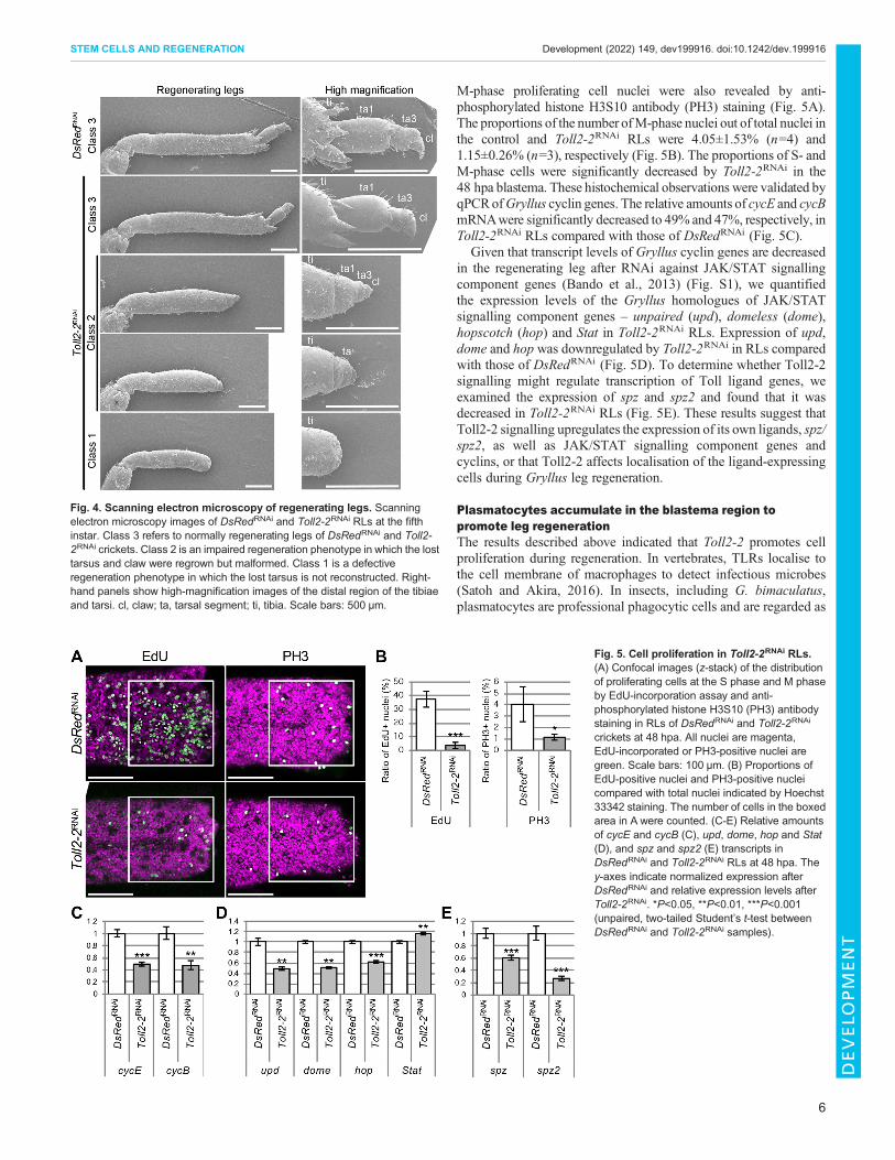
encoding Toll signalling molecules (Fig. 3B) at the fifth instar weredetermined and were categorised as class 1, class 2 and class 3(Fig. 3A,C, Fig. 4). Class 3 was the normally regenerated leg foundin the DsRedRNAi crickets. In this class, the lost portion of the tibiawas regrown, and tibial spurs were reconstructed at the distal end ofthe regrown tibia. The lost tarsus was reconstructed and segmentedinto tarsal segments 1 and 3 and the claws (Fig. 4). Class 2 wasdefined as an impaired regeneration phenotype. In this phenotype,the lost part of the tibia was not regrown, and tibial spurs were notreconstructed. The lost tarsus was reconstructed but was small and
displayed an abnormal morphology. Class 1 was defined as adefective regeneration phenotype, in which no regenerationoccurred, with no reconstruction of the lost tarsus (Fig. 3A,Fig. 4). In the RNAi experiments (Fig. 3C), >40% of cricketsdisplayed class 1 phenotypes when RNAi targeted Toll2-1 or Toll2-2 (Fig. 3C). In addition, 18.8% and 32% of Toll2-1RNAi and Toll2-2RNAi crickets exhibited the class 2 phenotype, respectively;therefore, >70% of these RNAi crickets showed abnormalitiesduring leg regeneration. These data, together with the observedexpression changes during regeneration (Fig. 2B), indicate thatToll2-1 and Toll2-2 are important in leg regeneration.
To analyse further the role of Toll signalling, RNAitargeting ligands and intracellular component genes of thesignalling (Fig. 3B) was performed. Whereas Drosophila has sixspz paralogues (Viljakainen, 2015), we found two spz paralogues,which encode Toll ligands, in theGryllus genome. Spz and Spz2 areparalogous toDrosophila Spz and Spz5, respectively. RNAi againstGryllus spz or spz2 resulted in class 1 phenotypes occurring at ratesof 21.1% and 16.7%, respectively (Fig. 3C,D), and those againstMyD88, tube, pelle or TRAF6, which encode intracellular signallingmolecules of Toll signalling (Fig. 3B), were 57.1%, 10.0%, 11.1%and 16.7%, respectively (Fig. 3C,D). The percentage of class 1 andclass 2 phenotypes after MyD88RNAi was 78.5%, which iscomparable to that of Toll2-1RNAi (75.1%) or Toll2-2RNAi
(72.0%) (Fig. 3C), suggesting that Toll2-1 and Toll2-2 may playcrucial roles in leg regeneration via MyD88.
Next, we performed RNAi against dorsal (dl) andDorsal-relatedimmunity factor (Dif ), both of which encode nuclear factor-kappa-B(NF-κB) transcription factors that function in the Toll signallingpathway (Fig. 3B) (Lindsay and Wasserman, 2014). Single RNAiagainst dl or Dif showed weak defects in leg regeneration. Forexample, 18.2% and 50.0% of DifRNAi cricket legs showed class 1
Fig. 1. Summary of RNA-seq results. Frequentlycounted gene ontology (GO) biological process termsby annotations of the transcripts with upregulatedexpression in RLs. The y-axis indicates the number ofcontigs.
Table 1. Process of short reads assembly and annotation of assembled contigs
Totallength (bp)
Averagelength (bp)
Number ofcontigs
Number of contigs (numberof reads ≥10, RPKM ≥5)
Number ofBLASTed contigs
Assembled transcripts 14,368,694 2252 20,317 8441Only expressed in RLs 908 59 32Upregulated in RLs 2565 957 549Similarly expressed in both RLs and NLs 10,669 6116Downregulated in RLs 2797 1271Only expressed in NLs 1194 38
3
STEM CELLS AND REGENERATION Development (2022) 149, dev199916. doi:10.1242/dev.199916
DEVELO
PM
ENT

and 2 phenotypes, respectively (Fig. 3C,D). The proportion of theclass 1 phenotype caused by DifRNAi was slightly increased incrickets with dual RNAi against dl and Dif (Fig. 3C). Given thatDl and Dif function downstream of both Toll and TNF signallingpathways (Igaki and Miura, 2014), we performed RNAi againsteiger (egr) and wengen (wgn), which encode TNFα and TNFreceptors, respectively. Leg regeneration occurred normally inegrRNAi and wgnRNAi crickets (Fig. S7A), despite the reductionof egr or wgn mRNA to approximately 10% compared withDsRedRNAi controls (Fig. S7B). These RNAi results indicate thatSpz and Spz2 could promote leg regeneration through Tollreceptors, MyD88 and Dl/Dif dimers.
Because Toll2-1RNAi and Toll2-2RNAi crickets showed the mostsevere defects in leg regeneration (Fig. 3C), we quantified themRNA levels of all Gryllus Toll genes in RLs of Toll2-1RNAi
and Toll2-2RNAi crickets at 48 hpa (Fig. S5A,B). In the Toll2-1RNAi
RLs, the relative amount of Toll2-2 mRNA was significantlydecreased to 54%, whereas Toll and Toll2-4 mRNAs weresignificantly increased to 160% and 152%, respectively(Fig. S5A). In the Toll2-2RNAi RLs, the relative amounts of Tolland Toll6-2 mRNA were slightly reduced to 75% and 71%,respectively (Fig. S5B), but expression levels of other cricket Tollgenes, including Toll2-1, were not significantly changed. Theseresults suggest that the RNAi phenotypes observed in Toll2-1RNAi
Fig. 2. Phylogenetic tree and temporal expression change ofGryllus Toll genes. (A) Phylogenetic tree of Toll genes ofGryllus bimaculatus and other insectsbased on the amino acid sequence alignment by CLUSTALW. Ame, Apis mellifera; Bge, Blattella germanica; Dme, Drosophila melanogaster; Gb, Gryllusbimaculatus; Ofa,Oncopeltus fasciatus; Tca, Tribolium castaneum; Zne, Zootermopsis nevadensis. Aqu (Amphimedon queenslandica) LRR was selected as anoutgroup. (B) Temporal expression changes of Gryllus Toll genes at 0, 3, 24 and 48 hpa during leg regeneration. The y-axes show normalised expression at the0 hpa and relative expression levels at 3, 24 and 48 hpa. *P<0.05 (Tukey’s test). Toll2-1, Toll2-2 and Toll2-5 (pink bars) were significantly upregulated and Toll2-4,Toll6-1, Toll6-2, Toll7, Toll8 and Toll9 (blue bars) were significantly downregulated at all time points. Genes for which expression changes were either less thantwofold (Toll) or less than 50% (Toll2-3) are shown in yellow.
4
STEM CELLS AND REGENERATION Development (2022) 149, dev199916. doi:10.1242/dev.199916
DEVELO
PM
ENT

crickets might be caused by an additional reduction in the Toll2-2mRNA level. Nucleotide alignment indicated that an off-targeteffect was unlikely (Fig. S5C), implying a possible epistaticregulation of Toll2-2 by Toll2-1.We next focused on Toll2-2 function during cricket leg
regeneration by carrying out single-gene functional analysis byRNAi. Notably, 28% of Toll2-2RNAi crickets exhibited a class 3phenotype (Fig. 3C) and 36% of endogenous Toll2-2 mRNAremained after Toll2-2RNAi (Fig. S4A), implying that RNAiagainst Toll2-2 could show dose dependency. We injected a largervolume of Toll2-2 dsRNA solution (483 nl) and compared theresulting phenotypes with those obtained from standard Toll2-2RNAi
(207 nl). RNA reduction and phenotype ratio were not significantlydifferent in either experimental condition (Fig. S6); therefore,
for subsequent analyses, we performed RNAi using standardconditions.
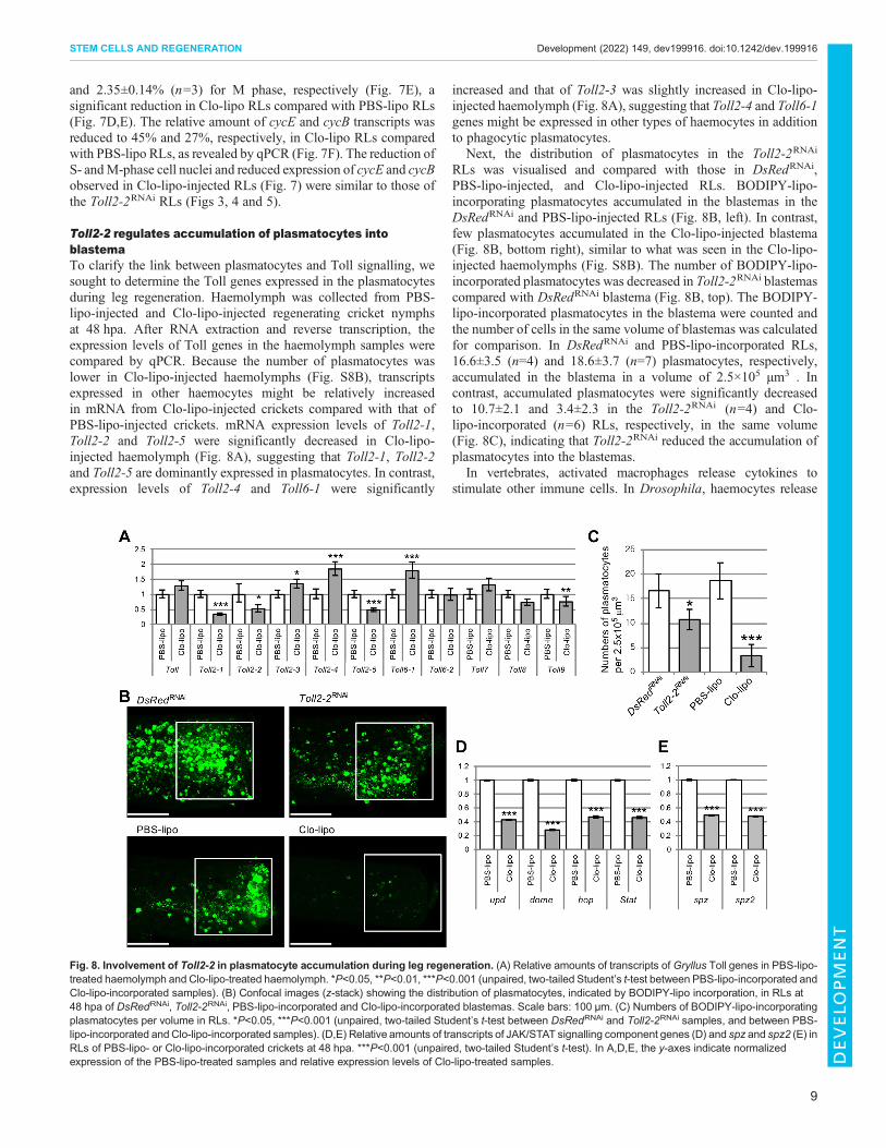
Toll2-2 regulates blastema cell proliferationTo examine whether the class 1 or 2 phenotype observed inToll2-2RNAi crickets (Fig. 3A,C, Fig. 4) was caused by abnormalregulation of cell proliferation in the blastema, we analysed cellproliferation in Toll2-2RNAi crickets at 48 hpa. The distribution ofS-phase proliferating cell nuclei and total nuclei, revealed by5-ethynyl-2-deoxyuridine (EdU) incorporation assay and Hoechst33342 staining, respectively, was determined in Toll2-2RNAi andDsRedRNAi RLs (Fig. 5A). The proportions of S-phase nuclei outof all nuclei in the Toll2-2RNAi and DsRedRNAi RLs were37.41±5.80% (n=4) and 3.63±2.26% (n=3), respectively (Fig. 5B).
Fig. 3. Phenotypes after RNAi against Toll signalling component genes. (A) Typical morphology of an unamputated leg and RLs of DsRedRNAi and Toll2-2RNAi crickets at the fifth instar. Class 3 is a normal regeneration phenotype in which the lost parts of the tibia, tarsus and claw were successfully reconstructed.Class 1 is a defective regeneration phenotype in which the lost tarsus was not reconstructed. Class 2 is an impaired regeneration phenotype in which the losttarsus and claw were regrown but were malformed. (B) Schematic of the molecules involved in Toll signalling. Gryllus genome has a single gene for each ofMyD88, tube, pelle and TRAF6 homologues, and there are two spz genes. (C) The percentage of RNAi phenotypes of class 1, class 2 and class 3. n=numbers ofRNAi-treated individuals. (D) Typical morphology of RLs of frequently observed phenotypes for RNAi-treated crickets at fifth instar.
5
STEM CELLS AND REGENERATION Development (2022) 149, dev199916. doi:10.1242/dev.199916
DEVELO
PM
ENT
M-phase proliferating cell nuclei were also revealed by anti-phosphorylated histone H3S10 antibody (PH3) staining (Fig. 5A).The proportions of the number ofM-phase nuclei out of total nuclei inthe control and Toll2-2RNAi RLs were 4.05±1.53% (n=4) and1.15±0.26% (n=3), respectively (Fig. 5B). The proportions of S- andM-phase cells were significantly decreased by Toll2-2RNAi in the48 hpa blastema. These histochemical observations were validated byqPCRofGryllus cyclin genes. The relative amounts of cycE and cycBmRNAwere significantly decreased to 49% and 47%, respectively, inToll2-2RNAi RLs compared with those of DsRedRNAi (Fig. 5C).
Given that transcript levels of Gryllus cyclin genes are decreasedin the regenerating leg after RNAi against JAK/STAT signallingcomponent genes (Bando et al., 2013) (Fig. S1), we quantifiedthe expression levels of the Gryllus homologues of JAK/STATsignalling component genes – unpaired (upd), domeless (dome),hopscotch (hop) and Stat in Toll2-2RNAi RLs. Expression of upd,dome and hop was downregulated by Toll2-2RNAi in RLs comparedwith those of DsRedRNAi (Fig. 5D). To determine whether Toll2-2signalling might regulate transcription of Toll ligand genes, weexamined the expression of spz and spz2 and found that it wasdecreased in Toll2-2RNAi RLs (Fig. 5E). These results suggest thatToll2-2 signalling upregulates the expression of its own ligands, spz/spz2, as well as JAK/STAT signalling component genes andcyclins, or that Toll2-2 affects localisation of the ligand-expressingcells during Gryllus leg regeneration.
Plasmatocytes accumulate in the blastema region topromote leg regenerationThe results described above indicated that Toll2-2 promotes cellproliferation during regeneration. In vertebrates, TLRs localise tothe cell membrane of macrophages to detect infectious microbes(Satoh and Akira, 2016). In insects, including G. bimaculatus,plasmatocytes are professional phagocytic cells and are regarded as
Fig. 4. Scanning electron microscopy of regenerating legs. Scanningelectron microscopy images of DsRedRNAi and Toll2-2RNAi RLs at the fifthinstar. Class 3 refers to normally regenerating legs of DsRedRNAi and Toll2-2RNAi crickets. Class 2 is an impaired regeneration phenotype in which the losttarsus and claw were regrown but malformed. Class 1 is a defectiveregeneration phenotype in which the lost tarsus is not reconstructed. Right-hand panels show high-magnification images of the distal region of the tibiaeand tarsi. cl, claw; ta, tarsal segment; ti, tibia. Scale bars: 500 µm.
Fig. 5. Cell proliferation in Toll2-2RNAi RLs.(A) Confocal images (z-stack) of the distributionof proliferating cells at the S phase and M phaseby EdU-incorporation assay and anti-phosphorylated histone H3S10 (PH3) antibodystaining in RLs of DsRedRNAi and Toll2-2RNAi
crickets at 48 hpa. All nuclei are magenta,EdU-incorporated or PH3-positive nuclei aregreen. Scale bars: 100 µm. (B) Proportions ofEdU-positive nuclei and PH3-positive nucleicompared with total nuclei indicated by Hoechst33342 staining. The number of cells in the boxedarea in A were counted. (C-E) Relative amountsof cycE and cycB (C), upd, dome, hop and Stat(D), and spz and spz2 (E) transcripts inDsRedRNAi and Toll2-2RNAi RLs at 48 hpa. They-axes indicate normalized expression afterDsRedRNAi and relative expression levels afterToll2-2RNAi. *P<0.05, **P<0.01, ***P<0.001(unpaired, two-tailed Student’s t-test betweenDsRedRNAi and Toll2-2RNAi samples).
6
STEM CELLS AND REGENERATION Development (2022) 149, dev199916. doi:10.1242/dev.199916
DEVELO
PM
ENT

insect macrophages (Cho and Cho, 2019). To visualise the cricketpresumptive plasmatocytes, India ink and boron-dipyrromethene-cholesterol within liposomes (BODIPY-lipo) were injectedinto the haemolymph of third instar nymph abdomens. Indiaink and BODIPY-lipo are characteristically incorporated intoplasmatocytes. Thus, Gryllus plasmatocytes were identified byblack droplets of India ink in Giemsa-stained smears and bygreen fluorescence of BODIPY-lipo (Fig. 6A). Phalloidin stainingrevealed the actin cytoskeleton of haemocytes and part of thehaemocytes incorporated with BODIPY-lipo (Fig. 6B). Duringleg regeneration, plasmatocytes accumulated near the blastemaregion of the regenerating tibia, and some plasmatocytes remainedin the femur (Fig. 6C). Accumulation of plasmatocytes into theblastema region was observed at 48 hpa compared with the patternat 0 hpa (Fig. 6D), suggesting that plasmatocytes accumulate at thewound region following amputation.To clarify the role of plasmatocytes in leg regeneration,
plasmatocytes were depleted using 100 nm diameter Clo-lipo.
The mean lifespan was estimated as the day when the survival ratewas 50% after injection of Clo-lipo and PBS-lipo (liposome-encapsulated PBS buffer for control experiments). The meanlifespan of Clo-lipo-injected crickets was 15 days (Fig. S8A).To confirm the depletion of plasmatocytes, BODIPY-lipo wasinjected 24 h after the injection of Clo-lipo or PBS-lipo. Thenumber of BODIPY-labelled plasmatocytes was decreased inthe haemolymph of Clo-lipo-injected crickets compared withPBS-lipo-injected crickets (Fig. S8B). We also confirmed thedepletion of plasmatocytes in the RLs at 48 hpa: Phalloidin-positive haemocytes were present beneath the epidermal cells inPBS-lipo- or Clo-lipo-injected RLs (Fig. 6E). However, BODIPY-incorporated plasmatocytes were not present in the blastema regionof Clo-lipo-injected RLs (Fig. 6E).
To clarify the relationship between plasmatocyte function and legregeneration, we observed the leg regeneration processes ofClo-lipo- and PBS-lipo-injected crickets. In PBS-lipo crickets, thewound surface was covered by a new cuticle at the fourth instar and
Fig. 6. Distribution of plasmatocytes in haemolymph and regenerating legs. (A) Smear samples of Gryllus haemolymphs were stained with Giemsa.Plasmatocytes (red arrowheads) are visualised by incorporation of India ink (top) and BODIPY-lipo (bottom). (B) Nuclei and actin cytoskeleton of haemocyteswere stained by Hoechst 33342 and Rhodamine phalloidin, respectively. Plasmatocytes were visualised by BODIPY-lipo incorporation. (C) Distribution ofplasmatocytes in a RL at 48 hpa was visualised by BODIPY-lipo incorporation. Blastema (Bl) is indicated by the bracket in the brightfield image. Dashed linemarks the leg outline in the BODIPY-lipo image. (D) Distribution of plasmatocytes in RLs at 0 and 48 hpa were visualised by BODIPY-lipo incorporation.(E) Confocal images (z-stack) of the distribution of plasmatocytes in the blastema region of RLs of PBS-lipo-treated and Clo-lipo-treated crickets at 48 hpa.Plasmatocytes were visualised by BODIPY-lipo (green) and Rhodamine phalloidin (red), and nuclei were stained with Hoechst 33342 (blue). Cells with strong redfluorescence are likely spreading plasmatocytes with increased F-action expression (Williams, 2009; Xu et al., 2012). Scale bars: 20 µm.
7
STEM CELLS AND REGENERATION Development (2022) 149, dev199916. doi:10.1242/dev.199916
DEVELO
PM
ENT

the lost parts of the legs were regenerated by the sixth instarin a regeneration process that was indistinguishable from thatof untreated crickets (Fig. 7A). In contrast, in Clo-lipo-injectedcrickets, the wound surface was covered by a new cuticle at thefourth instar, but the lost parts of the legs were not regenerated (class1 phenotype) in 30/39 (76.9%) crickets or were regenerated in asmall-malformed structure (class 2 phenotype) in 9/39 (23.1%)crickets (Fig. 7A). Reduction of plasmatocytes in the RLs at 48 hpawas quantified by qPCR for Gryllus homologues of croquemort(crq) and glial cells missing (Gcm), which are expressed inDrosophila plasmatocytes (Wang et al., 2014). A significantreduction in both gene expression was observed. Expression ofcrq was downregulated in Clo-lipo RLs compared with PBS-lipo
RLs (P<0.001 by Student’s t-test; Fig. 7B), indicating that Clo-lipoinjection led to a decrease in the number of plasmatocytes in RLs .
The distribution of proliferating cells in the S and M phaseswas examined using the EdU-incorporation assay and anti-phosphorylated histone H3S10 antibody staining, respectively(Fig. 7D). In cricket RLs, the blastema appears as a whitish tissuelocalised in the distal region (Fig. 7C, brackets). In Clo-lipo RLs,however, scabs adhered to the wound surface. Thus, the woundepidermis and tibial epidermis were fragile when we removedthe scab and cuticle from the RLs (Fig. 7C). The proportions ofS- and M-phase cell nuclei out of total cell nuclei in the PBS-lipo- and Clo-lipo-injected RLs at 48 hpa were 27.57±7.83% (n=3)and 9.73±5.07% (n=3) for S phase, and 5.72±1.173% (n=3)
Fig. 7. Involvement of plasmatocytes in cricket leg regeneration. (A) Typical regeneration process of PBS-lipo- and Clo-lipo-incorporated crickets at fourth,fifth and sixth instar nymph stages. (B) Relative amounts ofGcm and crq transcripts in RLs of PBS-lipo- or Clo-lipo-incorporated crickets at 48 hpa. (C) Blastemaformation in the PBS-lipo-incorporated RLs and scab formation in the Clo-lipo-incorporated RLs. Blastemas and scabs are indicated by brackets. (D) Confocalimages (z-stack) showing proliferating cells at the S phase and M phase detected by EdU incorporation and anti-phosphorylated histone H3S10 (PH3) antibodystaining, respectively, in PBS-lipo- or Clo-lipo-incorporated regenerating tibiae. Nuclei were stained with Hoechst 33342. All nuclei are magenta; EdU-incorporated or PH3-positive nuclei are green. Scale bars: 100 µm. (E) Proportions of EdU-positive nuclei and of PH3-positive nuclei compared with total nuclei.The number of cells in the boxed area in D was counted. (F) Relative amounts of cycE and cycB transcripts in RLs of PBS-lipo- or Clo-lipo-incorporated crickets.*P<0.05, ***P<0.001 (unpaired, two-tailed Student’s t-test between PBS-lipo-incorporated and Clo-lipo-incorporated samples). In B,F, the y-axes indicatenormalized expression of the PBS-lipo-treated samples and relative expression levels of Clo-lipo-treated samples.
8
STEM CELLS AND REGENERATION Development (2022) 149, dev199916. doi:10.1242/dev.199916
DEVELO
PM
ENT
and 2.35±0.14% (n=3) for M phase, respectively (Fig. 7E), asignificant reduction in Clo-lipo RLs compared with PBS-lipo RLs(Fig. 7D,E). The relative amount of cycE and cycB transcripts wasreduced to 45% and 27%, respectively, in Clo-lipo RLs comparedwith PBS-lipo RLs, as revealed by qPCR (Fig. 7F). The reduction ofS- andM-phase cell nuclei and reduced expression of cycE and cycBobserved in Clo-lipo-injected RLs (Fig. 7) were similar to those ofthe Toll2-2RNAi RLs (Figs 3, 4 and 5).
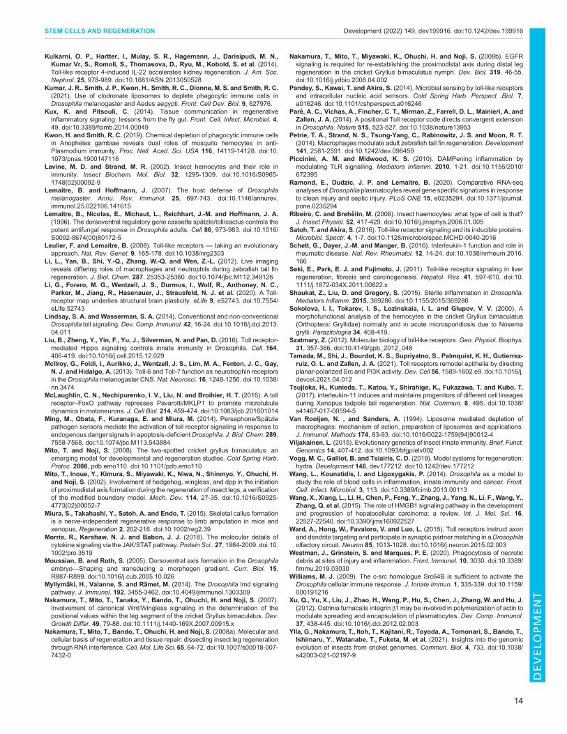
Toll2-2 regulates accumulation of plasmatocytes intoblastemaTo clarify the link between plasmatocytes and Toll signalling, wesought to determine the Toll genes expressed in the plasmatocytesduring leg regeneration. Haemolymph was collected from PBS-lipo-injected and Clo-lipo-injected regenerating cricket nymphsat 48 hpa. After RNA extraction and reverse transcription, theexpression levels of Toll genes in the haemolymph samples werecompared by qPCR. Because the number of plasmatocytes waslower in Clo-lipo-injected haemolymphs (Fig. S8B), transcriptsexpressed in other haemocytes might be relatively increasedin mRNA from Clo-lipo-injected crickets compared with that ofPBS-lipo-injected crickets. mRNA expression levels of Toll2-1,Toll2-2 and Toll2-5 were significantly decreased in Clo-lipo-injected haemolymph (Fig. 8A), suggesting that Toll2-1, Toll2-2and Toll2-5 are dominantly expressed in plasmatocytes. In contrast,expression levels of Toll2-4 and Toll6-1 were significantly
increased and that of Toll2-3 was slightly increased in Clo-lipo-injected haemolymph (Fig. 8A), suggesting that Toll2-4 and Toll6-1genes might be expressed in other types of haemocytes in additionto phagocytic plasmatocytes.
Next, the distribution of plasmatocytes in the Toll2-2RNAi
RLs was visualised and compared with those in DsRedRNAi,PBS-lipo-injected, and Clo-lipo-injected RLs. BODIPY-lipo-incorporating plasmatocytes accumulated in the blastemas in theDsRedRNAi and PBS-lipo-injected RLs (Fig. 8B, left). In contrast,few plasmatocytes accumulated in the Clo-lipo-injected blastema(Fig. 8B, bottom right), similar to what was seen in the Clo-lipo-injected haemolymphs (Fig. S8B). The number of BODIPY-lipo-incorporated plasmatocytes was decreased in Toll2-2RNAi blastemascompared with DsRedRNAi blastema (Fig. 8B, top). The BODIPY-lipo-incorporated plasmatocytes in the blastema were counted andthe number of cells in the same volume of blastemas was calculatedfor comparison. In DsRedRNAi and PBS-lipo-incorporated RLs,16.6±3.5 (n=4) and 18.6±3.7 (n=7) plasmatocytes, respectively,accumulated in the blastema in a volume of 2.5×105 µm3 . Incontrast, accumulated plasmatocytes were significantly decreasedto 10.7±2.1 and 3.4±2.3 in the Toll2-2RNAi (n=4) and Clo-lipo-incorporated (n=6) RLs, respectively, in the same volume(Fig. 8C), indicating that Toll2-2RNAi reduced the accumulation ofplasmatocytes into the blastemas.
In vertebrates, activated macrophages release cytokines tostimulate other immune cells. In Drosophila, haemocytes release
Fig. 8. Involvement of Toll2-2 in plasmatocyte accumulation during leg regeneration. (A) Relative amounts of transcripts of Gryllus Toll genes in PBS-lipo-treated haemolymph and Clo-lipo-treated haemolymph. *P<0.05, **P<0.01, ***P<0.001 (unpaired, two-tailed Student’s t-test between PBS-lipo-incorporated andClo-lipo-incorporated samples). (B) Confocal images (z-stack) showing the distribution of plasmatocytes, indicated by BODIPY-lipo incorporation, in RLs at48 hpa of DsRedRNAi, Toll2-2RNAi, PBS-lipo-incorporated and Clo-lipo-incorporated blastemas. Scale bars: 100 µm. (C) Numbers of BODIPY-lipo-incorporatingplasmatocytes per volume in RLs. *P<0.05, ***P<0.001 (unpaired, two-tailed Student’s t-test between DsRedRNAi and Toll2-2RNAi samples, and between PBS-lipo-incorporated andClo-lipo-incorporated samples). (D,E) Relative amounts of transcripts of JAK/STAT signalling component genes (D) and spz and spz2 (E) inRLs of PBS-lipo- or Clo-lipo-incorporated crickets at 48 hpa. ***P<0.001 (unpaired, two-tailed Student’s t-test). In A,D,E, the y-axes indicate normalizedexpression of the PBS-lipo-treated samples and relative expression levels of Clo-lipo-treated samples.
9
STEM CELLS AND REGENERATION Development (2022) 149, dev199916. doi:10.1242/dev.199916
DEVELO
PM
ENT

insect cytokines, including Upd and Spz, to induce an immuneresponse (Agaisse et al., 2003; Shaukat et al., 2015). In Clo-lipoRLs, the relative expression levels of upd, spz and spz2 weresignificantly decreased to 43%, 49% and 48%, respectively,compared with PBS-lipo RLs (Fig. 8D,E). Taken together withthe downregulation of upd, spz and spz2 in Toll2-2RNAi RLs(Fig. 5D,E) and defective regeneration phenotypes caused by RNAiof each of these genes (Fig. 3C,D), the collective data suggest thatUpd and Spz/Spz2 activate the JAK/STAT and Toll signallingpathways, respectively, during Gryllus leg regeneration. Thus,plasmatocytes that accumulate in RLs release cytokines, whichactivate the JAK/STAT and Toll signalling pathways, leading toblastemal cell proliferation during cricket leg regeneration.
Infectious microbes do not have a major role in legregenerationIn mammals, PAMPs, including lipopolysaccharides, lipoproteinsand flagellin, directly bind to TLRs (Pandey et al., 2014). In insects,PGRPs detect PAMPs, leading to the activation of Toll or Imdsignalling pathways to produce antimicrobial peptides (AMPs)(Lemaitre and Hoffmann, 2007; Lemaitre et al., 1996; Lindsay andWasserman, 2014) (Fig. S1). To identify whether pathogeninfection is a trigger for regeneration, we cloned partial cDNAfragments of Gryllus homologous genes of PGRP-SA and PGRP-SD, which recognise gram-positive bacteria, and PGRP-LC andimd, which recognise gram-negative bacteria, and performed RNAito observe phenotypes of regenerating legs. The Gryllus genomeencodes a single gene for each of the four genes. Determination ofleg regeneration phenotypes after PGRP-SARNAi, PGRP-SDRNAi,PGRP-LCRNAi and imdRNAi revealed that all crickets regeneratedlegs normally, in a similar manner to DsRedRNAi crickets(Fig. S9A). This finding indicates that infectious microbes do notplay a major role in leg regeneration. To further substantiate this, weexamined the gene expression change of the Gryllus homologueof defensin, which encodes an evolutionarily conserved AMP(Fig. S10). qPCR analysis revealed that the expression level ofGryllus defensin was decreased at 3, 24 and 48 hpa in RLs(Fig. S10C), suggesting that PAMP-mediated activation of Tollsignalling is suppressed during regeneration.Toll molecules recognise PAMPs and DAMPs, mediated by clip-
SP Persephone inDrosophila (Ming et al., 2014). We could not finda persephone ( psh) homologue in the Gryllus genome (Fig. S11).DAMPs are released from cells in response to injury or cell death,including apoptosis and necrosis. We focused on the function of thescavenger receptor CD36 homologue Crq, which recognises theapoptotic body and is required for phagocytosis of plasmatocytesin Drosophila. The Gryllus crq homologue was expressed inplasmatocytes (Fig. 7B). During leg regeneration, 16.7% and 45.8%of crqRNAi crickets had class 1 and class 2 phenotype, respectively(Fig. S9B-D). These results suggest that recognition and engulfmentof apoptotic bodies released from injured cells by Crq likely triggersleg regeneration in crickets.
DISCUSSIONToll-related signalling, rather than Imd signalling, is a majorpathway in cricket leg regeneration from the early phasesToll/TLR signalling is evolutionarily conserved from insects tohumans for the recognition of pathogens including viruses, gram-positive bacteria and fungi (Lindsay and Wasserman, 2014; Pandeyet al., 2014). Toll/TLRs induce gene expression of cytokines andantibacterial components via MyD88 and NF-κB (Lemaitre andHoffmann, 2007), although Drosophila Toll-2 does not require
MyD88 for cell proliferation (Li et al., 2020). In insect immunity,activation of Toll signalling is different from that of vertebrates;infectious microbes are detected by PGRPs that activate clip-SPs tocatalyse pro-Spz to Spz, which binds to a Toll receptor to activatesignalling (Fig. S1) (Krautz et al., 2014; Lindsay and Wasserman,2014). We previously reported that expression of Spz, Toll receptorsand NF-κB (Relish, dl) genes is upregulated in RLs at 24 hpa(Bando et al., 2013). In this study, RNA-seq data showed that theexpression of three Gryllus Toll genes (Toll2-2, Toll2-5, Toll8) andRel were upregulated as early as at 3 hpa in RLs. Upregulation ofToll2-1, Toll2-2 and Toll2-5 genes was confirmed by qPCR duringregeneration, suggesting that Toll signalling is involved in thecricket regeneration process from the early phases.
Imd signalling, another pathway involved in insect immunity(Fig. S1), detects infections by gram-negative bacteria. PGRP-LCand Imd together induce the expression of antibacterial componentsthrough the NF-κB transcription factor Rel (Krautz et al., 2014;Lemaitre and Hoffmann, 2007). Although expression of Rel wasupregulated in 3 hpa RLs (Fig. S2B), RNAi of PGRP-LC, imd andRel did not show obvious defects in leg regeneration (Fig. 3C,D,Fig. S9A), suggesting that Imd signalling is not essential for legregeneration in G. bimaculatus.
Our previous study showed that JAK/STAT signalling is involvedin leg regeneration (Bando et al., 2013). Ligands for JAK/STATsignalling are interleukins in vertebrates (Morris et al., 2018).In Xenopus tadpole tail regeneration, interleukin-11 inducesundifferentiation state (Tsujioka et al., 2017). In Drosophilamelanogaster, ligands of JAK/STAT signalling are Upds, whichact as cytokines (Arbouzova and Zeidler, 2006) and are required forthe regulation of stem cell proliferation during intestinalregeneration (Kux and Pitsouli, 2014). In the present study, theexpression of upd was decreased in plasmatocyte-depleted RLs.Lost parts of legs of updRNAi crickets were not fully regenerated,similar to observations after RNAi of JAK/STAT signalling genes(Bando et al., 2013). In a similar manner to the production ofinterleukins by macrophages to activate other immune cells invertebrates (Schett et al., 2016), plasmatocytes release insectcytokines to induce the production of antibacterial components inDrosophila (Lemaitre and Hoffmann, 2007; Ramond et al., 2020).Thus, it is likely thatGryllus plasmatocytes produce Upd to activateJAK/STAT signalling in leg regeneration.
Toll-related signalling promotes leg regeneration bycontrolling cell proliferation during blastema formationWe cloned 11 Toll genes from Gryllus, not all of which weredirectly homologous to those of the nine Toll genes of Drosophila(Hillyer, 2016). The Gryllus genome has five Toll2 paralogues(Toll2-1–5) that are not present in Drosophila, and two Toll6paralogues (Toll6-1–2). Gryllus has no genes homologous toDrosophila Toll3, Toll4 and Toll5. Drosophila Toll (McIlroy et al.,2013; McLaughlin et al., 2016; Ward et al., 2015) is importantfor defence against pathogens (Lemaitre and Hoffmann, 2007)and dorsoventral patterning during embryogenesis (Moussian andRoth, 2005). Toll-2, -6 and -8 are involved in the anteroposteriorpatterning of the fly (Paré et al., 2014). Drosophila Toll-2 regulatescell proliferation and planar cell polarity (Li et al., 2020; Tamadaet al., 2021) and Toll-6 promotes neuronal cell shape, survival andinteractions (McIlroy et al., 2013; McLaughlin et al., 2016; Wardet al., 2015). In Gryllus, Toll6-1, Toll6-2, Toll7 and Toll8are expressed during embryogenesis (Benton et al., 2016). Theirfunctions related to immunity and dorsoventral patterning areunknown. This study has clarified that diversified Gryllus Toll2
10
STEM CELLS AND REGENERATION Development (2022) 149, dev199916. doi:10.1242/dev.199916
DEVELO
PM
ENT

subfamily members play crucial roles in the early phase of legregeneration.Expression of Toll2-1, Toll2-2 and Toll2-5 was upregulated
during leg regeneration, in which >70% of Toll2-2RNAi cricketsshowed regeneration defects. The proportions of S- and M-phaseproliferating cells were decreased in Toll2-2RNAi RLs, indicatingthat Toll2-2 signalling is required for cell proliferation during legregeneration. In Toll2-2RNAi RLs, accumulation of plasmatocyteswas reduced and expression of updwas downregulated. Thus, Toll2-2 in plasmatocytes may induce upd expression, which activates theJAK/STAT signalling pathway (Bando et al., 2013) and cyclins,leading to cell proliferation in regenerating legs.During early leg regeneration processes, expression of Toll2-4,
Toll6-1, Toll6-2, Toll7, Toll8 and Toll9 was decreased (Fig. 2B).However, Toll2-4RNAi, Toll6-2RNAi, Toll7RNAi and Toll9RNAi cricketsshowed defective or impaired regeneration phenotypes (Fig. 3C).Toll2-4RNAi, Toll6-2RNAi, Toll7RNAi and Toll9RNAi decreased targetgene transcripts by >40%, verifying the RNAi efficiency (Fig. S4A).Our preliminary study showed that RNAi of cricket nymphscontinued to suppress target gene expression for 2 weeks.Therefore, it is possible that the continuous suppression of geneexpression caused defective or impaired regeneration phenotypes inToll2-4RNAi, Toll6-2RNAi, Toll7RNAi and Toll9RNAi crickets.Crosstalk between Toll signalling and Hippo signalling,
and between Hippo signalling and JAK/STAT signalling mayalso regulate tissue growth (Liu et al., 2016; Jiang et al., 2016).Specifically, the Hippo signalling component Warts, togetherwith protocadherins Fat and Dachsous, suppresses blastema cellproliferation, whereas Yorkie promotes proliferation during cricketleg regeneration (Bando et al., 2009). Therefore, Toll signalling mayinterfere with Hippo signalling to regulate blastema cellproliferation in crickets, although Drosophila Toll-2 cooperativelypromotes cell proliferation with Yki (Li et al., 2020).
Plasmatocytes (insect macrophages) promote legregeneration in the cricket, via Spz-Toll-related and Upd-JAK/STAT signallingInsect haemocytes play a major role in host defence (Lavine andStrand, 2002; Ribeiro and Brehélin, 2006), as insects have an openblood-vascular system and lack oxygen-carrying erythrocytes.Insect haemocytes vary depending on the species. For example,the cricket G. bimaculatus has plasmatocytes, granulocytes, andthree or four other haemocytes (Cho and Cho, 2019; Sokolova et al.,2000). Plasmatocytes in several insect species engulf pathogens andproduce inflammatory cytokines and antipathogenic components(Evans et al., 2003; Lavine and Strand, 2002). In the present study,plasmatocyte-depleted crickets did not regenerate missing leg parts.Given that cytokines promote regeneration in axolotl and zebrafish(Godwin et al., 2013; Petrie et al., 2014), cricket plasmatocytescould promote tissue regeneration, possibly by these evolutionarilyconserved molecular mechanisms. Although we had no directevidence for localisation of the Toll2-1, Toll2-2 and Toll2-5 genes inplasmatocytes, their reduced expression levels in plasmatocyte-depleted haemocytes strongly support this idea. Likewise, thefinding that cricket plasmatocytes express the cytokine genes upd,spz and spz2 is reminiscent of the secretion of interleukins fromaxolotl macrophages during regeneration (Godwin et al., 2013).
Toll-related signalling senses cell debris caused by injuryand promotes regenerationMammalian TLRs are pattern recognition receptors, butDrosophilaTolls are not, as they bind endogenous ligands instead of pathogens
(Krautz et al., 2014; Lindsay and Wasserman, 2014; Pandey et al.,2014). When pathogens are recognised by PGRPs, the PGRPscatalyse the maturation of pro-Spz to Spz. Spz subsequently binds toToll proteins to activate Toll signalling. In the present study, cricketssubjected to RNAi of the pathogen recognition protein-codinggenes PGRP-SA, PGRP-SD, PGRP-LC or imd showed normalregeneration (Fig. S9A). Expression of the antimicrobial peptidegene defensin was not upregulated during regeneration (Fig. S10C),although the upstream region of the defensin gene contains manyNF-κB-binding sites (Fig. S10D). This suggests that pathogeninfection is not required for leg regeneration. Thus, non-pathogenicmolecules may activate Gryllus Toll signalling during legregeneration. Candidate molecules include DAMPs (Krautz et al.,2014), which are endogenous proteins that are likely released frominjured cells, such as necrotic or apoptotic cells, and are recognisedby specific receptors, such as RAGE, TREM1 and TLRs, invertebrates (Piccinini and Midwood, 2010; Wang et al., 2015). Theevolutionarily conserved proteins HMGB1, S100, HSP, histone,actin, DNA and RNA are well-known DAMPs (Piccinini andMidwood, 2010). In the mammalian kidney, DAMPs released fromdying cells cause inflammatory responses and acute injury.Regeneration is accelerated by the TLR-mediated release ofinterleukins from macrophages or dendritic cells (Kulkarni et al.,2014). In the liver, denatured DNA from dying hepatocytesstimulates TLRs to mediate hepatocyte stem cell differentiation(Seki et al., 2011). RAGE and TREM1 are not conserved amonginsects; hence, Toll signalling is able to respond to DAMPs ininsects. In Drosophila, the clip-SP Psh is involved in DAMPrecognition and catalyses Pro-Spz to Spz to activate Tolls (Minget al., 2014). The psh homologue is not present in the Gryllusgenome (Ylla et al., 2021), but a snake homologue and three grassparalogues, which encode other clip-SPs, are (Fig. S11). Some ofthese genes may replace a particular role of psh.
Crq, which is a scavenger receptor CD36 homologue, isexpressed in the plasmatocytes and is required for engulfment ofapoptotic cells in Drosophila (Franc et al., 1996, 1999; Guillouet al., 2016). In mice, CD36 mediates phagocytosis by cooperatingwith TLR (Erdman et al., 2009). In the present study, crqRNAi
crickets showed defective and impaired regeneration phenotypes,similar to those found in Toll2-2RNAi crickets. As mentioned above,RNAi of PGRP genes and imd resulted in normal regeneration,implying that the defective and impaired regeneration phenotypescaused by crqRNAi and Toll2-2RNAi could occur as a result of defectsin phagocytosis of cell debris after injury. We propose that Toll2-2and Crq cooperatively recognise damage-associated molecules nearthe wound region (Fig. S12).
Our cricket leg amputation experiments were conductedwithout artificial pathogen inoculation. In the early phase of legregeneration, when blastema cells form, activated plasmatocytesmigrate to the injury site and facilitate the proliferation of blastemacells. These plasmatocytes are most likely polarised towardenhanced phagocytic function by regulating Toll-related and JAK/STAT signalling (Fig. S1). The extent of plasmatocyte polarisationwould be influenced by surrounding tissue damage and pathogenicstatus. Recent single-cell studies of plasmatocytes (Cattenoz et al.,2020) suggest that crq-expressing cells and Toll-2-2-expressingcells would not be identical subpopulations in the plasmatocytesduring early cricket leg regeneration, which will require furtherstudy.
In conclusion, this study provides new insights into the functionof Toll-related signalling for leg regeneration via plasmatocytes,cooperatively with JAK/STAT signalling. Recognition of apoptotic
11
STEM CELLS AND REGENERATION Development (2022) 149, dev199916. doi:10.1242/dev.199916
DEVELO
PM
ENT

cells via the scavenger receptor Crq on plasmatocytes also promotesleg regeneration.
MATERIALS AND METHODSAnimalsAll nymphal and adult two-spotted crickets (G. bimaculatus) were rearedunder standard conditions (Mito and Noji, 2008). Third instar nymphs wereused for RNAi and liposome injection to analyse the regeneration processes(Nakamura et al., 2008a).
RNA-seqThe distal one-third tibial regions of regenerating legs at 0 hpa (NLs) and3 hpa (RLs) were separately collected from a few hundred crickets. TotalRNA was extracted using ISOGEN II (311-07361, Nippon Gene). Poly(A)+RNAs were purified using a MicroPoly(A)Purist kit (AM1919, ThermoFisher Scientific). The cDNA libraries constructed frompoly(A)+RNAsweresequenced using a GS FLX Titanium next-generation sequencer (454 LifeSciences). To construct the assembled transcripts, the 595,425 reads of NLand 519,961 reads of RL samples (14,368,694 bp in total) that weresequenced were assembled into 20,317 contigs using GSDeNovo Assemblerv2.8 software. The average length of the assembled contigs was 2252 bp. Toestimate the normalized expression levels of each transcript, each readobtained from the RL and NL samples was mapped to the contigs to calculatethe RPKM value using GS Reference Mapper v2.8 software. Expressionchanges of assembled transcripts were calculated by dividing the RPKMvalues of RLs by corresponding RPKM value of NLs, which are listed inTable S1. Assembled transcripts that were upregulated more than twofold inRLs compared with NLs were annotated with the BLASTX program againstthe NCBI non-redundant protein sequence database, with an E-value cut-offof 0.001 (Table S2). Functional annotation of BLASTed transcripts wasperformed using Blast2GO software (https://www.blast2go.com/).
Cloning for Gryllus homologous genesGryllus homologues were cloned by PCR with Ex-taq or LA-taq with GCbuffer (RR006A or RR02AG, TaKaRa Bio). Primers used are listed inTable S3. Template cDNAs were reverse transcribed using the SuperScriptIII reverse transcription kit with random primers (18080051, Thermo FisherScientific) from total RNA extracted from the regenerating legs of cricketnymphs at the third instar. The isolated Gryllus cDNA fragments were usedas templates for the synthesis of dsRNAs.
RNAidsRNAs were synthesised using the MEGAScript T7 Kit (AMB13345,Thermo Fisher Scientific) and adjusted to 20 μM. Third instar nymphs wereanaesthetised on ice before RNAi. After injection of 207 nl of dsRNA into theabdomen of the third instar nymphs with an auto-nanoliter injector (NanojectII, #3-000-204, Drummond Scientific Company), their tibiae were amputatedbetween the second and third spines. Wound regions are usually covered withscab within a day in third instar. The third instar nymphs moult to fourth instarwithin 4 days. In the fourth instar, newly formed cuticles cover the woundregion instead of the scab. The fourth instar nymphsmoult to fifth instar within5 days and the lost leg tissues are reconstructed in miniature in the fifth instar.After RNAi and leg amputation, we observed RNAi phenotypes ofregenerating legs on the 10th day, which corresponds to the late fifth instar.As a negative control for RNAi experiments, we injected dsRNA for theDsRed2 exogenous gene. For dual RNAi experiments, a mixture of dsRNAsfor the two target geneswas used. In themixture, the final concentration of eachdsRNA was adjusted to 10 μM. Statistical differences in RNAi phenotypeswere analysed by Fisher’s exact test. For RNAi against Toll and Toll2-2, wegenerated two non-overlapping dsRNAs corresponding to either the 5′ or 3′portion of the coding region (Fig. S3B, denoted by double-headed arrows). TheRNAi experiments performed with these sequences were designatedToll(5′)RNAi, Toll(3′)RNAi, Toll2-2(5′)RNAi and Toll2-2(3′)RNAi, respectively.
qPCRRegenerating tibiae of control, RNAi-treated or liposome-injected nymphswere pooled into single tubes, and total RNA was extracted using the
RNAqueous-Micro Kit (AM1931, Thermo Fisher Scientific). Each pooledRNA sample was divided into two samples and reverse transcribed toprepare cDNA. Each cDNA was used for qPCR performed with theFastStart Essential DNA Green Master kit (06402712001, Roche) and theLightCycler Nano System (Roche). The relative proportions of thetranscripts were calculated using Gryllus β-actin as an internal control.Relative gene expression levels are shown as the mean±s.d. Statisticaldifferences were analysed by an unpaired, two-tailed Student’s t-testbetween two samples or Tukey’s test for more than three samples and areshown by asterisks (*P<0.05, **P<0.01, ***P<0.001). The qPCRexperiments were repeated twice for confirmation. The primers used arelisted in Table S4.
Cell proliferation assayProliferating cells in the S phase were detected using the Click-iT EdUAlexa Fluor 488 Imaging Kit (C10337, Thermo Fisher Scientific). EdU wasinjected into the abdomen of third instar cricket nymphs at 44 hpa, and theRLs were fixed at 48 hpa (4 h after EdU injection) with 2%paraformaldehyde (PFA) in PBS containing 0.05% Tween-20 (PBT)overnight at 4°C. EdU-incorporated cells were detected according to themanufacturer’s instructions. Hoechst 33342 (H3570, Thermo FisherScientific) was used for nuclear staining. Proliferating cells in the Mphase were detected by immunostaining of phospho-histone H3S10.Regenerating legs at 48 hpa were fixed with 2% PFA and cuticles wereremoved, then regenerating tibiae were washed with PBT and blocked with1% normal goat serum (NGS) in PBT for 1 h. Blocked samples wereincubated with primary antibody [rabbit polyclonal anti-phospho-histoneH3 (Ser10) antibody; 06-570, Millipore] at 1:500 in 1% NGS in PBTovernight at 4°C. The samples were washed with PBT and blocked with 1%NGS in PBT. The samples were incubated with secondary antibody (AlexaFluor 488-conjugated anti-rabbit IgG antibody; A-11008, MolecularProbes) at 1:750 in 1% NGS in PBT for 3 h at 25°C. Finally, the sampleswere washed with PBT and incubated with a 1:1000 dilution of Hoechst33342 in PBT for 15 min.
Macrophage depletion and detectionFor plasmatocyte depletion in crickets, 207 nl of Clo-lipo 100 (6.4 mg/mlclodronate) and Clo-lipo 300 (9.8 mg/ml clodronate) (160-0432-1 and 160-0430-1, Katayama Chemical Industries) was injected into the abdomen ofthird instar nymphs. PBS-encapsulated liposomes were used as controls.For plasmatocyte detection in the haemolymph, 207 nl India ink or207 nl BODIPY-lipo (130 μg/ml BODIPY-cholesterol, Katayama ChemicalIndustries) were injected into the abdomen of third instar nymphs. One dayafter the injection, an anticoagulant solution (20 mM EDTA in PBS) wasinjected into the abdomen of the nymphs and haemolymphs were suckedfrom the nymphs. Haemolymphs were fixed with 2%PFA in an anticoagulantsolution for 30 min at 25°C. Fixed haemocytes were washed withanticoagulant solution and then stained with a 1:2000 dilution of Hoechst33342 for 30 min at 25°C. The haemocytes were washed with ananticoagulant solution and stained with a 1:40 dilution of Rhodaminephalloidin (R415, Thermo Fisher Scientific) for 30 min at 25°C. Forplasmatocyte detection in the RLs, RLs of BODIPY-lipo-injected crickets at48 hpa were fixed with 2% PFA in PBT and the cuticles were removed.Stained haemocytes and cuticle-removed RLs were observed with afluorescent microscope DM5000 B (Leica Microsystems), and with aconfocal laser scanning microscope LSM780 (Carl Zeiss) at the CentralResearch Laboratory, Okayama UniversityMedical School, Okayama, Japan.
Scanning electron microscopyScanning electron microscopy images of RLs at the fifth instar wereobtained. RLs of control and Toll2-2RNAi were fixed in 4% PFA and 4%glutaraldehyde in PBS for 15 min at room temperature. Fixed legs werewashed with PBS, then dehydrated through an alcohol series (20%, 40% and60% ethanol in PBS, 80% ethanol in water, 45% ethanol and 45% tert-butylalcohol in water) for 30 min and in 100% tert-butyl alcohol for 1 h. Afterdehydration, the fixed legs were substituted with 100% tert-butyl alcohol,frozen at 4°C, and freeze-dried. Dried legs were coated with osmium with
12
STEM CELLS AND REGENERATION Development (2022) 149, dev199916. doi:10.1242/dev.199916
DEVELO
PM
ENT

osmium coater (HPC-1S; VACUUM DEVICE). Images were capturedusing a model S-4800 field emission scanning electronmicroscope (Hitachi)at the Central Research Laboratory, Okayama University Medical School,Okayama, Japan.
AcknowledgementsWe are grateful to Dr Itsuro Sugimura (Hokkaido System Science Co., Ltd.) forassistance with data analysis; Dr Takayuki Otani (Katayama Chemical IndustriesCo., Ltd.) for preparing PBS-lipo, Clo-lipo and BODIPY-lipo; and Nobuaki Fujimoriand Taiki Morino for their technical assistance.
Competing interestsThe authors declare no competing or financial interests.
Author contributionsConceptualization: T.B., M.O., K.A., S.N., H.O.; Methodology: T.B., M.O., Y.I., E.K.,T.I., K.A., S.N.; Validation: T.B., M.O., Y.B., M.H., Y.H., E.K., T.I.; Formal analysis:T.B., M.O., Y.B., M.H., Y.H., E.K., T.I.; Investigation: T.B., M.O., Y.B., M.H., Y.H., Y.I.,E.K., T.I.; Resources: T.B., M.O., Y.B., M.H., Y.H., E.K., T.I., S.N.; Data curation:T.B., M.O., Y.B., M.H., Y.H., Y.I., E.K., T.I., H.O.; Writing - original draft: T.B., M.O.;Writing - review & editing: T.B., Y.H., T.M., K.A., S.N., H.O.; Visualization: T.B., M.O.,Y.B., M.H.; Supervision: T.B., K.A., S.N., H.O.; Project administration: T.B., S.N.,H.O.; Funding acquisition: T.B., T.M., K.A., S.N., H.O.
FundingThis work was supported by a Grant-in-Aid for Scientific Research on InnovativeAreas (22124003 to S.N.) and a Grant-in-Aid for Creative Scientific Research(15K06897, 18K06184 to T.B.) from the Ministry of Education, Culture, Sports,Science, and Technology of Japan.
Data availabilityRNA-seq data have been deposited in the DDBJ Read Archive under accessionnumbers DRR287335 and DRR287334. Assembled transcriptome data have beendeposited in DDBJ Transcriptome Shotgun Assembly (TSA) division underaccession numbers ICRS01000001-ICRS01020314. Nucleotide sequences ofGryllus homologues of immune-related genes used in this study were deposited inGenBank under accession numbers LC422646-LC422679.
Peer review historyThe peer review history is available online at https://journals.biologists.com/dev/article-lookup/doi/10.1242/dev.199916
ReferencesAgaisse, H., Petersen, U.-M., Boutros, M., Mathey-Prevot, B. and Perrimon, N.(2003). Signaling role of hemocytes in Drosophila JAK/STAT-dependentresponse to septic injury. Dev. Cell 5, 441-450. doi:10.1016/S1534-5807(03)00244-2
Agata, K. and Inoue, T. (2012). Survey of the differences between regenerative andnon-regenerative animals. Dev. Growth Differ. 54, 143-152. doi:10.1111/j.1440-169X.2011.01323.x
Agata, K., Saito, Y. and Nakajima, E. (2007). Unifying principles of regeneration I:epimorphosis versus morphallaxis. Dev. Growth Differ. 49, 73-78. doi:10.1111/j.1440-169X.2007.00919.x
Anders, H.-J. and Schaefer, L. (2014). Beyond tissue injury— damage-associatedmolecular patterns, toll-like receptors, and Inflammasomes also driveregeneration and fibrosis. J. Am. Soc. Nephrol. 25, 1387-1400. doi:10.1681/ASN.2014010117
Anthoney, N., Foldi, I. and Hidalgo, A. (2018). Toll and Toll-like receptor signallingin development. Development 145, dev156018. doi:10.1242/dev.156018
Arbouzova, N. I. and Zeidler, M. P. (2006). JAK/STAT signalling in Drosophila:insights into conserved regulatory and cellular functions. Development 133,2605-2616. doi:10.1242/dev.02411
Bando, T., Mito, T., Maeda, Y., Nakamura, T., Ito, F., Watanabe, T., Ohuchi, H.and Noji, S. (2009). Regulation of leg size and shape by the Dachsous/Fatsignalling pathway during regeneration. Development 136, 2235-2245. doi:10.1242/dev.035204
Bando, T., Mito, T., Nakamura, T., Ohuchi, H. and Noji, S. (2011a). Regulation ofleg size and shape: involvement of the Dachsous-fat signaling pathway.Dev. Dyn.240, 1028-1041. doi:10.1002/dvdy.22590
Bando, T., Hamada, Y., Kurita, K., Nakamura, T., Mito, T., Ohuchi, H. and Noji, S.(2011b). Lowfat, a mammalian Lix1 homologue, regulates leg size and growthunder the Dachsous/Fat signaling pathway during tissue regeneration. Dev. Dyn.240, 1440-1453. doi:10.1002/dvdy.22647
Bando, T., Ishimaru, Y., Kida, T., Hamada, Y., Matsuoka, Y., Nakamura, T.,Ohuchi, H., Noji, S. and Mito, T. (2013). Analysis of RNA-Seq data reveals
involvement of JAK/STAT signalling during leg regeneration in the cricket Gryllusbimaculatus. Development 140, 959-964. doi:10.1242/dev.084590
Bando, T., Hamada, Y. and Noji, S. (2017). Leg formation and regeneration. In TheCricket as a Model Organism (ed. H. W. Horch, T. Mito, A. Popadic, H. Ohuchi andS. Noji), pp. 31-48. Springer.
Benton, M. A., Pechmann, M., Frey, N., Stappert, D., Conrads, K. H., Chen, Y.-T.,Stamataki, E., Pavlopoulos, A. and Roth, S. (2016). Toll genes have anancestral role in axis elongation. Curr. Biol. 26, 1609-1615. doi:10.1016/j.cub.2016.04.055
Bodo, K., Kellermayer, Z., Laszlo, Z., Boros, Á., Kokhanyuk, B., Nemeth, P. andEngelmann, P. (2021). Injury-induced innate immune response during segmentregeneration of the earthworm, eisenia andrei. Int. J. Mol. Sci. 22, 2363. doi:10.3390/ijms22052363
Browne, N., Heelan, M. and Kavanagh, K. (2013). An analysis of the structural andfunctional similarities of insect hemocytes andmammalian phagocytes. Virulence4, 597-603. doi:10.4161/viru.25906
Cattenoz, P. B., Sakr, R., Pavlidaki, A., Delaporte, C., Riba, A., Molina, N.,Hariharan, N., Mukherjee, T. and Giangrande, A. (2020). Temporal specificityand heterogeneity of Drosophila immune cells. EMBO J. 39, e104486. doi:10.15252/embj.2020104486
Cho, Y. and Cho, S. (2019). Hemocyte-hemocyte adhesion by granulocytes isassociated with cellular immunity in the cricket, Gryllus bimaculatus. Sci. Rep. 9,18066. doi:10.1038/s41598-019-54484-5
Debuque, R. J., Nowoshilow, S., Chan, K. E., Rosenthal, N. A. andGodwin, J.W.(2021). Distinct toll-like receptor signaling in the salamander response to tissuedamage. Dev. Dyn. Online Version of Record before inclusion in an is. doi:10.1002/dvdy.340
Erdman, L. K., Cosio, G., Helmers, A. J., Gowda, D. C., Grinstein, S. andKain, K. C. (2009). CD36 and TLR interactions in inflammation and phagocytosis:implications for malaria. J. Immunol. 183, 6452-6459. doi:10.4049/jimmunol.0901374
Evans, C. J., Hartenstein, V. and Banerjee, U. (2003). Thicker than blood:conserved mechanisms in Drosophila and vertebrate hematopoiesis. Dev. Cell 5,673-690. doi:10.1016/S1534-5807(03)00335-6
Franc, N. C., Dimarcq, J.-L., Lagueux, M., Hoffmann, J. and Ezekowitz, R. A. B.(1996). Croquemort, a novel Drosophila hemocyte/macrophage receptor thatrecognizes apoptotic cells. Immunity 4, 431-443. doi:10.1016/S1074-7613(00)80410-0
Franc, N. C., Heitzler, P., Ezekowitz, R. A. B. and White, K. (1999). Requirementfor croquemort in phagocytosis of apoptotic cells in Drosophila. Science 284,1991-1994. doi:10.1126/science.284.5422.1991
Godwin, J. W., Pinto, A. R. and Rosenthal, N. A. (2013). Macrophages arerequired for adult salamander limb regeneration. Proc. Natl. Acad. Sci. USA 110,9415-9420. doi:10.1073/pnas.1300290110
Guillou, A., Troha, K., Wang, H., Franc, N. C. and Buchon, N. (2016). TheDrosophila CD36 homologue croquemort is required to maintain immune and guthomeostasis during development and aging. PLoS Pathog. 12, e1005961. doi:10.1371/journal.ppat.1005961
Hamada, Y., Bando, T., Nakamura, T., Ishimaru, Y., Mito, T., Noji, S., Tomioka, K.and Ohuchi, H. (2015). Leg regeneration is epigenetically regulated by histoneH3K27 methylation in the cricket Gryllus bimaculatus. Development 142,2916-2927. doi:10.1242/dev.122598
Hasegawa, T., Hall, C. J., Crosier, P. S., Abe, G., Kawakami, K., Kudo, A. andKawakami, A. (2017). Transient inflammatory response mediated by interleukin-1β is required for proper regeneration in zebrafish fin fold. eLife 6, e22716. doi:10.7554/eLife.22716
Hillyer, J. F. (2016). Insect immunology and hematopoiesis. Dev. Comp. Immunol.58, 102-118. doi:10.1016/j.dci.2015.12.006
Hu, X., Chen, J., Wang, L. and Ivashkiv, L. B. (2007). Crosstalk among Jak-STAT,Toll-like receptor, and ITAM-dependent pathways in macrophage activation.J. Leukoc. Biol. 82, 237-243. doi:10.1189/jlb.1206763
Igaki, T. and Miura, M. (2014). The Drosophila TNF ortholog eiger: emergingphysiological roles and evolution of the TNF system. Semin. Immunol. 26,267-274. doi:10.1016/j.smim.2014.05.003
Ishimaru, Y., Nakamura, T., Bando, T., Matsuoka, Y., Ohuchi, H., Noji, S. andMito, T. (2015). Involvement of dachshund and Distal-less in distal patternformation of the cricket leg during regeneration. Sci. Rep. 5, 8387. doi:10.1038/srep08387
Ishimaru, Y., Bando, T., Ohuchi, H., Noji, S. and Mito, T. (2018). Bonemorphogenetic protein signaling in distal patterning and intercalation during legregeneration of the cricket, Gryllus bimaculatus. Dev. Growth Differ. 60, 377-386.doi:10.1111/dgd.12560
Jiang, H., Tian, A. and Jiang, J. (2016). Intestinal stem cell response to injury:lessons from Drosophila. Cell. Mol. Life Sci. 73, 3337-3349. doi:10.1007/s00018-016-2235-9
Kawai, T. and Akira, S. (2011). Toll-like receptors and their crosstalk with otherinnate receptors in infection and immunity. Immunity 34, 637-650. doi:10.1016/j.immuni.2011.05.006
Krautz, R., Arefin, B. and Theopold, U. (2014). Damage signals in the insectimmune response. Front. Plant Sci. 5, 342. doi:10.3389/fpls.2014.00342
13
STEM CELLS AND REGENERATION Development (2022) 149, dev199916. doi:10.1242/dev.199916
DEVELO
PM
ENT
Kulkarni, O. P., Hartter, I., Mulay, S. R., Hagemann, J., Darisipudi, M. N.,Kumar Vr, S., Romoli, S., Thomasova, D., Ryu, M., Kobold, S. et al. (2014).Toll-like receptor 4-induced IL-22 accelerates kidney regeneration. J. Am. Soc.Nephrol. 25, 978-989. doi:10.1681/ASN.2013050528
Kumar, J. R., Smith, J. P., Kwon, H., Smith, R. C., Dionne, M. S. and Smith, R. C.(2021). Use of clodronate liposomes to deplete phagocytic immune cells inDrosophila melanogaster and Aedes aegypti. Front. Cell Dev. Biol. 9, 627976.
Kux, K. and Pitsouli, C. (2014). Tissue communication in regenerativeinflammatory signaling: lessons from the fly gut. Front. Cell. Infect. Microbiol. 4,49. doi:10.3389/fcimb.2014.00049
Kwon, H. and Smith, R. C. (2019). Chemical depletion of phagocytic immune cellsin Anopheles gambiae reveals dual roles of mosquito hemocytes in anti-Plasmodium immunity. Proc. Natl. Acad. Sci. USA 116, 14119-14128. doi:10.1073/pnas.1900147116
Lavine, M. D. and Strand, M. R. (2002). Insect hemocytes and their role inimmunity. Insect Biochem. Mol. Biol. 32, 1295-1309. doi:10.1016/S0965-1748(02)00092-9
Lemaitre, B. and Hoffmann, J. (2007). The host defense of Drosophilamelanogaster. Annu. Rev. Immunol. 25, 697-743. doi:10.1146/annurev.immunol.25.022106.141615
Lemaitre, B., Nicolas, E., Michaut, L., Reichhart, J.-M. and Hoffmann, J. A.(1996). The dorsoventral regulatory gene cassette spatzle/toll/cactus controls thepotent antifungal response in Drosophila adults. Cell 86, 973-983. doi:10.1016/S0092-8674(00)80172-5
Leulier, F. and Lemaitre, B. (2008). Toll-like receptors — taking an evolutionaryapproach. Nat. Rev. Genet. 9, 165-178. doi:10.1038/nrg2303
Li, L., Yan, B., Shi, Y.-Q., Zhang, W.-Q. and Wen, Z.-L. (2012). Live imagingreveals differing roles of macrophages and neutrophils during zebrafish tail finregeneration. J. Biol. Chem. 287, 25353-25360. doi:10.1074/jbc.M112.349126
Li, G., Forero, M. G., Wentzell, J. S., Durmus, I., Wolf, R., Anthoney, N. C.,Parker, M., Jiang, R., Hasenauer, J., Strausfeld, N. J. et al. (2020). A Toll-receptor map underlies structural brain plasticity. eLife 9, e52743. doi:10.7554/eLife.52743
Lindsay, S. A. and Wasserman, S. A. (2014). Conventional and non-conventionalDrosophila toll signaling.Dev. Comp. Immunol. 42, 16-24. doi:10.1016/j.dci.2013.04.011
Liu, B., Zheng, Y., Yin, F., Yu, J., Silverman, N. and Pan, D. (2016). Toll receptor-mediated Hippo signaling controls innate immunity in Drosophila. Cell 164,406-419. doi:10.1016/j.cell.2015.12.029
McIlroy, G., Foldi, I., Aurikko, J., Wentzell, J. S., Lim, M. A., Fenton, J. C., Gay,N. J. and Hidalgo, A. (2013). Toll-6 and Toll-7 function as neurotrophin receptorsin the Drosophilamelanogaster CNS. Nat. Neurosci. 16, 1248-1256. doi:10.1038/nn.3474
McLaughlin, C. N., Nechipurenko, I. V., Liu, N. and Broihier, H. T. (2016). A tollreceptor–FoxO pathway represses Pavarotti/MKLP1 to promote microtubuledynamics in motoneurons. J. Cell Biol. 214, 459-474. doi:10.1083/jcb.201601014
Ming, M., Obata, F., Kuranaga, E. and Miura, M. (2014). Persephone/Spatzlepathogen sensors mediate the activation of toll receptor signaling in response toendogenous danger signals in apoptosis-deficientDrosophila. J. Biol. Chem. 289,7558-7568. doi:10.1074/jbc.M113.543884
Mito, T. and Noji, S. (2008). The two-spotted cricket gryllus bimaculatus: anemerging model for developmental and regeneration studies. Cold Spring Harb.Protoc. 2008, pdb.emo110. doi:10.1101/pdb.emo110
Mito, T., Inoue, Y., Kimura, S., Miyawaki, K., Niwa, N., Shinmyo, Y., Ohuchi, H.and Noji, S. (2002). Involvement of hedgehog, wingless, and dpp in the initiationof proximodistal axis formation during the regeneration of insect legs, a verificationof the modified boundary model. Mech. Dev. 114, 27-35. doi:10.1016/S0925-4773(02)00052-7
Miura, S., Takahashi, Y., Satoh, A. and Endo, T. (2015). Skeletal callus formationis a nerve-independent regenerative response to limb amputation in mice andxenopus. Regeneration 2, 202-216. doi:10.1002/reg2.39
Morris, R., Kershaw, N. J. and Babon, J. J. (2018). The molecular details ofcytokine signaling via the JAK/STAT pathway. Protein Sci.. 27, 1984-2009. doi:10.1002/pro.3519
Moussian, B. and Roth, S. (2005). Dorsoventral axis formation in the Drosophilaembryo—Shaping and transducing a morphogen gradient. Curr. Biol. 15,R887-R899. doi:10.1016/j.cub.2005.10.026
Myllymaki, H., Valanne, S. and Ramet, M. (2014). The Drosophila Imd signalingpathway. J. Immunol. 192, 3455-3462. doi:10.4049/jimmunol.1303309
Nakamura, T., Mito, T., Tanaka, Y., Bando, T., Ohuchi, H. and Noji, S. (2007).Involvement of canonical Wnt/Wingless signaling in the determination of thepositional values within the leg segment of the cricket Gryllus bimaculatus. Dev.Growth Differ. 49, 79-88. doi:10.1111/j.1440-169X.2007.00915.x
Nakamura, T., Mito, T., Bando, T., Ohuchi, H. and Noji, S. (2008a). Molecular andcellular basis of regeneration and tissue repair: dissecting insect leg regenerationthrough RNA interference.Cell. Mol. Life Sci. 65, 64-72. doi:10.1007/s00018-007-7432-0
Nakamura, T., Mito, T., Miyawaki, K., Ohuchi, H. and Noji, S. (2008b). EGFRsignaling is required for re-establishing the proximodistal axis during distal legregeneration in the cricket Gryllus bimaculatus nymph. Dev. Biol. 319, 46-55.doi:10.1016/j.ydbio.2008.04.002
Pandey, S., Kawai, T. and Akira, S. (2014). Microbial sensing by toll-like receptorsand intracellular nucleic acid sensors. Cold Spring Harb. Perspect .Biol. 7,a016246. doi:10.1101/cshperspect.a016246
Pare, A. C., Vichas, A., Fincher, C. T., Mirman, Z., Farrell, D. L., Mainieri, A. andZallen, J. A. (2014). A positional Toll receptor code directs convergent extensionin Drosophila. Nature 515, 523-527. doi:10.1038/nature13953
Petrie, T. A., Strand, N. S., Tsung-Yang, C., Rabinowitz, J. S. and Moon, R. T.(2014). Macrophages modulate adult zebrafish tail fin regeneration. Development141, 2581-2591. doi:10.1242/dev.098459
Piccinini, A. M. and Midwood, K. S. (2010). DAMPening inflammation bymodulating TLR signalling. Mediators Inflamm. 2010, 1-21. doi:10.1155/2010/672395
Ramond, E., Dudzic, J. P. and Lemaitre, B. (2020). Comparative RNA-seqanalyses ofDrosophila plasmatocytes reveal gene specific signatures in responseto clean injury and septic injury. PLoS ONE 15, e0235294. doi:10.1371/journal.pone.0235294
Ribeiro, C. and Brehelin, M. (2006). Insect haemocytes: what type of cell is that?J. Insect Physiol. 52, 417-429. doi:10.1016/j.jinsphys.2006.01.005
Satoh, T. andAkira, S. (2016). Toll-like receptor signaling and its inducible proteins.Microbiol. Spectr. 4, 1-7. doi:10.1128/microbiolspec.MCHD-0040-2016
Schett, G., Dayer, J.-M. and Manger, B. (2016). Interleukin-1 function and role inrheumatic disease. Nat. Rev. Rheumatol. 12, 14-24. doi:10.1038/nrrheum.2016.166
Seki, E., Park, E. J. and Fujimoto, J. (2011). Toll-like receptor signaling in liverregeneration, fibrosis and carcinogenesis. Hepatol. Res. 41, 597-610. doi:10.1111/j.1872-034X.2011.00822.x
Shaukat, Z., Liu, D. and Gregory, S. (2015). Sterile inflammation in Drosophila.Mediators Inflamm. 2015, 369286. doi:10.1155/2015/369286
Sokolova, I. I., Tokarev, I. S., Lozinskaia, I. L. and Glupov, V. V. (2000). Amorphofunctional analysis of the hemocytes in the cricket Gryllus bimaculatus(Orthoptera: Gryllidae) normally and in acute microsporidiosis due to Nosemagrylli. Parazitologiia 34, 408-419.
Szatmary, Z. (2012). Molecular biology of toll-like receptors. Gen. Physiol. Biophys.31, 357-366. doi:10.4149/gpb_2012_048
Tamada, M., Shi, J., Bourdot, K. S., Supriyatno, S., Palmquist, K. H., Gutierrez-ruiz, O. L. and Zallen, J. A. (2021). Toll receptors remodel epithelia by directingplanar-polarized Src and PI3K activity. Dev. Cell 56, 1589-1602.e9. doi:10.1016/j.devcel.2021.04.012
Tsujioka, H., Kunieda, T., Katou, Y., Shirahige, K., Fukazawa, T. and Kubo, T.(2017). interleukin-11 induces and maintains progenitors of different cell lineagesduring Xenopus tadpole tail regeneration. Nat. Commun. 8, 495. doi:10.1038/s41467-017-00594-5
Van Rooijen, N. , and Sanders, A. (1994). Liposome mediated depletion ofmacrophages: mechanism of action, preparation of liposomes and applications.J. Immunol. Methods 174, 83-93. doi:10.1016/0022-1759(94)90012-4
Viljakainen, L. (2015). Evolutionary genetics of insect innate immunity. Brief. Funct.Genomics 14, 407-412. doi:10.1093/bfgp/elv002
Vogg, M. C., Galliot, B. and Tsiairis, C. D. (2019). Model systems for regeneration:hydra. Development 146, dev177212. doi:10.1242/dev.177212
Wang, L., Kounatidis, I. and Ligoxygakis, P. (2014). Drosophila as a model tostudy the role of blood cells in inflammation, innate immunity and cancer. Front.Cell. Infect. Microbiol. 3, 113. doi:10.3389/fcimb.2013.00113
Wang, X., Xiang, L., Li, H., Chen, P., Feng, Y., Zhang, J., Yang, N., Li, F., Wang, Y.,Zhang, Q. et al. (2015). The role of HMGB1 signaling pathway in the developmentand progression of hepatocellular carcinoma: a review. Int. J. Mol. Sci. 16,22527-22540. doi:10.3390/ijms160922527
Ward, A., Hong, W., Favaloro, V. and Luo, L. (2015). Toll receptors instruct axonand dendrite targeting and participate in synaptic partner matching in aDrosophilaolfactory circuit. Neuron 85, 1013-1028. doi:10.1016/j.neuron.2015.02.003
Westman, J., Grinstein, S. and Marques, P. E. (2020). Phagocytosis of necroticdebris at sites of injury and inflammation. Front. Immunol. 10, 3030. doi:10.3389/fimmu.2019.03030
Williams, M. J. (2009). The c-src homologue Src64B is sufficient to activate theDrosophila cellular immune response. J. Innate Immun. 1, 335-339. doi:10.1159/000191216
Xu, Q., Yu, X., Liu, J., Zhao, H., Wang, P., Hu, S., Chen, J., Zhang, W. and Hu, J.(2012). Ostrinia furnacalis integrin β1 may be involved in polymerization of actin tomodulate spreading and encapsulation of plasmatocytes. Dev .Comp. Immunol.37, 438-445. doi:10.1016/j.dci.2012.02.003
Ylla, G., Nakamura, T., Itoh, T., Kajitani, R., Toyoda, A., Tomonari, S., Bando, T.,Ishimaru, Y., Watanabe, T., Fuketa, M. et al. (2021). Insights into the genomicevolution of insects from cricket genomes. Commun. Biol. 4, 733. doi:10.1038/s42003-021-02197-9
14
STEM CELLS AND REGENERATION Development (2022) 149, dev199916. doi:10.1242/dev.199916
DEVELO
PM
ENT
Related Documents