BioMed Central Page 1 of 12 (page number not for citation purposes) Reproductive Biology and Endocrinology Open Access Research Toll-like receptor and antimicrobial peptide expression in the bovine endometrium Darren Davies 1 , Kieran G Meade 2 , Shan Herath 1 , P David Eckersall 3 , Deyarina Gonzalez 4 , John O White 4 , R Steven Conlan 4 , Cliona O'Farrelly 2 and I Martin Sheldon* 4 Address: 1 Department of Veterinary Clinical Sciences, Royal Veterinary College, Royal College Street, London, NW1 0TU, UK, 2 Comparative Immunology Group, School of Biochemistry and Immunology, Trinity College Dublin, Dublin, Ireland, 3 Institute of Comparative Medicine, Faculty of Veterinary Medicine, University of Glasgow, Bearsden Rd, Glasgow, G61 1QH, UK and 4 Institute of Life Science, School of Medicine, Swansea University, Swansea, SA2 8PP, UK Email: Darren Davies - [email protected]; Kieran G Meade - [email protected]; Shan Herath - [email protected]; P David Eckersall - [email protected]; Deyarina Gonzalez - [email protected]; John O White - [email protected]; R Steven Conlan - [email protected]; Cliona O'Farrelly - [email protected]; I Martin Sheldon* - [email protected] * Corresponding author Abstract Background: The endometrium is commonly infected with bacteria leading to severe disease of the uterus in cattle and humans. The endometrial epithelium is the first line of defence for this mucosal surface against bacteria and Toll-like receptors (TLRs) are a critical component of the innate immune system for detection of pathogen associated molecular patterns (PAMPs). Antimicrobial peptides, acute phase proteins and Mucin-1 (MUC-1) also provide non-specific defences against microbes on mucosal surfaces. The present study examined the expression of innate immune defences in the bovine endometrium and tested the hypothesis that endometrial epithelial cells express functional receptors of the TLR family and the non-specific effector molecules for defence against bacteria. Methods: Bovine endometrial tissue and purified populations of primary epithelial and stromal cells were examined using RT-PCR for gene expression of TLRs, antimicrobial peptides and MUC-1. Functional responses were tested by evaluating the secretion of prostaglandin E 2 and acute phase proteins when cells were treated with bacterial PAMPs such as bacterial lipopolysaccharide (LPS) and lipoproteins. Results: The endometrium expressed TLRs 1 to 10, whilst purified populations of epithelial cells expressed TLRs 1 to 7 and 9, and stromal cells expressed TLRs 1 to 4, 6, 7, 9 and 10. The TLRs appear to be functional as epithelial cells secreted prostaglandin E 2 in response to bacterial PAMPs. In addition, the epithelial cells expressed antimicrobial peptides, such as Tracheal and Lingual Antimicrobial Peptides (TAP and LAP) and MUC-1, which were upregulated when the cells were treated with LPS. However, the epithelial cells did not express appreciable amounts of the acute phase proteins haptoglobin or serum amyloid A. Conclusion: Epithelial cells have an essential role in the orchestration of innate immune defence of the bovine endometrium and are likely to be the key to prevention of endometrial infection with bacteria. Published: 18 November 2008 Reproductive Biology and Endocrinology 2008, 6:53 doi:10.1186/1477-7827-6-53 Received: 18 September 2008 Accepted: 18 November 2008 This article is available from: http://www.rbej.com/content/6/1/53 © 2008 Davies et al; licensee BioMed Central Ltd. This is an Open Access article distributed under the terms of the Creative Commons Attribution License (http://creativecommons.org/licenses/by/2.0 ), which permits unrestricted use, distribution, and reproduction in any medium, provided the original work is properly cited.

Welcome message from author
This document is posted to help you gain knowledge. Please leave a comment to let me know what you think about it! Share it to your friends and learn new things together.
Transcript
BioMed Central
Reproductive Biology and Endocrinology
ss
Open AcceResearchToll-like receptor and antimicrobial peptide expression in the bovine endometriumDarren Davies1, Kieran G Meade2, Shan Herath1, P David Eckersall3, Deyarina Gonzalez4, John O White4, R Steven Conlan4, Cliona O'Farrelly2 and I Martin Sheldon*4Address: 1Department of Veterinary Clinical Sciences, Royal Veterinary College, Royal College Street, London, NW1 0TU, UK, 2Comparative Immunology Group, School of Biochemistry and Immunology, Trinity College Dublin, Dublin, Ireland, 3Institute of Comparative Medicine, Faculty of Veterinary Medicine, University of Glasgow, Bearsden Rd, Glasgow, G61 1QH, UK and 4Institute of Life Science, School of Medicine, Swansea University, Swansea, SA2 8PP, UK
Email: Darren Davies - [email protected]; Kieran G Meade - [email protected]; Shan Herath - [email protected]; P David Eckersall - [email protected]; Deyarina Gonzalez - [email protected]; John O White - [email protected]; R Steven Conlan - [email protected]; Cliona O'Farrelly - [email protected]; I Martin Sheldon* - [email protected]
* Corresponding author
AbstractBackground: The endometrium is commonly infected with bacteria leading to severe disease of theuterus in cattle and humans. The endometrial epithelium is the first line of defence for this mucosal surfaceagainst bacteria and Toll-like receptors (TLRs) are a critical component of the innate immune system fordetection of pathogen associated molecular patterns (PAMPs). Antimicrobial peptides, acute phaseproteins and Mucin-1 (MUC-1) also provide non-specific defences against microbes on mucosal surfaces.The present study examined the expression of innate immune defences in the bovine endometrium andtested the hypothesis that endometrial epithelial cells express functional receptors of the TLR family andthe non-specific effector molecules for defence against bacteria.
Methods: Bovine endometrial tissue and purified populations of primary epithelial and stromal cells wereexamined using RT-PCR for gene expression of TLRs, antimicrobial peptides and MUC-1. Functionalresponses were tested by evaluating the secretion of prostaglandin E2 and acute phase proteins when cellswere treated with bacterial PAMPs such as bacterial lipopolysaccharide (LPS) and lipoproteins.
Results: The endometrium expressed TLRs 1 to 10, whilst purified populations of epithelial cellsexpressed TLRs 1 to 7 and 9, and stromal cells expressed TLRs 1 to 4, 6, 7, 9 and 10. The TLRs appear tobe functional as epithelial cells secreted prostaglandin E2 in response to bacterial PAMPs. In addition, theepithelial cells expressed antimicrobial peptides, such as Tracheal and Lingual Antimicrobial Peptides (TAPand LAP) and MUC-1, which were upregulated when the cells were treated with LPS. However, theepithelial cells did not express appreciable amounts of the acute phase proteins haptoglobin or serumamyloid A.
Conclusion: Epithelial cells have an essential role in the orchestration of innate immune defence of thebovine endometrium and are likely to be the key to prevention of endometrial infection with bacteria.
Published: 18 November 2008
Reproductive Biology and Endocrinology 2008, 6:53 doi:10.1186/1477-7827-6-53
Received: 18 September 2008Accepted: 18 November 2008
This article is available from: http://www.rbej.com/content/6/1/53
© 2008 Davies et al; licensee BioMed Central Ltd. This is an Open Access article distributed under the terms of the Creative Commons Attribution License (http://creativecommons.org/licenses/by/2.0), which permits unrestricted use, distribution, and reproduction in any medium, provided the original work is properly cited.
Page 1 of 12(page number not for citation purposes)
Reproductive Biology and Endocrinology 2008, 6:53 http://www.rbej.com/content/6/1/53
BackgroundMicrobial infection of the female genital tract is an impor-tant cause of disease, infertility and mortality in mam-mals, particularly cattle and humans. Bos taurus is abiologically relevant model to study female genital tractdisease because infections are ubiquitous after parturi-tion, often leading to uterine disease. Up to 40% of ani-mals develop clinical metritis within 2 weeks ofparturition and endometritis persists for at least 3 weeksin about 20% of cattle [1,2]. The microbes most com-monly associated with postpartum uterine disease areEscherichia coli, Arcanobacterium pyogenes and bovine her-pesvirus 4 (BoHV-4) [2-4]. E. coli infection paves the wayfor subsequent infection with A. pyogenes and activatesBoHV-4 replication [5,6]. The first line of defence againstthese infections in the female genital tract is theendometrium, which is a mucosa comprising of a layer ofsingle columnar epithelial cells overlying a stroma thatcontains blood vessels and immune cells as well asendometrial stromal cells. The initial defence of theendometrium against microbes is dependent on innateimmune systems, including Toll-like receptors (TLRs),antimicrobial peptides (AMPs), and acute phase proteins(APPs) [7,8]. Furthermore, the immune defence of theendometrium is regulated by the ovarian steroids, oestra-diol and progesterone [9]. Bos taurus endometriumpresents an advantage over human or mouse models forstudying innate immunity because it is possible to obtainpurified populations of epithelial or stromal cells that arenot contaminated by other immune cells [10].
TLRs recognise pathogen associated molecular patterns(PAMPs), and 10 members of the receptor family arewidely expressed in the mammalian genome [11]. TLR1,TLR2, and TLR6 recognise bacterial lipids such as lipotei-choic acid (LTA), whereas TLR3, TLR7, TLR8, and TLR9recognize nucleic acids, often from viruses, although TLR9also recognises bacterial DNA. TLR4 recognizes lipopoly-saccharide (LPS) from Gram-negative bacteria such as E.coli, and TLR5 binds flagellin, but the ligand for TLR10 isstill not known. The expression of TLRs in theendometrium has been examined in humans [12,13].However, in cattle, only TLR4 has been studied in detail inendometrial cells, where LPS treatment was characterisedby the secretion of prostaglandin E2 [14].
AMPs are an ancient component of the immune system,of which β-defensins are the main family, and are particu-larly important for mucosal immunity [15]. Indeed, thefirst mammalian β-defensin, Tracheal Antimicrobial Pep-tide (TAP), was identified in cattle. The Bos taurus genomesequence has been exploited to expand the AMP family to57 putative β-defensin genes – the most in any mammal[16]. In a study screening several bovine tissues, the uteruswas found to express genes encoding Lingual Antimicro-
bial Peptide (LAP), Bovine neutrophil β-defensins(BNBD4, DEFB5), and novel bovine β-defensins (BBD19,BBD123 and BBD124) [17]. However, such uterine sam-ples also contain a complex range of cell types, so it is notclear if AMPs are expressed by endometrial cells and havea role in the immune defence of the endometrium.
Acute phase proteins (APPs) such as haptoglobin andserum amyloid A provide non-specific protection againstmicrobes [18]. These APPs are usually produced by theliver, but localised expression has been found in the geni-tal tract of rodents and expression was regulated by the sexsteroids [19,20]. Mucin-1 (MUC-1) is an epithelial cellglycosylated transmembrane protein that may also have arole in microbial defence of the endometrium [21].Although MUC-1 expression has not been evaluated inthe bovine uterus, it is expressed by epithelial cells of thehuman and ovine endometrium [22,23].
Our central paradigm is that the endocrine cells of theendometrium play the key role in the defence of theuterus against microbial infection. The present studytested the hypothesis that endometrial epithelial cellsexpress functional receptors of the TLR family and thenon-specific effector molecules for defence against bacte-ria. The first objective was to establish if bovineendometrium expresses TLRs. The next question waswhich endometrial cells express functional receptors ofthe TLR family and the non-specific effector moleculessuch as AMPs, APPs, and MUC-1 for defence against bac-teria.
MethodsTissues and cellsUteri from postpubertal non-pregnant cattle were col-lected at a local abattoir immediately after slaughter andkept on ice until further processing in the laboratory.These animals had no evidence of genital disease based onvisual inspection and attempted culture of bacteria usingstandard microbiological techniques. The physiologicalstage of the reproductive cycle for each genital tract wasdetermined by observation of the ovarian morphology[24]. Tracts with an early corpus luteum (Stage 1) wereselected for endometrial tissue and cell culture.Endometrium was carefully dissected free from the under-lying tissue and immediately used for cell isolation or putin RNAlater (Qiagen, Crawley, UK) for isolation of mRNAto examine gene expression.
Cell isolation was performed as previously describedusing only the endometrium from the horn ipsilateral tothe corpus luteum [14]. Briefly, tissue was digested in 25ml sterile filtered digestive solution, which was made bydissolving 50 mg trypsin III (Roche, Lewes, UK), 50 mgcollagenase II (Sigma, Poole, UK), 100 mg BSA (Sigma),
Page 2 of 12(page number not for citation purposes)
Reproductive Biology and Endocrinology 2008, 6:53 http://www.rbej.com/content/6/1/53
and 10 mg DNase I (Sigma) in 100 ml phenol-red-freeHanks Balanced Salt Solution (HBSS; Sigma). After a 1.5-h incubation in a shaking water bath at 37°C, the cell sus-pension was filtered through a 40-μm mesh (Fisher Scien-tific, Loughborough, UK) to remove undigested material,and the filtrate was resuspended in phenol-red-free HBSScontaining 10% fetal bovine serum (FBS; Sigma) and 3μg/ml trypsin inhibitor (Sigma) (washing medium). Thesuspension was centrifuged at 100 × g for 10 min and,after two further washes in washing medium, the cellswere resuspended in RPMI 1640 medium (Sigma) con-taining 10% fetal bovine serum (PAA laboratories), 50IU/ml penicillin, 50 μg/ml streptomycin, and 2.5 μg/mlamphotericin B (Sigma). The cells were plated at a densityof 1 × 10⁵ cells in 2 ml per well using 24-well plates(Nunc, Rochester, NY, USA). To obtain separate stromaland epithelial cell populations, the cell suspension wasremoved 18 h after plating, which allowed selectiveattachment of stromal cells, and the removed cell suspen-sion was then replated and incubated allowing epithelialcells to adhere. Stromal and epithelial cell populationswere distinguished by cell morphology and the purity wasgreater than 95% as determined by microscopy and thedifferential production of prostaglandins – stromal cellsdo not produce prostaglandin F2α [14]. The culture mediawas changed every 48 h until the cells reached confluence.All cultures were maintained at 37°C, 5% CO2 in air, in ahumidified incubator.
Purified populations of stromal and epithelial cells werecollected and stored immediately in RNAlater for mRNAisolation to examine gene expression. To examine if theTLRs were function, 90% confluent stromal and epithelialcells were challenged for 24 h with bacterial PAMPs atconcentrations recommended by the manufacture. ThePAMPs were 1 μg/ml O55:B5 LPS (Sigma) or 1 μg/mlultrapurified O111:B4 LPS (InvivoGen, San Diego, CA,USA) for TLR4; 1 μg/ml Pam3CSK4 synthetic bacteriallipoprotein for TLR2 and TLR1 (InvivoGen); 1 μg/ml puri-fied lipoteichoic acid from Staphylococcus aureus for TLR2(LTA, InvivoGen); or, 5 μg/ml LPS-free DNA from E. colifor TLR9 (DNA, InvivoGen). The culture supernatantswere harvested and frozen at -20°C until used to measureprostaglandin E2 as previously described [14]. Briefly,samples were diluted in 0.05 M Tris buffer containing0.1% gelatin and 0.01% sodium azide. Standards and tri-tiated tracers for the prostaglandin were purchased fromSigma and Amersham International PLC (Amersham, Lit-tle Chalfont, Buckinghamshire, UK), respectively. Theantiserum was a generous gift from Prof. N. L. Poyser(University of Edinburgh, Edinburgh, UK), the limit ofdetection was 2 pg/tube, and intra-assay and inter-assaycoefficients of variation were 4.4 and 7.8%, respectively.The effect of the PAMPs on cell survival was evaluated bycounting the number of live epithelial and stromal cell
using a haemocytometer and the Trypan Blue exclusionmethod.
To examine AMP, MUC-1 and APP expression stromaland epithelial cells were collected and stored immediatelyin RNAlater for mRNA isolation. To test if these moleculeshave a role in the endometrial response to bacteria, thecells were also treated for 24 h with 1 μg/ml O55:B5 LPS(Sigma), progesterone (Sigma), or oestradiol (Sigma), asindicated in Results.
Toll-like receptorsTotal RNA was isolated with the RNeasy Mini kit (Qiagen)from the samples stored in RNAlater, and DNase treatedwith RQ1 RNase-free DNase (Promega, Madison, USA).The RNA quality and quantity was determined by spectro-photometry using a NanoDrop-1000 (Labtech Int. Ltd,Ringmer, UK) and 1 μg reverse transcribed using Super-Script II RT (Invitrogen, Paisley, UK) to synthesise firststrand cDNA, according to the manufacturers' instruc-tions.
Intron-spanning gene-specific primers for real-timepolymerase chain reaction (RT-PCR) were designed forTLRs 1 to 10 coding sequences published in the NationalCenter for Biotechnology Information database (NCBI,Bethesda, MD, USA) with the aid of Primer3 software[25], and purchased from MWG (Ebersberg, Germany).Details of the PCR primer sequences are provided in Table1, and each product was sequenced to confirm specificity.Amplification of cDNA was performed using the follow-ing conditions: an initial denaturation at 94°C for 5 min;followed by 38 cycles of 94°C for 30 sec, 55–56°C(depending on primer pair TM) for 30 sec and 72°C for 30sec; with a final extension at 72°C for 5 min. ResultingPCR amplicons were separated on 2% agarose gels to con-firm the amplification of distinct bands and to assess theexpression of each gene.
Antimicrobial peptidesIntron-spanning gene-specific primers for RT-PCR weredesigned, using the Vector NTI Advance™ software pack-age (Invitrogen, Paisley, UK) and commercially synthe-sised (Invitrogen) for the following AMPs: LAP, TAP,BNBD4, DEFB5, and BBD119, BBD120, BBD122,BBD122a, BBD123, BBD124, BBD142, as previouslydescribed in detail [17]. The AMPs selected were chosenbecause they had been identified previously in RNAextracted from a homogenate of uterine tissue [17]. Forthose gene transcripts identified in the endometrial cellsin the present study, quantitative PCR was performed.Each reaction was carried out in duplicate in a total vol-ume of 25 μl with 2 μl of cDNA (20 ng/μl), 12.5 μl 2 ×PCR master mix (Stratagene Corp., La Jolla, CA, USA), and10.5 μl primer/H2O. Optimal concentrations of primers
Page 3 of 12(page number not for citation purposes)
Reproductive Biology and Endocrinology 2008, 6:53 http://www.rbej.com/content/6/1/53
were determined by titrating 100, 300 and 900 nM finalconcentrations of the forward and reverse primers. Realtime qRT-PCR was performed using an MX3000P® quanti-tative PCR system (Stratagene Corp.) with the followingcycling parameters: 95°C for 10 min followed by 40 cyclesof 95°C for 30 sec, 60°C for 1 min and 72°C for 30 secfollowed by amplicon dissociation. Resulting PCR ampli-cons were separated on 2% agarose gels to confirm theamplification of distinct bands and to assess the expres-sion of each gene. The 2-ΔΔCt method was used to deter-mine median fold changes in gene expression.
MUC-1Total RNA was isolated as above, with intermediate on-column DnaseI digestion step (Qiagen, UK) and 1 μg RNAwas reverse transcribed using random decamer primers(RETROscript™, Ambion, UK). Gene specific primer pairswere designed (Beacon Design 2.0, Premier Biosoft, USA)for MUC-1 (sense 5'-TGTGGTGGTAGAATTAACTC-3';antisense 5'-ACTAACTCCGCTGATGG-3'; 120 bp) and β-ACTIN (sense 5'-ATCGGCAATGAGCGGTTCC-3'; anti-sense 5'-GTGTTGGCGTAGAGGTCCTTG-3'; 143 bp) as areference amplicon. Amplification reactions were pre-pared in a volume of 20 μL by adding 10 μL of SYBR-Green Supermix 2× containing the Thermo-Start® DNAPolymerase (ABgene), 2.5 μL of each primer (4 μM) and 5μL of serial dilutions of cDNA. RT-PCR amplificationswere done in triplicate in 96-well optical reaction platesand run in the BioRad IQ iCycler; genomic DNA and RNAwere used as positive and negative controls, respectively.Plates were heated first to 95°C for 15 min to activate the
Thermo-Start® DNA Polymerase enzyme and run for 50cycles of 15 sec at 95°C, 30 sec at the optimal annealingtemperature for each primer pair and 30 sec at 72°C, fol-lowed by 1 cycle of annealing at 55°C for 30 sec and 1cycle of denaturation at 95°C for 30 sec. To obtain themelting curves for each sample a final step of 40 cycles wasperformed for 10s at 53°C, increasing the set point tem-perature by 1°C per cycle up to a maximum temperatureof 94°C. No amplicons were obtained using RNA directlyin the PCR reaction. Relative quantification of geneexpression data was determined from threshold cycle (Tc)values for each sample. Serial dilutions of cDNA wereused to plot a calibration curve, and gene expression levelsquantified by plotting Tc values on the curve. Expressionlevels were normalised with values obtained for the inter-nal reference gene, and fold expression calculated as aratio of transcript levels between treated and control sam-ples.
Acute phase proteinsThe concentration of haptoglobin was determined using ahaptoglobin-haemoglobin binding assay as describedpreviously [26]. The concentrations of serum amyloidwere determined using an enzyme linked immunosorbentassay kit (Tridelta Development PLC, Dublin, Ireland)according to the manufacturer's instructions [26]. Thelimits of detection of the haptoglobin and serum amyloidA assays were 20 μg/ml and 0.33 μg/ml, respectively.
Table 1: Genebank accession numbers, product sizes and nucleotide sequence details of primers for RT-PCR analysis of mRNA expression of bovine TLR 1 to 10.
Gene Primer Sequence 5'>3' Product Size (bp) Accession No.
TLR1 Sense ACT TGG AAT TCC TTC TTC ACG A 176 NM_001046504Anti-sense GGA AGA CTG AAC ACA TCA TGG A
TLR2 Sense GGT TTT AAG GCA GAA TCG TTT G 190 NM_174197Anti-sense AAG GCA CTG GGT TAA ACT GTG T
TLR3 Sense GAT GTA TCA CCC TGC AAA GAC A 195 NM_001008664Anti-sense TGC ATA TTC AAA CTG CTC TGC T
TLR4 Sense CTT GCG TAC AGG TTG TTC CTA A 153 NM_174198Anti-sense CTG GGA AGC TGG AGA AGT TAT G
TLR5 Sense CCT CCT GCT CAG CTT CAA CTA T 172 AY634631Anti-sense TAT CTG ACT TCC ACC CAG GTC T
TLR6 Sense CCT TGT TTT TCA CCC AAA TAG C 154 NM_001001159Anti-sense TAA GGT TGG TCC TCC AGT GAG T
TLR7 Sense TCT TGA GGA AAG GGA CTG GTT A 205 DQ333225Anti-sense AAG GGG CTT CTC AAG GAA TAT C
TLR8 Sense TAA CCT TCG GAA TGT CTC CAG T 232 NM_001033937Anti-sense GTG GGA AAT TCT GTT TCG ACT C
TLR9 Sense CTG ACA CCT TCA GTC ACC TGA G 156 NM_183081Anti-sense TGG TGG TCT TGG TGA TGT AGT C
TLR10 Sense ATG GTG CCA TTA TGA ACC CTA C 248 NM_001076918Anti-sense CAC ATG TCC CTC TGG TGT CTA A
Page 4 of 12(page number not for citation purposes)
Reproductive Biology and Endocrinology 2008, 6:53 http://www.rbej.com/content/6/1/53
AnalysisComparisons between treatment groups were made usingANOVA with Bonferroni Posthoc tests for normally dis-tributed data, and Kruskal-Wallis tests for fold changes ingene expression. Significance was attributed when P <0.05 and data are reported as mean ± SEM.
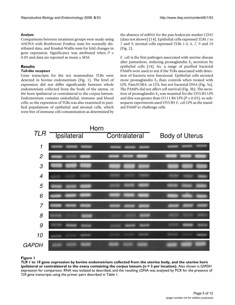
ResultsToll-like receptorsGene transcripts for the ten mammalian TLRs weredetected in bovine endometrium (Fig. 1). The level ofexpression did not differ significantly between wholeendometrium collected from the body of the uterus, orthe horn ipsilateral or contralateral to the corpus luteum.Endometrium contains endothelial, immune and bloodcells, so the expression of TLRs was also examined in puri-fied populations of epithelial and stromal cells, whichwere free of immune cell contamination as determined by
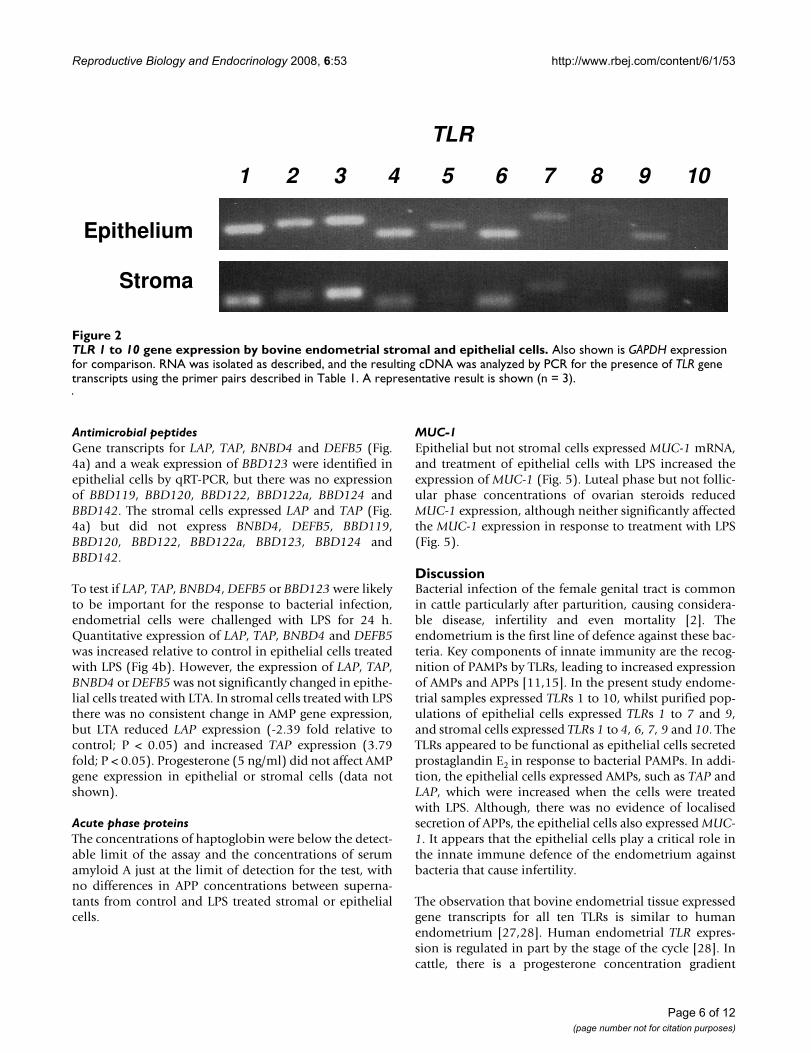
the absence of mRNA for the pan-leukocyte marker CD45(data not shown) [14]. Epithelial cells expressed TLRs 1 to7 and 9; stromal cells expressed TLRs 1-4, 6, 7, 9 and 10(Fig. 2).
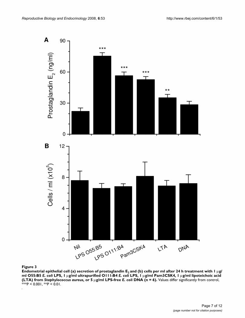
E. coli is the first pathogen associated with uterine diseaseafter parturition, inducing prostaglandin E2 secretion byepithelial cells [14]. So, a range of purified bacterialPAMPs were used to test if the TLRs associated with detec-tion of bacteria were functional. Epithelial cells secretedmore prostaglandin E2 than controls when treated withLPS, Pam3CSK4, or LTA, but not bacterial DNA (Fig. 3a).The PAMPs did not affect cell survival (Fig. 3b). The secre-tion of prostaglandin E2 was maximal for the O55:B5 LPSand this was greater than O111:B4 LPS (P < 0.05), so sub-sequent experiments used O55:B5 E. coli LPS as the stand-ard PAMP to challenge cells.
TLR 1 to 10 gene expression by bovine endometrium collected from the uterine body, and the uterine horn ipsilateral or con-tralateral to the ovary containing the corpus luteum (n = 3 per location)Figure 1TLR 1 to 10 gene expression by bovine endometrium collected from the uterine body, and the uterine horn ipsilateral or contralateral to the ovary containing the corpus luteum (n = 3 per location). Also shown is GAPDH expression for comparison. RNA was isolated as described, and the resulting cDNA was analyzed by PCR for the presence of TLR gene transcripts using the primer pairs described in Table 1.
Horn Ipsilateral Contralateral Body of Uterus TLR
1
2
3
4
5
6
7
8
9
10
GAPDH
Page 5 of 12(page number not for citation purposes)
Reproductive Biology and Endocrinology 2008, 6:53 http://www.rbej.com/content/6/1/53
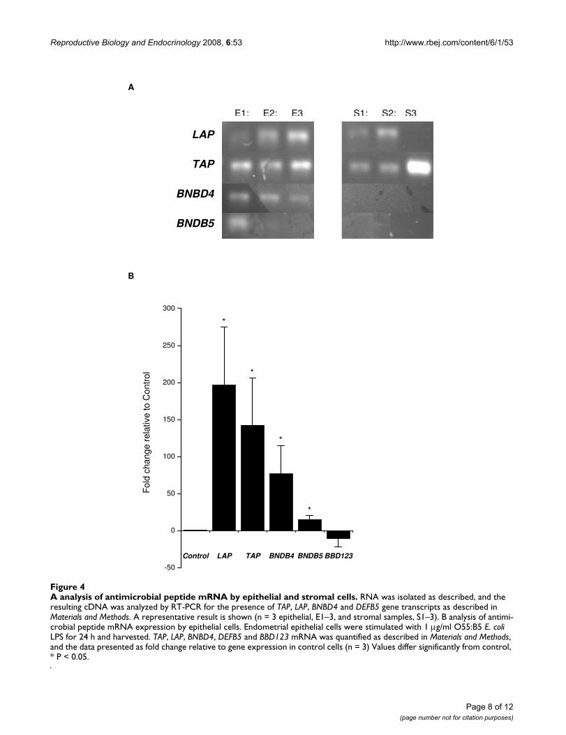
Antimicrobial peptidesGene transcripts for LAP, TAP, BNBD4 and DEFB5 (Fig.4a) and a weak expression of BBD123 were identified inepithelial cells by qRT-PCR, but there was no expressionof BBD119, BBD120, BBD122, BBD122a, BBD124 andBBD142. The stromal cells expressed LAP and TAP (Fig.4a) but did not express BNBD4, DEFB5, BBD119,BBD120, BBD122, BBD122a, BBD123, BBD124 andBBD142.
To test if LAP, TAP, BNBD4, DEFB5 or BBD123 were likelyto be important for the response to bacterial infection,endometrial cells were challenged with LPS for 24 h.Quantitative expression of LAP, TAP, BNBD4 and DEFB5was increased relative to control in epithelial cells treatedwith LPS (Fig 4b). However, the expression of LAP, TAP,BNBD4 or DEFB5 was not significantly changed in epithe-lial cells treated with LTA. In stromal cells treated with LPSthere was no consistent change in AMP gene expression,but LTA reduced LAP expression (-2.39 fold relative tocontrol; P < 0.05) and increased TAP expression (3.79fold; P < 0.05). Progesterone (5 ng/ml) did not affect AMPgene expression in epithelial or stromal cells (data notshown).
Acute phase proteinsThe concentrations of haptoglobin were below the detect-able limit of the assay and the concentrations of serumamyloid A just at the limit of detection for the test, withno differences in APP concentrations between superna-tants from control and LPS treated stromal or epithelialcells.
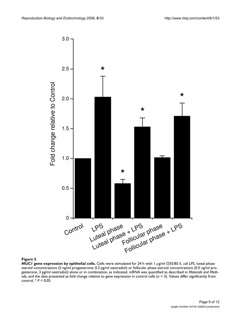
MUC-1Epithelial but not stromal cells expressed MUC-1 mRNA,and treatment of epithelial cells with LPS increased theexpression of MUC-1 (Fig. 5). Luteal phase but not follic-ular phase concentrations of ovarian steroids reducedMUC-1 expression, although neither significantly affectedthe MUC-1 expression in response to treatment with LPS(Fig. 5).
DiscussionBacterial infection of the female genital tract is commonin cattle particularly after parturition, causing considera-ble disease, infertility and even mortality [2]. Theendometrium is the first line of defence against these bac-teria. Key components of innate immunity are the recog-nition of PAMPs by TLRs, leading to increased expressionof AMPs and APPs [11,15]. In the present study endome-trial samples expressed TLRs 1 to 10, whilst purified pop-ulations of epithelial cells expressed TLRs 1 to 7 and 9,and stromal cells expressed TLRs 1 to 4, 6, 7, 9 and 10. TheTLRs appeared to be functional as epithelial cells secretedprostaglandin E2 in response to bacterial PAMPs. In addi-tion, the epithelial cells expressed AMPs, such as TAP andLAP, which were increased when the cells were treatedwith LPS. Although, there was no evidence of localisedsecretion of APPs, the epithelial cells also expressed MUC-1. It appears that the epithelial cells play a critical role inthe innate immune defence of the endometrium againstbacteria that cause infertility.
The observation that bovine endometrial tissue expressedgene transcripts for all ten TLRs is similar to humanendometrium [27,28]. Human endometrial TLR expres-sion is regulated in part by the stage of the cycle [28]. Incattle, there is a progesterone concentration gradient
TLR 1 to 10 gene expression by bovine endometrial stromal and epithelial cellsFigure 2TLR 1 to 10 gene expression by bovine endometrial stromal and epithelial cells. Also shown is GAPDH expression for comparison. RNA was isolated as described, and the resulting cDNA was analyzed by PCR for the presence of TLR gene transcripts using the primer pairs described in Table 1. A representative result is shown (n = 3).
TLR
1 2 3 4 5 6 7 8 9 10
Epithelium
Stroma
Page 6 of 12(page number not for citation purposes)
Reproductive Biology and Endocrinology 2008, 6:53 http://www.rbej.com/content/6/1/53
Page 7 of 12(page number not for citation purposes)
Endometrial epithelial cell (a) secretion of prostaglandin E2 and (b) cells per ml after 24 h treatment with 1 μg/ml O55:B5 E. coli LPS, 1 μg/ml ultrapurified O111:B4 E. coli LPS, 1 μg/ml Pam3CSK4, 1 μg/ml lipoteichoic acid (LTA) from Staphylococcus aureus, or 5 μg/ml LPS-free E. coli DNA (n = 6)Figure 3Endometrial epithelial cell (a) secretion of prostaglandin E2 and (b) cells per ml after 24 h treatment with 1 μg/ml O55:B5 E. coli LPS, 1 μg/ml ultrapurified O111:B4 E. coli LPS, 1 μg/ml Pam3CSK4, 1 μg/ml lipoteichoic acid (LTA) from Staphylococcus aureus, or 5 μg/ml LPS-free E. coli DNA (n = 6). Values differ significantly from control, ***P < 0.001, **P < 0.01.
0
30
60
90
B
**
******
Pro
stag
land
in E
2 (ng
/ml)
***
A
Nil
LPS O55:B5
LPS O111:B4
Pam3CSK4 LTADNA
0
4
8
12
Cel
ls /
ml (
x105 )
Reproductive Biology and Endocrinology 2008, 6:53 http://www.rbej.com/content/6/1/53
Page 8 of 12(page number not for citation purposes)
A Analysis of antimicrobial peptide mRNA by epithelial and stromal cellsFigure 4A analysis of antimicrobial peptide mRNA by epithelial and stromal cells. RNA was isolated as described, and the resulting cDNA was analyzed by RT-PCR for the presence of TAP, LAP, BNBD4 and DEFB5 gene transcripts as described in Materials and Methods. A representative result is shown (n = 3 epithelial, E1–3, and stromal samples, S1–3). B analysis of antimi-crobial peptide mRNA expression by epithelial cells. Endometrial epithelial cells were stimulated with 1 μg/ml O55:B5 E. coli LPS for 24 h and harvested. TAP, LAP, BNBD4, DEFB5 and BBD123 mRNA was quantified as described in Materials and Methods, and the data presented as fold change relative to gene expression in control cells (n = 3) Values differ significantly from control, * P < 0.05.
A
B
Control LAP TAP BNDB4 BNDB5 BBD123
-50
0
50
100
150
200
250
300
*
*
*
Fol
d ch
ange
rel
ativ
e to
Con
trol
*
E1; E2; E3 S1; S2; S3
LAP
TAP
BNBD4
BNDB5
Reproductive Biology and Endocrinology 2008, 6:53 http://www.rbej.com/content/6/1/53
Page 9 of 12(page number not for citation purposes)
MUC1 gene expression by epithelial cellsFigure 5MUC1 gene expression by epithelial cells. Cells were stimulated for 24 h with 1 μg/ml O55:B5 E. coli LPS, luteal phase steroid concentrations (5 ng/ml progesterone; 0.3 pg/ml oestradiol) or follicular phase steroid concentrations (0.5 ng/ml pro-gesterone, 3 pg/ml oestradiol) alone or in combination, as indicated. mRNA was quantified as described in Materials and Meth-ods, and the data presented as fold change relative to gene expression in control cells (n = 3). Values differ significantly from control, * P < 0.05.
ControlLPS
Luteal phase
Luteal phase + LPS
Follicular phase
Follicular phase + LPS
0
0.5
1.0
1.5
2.0
2.5
3.0
*
**
*
F
old
chan
ge r
elat
ive
to C
ontr
ol
*
Reproductive Biology and Endocrinology 2008, 6:53 http://www.rbej.com/content/6/1/53
across the uterus with the highest concentration at the tipof the horn ipsilateral to the corpus luteum and the lowestin the contralateral uterine horn [29]. Progesterone con-centrations are important because progesterone sup-presses immune defences in the bovine endometrium [9].However, there were no obvious differences in gene tran-script expression for any of the TLRs between differentlocations in the uterus.
Endometrial tissue samples contain several cell typesincluding immune cells, so to test the concept that theendocrine cells have a role in immunity, we explored TLRexpression in purified populations of epithelial and stro-mal cells, which are free of immune cell contamination incattle [14]. The epithelial cell expression of TLRs 1 to 7and 9, and stromal cells expression of TLRs 1 to 4, 6, 7, 9and 10, is similar to humans, where expression of TLR 1to 9 has been reported in endometrial cell lines and in pri-mary uterine epithelial cell cultures [30,31]. The epithelialcell expression of TLRs 1, 2, 4, 6 and 9 is importantbecause they are required to detect bacterial PAMPs [11].Bacterial infection is the predominant cause of uterine dis-ease in cattle, and E. coli paves the way for other pathogensto cause damage to the endometrium, as well as disrupt-ing endocrine function [5].
Bacterial PAMPs include LPS, LTA and DNA, so we testedthe effects of these and the synthetic Pam3CSK4, to gainan insight into whether the cognate TLRs 1, 2, 4, 6 and 9are functional in the epithelial cells. We chose to evaluateprostaglandin E2 concentrations because it is a clearmarker of bacterial infection in the bovine endometrium,as well as having an important endocrine role for regulat-ing ovarian cycles and implantation [14,32]. E. coli or LPSswitches epithelial cell secretion from prostaglandin F2α topredominantly prostaglandin E2, mediated by the TLR4,MD2, CD14 signalling complex expressed by epithelialcells [14]. The predominant secretion of prostaglandin E2rather than prostaglandin F2α is dependent on bacteria orPAMPs stimulating changes in the eicosaniod synthesispathway, which includes phospholipases and prostaglan-din synthases (Herath, unpublished data). This switch infunction is important as prostaglandin F2α initiates luteol-ysis whereas prostaglandin E2 is luteotrophic in ruminants[33]. Prostaglandin E2 is also an important component ofthe immune response to bacteria and regulates or sup-presses inflammation in many tissues [34]. In the presentstudy, LPS, LTA and Pam3CSK4 increased prostaglandinE2 secretion and none of the ligands affected epithelial cellsurvival, extending earlier observations that LPS wasdetected by human and bovine endometrial cells [14,35].This stimulation of epithelial cell secretion of prostaglan-din E2 by several bacterial PAMPs may explain the associ-ation between uterine disease and extended luteal phases[36,37]. The secretion of prostaglandin E2 was greater for
the LPS from O55:B5 than O111:B4 E. coli, which mayreflect the greater purity of the latter preparation. On theother hand, the O55:B5 LPS provided a potent PAMP toevaluate AMP expression in subsequent experiments.
The AMPs are an important arm of the innate immunedefence against bacteria and TLRs mediate their inductionin many mamalian tissues [15,38]. In the present study,bovine endometrial epithelial cells expressed severalAMPs including LAP, TAP, BNBD4 and DEFB5, whilststromal cells expressed mainly LAP and TAP. The predom-inant expression of TAP and LAP by the epithelial cells issimilar to the bovine mammary gland [39]. In humans, β-defensins have also been detected in the endometrium(HBD1, 2, 3 and 4), although there are some differencesbetween studies [40,41]. In the present study, the epithe-lial cell AMP expression appeared to be of functionalimportance as the gene expression was increased in epi-thelial cells treated with LPS. The epithelial cells were con-sistently more responsive than the stromal cells, and LPSstimulated a greater response than LTA. Similarly inhumans, endometrial HBD2 mRNA expression isincreased after 24 h treatment with LPS, although unlikestudies with human cells we did not find that progester-one regulated AMP expression in endometrial cells[40,41]. However, the substantial induction of epithelialLAP and TAP by LPS treatment probably reflects theirdefensive role against E. coli in cattle, and agrees with theconcept that TLRs mediate induction of AMPs in responseto PAMPs [15].
The concentrations of APPs, including haptoglobin andserum amyloid A, are substantially increased in theperipheral plasma of cow with bacterial infection of theendometrium [5]. Similar to the AMPs, the APPs such ashaptoglobin and serum amyloid A provide non-specificprotection against microbes [18]. Although usually pro-duced by the liver, there is evidence of localised APPexpression in the genital tract of rodents, regulated by thesex steroids [19,20]. However, in the present study theconcentrations of haptoglobin and serum amyloid A pro-teins were barely detectable in epithelial or stromal cells,and not affected by treatment of cells with LPS. In vivoperipheral plasma concentrations are at least 25 timesthose of the culture supernatants [42]. So, it appearsunlikely that localised secretion of APPs plays a major rolein endometrial immunity in cattle.
MUC-1 is a glycosylated transmembrane protein com-monly expressed by the epithelial cells of mucosal sur-faces, including the reproductive tract, gut, testis andmammary gland [21,43]. MUC-1 is expressed in thehuman and ovine endometrium and has important rolesin endometrial receptivity for embryo implantation, andis a marker of endometrial health and fertility in humans
Page 10 of 12(page number not for citation purposes)
Reproductive Biology and Endocrinology 2008, 6:53 http://www.rbej.com/content/6/1/53
[22,23]. However, like AMPs and APPs, MUC-1 also has arole in protecting mucosal surfaces against bacteria. MUC-1 sterically inhibits microbial access to the cell surface andregulates inflammation [21,43]. In the present study,MUC-1 expression was detected in the bovine epithelialcells and LPS increased the mRNA expression. In thehuman endometrium, MUC-1 is highly regulated by theovarian sex steroids [44]. Although luteal phase concen-trations of ovarian steroids decreased MUC-1 expression,follicular phase concentrations had no effect in thepresent study. Further, neither steroid combinationaffected the increase of MUC-1 expression stimulated byLPS. This is in contrast to the reduction in LPS-stimulatedprostaglandin E2 secretion associated with progesteroneor oestradiol [14].
ConclusionIn conclusion, the endometrium is an important first lineof defence against invading bacteria that cause disease incattle and endometrial samples expressed TLRs 1 to 10.The present study explored the concept that the endocrinecells play an important mechanistic role in the defence ofthe endometrium against bacteria. Purified populationsof endometrial epithelial cells expressed TLRs 1 to 7 and9, and stromal cells expressed TLRs 1 to 4, 6, 7, 9 and 10.The TLRs appear to be functional as epithelial cellssecreted prostaglandin E2 in response to bacterial PAMPs.In addition, the epithelial cells expressed AMPs, such asTAP and LAP, and MUC-1, which were upregulated whenthe cells were treated with LPS. Thus, innate immunedefence systems in the epithelial cells of the bovineendometrium are likely to play a key role in the preven-tion of endometrial infection with bacteria and mediatechanges in endocrine function.
Competing interestsIMS holds research funding under a Department for Envi-ronment Food and Rural Affairs (DEFRA) LINK awardfrom Pfizer Animal Health and the Biotechnology andBiological Sciences Research Council (BBSRC; F005121).Darren Davies was supported by a BBSRC CASE Student-ship partly funded by Pfizer Animal Health (BBS/S/N/2005/12367). The remaining authors declare that theyhave no competing interests.
Authors' contributionsIMS was awarded the grants to fund the work, devisedexperiments, collated the data and wrote the manuscript.DD performed cell culture work and the molecular biol-ogy for TLR analysis. KGM and COF performed the AMPexpression work and contributed to the manuscript. SHperformed the cell culture PAMP treatments and prostag-landin measurements. PDE performed the acute phaseprotein measurements. DG, JOW, and RSC completed the
MUC-1 analysis and contributed to the manuscript. Allauthors read and approved the final manuscript.
AcknowledgementsMartin Sheldon is a BBSRC Research Development Fellow (Grant No. BB/D02028X/1) and Darren Davies was a BBSRC CASE Student (BBS/S/N/2005/12367). The work was partly funded under a DEFRA LINK award by Pfizer Animal Health and BBSRC (F005121).
References1. Sheldon IM, Lewis GS, LeBlanc SJ, Gilbert RO: Defining postpar-
tum uterine disease in cattle. Theriogenology 2006, 65:1516-1530.2. Sheldon IM, Williams EJ, Miller ANA, Nash DM, Herath S: Uterine
diseases in cattle after parturition. Veterinary Journal 2008,176:115-121.
3. Sheldon IM, Noakes DE, Rycroft AN, Pfeiffer DU, Dobson H: Influ-ence of uterine bacterial contamination after parturition onovarian dominant follicle selection and follicle growth andfunction in cattle. Reproduction 2002, 123:837-845.
4. Donofrio G, Herath S, Sartori C, Cavirani S, Flammini CF, Sheldon IM:Bovine herpesvirus 4 (BoHV-4) is tropic for bovine endome-trial cells and modulates endocrine function. Reproduction2007, 134:183-197.
5. Williams EJ, Fischer DP, Noakes DE, England GC, Rycroft A, DobsonH, Sheldon IM: The relationship between uterine pathogengrowth density and ovarian function in the postpartum dairycow. Theriogenology 2007, 68:549-559.
6. Donofrio G, Ravaneti L, Cavirani S, Herath S, Capocefalo A, SheldonIM: Bacterial infection of endometrial stromal cells influ-ences bovine herpesvirus 4 immediate early gene activation:a new insight into bacterial and viral interaction for uterinedisease. Reproduction 2008, 136:361-366.
7. Wira CR, Grant-Tschudy KS, Crane-Godreau MA: Epithelial cellsin the female reproductive tract: a central role as sentinelsof immune protection. Am J Reprod Immunol 2005, 53:65-76.
8. King AE, Critchley HO, Kelly RW: Innate immune defences inthe human endometrium. Reproductive Biology and Endocrinology2003, 1:116.
9. Lewis GS: Steroidal regulation of uterine resistance to bacte-rial infection in livestock. Reproductive Biology and Endocrinology2003, 1:117.
10. Herath S, Dobson H, Bryant CE, Sheldon IM: Use of the cow as alarge animal model of uterine infection and immunity. Journalof Reproductive Immunology 2006, 69:13-22.
11. Akira S, Uematsu S, Takeuchi O: Pathogen recognition andinnate immunity. Cell 2006, 124:783-801.
12. Hirata T, Osuga Y, Hamasaki K, Hirota Y, Nose E, Morimoto C,Harada M, Takemura Y, Koga K, Yoshino O, Tajima T, Hasegawa A,Yano T, Taketani Y: Expression of toll-like receptors 2, 3, 4, and9 genes in the human endometrium during the menstrualcycle. Journal of Reproductive Immunology 2007, 74:53-60.
13. Fazeli A, Bruce C, Anumba DO: Characterization of Toll-likereceptors in the female reproductive tract in humans.Human Reproduction 2005, 20:1372-1378.
14. Herath S, Fischer DP, Werling D, Williams EJ, Lilly ST, Dobson H,Bryant CE, Sheldon IM: Expression and function of Toll-likereceptor 4 in the endometrial cells of the uterus. Endocrinology2006, 147:562-570.
15. Selsted ME, Ouellette AJ: Mammalian defensins in the antimi-crobial immune response. Nature Immunology 2005, 6:551-557.
16. Diamond G, Zasloff M, Eck H, Brasseur M, Maloy WL, Bevins CL:Tracheal antimicrobial peptide, a cysteine-rich peptide frommammalian tracheal mucosa: peptide isolation and cloningof a cDNA. Proc Natl Acad Sci U S A 1991, 88:3952-3956.
17. Cormican P, Meade KG, Cahalane S, Narciandi F, Chapwanya A, LloydAT, O'Farrelly C: Evolution, expression and effectiveness in acluster of novel bovine beta-defensins. Immunogenetics 2008,60:147-156.
18. Baumann H, Gauldie J: The acute phase response. ImmunologyToday 1994, 15:74-80.
19. Son DS, Arai KY, Roby KF, Terranova PF: Tumor necrosis factoralpha (TNF) increases granulosa cell proliferation: depend-
Page 11 of 12(page number not for citation purposes)
http://www.ncbi.nlm.nih.gov/entrez/query.fcgi?cmd=Retrieve&db=PubMed&dopt=Abstract&list_uids=2023943
http://www.ncbi.nlm.nih.gov/entrez/query.fcgi?cmd=Retrieve&db=PubMed&dopt=Abstract&list_uids=2023943
http://www.ncbi.nlm.nih.gov/entrez/query.fcgi?cmd=Retrieve&db=PubMed&dopt=Abstract&list_uids=2023943
Reproductive Biology and Endocrinology 2008, 6:53 http://www.rbej.com/content/6/1/53
Publish with BioMed Central and every scientist can read your work free of charge
"BioMed Central will be the most significant development for disseminating the results of biomedical research in our lifetime."
Sir Paul Nurse, Cancer Research UK
Your research papers will be:
available free of charge to the entire biomedical community
peer reviewed and published immediately upon acceptance
cited in PubMed and archived on PubMed Central
yours — you keep the copyright
Submit your manuscript here:http://www.biomedcentral.com/info/publishing_adv.asp
BioMedcentral
ence on c-Jun and TNF receptor type 1. Endocrinology 2004,145:1218-1226.
20. Lebreton JP, Hiron M, Biou D, Daveau M: Regulation of alpha 1-acid glycoprotein plasma concentration by sex steroids andadrenal-cortical hormones during experimental inflamma-tion in the rat. Inflammation 1988, 12:413-424.
21. Brayman M, Thathiah A, Carson DD: MUC1: a multifunctionalcell surface component of reproductive tissue epithelia.Reproductive Biology and Endocrinology 2004, 2:1-9.
22. Johnson GA, Bazer FW, Jaeger LA, Ka H, Garlow JE, Pfarrer C, Spen-cer TE, Burghardt RC: MUC-1, integrin, and osteopontinexpression during the implantation cascade in sheep. Biologyof reproduction 2001, 65:820-828.
23. Horne AW, Lalani EN, Margara RA, Ryder TA, Mobberley MA, WhiteJO: The expression pattern of MUC1 glycoforms and otherbiomarkers of endometrial receptivity in fertile and infertilewomen. Mol Reprod Dev 2005, 72:216-229.
24. Ireland JJ, Murphee RL, Coulson PB: Accuracy of predictingstages of bovine estrous cycle by gross appearance of thecorpus luteum. Journal of Dairy Science 1980, 63:155-160.
25. Rozen S, Skaletsky H: Primer3 on the WWW for general usersand for biologist programmers. In Bioinformatics Methods and Pro-tocols: Methods in Molecular Biology Edited by: Krawetz, Misener, S.Totowa, NJ: Humana Press; 2000:365-386.
26. Eckersall PD, Young FJ, Nolan AM, Knight CH, McComb C, Water-ston MM, Hogarth CJ, Scott EM, Fitzpatrick JL: Acute phase pro-teins in bovine milk in an experimental model ofStaphylococcus aureus subclinical mastitis. Journal of Dairy Sci-ence 2006, 89:1488-1501.
27. Young SL, Lyddon TD, Jorgenson RL, Misfeldt ML: Expression ofToll-like Receptors in human endometrial epithelial cells andcell lines. American Journal of Reproductive Immunology 2004,52:67-73.
28. Aflatoonian R, Tuckerman E, Elliott SL, Bruce C, Aflatoonian A, Li TC,Fazeli A: Menstrual cycle-dependent changes of Toll-likereceptors in endometrium. Human Reproduction 2007,22:586-593.
29. Weems CW, Lee CN, Weems YS, Vincent DL: Distribution of pro-gesterone to the uterus and associated vasculature of cattle.Endocrinol Jpn 1988, 35(4):625-630.
30. Schaefer TM, Desouza K, Fahey JV, Beagley KW, Wira CR: Toll-likereceptor (TLR) expression and TLR-mediated cytokine/chemokine production by human uterine epithelial cells.Immunology 2004, 112:428-436.
31. Schaefer TM, Fahey JV, Wright JA, Wira CR: Innate Immunity inthe Human Female Reproductive Tract: Antiviral Responseof Uterine Epithelial Cells to the TLR3 Agonist Poly(I:C). JImmunol 2005, 174(2):992-1002.
32. Miller AN, Williams EJ, Sibley K, Herath S, Lane EA, Fishwick J, NashDM, Rycroft AN, Dobson H, Bryant CE, Sheldon IM: The effects ofArcanobacterium pyogenes on endometrial function invitro, and on uterine and ovarian function in vivo. Theriogenol-ogy 2007, 68:972-980.
33. Weems CW, Vincent DL, Weems YS: Roles of prostaglandins(PG) F2 alpha, E1, E2, adenosine, oestradiol-17 beta, his-tone-H2A and progesterone of conceptus, uterine or ovarianorigin during early and mid pregnancy in the ewe. Reproduc-tion Fertility and Development 1992, 4:289-295.
34. Narumiya S: Prostanoids in immunity: roles revealed by micedeficient in their receptors. Life Sciences 2003, 74:391-395.
35. Hirata T, Osuga Y, Hirota Y, Koga K, Yoshino O, Harada M, Morim-oto C, Yano T, Nishii O, Tsutsumi O, Taketani Y: Evidence for thepresence of toll-like receptor 4 system in the humanendometrium. J Clin Endocrinol Metab 2005, 90:548-556.
36. Poyser NL: The control of prostaglandin production by theendometrium in relation to luteolysis and menstruation.Prostaglandins Leukot Essent Fatty Acids 1995, 53:147-195.
37. Opsomer G, Grohn YT, Hertl J, Coryn M, Deluyker H, de Kruif A:Risk factors for post partum ovarian dysfunction in high pro-ducing dairy cows in Belgium: a field study. Theriogenology2000, 53:841-857.
38. Ganz T: Defensins and host defense. Science 1999, 286:420-421.39. Roosen S, Exner K, Paul S, Schroder JM, Kalm E, Looft C: Bovine
beta-defensins: identification and characterization of novelbovine beta-defensin genes and their expression in mam-mary gland tissue. Mammalian Genome 2004, 15:834-842.
40. King AE, Fleming DC, Critchley HO, Kelly RW: Regulation of nat-ural antibiotic expression by inflammatory mediators andmimics of infection in human endometrial epithelial cells.Molecular Human Reproduction 2002, 8:341-349.
41. King AE, Fleming DC, Critchley HO, Kelly RW: Differentialexpression of the natural antimicrobials, beta-defensins 3and 4, in human endometrium. Journal of Reproductive Immunol-ogy 2003, 59:1-16.
42. Sheldon IM, Noakes DE, Rycroft A, Dobson H: Acute phase pro-tein response to postpartum uterine bacterial contamina-tion in cattle. Veterinary Record 2001, 148:172-175.
43. Beatty PL, Plevy SE, Sepulveda AR, Finn OJ: Cutting edge: trans-genic expression of human MUC1 in IL-10-/- mice acceler-ates inflammatory bowel disease and progression to coloncancer. J Immunol 2007, 179(2):735-739.
44. Horne AW, Lalani EN, Margara RA, White JO: The effects of sexsteroid hormones and interleukin-1-beta on MUC1 expres-sion in endometrial epithelial cell lines. Reproduction 2006,131:733-742.
Page 12 of 12(page number not for citation purposes)
http://www.ncbi.nlm.nih.gov/entrez/query.fcgi?cmd=Retrieve&db=PubMed&dopt=Abstract&list_uids=3198247
http://www.ncbi.nlm.nih.gov/entrez/query.fcgi?cmd=Retrieve&db=PubMed&dopt=Abstract&list_uids=3198247
http://www.ncbi.nlm.nih.gov/entrez/query.fcgi?cmd=Retrieve&db=PubMed&dopt=Abstract&list_uids=3198247
http://www.ncbi.nlm.nih.gov/entrez/query.fcgi?cmd=Retrieve&db=PubMed&dopt=Abstract&list_uids=7372895
http://www.ncbi.nlm.nih.gov/entrez/query.fcgi?cmd=Retrieve&db=PubMed&dopt=Abstract&list_uids=7372895
http://www.ncbi.nlm.nih.gov/entrez/query.fcgi?cmd=Retrieve&db=PubMed&dopt=Abstract&list_uids=7372895
http://www.ncbi.nlm.nih.gov/entrez/query.fcgi?cmd=Retrieve&db=PubMed&dopt=Abstract&list_uids=3215149
http://www.ncbi.nlm.nih.gov/entrez/query.fcgi?cmd=Retrieve&db=PubMed&dopt=Abstract&list_uids=3215149
http://www.ncbi.nlm.nih.gov/entrez/query.fcgi?cmd=Retrieve&db=PubMed&dopt=Abstract&list_uids=7480081
Related Documents


![Bovine placentome development during early pregnancy · 2017-10-19 · embryos and fetuses were removed for CRL evaluation [21] and the mesometrial side of respective endometrium](https://static.cupdf.com/doc/110x72/5e41eb8fe85f120fd757723a/bovine-placentome-development-during-early-pregnancy-2017-10-19-embryos-and-fetuses.jpg)
