RESEARCH ARTICLE Toll-Like Receptor 4 Promotes Autonomic Dysfunction, Inflammation and Microglia Activation in the Hypothalamic Paraventricular Nucleus: Role of Endoplasmic Reticulum Stress Gustavo S. Masson 1,2☯ , Anand R. Nair 1☯ , Rahul B. Dange 1 , Pedro Paulo Silva-Soares 3 , Lisete C. Michelini 2 , Joseph Francis 1 * 1 Comparative Biomedical Sciences, School of Veterinary Medicine, Louisiana State University, Baton Rouge, Louisiana, United States of America, 2 Department of Physiology and Biophysics, Biomedical Sciences Institute, University of Sao Paulo, Sao Paulo, Brazil, 3 Department of Pharmacology, Fluminense Federal University, Rio De Janeiro, Brazil ☯ These authors contributed equally to this work. * [email protected] Abstract Background & Purpose Toll-like receptor 4 (TLR4) signaling induces tissue pro-inflammatory cytokine release and endoplasmic reticulum (ER) stress. We examined the role of TLR4 in autonomic dysfunction and the contribution of ER stress. Experimental approach Our study included animals divided in 6 experimental groups: rats treated with saline (i.v., 0.9%), LPS (i.v., 10mg/kg), VIPER (i.v., 0.1 mg/kg), or 4-PBA (i.p., 10 mg/kg). Two other groups were pretreated either with VIPER (TLR4 viral inhibitory peptide) LPS + VIPER (i.v., 0.1 mg/kg) or 4-Phenyl butyric acid (4-PBA) LPS + PBA (i.p., 10 mg/kg). Arterial pressure (AP) and heart rate (HR) were measured in conscious Sprague-Dawley rats. AP, HR vari- ability, as well as baroreflex sensitivity (BrS), was determined after LPS or saline treatment for 2 hours. Immunofluorescence staining for NeuN, Ib1a, TLR4 and GRP78 in the hypotha- lamic paraventricular nucleus (PVN) was performed. TNF-α, TLR4 and GRP78 protein ex- pression in the PVN were evaluated by western blot. Plasma norepinephrine levels were determined by ELISA. Key Results Acute LPS treatment increased HR and plasma norepinephrine concentration. It also de- creased HR variability and high frequency (HF) components of HR variability, as well BrS. Acute LPS treatment increased TLR4 and TNF-α protein expression in the PVN. These PLOS ONE | DOI:10.1371/journal.pone.0122850 March 26, 2015 1 / 15 OPEN ACCESS Citation: Masson GS, Nair AR, Dange RB, Silva- Soares PP, Michelini LC, Francis J (2015) Toll-Like Receptor 4 Promotes Autonomic Dysfunction, Inflammation and Microglia Activation in the Hypothalamic Paraventricular Nucleus: Role of Endoplasmic Reticulum Stress. PLoS ONE 10(3): e0122850. doi:10.1371/journal.pone.0122850 Academic Editor: Tony Wang, SRI International, UNITED STATES Received: October 21, 2014 Accepted: February 23, 2015 Published: March 26, 2015 Copyright: © 2015 Masson et al. This is an open access article distributed under the terms of the Creative Commons Attribution License, which permits unrestricted use, distribution, and reproduction in any medium, provided the original author and source are credited. Data Availability Statement: All relevant data are within the paper and its Supporting Information. Funding: The author(s) received no specific funding for this work Competing Interests: The authors have declared that no competing interests exist.

Welcome message from author
This document is posted to help you gain knowledge. Please leave a comment to let me know what you think about it! Share it to your friends and learn new things together.
Transcript

RESEARCH ARTICLE
Toll-Like Receptor 4 Promotes AutonomicDysfunction, Inflammation and MicrogliaActivation in the HypothalamicParaventricular Nucleus: Role of EndoplasmicReticulum StressGustavo S. Masson1,2☯, Anand R. Nair1☯, Rahul B. Dange1, Pedro Paulo Silva-Soares3,Lisete C. Michelini2, Joseph Francis1*
1 Comparative Biomedical Sciences, School of Veterinary Medicine, Louisiana State University, BatonRouge, Louisiana, United States of America, 2 Department of Physiology and Biophysics, BiomedicalSciences Institute, University of Sao Paulo, Sao Paulo, Brazil, 3 Department of Pharmacology, FluminenseFederal University, Rio De Janeiro, Brazil
☯ These authors contributed equally to this work.* [email protected]
Abstract
Background & Purpose
Toll-like receptor 4 (TLR4) signaling induces tissue pro-inflammatory cytokine release and
endoplasmic reticulum (ER) stress. We examined the role of TLR4 in autonomic dysfunction
and the contribution of ER stress.
Experimental approach
Our study included animals divided in 6 experimental groups: rats treated with saline (i.v.,
0.9%), LPS (i.v., 10mg/kg), VIPER (i.v., 0.1 mg/kg), or 4-PBA (i.p., 10 mg/kg). Two other
groups were pretreated either with VIPER (TLR4 viral inhibitory peptide) LPS + VIPER (i.v.,
0.1 mg/kg) or 4-Phenyl butyric acid (4-PBA) LPS + PBA (i.p., 10 mg/kg). Arterial pressure
(AP) and heart rate (HR) were measured in conscious Sprague-Dawley rats. AP, HR vari-
ability, as well as baroreflex sensitivity (BrS), was determined after LPS or saline treatment
for 2 hours. Immunofluorescence staining for NeuN, Ib1a, TLR4 and GRP78 in the hypotha-
lamic paraventricular nucleus (PVN) was performed. TNF-α, TLR4 and GRP78 protein ex-
pression in the PVN were evaluated by western blot. Plasma norepinephrine levels were
determined by ELISA.
Key Results
Acute LPS treatment increased HR and plasma norepinephrine concentration. It also de-
creased HR variability and high frequency (HF) components of HR variability, as well BrS.
Acute LPS treatment increased TLR4 and TNF-α protein expression in the PVN. These
PLOSONE | DOI:10.1371/journal.pone.0122850 March 26, 2015 1 / 15
OPEN ACCESS
Citation: Masson GS, Nair AR, Dange RB, Silva-Soares PP, Michelini LC, Francis J (2015) Toll-LikeReceptor 4 Promotes Autonomic Dysfunction,Inflammation and Microglia Activation in theHypothalamic Paraventricular Nucleus: Role ofEndoplasmic Reticulum Stress. PLoS ONE 10(3):e0122850. doi:10.1371/journal.pone.0122850
Academic Editor: Tony Wang, SRI International,UNITED STATES
Received: October 21, 2014
Accepted: February 23, 2015
Published: March 26, 2015
Copyright: © 2015 Masson et al. This is an openaccess article distributed under the terms of theCreative Commons Attribution License, which permitsunrestricted use, distribution, and reproduction in anymedium, provided the original author and source arecredited.
Data Availability Statement: All relevant data arewithin the paper and its Supporting Information.
Funding: The author(s) received no specific fundingfor this work
Competing Interests: The authors have declaredthat no competing interests exist.

hemodynamic and molecular effects were partially abrogated with TLR4 blocker or ER
stress inhibitor pretreatment. In addition, immunofluorescence study showed that TLR4 is
co-localized with GRP78in the neurons. Further inhibition of TLR4 or ER stress was able to
attenuate the LPS-induced microglia activation.
Conclusions & Implications
TLR4 signaling promotes autonomic dysfunction, inflammation and microglia activation,
through neuronal ER stress, in the PVN.
IntroductionAutonomic dysfunction is implicated in several cardiovascular diseases, such as heart failure[1] and hypertension [2], with prognostic implications. It is characterized by reduced barore-flex sensitivity (BrS), heart rate (HR) variability and cardiac vagal output, as well as increasedsympathetic activity. Attenuating autonomic dysfunction is a common theme of many phar-macological agents in treating cardiovascular diseases, using renin-angiotensin system andbeta-receptor blockade [3].
Brain inflammation has been identified as a causative pathophysiologic marker in autonom-ic dysfunction. Several studies from our lab and other groups reported that pro-inflammatorycytokine levels, as tumor necrosis factor-α (TNF-α) and interleukin-1β, are increased in auto-nomic control areas, such as the paraventricular nucleus (PVN) of the hypothalamus, in exper-imental models of hypertension and heart failure [4–8]. Acute increase of arterial pressure(AP) and renal sympathetic activity was also reported after pro-inflammatory cytokine injec-tions into the PVN in anesthetized rats [9]. In addition, blockade of pro-inflammatory cyto-kines or nuclear factor-kappaB (NF-κB), within the PVN, prevented the increase of mean APand cardiac dysfunction in Angiotensin II induced-hypertension and ischemic cardiomyopa-thy [4–7]. These findings indicate a major role for inflammatory molecules in cardiovasculardiseases.
Microglial activated cells have been suggested as a major cellular source of pro-inflammato-ry cytokines in experimental models of hypertension and heart failure [10, 11]. Chronic block-ade of microglial activation decreased AP and prevented cardiac hypertrophy, as well asreduced pro-inflammatory cytokine expression in the PVN in Angiotensin II induced-hyper-tension [10]. From a molecular view, role of Toll-like receptor 4 (TLR4) in microglial activationwas proposed in several stressor conditions, for instance in ischemia-reperfusion injury [12],chronic alcohol consumption [13] and sepsis [14]. Very few studies have examined the role ofTLR4 in cardiovascular disorders. Recently, it was demonstrated that chronic brain TLR4blockade prevented cardiac remodeling in heart failure [15] and hypertensive rats [16].
In addition to tissue inflammation, persistent endoplasmic reticulum (ER) stress has recent-ly been suggested as a causative agent of hypertension, since the 78kDa glucose regulated pro-tein (GRP78) was over-expressed in the subfornical organ [17] and rostral ventrolateralmedulla during a hypertensive response [18]. These studies also identified increased expressionof ER stress markers in both autonomic control areas of Angiotensin II induced-hypertensiveanimals [17, 18]. The cross talk between TLR4 signaling and ER stress is demonstrated by thefinding that high fat feeding failed to induce ER stress in TLR4 knock-out mice liver, adiposeand skeletal muscle [19]. In addition, Yao et al [20] observed that activation of TLR4 by modi-fied low-density lipoprotein is essential to induce ER stress in macrophages. Considering these
TLR4 Induces Autonomic Dysfunction
PLOS ONE | DOI:10.1371/journal.pone.0122850 March 26, 2015 2 / 15

data, we hypothesized that TLR4 activation induces acute autonomic dysfunction through ERstress in the PVN. To test our hypothesis, we conducted acute in vivo studies in conscious rats,and analyzed cardiovascular and autonomic parameters in response to lipopolysaccharide(LPS), a specific ligand for TLR4. We also performed ex vivo experiments and used pharmaco-logical and molecular biology techniques to address the crosstalk between TLR4-induced in-flammation and ER stress in the PVN.
Materials and Methods
Animals and Experimental ProtocolAnimal studies were in accordance with the Guide for the Care and Use of Laboratory Animalsas adopted and promulgated by the U.S. National Institutes of Health. Sprague-Dawley rats (2–3 months-old, 250–300 g) were housed in a temperature-controlled room (25±1°C) and main-tained on a 12:12 hour light:dark cycle with free access to food and water. Rats were random-ized into groups consisting of: LPS (Sigma-Aldrich, L3129, LPS from E. coli 0127:B8), a specificligand for TLR4; LPS + TLR4 blocker (VIPER-TLR4 Viral Inhibitory Peptide); LPS + ER stressinhibitor (PBA—4-Phenyl butyric acid); and controls (saline, VIPER and 4-PBA groups). LPS-treated animals received a bolus injection (i.v., 10mg/kg). VIPER (Imgenex, USA) was injected(i.v., 0.1 mg/kg) two hours before LPS injection [21]. PBA (Sigma Aldrich, USA), an ER stressinhibitor, was administered 16–18 hours (i.p., 10 mg/kg) before LPS injection. Optimal PBAand VIPER doses were chosen after a preliminary study. Rats in the control group received asaline (i.v., 0.9%) bolus injection.
Cardiovascular parameters measurementsRats (n = 9–10 rats) per group were anesthetized with a ketamine (90 mg/kg) and xylazine(10 mg/kg) mixture (i.p.) for chronic implantation of catheters in the left femoral artery andvein. The adequacy of anesthesia was monitored by limb withdrawal response to toe pinching.Rats were treated with antibiotics and analgesics and allowed to recover for 3 days. AP and HRwere continuously recorded in conscious freely moving rats three days post catheter placement.The arterial catheter was connected to the recording system (transducer, Deltran1 II, UtahMedical Products, MidVale, UT, USA + Power Lab system, AD Instruments, Bella Vista NSW,Australia) and 60 min were allowed for the stabilization of cardiovascular parameters (2000 Hzsampling frequency, LabChart 2.0.1). HR was determined from AP pulse interval. After thebaseline period, LPS or saline were administrated and cardiovascular parameters were mea-sured for 120 minutes.
Autonomic parameters measurementsTime series of systolic arterial pressure (SAP) and pulse interval (PI) were generated from eachten minute period after LPS or saline acute treatment. Systolic AP (SAPVar) and HR variability(HRVar) were analyzed in frequency domain. PI is related to the R-R frequency that expressesHRVar. Following linear trend removal, power spectral density was obtained by the Fast Fou-rier Transformation. Spectral power for low- (LF: 0.20–0.75 Hz) and high (HF: 0.75–4.0 Hz)frequency bands was calculated by means of power spectrum density integration within eachfrequency bandwidth, using a customized routine (MATLAB 6.0, Mathworks). Coherence be-tween the PI and SAP variability signals was assessed by means of cross-spectral analysis [22].
At the end of 120 minutes of continuous recording, BrS was determined by loading / un-loading baroreceptors with graded iv. doses of phenylephrine and sodium nitroprusside (0.1–6.4 and 0.2–12.8 μg/kg, respectively, 100 μL bolus injection); subsequent injections were not
TLR4 Induces Autonomic Dysfunction
PLOS ONE | DOI:10.1371/journal.pone.0122850 March 26, 2015 3 / 15

made until the recorded parameters had returned to pre-injection levels. Mean AP and HR val-ues were measured before (control) and at the peak of each response. Baroreceptor reflex con-trol of HR, determined for each rat, was estimated by the sigmoidal logistic equation fitted todata points, as described previously [21]. The equation linking HR responses to pressurechanges was: HR = P1 + P2/[1 + eP3(BP–P4)], where P1 = lower HR plateau, P2 = HR range,P3 = the curvature coefficient and P4 = BP50 (the value of AP at half of the HR range). Theaverage gain of BrS was calculated as BrS = −(P2 ×P3)/4.Since PBA or VIPER alone did notalter cardiovascular or autonomic function we did not include this group in our molecularcharacterization.
Plasma Norepinephrine measurementsBefore rats were euthanized, blood was collected from the femoral artery in unanesthetizedconditions (n = 9–10 rats). Blood samples were centrifuged at 1800 rpm, 20 minutes, 4°C andthe plasma was collected. Norepinephrine (NE) concentration was evaluated through ELISAkit (KA1891; Abnova; USA), according to the manufacturers protocol.
ImmunoblottingWestern blot analysis was performed according to standard protocols as described previously[4, 7]. Since no changes were observed on the cardiovascular and autonomic parameters in therats treated with TLR4 or ER stress inhibitors alone, we did not apply molecular techniques inthese groups. Rat brains (n = 6) were quickly removed and frozen for later procedures. PVNpunches were performed in frozen brains sections and homogenized with RIPA lysis buffer.The protein concentration was measured using a bicinchioninic acid protein assay kit (Pierce,USA). Equal amounts of protein (15 μg) were separated by SDS-PAGE on 10% gels, transferredon to PVDF membrane (Immobilon-P, Millipore), and blocked with 1% BSA in TBS-T atroom temperature for 60 min. The membranes were incubated with anti-GRP 78 (ab21685;Abcam, 1:1000), anti-TNF-α (sc-8301; Santa Cruz Biotechnology, 1:1000), anti-TLR4(ab22048; Abcam, 1:500) and anti-actin (sc-1616; Santa Cruz Biotechnology, 1:1000) over-night, at 4 C. The membranes were washed and incubated with anti-rabbit (sc-2004; SantaCruz Biotechnology, 1:2000) or anti-mouse (sc-2314; Santa Cruz Biotechnology, 1:2000) sec-ondary antibodies for 1 hour at room temperature. Specific bands were detected using an en-hanced chemiluminescence kit (GE Life sciences, USA). The bands were visualized usingChemidoc XRS system and Quantity-One software (Bio-Rad) and analyzed using ImageJsoftware.
ImmunofluorescenceImmunofluorescence staining was performed as described previously [4, 5, 7, 21]. Briefly, se-quential hypothalamic coronal sections (30μm, -1.80 to -2.12 caudal to the Bregma) were cutwith a cryostat (Leica CM 1850; Nussloch, Germany) and collected in tissue culture wells with0.1M phosphate buffer (n = 3). Free-floating sections were pretreated with 1% H2O2-20% etha-nol for 30 min, washed with 0.1M phosphate buffer for 25 min and blocked with 2% normalgoat serum for 30 min. For the immunofluorescence reaction, the sections were incubated for48 hours at 4°C, with anti-GRP78 (ab21685, Abcam, 1:400), anti-TLR4 (ab22048 or ab13556,Abcam, 1:250), anti-NeuN (ab104224 Abcam, Ab, 1:1000) and anti-Ib1a (019–19741 Wako,1:800). Slices were washed with PB 0.1 M for 25 min and incubated for 1 hour at room temper-ature with Alexa 488 conjugated anti-rabbit (A11034; Invitrogen) or/and Alexa 594 conjugatedanti-mouse (A11005; Invitrogen). Four to six slices were placed in each slide and mountedwith a coverslip and Vectashield. Negative controls without the primary or secondary antibody
TLR4 Induces Autonomic Dysfunction
PLOS ONE | DOI:10.1371/journal.pone.0122850 March 26, 2015 4 / 15
were used to determine the background staining. Image analysis was performed with ImageProPlus software. The sections were examined to localize PVN in to 10X and 40X magnifica-tion with the same settings for all groups (exposure time-GRP78: 486.88 ms; TLR4: 336.02 ms;NeuN: 236.02 ms; Ib1a: 418.08 ms and sensitivity: ISO16000).
Statistical AnalysisAll data are presented as mean ± SEM and P-values less than 0.05 were considered statisticallysignificant. Statistical analyses were performed using Prism (GraphPad Software, Inc; version5.0). TWO-way ANOVA (repeated measurements), followed by Bonferroni’s post hoc, wasused to analyze cardiovascular and autonomic parameters after LPS or saline infusion (AP,HR, SAPVar and HRVar, as well theirs low and high frequency components). While, for theBrS, plasma NE and all immunoblotting data, One-way ANOVA, with Tukey’s post hoc test,was applied to perform the statistical analyses.
Results
TLR4 activation induced autonomic dysfunctionAcute treatment with LPS increased HR from 10 minutes to the end of the protocol, which wasinhibited by pretreatment with TLR4 blocker from 60 minutes of the protocol (Fig. 1A). LPSdid not significantly change mean AP (Fig. 1B). In addition, acute LPS treatment promptly de-creased HRVar and high frequency components of HRVar (cardiac vagal activity marker)from 10 minutes to the end of the experimental protocol. Both effects of LPS treatment wereblunted by TLR4 blockade from 60 minutes to the end of the experimental protocol (Fig. 1Cand 1D). Although LPS treatment transitorily increased SAPVar at minutes 40, 70 and 110, itincreased the low frequency component of SAPVar (peripheral sympathetic marker) at min-utes 80 and 110. TLR4 blockade inhibited the increase of SAPVar, as well as its low frequencycomponent, induced by LPS (Fig. 1E and 1F). Consistent with HRVar, we observed that acuteLPS treatment decreased BrS and TLR4 blockade inhibited this effect (Fig. 1G). Applying amathematical model, we plotted the baroreflex curve and we identified the upward and rightshift of LPS treated-rats baroreflex curve when compared to saline treated-rats. Interestingly,VIPER pre-treatment caused a downward and left shift of baroreflex curve, which indicates animprovement in baroreflex function (Fig. 1H). Administration of VIPER alone did not changeany cardiovascular and autonomic variables measured.
Role of ER stress in TLR4 induced-autonomic dysfunctionAs demonstrated above, TLR4 ligand LPS, induced acute autonomic dysfunction. Interestingly,ER stress inhibitor PBA pre-treatment was also able to block the HR increase induced by LPS(Fig. 2A). ER stress inhibition blunted the decrease of HRVar evoked by LPS from 60 minutesof experimental protocol (Fig. 2C). We also observed that PBA prevented the decrease of HFcomponent of HRVar induced by LPS during all experimental protocols (Fig. 2D). ER stress in-hibition prevented the increase of SAPVar, as well as its low frequency component, induced byLPS (Fig. 2E and 2F). In agreement with HRVar findings, ER stress inhibition prevented acutebaroreflex dysfunction evoked by LPS (Fig. 2G). Similarly to TLR4 blockade, PBA treated-ratsalso exhibited a downward and left shift of baroreflex curve, which suggests an improvement inbaroreflex function (Fig. 2H). PBA treatment alone did not affect autonomic or cardiovascularfunction measurements.
TLR4 Induces Autonomic Dysfunction
PLOS ONE | DOI:10.1371/journal.pone.0122850 March 26, 2015 5 / 15

TLR4 activation increased norepinephrine (NE) releaseConsistent with our HR data, acute LPS treatment increased plasma NE concentration whencompared to saline treated animals (136.4±5 vs 52.7±14 ng/mL, p<0.05). TLR4 blockade withVIPER abolished this effect (88.7±7 vs 136.4±5 ng/mL, p<0.05). Interestingly, ER stress inhibi-tor pre-treatment partially attenuated the increase of plasma NE induced by LPS (125.1±3 vs136.4±5 ng/mL, p<0.05), since PBA treat rats still exhibited a higher plasma NE compared tosaline treated rats (125.1±3 vs 52.7±14 ng/mL, p<0.05) (Fig. 3).
Fig 1. TLR4 signaling induce autonomic dysfunction. (A) Effects of TLR4 blockade (iv, 0.1 mg/kg) in LPS-induced changes on heart rate. (B) Effects ofTLR4 blockade (iv, 0.1 mg/kg) in LPS-induced changes on mean arterial pressure. (C) Effects of TLR4 blockade (iv, 0.1 mg/kg) in LPS-induced changes onheart rate variability. (D) Effects of TLR4 blockade (iv, 0.1 mg/kg) in LPS-induced changes on high-frequency component of heart rate variability. (E) Effectsof TLR4 blockade (iv, 0.1 mg/kg) in LPS-induced changes on systolic arterial pressure variability. (F) Effects of TLR4 blockade (iv, 0.1 mg/kg) in LPS-inducedchanges on low-frequency component of systolic arterial pressure variability. (G) Effects of TLR4 blockade (iv, 0.1 mg/kg) in LPS-induced changes onbaroreflex sensitivity. (H) Effects of TLR4 blockade (iv, 0.1 mg/kg) in LPS-induced changes on baroreflex sigmoidal curve. #—p<0.05 saline vs LPS treated-rats; *—p<0.05 LPS vs LPS+ Viper; n = 9–10 rats.
doi:10.1371/journal.pone.0122850.g001
TLR4 Induces Autonomic Dysfunction
PLOS ONE | DOI:10.1371/journal.pone.0122850 March 26, 2015 6 / 15

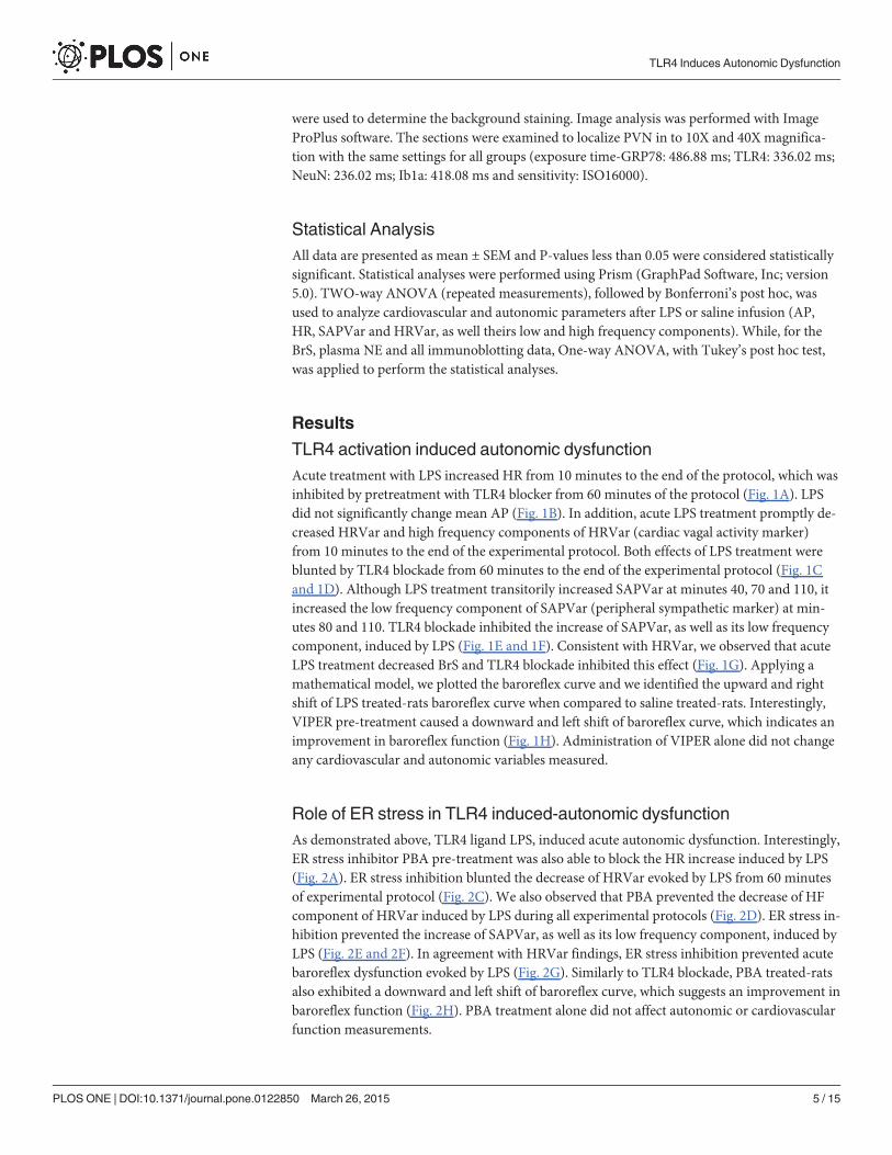
TLR4 activation by LPS increased ER stress marker GRP78 expressionin the PVNAcute treatment with LPS, when compared to saline treated animals, increased GRP78 proteinexpression in the PVN (1.97±0.04 vs 1.18±0.02 AU, p<0.05). Pre-treatment with TLR4 blocker(1.43±0.01 vs 1.97±0.04 AU, p<0.05) or ER stress inhibitor (0.81±0.02 vs 1.97±0.04 AU,p<0.05) were able to completely inhibit the increase of GRP78 protein expression induced byLPS in the PVN (Fig. 4A and 4B).
Fig 2. ER stress inhibition prevents autonomic dysfunction induced by TLR4 signaling. (A) Effects of ER stress inhibition (ip, 10 mg/kg) in LPS-induced changes on heart rate. (B) Effects of ER stress inhibition (ip, 10 mg/kg) in LPS-induced changes on mean arterial pressure. (C) Effects of ER stressinhibition (ip, 10 mg/kg) in LPS-induced changes on heart rate variability. (D) Effects of ER stress inhibition (ip, 0.1 mg/kg) in LPS-induced changes on high-frequency component of heart rate variability. (E) Effects of ER stress inhibition (ip, 10 mg/kg) in LPS-induced changes on systolic arterial pressurevariability. (F) Effects of ER stress inhibition (ip, 10 mg/kg) in LPS-induced changes on low-frequency component of systolic arterial pressure variability. (G)Effects of ER stress inhibition (ip, 10 mg/kg) in LPS-induced changes on baroreflex sensitivity. (H) Effects of ER stress inhibition (ip, 10 mg/kg) in LPS-induced changes on baroreflex sigmoidal curve. #—p<0.05 saline vs LPS treated-rats; *—p<0.05 LPS vs LPS+ PBA; n = 9–10 rats.
doi:10.1371/journal.pone.0122850.g002
TLR4 Induces Autonomic Dysfunction
PLOS ONE | DOI:10.1371/journal.pone.0122850 March 26, 2015 7 / 15
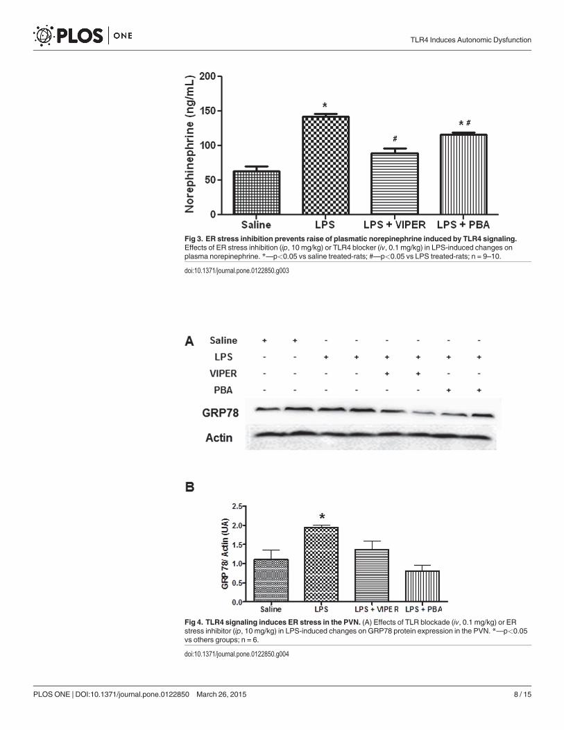
Fig 3. ER stress inhibition prevents raise of plasmatic norepinephrine induced by TLR4 signaling.Effects of ER stress inhibition (ip, 10 mg/kg) or TLR4 blocker (iv, 0.1 mg/kg) in LPS-induced changes onplasma norepinephrine. *—p<0.05 vs saline treated-rats; #—p<0.05 vs LPS treated-rats; n = 9–10.
doi:10.1371/journal.pone.0122850.g003
Fig 4. TLR4 signaling induces ER stress in the PVN. (A) Effects of TLR blockade (iv, 0.1 mg/kg) or ERstress inhibitor (ip, 10 mg/kg) in LPS-induced changes on GRP78 protein expression in the PVN. *—p<0.05vs others groups; n = 6.
doi:10.1371/journal.pone.0122850.g004
TLR4 Induces Autonomic Dysfunction
PLOS ONE | DOI:10.1371/journal.pone.0122850 March 26, 2015 8 / 15

Inflammation induced by TLR4 activation is suppressed by ER Stressblocker in the PVNAcute treatment with LPS increased TLR4 (1.3±0.08 vs 0.53±0.08 AU, p<0.05) (and TNF-α(1.09±0.16 vs 0.58±0.15 AU, p<0.05) protein expression in the PVN when compared to the sa-line group. TLR4 blockade was able to reduce the TLR4 (0.97±0.09 vs 1.3±0.08 AU, p<0.05)and TNF-α (0.65±0.08 vs 1.09±0.16 AU, p<0.05) protein expression induced by LPS treat-ment. Interestingly, ER stress inhibition was able to blunt LPS effects on TLR4 (0.60±0.21 vs1.3±0.08 AU, p<0.05) and TNF-α (0.58±0.07 vs 1.09±0.16 AU, p<0.05) protein expression(Fig. 5A and 5B).
TLR4 activation induces neuronal ER Stress in the PVNIn order to identify cellular location of ER stress marker GRP78, as well as cellular crosstalkwith TLR4, we performed immunofluorescence double-staining in the PVN. Consistent withour immunoblot data, we identified a dramatic increase of GRP78 (Fig. 6A) and TLR4(Fig. 6B) staining induced by LPS treatment, particularly in the ventral parvocellular area (anautonomic control region). However, VIPER and ER stress inhibitor treatment before LPS ad-ministration significantly reduced the increase in GRP78 and TLR4 expression. We observed aco-localization between GRP78 and TLR4 in LPS treated-rats (arrows, Fig. 6C). In addition,GRP78 was co-localized with NeuN, neuronal marker (arrows, Fig. 6D) in LPS treated-rats.
TLR4 agonist induced neuronal ER stress and microglia activation in thePVNIn order to identify a possible source of pro-inflammatory cytokines, we performed immuno-fluorescence staining for Iba1 (microglia marker) to analyze microglia activation in the PVN.In saline treated-animals, microglia exhibited a higher number of normal morphology cellswith numerous long branches and multiple filopodia. Acute LPS treatment induced microgliaactivation, which was characterized for a dramatic reduction in the cellular branches. TLR4blocker or ER stress inhibitor pre-treatment were able to decrease microglia activation inducedby LPS (Fig. 7A and 7B).
DiscussionIn the present study, we demonstrate for the first time, that acute LPS-induced TLR4 activa-tion, in the PVN, decreases HRVar, cardiac vagal activity and induced baroreflex dysfunctionin conscious rats, as well as increases plasma NE. These effects seem to be dependent on ERstress. This is the first report to show that acute LPS (a ligand for TLR4) induces TLR4 and ERstress within the PVN and contributes to autonomic dysfunction. Furthermore, blockade of ERstress attenuates LPS-induced neuroinflammation and TLR4 expression in the PVN, demon-strating a possible role for TLR4-induced brain inflammation and ER stress in autonomic dys-function. In addition, our immunofluorescence data revealed that acute LPS, a specific TLR4agonist, induce microglia activation, inflammation and neuronal ER stress in the PVN. Thesedata suggest that TLR4 signaling in the PVN induces autonomic dysfunction through brain in-flammation and neuronal ER stress.
Decreased parasympathetic activity and baroreflex function are the hallmark of autonomicdysfunction, which has prognostic value in cardiovascular diseases [1, 2]. The role of brainTLR4 has been recently suggested to modulate cardiac remodeling in heart failure rats [15] andhypertension [16]. Furthermore, chronic LPS treatment has been shown to increase mean APin healthy rats [23]. In the present study, we observed that acute LPS treatment decreased
TLR4 Induces Autonomic Dysfunction
PLOS ONE | DOI:10.1371/journal.pone.0122850 March 26, 2015 9 / 15

HRVar, HF component of HRVar (cardiac parasympathetic marker) and BrS. Although SAP-Var and its LF components (peripheral sympathetic marker) were increased significantly onlyat 75 and 115 minutes post-LPS treatment, it evoked an increase of plasma NE, which again
Fig 5. Inflammation induced through TLR4 signaling is dependent to ER stress in the PVN. (A)Representative blots of effects of TLR blockade (iv, 0.1 mg/kg) or ER stress inhibitor (ip, 10 mg/kg) in LPS-induced changes on TNF-α and TLR4 protein expression in the PVN. (B) Effects of TLR blockade (iv, 0.1 mg/kg) or ER stress inhibitor (ip, 10 mg/kg) in LPS-induced changes on TNF-α protein expression in the PVN. (C)Effects of TLR blockade (iv, 0.1 mg/kg) or ER stress inhibitor (ip, 10 mg/kg) in LPS-induced changes on TLR4protein expression in the PVN. *—p<0.05 vs others groups; n = 6.
doi:10.1371/journal.pone.0122850.g005
TLR4 Induces Autonomic Dysfunction
PLOS ONE | DOI:10.1371/journal.pone.0122850 March 26, 2015 10 / 15

indicates an increase in sympathetic activity. The effects induced by LPS were abolished withpretreatment with TLR4 blocker VIPER, indicating a direct role for LPS-induced autonomicdysfunction. Taken together, our data demonstrates, for the first time, that in an acute condi-tion, TLR4 signaling promotes a decrease in cardiac parasympathetic activity and baroreflexfunction, and increases sympathetic activity.
In addition to autonomic dysfunction induced through TLR4, recent studies have identifieda causative role for ER stress in hypertension [17, 18]. In our study, we identified that pre-treat-ment with an ER stress inhibitor blunted the decrease of HRVar, HF component of HRVar(cardiac parasympathetic marker) and BrS induced by TLR4 activation. In summary, we showfor the first time, that TLR4 promotes acute autonomic dysfunction through the induction ofER stress. However, ER stress inhibitor pre-treatment was not able to fully prevent the increaseof plasma NE induced by LPS, which demonstrates that LPS induced sympathetic activation isnot completely ER stress-dependent.
Recent studies have shown that TLR4 signaling is crucial to development of ER stress [19,20]. In our study, we identified LPS treatment increases GRP78 protein expression in the PVN,
Fig 6. TLR4 and GRP78 immunofluorescence in the PVN. (A) Representative fluorescence microscopy illustrations showing GRP78 single-labeling in thePVN of four experimental groups. (B) Representative fluorescence microscopy illustrations showing TLR4 single-labeling in the PVN of saline or LPS treatedrats. (C) Representative fluorescence microscopy illustrations showing TLR4 or GRP78 double-labeling in the PVN of LPS treated rats. Arrows indicatepositive TLR4 and GRP78 cells. (D) Representative fluorescence microscopy illustrations showing GRP78 and NeuN double-labeling in the PVN. Arrowsindicate positive GRP78 neurons; n = 3.
doi:10.1371/journal.pone.0122850.g006
TLR4 Induces Autonomic Dysfunction
PLOS ONE | DOI:10.1371/journal.pone.0122850 March 26, 2015 11 / 15
a major autonomic and neuroendocrine region that regulates salt appetite, sympathetic outflowand baroreflex function. LPS-induced GRP78 expression was abolished with TLR4 blockade orER stress inhibitor indicating a cross talk between TLR4 and ER stress. Taken together withour physiological data, we propose that LPS induced autonomic dysfunction is at least in partmodulated by ER stress in the PVN.
TLR4 signaling is a major source of pro-inflammatory cytokines [24], such as TNF-α [25],and blockade of TLR4 in the brain has been shown to prevent cardiac dysfunction in
Fig 7. TLR4 activation inducedmicroglia activation through ER stress. (A) Representative fluorescence microscopy illustrations showing Ib1a single-labeling in the PVN of four experimental groups. Arrows indicate amplified microgial cell. (B) Representative fluorescence microscopy illustrations showingmicroglia in different morphologic levels of activation in four experimental groups; n = 3.
doi:10.1371/journal.pone.0122850.g007
TLR4 Induces Autonomic Dysfunction
PLOS ONE | DOI:10.1371/journal.pone.0122850 March 26, 2015 12 / 15
experimental models of heart failure [15] and hypertension [16]. Moreover, chronic LPS treat-ment was able to increase mean AP and oxidative stress in the rostral ventrolateral medulla[23] indicating a role for TLR4 in sympathetic hyperactivity. Data from our lab demonstratedthe role of pro-inflammatory cytokines in the PVN in hypertension and heart failure [4–7]. Inour present study, we report that acute LPS treatment increased TNF-α and TLR4 protein ex-pression in the PVN, which were prevented by TLR4 blocker or ER stress inhibitor. These find-ings indicate that inflammation and ER stress set a positive feedback mechanism, onceinflammation elicits ER stress and this cellular condition is able to induce pro-inflammatorycytokines release leading to vicious cycle [26, 27] thereby contributing to autonomic dysfunc-tion. This is supported by our findings that ER stress inhibition attenuated the increase ofTNF-α, TLR4 and GRP78. Based on these findings, we suggest that TLR4 signaling-inducedER stress contribute to autonomic dysfunction and brain inflammation.
In addition to our cardiovascular and protein expression data, we performed immunofluo-rescence staining to characterize the crosstalk between TLR4 signaling and ER stress in thePVN. Acute LPS treatment increased GRP78 and TLR4 protein expression, especially in theventral parvocellular area, an important cardiovascular autonomic control region in the PVN.Furthermore, in LPS treated rats, TLR4 was co-localized with GRP78, which indicate a possibleautocrine mechanism between TLR4 activation and development of ER stress, and these find-ings are in accordance with previous studies [12–14, 27]. Since we observed GRP78 positiveneurons co-localized in the PVN, we can suggest that LPS promotes neuronal ER stressthrough TLR4 activation. In addition, blockade with TLR4 or ER stress inhibitor decreased co-localization of these markers in the PVN reinforcing a direct crosstalk between TLR4 andER stress.
Beside the neuronal ER stress induced through TLR4 activation in the PVN, we identifiedmicroglia activation, an important pro-inflammatory cytokine source in the PVN. Microglia, asupporting cell in the brain, communicates with neurons and contributes to neuroinflamma-tion in pathological conditions. Here we demonstrate that LPS treatment also increases micro-glial activation. In saline treated rats, microglia showed numerous long branches whichcorresponds to an inactivated state. However, LPS modified the cellular morphology to anamoeboid-shape with a decreased number of cellular branches which is related with the pro-in-flammatory cytokines secretion [28]. TLR4 blockade or ER stress inhibitor pre-treatment wasable to decrease these morphologic modifications in microglia cells induced by LPS in thePVN. Since ER stress inhibitor was able to decrease the microglial activation and PVN cyto-kines, we can suggest that microglia activation induced through TLR4 is, at least in part, depen-dent on ER stress. Taken together, our data demonstrated TLR4 activation promotes neuronalinflammation and microglia activation, in the PVN, which contributes to autonomic dysfunc-tion and this effect seems to be, at least in part, ER stress-dependent.
ConclusionWe performed an acute LPS treatment in conscious rats and utilized pharmacological agents todemonstrate that TLR4 signaling decreases HRVar, cardiac vagal activity and BrS, as well anincrease in sympathetic activity through ER stress, neuronal inflammation and microglia acti-vation in the PVN. Our finding suggests that TLR4 signaling pathway and ER stress could beimportant pharmacological targets for treating some of the autonomic dysfunction in cardio-vascular disease.
TLR4 Induces Autonomic Dysfunction
PLOS ONE | DOI:10.1371/journal.pone.0122850 March 26, 2015 13 / 15
Supporting InformationS1 Fig. TLR4 immunoflourescence in the PVN. Representative fluorescence microscopy illus-trations showing TLR4 single-labeling in the PVN of four experimental groups.(TIF)
Author ContributionsConceived and designed the experiments: GSM ARN LCM JF. Performed the experiments:GSM ARN RBD. Analyzed the data: GSM ARN RBD PPSS. Contributed reagents/materials/analysis tools: GSM ARN RBD PPSS JF. Wrote the paper: GSM ARN LCM JF.
References1. La Rovere MT, Bersano C, Gnemmi M, Specchia G, Schwartz PJ. Exercise-induced increase in barore-
flex sensitivity predicts improved prognosis after myocardial infarction. Circulation. 2002; 106(8):945–9. Epub 2002/08/21. PubMed PMID: 12186798.
2. Ormezzano O, Cracowski JL, Quesada JL, Pierre H, Mallion JM, Baguet JP. EVAluation of the prog-nostic value of BARoreflex sensitivity in hypertensive patients: the EVABAR study. J Hypertens. 2008;26(7):1373–8. Epub 2008/06/14. doi: 10.1097/HJH.0b013e3283015e5a PubMed PMID: 18551013.
3. Cohuet G, Struijker-Boudier H. Mechanisms of target organ damage caused by hypertension: thera-peutic potential. Pharmacol Ther. 2006; 111(1):81–98. Epub 2005/11/18. doi: 10.1016/j.pharmthera.2005.09.002 PubMed PMID: 16288809.
4. Sriramula S, Cardinale JP, Francis J. Inhibition of TNF in the brain reverses alterations in RAS compo-nents and attenuates angiotensin II-induced hypertension. PLoS One. 2013; 8(5):e63847. Epub 2013/05/22. doi: 10.1371/journal.pone.0063847 PubMed PMID: 23691105; PubMed Central PMCID:PMCPmc3655013.
5. Cardinale JP, Sriramula S, Mariappan N, Agarwal D, Francis J. Angiotensin II-induced hypertension ismodulated by nuclear factor-kappaBin the paraventricular nucleus. Hypertension. 2012; 59(1):113–21.Epub 2011/11/23. doi: 10.1161/hypertensionaha.111.182154 PubMed PMID: 22106405; PubMed Cen-tral PMCID: PMCPmc3268075.
6. Kang YM, Ma Y, Zheng JP, Elks C, Sriramula S, Yang ZM, et al. Brain nuclear factor-kappa B activationcontributes to neurohumoral excitation in angiotensin II-induced hypertension. Cardiovasc Res. 2009;82(3):503–12. Epub 2009/02/28. doi: 10.1093/cvr/cvp073 PubMed PMID: 19246475; PubMed CentralPMCID: PMCPmc2682616.
7. Kang YM, He RL, Yang LM, Qin DN, Guggilam A, Elks C, et al. Brain tumour necrosis factor-alpha mod-ulates neurotransmitters in hypothalamic paraventricular nucleus in heart failure. Cardiovasc Res.2009; 83(4):737–46. Epub 2009/05/22. doi: 10.1093/cvr/cvp160 PubMed PMID: 19457890; PubMedCentral PMCID: PMCPmc2725776.
8. Guggilam A, Patel KP, Haque M, Ebenezer PJ, Kapusta DR, Francis J. Cytokine blockade attenuatessympathoexcitation in heart failure: cross-talk between nNOS, AT-1R and cytokines in the hypothalam-ic paraventricular nucleus. Eur J Heart Fail. 2008; 10(7):625–34. Epub 2008/06/14. doi: 10.1016/j.ejheart.2008.05.004 PubMed PMID: 18550427; PubMed Central PMCID: PMCPmc2593148.
9. Shi Z, Gan XB, Fan ZD, Zhang F, Zhou YB, Gao XY, et al. Inflammatory cytokines in paraventricular nu-cleus modulate sympathetic activity and cardiac sympathetic afferent reflex in rats. Acta Physiol (Oxf).2011; 203(2):289–97. Epub 2011/06/01. doi: 10.1111/j.1748-1716.2011.02313.x PubMed PMID:21624097.
10. Shi P, Diez-Freire C, Jun JY, Qi Y, Katovich MJ, Li Q, et al. Brain microglial cytokines in neurogenic hy-pertension. Hypertension. 2010; 56(2):297–303. Epub 2010/06/16. doi: 10.1161/hypertensionaha.110.150409 PubMed PMID: 20547972; PubMed Central PMCID: PMCPmc2929640.
11. Rana I, Stebbing M, Kompa A, Kelly DJ, Krum H, Badoer E. Microglia activation in the hypothalamicPVN following myocardial infarction. Brain Res. 2010; 1326:96–104. Epub 2010/02/17. doi: 10.1016/j.brainres.2010.02.028 PubMed PMID: 20156424.
12. Bell MT, Puskas F, Agoston VA, Cleveland JC Jr., Freeman KA, Gamboni F, et al. Toll-like receptor 4-dependent microglial activation mediates spinal cord ischemia-reperfusion injury. Circulation. 2013;128(11 Suppl 1):S152–6. Epub 2013/09/18. doi: 10.1161/circulationaha.112.000024 PubMed PMID:24030400.
13. Lippai D, Bala S, Csak T, Kurt-Jones EA, Szabo G. Chronic alcohol-induced microRNA-155 contributesto neuroinflammation in a TLR4-dependent manner in mice. PLoS One. 2013; 8(8):e70945. Epub
TLR4 Induces Autonomic Dysfunction
PLOS ONE | DOI:10.1371/journal.pone.0122850 March 26, 2015 14 / 15
2013/08/21. doi: 10.1371/journal.pone.0070945 PubMed PMID: 23951048; PubMed Central PMCID:PMCPmc3739772.
14. Hines DJ, Choi HB, Hines RM, Phillips AG, MacVicar BA. Prevention of LPS-induced microglia activa-tion, cytokine production and sickness behavior with TLR4 receptor interfering peptides. PLoS One.2013; 8(3):e60388. Epub 2013/04/05. doi: 10.1371/journal.pone.0060388 PubMed PMID: 23555964;PubMed Central PMCID: PMCPmc3610686.
15. Ogawa K, Hirooka Y, Kishi T, Ide T, Sunagawa K. Partially silencing brain toll-like receptor 4 prevents inpart left ventricular remodeling with sympathoinhibition in rats with myocardial infarction-induced heartfailure. PMID: PLoS One. 2013; 8(7):e69053. Epub 2013/07/23. doi: 10.1371/journal.pone.0069053PubMed PMID: 23874864; PubMed Central PMCID: PMCPmc3706428.
16. Dange RB, Agarwal D, Masson GS, Vila J, Wilson B, Nair A, et al. Central blockade of TLR4 improvescardiac function and attenuates myocardial inflammation in angiotensin II-induced hypertension. Cardi-ovasc Res. 2014; 103(1):17–27. Epub 2014/03/29. doi: 10.1093/cvr/cvu067 PubMed PMID: 24667851.
17. Young CN, Cao X, Guruju MR, Pierce JP, Morgan DA, Wang G, et al. ER stress in the brain subfornicalorgan mediates angiotensin-dependent hypertension. J Clin Invest. 2012; 122(11):3960–4. Epub 2012/10/16. doi: 10.1172/jci64583 PubMed PMID: 23064361; PubMed Central PMCID: PMCPmc3484457.
18. Chao YM, Lai MD, Chan JY. Redox-sensitive endoplasmic reticulum stress and autophagy at rostralventrolateral medulla contribute to hypertension in spontaneously hypertensive rats. Hypertension.2013; 61(6):1270–80. Epub 2013/04/24. doi: 10.1161/hypertensionaha.111.00469 PubMed PMID:23608659.
19. Pierre N, Deldicque L, Barbe C, Naslain D, Cani PD, Francaux M. Toll-like receptor 4 knockout miceare protected against endoplasmic reticulum stress induced by a high-fat diet. PLoS One. 2013; 8(5):e65061. Epub 2013/06/07. doi: 10.1371/journal.pone.0065061 PubMed PMID: 23741455; PubMedCentral PMCID: PMCPmc3669084.
20. Yao L, Kan EM, Lu J, Hao A, Dheen ST, Kaur C, et al. Toll-like receptor 4 mediates microglial activationand production of inflammatory mediators in neonatal rat brain following hypoxia: role of TLR4 in hypox-ic microglia. J Neuroinflammation. 2013; 10:23. Epub 2013/02/08. doi: 10.1186/1742-2094-10-23PubMed PMID: 23388509; PubMed Central PMCID: PMCPmc3575244.
21. Nair AR, Masson GS, Ebenezer PJ, Del Piero F, Francis J. Role of TLR4 in lipopolysaccharide-inducedacute kidney injury: protection by blueberry. Free Radic Biol Med. 2014; 71:16–25. Epub 2014/03/25.doi: 10.1016/j.freeradbiomed.2014.03.012 PubMed PMID: 24657730.
22. Ceroni A, Chaar LJ, Bombein RL, Michelini LC. Chronic absence of baroreceptor inputs prevents train-ing-induced cardiovascular adjustments in normotensive and spontaneously hypertensive rats. ExpPhysiol. 2009; 94(6):630–40. Epub 2009/03/03. doi: 10.1113/expphysiol.2008.046128 PubMed PMID:19251981.
23. Wu KL, Chan SH, Chan JY. Neuroinflammation and oxidative stress in rostral ventrolateral medullacontribute to neurogenic hypertension induced by systemic inflammation. J Neuroinflammation. 2012;9(1):212. Epub 2012/09/11. doi: 10.1186/1742-2094-9-212 PubMed PMID: 22958438; PubMed CentralPMCID: PMCPmc3462714.
24. Nair AR, Elks CM, Vila J, Del Piero F, Paulsen DB, Francis J. A blueberry-enriched diet improves renalfunction and reduces oxidative stress in metabolic syndrome animals: potential mechanism of TLR4-MAPK signaling pathway. PLoS One. 2014; 9(11):e111976. Epub 2014/11/06. doi: 10.1371/journal.pone.0111976 PubMed PMID: 25372283; PubMed Central PMCID: PMCPmc4221362.
25. Salomao R, Brunialti MK, Rapozo MM, Baggio-Zappia GL, Galanos C, Freudenberg M. Bacterial sens-ing, cell signaling, and modulation of the immune response during sepsis. Shock. 2012; 38(3):227–42.Epub 2012/07/11. doi: 10.1097/SHK.0b013e318262c4b0 PubMed PMID: 22777111.
26. O'Neill CM, Lu C, Corbin KL, Sharma PR, Dula SB, Carter JD, et al. Circulating levels of IL-1B+IL-6cause ER stress and dysfunction in islets from prediabetic male mice. Endocrinology. 2013; 154(9):3077–88. Epub 2013/07/10. doi: 10.1210/en.2012-2138 PubMed PMID: 23836031; PubMed Cen-tral PMCID: PMCPmc3749476.
27. Kim S, Joe Y, Jeong SO, Zheng M, Back SH, Park SW, et al. Endoplasmic reticulum stress is sufficientfor the induction of IL-1beta production via activation of the NF-kappaB and inflammasome pathways.Innate Immun. 2014; 20(8):799–815. Epub 2013/11/13. doi: 10.1177/1753425913508593 PubMedPMID: 24217221.
28. Buttini M, Limonta S, Boddeke HW. Peripheral administration of lipopolysaccharide induces activationof microglial cells in rat brain. Neurochem Int. 1996; 29(1):25–35. Epub 1996/07/01. PubMed PMID:8808786.
TLR4 Induces Autonomic Dysfunction
PLOS ONE | DOI:10.1371/journal.pone.0122850 March 26, 2015 15 / 15
Related Documents